Embed Size (px)
Citation preview

ARTICLE
Received 2 Nov 2012 | Accepted 26 Jun 2013 | Published 30 Jul 2013
Metformin improves healthspan and lifespanin miceAlejandro Martin-Montalvo1,*, Evi M. Mercken1,*, Sarah J. Mitchell1,2,3, Hector H. Palacios1, Patricia L. Mote4,
Morten Scheibye-Knudsen5, Ana P. Gomes6, Theresa M. Ward1, Robin K. Minor1, Marie-Jose Blouin7,
Matthias Schwab8, Michael Pollak7, Yongqing Zhang9, Yinbing Yu10, Kevin G. Becker9, Vilhelm A. Bohr5,
Donald K. Ingram11, David A. Sinclair6, Norman S. Wolf12, Stephen R. Spindler4, Michel Bernier1 & Rafael de Cabo1
Metformin is a drug commonly prescribed to treat patients with type 2 diabetes. Here we
show that long-term treatment with metformin (0.1% w/w in diet) starting at middle age
extends healthspan and lifespan in male mice, while a higher dose (1% w/w) was toxic.
Treatment with metformin mimics some of the benefits of calorie restriction, such as
improved physical performance, increased insulin sensitivity, and reduced low-density
lipoprotein and cholesterol levels without a decrease in caloric intake. At a molecular level,
metformin increases AMP-activated protein kinase activity and increases antioxidant
protection, resulting in reductions in both oxidative damage accumulation and chronic
inflammation. Our results indicate that these actions may contribute to the beneficial effects
of metformin on healthspan and lifespan. These findings are in agreement with current
epidemiological data and raise the possibility of metformin-based interventions to promote
healthy aging.
DOI: 10.1038/ncomms3192
1 Translational Gerontology Branch, National Institute on Aging, National Institutes of Health, 251 Bayview Boulevard, Baltimore, Maryland 21224, USA.2 Kolling Institute of Medical Research, Royal North Shore Hospital, St Leonards, New South Wales 2065, Australia. 3 Sydney Medical School, University ofSydney, Sydney, New South Wales 2006, Australia. 4 Department of Biochemistry, University of California Riverside, 3401 Watkins Drive, Riverside, California92521, USA. 5 Laboratory of Molecular Gerontology, National Institute on Aging, National Institutes of Health, 251 Bayview Boulevard, Baltimore, Maryland21224, USA. 6 Department of Genetics, Paul F. Glenn Laboratories for the Biological Mechanisms of Aging, Harvard Medical School, 77 Avenue Louis Pasteur,Boston, Massachusetts 02115, USA. 7 Department of Medicine and Oncology, McGill University, Montreal, Quebec H3A 2B3, Canada. 8 Dr MargareteFischer-Bosch Institute of Clinical Pharmacology, Stuttgart and University of Tubingen, Auerbachstrasse 112, Stuttgart 70376, Germany. 9 Gene Expressionand Genomics Unit, National Institute on Aging, National Institutes of Health, 251 Bayview Boulevard, Baltimore, Maryland 21224, USA. 10 Laboratory ofEpidemiology, Demography and Biometry, National Institute on Aging, Bethesda, Maryland 20892, USA. 11 Nutritional Neuroscience and Aging Laboratory,Pennington Biomedical Research Center, Louisiana State University System, 6400 Perkins Road, Baton Rouge, Louisiana 70808, USA. 12 Department ofPathology, University of Washington, Seattle, Washington 98195-7470, USA. * These authors contributed equally to this work. Correspondence and requestsfor materials should be addressed to R.d.C. (email: [email protected]).
NATURE COMMUNICATIONS | 4:2192 | DOI: 10.1038/ncomms3192 | www.nature.com/naturecommunications 1
& 2013 Macmillan Publishers Limited. All rights reserved.
Metformin is a biguanide used since the 1960s in thetreatment of type 2 diabetes and metabolic syndrome. Itenhances insulin sensitivity, induces glycolysis and
suppresses gluconeogenesis in the liver1–3. Patients withmetabolic syndrome exhibit many manifestations of acceleratedaging, such as cardiovascular disease, cancer and inflammatorydisorders, all of which reduce lifespan. The fact that metformintreatment has been associated with reduced risk of cancer4 andcardiovascular disease raises the possibility of a beneficial role ofmetformin for other age-related diseases5. Recently, we andothers have studied pharmacological interventions that can delayaging and the incidence of age-related diseases6,7. Many of theseinterventions are based on the study of calorie restriction (CR)mimetics8,9. CR mimetics involve interventions that reproducephysiological and anti-aging effects found in CR animals. Severalreports suggest that the actions of metformin resemble the effectsof CR to some extent; microarray analyses have shown thatmetformin induces a gene expression profile that aligns with thatof CR, although conflicting results have been shown in lifespanextension9–15.
The mechanism of action of metformin involves, at least inpart, activation of adenosine monophosphate-activated proteinkinase (AMPK), an enzyme involved in cellular and whole-organism energy balance, as well as glucose and fat metabolism16,17.Separate evidence suggests that metformin may also act via AMPK-independent mechanisms3,18,19. The activation of AMPK is a well-known process triggered by an increase of the AMP/ATP ratio20.Several studies provide evidence that metformin partially inhibitscomplex I of the electron transport chain (ETC) with subsequentalteration of the mitochondrial performance, but the molecularmechanisms underlying this process have not been characterized indetail21–23. Thus, metformin may compromise ATP production inmitochondria leading to an increase of the AMP/ATP ratio. As aconsequence of energy depletion, glycolysis is induced to maintaincellular metabolism. Even though mitochondrial poisons increaseoxidative cellular damage by mechanisms involving increasedreactive oxygen species24,25, there is no evidence that metformininduces the generation of reactive oxygen species and/oraccumulation of oxidative damage26,27. In fact, the transcriptionfactor SKN-1/Nrf2 is activated upon metformin treatment,resulting in increased expression of antioxidant genes in cells andanimal models10. Reduced accumulation of oxidative damage maycontribute to the inhibitory effects of metformin treatment incarcinogenesis models3,28,29.
The ability of metformin to extend lifespan in the nematodeCaenorhabditis elegans and the conflicting results in drosophilaand mammals led us to study chronic metformin supplementa-tion in laboratory mice9,10,12. Cohorts of middle-aged maleC57BL/6 and B6C3F1 mice were provided with either a standarddiet (SD) or SD supplemented with 0.1% (w/w) or 1% (w/w)metformin for the remainder of their lives. Our data show a levelof chronic metformin exposure that leads to healthier and longerlives in mice, justifying further studies to determine if there is anexposure level that leads to improvement in healthspan andlifespan in humans.
ResultsMetformin increases healthspan and longevity of mice. Wedetermined the long-term effects of two doses of metformin inmale C57BL/6 mice. The first dose consisted of 0.1% metformin(w/w) supplemented in diet, which yielded a concentration of0.45±0.09 mM in serum and 0.49±0.06 nmoles mg� 1 protein inthe liver. The second dose (1% w/w) yielded a concentration of5.03±0.87 mM in serum and 3.67±0.32 nmoles mg� 1 protein inthe liver (n¼ 6–8 per group; age¼ 84 weeks; diet¼ 30 weeks;
values presented as mean±s.e.m.). The survival curves of controland metformin-treated male mice separated shortly after the onsetof the treatment. Diet supplementation with 0.1% metformin ledto a 5.83% extension of mean lifespan (Fig. 1a), w2¼ 5.46 andP¼ 0.02 in Gehan–Breslow survival test. In agreement with thesedata, a different strain of male mice (B6C3F1) supplemented withthe same dose of metformin (0.1% w/w) resulted in a 4.15%extension of mean lifespan (Supplementary Fig. S1), w2¼ 3.43 andP¼ 0.064 in Gehan–Breslow survival test, which although is notsignificant suggests that the effects of metformin in longevity arenot strain-specific. On the other hand, a higher concentration ofthe drug (1% w/w) was toxic and significantly shortened meanlifespan of C57BL/6 mice by 14.4% (Fig. 1b), likely owing to renalfailure (Supplementary Fig. S1 and Supplementary Table S1),w2¼ 51,70 and Po0.001 in Gehan–Breslow survival test. Highdoses of metformin have been associated with the development oflactic acidosis and the drug is contraindicated in patients withkidney dysfunction30. Importantly, male mice treated with 0.1%metformin in both longevity studies did not show any majordifference in pathologies at 115 weeks of age nor obvious causes ofdeath in the necropsies compared with SD-fed animals(Supplementary Tables S1–S3).
Adult male C57BL/6 mice (from week 72 to 90) treated with0.1% metformin were lighter than control animals (Fig. 1c). Miceon metformin tended to preserve body weight with advancingage, which has been associated with increased survival in rodentand human studies31,32. By 124 weeks of age, the average weightof metformin-treated mice was higher than SD-fed mice,although these observations were not found in B6C3F1 mice,probably because food intake in this strain was controlled(Supplementary Fig. S1)33. Determinations of body compositionduring the lifetime of the animals revealed no significantdifferences in the percentage of fat mass and lean body mass orin the lean-to-fat ratio (Supplementary Table S4). AlthoughC57BL6/ mice fed with metformin were lighter, this groupconsumed more calories than their SD counterparts, indicating ashift in energy metabolism (Fig. 1d). The energy content of thefaeces was identical in both groups, suggesting that metformindid not alter intestinal absorption of nutrients (SupplementaryFig. S1). However, the beneficial effects of metformin might beexplained, at least in part, by changes in the gut microbiota34, aprocess that has not been addressed in our study. Nevertheless,the effect of metformin on food consumption provides evidenceagainst the possibility that metformin acts in a trivial fashion byleading to caloric restriction attributable to lack of palatability ofmetformin-supplemented chow.
We next examined whether the increase in calorie intake wasassociated with higher energy expenditure. Indirect calorimetryrevealed that metformin-treated animals showed higher heatproduction during the dark phase, when mice are normally moreactive (Fig. 1e). In accordance with previous reports demon-strating that metformin increases the use of lipids as a source ofenergy35, the respiratory exchange ratio was decreased duringboth the light and dark phases, and liver glyceride content wasreduced in metformin-treated mice, indicating the preference forfat utilization (Fig. 1f and Supplementary Fig. S1). Consistentwith this hypothesis, b-oxidation of fatty acids was found to beincreased while lipid synthesis was decreased in primaryhepatocytes and mouse embryonic fibroblasts (MEFs) treatedwith metformin (Supplementary Fig. S1). Interestingly, totallevels of oxygen consumption and carbon dioxide productionwere not modified significantly by metformin in mice(Supplementary Fig. S1). Moreover, there was no difference inspontaneous locomotion activity, suggesting that metforminshifts energy homeostasis of mice rather than their behaviour(Supplementary Fig. S1).
ARTICLE NATURE COMMUNICATIONS | DOI: 10.1038/ncomms3192
2 NATURE COMMUNICATIONS | 4:2192 | DOI: 10.1038/ncomms3192 | www.nature.com/naturecommunications
& 2013 Macmillan Publishers Limited. All rights reserved.

While the effect of metformin on longevity was obvious, it wasimportant to determine whether the healthspan of the animalswas preserved. We used several approaches to ascertain physicalhealth of the animals. Rotarod, treadmill and open-field testsindicated that metformin improved the general fitness oflaboratory mice (Fig. 1g–i). C57BL/6 mice are known to developcataracts as they age32. Metformin supplementation led tosignificant reduction in lens opacity of 105-week-old mice(Fig. 1j), consistent with an overall improvement in healthspan.
As mentioned earlier, metformin is widely prescribed toimprove insulin sensitivity. Mice treated with 0.1% metforminshowed lower glycated haemoglobin (Hb1Ac) levels 66 weeksafter the treatment was initiated (Table 1). Metformin-fed miceand mice on SD displayed comparable glucose homeostasis when
measured by oral glucose tolerance test (OGTT) and insulintolerance test (ITT) at 81 and 93 weeks of age, respectively(Fig. 1k–n). At 100 weeks of age, metformin-treated mice showedimprovements in serum metabolite levels that are associated withdiabetes compared with their control counterparts, with areduction in insulin levels, total cholesterol, low-density lipopro-teins and the homeostatic model assessment index-insulinresistance (Table 1). Taken together, these results strongly suggestthat metformin prevents the onset of metabolic syndrome.
Metformin mimics CR transcriptome. We have previouslyassociated the induction of similar transcriptome to animalsunder CR in liver tissue, even in a 8-week treatment15. To test
ITT
Time (min)0 20 40 60 80 100 120
Glu
cose
(m
g.dl
–1)
0
50
100
150
200
SD0.1% Met
OGTT
Time (min)0 20 40 60 80 100 120
Glu
cose
(m
g.dl
–1)
0
150
300
450
SD0.1% Met
Light Dark
Hea
t (kc
al.h
–1)
0.0
0.2
0.4
0.6
0.8 SD0.1% Met *
Light Dark
RE
R
0.0
0.2
0.4
0.6
0.8
1.0
* *
SD 0.1% Met
Tre
adm
ill (
m)
050
100150200250300350 *
SD 0.1% Met
Rot
arod
(s)
0
50
100
150
200*
SD 0.1% Met
Ope
n fie
ld (
m.m
in–1
)
0.00
0.01
0.02
0.03*
SD 0.1% Met
Cat
arac
ts in
dex
(a.u
.)
0.00.51.01.52.02.53.0
*
SD 0.1% Met
A.U
.C. (
a.u.
)
0
3,000
6,000
9,000
12,000
15,000
SD 0.1% Met
A.U
.C. (
a.u.
)
0
10,000
20,000
30,000
40,000
Age (weeks)
0 20 40 60 80 100 120 140 160
Sur
viva
l (%
)0
20
40
60
80
100
SD
0.1% Met
Age (weeks)
0 20 40 60 80 100 120 140 160
Sur
viva
l (%
)
0
20
40
60
80
100
SD
1 % Met
Age (weeks)60 80 100 120 140 160
Bod
y w
eigh
t (g)
20253035404550 SD
0.1% Met
*72–90
SD 0.1% Met
Foo
d in
take
(g. d
ay–1
)
0.00.51.01.52.02.53.03.5
*
Figure 1 | Metformin increases survival and improves physical performance. (a,b) Kaplan–Meier survival curve for mice treated either with 0.1 or 1%
metformin. n¼ 64 for metformin 0.1% group and n¼ 83 for their untreated counterparts; n¼90 for metformin 1% group and n¼88 for their untreated
counterparts. The arrows at 54 weeks indicate the age at which metformin treatment was initiated. (c) Body weights. (d) Food consumption. (e,f) In vivo
metabolic response to 0.1% metformin treatment. n¼9 per group. (e) Energy expenditure. (f) Respiratory exchange ratio. (g) Time to fall from an
accelerating rotarod. n¼ 16 per group. (h) Distance ran on treadmill performance. n¼ 9 per group. (i) Average speed of animals in the open-field test.
n¼ 15–16 per group. (j) Metformin treatment delayed the onset of age-related cataracts. n¼93–124 eyes per group. (k) Plasma levels of glucose after
oral glucose load (OGTT). n¼8 per group. (l) Area under OGTT curve. (m) Plasma levels of glucose after intraperitoneal insulin injection (ITT). n¼ 9 per
group. (n) Area under ITT curve. Met, metformin. Unless otherwise stated, n¼ all live animals in the study. Data are represented as the mean±s.e.m.
*Po0.05 compared with SD-fed mice (t-test two tailed).
NATURE COMMUNICATIONS | DOI: 10.1038/ncomms3192 ARTICLE
NATURE COMMUNICATIONS | 4:2192 | DOI: 10.1038/ncomms3192 | www.nature.com/naturecommunications 3
& 2013 Macmillan Publishers Limited. All rights reserved.
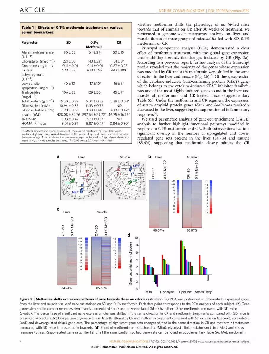
whether metformin shifts the physiology of ad lib-fed micetowards that of animals on CR after 30 weeks of treatment, weperformed a genome-wide microarray analysis on liver andmuscle tissues of three groups of mice ad lib-fed with SD, 0.1%metformin or CR.
Principal component analysis (PCA) demonstrated a cleareffect of metformin treatment, with the global gene expressionprofile shifting towards the changes induced by CR (Fig. 2a).According to a previous report, further analysis of the transcriptprofile revealed that the majority of the genes whose expressionwas modified by CR and 0.1% metformin were shifted in the samedirection in the liver and muscle (Fig. 2b)15. Of these, expressionof the cytokine-inducible SH2-containing protein (CISH) gene,which belongs to the cytokine-induced STAT inhibitor family27,was one of the most highly induced genes found in the liver andmuscle of metformin- and CR-treated mice (SupplementaryTable S5). Under the metformin and CR regimen, the expressionof serum amyloid protein genes (Saa1 and Saa2) was markedlydecreased in the liver, suggesting the suppression of inflammatoryresponses36.
We used parametric analysis of gene-set enrichment (PAGE)analysis to further highlight functional pathways modified inresponse to 0.1% metformin and CR. Both interventions led to asignificant overlap in the number of upregulated and down-regulated gene sets present in the liver (84.7%) and muscle(85.6%), supporting that metformin closely mimics the CR
Table 1 | Effects of 0.1% metformin treatment on variousserum biomarkers.
Parameter SD 0.1%Metformin
CR
Ala aminotransferase(U l� 1)
90±58 64±29 50±15
Cholesterol (mg dl� 1) 221±30 143±33* 101±8*Creatinine (mg dl� 1) 0.11±0.01 0.11±0.01 0.27±0.25Lactatedehydrogenase(U l� 1)
573±82 623±165 443±109
Low-densitylipoprotein (mg dl� 1)
40±10 17±10* 16±5*
Triglycerides(mg dl� 1)
106±28 129±50 45±7*
Total protein (g dl� 1) 6.00±0.39 6.04±0.32 5.28±0.04*Glucose-fed (mM) 10.94±0.35 11.33±0.76 NDGlucose-fasted (mM) 8.23±0.65 8.80±0.43 4.10±0.42*Insulin (pM) 428.08±34.26 297.64±29.72* 46.75±16.76*% HbA1c 6.33±0.47 5.81±0.57* NDHOMA-IR index 8.01±0.57 5.87±0.47* 0.84±0.30*
HOMA-IR, homeostatic model assessment index-insulin resistance; ND, not determined.Insulin and glucose levels were determined at 100 weeks of age and HbA1c was determined at66 weeks of age. All other determinations were assayed at 74 weeks of age. Values shown aremean±s.d.; n¼4–16 samples per group. *Po0.05 versus SD (t-test two tailed).
Liver Muscle
MET
CR
SDSD
CR
MET
–4
–2
0
2
4
6
8
10
Liver Muscle
–20–16–12–8–4048
121620
–20 –10
10
–8–6
6
–4–2
2
–16–12–8–4
0 0
448
8121620
–15–12–9–6–30369
1215
0.1%
Met
-SD
0.1%
Met
-SD
CR
–SD
CR
–SD
Liver Muscle
CR
–SD
0.1%
Met
-SD
CR
–SD
0.1%
Met
-SD
10 10
10
–10 –10
–10–10
–8
–8
–6
–6
–4
–4
–2
–2
0
0
–10
–10–10
107.57.5
55
2.52.5
–2.5–2.5–5–5
–10
–5 –5
–5
4
4
2
2
–5
–5
–5 –5
–5
55
55
5
55
1010
10
5
0 0
0
0
0 0
00
0
PC
2 (2
1.8%
)
PC
2 (2
1.8%
)
PC1 (38.7%)
PC
3 (9
%)
PC1 (27.9%)
PC3 (1
4.4%
)
Stress RespLipid MetGlycolysisMito85.63%84.74%
83.97%86.67%
Gen
e se
t enr
ichm
ent (Z
sco
re)
–10
PC
3 (9
%)
PC1 (38.7%) PC1 (27.9%)
PC2 (2
0.1%
)
PC
2 (2
0.1%
)
–10
PC
3 (1
4.4%
)
10
Figure 2 | Metformin shifts expression patterns of mice towards those on calorie restriction. (a) PCA was performed on differentially expressed genes
from the liver and muscle tissue of mice maintained on SD and 0.1% metformin. Each data point corresponds to the PCA analysis of each subject. (b) Gene
expression profile comparing genes significantly upregulated (red) and downregulated (blue) by either CR or metformin compared with SD mice
(z-ratio). The percentage of significant gene expression changes shifted in the same direction in CR and metformin treatments compared with SD mice is
presented in brackets. (c) Comparison of gene sets significantly altered by CR and metformin treatment compared with SD expression (z-score); upregulated
(red) and downregulated (blue) gene sets. The percentage of significant gene sets changes shifted in the same direction in CR and metformin treatments
compared with SD mice is presented in brackets. (d) Effect of metformin on mitochondria (Mito), glycolysis, lipid metabolism (Lipid Met) and stress
response (Stress Resp)-related gene sets. The list of all the significantly modified gene sets can be found in Supplementary Table S6. Met, metformin.
ARTICLE NATURE COMMUNICATIONS | DOI: 10.1038/ncomms3192
4 NATURE COMMUNICATIONS | 4:2192 | DOI: 10.1038/ncomms3192 | www.nature.com/naturecommunications
& 2013 Macmillan Publishers Limited. All rights reserved.

transcriptome (Fig. 2c)15. The liver has a central role in themaintenance of glucose homeostasis and has been proposed asone of the main targets of metformin37. In the livers ofmetformin-treated mice, PAGE analysis showed a significantalteration in 220 gene sets (Supplementary Table S6), many ofthese were related to mitochondrial function, glycolysis, lipidbiosynthesis and stress response (Fig. 2d). In accordance withmicroarray data, quantitative PCR indicated the increase in themRNA levels of several genes encoding transcriptional regulatorsand enzymes involved in mitochondrial energetics, glycolysis andfatty acid metabolism (Supplementary Fig. S2). Moreover, therewas a significant downregulation of gene sets related toautophagy, apoptosis and ubiquitin cycle in the liver ofmetformin-fed mice. Several stress response pathways wereinduced by metformin as shown by Ingenuity Pathway Analysis(Supplementary Fig. S3). Nrf2 is a transcription factor thatregulates the expression of multiple antioxidant genes and is alsorequired for the beneficial effects of metformin in C. elegans10.Here, metformin treatment also induced Nrf2 target geneactivation. The overall interpretation of these results is thatmetformin exerts CR-like genomic and metabolic responses byinducing longevity-associated pathways in laboratory mice.
Preservation of mitochondrial function by metformin. It hasbeen proposed that mild inhibition of the mitochondrial ETCcomplex I activity by metformin21,22 reduces ATP productionand contributes to increase the AMP/ATP ratio, which, in turn,results in AMPK activation. AMPK has an important role in theregulation of energy metabolism38. In cultured MEFs and in liversof treated mice, metformin increased the relative levels ofphosphoactive AMPK (Thr172) by 27% (Po0.05, t-test twotailed), which led to increased phosphorylation at Ser-79 of itsdirect target, acetyl-CoA carboxylase, by 169% (Po0.01, t-testtwo tailed) (Fig. 3a,b, Supplementary Fig. S4, SupplementaryTables S7 and S8 for entire western blot images and densitometricanalyses). The ATP content in the liver of metformin-treatedmice was unaffected, which may stem from increased hepaticglycolysis (Supplementary Fig. S5). In parallel, we examinedmitochondrial performance in cultured MEFs in response tometformin (Fig. 3c) and found a decrease in oxygen consumptionrate, in agreement with earlier reports39. Oxygen consumptionwas also decreased following the addition of the mitochondrialuncoupler, carbonyl cyanide-4-(trifluoromethoxy)phenylhydra-zone, which indicates that metformin altered maximal oxygenconsumption in MEFs. Cell culture media showed a reduction inpH on exposure of cells to metformin (Supplementary Fig. S5),which is consistent with reports showing increased lactic acidproduction due to an increase in glycolysis and studies thatassociate the risk of lactic acidosis to metformin administration todiabetic patients40. Furthermore, metformin induced theexpression of cytosolic lactate dehydrogenase, the enzymeresponsible for lactic acid production, both in MEFs and in theliver of treated animals (Supplementary Fig. S5).
Our microarray and quantitative PCR analyses showedincreased expression of several mitochondrial genes in the liversof metformin-treated mice. However, metformin lowered oxygenconsumption in MEFs, suggesting that these alterations inmitochondrial bioenergetic function may occur in cell cultures.The administration of this anti-diabetic drug did not alter severalmarkers of mitochondrial content in both MEFs and the liver oftreated mice (Fig. 3d–f), while causing only moderate increase, ifany, in the expression of several subunits in the ETC complexes(Fig. 3g,h). Among key enzymes of the tricarboxylic acid cycleand ETC that were assayed, complex I activity was significantlylower in metformin-treated MEFs (Fig. 3i). In contrast, mice
treated with metformin had a remarkable increase in hepaticcomplex I activity (Fig. 3j). The activity for complexes III and IVwas significantly reduced by metformin in MEFs, but remainedunchanged in liver lysates of metformin-treated mice. The
SD 0.1% Met CR
mtD
NA
/nD
NA
0.00.20.40.60.81.01.21.41.6 *
SD 1 mM Met
TM
RM
(M
FI)
0
10,000
20,000
30,000
40,000
SD 1 mM Met
Mito
Tra
cker
Gre
en(M
FI)
0.0
1.5e+5
3.0e+5
4.5e+5
CS
Complex I
Complex II
Complex III
Complex IV
Mito
chon
dria
l act
iviti
es(%
of a
ctiv
ity u
ntre
ated
ME
Fs)
0
20
40
60
80
100
120Untreated1 mM Met
***
*
CS
Complex I
Complex II
Complex III
Complex IV
Mito
chon
dria
l act
iviti
es (
% o
f act
ivity
SD
mic
e)
0
30
60
90
120
150
180SD0.1% MetCR
** **
**
Time (min)0 20 40 60 80 100
Oxy
gen
cons
umpt
ion
(pm
ol m
in–1
per
1,0
00ce
lls)
01234567
Untreated1 mM Met
**
*
Ut Met Ut Met Ut Met
AMPK
pAMPK
pACC
ACC
β-Actin
Ut Met Ut Met Ut Met
Pgc1α/β
Complex I
Complex V
Complex III
β-Actin
SD
Pgc1α/β
Complex I
Complex V
Complex III
β-Actin
SD
AMPK
pAMPK
pACC
ACC
β-Actin
76a b
c d
e f
g h
i j
5276
52
225
225
38
52
76
5276
52
225
225
38
52
38
52
38
52
5252
3838
5252
76
102
76
102
CR0.1% met
CR0.1% Met
Figure 3 | Metformin activates AMPK without altering in vivo ETC
activities. (a) Activation of AMPK by metformin in MEFs. AMPK and
acetyl-coA carboxylase (ACC) phosphorylation by metformin. n¼ 3 per
group. (b) Activation of AMPK and ACC phosphorylation in the liver of
0.1% metformin-treated mice. n¼4–6 per group. (c) Oxygen consumption
in MEFs treated with 1 mM metformin. n¼ 3 per group. (d,e) Mitochondrial
content in MEFs treated with metformin was determined by TMRM (d) and
MitoTracker green (e) staining, MFI, mean fluorescence intensity. n¼ 3 per
group. (f) Mitochondrial DNA content analysed by quantitative PCR in the
liver. n¼ 5–8 per group. (g) Mitochondrial protein levels in MEFs treated
with metformin. n¼ 3 per group. (h) Mitochondrial protein levels in the
liver from 0.1% metformin-treated mice. n¼4–6 per group. (i,j) Effect of
metformin on mitochondrial enzymatic activities. (i) MEFs treated with
1 mM metformin (n¼ 3 per group) and (j) liver lysates from 0.1%
metformin-treated mice (n¼ 5–6 per group). Met, metformin; Ut,
untreated. Data are represented as the mean±s.e.m. *Po0.05 versus
untreated controls or SD-fed mice (t-test two tailed).
NATURE COMMUNICATIONS | DOI: 10.1038/ncomms3192 ARTICLE
NATURE COMMUNICATIONS | 4:2192 | DOI: 10.1038/ncomms3192 | www.nature.com/naturecommunications 5
& 2013 Macmillan Publishers Limited. All rights reserved.

differential effects of metformin in vitro and in vivo may be owingto the hepatocytes adapting to long-term inhibition of complex Iactivity. Interestingly, and in agreement with our findings,preservation of mitochondrial complex I activity, at least inmuscle, has been recently reported after long-term metformintreatment in humans41. Furthermore, the inhibition ofmitochondrial respiration by metformin is reported to beconcentration-dependent, with no effect at concentrations lowerthan 1 mM42. Serum level of metformin was 0.45±0.09 mM(mean±s.e.m.) in 0.1% metformin-treated mice, which isconsiderably higher than seen in the serum of diabetic patientstreated with metformin43, but lower than those used in mostin vitro models that demonstrate inhibition of mitochondrialrespiration by this compound21,22,42. The degree of inhibition ofoxidative phosphorylation as a function of metformin serumlevels cannot be quantitated because of the fact that active uptakeof metformin occurs via transporters on the cell surface and,possibly, mitochondria.
Metformin inhibits chronic inflammation. The activation of theSKN-1/Nrf2-dependent antioxidant response is required for thebeneficial effects of metformin in C. elegans10. There was a trendtowards reduction in superoxide production in mitochondrialcomplexes in the livers of metformin-treated mice (P¼ 0.09,t-test two tailed) (Supplementary Fig. S6). When divided by thetotal activity of mitochondrial complexes I–II to III, theproportion of superoxide leakage was significantly decreased bymetformin (Fig. 4a), indicating greater efficiency of mitochondrialcomplexes in transferring electrons to their expected acceptors inthe ETC. Moreover, the marked reduction in the amount oflysine-4-hydroxynonenal-modified proteins and 8-iso-PGF2a, amarker of lipid peroxidation, was consistent with decreasedoxidative stress damage in the liver of metformin-treated mice(Fig. 4b,c). To investigate whether metformin activates the Nrf2-dependent antioxidant response, HepG2 cells stably expressing aNrf2-responsive antioxidant response element (ARE) luciferasereporter construct were treated with increasing concentrations ofmetformin for 16 h (Fig. 4d). The increase in Nrf2/ARE reporteractivity occurred with an ED50 of B1.5 mM metformin withoutreduction in cell survival (Supplementary Fig. S6). The cellularreduction of the tetrazolium dye MTT (3-(4,5-dimethylthiazol-2-yl)-2,5-diphenyltetrazolium bromide), an assay that measuresNADH oxidase activity, was lower in metformin-treated cells,with an IC50 of B3.3 mM (Supplementary Fig. S6). Finally, in thelivers of treated mice, 0.1% metformin contributed to an increasein the level of antioxidant and stress response proteins, includingSOD2, TrxR1, NQO1 and NQO2 (Fig. 4e), further contributing tothe reduction in hepatic oxidative stress damage.
The development of chronic inflammation is one of thehallmarks of oxidative damage accumulation. Inflammatoryprocesses contribute to liver dysfunction in aging and areknown to be suppressed by CR44. In this context, increases inthe relative levels of phosphoactive forms of NF-kB and JNK havebeen shown to contribute to pro-inflammatory signalling45,46.Interestingly, metformin reduces the production of inflammatorymarkers in human liver cells36. In the livers of metformin-treatedmice, a decline in pNF-kB by 64% (Po0.01, t-test two tailed) andpJNK by 79% (Po0.01, t-test two tailed) levels was observed(Fig. 4f) together with attenuated expression of NF-kB gene(Fig. 4f). An additional gene expression signature associatedwith metformin treatment was the increased expression of severalanti-inflammatory genes (Fig. 4g). These results are in agreementwith the fact that metformin reduces the number of activatedmacrophages, although we could not detect significant differencesin the number of infiltrated macrophages in the liver of the mice
(Supplementary Fig. S6). Further research will be required toaddress this fact. In summary, the ability of metformin to reduceoxidative stress and inflammation suggests that this could partlybe the reason why this drug confers health and lifespan benefits inlaboratory mice.
DiscussionOur model reveals a level of chronic metformin exposure thatlengthens lifespan and also attenuates the deleterious effects ofaging in male mice. The effects of metformin resembled to someextent the effects of caloric restriction, even though food intakewas increased. While it is clear that in short-term in vitro models
SD
a b
c d
e f
g
0.1% Met CR
Sup
erox
ide
gene
ratio
n ra
tio
0.00
0.02
0.04
0.06
0.08* *
IL6IL10
NFκBTNFa
CISHSocs2
mR
NA
exp
ress
ion
(a.u
. ver
sus
SD
)
0
2
48
10
12SD0.1% Met
*
*
**
Lysi
ne 4
-HN
E
Ut0.
30.
6 1.3
2.5 5 10 20 40
tBHQ
AR
E-N
RF
2ac
tivat
ion
(r.u
.)
02,0004,0006,0008,000
10,00012,00014,000
*
*
Met (mM)
SD 0.1% Met CR
8-is
o-P
GF
2α(p
g m
g of
pro
tein
–1)
020406080
100120140160
**
SOD2
TrxR1
NQO1
NQO2
β-Actin
NFκB
pNFκB
pJNK
JNK
β-Actin38
52
38
52
7652
5231 5224
52
3124
31
24
76SD 0.1% Met CR SD 0.1% Met CR
SD 0.1% Met CR
Figure 4 | Metformin enhances antioxidant defenses and inhibits
inflammation. (a) Rate of electrons derived to superoxide generation in
mitochondrial complexes I and II to III in the liver of 0.1% metformin-treated
mice. n¼ 5–6 per group. (b) Oxidative damage in proteins determined by
lysine-4-hydroxinonenal levels in the liver of 0.1% metformin-treated mice
(n¼4–6 per group). (c) Oxidative damage in lipids determined by 8-iso-
PGF2a levels in the liver of 0.1% metformin-treated mice (n¼4–6 per
group). (d) Nrf2–ARE assay determining Nrf2–ARE-dependent expression
in metformin-treated HepG2 cells. tBHQ was added as positive control for
NRF2–ARE induction (n¼ 3 per group). (e) Antioxidant and stress response
protein levels in the liver of 0.1% metformin-treated mice (n¼4–6 per
group). (f) Activation of pro-inflammatory signalling pathways in the liver
of 0.1% metformin-treated mice (n¼4–6 per group). (g) Expression of
multiple inflammatory-related genes in the liver of 0.1% metformin-treated
mice (n¼ 5 per group). Met, metformin. Data are represented as the
mean±s.e.m. *Po0.05 versus SD-fed mice (t-test two tailed).
ARTICLE NATURE COMMUNICATIONS | DOI: 10.1038/ncomms3192
6 NATURE COMMUNICATIONS | 4:2192 | DOI: 10.1038/ncomms3192 | www.nature.com/naturecommunications
& 2013 Macmillan Publishers Limited. All rights reserved.

metformin partially inhibits oxidative phosphorylation, we findno evidence for this with long-term exposure in vivo, suggestingthat adaptation to metformin occurs, and is associated withbenefits, including reduced oxidative stress and increasedantioxidant defenses, leading to lower oxidative damage accu-mulation and inhibition of chronic inflammation. The doseassociated with these beneficial effects was well tolerated in mice,but lead to serum levels an order of magnitude higher than thoseconventionally used in treatment of diabetes in human patients.Thus, while the pleotropic effects of metformin in vivo presentschallenges in the identification of the specific mechanisms thatare critical for the observed improvements on health and aging,pharmacokinetic issues must be addressed before results from ourmodel can be extrapolated to other species. Further studies arerequired to determine the effects of chronic exposure levels tobiguanides in health and aging in humans.
MethodsAnimal models and diets. Animal procedures, housing and diets were inaccordance with the guidelines issued by the Intramural Research Program of theNational Institutes of Health protocol number 352TGB2013. Male C57BL/6 mice ateither 6 months of age (for 0.1% metformin and CR) or 11 months of age (for 1%metformin dose) were purchased from the National Institute on Aging AgedRodent Colony from Charles River. The C57BL/6 mice were maintained on astandard purified mouse diet (AIN-93G) until they reached 1 year of age when thetreatments started. The CR animals were subjected to a lifelong restriction on AIN-93G diet, starting at 27 weeks of age, with a daily food allotment of 60% of thateaten by the ad lib animals. Besides CR animals, the C57BL/6 groups in this studywere fed the standard AIN-93G diet (SD) or AIN-93G plus 0.1% metformin or 1%metformin ad lib for the remainder of their lives. Pure metformin was obtainedfrom Farmhispania (Farmhispania S.A., Barcelona, Spain) and mixed to homo-geneity during manufacturing of the diets (Dyets Inc., Bethlehem, PA). For B6C3F1mice longevity study, male mice (Harlan Breeders, Indianapolis) were randomlyassigned to treatment groups at 12 months of age. In the control group, mice wereshifted from ad lib chow feeding (Diet # 5001, Purina Mills, Richmond, IN) to dailyfeeding with 13.3 kcal per day per mouse of control diet (AIN-93M, Diet No.F05312; BioServ, Frenchtown, NJ). For the metformin B6C3F1 group, mice wereshifted to daily feeding with an identical quantity of control AIN-93M dietsupplemented with metformin (Caraco Pharmaceutical Laboratories, Detroit, MI)at 100 mg kg� 1 diet (0.1% w/w) (B10.6 mg kg� 1 bw per day. All mice were feddaily and, with rare exceptions, all food was eaten each day. Metformin was mixedwith powdered diet and cold pressed into 1-g pellets by BioServ. The chow wasproduced every 3 months during the length of the study, never was permitted toexceed 50 �C and was kept away from light whenever possible to ensure the stabilityof metformin (the light/dark cycle in the mouse facility was not altered). Stockmetformin and all chow were stored in the dark at 4 �C. The mice were on alight:dark 12:12-h schedule and maintained between 20–22 �C according to animalprotocols and NIH guidelines. Food intake and body weight were measured on abiweekly or bimonthly basis for the duration of the study. Survival curves wereplotted using the Kaplan–Meier method, which includes all available animals ateach time point. The criteria for euthanasia was based on an independent assess-ment by a veterinarian, according to AAALAC guidelines and only cases, where thecondition of the animal was considered incompatible with continued survival, arerepresented in the curves. Every animal found dead or euthanized was necropsiedfor pathology score. The C57BL/6 0.1% metformin group started with 64 mice andthe corresponding 83 corresponding SD control mice. CR group: 76 mice. TheC57BL/6J 1% metformin group: 90 mice; SD-matched group: 88 mice. The B6C3F10.1% metformin group: 36 mice; SD-matched group: 297 mice.
Faecal analysis. Total energy in faeces was determined in 0.6±0.04 g of faecalsamples in a 1281 Oxygen Bomb Calorimeter (Parr Instrument, Moline, IL)according to the manufacturer’s protocol. (n¼ 3–5 per group; age¼ 66 weeks;diet¼ 12 weeks).
Metabolic clamps. Mouse metabolic rate was assessed by indirect calorimetry inopen-circuit oxymax chambers using the Comprehensive Lab Animal MonitoringSystem (CLAMS; Columbus Instruments, Columbus, OH). Mice were housed singlywith water and food available ad lib, except for CR group, and maintained at20–22 �C under a 12:12-h light–dark cycle (light period 0600–1800 hours). All micewere acclimatized to monitoring cages for 3–6 h before recording. Sample air waspassed through an oxygen sensor for determination of oxygen content. Oxygenconsumption was determined by measuring oxygen concentration in air enteringthe chamber compared with air leaving the chamber. The sensor was calibratedagainst a standard gas mix containing defined quantities of oxygen, carbon dioxideand nitrogen. Constant airflow (0.6 l min� 1) was drawn through the chamber and
monitored by a mass-sensitive flowmeter. The concentrations of oxygen and carbondioxide were monitored at the inlet and outlet of the sealed chambers to calculateoxygen consumption. Measurement in each chamber was recorded for 30 s at 30-min intervals for a total of 60 h. The second dark:light cycle is represented in theplots. Movement (both horizontal and vertical) was also monitored. The system hasbeams 0.5 inches apart on the horizontal plane, providing a high-resolution gridcovering the XY planes and the software provides counts of beam breaks by themouse in 30-s epochs (n¼ 9 per group; age¼ 71 weeks; diet¼ 17 weeks).
Physical performance tests. Results from rotarod, treadmill and open-field arepresented as follows: time to fall from an accelerating rotarod (4–40 r.p.m. over5 min); total distance ran in the treadmill test until exhaustion; and the speed of theanimals in the open-field. Rotarod: mice were given a habituation trial on day 1where they were placed on the rotarod at a constant speed (4 r.p.m.) and had toremain on the rotarod for 1 min. Results shown are the average of three trials permouse, measuring time to fall from an accelerating rotarod (4–40 r.p.m. over5 min). The maximum trial length was 5 min, and there was a 30-min rest periodbetween each trial (n¼ 16 per group; age¼ 95 weeks; diet¼ 41 weeks). For thetreadmill test, mice were required to exercise on the treadmill until exhaustion. Thetreadmill was horizontal (0� incline) and mice ran in groups of six. Subjects werehabituated at a constant speed of 4 m min� 1 for 5 min. The following day, eachmouse was given a trial starting at 7 m min� 1 for 0–3 min, 12 m min� 1 for3–7 min, 15 m min� 1 for 7–25 min and 19 m min� 1 for 25 min (n¼ 9 per group;age¼ 73 weeks; diet¼ 19 weeks). For open-field determinations, mice were placedon the field and movement was recorded for 300 s using Field 2020 trackingsoftware from HVS Image. Results were averaged for movement speed (n¼ 15–16per group; age¼ 95 weeks; diet¼ 41 weeks).
Insulin, OGTT and ITT. To determine glucose and insulin levels, blood sampleswere collected by venipuncture and analysed as described in SupplementaryInformation7.
Serum markers and hormones. Serum metabolites were quantified using aCOBAS Integra 400 instrument according to the manufacturer’s instructions(Roche, Indianapolis, IN). HbA1c levels were determined according to themanufacturer’s protocol using the mouse HbA1c kit (Crystal Chem, DownersGrove, IL).
Microarray analysis. RNA from tissues was isolated using the RNeasy kit (Qiagen,Valencia, CA) and then hybridized to BD-202-0202 Illumina Beadchips. Raw datawere subjected to Z-normalization, as described elsewhere47,48. PCA, performed onthe normalized Z-scores of all of the detectable probes in the samples, wasperformed by using the DIANE 6.0 software (http://www.grc.nia.nih.gov/branches/rrb/dna/diane_software.pdf). Significant genes were selected by the z-test o0.05,false discovery rate o0.30, as well as z-ratio 41.5 in both directions and analysis ofvariance P-value o0.05. PAGE was analysed as previously described49. Generegulatory network and canonic pathway analysis was performed by usingIngenuity Pathway Analysis.
Oxygen consumption. Oxygen consumption in MEFs was measured using theSeahorse 24� F instrument (Seahorse Biosciences, North Billerica, MA). In brief,cells were seeded into Seahorse tissue culture plates and treated without or with1 mM metformin for 16 h. Then, the medium was changed and the assay was run.A detailed explanation of the protocol is included in Supplemental Information.
Nrf2/ARE pathway activity. Nrf2 induction was determined according to themanufacturer’s protocols (Invitrogen, La Jolla, CA). A detailed explanation of theprotocols is described in Supplementary Information.
Mitochondrial mass determination. MEFs (2� 106) were seeded in a 100-mmculture dish. After 24 h, cells were washed once in PBS, trypsinized and thenresuspended in phenol red-free DMEM supplemented with 10% fetal bovine serumcontaining either 50 nM MitoTracker Green FM (Invitrogen) or 20 nM tetramethylrhodamine methyl ester (TMRM) (Invitrogen). Cells stained with TMRM wereincubated for 15 min at 37 �C while 30-min incubations were used for MitoTrackerGreen FM. Fluorescence was measured by a flow cytometer (C6 Flow Cytometer,Accuri, MI).
Superoxide generation. Superoxide production was determined in liver samplesby the reduction of acetylated cytochrome c50 according to a method describedpreviously for succinate:NADH-cytochrome c reductase with minormodifications51.
Lipid peroxidation. Lipid peroxidation was determined by quantifing the levelsof 8-iso-prostaglandin F2a according to the manufacturer’s protocol using the8-iso-PGF2a ELISA kit (Enzo Life Sciences, Farmington, NY).
NATURE COMMUNICATIONS | DOI: 10.1038/ncomms3192 ARTICLE
NATURE COMMUNICATIONS | 4:2192 | DOI: 10.1038/ncomms3192 | www.nature.com/naturecommunications 7
& 2013 Macmillan Publishers Limited. All rights reserved.

Body composition. Several times during the length of the study, the amounts ofbody fluid, fat and lean body mass were determined in whole live mice. Theassessment was acquired by NMR using the Minispec LF90 (Bruker Optics,Billerica, MA).
Statistical analysis. Unless otherwise stated, Student’s t-test two tailed assumingunequal variances was used. Analyses were performed using Excel 2010 (MicrosoftCorp., Redmond, WA). For longevity studies, Gehan–Breslow statistical test wasused and analyses were performed using sigmastat 3.5 (Systat Software Inc.,San Jose, CA). Microarray data are accessible in GEO database under accessioncode GSE40936. In all experiments, results are represented as the mean±s.e.m.In all cases, P-values r0.05 were considered significant.
References1. He, L. et al. Metformin and insulin suppress hepatic gluconeogenesis through
phosphorylation of CREB binding protein. Cell 137, 635–646 (2009).2. Correia, S. et al. Mechanisms of action of metformin in type 2 diabetes and
associated complications: an overview. Mini. Rev. Med. Chem. 8, 1343–1354(2008).
3. Pollak, M. N. Investigating metformin for cancer prevention and treatment: theend of the beginning. Cancer Discov. 9, 778–790 (2012).
4. Libby, G. et al. New users of metformin are at low risk of incident cancer: acohort study among people with type 2 diabetes. Diabetes Care 32, 1620–1625(2009).
5. Giovannucci, E. et al. Diabetes and cancer: a consensus report. Diabetes Care33, 1674–1685 (2010).
6. Harrison, D. E. et al. Rapamycin fed late in life extends lifespan in geneticallyheterogeneous mice. Nature 460, 392–395 (2009).
7. Baur, J. A. et al. Resveratrol improves health and survival of mice on a high-calorie diet. Nature 444, 337–342 (2006).
8. Martin-Montalvo, A., Villalba, J. M., Navas, P. & de Cabo, R. NRF2, cancer andcalorie restriction. Oncogene 30, 505–520 (2011).
9. Anisimov, V. N. et al. If started early in life, metformin treatment increases lifespan and postpones tumors in female SHR mice. Aging 3, 148–157 (2011).
10. Onken, B. & Driscoll, M. Metformin induces a dietary restriction-like state andthe oxidative stress response to extend C. elegans healthspan via AMPK, LKB1,and SKN-1. PLoS One 5, e8758 (2010).
11. Smith, Jr D. L. et al. Metformin supplementation and life span in Fischer-344rats. J. Gerontol. A Biol. Sci. Med. Sci. 65, 468–474 (2010).
12. Anisimov, V. N. et al. Gender differences in metformin effect on aging, life spanand spontaneous tumorigenesis in 129/Sv mice. Aging (Albany NY) 2, 945–958(2010).
13. Slack, C., Foley, A. & Partridge, L. Activation of AMPK by the putative dietaryrestriction mimetic metformin is insufficient to extend lifespan in Drosophila.PLoS ONE 7, e47699 (2012).
14. Dhahbi, J. M., Mote, P. L., Fahy, G. M. & Spindler, S. R. Identification ofpotential caloric restriction mimetics by microarray profiling. Physiol. Genomics23, 343–350 (2005).
15. Spindler, S. R. Use of microarray biomarkers to identify longevity therapeutics.Aging Cell 5, 39–50 (2006).
16. Zhou, G. et al. Role of AMP-activated protein kinase in mechanism ofmetformin action. J. Clin. Invest. 108, 1167–1174 (2001).
17. Larsson, O. et al. Distinct perturbation of the translatome by the antidiabeticdrug metformin. Proc. Natl Acad. Sci. USA 109, 8977–8982 (2012).
18. Saeedi, R. et al. Metabolic actions of metformin in the heart can occur byAMPK-independent mechanisms. Am. J. Physiol. 294, H2497–H2506 (2008).
19. Ben Sahra, I. et al. Metformin, independent of AMPK, induces mTORinhibition and cell-cycle arrest through REDD1. Cancer Res. 71, 4366–4372(2011).
20. Hardie, D. G., Ross, F. A. & Hawley, S. A. AMPK: a nutrient and energy sensorthat maintains energy homeostasis. Nat. Rev. Mol. Cell Biol. 13, 251–262(2012).
21. Brunmair, B. et al. Thiazolidinediones, like metformin, inhibit respiratorycomplex I: a common mechanism contributing to their antidiabetic actions?Diabetes 53, 1052–1059 (2004).
22. El-Mir, M. Y. et al. Dimethylbiguanide inhibits cell respiration via an indirecteffect targeted on the respiratory chain complex I. J. Biol. Chem. 275, 223–238(2000).
23. Martin-Montalvo, A. & de Cabo, R. Mitochondrial metabolic reprogramminginduced by calorie restriction. Antioxid. Redox Signal 19, 310–320 (2012).
24. Testa, C. M., Sherer, T. B. & Greenamyre, J. T. Rotenone induces oxidativestress and dopaminergic neuron damage in organotypic substantia nigracultures. Brain Res. 134, 109–118 (2005).
25. Hariharakrishnan, J., Satpute, R. M., Prasad, G. B. & Bhattacharya, R. Oxidativestress mediated cytotoxicity of cyanide in LLC-MK2 cells and its attenuation byalpha-ketoglutarate and N-acetyl cysteine. Toxicol. Lett. 185, 132–141 (2009).
26. Algire, C. et al. Metformin reduces endogenous reactive oxygen species andassociated DNA damage. Cancer Prev. Res. (Phila) 5, 536–543 (2012).
27. Halicka, H. D. et al. Genome protective effect of metformin as revealed byreduced level of constitutive DNA damage signaling. Aging 3, 1028–1038(2011).
28. Hou, X. et al. Metformin reduces intracellular reactive oxygen species levels byupregulating expression of the antioxidant thioredoxin via the AMPK-FOXO3pathway. Biochem. Biophys. Res. Commun. 396, 199–205 (2010).
29. Pierotti, M. A. et al. Targeting metabolism for cancer treatment and prevention:metformin, an old drug with multi-faceted effects. Oncogene 32, 1475–1487(2013).
30. Holst, H., Eldrup, E., Guldstad, N. H., Bulow, H. H. & Christensen, H. R.[Metformin associated with lactic acidosis in treatment of type 2 diabetes.].Ugeskr. Laeger. 174, 1598–1602 (2012).
31. Alley, D. E. et al. Changes in weight at the end of life: characterizing weight lossby time to death in a cohort study of older men. Am. J. Epidemiol. 172, 558–565(2010).
32. Pearson, K. J. et al. Resveratrol delays age-related deterioration and mimicstranscriptional aspects of dietary restriction without extending life span. Cell.Metab. 8, 157–168 (2008).
33. Sheldon, W. G., Bucci, T. J., Hart, R. W. & Turturro, A. Age-related neoplasia ina lifetime study of ad libitum-fed and food-restricted B6C3F1 mice. Toxicol.Pathol. 23, 458–476 (1995).
34. Pyra, K. A., Saha, D. C. & Reimer, R. A. Prebiotic fiber increases hepatic acetylCoA carboxylase phosphorylation and suppresses glucose-dependentinsulinotropic polypeptide secretion more effectively when used withmetformin in obese rats. J. Nutr. 142, 213–220 (2012).
35. Collier, C. A., Bruce, C. R., Smith, A. C., Lopaschuk, G. & Dyck, D. J.Metformin counters the insulin-induced suppression of fatty acid oxidation andstimulation of triacylglycerol storage in rodent skeletal muscle. Am. J. Physiol.Endocrinol. Metab. 291, E182–E189 (2006).
36. Nerstedt, A. et al. AMP-activated protein kinase inhibits IL-6-stimulatedinflammatory response in human liver cells by suppressing phosphorylation ofsignal transducer and activator of transcription 3 (STAT3). Diabetologia 53,2406–2416 (2010).
37. Radziuk, J., Bailey, C. J., Wiernsperger, N. F. & Yudkin, J. S. Metformin and itsliver targets in the treatment of type 2 diabetes. Curr. Drug Targets ImmuneEndocr. Metabol. Disord. 3, 151–169 (2003).
38. Kim, Y. D. et al. Metformin inhibits hepatic gluconeogenesis through AMP-activated protein kinase-dependent regulation of the orphan nuclear receptorSHP. Diabetes 57, 306–314 (2008).
39. Gonzalez-Barroso, M. M. et al. Fatty acids revert the inhibition of respirationcaused by the antidiabetic drug metformin to facilitate their mitochondrialbeta-oxidation. Biochim. Biophys. Acta 1817, 1768–1775 (2012).
40. Vives, M. et al. Metformin-associated lactic acidosis: incidence, diagnosis,prognostic factors and treatment. Rev. Esp. Anestesiol. Reanim. 59, 276–279(2012).
41. Larsen, S. et al. Metformin-treated patients with type 2 diabetes have normalmitochondrial complex I respiration. Diabetologia 55, 443–449 (2012).
42. Kane, D. A. et al. Metformin selectively attenuates mitochondrial H2O2emission without affecting respiratory capacity in skeletal muscle of obese rats.Free Radic. Biol. Med. 49, 1082–1087 (2010).
43. Sum, C. F. et al. The effect of intravenous metformin on glucose metabolismduring hyperglycaemia in type 2 diabetes. Diabet. Med. 9, 61–65 (1992).
44. Szabo, G. & Csak, T. Inflammasomes in liver diseases. J. Hepatol. 57, 642–654(2012).
45. Brasier, A. R. The nuclear factor-kappaB-interleukin-6 signalling pathwaymediating vascular inflammation. Cardiovasc. Res. 86, 211–218 (2010).
46. De Luca, C. & Olefsky, J. M. Inflammation and insulin resistance. FEBS Lett.582, 97–105 (2008).
47. Cheadle, C., Vawter, M. P., Freed, W. J. & Becker, K. G. Analysis of microarraydata using Z score transformation. J. Mol. Diagn. 5, 73–81 (2003).
48. Lee, C. et al. Fasting cycles retard growth of tumors and sensitize a range ofcancer cell types to chemotherapy. Sci. Transl. Med. 4, 124–127 (2012).
49. Kim, S. Y. & Volsky, D. J. PAGE: parametric analysis of gene set enrichment.BMC Bioinform. 6, 144 (2005).
50. Padilla, S. et al. Demethoxy-Q, an intermediate of coenzyme Q biosynthesis,fails to support respiration in Saccharomyces cerevisiae and lacks antioxidantactivity. J. Biol. Chem. 279, 25995–26004 (2004).
51. Bernier, M. et al. Negative regulation of STAT3 protein-mediated cellularrespiration by SIRT1 protein. J. Biol. Chem. 286, 19270–19279 (2011).
AcknowledgementsWe thank Federico Butelman from Farmhispania S.A., a FDA-approved cGMP company,for providing us with the metformin used in C57BL/6 mouse study; W. Wood andE. Lehrmann for microarray assistance; D. Phillips-Boyer, D. Nines and J. Lucas foranimal care; and O. Carlson for insulin measurements. This research was supported, inpart, by the Intramural Research Program of the NIA, NIH, and parts of this work wasdone under a CRADA with SIRTRIS, a GlaxoSmithKline (GSK) company. A.P.G. is therecipient of an individual fellowship from the Portuguese Foundation for Science and
ARTICLE NATURE COMMUNICATIONS | DOI: 10.1038/ncomms3192
8 NATURE COMMUNICATIONS | 4:2192 | DOI: 10.1038/ncomms3192 | www.nature.com/naturecommunications
& 2013 Macmillan Publishers Limited. All rights reserved.

Technology (SFRH/BD//44674/ 2008). S.J.M. is supported by a National Health andMedical Research Council of Australia CJ Martin Early Career Fellowship (RIMS ProjectID 2010-01671).
Author contributionsAll experiments were designed by A.M.-M., R.K.M., D.K.I. and R.de C. The experimentswere carried out by A.M.-M., H.H.P., T.M.W., E.M.M., S.J.M., M.S.-K. and A.P.G. S.R.S. andP.L.M. performed the longevity study in B6C3F1 mice. Y.Z. and K.G.B. developed andapplied the computational methods for analysis of microarray data. M.P. and M.-J.B.determined metformin concentration in plasma and liver tissues. N.S.W. determined cataractdevelopment. Y.Y. applied the statistical analysis of survival. A.M.-M., M.B., R.de C., V.A.B.,D.A.S. and M.P. interpreted the data. A.M.-M., M.B. and R.de C. wrote the manuscript.
Additional informationAccession codes: Microarray data have been deposited in the Gene Expression Omnibusdatabase under accession code GSE40936.
Supplementary Information accompanies this paper at http://www.nature.com/naturecommunications.
Competing financial interests: The authors declare no competing financial interests.
Reprints and permission information is available online at http://npg.nature.com/reprintsandpermissions/
How to cite this article: Martin-Montalvo A. et al. Metformin improves healthspan andlifespan in mice. Nat. Commun. 4:2192 doi: 10.1038/3192 (2013).
NATURE COMMUNICATIONS | DOI: 10.1038/ncomms3192 ARTICLE
NATURE COMMUNICATIONS | 4:2192 | DOI: 10.1038/ncomms3192 | www.nature.com/naturecommunications 9
& 2013 Macmillan Publishers Limited. All rights reserved.


