Embed Size (px)
Citation preview
foods
Review
Microalgae Xanthophylls: From Biosynthesis Pathway andProduction Techniques to Encapsulation Development
Slim Smaoui 1 , Mohamed Barkallah 2, Hajer Ben Hlima 2 , Imen Fendri 3 , Amin Mousavi Khaneghah 4 ,Philippe Michaud 5,* and Slim Abdelkafi 2,*
�����������������
Citation: Smaoui, S.; Barkallah, M.;
Ben Hlima, H.; Fendri, I.; Mousavi
Khaneghah, A.; Michaud, P.;
Abdelkafi, S. Microalgae
Xanthophylls: From Biosynthesis
Pathway and Production Techniques
to Encapsulation Development. Foods
2021, 10, 2835. https://doi.org/
10.3390/foods10112835
Academic Editor: Massimo Castellari
Received: 20 October 2021
Accepted: 10 November 2021
Published: 17 November 2021
Publisher’s Note: MDPI stays neutral
with regard to jurisdictional claims in
published maps and institutional affil-
iations.
Copyright: © 2021 by the authors.
Licensee MDPI, Basel, Switzerland.
This article is an open access article
distributed under the terms and
conditions of the Creative Commons
Attribution (CC BY) license (https://
creativecommons.org/licenses/by/
4.0/).
1 Laboratoire de Microorganismes et de Biomolécules, Centre de Biotechnologie de Sfax, Route Sidi MansourKm 6 B.P. 117, Sfax 3018, Tunisia; [email protected]
2 Laboratoire de Génie Enzymatique et Microbiologie, Equipe de Biotechnologie des Algues, Ecole Nationaled’Ingénieurs de Sfax, Université de Sfax, Sfax 3038, Tunisia; [email protected] (M.B.);[email protected] (H.B.H.)
3 Laboratoire de Biotechnologie Végétale Appliquée à l’Amélioration des Cultures, Faculté des Sciences deSfax, Université de Sfax, Sfax 3038, Tunisia; [email protected]
4 Department of Food Science and Nutrition, Faculty of Food Engineering, University ofCampinas (UNICAMP), Campinas 13083-862, SP, Brazil; [email protected]
5 Institut Pascal, Université Clermont Auvergne, CNRS, Clermont Auvergne INP,F-63000 Clermont-Ferrand, France
* Correspondence: [email protected] (P.M.); [email protected] (S.A.)
Abstract: In the last 20 years, xanthophylls from microalgae have gained increased scientific andindustrial interests. This review highlights the essential issues that concern this class of high valuecompounds. Firstly, their chemical diversity as the producer microorganisms was detailed. Then, theuse of conventional and innovative extraction techniques was discussed. Upgraded knowledge onthe biosynthetic pathway of the main xanthophylls produced by photosynthetic microorganisms wasreviewed in depth, providing new insightful ideas, clarifying the function of these active biomolecules.In addition, the recent advances in encapsulation techniques of astaxanthin and fucoxanthin, suchas spray and freeze drying, gelation, emulsification and coacervation were updated. Providinginformation about these topics and their applications and advances could be a help to students andyoung researchers who are interested in chemical and metabolic engineering, chemistry and naturalproducts communities to approach the complex thematic of xanthophylls.
Keywords: xanthophylls; microalgae; biosynthesis; processes; extraction; encapsulation
1. Introduction
Microalgae and cyanobacteria are a highly diverse group of photoautotrophs. Thesemicroorganisms are present across all aquatic environments [1]. Common microalgae an-cestors lived in an aquatic environment approximately 3 billion years ago and in this period,microalgae evolved and diversified [2]. Nowadays, microalgae and cyanobacteria are listedin 72,000 species and 16 classes [3]. The largest algae groups are green algae (Chlorophyceae),diatoms (Bacillariophyceae) and golden algae (Chrysophyceae). These microorganisms occupythe base of the aquatic food chain. Some are adapted to growth under a broad spectrumof environmental stressors including cold, heat, drought, salinity, anaerobiosis and os-motic pressure [4]. They have the intrinsic capacity to fix dioxide (CO2) with the aid ofsunlight, and notably contribute to the production of oxygen on earth by photosynthesis.Besides their essential role in ecosystems, cyanobacteria and microalgae are being exploitedcommercially thanks to their richness in bioactive health beneficial compounds such aspolysaccharides, proteins, pigments (chlorophylls, carotenoids and phycobiliproteins),lipids (including oils and polyunsaturated fatty acids) and vitamins [5–10]. Geographically,the principal producers of commercial microalgae biomass are in the United States, Taiwan,China, Japan, Spain, Brazil and Germany, comprising an annual production of 19,500 tons
Foods 2021, 10, 2835. https://doi.org/10.3390/foods10112835 https://www.mdpi.com/journal/foods
Foods 2021, 10, 2835 2 of 28
of biomass, and generating global revenue of about 5.8 billion USD [11]. Among the largediversity of high-value added compounds from microalgae, carotenoids are one of themost pertinent groups to be valorized. The anti-oxidants α-carotene, β-carotene, lycopene,astaxanthin (ASX), lutein and canthaxanthin are the main high value carotenoids frommicroalgae. Chemically, carotenoids have a general C40 backbone structure composed ofisoprene units (terpenoid), characterized by a color that turns from yellow to red [12].
These pigments are classified into carotenes (do not containing O2) and xanthophylls(containing O2) [13]. Xanthophylls have received a lot of attention because of their di-verse biological functions in all living organisms [14,15]. Their most interesting biologicalroles are associated with their antioxidant properties, depending on their molecular struc-ture [16]. Xanthophylls play the role of potent scavengers of reactive oxygen (ROS) andreactive nitrogen (RNS) species and effective chain-breaking antioxidants [17]. The ad-dition of xanthophylls in foods is very beneficial as they are able to protect cells againstoxidative damage [18]. They are also of interest because they protect the quality of foodproducts during processing and storage. Among marine xanthophylls, the most interestingand abundant is fucoxanthin (C42H58O6), accounting for about 10% of total carotenoidproduction [19]. High concentrations of this xanthophyll are found in the chloroplasts ofseveral brown algae, giving them an olive-green or brown color, and in diatoms (Bacillario-phyta) [20,21]. The second xanthophyll of interest is ASX (C40H52O4), which is a red-pinkcarotenoid. It is known as a powerful antioxidant since it has about ten times more antioxi-dant activity than other carotenoids [11]. The principal natural source of this xanthophyll isthe microalgae Haematococcus pluvialis, which is already produced on an industrial scale [20].Lutein and zeaxanthin (C40H56O2), two isomers, are yellow pigments found in several mi-croalga species such as Chlorella minutissima and Nannochloropsis oculata. The cyanobacteria,Spirulina platensis pacifica is also a relevant source of the red β-cryptoxanthin (C40H56O)and zeaxanthin. Today, the most available pigments on the market are fucoxanthin andASX, followed by lutein (C40H52O2), a yellow isomer of zeaxanthin and canthaxanthin(C40H52O2) from green algae [5,22]. Up to now, most commercial xanthophylls have beenproduced artificially [16]. However, interest in natural foods has increased the demand fornatural sources of xanthophylls [23].
Microalgae are a sustainable origin for xanthophyll production and have numerousbenefits in comparison to many other natural sources. In order to obtain high concentrationsof specific xanthophylls, some environmental stresses and culture conditions can be appliedto modulate the biochemical composition of microalgae [24]. However, under basic growthconditions, the concentration of produced xanthophylls is usually too low, making theproduction of carotenoids from microalgae economically unprofitable [25]. To improve itseconomic profitability, it is necessary to explore the metabolic pathways and to understandhow environmental factors and the integration of nutrients into microalgae cultures affectthe production of xanthophylls.
Today, there is a great deal of interest in investigating the beneficial effects of the majorxanthophylls in the human diet through their use as feed additives, dietary supplementsand food colorants in several sorts of food. In this review, we will first describe the chemicalstructures of the principal commercial xanthophylls and their different synthetic pathwaysin microalgae. Secondly, we will analyze the important strategies implemented to optimizetheir biotechnological production, both by the manipulation of the culture conditions aswell as by genetic engineering. In particular, this review suitably details the recent advancesin the use of new technologies to recover xanthophylls from microalgae. Finally, we willdescribe the current encapsulation processes of xanthophylls and their effects on theirbioactive properties when used as food ingredients.
2. Main Xanthophylls Present in Microalgae
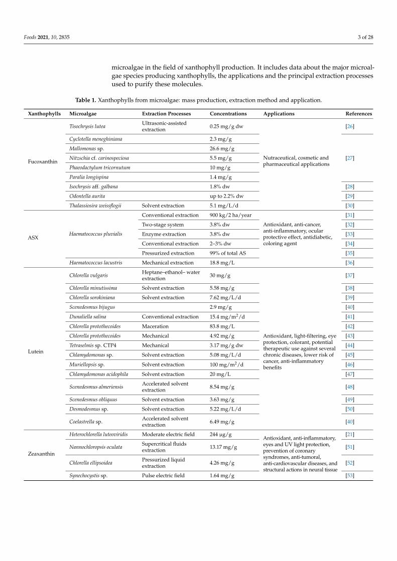
Microalgae present a raw material of interest because of their pigment content, whichis known by their biological activities. Currently, few species are used to produce xan-thophylls, as their industrial exploitation is rare. Table 1 illustrates the current use of
Foods 2021, 10, 2835 3 of 28
microalgae in the field of xanthophyll production. It includes data about the major microal-gae species producing xanthophylls, the applications and the principal extraction processesused to purify these molecules.
Table 1. Xanthophylls from microalgae: mass production, extraction method and application.
Xanthophylls Microalgae Extraction Processes Concentrations Applications References
Fucoxanthin
Tisochrysis lutea Ultrasonic-assistedextraction 0.25 mg/g dw
Nutraceutical, cosmetic andpharmaceutical applications
[26]
Cyclotella meneghiniana 2.3 mg/g
[27]
Mallomonas sp. 26.6 mg/g
Nitzschia cf. carinospeciosa 5.5 mg/g
Phaeodactylum tricornutum 10 mg/g
Paralia longispina 1.4 mg/g
Isochrysis aff. galbana 1.8% dw [28]
Odontella aurita up to 2.2% dw [29]
Thalassiosira weissflogii Solvent extraction 5.1 mg/L/d [30]
ASXHaematococcus pluvialis
Conventional extraction 900 kg/2 ha/year
Antioxidant, anti-cancer,anti-inflammatory, ocularprotective effect, antidiabetic,coloring agent
[31]
Two-stage system 3.8% dw [32]
Enzyme extraction 3.8% dw [33]
Conventional extraction 2–3% dw [34]
Pressurized extraction 99% of total AS [35]
Haematococcus lacustris Mechanical extraction 18.8 mg/L [36]
Lutein
Chlorella vulgaris Heptane–ethanol– waterextraction 30 mg/g
Antioxidant, light-filtering, eyeprotection, colorant, potentialtherapeutic use against severalchronic diseases, lower risk ofcancer, anti-inflammatorybenefits
[37]
Chlorella minutissima Solvent extraction 5.58 mg/g [38]
Chlorella sorokiniana Solvent extraction 7.62 mg/L/d [39]
Scenedesmus bijugus 2.9 mg/g [40]
Dunaliella salina Conventional extraction 15.4 mg/m2/d [41]
Chlorella protothecoides Maceration 83.8 mg/L [42]
Chlorella protothecoides Mechanical 4.92 mg/g [43]
Tetraselmis sp. CTP4 Mechanical 3.17 mg/g dw [44]
Chlamydomonas sp. Solvent extraction 5.08 mg/L/d [45]
Muriellopsis sp. Solvent extraction 100 mg/m2/d [46]
Chlamydomonas acidophila Solvent extraction 20 mg/L [47]
Scenedesmus almeriensis Accelerated solventextraction 8.54 mg/g [48]
Scenedesmus obliquus Solvent extraction 3.63 mg/g [49]
Desmodesmus sp. Solvent extraction 5.22 mg/L/d [50]
Coelastrella sp. Accelerated solventextraction 6.49 mg/g [40]
Zeaxanthin
Heterochlorella luteoviridis Moderate electric field 244 µg/gAntioxidant, anti-inflammatory,eyes and UV light protection,prevention of coronarysyndromes, anti-tumoral,anti-cardiovascular diseases, andstructural actions in neural tissue
[21]
Nannochloropsis oculata Supercritical fluidsextraction 13.17 mg/g [51]
Chlorella ellipsoidea Pressurized liquidextraction 4.26 mg/g [52]
Synechocystis sp. Pulse electric field 1.64 mg/g [53]
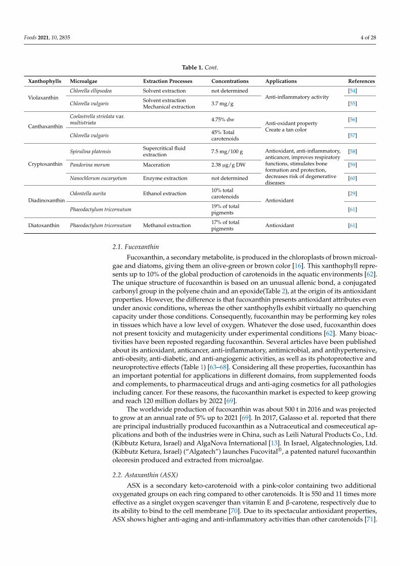
Foods 2021, 10, 2835 4 of 28
Table 1. Cont.
Xanthophylls Microalgae Extraction Processes Concentrations Applications References
ViolaxanthinChlorella ellipsodea Solvent extraction not determined
Anti-inflammatory activity[54]
Chlorella vulgaris Solvent extractionMechanical extraction 3.7 mg/g [55]
Canthaxanthin
Coelastrella striolata var.multistriata 4.75% dw
Anti-oxidant propertyCreate a tan color
[56]
Chlorella vulgaris 45% Totalcarotenoids [57]
Cryptoxanthin
Spirulina platensis Supercritical fluidextraction 7.5 mg/100 g Antioxidant, anti-inflammatory,
anticancer, improves respiratoryfunctions, stimulates boneformation and protection,decreases risk of degenerativediseases
[58]
Pandorina morum Maceration 2.38 µg/g DW [59]
Nanochlorum eucaryotum Enzyme extraction not determined [60]
DiadinoxanthinOdontella aurita Ethanol extraction 10% total
carotenoidsAntioxidant
[29]
Phaeodactylum tricornutum 19% of totalpigments [61]
Diatoxanthin Phaeodactylum tricornutum Methanol extraction 17% of totalpigments Antioxidant [61]
2.1. Fucoxanthin
Fucoxanthin, a secondary metabolite, is produced in the chloroplasts of brown microal-gae and diatoms, giving them an olive-green or brown color [16]. This xanthophyll repre-sents up to 10% of the global production of carotenoids in the aquatic environments [62].The unique structure of fucoxanthin is based on an unusual allenic bond, a conjugatedcarbonyl group in the polyene chain and an epoxide(Table 2), at the origin of its antioxidantproperties. However, the difference is that fucoxanthin presents antioxidant attributes evenunder anoxic conditions, whereas the other xanthophylls exhibit virtually no quenchingcapacity under those conditions. Consequently, fucoxanthin may be performing key rolesin tissues which have a low level of oxygen. Whatever the dose used, fucoxanthin doesnot present toxicity and mutagenicity under experimental conditions [62]. Many bioac-tivities have been reposted regarding fucoxanthin. Several articles have been publishedabout its antioxidant, anticancer, anti-inflammatory, antimicrobial, and antihypertensive,anti-obesity, anti-diabetic, and anti-angiogenic activities, as well as its photoprotective andneuroprotective effects (Table 1) [63–68]. Considering all these properties, fucoxanthin hasan important potential for applications in different domains, from supplemented foodsand complements, to pharmaceutical drugs and anti-aging cosmetics for all pathologiesincluding cancer. For these reasons, the fucoxanthin market is expected to keep growingand reach 120 million dollars by 2022 [69].
The worldwide production of fucoxanthin was about 500 t in 2016 and was projectedto grow at an annual rate of 5% up to 2021 [69]. In 2017, Galasso et al. reported that thereare principal industrially produced fucoxanthin as a Nutraceutical and cosmeceutical ap-plications and both of the industries were in China, such as Leili Natural Products Co., Ltd.(Kibbutz Ketura, Israel) and AlgaNova International [13]. In Israel, Algatechnologies, Ltd.(Kibbutz Ketura, Israel) (“Algatech”) launches Fucovital®, a patented naturel fucoxanthinoleoresin produced and extracted from microalgae.
2.2. Astaxanthin (ASX)
ASX is a secondary keto-carotenoid with a pink-color containing two additionaloxygenated groups on each ring compared to other carotenoids. It is 550 and 11 times moreeffective as a singlet oxygen scavenger than vitamin E and β-carotene, respectively due toits ability to bind to the cell membrane [70]. Due to its spectacular antioxidant properties,ASX shows higher anti-aging and anti-inflammatory activities than other carotenoids [71].
Foods 2021, 10, 2835 5 of 28
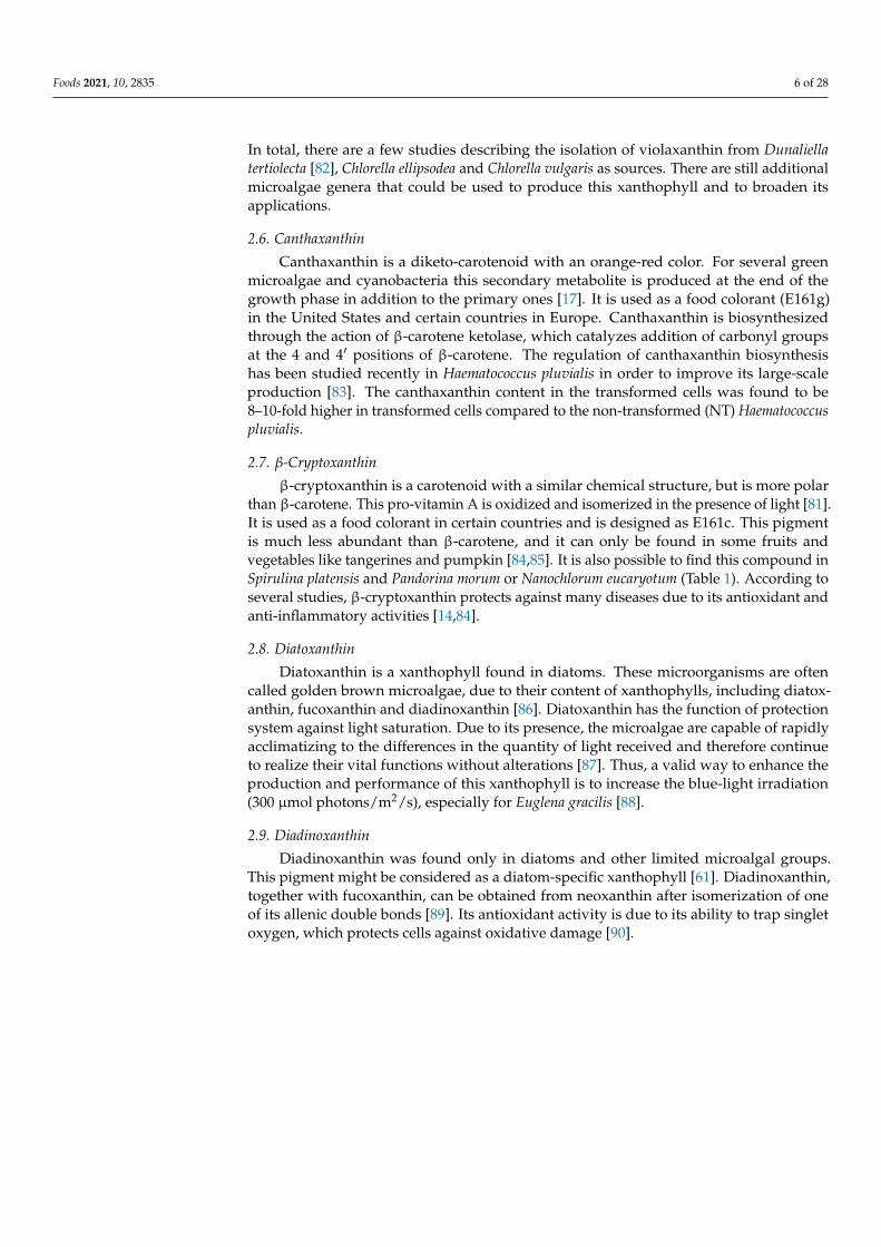
This xanthophyll occurs naturally in a large variety of microalgae such as Haematococcuspluvialis and Chlorella zofingiensis [72]. Haematococcus pluvialis is the most widely usednatural source for the industrial production of this pigment with yields of up to 3.8%DW [32,33,73]. It is relatively easy to purify ASX from microalgae because it represents90% of the total carotenoid content [74]. For all these reasons, various products containingASX are already available on the international market in diver forms such as soft, oils,syrups, creams, capsules with a market value of USD 1.0 billion in 2019 [16]. Two examplesare AstaPure® (Algatech Ltd., Kibbutz Ketura D.N, Israel, https://www.algatech.com/,accessed on 15 April 2021) and BioAstin (Cynotech Corporation, Kailua-Kona, HI, USA,https://www.Cyanotech.com/, accessed on 15 April 2021) produced from the microalgaeHaematococcus pluvialis.
2.3. Lutein
Lutein is one of the two essential compounds of the macular pigment of the retina [75].This xanthophyll is synthesized only by algae, is abundant in green microalgae [16]. Itacts as a strong antioxidant able to filter phototoxic blue-light radiation [76]. Chlorella is aneffective source of lutein and a good candidate for its production [43,77]. The results of thesestudies showed that nitrogen limitation and high temperature stress have been identified asthe main parameters impacting lutein accumulation. However, the cultivation conditionsof other microalgae species with significant lutein contents such as Scenedesmus almeriensis,Dunaliella salina and Galdieria sulphuraria have also been an optimized production of thispigment. According to the results of these optimizations, the contribution of nutrients hasa lesser effect due to the high tolerance of these microalgae to large ranges of salinity, pH,temperature and nutrient concentration [78,79].
Up to now, there are no production systems for the commercial production of luteinfrom microalgae. Outdoor production systems of Muriellopsis sp have been installed ata pilot scale. In it Muriellopsis sp. was grown in 50 L tubular PBRs which produced40 g/m2/d of lutein. Scenedesmus almeriensis biomass wasproduced in a 4000 L tubular PBRfor lutein production and 290 mg/m2/d of lutein was obtained [79]. The lutein market isexpected to reach USD 410 million in 2027 at a compound annual growth rate of 6.1% overthe planned period 2020–2027.
2.4. Zeaxanthin
Zeaxanthin is present in large quantities in plants and algae. It plays a major role inthe xanthophyll cycle. Zeaxanthin is a structural isomer of lutein, and the two xanthophyllsdiffer from each other only in the location of a single double bond. Indeed, in zeaxanthin,all the double bonds are conjugated. This pigment performs an essential role in theprevention of age-associated macular degeneration; one of the major blindness causes [80].Furthermore, this compound may also be used in cancer prevention via its powerfulanti-inflammatory effect [81]. For these reasons, its extraction from algae, microalgae andcyanobacteria is of great interest. There are several studies that have investigated theproduction of this pigment from some microalgae species. Among them, one publicationdescribes a genetically modified strain of Nannochloropsis oculata, which accumulates 13 mgof zeaxanthin per gram of dried biomass [51]. Other species synthetizing zeaxanthininclude Chlorella ellipsoidea and Synechocystis sp. (Table 1). These microalgae can accumulatezeaxanthin up to nine times higher than red peppers which are the traditional source ofthis pigment. In addition, microalgae have the advantage over plants that the zeaxanthinis present in a free form, while it is present as a monoester and a diester of zeaxanthin inplants [52]. For this reason, numerous works focused on processes to produce zeaxanthinat a large scale from microalgae [81].
2.5. Violaxanthin
Violaxanthin is an orange xanthophyll pigment. It is present in diverse groups ofmicroalgae (Table 1). This xanthophyll is biosynthesized through epoxidation of zeaxanthin.
Foods 2021, 10, 2835 6 of 28
In total, there are a few studies describing the isolation of violaxanthin from Dunaliellatertiolecta [82], Chlorella ellipsodea and Chlorella vulgaris as sources. There are still additionalmicroalgae genera that could be used to produce this xanthophyll and to broaden itsapplications.
2.6. Canthaxanthin
Canthaxanthin is a diketo-carotenoid with an orange-red color. For several greenmicroalgae and cyanobacteria this secondary metabolite is produced at the end of thegrowth phase in addition to the primary ones [17]. It is used as a food colorant (E161g)in the United States and certain countries in Europe. Canthaxanthin is biosynthesizedthrough the action of β-carotene ketolase, which catalyzes addition of carbonyl groupsat the 4 and 4′ positions of β-carotene. The regulation of canthaxanthin biosynthesishas been studied recently in Haematococcus pluvialis in order to improve its large-scaleproduction [83]. The canthaxanthin content in the transformed cells was found to be8–10-fold higher in transformed cells compared to the non-transformed (NT) Haematococcuspluvialis.
2.7. β-Cryptoxanthin
β-cryptoxanthin is a carotenoid with a similar chemical structure, but is more polarthan β-carotene. This pro-vitamin A is oxidized and isomerized in the presence of light [81].It is used as a food colorant in certain countries and is designed as E161c. This pigmentis much less abundant than β-carotene, and it can only be found in some fruits andvegetables like tangerines and pumpkin [84,85]. It is also possible to find this compound inSpirulina platensis and Pandorina morum or Nanochlorum eucaryotum (Table 1). According toseveral studies, β-cryptoxanthin protects against many diseases due to its antioxidant andanti-inflammatory activities [14,84].
2.8. Diatoxanthin
Diatoxanthin is a xanthophyll found in diatoms. These microorganisms are oftencalled golden brown microalgae, due to their content of xanthophylls, including diatox-anthin, fucoxanthin and diadinoxanthin [86]. Diatoxanthin has the function of protectionsystem against light saturation. Due to its presence, the microalgae are capable of rapidlyacclimatizing to the differences in the quantity of light received and therefore continueto realize their vital functions without alterations [87]. Thus, a valid way to enhance theproduction and performance of this xanthophyll is to increase the blue-light irradiation(300 µmol photons/m2/s), especially for Euglena gracilis [88].
2.9. Diadinoxanthin
Diadinoxanthin was found only in diatoms and other limited microalgal groups.This pigment might be considered as a diatom-specific xanthophyll [61]. Diadinoxanthin,together with fucoxanthin, can be obtained from neoxanthin after isomerization of oneof its allenic double bonds [89]. Its antioxidant activity is due to its ability to trap singletoxygen, which protects cells against oxidative damage [90].
Foods 2021, 10, 2835 7 of 28
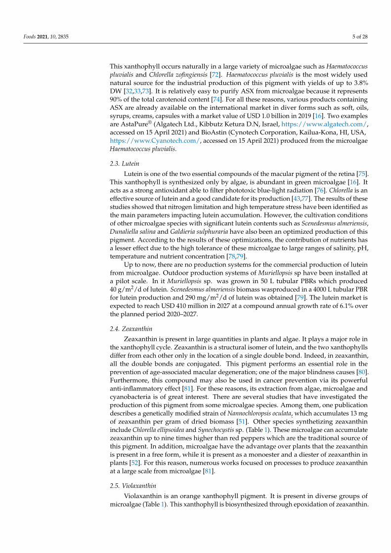
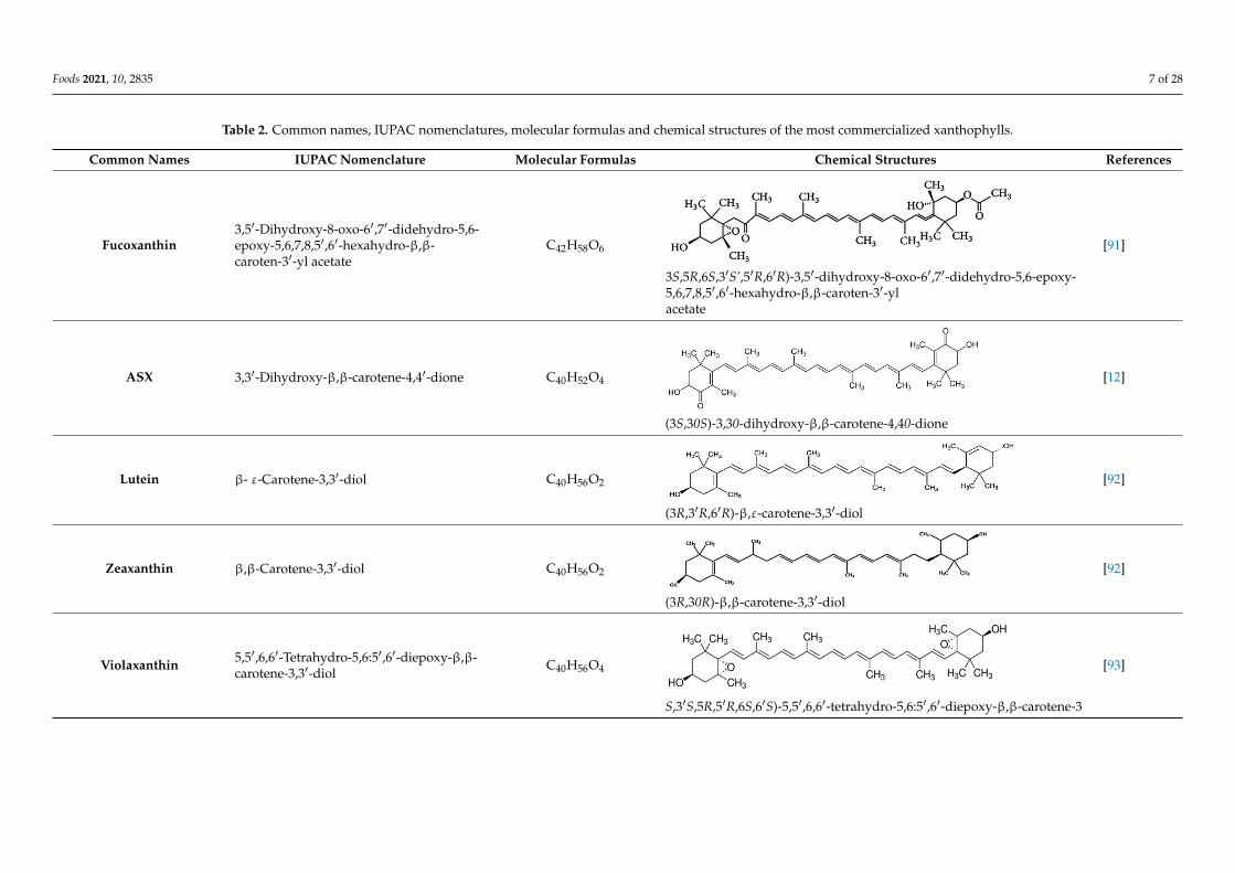
Table 2. Common names, IUPAC nomenclatures, molecular formulas and chemical structures of the most commercialized xanthophylls.
Common Names IUPAC Nomenclature Molecular Formulas Chemical Structures References
Fucoxanthin3,5′-Dihydroxy-8-oxo-6′,7′-didehydro-5,6-epoxy-5,6,7,8,5′,6′-hexahydro-β,β-caroten-3′-yl acetate
C42H58O6
Foods 2021, 10, x FOR PEER REVIEW 8 of 30
Table 2. Common names, IUPAC nomenclatures, molecular formulas and chemical structures of the most commercialized xanthophylls.
Common Names IUPAC Nomenclature Molecular Formulas Chemical Structures References
Fucoxanthin 3,5′-Dihydroxy-8-oxo-6′,7′- didehydro-5,6-epoxy-5,6,7,8,5′,6′- hexahydro-β,β-caroten-3′-yl acetate
C42H58O6
3S,5R,6S,3′S’,5′R,6′R)-3,5′-dihydroxy-8-oxo-6′,7′-didehydro-5,6-epoxy-5,6,7,8,5′,6′-hexahydro-β,β-caroten-3′-yl acetate
[91]
ASX 3,3′-Dihydroxy-β,β-carotene-4,4′-dione C40H52O4
(3S,30S)-3,30-dihydroxy-β,β-carotene-4,40-dione
[12]
Lutein β- ε-Carotene-3,3′-diol C40H56O2
(3R,3′R,6′R)-β,ε-carotene-3,3′-diol
[92]
Zeaxanthin β,β-Carotene-3,3′-diol C40H56O2
(3R,30R)-β,β-carotene-3,3′-diol
[92]
3S,5R,6S,3′S’,5′R,6′R)-3,5′-dihydroxy-8-oxo-6′,7′-didehydro-5,6-epoxy-5,6,7,8,5′,6′-hexahydro-β,β-caroten-3′-ylacetate
[91]
ASX 3,3′-Dihydroxy-β,β-carotene-4,4′-dione C40H52O4
Foods 2021, 10, x FOR PEER REVIEW 8 of 30
Table 2. Common names, IUPAC nomenclatures, molecular formulas and chemical structures of the most commercialized xanthophylls.
Common Names IUPAC Nomenclature Molecular Formulas Chemical Structures References
Fucoxanthin 3,5′-Dihydroxy-8-oxo-6′,7′- didehydro-5,6-epoxy-5,6,7,8,5′,6′- hexahydro-β,β-caroten-3′-yl acetate
C42H58O6
3S,5R,6S,3′S’,5′R,6′R)-3,5′-dihydroxy-8-oxo-6′,7′-didehydro-5,6-epoxy-5,6,7,8,5′,6′-hexahydro-β,β-caroten-3′-yl acetate
[91]
ASX 3,3′-Dihydroxy-β,β-carotene-4,4′-dione C40H52O4
(3S,30S)-3,30-dihydroxy-β,β-carotene-4,40-dione
[12]
Lutein β- ε-Carotene-3,3′-diol C40H56O2
(3R,3′R,6′R)-β,ε-carotene-3,3′-diol
[92]
Zeaxanthin β,β-Carotene-3,3′-diol C40H56O2
(3R,30R)-β,β-carotene-3,3′-diol
[92]
(3S,30S)-3,30-dihydroxy-β,β-carotene-4,40-dione
[12]
Lutein β- ε-Carotene-3,3′-diol C40H56O2
Foods 2021, 10, x FOR PEER REVIEW 8 of 30
Table 2. Common names, IUPAC nomenclatures, molecular formulas and chemical structures of the most commercialized xanthophylls.
Common Names IUPAC Nomenclature Molecular Formulas Chemical Structures References
Fucoxanthin 3,5′-Dihydroxy-8-oxo-6′,7′- didehydro-5,6-epoxy-5,6,7,8,5′,6′- hexahydro-β,β-caroten-3′-yl acetate
C42H58O6
3S,5R,6S,3′S’,5′R,6′R)-3,5′-dihydroxy-8-oxo-6′,7′-didehydro-5,6-epoxy-5,6,7,8,5′,6′-hexahydro-β,β-caroten-3′-yl acetate
[91]
ASX 3,3′-Dihydroxy-β,β-carotene-4,4′-dione C40H52O4
(3S,30S)-3,30-dihydroxy-β,β-carotene-4,40-dione
[12]
Lutein β- ε-Carotene-3,3′-diol C40H56O2
(3R,3′R,6′R)-β,ε-carotene-3,3′-diol
[92]
Zeaxanthin β,β-Carotene-3,3′-diol C40H56O2
(3R,30R)-β,β-carotene-3,3′-diol
[92]
(3R,3′R,6′R)-β,ε-carotene-3,3′-diol
[92]
Zeaxanthin β,β-Carotene-3,3′-diol C40H56O2
Foods 2021, 10, x FOR PEER REVIEW 8 of 30
Table 2. Common names, IUPAC nomenclatures, molecular formulas and chemical structures of the most commercialized xanthophylls.
Common Names IUPAC Nomenclature Molecular Formulas Chemical Structures References
Fucoxanthin 3,5′-Dihydroxy-8-oxo-6′,7′- didehydro-5,6-epoxy-5,6,7,8,5′,6′- hexahydro-β,β-caroten-3′-yl acetate
C42H58O6
3S,5R,6S,3′S’,5′R,6′R)-3,5′-dihydroxy-8-oxo-6′,7′-didehydro-5,6-epoxy-5,6,7,8,5′,6′-hexahydro-β,β-caroten-3′-yl acetate
[91]
ASX 3,3′-Dihydroxy-β,β-carotene-4,4′-dione C40H52O4
(3S,30S)-3,30-dihydroxy-β,β-carotene-4,40-dione
[12]
Lutein β- ε-Carotene-3,3′-diol C40H56O2
(3R,3′R,6′R)-β,ε-carotene-3,3′-diol
[92]
Zeaxanthin β,β-Carotene-3,3′-diol C40H56O2
(3R,30R)-β,β-carotene-3,3′-diol
[92]
(3R,30R)-β,β-carotene-3,3′-diol
[92]
Violaxanthin 5,5′,6,6′-Tetrahydro-5,6:5′,6′-diepoxy-β,β-carotene-3,3′-diol
C40H56O4
Foods 2021, 10, x FOR PEER REVIEW 9 of 30
Violaxanthin 5,5′,6,6′-Tetrahydro-5,6:5′,6′- diepoxy-β,β-carotene-3,3′-diol
C40H56O4
S,3′S,5R,5′R,6S,6′S)-5,5′,6,6′-tetrahydro-5,6:5′,6′-diepoxy-β,β-carotene-3
[93]
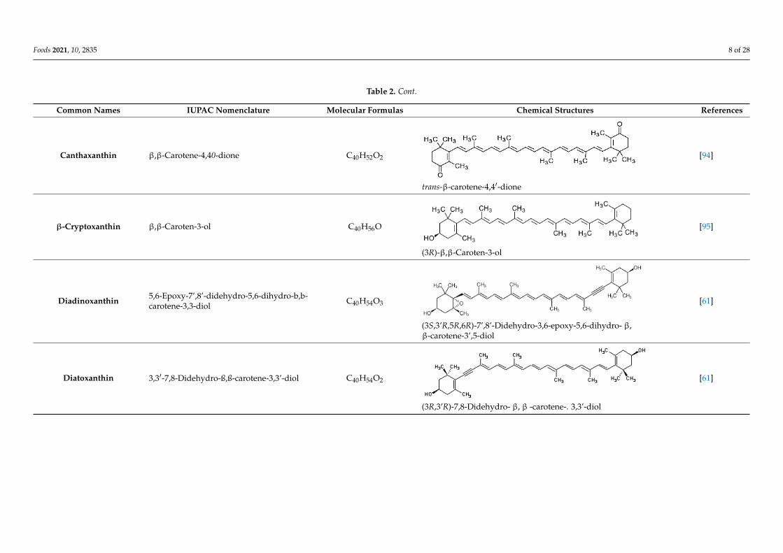
Canthaxanthin β,β-Carotene-4,40-dione C40H52O2
trans-β-carotene-4,4′-dione
[94]
β-Cryptoxanthin β,β-Caroten-3-ol C40H56O
(3R)-β,β-Caroten-3-ol
[95]
Diadinoxanthin 5,6-Epoxy-7’,8’-didehydro-5,6-dihydro-b,b-carotene-3,3-diol
C40H54O3
(3S,3’R,5R,6R)-7’,8’-Didehydro-3,6-epoxy-5,6-dihydro- β, β -carotene-3’,5-diol
[61]
Diatoxanthin 3,3′-7,8-Didehydro-ß,ß-carotene-3,3’-diol C40H54O2
(3R,3’R)-7,8-Didehydro- β, β -carotene-. 3,3’-diol
[61]
S,3′S,5R,5′R,6S,6′S)-5,5′,6,6′-tetrahydro-5,6:5′,6′-diepoxy-β,β-carotene-3
[93]
Foods 2021, 10, 2835 8 of 28
Table 2. Cont.
Common Names IUPAC Nomenclature Molecular Formulas Chemical Structures References
Canthaxanthin β,β-Carotene-4,40-dione C40H52O2
Foods 2021, 10, x FOR PEER REVIEW 9 of 30
Violaxanthin 5,5′,6,6′-Tetrahydro-5,6:5′,6′- diepoxy-β,β-carotene-3,3′-diol
C40H56O4
S,3′S,5R,5′R,6S,6′S)-5,5′,6,6′-tetrahydro-5,6:5′,6′-diepoxy-β,β-carotene-3
[93]
Canthaxanthin β,β-Carotene-4,40-dione C40H52O2
trans-β-carotene-4,4′-dione
[94]
β-Cryptoxanthin β,β-Caroten-3-ol C40H56O
(3R)-β,β-Caroten-3-ol
[95]
Diadinoxanthin 5,6-Epoxy-7’,8’-didehydro-5,6-dihydro-b,b-carotene-3,3-diol
C40H54O3
(3S,3’R,5R,6R)-7’,8’-Didehydro-3,6-epoxy-5,6-dihydro- β, β -carotene-3’,5-diol
[61]
Diatoxanthin 3,3′-7,8-Didehydro-ß,ß-carotene-3,3’-diol C40H54O2
(3R,3’R)-7,8-Didehydro- β, β -carotene-. 3,3’-diol
[61]
trans-β-carotene-4,4′-dione
[94]
β-Cryptoxanthin β,β-Caroten-3-ol C40H56O
Foods 2021, 10, x FOR PEER REVIEW 9 of 30
Violaxanthin 5,5′,6,6′-Tetrahydro-5,6:5′,6′- diepoxy-β,β-carotene-3,3′-diol
C40H56O4
S,3′S,5R,5′R,6S,6′S)-5,5′,6,6′-tetrahydro-5,6:5′,6′-diepoxy-β,β-carotene-3
[93]
Canthaxanthin β,β-Carotene-4,40-dione C40H52O2
trans-β-carotene-4,4′-dione
[94]
β-Cryptoxanthin β,β-Caroten-3-ol C40H56O
(3R)-β,β-Caroten-3-ol
[95]
Diadinoxanthin 5,6-Epoxy-7’,8’-didehydro-5,6-dihydro-b,b-carotene-3,3-diol
C40H54O3
(3S,3’R,5R,6R)-7’,8’-Didehydro-3,6-epoxy-5,6-dihydro- β, β -carotene-3’,5-diol
[61]
Diatoxanthin 3,3′-7,8-Didehydro-ß,ß-carotene-3,3’-diol C40H54O2
(3R,3’R)-7,8-Didehydro- β, β -carotene-. 3,3’-diol
[61]
(3R)-β,β-Caroten-3-ol
[95]
Diadinoxanthin 5,6-Epoxy-7’,8’-didehydro-5,6-dihydro-b,b-carotene-3,3-diol C40H54O3
Foods 2021, 10, x FOR PEER REVIEW 9 of 30
Violaxanthin 5,5′,6,6′-Tetrahydro-5,6:5′,6′- diepoxy-β,β-carotene-3,3′-diol
C40H56O4
S,3′S,5R,5′R,6S,6′S)-5,5′,6,6′-tetrahydro-5,6:5′,6′-diepoxy-β,β-carotene-3
[93]
Canthaxanthin β,β-Carotene-4,40-dione C40H52O2
trans-β-carotene-4,4′-dione
[94]
β-Cryptoxanthin β,β-Caroten-3-ol C40H56O
(3R)-β,β-Caroten-3-ol
[95]
Diadinoxanthin 5,6-Epoxy-7’,8’-didehydro-5,6-dihydro-b,b-carotene-3,3-diol
C40H54O3
(3S,3’R,5R,6R)-7’,8’-Didehydro-3,6-epoxy-5,6-dihydro- β, β -carotene-3’,5-diol
[61]
Diatoxanthin 3,3′-7,8-Didehydro-ß,ß-carotene-3,3’-diol C40H54O2
(3R,3’R)-7,8-Didehydro- β, β -carotene-. 3,3’-diol
[61]
(3S,3’R,5R,6R)-7’,8’-Didehydro-3,6-epoxy-5,6-dihydro- β,β-carotene-3’,5-diol
[61]
Diatoxanthin 3,3′-7,8-Didehydro-ß,ß-carotene-3,3’-diol C40H54O2
Foods 2021, 10, x FOR PEER REVIEW 9 of 30
Violaxanthin 5,5′,6,6′-Tetrahydro-5,6:5′,6′- diepoxy-β,β-carotene-3,3′-diol
C40H56O4
S,3′S,5R,5′R,6S,6′S)-5,5′,6,6′-tetrahydro-5,6:5′,6′-diepoxy-β,β-carotene-3
[93]
Canthaxanthin β,β-Carotene-4,40-dione C40H52O2
trans-β-carotene-4,4′-dione
[94]
β-Cryptoxanthin β,β-Caroten-3-ol C40H56O
(3R)-β,β-Caroten-3-ol
[95]
Diadinoxanthin 5,6-Epoxy-7’,8’-didehydro-5,6-dihydro-b,b-carotene-3,3-diol
C40H54O3
(3S,3’R,5R,6R)-7’,8’-Didehydro-3,6-epoxy-5,6-dihydro- β, β -carotene-3’,5-diol
[61]
Diatoxanthin 3,3′-7,8-Didehydro-ß,ß-carotene-3,3’-diol C40H54O2
(3R,3’R)-7,8-Didehydro- β, β -carotene-. 3,3’-diol
[61]
(3R,3’R)-7,8-Didehydro- β, β -carotene-. 3,3’-diol
[61]
Foods 2021, 10, 2835 9 of 28
3. Structures of Xanthophylls
Carotenoids are the most diverse and widespread lipophilic compounds found innature and are generally colored yellow, orange or red [96]. Most carotenoids share acommon C40 backbone structure of isoprene units (termed terpenoid) and are dividedinto two groups: carotenes and xanthophylls. Each of the carotenoids consist of differenttrans and cis isomers. Xanthophylls are oxidized derivatives of carotenes (which arehydrocarbons). The molecules most representative of xanthophylls are lutein, fucoxanthin,β-cryptoxanthin, ASX and zeaxanthin. They are more polar than carotenes due to thepresence of oxygen at the ends of their rings in the form of a methoxy, keto, hydroxy,epoxy and carboxy groups [5]. Indeed, lutein and zeaxanthin are characterized by thepresence of -OH groups in their structures, while canthaxanthin and echinenone contain= O groups. ASX is a xanthophyll which has both -OH and =O groups in its structure.There are xanthophylls such as violaxanthin and diadinoxanthin, which contain epoxygroups in their structures, while others such as dinoxanthin and fucoxanthin have acetylgroups. In addition, the two acetyl-containing carotenoids also contain the group C=C=C(allene), which is exceptional in natural products [97]. Certain carotenoids such as hetero-,allo-, diadino-, diato-, pyro- croco- and monadoxanthine present in their structures C≡C(acetylene) groups. As powerful antioxidants, xanthophylls are generally sensitive toseveral factors such as light, oxygen and heat, leading to difficulties in their purificationand storage. The structures of the most abundant xanthophylls are shown in Table 2.
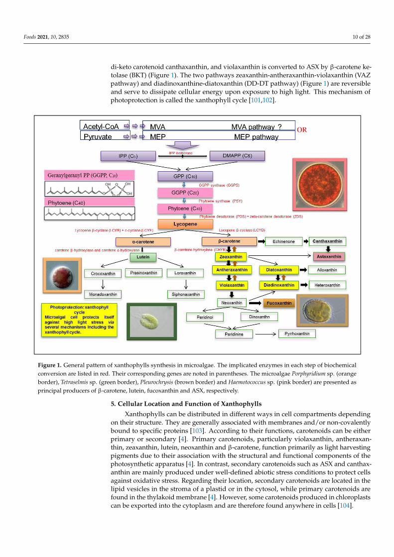
4. Biosynthesis of Xanthophylls
All carotenoids are synthetized from a common precursor to all isoprenoids, theisopentenyl pyrophosphate (IPP). The biosynthesis of IPP may generally be performed byone of two different pathways (Figure 1): the mevalonate pathway (MVA) in euglenophytesand the plastidial 2-C-methyl-D-erythriol-4-phosphate (MEP) pathway in Chlorophyceaeand Cyanophyceae [42,67,96,98]. The MEP pathway is considered to be the main biosyn-thetic pathway of carotenoids in several microalgae species such as Chlorella vulgaris,Dunaliella salina, Scenedesmus sp., and Haematococcus pluvialis [98,99]. In this pathway, thecarotenoid biosynthesis begins through IPP, which isomerises to dimethylallyl pyrophos-phate (DMAPP) by the action of the IPP isomerase. Elongation of the carbon chain tookplace through continuous head-to-tail condensation of IPP to DMAPP followed by growingof the polyprenyl pyrophosphate chain by the action of the prenyltransferase [55,100]. Thesecond committing step in carotenoid biosynthesis is the condensation of two moleculesof geranyl pyrophosphate (GPP, C10), catalyzed by the GGPP synthase, to yield geranyl-geranyl PP (GGPP, C20) [42]. After that, the colourless C40 carotenoid phytoene is formedthrough the condensation of two GGPP (C20) molecules by the action of phytoene synthase(PSY) [42]. From this step onwards, phytoene undergoes a series of sequential desatura-tions (four desaturation steps) catalyzed by phytoene desaturase (PDS) and zeta-carotenedesaturase (ZDS)leading to the formation of pro-lycopene [42]. Pro-lycopene is then iso-merized by a specific carotenoid isomerase (CRTISO) into all-trans-lycopene. At this level,the pathway is divided into two branches. A first branch leads to the formation of β-carotene and its xanthophyll derivatives, such as zeaxanthin and ASX, and the secondbranch leads to the formation of α-carotene and lutein in microalgae. The formation of βand ε rings at both ends of lycopene results in a yield of α-carotene under the catalysisof lycopene β-cyclase (LCYB) and ε-cyclase (LCYE) respectively, whereas the formationof two β-rings at the two ends of lycopene gives rise to β-carotene catalyzed by LCY-balone [42]. B-carotene can be further hydroxylated by the β-carotene hydroxylase (CHYB)to give zeaxanthin. The amounts of carotenoids produced by each branch of the pathwayare dependent on activities of LCYE and LCYB. α-carotene hydroxylation is assured bytwo heme-containing cytochrome P450 monoxygenases (namely, β carotene and caroteneε-hydroxylases), which formslutein. In a first branch, zeaxanthin is transformed into violax-anthin by the enzyme zeaxanthin epoxidase (ZEP), which incorporates two epoxy groupsat positions C-5,6 and C-5′,6′. Zeaxanthin is also transformed in a second branch into
Foods 2021, 10, 2835 10 of 28
di-keto carotenoid canthaxanthin, and violaxanthin is converted to ASX by β-carotene ke-tolase (BKT) (Figure 1). The two pathways zeaxanthin-antheraxanthin-violaxanthin (VAZpathway) and diadinoxanthine-diatoxanthin (DD-DT pathway) (Figure 1) are reversibleand serve to dissipate cellular energy upon exposure to high light. This mechanism ofphotoprotection is called the xanthophyll cycle [101,102].
Foods 2021, 10, x FOR PEER REVIEW 12 of 30
which incorporates two epoxy groups at positions C-5,6 and C-5′,6′. Zeaxanthin is also transformed in a second branch into di-keto carotenoid canthaxanthin, and violaxanthin is converted to ASX by β-carotene ketolase (BKT) (Figure 1). The two pathways zeaxan-thin-antheraxanthin-violaxanthin (VAZ pathway) and diadinoxanthine-diatoxanthin (DD-DT pathway) (Figure 1) are reversible and serve to dissipate cellular energy upon exposure to high light. This mechanism of photoprotection is called the xanthophyll cycle [101,102].
Figure 1. general pattern of xanthophylls synthesis in microalgae. The implicated enzymes in each step of biochemical conversion are listed in red. Their corresponding genes are noted in parentheses. The microalgae Porphyridium sp. (orange border), Tetraselmis sp. (green border), Pleurochrysis (brown border) and Haemotococcus sp. (pink border) are presented as principal producers of β-carotene, lutein, fucoxanthin and ASX, respectively.
5. Cellular Location and Function of Xanthophylls Xanthophylls can be distributed in different ways in cell compartments depending
on their structure. They are generally associated with membranes and/or non-covalently bound to specific proteins [103]. According to their functions, carotenoids can be either primary or secondary [4]. Primary carotenoids, particularly violaxanthin, antheraxanthin, zeaxanthin, lutein, neoxanthin and β-carotene, function primarily as light harvesting pig-ments due to their association with the structural and functional components of the pho-tosynthetic apparatus [4]. In contrast, secondary carotenoids such as ASX and canthaxan-thin are mainly produced under well-defined abiotic stress conditions to protect cells against oxidative stress. Regarding their location, secondary carotenoids are located in the lipid vesicles in the stroma of a plastid or in the cytosol, while primary carotenoids are found in the thylakoid membrane [4]. However, some carotenoids produced in chloro-plasts can be exported into the cytoplasm and are therefore found anywhere in cells [104].
Figure 1. General pattern of xanthophylls synthesis in microalgae. The implicated enzymes in each step of biochemicalconversion are listed in red. Their corresponding genes are noted in parentheses. The microalgae Porphyridium sp. (orangeborder), Tetraselmis sp. (green border), Pleurochrysis (brown border) and Haemotococcus sp. (pink border) are presented asprincipal producers of β-carotene, lutein, fucoxanthin and ASX, respectively.
5. Cellular Location and Function of Xanthophylls
Xanthophylls can be distributed in different ways in cell compartments dependingon their structure. They are generally associated with membranes and/or non-covalentlybound to specific proteins [103]. According to their functions, carotenoids can be eitherprimary or secondary [4]. Primary carotenoids, particularly violaxanthin, antheraxan-thin, zeaxanthin, lutein, neoxanthin and β-carotene, function primarily as light harvestingpigments due to their association with the structural and functional components of thephotosynthetic apparatus [4]. In contrast, secondary carotenoids such as ASX and canthax-anthin are mainly produced under well-defined abiotic stress conditions to protect cellsagainst oxidative stress. Regarding their location, secondary carotenoids are located in thelipid vesicles in the stroma of a plastid or in the cytosol, while primary carotenoids arefound in the thylakoid membrane [4]. However, some carotenoids produced in chloroplastscan be exported into the cytoplasm and are therefore found anywhere in cells [104].
Foods 2021, 10, 2835 11 of 28
Xanthophylls assure various physiological functions in microalgae: they are primarilyinvolved in light harvesting, but also participate to stabilizing the structure and aid in thefunction of photosynthetic complexes besides protecting chlorophyll from being damagedby visible light or near-UV radiation [105]. They safeguard unsaturated fatty acids (UFAs)contained in the cellular membrane from photo- and pero-oxidations [103]. Furthermore,they act as efficient antioxidants by scavenging free radicals, which may attack DNA andRNA, as well as metabolites such as membrane proteins [103].
6. Recent Applications in Metabolic Engineering for Xanthophylls Production
Microalgae are considered to be ideal model hosts for metabolic engineering sincethey offer many advantages, such as a simplicity of culture and fast growth rates comparedwith plants. Furthermore, eukaryotic microalgae present more genetic and physiologicalsimilarities with plant cells than bacteria [96]. In addition, many microalgae have an activecentral terpenoid metabolism, allowing a great enough supply of precursors and a highcapacity for xanthophylls storage [96]. Nevertheless, studies into the modification of thesepigment pathways in microalgae are infrequent.
Mutagenesis, with its various methods, has been applied to wild strains of microalgaeto improve their xanthophyll production. In 2001, Jin and collaborators used mutagenesisto increase production of zeaxanthin by Dunaliella salina and ultimately succeeded in gener-ating two overproducing strains of zeaxanthin. One of these zeaxanthin epoxidase mutantswas recognized in the study carried out by Jin and Melis in 2003 [106]. Similar mutationshave also been conducted in other microalgae strains, such as Scenedesmus obliquus andChlamydomonas reinhardtii [107]. The zeaxanthin content (per cell) is 15-fold higher than thewild type under non-stressed conditions [108]. Zeaxanthin has previously been engineeredby chemical mutagenesis into Chlorella zofingiensis [109]. Its production reached 4 and7 mg/g DW in mixotrophic and photosynthetic conditions, respectively [109]. Manipu-lation of microalgae to make them able to produce higher quantities of xanthophylls canalso been carried out by the inactivation or the overexpression of own genes or by theexpression of genes from other species [16]. This would allow us to obtain geneticallymodified species of microalgae with ameliorated or decreased enzymatic activities and ableto accumulate the desired pigments. Recently, in 2018, Sarnaik et al. introduced the exoge-nous β-carotene oxygenase gene (CrtR) from the strain Synechoccocus elongatus (PCC 7002),to prolong carotenoid pathway toward zeaxanthin production in vivo [110]. Associatingthe CrtR gene with the insertion of the galactose transporter from Escherichia coli led to re-spective zeaxanthin yields of 9 mg/g DW and 8 mg/g DW in autotrophic and mixotrophicconditions [110]. ASX is commercially produced, generally for its human health benefits,and is used as an anti-inflammatory compound and as a skin protector. It can also be usedas an essential component of aquaculture feed [71,111]. Haematococcus pluvialis, the mostcommon strain of microalgae, used for the production of ASX, accumulates between 9 and36 mg/g DW of this pigment depending on culture conditions. However, production ofASX by other species is currently under study to bypass the constraints of culture observedwith Haematococcus strains [112]. Even if Synechocystis sp., a cyanobacteria, does not natu-rally express the genes encoding for BKT and CRTR-B, and the enzymes can be engineeredusing genes from Haematococcus pluvialis under the promoters cpc560 for BKT and psbA2CrtR-B [113]. These successful gene expressions led to the conversion of ASX precursorsechinenone and zeaxanthin, into 4.8 mg/g DW ASX in nitrogen deprivation [113]. Similarly,the Chlamydomonas reinhardtii UVM4 strain mutant has become able to produce ASX after aredesign by codon optimization of the Bkt gene. The latter was a dysfunctional endogenousgene [114] which was targeted at the chloroplast with a psaD transit sequence. Hence, ASXproduction yields of 1 and 3 mg/L/day in autotrophy and mixotrophy conditions wererespectively achieved [114]. Additionally, the endogenous PDS was recently overexpressedin the chloroplast of Haematococcus pluvialis by optimizing codons and using the psbA pro-moter and UTR [115], demonstrating the capacity of expressing a nuclear gene successfullyin Haematococcus pluvialis chloroplast. ASX yields reached in the efficient transformants
Foods 2021, 10, 2835 12 of 28
were up to 34 mg/L instead of 18 mg/L in the wild strain microalgae [115]. Fucoxanthin isanother highly researched carotenoid due to its anti-obesity, anti-diabetic and anti-cancerproperties. It is also well established as strong antioxidant compounds [29]. With the aimof increasing fucoxanthin production, Manfellotto et al. (2020) transformed Phaeodacty-lum tricornutum with single plasmids or combinations of them for the overexpression ofgenes with a putative role in xanthophylls biosynthesis [116]. They obtained two tripletransformant genes: Vde-related (VDR),Zeaxanthin epoxidase 3 (ZEP3) and Violaxanthinde-epoxidase (VDE), were over expressed, allowing the carotenoids accumulation with afour-fold increase in the fucoxanthin content compared to the wild strain [116]. Similarly,overexpression of the Psy gene in Phaeodactylum tricornutum allowed a fucoxanthin content1.45 times higher than that in the wild type strain [117]. In the same way, overexpressionof Dxs and Psy genes led to an increase in fucoxanthin content of 2.4 fold and 1.8-foldrespectively [117,118]. Similar metabolic engineering studies were realized for the overproduction of lutein.
Chlamydomonas reinhardtii was transformed using the genes of Dunaliella salina andChlorella zofingiensis, producing respectively 2.6- and 2.2-fold higher yields of lutein [119].A point mutation was introduced in the endogenous gene encoding PDS of Chlamydomonasreinhardtii to enhance its expression. Concomitantly to it, the increase of lutein productionwas observed [120]. In a very similar approach using Chlamydomonas reinhardtii, the geneencoding for PBS from Xanthophyllomyces dendrorhous was introduced into pMS188 plasmidand the nuclear transformed [121]. The mutated microalga possesses a bifunctional enzymewith the two PSY and LCYB activities. This allowed carotenoid accumulation for the firsttime using an heterologous expression system, leading to a simultaneous increase of about60% in lutein biosynthesis under low light culture conditions. Random mutagenesis hasbeen effectively used for the production of Chlorella sorokiniana mutants with high contentsof lutein [119].
In another study, ethyl methane sulfonate and N-methyl-N′-nitro-N-nitrosoguanidinehave been used as chemical mutagenes for generating a lutein-deficient Chlorella vulgaris(CvLD), which was found to be an enhanced producer of the pigment violaxanthin [55]. Thesequencing of the lcy-e gene of this lutein-deficient Chlorella vulgaris led to the identificationof a single mutation at the position 336. The mutated Valine, instead of an Alanine, mighthave occurred in the active site of the lycopene ε-cyclase, decreasing its activity.
7. Bioprocess for Xanthophylls Production by Microalgae
To acquire high xanthophyll productivity, both biomass production and its pigmentcontent need to be optimized.
7.1. Cultivation Systems
At this time, xanthophyll production from microalgae is achieved in open pondsystems or the closed photo-bioreactors (PBRs).
7.1.1. Open Systems
The cost of construction and operation in open systems are reported to be much lowerthan for closed PBRs and the cultivation process is also simpler. Open ponds is the mostcommercially used method for cultures of microalgae, in which the medium flow occursthrough a system of paddle-wheels. The latter keeps the cells in suspension and providesbetter mass transfer [122]. The low deepness in these open systems ensures the light pene-tration efficiency. The system flow is continuous, so nutrients are continuously suppliedand microalgae are harvested at the same time [123]. This system is ideal for the growth ofmicroorganisms that tolerate and can grow under extreme environment conditions suchas high alkalinity (Spirulina sp.), high salinity (Dunaliella salina) and nutrient-rich media(Chlorella sp.). Indeed, the photoautotrophic culture mode has been extensively employedfor Dunaliella with the aim of carotenoid overproduction [124]. A two-step system, namely“intensive cultivation”, allowed large-scale carotenoid production in Dunaliella. The aim of
Foods 2021, 10, 2835 13 of 28
stage one is to promote biomass accumulation with a weak β-carotene-chlorophyll ratio;and in stage two, Dunaliella culture is diluted three times to increase the light penetra-tion to cells, and carotenogenesis after nitrogen depletion [79]. Therefore, it is alwaysrecommended to use open and raceway ponds for cultivating microalgae using a photoau-totrophic growing condition to minimize contamination issues. However, microalgae thatcannot grow in these specific environmental conditions, such as Tetraselmis sp., Isochrysissp., Crypthecodinium sp., and Skeletonema sp., should not be cultured with this type ofapproach [125]. Some drawbacks are observed from this method; for example, it is difficultto control the conditions around the tank, such as the temperature and light, and there isa high risk of contamination by other algal/bacterial strains [126]. Other crucial factorscould have a great influence on these systems amongst other large losses of water afterevaporation, CO2 diffusion into the atmosphere, and the need for large land areas [122].Therefore, the closed systems seem to be preferred.
7.1.2. Closed Systems
Open system problems have led to the design of closed systems. PBRs representthe most successful approach to attain better control of important culture parameterslike pH, light, temperature, loss of H2O, capture of CO2 and biomass productivity [127].Furthermore, the low contamination risk is a main asset that would permit a higher controland production of molecules with high-commercial values, such as xanthophylls. PBRswere designed for the cultivation of microalgae [123,126], including the following types:
- The tubular type is the most appropriate kind of PBR for producing satisfying high-quality cyanobacteria and microalgae biomasses in outdoor environments [123,128].It is generally built with glass or plastic tubes, allowing a large illuminated surfacearea. In this system, the culture homogenization is generally assured by means of airpumps. It is characterized by some defects, such as pH variation, dissolved oxygen,fouling, and CO2 heterogeneity. There are many studies indicating the suitability ofusing this PBR kind for high-quality microalgae and cyanobacteria productions.
- Flat PBRs have a large surface exposed to light and are characterized by high algalproductivities, which is generally greater than those produced by tubular PBR. Thisculture system is constructed from a rigid transparent material to optimize lightcapture and to facilitate sterilization. It is suitable for outdoor cultivation, ideal forcell immobilization and is relatively inexpensive. The only drawback of this typeof system is the difficulty in controlling the temperature of algal cultures [129]. FlatPBRs have been tested for culturing the marine diatom Phaeodactylum tricornutumfor the production of fucoxanthin and chrysolaminarin [130]. The AlgaTechnologiesindustry (https://www.algatech.com/, accessed on 15 April 2021) also establisheda Haematococcus cultivation facility back in the late 1990s. Quite different to otherAmerican industries, the AlgaTechnologies Company used glass tubular PBRs forboth green and red phases to phototrophically cultivate Haematococcus [131].
Problems that are associated with a limited light source that hinders high cell densityin large-scale PBRs during photoautotrophic growth can be avoided by using heterotrophiccultivations [132]. The elimination of light restriction led to a higher microalgae cellgrowth rate and a greater cell mass content can be reached faster. So far, the maximumproduced biomass in photoautotrophic conditions was about 40 g/L of microalga dryweight [133] and this content was lower than in that obtained in heterotrophic conditions(150 g/L) [134]. Heterotrophic culture mode is mainly used for the production of highvalue-added xanthophylls (lutein, ASX . . . ) from microalgae, due to its high cost [79]. Thedry weight of the green microalgae Chlorella protothecoides and its lutein content attained19 g/L and 84 mg/L respectively, when it was cultured heterotrophically. These valuesreached 47 g/L and 225 mg/L using a culture with the fed-batch system [135]. Wu and Shi(2006) found the highest biomass concentrations and a maximum productivity of 105 g/Lcell dry weight and 0.613 g/L/h, respectively, when Chlorella pyrenoidosa was grown inheterotrophic conditions [136].
Foods 2021, 10, 2835 14 of 28
Hybrid systems are deployed for the large-scale production of marine microalgaeto produce some molecules of commercial interest. In 2015, The diatom Staurosina sp.and the chlorophyte Desmodesmus sp. were cultured in a hybrid system combining PBRs(25 m3) and open basins (400 m2) [137]. In this system, the PBRs permanently ensured asource of uncontaminated inoculum for the short-lived batch culture in an open pond. Thelatter ensures a large-scale biomass production in a competitive cost- and time-consumingoperation. For the production of ASX from the green microalgae Haematococcus pluvialis,Cyanotech corporation https://www.cyanotech.com/is, (accessed on 15 April 2021) anexample of industries that use PBRs for the green phase (vegetative growth) and pondsas open systems for the red phase (production of xanthophylls). Mera PharmaceuticalsInc. (http://www.merapharma.com/, accessed on 15 April 2021) was among the firstcompanies which established large scale ASX production facilities globally. The companywas located where weather conditions are extremely suitable for outdoor Haematococcuscultivation [138]. Based on the same system, the company employed a two-step autotrophiccultivation approach. The green phase of microalgae growth was conducted in 25,000 Lcomputerized outdoor PBRs, and the red phase of ASX accumulation in raceway ponds.Mixotrophic culture of Haematococcus has long been sought as an alternative approachfor the traditional two-step ASX production process [139,140]. In fact, the world’s firstcommercial ASX production facility from the microalga Haematococcus appears to have beenbased on mixotrophic culture technologies in 1995 [138]. However, since then, phototrophicculture approaches have been amply developed and have become the main strategy forthe production of ASX from microalgae. The advantage of mixotrophic cultivation isthat the production can be carried out indoors and under optimal controlled conditions.However, the cost of materials and energy consumption might be too high to compete withphototrophic culture using sunlight. Recently, a novel ASX production process based on amixotrophic mode has been developed with a heuristic multilevel LED light regime andthe highest content of 3.3% was achieved at the white-blue regime [141]. Polyol alcohols(glycerol and mannitol) have also been shown to be more efficient carbon sources thanacetate for the efficient and cost-effective ASX production from Haematococcus [142].
7.2. Factors Determining Xanthophylls Production
Secondary xanthophylls production is monitored by changes in culture conditions anddifferent stress factors [143]. Xanthophyll production is improved by ROS, under stress con-ditions like high light intensity and salinity [144]. For this reason, ASX is assumed to protectorganisms from free radical-linked diseases like cancer [145]. Several disadvantageousenvironmental conditions like nutrient deprivation, excessive photosynthesis and extremeirradiationreduce the incidence of electron transfer and, therefore, photo-oxidative dam-age [146]. Primary xanthophylls, like lutein, deteriorate under stress conditions, hence theircontent in cell biomass is diminished. Combined effects of numerous stress factors haveameliorated the ASX production in many microalgae such as Haematococcus pluvialis [147].
7.2.1. Light
Light availability constitutes the most effective controlling factor for the productionof numerous xanthophylls [119]. Light intensity and photoperiods affect the growthof cells, biomass and production of several high-value metabolites in many microalgaespecies. Higher light intensity caused a threefold increase in Haematococcus pluvialis ASXcontent. Besides, ASX and lutein contents are also changed under a high intensity oflight [148]. Muriellopsis sp. lutein content reached the maximum at 460 µmol photons/m2/s.Maximum lutein productivity (3.6 mg/L/day) was obtained under high light intensitywith Desmodesmus sp. [149] The lutein synthesis and accumulation were studied in Chlorellasorokiniana [119] and Scenedesmus sp. [78] because they both have high growth rates andhigh lutein production ability. Light stress was applied concomitantly with other stressorssuch as nitrogen [150], salinity [151] and temperature [152]. In addition, Zhao et al. (2018)demonstrated that of nitrogen lack and light stress together enhanced the ASX accumulation
Foods 2021, 10, 2835 15 of 28
in Haematococcus pluvialis, which reached 1.85% of the cell dry weight [153]. Light stress canstimulate the expression of the lycopene beta-cyclase gene [154], which is the key enzymefor carotenoid accumulation in microalgae. Moreover, Coesel et al. (2008) also showedthat high light intensity can regulate the activities of phytoene synthase and phytoenedesaturase [155].
7.2.2. Temperature
High temperatures play an important role in the accumulation of xanthophylls inmicroalgae due to the photooxidative stress [40]. High temperatures affect the synthe-sis of ASX in Haematococcus pluvialis [156] and Chromochloris zofingiensis [40]. Indeed,temperatures above 28 ◦C minimize the total productivity of ASX in Chromochloris zofin-giensis [148] because the ROSs are more assimilated through photosynthesis, which stim-ulates the buildup of ASX [157]. High temperatures also result in the accumulation oflutein in Dunaliella salina [158]. Similar observations were found for Chlorella protothecoidesstrains [135]. However, low temperatures decrease the nutrient uptake rate and slow thelutein accumulation [159].
7.2.3. Salinity
The salinity effect on microalgae growth and xanthophyll formation is complex. Manymicroalgae species are able to tolerate high salinity levels because of their osmoregulationcapacity, which involves constant glycerol synthesis [160]. Indeed, salt stress has a positiveeffect on the production of secondary xanthophylls such as ASX. For example, an ASXcontent of 5 mg/g was obtained with Haematococcus pluvialis when treated with 0.2 g/Lof NaCl, which was 42% higher than that of the control [161]. A general increase in ASXproduction was also observed with Chlorella zofingiensis grown at NaCl concentrationsof up to 0.2 M [148]. Furthermore, salinity stress alone did not change the content oflutein in Dunaliella salina [162], contrary to the combination of nitrate concentration andsalinity [163].
7.2.4. Nutrient-Related StressesNitrogen Starvation
The concentration of nitrogen in the culture medium and xanthophyll content ofmicroalgae were correlated in many species [164]. The production of xanthophylls wasincreased by nitrogen limitation in Neochloris oleoabundans, Chlorella zofingiensis, Dunaliellasalina, and Muriellopsis sp. [25,165,166]. In addition to ASX and lutein, β-carotene levelwas enhanced to 2.7% DW in Dunaliella salina cells in nitrogen-depletion [167]. Under thesame conditions, xanthophylls, namely ASX and canthaxanthin, initiate their accumulationin aerial microalgae (Coelastrella sp.); after this, the color of the cells changes from greento red [56]. Nitrogen starvation can promote the concomitant accumulation of ASX inmicroalgae [120,168] as well as lutein accumulation. The latter is highly dependent onnitrogen concentration in the culture medium [165]. In general, nitrogen deficiency hasa greater impact than excess nitrogen on carotenoid production, mainly that of ASX inHaematococcus pluvialis, because a culture growing in a nitrogen-rich medium requirescarbon to assimilate the nitrogen. On the other hand, high competition for carbon requiredfor xanthophylls synthesis would be established in low nitrogen concentrations [169]. Amixture of urea and other nitrogen sources has led to a maximal lutein production inAuxenochlorella protothecoides [77]. A two-step mode where nitrogen enriched and nitrogendeficient media were used consecutively stimulated the microalgae growth during the firstphase and carotenoid enrichment during the second phase.
Iron Supplementation
Iron is needed for microalgae growth such as Dunaliella. Of all the micronutrients, ironwas the best for accumulation of ASX in Haematococcus pluvialis cysts [144] because ironacts as a chelating agent and can scavenge hydroxyl radicals in the Fenton reaction, which
Foods 2021, 10, 2835 16 of 28
is widely used in the enzyme system of animals, microbes, and plants [170]. It acts as alimiting factor under hyper saline conditions. The site for assimilation of iron is usuallythe plasma membrane [171]. He et al. (2007) reported that ASX accumulation depends onmany nutrients, such as phosphorus, iron, and sulfur [171]. However, adding Fe2+- EDTAto culture medium produced ASX up to 3.1% dry cell in Haematococcus pluvialis [172].
Sulfur Limitation
Sulfur limitation is beneficial for xanthophylls production by microalgae and itsstarvation is more efficient than that of iron for high level accumulation of ASX [173].
Sulfur is essential for the glutathione biosynthesis [174]. Glutathione acts in theoxidative stress response as a ROS scavenger. So, an increase of ROS caused by sulfurlimitation led to decrease of GSH concentration and can enhance the carotenoids production.Additionally, many authors suggested that glutathione might act as a “sensor” of the cell’ssulfur status, thus regulating the rate of sulfur assimilation [174,175].
8. Encapsulation of Xanthophylls
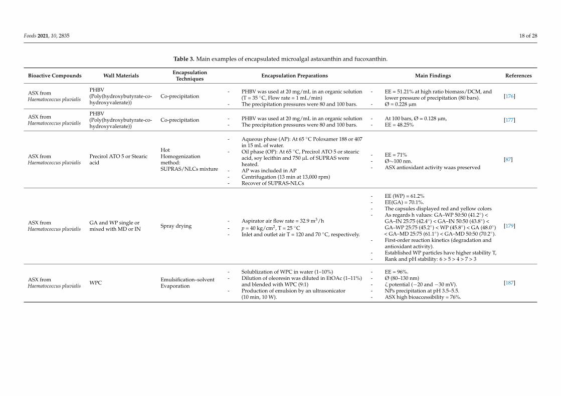
As summarized in Table 3, numerous attempts have been made concerning the en-capsulation of xanthophylls from microalgae. Machado et al. (2014 and 2016) proved thatan aqueous fluid of andrographolide made by particle engineering Supercritical (SEDS)is a promising approach for the encapsulation of ASX isolated from Haematococcus pluvi-alis [176,177]. ASX encapsulation based on a co-precipitation of PHBV with supercriticalCO2 and DCM respectively as an anti-solvent organic solvent was studied by Machadoet al. (2014 and 2016) [176,177]. Mean particle sizes of 0.128 µm was achieved with thea maximum Presure = 100 bars [176]. In addition, at the carotenoid extraction phase, amaximum of encapsulation efficiency (EE) of 48.25% was realized utilizing [B/DCM] = 10mg/mL. At [B/DCM] = 10 mg/mL and 80 bars, globular drops with 0.228 µm were gained.Overall, a pressure extension caused a reduction in EE [177]. By using a spraying technique,Park et al. in 2014 reviewed the EE of ASX-extracted from Xanthophyllomyces dendrorhous.These authors confirmed that the microparticles varied from 10 to 800 µm, with an averageof 210.26 µm and EE was within 68 and 79% [178]. Bustos-Garza et al. (2013) studied thepH-stability and thermal properties of ASX encapsulated by spray drying [179]. Theseauthors used gum Arabica (GA) and whey protein (WP) individually or in association withmaltodextrin (MD) or inulin (IN) as wall materials, and established circular micro particles,with a size that ranged between 1 and 10 µm. In another study, Higüera-Ciapara et al.(2004) examined the ASX/CT matrix microencapsulation [180]. The fabricated product(microcapsules) had a Ø of 5–50 µm. In a study by Kittikaiwan et al. (2007), encapsulatedASX was evaluated against oxidative stress [181]. Haematococcus pluvialis was entrappedinto beads, which were then coated with 5 layers of CT film, resulting in CT-algae capsulesthat have a Ø of 0.43 cm and the thickness of the film was ~100 µm. By precipitationprocesses using supercritical fluid (200 bars and 35 ◦C), Hong et al. (2009) studied H.pluvialis ASX, and obtained particles with Ø of 0.5 and 3 µm [182]. Similar findings wereobtained by Tachaprutinun et al. (2009) [183].
By using an external ionic gelation technique, Niizawa et al. (2019) evaluated the effectof five independent formulations (Tconcentrations of: CaCl2, oleoresin, alginate/oleoresin,alginate and surfactant) for natural ASX oleoresin encapsulation [184]. Mathematicalmodels were developed to predict size of particle, ASX t1/2 release and EE. If oleoresin-enriched beads Ø were linked to alginate and alginate/oleoresin levels, EE was manneredby surfactant and alginate concentrations. These parameters have an impact on the kineticmodeling on ASX release under an intestinal micro bioassay. Lin et al. (2016) reported thesame results [185].
According to Boonlao et al. (2020), ASX-enriched O/W emulsion was controlledby WPI (2–5 wt %) and XG (0.25 and 0.5 wt %) [186]. Compared to blends supportedby WPI, XG addition increased the stability of the emulsion. The ASX enclosed in anWPI-XG structure was more constant, at 5, 25 and 37 ◦C. Through simulated digestion,
Foods 2021, 10, 2835 17 of 28
WPI-XG trials showed a lesser globule dimension inside the gastric and intestinal stage,demonstrating that the XG enhances the emulsion. XG blended with WPI established lesserlipid digestibility and restricted the free fatty acid composition.
Through nanoencapsulation, Zanoni et al. (2019) developed a method to stabilizethe ASX of Haematococcus pluvialis to improve its nutritional properties and to increaseits bioavailability [187]. Nanoparticles (NPs) were prepared by an emulsification–solventevaporation technique, and oleoresin at 1%. At this concentration (1%), NP Ø was equal to90 nm. Regarding NPs Zeta-potential (ζ potential), values were due to the WP coveringthat is negatively charged at a neutral pH. The stability of the NPs was examined througha panel of stress experiments (Fe3+ exposition, heat at 65 ◦C, extreme pH and UV radia-tion, and). Simulated gastroenteric digestion was carried out to examine ASX release inphysiological terms and was presented at a high bioaccessibility (76%).
In another study, Zhou et al. (2018) made, through electrostatic complexation ofWP and GA by adjusting the pH to 4.0, esterified ASX microcapsules [188]. Biochemicalcharacteristics of the esterified ASX microcapsules were evaluated, and the gastrointestinalpotential fate and bioavailability were observed through in vivo and in vitro digestiontrials. At a stabilized system, Ø was equal to 15.4 µm, and EE was 95.3%. For in vivoexperiments, after breads oral gavage, the area under the curve (AUC0-t) was 8.23 h·µg/mLand was twofold greater than those of oleoresin (3.72 h·µg/mL) [188].
By using the vibrating nozzle technology, ASX-enriched oil was encapsulated in al-ginate and low-methoxyl pectin [189]. Authors studied the ASX degradation kinetics byfitting the data with deferred zero-, first- and second-order kinetic models. Interestingly,low methoxyl pectin exposed the appropriate ASX-enriched oil encapsulation. Previouslystudied conducted by Pu et al. (2011) [190], Niamnuy et al. (2008) [191] and Takeungwong-trakul and Benjakul (2016) [192] and Bustamante et al. (2016) [193] selected the degradationof ASX as a first order reaction.
Foods 2021, 10, 2835 18 of 28
Table 3. Main examples of encapsulated microalgal astaxanthin and fucoxanthin.
Bioactive Compounds Wall Materials EncapsulationTechniques Encapsulation Preparations Main Findings References
ASX fromHaematococcus pluvialis
PHBV(Poly(hydroxybutyrate-co-hydroxyvalerate))
Co-precipitation- PHBV was used at 20 mg/mL in an organic solution
(T = 35 ◦C, Flow rate = 1 mL/min)- The precipitation pressures were 80 and 100 bars.
- EE = 51.21% at high ratio biomass/DCM, andlower pressure of precipitation (80 bars).
- Ø = 0.228 µm[176]
ASX fromHaematococcus pluvialis
PHBV(Poly(hydroxybutyrate-co-hydroxyvalerate))
Co-precipitation - PHBV was used at 20 mg/mL in an organic solution- The precipitation pressures were 80 and 100 bars.
- At 100 bars, Ø = 0.128 µm,- EE = 48.25%
[177]
ASX fromHaematococcus pluvialis
Precirol ATO 5 or Stearicacid
HotHomogenizationmethod:SUPRAS/NLCs mixture
- Aqueous phase (AP): At 65 ◦C Poloxamer 188 or 407in 15 mL of water.
- Oil phase (OP): At 65 ◦C, Precirol ATO 5 or stearicacid, soy lecithin and 750 µL of SUPRAS wereheated.
- AP was included in AP- Centrifugation (13 min at 13,000 rpm)- Recover of SUPRAS-NLCs
- EE = 71%- Ø∼100 nm.- ASX antioxidant activity waas preserved
[87]
ASX fromHaematococcus pluvialis
GA and WP single ormixed with MD or IN Spray drying
- Aspirator air flow rate = 32.9 m3/h- p = 40 kg/cm2, T = 25 ◦C- Inlet and outlet air T = 120 and 70 ◦C, respectively.
- EE (WP) = 61.2%- EE(GA) = 70.1%.- The capsules displayed red and yellow colors- As regards h values: GA–WP 50:50 (41.2◦) <
GA–IN 25:75 (42.4◦) < GA–IN 50:50 (43.8◦) <GA–WP 25:75 (45.2◦) < WP (45.8◦) < GA (48.0◦)< GA–MD 25:75 (61.1◦) < GA–MD 50:50 (70.2◦).
- First-order reaction kinetics (degradation andantioxidant activity).
- Established WP particles have higher stability T,- Rank and pH stability: 6 > 5 > 4 > 7 > 3
[179]
ASX fromHaematococcus pluvialis WPC Emulsification–solvent
Evaporation
- Solublization of WPC in water (1–10%)- Dilution of oleoresin was diluted in EtOAc (1–11%)
and blended with WPC (9:1)- Production of emulsion by an ultrasonicator
(10 min, 10 W).
- EE = 96%.- Ø (80–130 nm)- ζ potential (−20 and −30 mV).- NPs precipitation at pH 3.5–5.5.- ASX high bioaccessibility = 76%.
[187]
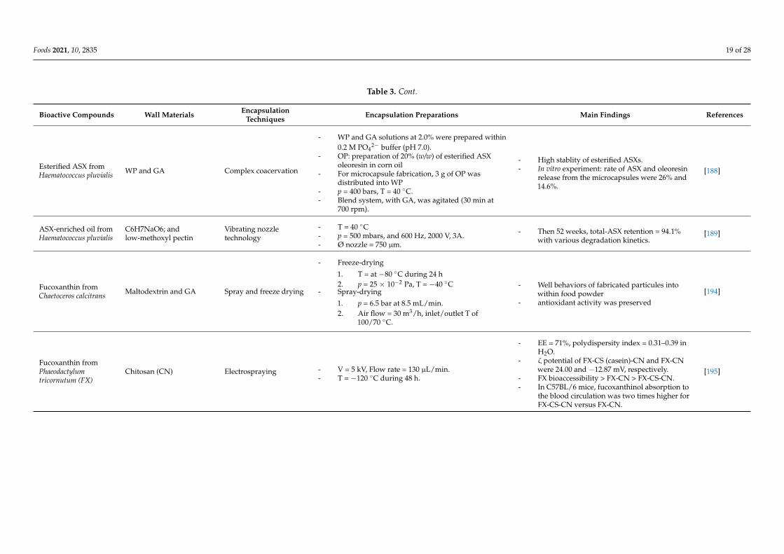
Foods 2021, 10, 2835 19 of 28
Table 3. Cont.
Bioactive Compounds Wall Materials EncapsulationTechniques Encapsulation Preparations Main Findings References
Esterified ASX fromHaematococcus pluvialis WP and GA Complex coacervation
- WP and GA solutions at 2.0% were prepared within0.2 M PO4
2− buffer (pH 7.0).- OP: preparation of 20% (w/w) of esterified ASX
oleoresin in corn oil- For microcapsule fabrication, 3 g of OP was
distributed into WP- p = 400 bars, T = 40 ◦C.- Blend system, with GA, was agitated (30 min at
700 rpm).
- High stablity of esterified ASXs.- In vitro experiment: rate of ASX and oleoresin
release from the microcapsules were 26% and14.6%.
[188]
ASX-enriched oil fromHaematococcus pluvialis
C6H7NaO6; andlow-methoxyl pectin
Vibrating nozzletechnology
- T = 40 ◦C- p = 500 mbars, and 600 Hz, 2000 V, 3A.- Ø nozzle = 750 µm.
- Then 52 weeks, total-ASX retention = 94.1%with various degradation kinetics.
[189]
Fucoxanthin fromChaetoceros calcitrans Maltodextrin and GA Spray and freeze drying
- Freeze-drying
1. T = at −80 ◦C during 24 h2. p = 25 × 10−2 Pa, T = −40 ◦C
- Spray-drying
1. p = 6.5 bar at 8.5 mL/min.2. Air flow = 30 m3/h, inlet/outlet T of
100/70 ◦C.
- Well behaviors of fabricated particules intowithin food powder
- antioxidant activity was preserved[194]
Fucoxanthin fromPhaeodactylumtricornutum (FX)
Chitosan (CN) Electrospraying - V = 5 kV, Flow rate = 130 µL/min.- T = −120 ◦C during 48 h.
- EE = 71%, polydispersity index = 0.31–0.39 inH2O.
- ζ potential of FX-CS (casein)-CN and FX-CNwere 24.00 and −12.87 mV, respectively.
- FX bioaccessibility > FX-CN > FX-CS-CN.- In C57BL/6 mice, fucoxanthinol absorption to
the blood circulation was two times higher forFX-CS-CN versus FX-CN.
[195]
Foods 2021, 10, 2835 20 of 28
9. Conclusions and Perspectives
By virtue of their importance to the food industry and human health, xanthophyllsproduced by microalgae have been extensively studied in the past two decades. Numerousstudies have been led to evaluate the efficiency of several conventional and innovativeextraction techniques for the isolation of various xanthophylls from diverse species of mi-croalgae. Many challenges still remain, as it is necessary to combine nonthermal processingtechnologies to achieve sustainable processing and assure safe outputs, which may offer anew way to obtain xanthophylls with high quality. More researches are needed to provide“environmentally friendly processes”.
Enzymes and genes of the biosynthetic pathway of xanthophylls have been widelyinvestigated. Nevertheless, regulation remains to be completely elucidated. Functionalstudies of identified putative xanthophyll regulators of the several species are mandatoryto increase a deeper study of their metabolism. Equally, promotor examination will besupportive for the identification of novel transcriptional regulators of late xanthophyllsbiosynthetic pathway genes. In this regard, integrative analysis of multi-omics data suchas genomics, transcriptomics, metabolomics and proteomics will allow us to have a betterunderstanding of the expression profiles of xanthophyll biosynthetic pathway genes inmicroalgae.
On the other hand, future research into newer capsules should also command at-tention regarding the widening range of hues that can be gained, and on promoting thexanthophylls with linked health-beneficial features. Further studies are required on xan-thophylls stabilization, which, to date, has been treated using diverse methods founded onencapsulation.
Author Contributions: Conceptualization, S.S., M.B., H.B.H., I.F. and S.A.; methodology, S.S., M.B.,and H.B.H.; validation, I.F., A.M.K., P.M. and S.A.; formal Analysis, S.S., M.B., I.F., P.M. and S.A.;investigation, S.S.; M.B. and H.B.H.; resources, S.S. and M.B.; data curation, S.S., M.B. and H.B.H.;writing—original draft preparation, S.S., M.B. and H.B.H.; writing—review and editing, H.B.H.,I.F., A.M.K., P.M. and S.A.; visualization, S.S., M.B., H.B.H.; supervision, I.F., P.M. and S.A.; projectadministration, P.M. and S.A; funding acquisition, S.A. All authors have read and agreed to thepublished version of the manuscript.
Funding: This work was supported by Grants from the Ministry of Higher Education and ScientificResearch of Tunisia.
Institutional Review Board Statement: Not applicable.
Informed Consent Statement: Not applicable.
Data Availability Statement: The data are included in the manuscript.
Conflicts of Interest: The authors declare no conflict of interest.
References1. Afreen, R.; Tyagi, S.; Singh, G.P.; Singh, M. Challenges and Perspectives of Polyhydroxyalkanoate Production from Microal-
gae/Cyanobacteria and Bacteria as Microbial Factories: An Assessment of Hybrid Biological System. Front. Bioeng. Biotechnol.2021, 9, 109. [CrossRef] [PubMed]
2. Khan, A.K.; Kausar, H.; Jaferi, S.S.; Drouet, S.; Hano, C.; Abbasi, B.H.; Anjum, S. An insight into the algal evolution and genomics.Biomolecules 2020, 10, 1524. [CrossRef] [PubMed]
3. Shrestha, K.K.; Bhattarai, S.; Bhandari, P. Handbook of Flowering Plants of Nepal (Vol. 1 Gymnosperms and Angiosperms: Cycadaceae-Betulaceae); Scientific Publishers: Norwood, NJ, USA, 2018.
4. Novoveská, L.; Ross, M.E.; Stanley, M.S.; Pradelles, R.; Wasiolek, V.; Sassi, J.F. Microalgal carotenoids: A review of production,current markets, regulations, and future direction. Mar. Drugs 2019, 17, 640. [CrossRef] [PubMed]
5. Gong, M.; Bassi, A. Carotenoids from microalgae: A review of recent developments. Biotechnol. Adv. 2016, 34, 1396–1412.[CrossRef] [PubMed]
6. Dammak, M.; Haase, S.M.; Miladi, R.; Ben Amor, F.; Barkallah, M.; Gosset, D.; Pichon, C.; Huchzermeyer, B.; Fendri, I.; Denis, M.;et al. Enhanced lipid and biomass production by a newly isolated and identified marine microalga. Lipids Health Dis. 2016, 15,209. [CrossRef]
Foods 2021, 10, 2835 21 of 28
7. Dammak, M.; Hadrich, B.; Barkallah, M.; Hentati, F.; Ben Hlima, H.; Pichon, C.; Denis, M.; Fendri, I.; Michaud, P.; Abdelkafi, S.Modelling Tetraselmis sp. growth-kinetics and optimizing bioactive-compound production through environmental conditions.Bioresour. Technol. 2018, 249, 510–518. [CrossRef]
8. Ben Hlima, H.; Bohli, T.; Kraiem, M.; Ouederni, A.; Mellouli, L.; Michaud, P.; Abdelkafi, S.; Smaoui, S. Combined effect ofSpirulina platensis and Punica granatum peel extacts: Phytochemical content and antiphytophatogenic activity. Appl. Sci. 2019, 9,5475.
9. Elleuch, F.; Ben Hlima, H.; Barkallah, M.; Baril, P.; Abdelkafi, S.; Pichon, C.; Fendri, I. Carotenoids overproduction in Dunaliellasp.: Transcriptional changes and new insights through lycopene cyclase regulation. Appl. Sci. 2019, 9, 5389. [CrossRef]
10. Barkallah, M.; Ben Slima, A.; Fendri, I.; Pichon, C.; Abdelkafi, S.; Baril, P. Protective role of Spirulina platensis against bifenthrin-induced reprotoxicity in adult male mice by reversing expression of altered histological, biochemical, and molecular markersincluding microRNAs. Biomolecules 2020, 10, 7539. [CrossRef]
11. Jacob-Lopes, E.; Maroneze, M.M.; Deprá, M.C.; Sartori, R.B.; Dias, R.R.; Zepka, L.Q. Bioactive food compounds from microalgae:An innovative framework on industrial biorefineries. Curr. Opin. Food Sci. 2019, 25, 1–7. [CrossRef]
12. Nisar, N.; Li, L.; Lu, S.; Khin, N.C.; Pogson, B.J. Carotenoid metabolism in plants. Mol. Plant. 2015, 8, 68–82. [CrossRef]13. Galasso, C.; Corinaldesi, C.; Sansone, C. Carotenoids from marine organisms: Biological functions and industrial applications.
Antioxidants 2017, 6, 96. [CrossRef]14. Gammone, M.A.; Riccioni, G.; D’Orazio, N. Carotenoids: Potential allies of cardiovascular health? Food. Nutr. Res. 2015, 59, 26762.
[PubMed]15. LaFountain, A.M.; Prum, R.O.; Frank, H.A. Diversity, physiology, and evolution of avian plumage carotenoids and the role of
carotenoid–protein interactions in plumage color appearance. Arch. Biochem. Biophys. 2015, 572, 201–212. [CrossRef]16. Pereira, A.G.; Otero, P.; Echave, J.; Carreira-Casais, A.; Chamorro, F.; Collazo, N.; Jaboui, A.; Lourenço-Lopes, C.; Simal-Gandara,
J.; Prieto, M.A. Xanthophylls from the Sea: Algae as Source of Bioactive Carotenoids. Mar. Drugs 2021, 19, 188. [CrossRef][PubMed]
17. Torregrosa-Crespo, J.; Montero, Z.; Fuentes, J.L.; García-Galbis, M.R.; Garbayo, I.; Vílchez, C.; Martínez-Espinosa, R.M. Exploringthe Valuable Carotenoids for the Large-Scale Production by Marine Microorganisms. Mar. Drugs 2018, 16, 203. [CrossRef]
18. Carbonell-Capella, J.M.; Buniowska, M.; Barba, F.J.; Esteve, M.J.; Frigola, A. Analytical methods for determining bio-availabilityandbio-accessibility of bioactive compounds from fruits and vegetables: A review. Compr. Rev. Food Sci. Food Saf. 2014, 13,155–171. [CrossRef]
19. Pangestuti, R.; Kim, S.K. Biological activities and health benefit effects of natural pigments derived from marine algae. J. Funct.Foods 2011, 3, 255–266. [CrossRef]
20. Ojulari, O.V.; Gi Lee, S.; Nam, J.O. Therapeutic Effect of Seaweed Derived Xanthophyl Carotenoid on Obesity Management;Overview of the Last Decade. Int. J. Mol. Sci. 2020, 21, 2502. [CrossRef]
21. Poojary, M.M.; Barba, F.J.; Aliakbarian, B.; Donsì, F.; Pataro, G.; Dias, D.A.; Juliano, P. Inno Gonçalves vative alternativetechnologies to extract carotenoids from microalgae and seaweeds. Mar. Drugs 2016, 14, 214. [CrossRef] [PubMed]
22. Silva, S.C.; Ferreira, I.C.F.R.; Dias, M.M.; Barreiro, M.F. Microalgae-Derived Pigments: A 10-Year Bibliometric Review andIndustryand Market Trend Analysis. Molecules 2020, 25, 3406. [CrossRef]
23. Jain, A.; Sirisha, V.L. Algal Carotenoids: Understanding Their Structure, Distribution and Potential Applications in HumanHealth. Encycl. Mar. Biotechnol. 2020, 1, 33–64.
24. da Silva Vaz, B.; Moreira, J.B.; de Morais, M.G.; Costa, J.A.V. Microalgae as a new source of bioactive compounds in foodsupplements. Curr. Opin. Food Sci. 2016, 7, 73–77.
25. Mulders, K.J.M.; Lamers, P.P.; Martens, D.E.; Wijffels, R.H. Phototrophic Pigment Production with Microalgae: BiologicalConstraints and Opportunities. J. Phycol. 2014, 50, 229–242. [CrossRef]
26. de Oliveira-Júnior, R.G.; Grougnet, R.; Bodet, P.-E.; Bonnet, A.; Nicolau, E.; Jebali, A.; Rumin, J.; Picot, L. Updated pigmentcomposition of Tisochrysis lutea and purification of fucoxanthin using centrifugal partition chromatography coupled to flashchromatography for the chemosensitization of melanoma cells. Algal Res. 2020, 51, 102035. [CrossRef]
27. Petrushkina, M.; Gusev, E.; Sorokin, B.; Zotko, N.; Mamaeva, A.; Filimonova, A.; Kulikovskiy, M.; Maltsev, Y.; Yampolsky, I.;Guglya, E.; et al. Fucoxanthin production by heterokont microalgae. Algal Res. 2017, 24, 387–393. [CrossRef]
28. Kim, S.M.; Kang, S.W.; Kwon, O.N.; Chung, D.; Pan, C.H. Fucoxanthin as a major carotenoid in Isochrysis aff. galbana: Characteri-zation of extraction for commercial application. J. Korean Soc. Appl. Biol. Chem. 2012, 55, 477–483. [CrossRef]
29. Xia, S.; Wang, K.; Wan, L.; Li, A.; Hu, Q.; Zhang, C. Production, characterization, and antioxidant activity of fucoxanthin from themarine diatom Odontella aurita. Mar. Drugs 2013, 11, 2667–2681. [CrossRef]
30. Marella, T.K.; Tiwari, A. Marine diatom Thalassiosira weissflogii based biorefinery for co-production of eicosapentaenoic acid andfucoxanthin. Bioresour. Technol. 2020, 307, 123245. [CrossRef]
31. Panis, G.; Carreon, J.R. Commercial astaxanthin production derived by green alga Haematococcus pluvialis: A microalgae processmodel and a techno-economic assessment all through production line. Algal Res. 2016, 18, 175–190. [CrossRef]
32. Aflalo, C.; Meshulam, Y.; Zarka, A.; Boussiba, S. On the relative efficiency of two-vs. one-stage production of astaxanthin by thegreen alga Haematococcus pluvialis. Biotechnol. Bioeng. 2007, 98, 300–305. [CrossRef]
Foods 2021, 10, 2835 22 of 28
33. Torzillo, G.; Goksan, T.; Faraloni, C.; Kopecky, J.; Masojídek, J. Interplay between Photochemical Activities and PigmentComposition in an Outdoor Culture of Haematococcus pluvialis during the Shift from the Green to Red Stage. J. Appl. Phycol. 2003,15, 127–136. [CrossRef]
34. Ranga, R.; Sarada, A.R.; Baskaran, V.; Ravishankar, G.A. Identification of carotenoids from green alga Haematococcus pluvialis byHPLC and LC-MS (APCI) and their antioxidant properties. J. Microbiol. Biotechnol. 2009, 19, 1333–1341.
35. Molino, A.; Rimauro, J.; Casella, P.; Cerbone, A.; Larocca, V.; Chianese, S.; Karatza, D.; Mehariya, S.; Ferraro, A.; Hristoforou, E.Extraction of Astaxanthin from Microalga Haematococcus pluvialis in Red Phase by Using Generally Recognized as Safe Solventsand Accelerated Extraction. J. Biotechnol. 2018, 283, 51–61. [CrossRef] [PubMed]
36. Han, S.; Yao, J.; Lee, C.; Park, J.; Choi, Y. A novel approach to enhance astaxanthin production in Haematococcus lacustris using amicrostructure-based culture platform. Algal Res. 2019, 39, 101464. [CrossRef]
37. Fábryová, T.; Cheel, J.; Kubác, D.; Hrouzek, P.; Tumová, L.; Kopecký, J. Purification of lutein from the green microalgae Chlorellavulgaris by integrated use of a new extraction protocol and a multi-injection high performance counter-current chromatography(HPCCC). Algal Res. 2019, 41, 101574. [CrossRef]
38. Dineshkumar, R.; Dhanarajan, G.; Dash, S.K.; Sen, R. An advanced hybrid medium optimization strategy for the enhancedproductivity of lutein in Chlorella minutissima. Algal Res. 2015, 7, 24–32. [CrossRef]
39. Chen, C.-Y.; Liu, C.-C. Optimization of lutein production with a two-stage mixotrophic cultivation system with Chlorellasorokiniana MB-1. Bioresour. Technol. 2018, 262, 74–79. [CrossRef] [PubMed]
40. Minhas, A.K.; Hodgson, P.; Barrow, C.J.; Sashidhar, B.; Adholeya, A. The isolation and identification of new microalgal strainsproducing oil and carotenoid simultaneously with biofuel potential. Bioresour. Technol. 2016, 211, 556–565. [CrossRef]
41. Serejo, M.L.; Posadas, E.; Boncz, M.A.; Blanco, S.; García-Encina, P.; Muñoz, R. Influence of Biogas Flow Rate on BiomassComposition during the Optimization of Biogas Upgrading in Microalgal-Bacterial Processes. Environ. Sci. Technol. 2015, 49,3228–3236.
42. Sun, Z.; Li, T.; Zhou, Z.G.; Jiang, Y. Microalgae as a source of lutein: Chemistry, biosynthesis, and carotenogenesis. MicroalgaeBiotechnol. 2015, 37–58.
43. Shi, X.M.; Jiang, Y.; Chen, F. High-Yield Production of Lutein by the Green Microalga Chlorella Protothecoides in HeterotrophicFed-Batch Culture. Biotechnol. Prog. 2002, 18, 723–727. [CrossRef]
44. Schüler, L.M.; Gangadhar, K.N.; Duarte, P.; Placines, C.; Molina-Márquez, A.M.; Léon-Bañares, R.; Sousa, V.S.; Varela, J.; Barreira,L. Improvement of carotenoid extraction from a recently isolated, robust microalga, Tetraselmis sp. CTP4 (chlorophyta). Bioprocess.Biosyst. Eng. 2020, 43, 785–796. [CrossRef]
45. Ma, R.; Zhao, X.; Xie, Y.; Ho, S.H.; Chen, J. Enhancing lutein productivity of Chlamydomonas sp. via high-intensity light exposurewith corresponding carotenogenic genes expression profiles. Bioresour. Technol. 2019, 275, 416–420. [CrossRef] [PubMed]
46. Blanco, A.M.; Moreno, J.; Del Campo, J.A.; Rivas, J.; Guerrero, M.G. Outdoor cultivation of lutein-rich cells of Muriellopsis sp. inopen ponds. Appl. Microbiol. Biotechnol. 2007, 73, 1259–1266. [CrossRef]
47. Garbayo, I.; Cuaresma, M.; Vílchez, C.; Vega, J.M. Effect of abiotic stress on the production of lutein and β-carotene byChlamydomonas acidophila. Process. Biochem. 2008, 43, 1158–1161. [CrossRef]
48. Molino, A.; Mehariya, S.; Karatza, D.; Chianese, S.; Iovine, A.; Casella, P. Bench-Scale Cultivation of Microalgae Scenedesmusalmeriensis for CO2 Capture and Lutein Production. Energies 2019, 12, 2806. [CrossRef]
49. Ho, S.H.; Chan, M.C.; Liu, C.C.; Chen, C.Y.; Lee, W.L.; Lee, D.J.; Chang, J.S. Enhancing lutein productivity of an indigenousmicroalga Scenedesmus obliquus FSP-3 using light-related strategies. Bioresour. Technol. 2014, 152, 275–282. [CrossRef]
50. Xie, Y.; Zhao, X.; Chen, J.; Yang, X.; Ho, S.H.; Wang, B.; Chang, J.S.; Shen, Y. Enhancing cell growth and lutein productivity ofDesmodesmus sp. F51 by optimal utilization of inorganic carbon sources and ammonium salt. Bioresour. Technol. 2017, 244, 664–671.[CrossRef] [PubMed]
51. Liau, B.C.; Hong, S.E.; Chang, L.P.; Shen, C.T.; Li, Y.C.; Wu, Y.P.; Jong, T.T.; Shieh, C.J.; Hsu, S.L.; Chang, C.M.J. Separationof Sight-Protecting Zeaxanthin from Nannochloropsis Oculata by Using Supercritical Fluids Extraction Coupled with ElutionChromatography. Sep. Purif. Technol. 2011, 78, 1–8. [CrossRef]
52. Koo, S.Y.; Cha, K.H.; Song, D.G.; Chung, D.; Pan, C.H. Optimization of Pressurized Liquid Extraction of Zeaxanthin from ChlorellaEllipsoidea. J. Appl. Phycol. 2012, 24, 725–730. [CrossRef]
53. Plaza, M.; Santoyo, S.; Jaime, L.; García-Blairsy Reina, G.; Herrero, M.; Señoráns, F.J.; Ibáñez, E. Screening for Bioactive Compoundsfrom Algae. J. Pharm. Biomed. Anal. 2010, 51, 450–455. [CrossRef]
54. Soontornchaiboon, W.; Joo, S.S.; Kim, S.M. Anti-inflammatory effects of violaxanthin isolated from microalga Chlorella ellipsoideain RAW 264.7 macrophages. Biol. Pharm. Bull. 2012, 35, 1137–1144. [CrossRef]
55. Kim, J.; Lee, S.; Baek, K.; Jin, E. Site-specific gene knock-out and on-site heterologous gene overexpression in Chlamydomonasreinhardtii via a CRISPR-Cas9-mediated knock-in method. Front. Plant. Sci. 2020, 11, 306. [CrossRef] [PubMed]
56. Abe, K.; Hattori, H.; Hirano, M. Accumulation and antioxidant activity of secondary carotenoids in the aerial microalga Coelastrellastriolata var. multistriata. Food Chem. 2007, 100, 656–661. [CrossRef]
57. Cha, K.H.; Koo, S.Y.; Lee, D.U. Antiproliferative effects of carotenoids extracted from Chlorella ellipsoidea and Chlorella vulgaris onhuman colon cancer cells. J. Agric. Food Chem. 2008, 56, 10521–10526. [CrossRef] [PubMed]
Foods 2021, 10, 2835 23 of 28
58. Careri, M.; Furlattini, L.; Mangia, A.; Musci, M.; Anklam, E.; Theobald, A.; Von Holst, C. Supercritical Fluid Extraction for LiquidChromatographic Determination of Carotenoids in Spirulina pacifica Algae: A Chemometric Approach. J. Chromatogr. A 2001, 912,61–71. [CrossRef]
59. Othman, R.; Noh, N.H.; Hatta, F.A.M.; Jamaludin, M.A. Natural Carotenoid Pigments of 6 Chlorophyta Freshwater Green AlgaeSpecies. J. Pharm. Nutr. Sci. 2018, 8, 1–5. [CrossRef]
60. Geisert, M.; Rose, T.; Bauer, W.; Zahn, R.K. Occurrence of carotenoids and sporopollenin in Nanochlorum eucaryotum, a novelmarine alga with unusual characteristics. Biosystems 1987, 20, 133–142. [CrossRef]
61. Kuczynska, P.; Jemiola-Rzeminska, M. Isolation and Purification of All-Trans Diadinoxanthin and All-Trans Diatoxanthin fromDiatom Phaeodactylum tricornutum. J. Appl. Phycol. 2017, 29, 79–87. [CrossRef]
62. Tanaka, T.; Shnimizu, M.; Moriwaki, H. Cancer chemoprevention by carotenoids. Molecules 2012, 17, 3202–3242. [CrossRef]63. Chuyen, H.V.; Eun, J.B. Marine carotenoids: Bioactivities and potential benefits to human health. Crit. Rev. Food Sci. Nutr. 2017,
57, 2600–2610. [CrossRef] [PubMed]64. Kotake-Nara, E.; Yonekura, L.; Nagao, A. Lysoglyceroglycolipids improve the intestinal absorption of micellar fucoxanthin by
Caco-2 cells. J. Oleo Sci. 2015, 64, 1207–1211. [CrossRef] [PubMed]65. Kumar, S.R.; Hosokawa, M.; Miyashita, K. Fucoxanthin: A Marine Carotenoid Exerting Anti-Cancer Effects by Affecting Multiple
Mechanisms. Mar. Drugs 2013, 5130–5147. [CrossRef]66. Maoka, T.; Fujiwara, Y.; Hashimoto, K.; Akimoto, N. Characterization of Fucoxanthin and Fucoxanthinol Esters in the Chinese
Surf Clam, Mactra Chinensis. J. Agric. Food Chem. 2007, 55, 1563–1567. [CrossRef]67. Mikami, K.; Hosokawa, M. Biosynthetic pathway and health benefits of fucoxanthin, an algae-specific xanthophyll in brown
seaweeds. Int. J. Mol. Sci. 2013, 14, 13763–13781. [CrossRef]68. Heo, S.-J.; Ko, S.-C.; Kang, S.-M.; Kang, H.-S.; Kim, J.-P.; Kim, S.-H.; Lee, K.-W.; Cho, M.-G.; Jeon, Y.-J. Cytoprotective Effect of
Fucoxanthin Isolated from Brown Algae Sargassum Siliquastrum against H2O2-Induced Cell Damage. Eur. Food Res. Technol. 2008,228, 145–151.
69. Market Reports World. Global Fucoxanthin Market Report 2017; Market Reports World: Pune, India, 2017.70. Fassett, R.G.; Coombes, J.S. Astaxanthin in cardiovascular health and disease. Molecules 2012, 17, 2030–2048. [CrossRef] [PubMed]71. Davinelli, S.; Nielsen, M.E.; Scapagnini, G. Astaxanthin in skin health, repair, and disease: A comprehensive review. Nutrients
2018, 10, 522. [CrossRef]72. Han, D.; Li, Y.; Hu, Q. Astaxanthin in Microalgae: Pathways, Functions and Biotechnological Implications. Algae 2013, 28, 131–147.
[CrossRef]73. Ambati, R.R.; Phang, S.M.; Ravi, S.; Aswathanarayana, R.G. Astaxanthin: Sources, extraction, stability, biological activities and its
commercial applications—A review. Mar. Drugs 2014, 2, 128–152. [CrossRef]74. Butler, T.; Golan, Y. Astaxanthin production from microalgae. In Microalgae Biotechnology for Food, Health and High Value Products;
Springer: Singapore, 2020; pp. 175–242.75. Naziri, D.; Hamidi, M.; Hassanzadeh, S.; Tarhriz, V.; Maleki Zanjani, B.; Nazemyieh, H.; Hejazi, M.A.; Hejazi, M.S. Analysis of
carotenoid production by Halorubrum sp. TBZ126; an extremely halophilic archeon from Urmia Lake. Adv. Pharm. Bull. 2014, 4,61–67.
76. Cuaresma, M.; Casal, C.; Forján, E.; Vílchez, C. Productivity and selective accumulation of carotenoids of the novel extremophilemicroalga Chlamydomonas acidophila grown with different carbon sources in batch systems. J. Ind. Microbiol. Biotechnol. 2011, 38,167–177. [CrossRef]
77. Shi, X.-M.; Zhang, X.-W.; Chen, F. Heterotrophic production of biomass and lutein by Chlorella protothecoides on various nitrogensources. Enzyme Microb. Technol. 2000, 27, 312–318. [CrossRef]
78. Sánchez, J.F.; Fernández-Sevilla, J.M.; Acién, F.G.; Cerón, M.C.; Pérez-Parra, J.; Molina-Grima, E. Biomass and lutein productivityof Scenedesmus almeriensis: Influence of irradiance, dilution rate and temperature. Appl. Microbiol. Biotechnol. 2008, 79, 719–729.[CrossRef]
79. Sun, H.; Kong, Q.; Geng, Z.; Duan, L.; Yang, M.; Guan, B. Enhancement of cell biomass and cell activity of astaxanthin-richHaematococcus pluvialis. Bioresour. Technol. 2015, 186, 67–73. [CrossRef] [PubMed]
80. Firdous, A.P.; Kuttan, G.; Kuttan, R. Anti-inflammatory potential of carotenoid meso-zeaxanthin and its mode of action. Pharm.Biol. 2015, 53, 961–967. [CrossRef]
81. Li, X.R.; Tian, G.Q.; Shen, H.J.; Liu, J.Z. Metabolic Engineering of Escherichia Coli to Produce Zeaxanthin. J. Ind. Microbiol.Biotechnol. 2015, 42, 627–636. [CrossRef] [PubMed]
82. Pasquet, V.; Morisset, P.; Ihammouine, S.; Chepied, A.; Aumailley, L.; Berard, J.B.; Serive, B.; Kaas, R.; Lanneluc, I.; Thiery, V.; et al.Antiproliferative activity of violaxanthin isolated from bioguided fractionation of Dunaliella tertiolecta extracts. Mar. Drugs 2011, 9,819–831. [CrossRef]
83. Kathiresan, S.; Chandrashekar, A.; Ravishankar, G.A.; Sarada, R. Regulation of astaxanthin and its intermediates through cloningand genetic transformation of β-carotene ketolase in Haematococcus pluvialis. J. Biotechnol. 2015, 196–197, 33–41. [CrossRef][PubMed]
84. Jiao, Y.; Reuss, L.; Wang, Y. β-Cryptoxanthin: Chemistry, occurrence, and potential health benefits. Curr. Pharmacol. Rep. 2019, 5,20–34. [CrossRef]
Foods 2021, 10, 2835 24 of 28
85. Burri, B.J.; La Frano, M.R.; Zhu, C. Absorption, metabolism, and functions of β-cryptoxanthin. Nutr. Rev. 2016, 74, 69–82.[CrossRef]
86. Gastineau, R.; Davidovich, N.; Hansen, G.; Rines, J.; Wulff, A.; Kaczmarska, I.; Ehrman, J.; Hermann, D.; Maumus, F.; Hardivillier,Y.; et al. Haslea ostrearia-like diatoms: Biodiversity out of the blue. Adv. Bot. Res. 2014, 71, 441–465.
87. Kooistra, W.H.; Gersonde, R.; Medlin, L.K.; Mann, D.G. The origin and evolution of the diatoms: Their adaptation to a planktonicexistence. Evol. Prim. Prod. Sea 2007, 207–249. [CrossRef]
88. Tanno, Y.; Kato, S.; Takahashi, S.; Tamaki, S.; Takaichi, S.; Kodama, Y.; Sonoike, K.; Shinomura, T. Light Dependent Accumulationof β-Carotene Enhances Photo-Acclimation of Euglena Gracilis. J. Photochem. Photobiol. B Biol. 2020, 209, 111950. [CrossRef]
89. Dambek, M.; Eilers, U.; Breitenbach, J.; Steiger, S.; Büchel, C.; Sandmann, G. Biosynthesis of Fucoxanthin and Diadinoxanthin andFunction of Initial Pathway Genes in Phaeodactylum tricornutum. J. Exp. Bot. 2012, 63, 5607–5612. [CrossRef]
90. Faraloni, C.; Torzillo, G. Synthesis of antioxidant carotenoids in microalgae in response to physiological stress. In Carotenoids;InTechOpen: London, UK, 2017; pp. 143–157.
91. Goodwin, T.W.; Britton, G. Distribution and analysis of carotenoids. Plant Pigment. 1988, 61–132.92. Mata-Gómez, L.C.; Montañez, J.C.; Méndez-Zavala, A.; Aguilar, C.N. Biotechnological production of carotenoids by yeasts: An
overview. Microb. Cell Fact. 2014, 13, 1–11.93. Rodrigo-Baños, M.; Garbayo, I.; Vílchez, C.; Bonete, M.J.; Martínez-Espinosa, R.M. Carotenoids from Haloarchaea and their
potential in biotechnology. Mar. Drugs 2015, 13, 5508–5532. [CrossRef] [PubMed]94. Fiedor, J.; Burda, K. Potential role of carotenoids as antioxidants in human health and disease. Nutrients 2014, 6, 466–468.
[PubMed]95. Yatsunami, R.; Ando, A.; Yang, Y.; Takaichi, S.; Kohno, M.; Matsumura, Y.; Ikeda, H.; Fukui, T.; Nakasone, K.; Fujita, N.
Identification of carotenoids from the extremely halophilic archaeon Haloarcula japonica. Front. Microbiol. 2014, 5, 100–105.[CrossRef]
96. Varela, J.C.; Pereira, H.; Vila, M.; León, R. Production of carotenoids by microalgae: Achievements and challenges. Photosynth.Res. 2015, 125, 423–436. [CrossRef]
97. Dembitsky, V.M.; Maoka, T. Allenic and cumulenic lipids. Prog. Lipid Res. 2007, 46, 328–375. [CrossRef] [PubMed]98. Raposo, M.F.D.J.; De Morais, A.M.M.B.; De Morais, R.M.S.C. Carotenoids from marine microalgae: A valuable natural source for
the prevention of chronic diseases. Mar. Drugs 2015, 13, 5128–5155. [CrossRef]99. Barredo, J.-L. Microbial Carotenoids from Bacteria and Microalgae. Methods and Protocols. Methods Mol. Biol. 2012, 892, 133–141.100. Gwak, Y.; Hwang, Y.S.; Wang, B.; Kim, M.; Jeong, J.; Lee, C.G.; Hu, Q.; Han, D.; Jin, E. Comparative analyses of lipidomes and
transcriptomes reveal a concerted action of multiple defensive systems against photooxidative stress in Haematococcus pluvialis. J.Exp. Bot. 2014, 65, 4317–4334. [CrossRef] [PubMed]
101. Hunter, W.N. The non-mevalonate pathway of isoprenoid precursor biosynthesis. J. Biol. Chem. 2007, 282, 21573–21577. [CrossRef]102. Jahns, P.; Latowski, D.; Strzalka, K. Mechanism and regulation of the violaxanthin cycle: The role of antenna proteins and
membrane lipids. Biochim. Biophys. Acta. 2009, 1787, 3–14. [CrossRef]103. Huang, J.J.; Lin, S.; Xu, W.; Cheung, P.C.K. Occurrence and biosynthesis of carotenoids in phytoplankton. Biotechnol. Adv. 2017,
35, 597–618. [CrossRef]104. Rabbani, S.; Beyer, P.; Lintig, J.V.; Hugueney, P.; Kleinig, H. Induced β-carotene synthesis driven by triacylglycerol deposition in
the unicellular alga Dunaliella bardawil. Plant Phys. 1998, 116, 1239–1248. [CrossRef]105. Guedes, A.C.; Amaro, H.M.; Malcata, F.X. Microalgae as sources of carotenoids. Mar. Drugs 2011, 9, 625–644. [CrossRef] [PubMed]106. Jin, E.; Feth, B.; Melis, A. A mutant of the green alga Dunaliella salina constitutively accumulates zeaxanthin under all growth
conditions. Biotechnol. Bioeng. 2003, 81, 115–124. [CrossRef] [PubMed]107. Abu-Ghosh, S.; Fixler, D.; Dubinsky, Z.; Iluz, D. Flashing light in microalgae biotechnology. Bioresour. Technol. 2016, 203, 357–363.
[CrossRef]108. Polle, J.E.; Qin, S. Development of Genetics and Molecular Tool Kits for Species of the Unicellular Green Alga Dunaliella
(Chlorophyta). Alga Dunaliella 2009, 17, 403–422.109. Huang, W.; Lin, Y.; He, M.; Gong, Y.; Huang, J. Induced high-yield production of zeaxanthin, lutein, and β-carotene by a mutant
of Chlorella zofingiensis. J. Agric. Food Chem. 2018, 66, 891–897. [CrossRef]110. Sarnaik, V.; Nambissan, R.; Pandit, A.; Lali, A. Recombinant Synechococcus elongatus PCC 7942 for improved zeaxanthin production
under natural light conditions. Algal Res. 2018, 36, 139–151. [CrossRef]111. Lim, K.C.; Yusoff, F.M.; Shariff, M.; Kamarudin, M.S. Astaxanthin as feed supplement in aquatic animals. Rev. Aquac. 2018, 10,
738–773. [CrossRef]112. Sproles, A.E.; Fields, F.J.; Smalley, T.N.; Le, C.H.; Badary, A.; Mayfield, S.P. Recent advancements in the genetic engineering of
microalgae. Algal Res. 2021, 53, 102158. [CrossRef]113. Liu, Y.; Cui, Y.; Chen, J.; Qin, S.; Chen, G. Metabolic engineering of Synechocystis sp. PCC6803 to produce astaxanthin. Algal Res.
2019, 44, 101679. [CrossRef]114. Perozeni, F.; Cazzaniga, S.; Baier, T.; Zanoni, F.; Zoccatelli, G.; Lauersen, K.J.; Wobbe, L.; Ballottari, M. Turning a green alga red:
Engineering astaxanthin biosynthesis by intragenic pseudogene revival in Chlamydomonas reinhardtii. Plant Biotechnol. J. 2020,18, 2053–2067. [CrossRef] [PubMed]
Foods 2021, 10, 2835 25 of 28
115. Galarza, J.I.; Gimpel, J.A.; Rojas, V.; Arredondo-Vega, B.O.; Henríquez, V. Over-accumulation of astaxanthin in Haematococcuspluvialis through chloroplast genetic engineering. Algal Res. 2018, 31, 291–297. [CrossRef]
116. Manfellotto, F.; Stella, G.R.; Falciatore, A.; Brunet, C.; Ferrante, M.I. Engineering the Unicellular Alga Phaeodactylum tricornutumfor Enhancing Carotenoid Production. Antioxidants 2020, 9, 757. [CrossRef]
117. Kadono, T.; Kira, N.; Suzuki, K.; Iwata, O.; Ohama, T.; Okada, S.; Nishimura, T.; Akakabe, M.; Tsuda, M.; Adachi, M. Effect of anintroduced phytoene synthase gene expression on carotenoid biosynthesis in the marine diatom Phaeodactylum tricornutum. Mar.Drugs 2015, 13, 5334–5357. [CrossRef] [PubMed]
118. Eilers, U.; Bikoulis, A.; Breitenbach, J.; Büchel, C.; Sandmann, G. Limitations in the biosynthesis of fucoxanthin as targets forgenetic engineering in Phaeodactylum tricornutum. J. Appl. Phycol. 2016, 28, 123–129. [CrossRef]
119. Cordero, B.F.; Obraztsova, I.; Couso, I.; Leon, R.; Vargas, M.A.; Rodriguez, H. Enhancement of lutein production in Chlorellasorokiniana (Chorophyta) by improvement of culture conditions and random mutagenesis. Mar. Drugs 2011, 9, 1607–1624.[CrossRef] [PubMed]
120. Liu, J.; Mao, X.; Zhou, W.; Guarnieri, M.T. Simultaneous production of triacylglycerol and high-value carotenoids by theastaxanthin-producing oleaginous green microalga Chlorella zofingiensis. Bioresour. Technol. 2016, 214, 319–327. [CrossRef]
121. Rathod, J.P.; Vira, C.; Lali, A.M.; Prakash, G. Metabolic Engineering of Chlamydomonas reinhardtii for Enhanced β-Carotene andLutein Production. Appl. Biochem. Biotechnol. 2020, 190, 1457–1469. [CrossRef]
122. Cezare-Gomes, E.A.; Mejia-da-Silva, L.D.C.; Pérez-Mora, L.S.; Matsudo, M.C.; Ferreira-Camargo, L.S.; Singh, A.K.; de Carvalho,J.C.M. Potential of microalgae carotenoids for industrial application. Appl. Biochem. Biotechnol. 2019, 1188, 602–634. [CrossRef]
123. Carvalho, J.C.; Bezerra, R.P.; Matsudo, M.C.; Sato, S. Cultivation of Arthrospira (Spirulina) platensis by fed-batch process. InAdvanced Biofuels and Bioproducts; Springer: New York, NY, USA, 2013; pp. 781–805.
124. Borowitzka, M.A. High-value products from microalgae—Their development and commercialisation. J. Appl. Phycol. 2013, 25,743–756. [CrossRef]
125. Borowitzka, M.A. Commercial production of microalgae: Ponds, tanks, tubes and fermenters. J. Biotechnol. 1999, 70, 313–321.[CrossRef]
126. Singh, R.N.; Sharma, S. Development of suitable photobioreactor for algae production—A review. Renew. Sust. Energ. Rev. 2012,16, 2347–2353. [CrossRef]
127. Chia, S.R.; Chew, K.W.; Leong, H.Y.; Ho, S.H.; Munawaroh, H.S.H.; Show, P.L. CO2 mitigation and phycoremediation of industrialflue gas and wastewater via microalgae-bacteria consortium: Possibilities and challenges. Chem. Eng. Sci. 2021, 425, 131436.[CrossRef]
128. Ferreira, L.S.; Rodrigues, M.S.; Converti, A.; Sato, S.; Carvalho, J.C. Kinetic and growth parameters of Arthrospira (Spirulina)platensis cultivated in tubular photobioreactor under different cell circulation systems. Biotechnol. Bioeng. 2012, 109, 444–450.[CrossRef] [PubMed]
129. Eze, C.N.; Ogbonna, J.C.; Ogbonna, I.O.; Aoyagi, H. A novel flat plate air-lift photobioreactor with inclined reflective brothcirculation guide for improved biomass and lipid productivity by Desmodesmus subspicatus LC172266. J. Appl. Phycol. 2017, 29,2745–2754. [CrossRef]
130. Gao, B.; Xia, S.; Lei, X.; Zhang, C. Combined effects of different nitrogen sources and levels and light intensities on growth andfatty acid and lipid production of oleaginous Eustigmatophycean microalga Eustigmatos cf. polyphem. J. Appl. Phycol. 2017, 30,215–229. [CrossRef]
131. Boussiba, S. Carotenogenesis in the green alga Haematococcus pluvialis: Cellular physiology and stress response. Physiol. Plantarum.2000, 108, 111–117. [CrossRef]
132. Huang, G.; Chen, F.; Wei, D.; Zhang, X.; Chen, G. Biodiesel production by microalgal biotechnology. Appl. Energy 2010, 87, 38–46.[CrossRef]
133. Doucha, J.; Lívanský, K. Production of high-density Chlorella culture grown in fermenters. J. Appl. Phycol. 2011, 24, 35–43.[CrossRef]
134. De Swaaf, M.E.; Sijtsma, L.; Pronk, J.T. High-cell-density fed-batch cultivation of the docosahexaenoic acid producing marine algaCrypthecodinium cohnii. Biotechnol. Bioeng. 2003, 81, 666–672. [CrossRef]
135. Shi, X.; Wu, Z.; Chen, F. Kinetic modeling of lutein production by heterotrophic Chlorella at various pH and temperatures. Mol.Nutr. Food Res. 2006, 50, 763–768. [CrossRef]
136. Wu, Z.; Shi, X. Optimization for high-density cultivation of heterotrophic Chlorella based on a hybrid neural network model. Lett.Appl. Microbiol. 2007, 44, 13–18. [CrossRef] [PubMed]
137. Huntley, M.E.; Johnson, Z.I.; Brown, S.L.; Sills, D.L.; Gerber, L.; Archibald, I.; Machesky, S.C.; Granados, J.; Beal, C.; Greene, C.H.Demonstrated large-scale production of marine microalgae for fuels and feed. Algal Res. 2015, 10, 249–265. [CrossRef]
138. Olaizola, M.; Huntley, M.E. Recent advances in commercial production of astaxanthin from microalgae. In Recent Advances inMarine Biotechnology; Fingerman, M., Nagabbushaman, R., Eds.; Science Publishers: Enfield, NH, USA, 2003; pp. 143–164.
139. Jeon, Y.C.; Cho, C.W.; Yun, Y.S. Combined effects of light intensity and acetate concentration on the growth of unicellularmicroalga Haematococcus pluvialis. Enzym. Microb. Technol. 2006, 39, 490–495. [CrossRef]
140. Jeon, Y.C.; Cho, C.W.; Yun, Y.S. Oxygen evolution rate of photosynthetic microalga Haematococcus pluvialis depending on lightintensity and quality. In Studies in Surface Science and Catalysis; Rhee, H.K., Nam, I.-S., Park, J.M., Eds.; Elsevier: Amsterdam, TheNetherlands, 2006; pp. 157–160.
Foods 2021, 10, 2835 26 of 28
141. Pang, N.; Fu, X.; Fernandez, J.S.M.; Chen, S. Multilevel heuristic LED regime for stimulating lipid and bioproducts biosynthesisin Haematococcus pluvialis under mixotrophic conditions. Bioresour. Technol. 2019, 288, 121525. [CrossRef]
142. Azizi, M.; Hejazi, M.A.; Hashemi, M. Supplementation with polyalcohols and se-quential mixotrophy dilution photoinductionstrategy boost the accumulation of astaxanthin by Haematococcus pluvialis. Aquaculture 2019, 511, 734225. [CrossRef]
143. Lemoine, Y.; Schoefs, B. Secondary ketocarotenoid astaxanthin biosynthesis in algae: A multifunctional response to stress.Photosynth. Res. 2010, 106, 155–177. [CrossRef] [PubMed]
144. Kobayashi, M.; Kakizono, T.; Nagai, S. Enhanced carotenoid biosynthesis by oxidative stress in acetate-induced cyst cells of agreen unicellular alga, Haematococcus pluvialis. Appl. Environ. Microbiol. 1993, 59, 867–873. [CrossRef] [PubMed]
145. Guerin, M.; Huntley, M.E.; Olaizola, M. Haematococcus astaxanthin: Applications for human health and nutrition. Trends Biotechnol.2003, 21, 210–216. [CrossRef]
146. Solovchenko, A.E.; Khozin-Goldberg, I.; Recht, L.; Boussiba, S. Stressinduced changes in optical properties, pigment and fattyacid content of Nannochloropsis sp.: Implications for non-destructive assay of total fatty acids. Mar. Biotechnol. 2011, 13, 527–535.[CrossRef]
147. Ben-Amotz, A. Industrial production of microalgal cell-mass and secondary products-major industrial species. Handb. MicroalgalCult. Biotechnol. Appl. Phycol. 2004, 255, 273.
148. Del Campo, J.A.; Rodríguez, H.; Moreno, J.; Vargas, M.A.; Rivas, J.; Guerrero, M.G. Accumulation of astaxanthin and lutein inChlorella zofingiensis (Chlorophyta). Appl. Microbiol. Biotechnol. 2004, 64, 848–854. [CrossRef] [PubMed]
149. Xie, Y.; Ho, S.H.; Chen, C.N.N.; Chen, C.Y.; Ng, I.S.; Jing, K.J.; Chang, J.S.; Lu, Y. Phototrophic cultivation of a thermo-tolerantDesmodesmus sp. for lutein production: Effects of nitrate concentration, light intensity and fed-batch operation. Bioresour. Technol.2013, 144, 435–444. [CrossRef] [PubMed]
150. Remmers, I.M.; Martens, D.E.; Wijffels, R.H.; Lamers, P.P. Dynamics of triacylglycerol and EPA production in Phaeodactylumtricornutum under nitrogen starvation at different light intensities. PLoS ONE 2017, 12, e0175630. [CrossRef]
151. Pal, D.; Khozin-Goldberg, I.; Cohen, Z.; Boussiba, S. The effect of light, salinity, and nitrogen availability on lipid production byNannochloropsis sp. Appl. Microbiol. Biotechnol. 2011, 90, 1429–1441. [CrossRef] [PubMed]
152. Kurpan Nogueira, D.P.; Silva, A.F.; Araujo, O.Q.F.; Chaloub, R.M. Impact of temperature and light intensity on triacylglycerolaccumulation in marine microalgae. Biomass Bioenergy. 2015, 72, 280–287. [CrossRef]
153. Zhao, Y.; Yue, C.; Ding, W.; Li, T.; Xu, J.-W.; Zhao, P. Butylated hydroxytoluene induces astaxanthin and lipid production inHaematococcus pluvialis under high-light and nitrogen-deficiency conditions. Bioresour. Technol. 2018, 266, 315–321. [CrossRef][PubMed]
154. Ramos, A.; Coesel, S.; Marques, A.; Rodrigues, M.; Baumgartner, A.; Noronha, J.; Rauter, A.; Brenig, B.; Varela, J. Isolation andcharacterization of a stress-inducible Dunaliella salina Lcy-b gene encoding a functional lycopene b-cyclase. Appl. Microbiol. Biot.2008, 79, 819–828. [CrossRef]
155. Coesel, S.N.; Baumgartner, A.C.; Teles, L.M.; Ramos, A.A.; Henriques, N.M.; Cancela, L.; Varela, J.C.S. Nutrient limitation isthe main regulatory factor for carotenoid accumulation and for Psy and Pds steady state transcript levels in Dunaliella salina(Chlorophyta) exposed to high light and salt stress. Mar. Biotechnol. 2008, 10, 602–611. [CrossRef]
156. Chekanov, K.; Lobakova, E.; Selyakh, I.; Semenova, L.; Sidorov, R.; Solovchenko, A. Accumulation of astaxanthin by a newHaematococcus pluvialis strain BM1 from the White Sea coastal rocks (Russia). Mar. Drugs 2014, 12, 4504–4520. [CrossRef]
157. Kovacic, P. Review of free radicals in biology and medicine Barry Halliwell and John M. C. Gutteridge. The Clarendon Press,Oxford University Press, New York, NY 10016. J. Pharm. Sci. 1986, 75, 105–106. [CrossRef]
158. García-González, M.; Moreno, J.; Manzano, C.; Florencio, F.J.; Guerrero, M.G. Production of Dunaliella salina biomass rich in 9-cisβ-carotene and lutein in a closed tubular photobioreactor. J. Biotechnol. 2005, 115, 81–90. [CrossRef]
159. Bhosale, P. Environmental and cultural stimulants in the production of carotenoids from microorganisms. Appl. Microbiol.Biotechnol. 2004, 63, 351–361. [CrossRef] [PubMed]
160. Pick, U. Adaptation of the halotolerant alga Dunaliella to high salinity. In Salinity: Environment–Plants–Molecules; Läuchli, A.,Lüttge, U., Eds.; Springer: Berlin/Heidelberg, Germany, 2002; pp. 97–112.
161. Ding, W.; Cui, J.; Zhao, Y.T.; Han, B.Y.; Li, T.; Zhao, P.; Xu, J.W.; Yu, X. Enhancing Haematococcus pluvialis biomass and g-aminobutyric acid accumulation by two-step cultivation and salt supplementation. Bioresour. Technol. 2019, 285, 121334.[CrossRef] [PubMed]
162. Lamers, P.P.; Janssen, M.; De Vos, R.C.; Bino, R.J.; Wijffels, R.H. Exploring and exploiting carotenoid accumulation in Dunaliellasalina for cell-factory applications. Trends Biotechnol. 2008, 26, 631–638. [CrossRef]
163. Fu, L.; Cui, X.; Li, Y.; Xu, L.; Zhang, C.; Xiong, R.; Zhou, D.; Crittenden, J.C. Excessive phosphorus enhances Chlorella regularislipid production under nitrogen starvation stress during glucose heterotrophic cultivation. Chem. Eng. J. 2017, 330, 566–572.[CrossRef]
164. Menegol, T.; Diprat, A.B.; Rodrigues, E.; Rech, R. Effect of temperature and nitrogen concentration on biomass composition ofHeterochlorella luteoviridis. Food Sci. Technol. 2017, 37, 28–37. [CrossRef]
165. Del Campo, J.A.; Moreno, J.; Rodriguez, H.; Vargas, M.A.; Rivas, J.; Guerrero, M.G. Carotenoid content of chlorophyceanmicroalgae: Factors determining lutein accumulation in Muriellopsis sp. (Chlorophyta). J. Biotechnol. 2000, 76, 51–59. [CrossRef]
Foods 2021, 10, 2835 27 of 28
166. Urreta, I.; Ikaran, Z.; Janices, I.; Ibanez, E.; Castro-Puyana, M.; Castanon, S.; Suárez-Alvarez, S. Revalorization of Neochlorisoleoabundans biomass as source of biodiesel by concurrent production of lipids and carotenoids. Algal Res. 2014, 5, 16–22.[CrossRef]
167. Lamers, P.P.; Janssen, M.; De Vos, R.C.H.; Bino, R.J.; Wijffels, R.H. Carotenoid and fatty acid metabolism in nitrogen-starvedDunaliella salina, a unicellular green microalga. J. Biotechnol. 2012, 162, 21–27. [CrossRef]
168. Chen, G.; Wang, B.; Han, D.; Sommerfeld, M.; Lu, Y.; Chen, F.; Hu, Q. Molecular mechanisms of the coordination betweenastaxanthin and fatty acid biosynthesis in Haematococcus pluvialis (Chlorophyceae). Plant J. 2015, 81, 95–107. [CrossRef]
169. Borowitzka, M.A.; Huisman, J.M.; Osborn, A. Culture of the astaxanthin-producing green alga Haematococcus pluvialis 1. Effects ofnutrients on growth and cell type. J. Appl. Phycol. 1991, 3, 295–304. [CrossRef]
170. Gulcin, I. Antioxidants and antioxidant methods: An updated overview. Arch. Toxicol. 2020, 94, 651–715. [CrossRef] [PubMed]171. He, P.; Duncan, J.; Barber, J. Astaxanthin accumulation in the green alga Haematococcus pluvialis: Effects of cultivation parameters.
J. Integr. Plant Biol. 2007, 49, 447–451. [CrossRef]172. Cai, M.; Li, Z.; Qi, A. Effects of iron electrovalence and species on growth and astaxanthin production of Haematococcus pluvialis.
Chin. J. Oceanol. Limnol. 2009, 27, 370–375. [CrossRef]173. Boussiba, S.; Vonshak, A. Astaxanthin accumulation in the green alga Haematococcus pluvialis 1. Plant Cell Physiol. 1991, 32,
1077–1082. [CrossRef]174. Carfagna, S.; Bottone, C.; Cataletto, P.R.; Petriccione, M.; Pinto, G.; Salbitani, G.; Vona, V.; Ciniglia, C. Impact of sulfur starvation
in autotrophic and heterotrophic cultures of the extremophilic microalga Galdieria phlegrea (Cyanidiophyceae). Plant. Cell. Physiol.2016, 57, 1890–1898. [CrossRef]
175. Carmen Ruiz-Dominguez, M.; Vaquero, I.; Obregon, V.; de la Morena, B.; Vilchez, C.; Vega, J.M. Lipid accumulation andantioxidant activity in the eukaryotic acidophilic microalga Coccomyxa sp. (strain onubensis) under nutrient starvation. J. Appl.Phycol. 2015, 27, 1099–1108. [CrossRef]
176. Machado, F.R., Jr.; Trevisol, T.C.; Boschetto, D.L.; Burkert, J.F.; Ferreira, S.R.; Oliveira, J.V.; Burkert, C.A.V. Technological processfor cell disruption, extraction and encapsulation of astaxanthin from Haematococcus pluvialis. J. Biotechnol. 2016, 218, 108–114.[CrossRef] [PubMed]
177. Machado, F.R., Jr.; Reis, D.F.; Boschetto, D.L.; Burkert, J.F.; Ferreira, S.R.; Oliveira, J.V.; Burkert, C.A.V. Encapsulation of astaxanthinfrom Haematococcus pluvialis in PHBV by means of SEDS technique using supercritical CO2. Ind. Crops Prod. 2014, 54, 17–21.[CrossRef]
178. Park, S.A.; Ahn, J.B.; Choi, S.H.; Lee, J.S.; Lee, H.G. The effects of particle size on the physicochemical properties of optimizedastaxanthin-rich Xanthophyllomyces dendrorhous-loaded microparticles. LWT-Food Sci. Technol. 2014, 55, 638–644. [CrossRef]
179. Bustos-Garza, C.; Yáñez-Fernández, J.; Barragán-Huerta, B.E. Thermal and pH stability of spray-dried encapsulated astaxanthinoleoresin from Haematococcus pluvialis using several encapsulation wall materials. Food Res. Int. 2013, 54, 641–649. [CrossRef]
180. Higuera-Ciapara, I.; Felix-Valenzuela, L.; Goycoolea, F.M.; Argüelles-Monal, W. Microencapsulation of astaxanthin in a chitosanmatrix. Carbohydr. Polym. 2004, 56, 41–45. [CrossRef]
181. Kittikaiwan, P.; Powthongsook, S.; Pavasant, P.; Shotipruk, A. Encapsulation of Haematococcus pluvialis using chitosan forastaxanthin stability enhancement. Carbohydr. Polym. 2007, 70, 378–385. [CrossRef]
182. Hong, H.L.; Suo, Q.L.; Han, L.M.; Li, C.P. Study on precipitation of astaxanthin in supercritical fluid. Powder Technol. 2009, 191,294–298. [CrossRef]
183. Tachaprutinun, A.; Udomsup, T.; Luadthong, C.; Wanichwecharungruang, S. Preventing the thermal degradation of astaxanthinthrough nanoencapsulation. Int. J. Pharm. 2009, 374, 119–124. [CrossRef] [PubMed]
184. Niizawa, I.; Espinaco, B.Y.; Zorrilla, S.E.; Sihufe, G.A. Natural astaxanthin encapsulation: Use of response surface methodologyfor the design of alginate beads. Int. J. Biol. Macromol. 2019, 121, 601–608. [CrossRef]
185. Lin, S.F.; Chen, Y.C.; Chen, R.N.; Chen, L.C.; Ho, H.O.; Tsung, Y.H.; Sheu, M.T.; Liu, D.Z. Improving the stability of astaxanthin bymicroencapsulation in calcium alginate beads. PLoS ONE 2016, 11, e0153685. [CrossRef]
186. Boonlao, N.; Shrestha, S.; Sadiq, M.B.; Anal, A.K. Influence of whey protein-xanthan gum stabilized emulsion on stability andin vitro digestibility of encapsulated astaxanthin. J. Food Eng. 2020, 272, 109859. [CrossRef]
187. Zanoni, F.; Vakarelova, M.; Zoccatelli, G. Development and characterization of astaxanthin-containing whey protein-basednanoparticles. Mar. Drugs 2019, 17, 627. [CrossRef] [PubMed]
188. Zhou, Q.; Yang, L.; Xu, J.; Qiao, X.; Li, Z.; Wang, Y.; Xue, C. Evaluation of the physicochemical stability and digestibility ofmicroencapsulated esterified astaxanthins using in vitro and in vivo models. Food Chem. 2018, 260, 73–81. [CrossRef]
189. Vakarelova, M.; Zanoni, F.; Lardo, P.; Rossin, G.; Mainente, F.; Chignola, R.; Menin, A.; Rizzi, C.; Zoccatelli, G. Production ofstable food-grade microencapsulated astaxanthin by vibrating nozzle technology. Food Chem. 2017, 221, 289–295. [CrossRef]
190. Pu, J.; Bankston, J.D.; Sathivel, S. Production of microencapsulated crawfish (Procambarus clarkii) astaxanthin in oil by spraydrying technology. Dry. Technol. 2011, 29, 1150–1160. [CrossRef]
191. Niamnuy, C.; Devahastin, S.; Soponronnarit, S.; Raghavan, G.V. Kinetics of astaxanthin degradation and color changes of driedshrimp during storage. J. Food. Eng. 2008, 87, 591–600. [CrossRef]
192. Takeungwongtrakul, S.; Benjakul, S. Astaxanthin degradation and lipid oxidation of Pacific white shrimp oil: Kinetics study andstability as affected by storage conditions. Int. Aquat. Res. 2016, 8, 15–27. [CrossRef]

Foods 2021, 10, 2835 28 of 28
193. Bustamante, A.; Masson, L.; Velasco, J.; Del Valle, J.M.; Robert, P. Microencapsulation of H. pluvialis oleoresins with different fattyacid composition: Kinetic stability of astaxanthin and alpha-tocopherol. Food Chem. 2016, 190, 1013–1021. [CrossRef] [PubMed]
194. Foo, S.C.; Khong, N.M.; Yusoff, F.M. Physicochemical, microstructure and antioxidant properties of microalgae-derived fucoxan-thin rich microcapsules. Algal Res. 2020, 51, 102061. [CrossRef]
195. Koo, S.Y.; Mok, I.K.; Pan, C.H.; Kim, S.M. Preparation of fucoxanthin-loaded nanoparticles composed of casein and chitosan withimproved fucoxanthin bioavailability. J. Agric. Food. Chem. 2016, 64, 9428–9435. [CrossRef]


