Embed Size (px)
Citation preview

REVIEW Open Access
MicroRNAs in rhabdomyosarcoma: pathogeneticimplications and translational potentialityRossella Rota1*, Roberta Ciarapica1, Antonio Giordano2,3, Lucio Miele4 and Franco Locatelli1,5
Abstract
There is growing evidence that interconnections among molecular pathways governing tissue differentiation arenodal points for malignant transformation. In this scenario, microRNAs appear as crucial players. This class of non-coding small regulatory RNA molecules controls developmental programs by modulating gene expression throughpost-transcriptional silencing of target mRNAs. During myogenesis, muscle-specific and ubiquitously-expressedmicroRNAs tightly control muscle tissue differentiation. In recent years, microRNAs have emerged as prominentplayers in cancer as well. Rhabdomyosarcoma is a pediatric skeletal muscle-derived soft-tissue sarcoma thatoriginates from myogenic precursors arrested at different stages of differentiation and that continue to proliferateindefinitely. MicroRNAs involved in muscle cell fate determination appear down-regulated in rhabdomyosarcomaprimary tumors and cell lines compared to their normal counterparts. More importantly, they behave as tumorsuppressors in this malignancy, as their re-expression is sufficient to restore the differentiation capability of tumorcells and to prevent tumor growth in vivo. In addition, up-regulation of pro-oncogenic microRNAs has also beenrecently detected in rhabdomyosarcoma.In this review, we provide an overview of current knowledge on microRNAs de-regulation in rhabdomyosarcoma.Additionally, we examine the potential of microRNAs as prognostic and diagnostic markers in this soft-tissuesarcoma, and discuss possible therapeutic applications and challenges of a “microRNA therapy”.
IntroductionSince the discovery of the function of lin-4, the first dis-covered canonical microRNA (miRNA) in Caenorhabditiselegans [1-3], more than 1400 miRNAs have been identi-fied in mammals (miRBASE, http://www.mirbase.org),most of which with unknown functions.Mature miRNAs are a class of non-coding ~ 19-25
nucleotide (nt) single-strand RNAs highly conservedacross species. They act by binding complementarysequences in the 3’-untranslated regions (UTRs) of amessenger RNA (mRNA) through their “seed” sequence(nt 2-8 at the 5’ end), with either incomplete or completebase-pairing. This leads to either translational repressionor transcriptional degradation of target mRNAs [4]. Theend result is post-transcriptional silencing of selectedgenes that provides an additional layer of gene expressioncontrol and enhances the flexibility of gene regulation. Arelatively low stringency requirement for base pairing
between a particular miRNA and its target 3’UTRsequences results in the capacity of each miRNA tosilence several mRNAs [5]. Consequently, small changesin miRNAs expression can have significant effects on cel-lular phenotype. Conversely, the same mRNA can betargeted by several miRNAs.Genes encoding for miRNAs are evolutionarily con-
served and the majority of these are located in intergenicregions or in antisense orientation, suggesting that theybehave as independent transcription units. Other miR-NAs can be present in intronic regions and transcribedas part of annotated genes. miRNAs can form clusterstranscribed as polycistronic transcripts by RNA polymer-ase II and/or III [6,7], which undergo sequential steps ofmaturation (Figure 1) [4,8]. The first step is catalyzedwithin the nucleus by RNase III Drosha that generatespre-miRNAs molecules of ~ 70 nt. After shuttling to thecytoplasm, pre-miRNAs are further processed by RNaseIII Dicer. The result is a 19 to 25-nt double-strand RNA.Of the 2 RNA strands the less stable one is the maturemiRNA, which is incorporated into the RNA-inducedsilencing complex (RISC). The RISC is necessary for the
* Correspondence: [email protected] of Oncohematology, Ospedale Pediatrico Bambino Gesù, IRCCS,Roma, ItalyFull list of author information is available at the end of the article
Rota et al. Molecular Cancer 2011, 10:120http://www.molecular-cancer.com/content/10/1/120
© 2011 Rota et al; licensee BioMed Central Ltd. This is an Open Access article distributed under the terms of the Creative CommonsAttribution License (http://creativecommons.org/licenses/by/2.0), which permits unrestricted use, distribution, and reproduction inany medium, provided the original work is properly cited.

annealing of miRNAs to the 3’UTR regions of targetmRNAs [4,9]. The complementary strand, which is gen-erally indicated with an asterisk, is released and generally,though not always, degraded [10]. The expression ofmature miRNAs is subjected to stringent transcriptionaland post-transcriptional regulation.A connection between miRNAs and differentiative
processes emerged initially from studies on worms andDrosophila, where miRNAs play fundamental roles indevelopmental timing and tissue differentiation [3,11].Subsequent loss-of-function studies on miRNAs, or onproteins responsible for miRNA maturation, confirmedthat these small RNAs are crucial regulators of develop-ment, stem cell fate and maintenance of tissue identityin vertebrates as well [12-16].In mammals, miRNAs participate in the organization
of tissue and organ diversity during the embryonal life.Physiologically, the majority of miRNAs function in atissue-specific manner, by preventing the expression ofgenes that should not be expressed in a particular tissuecontext and the inappropriate expansion of tissue pre-cursors. To date, miRNAs have been involved in a con-siderable number of physiologic and pathologicprocesses such as aging, cancer, metastasis, angiogenesisand immune regulation [17-19]. De-regulation of miR-NAs expression in cancer was first reported for chroniclymphocytic leukemia [20]. Although some miRNAs can
act as oncogenes, miRNAs identified as de-regulated incancer are more commonly tumor suppressors. Thesetumor suppressor miRNAs are globally down-regulatedby several mechanisms involving rearrangements ofunstable chromosomal regions, mutations or epigeneticsilencing [21-23]. Several studies have suggested thatnormalization of miRNAs expression could be used as adifferentiation therapy in cancer [24-29].Rhabdomyosarcoma (RMS), the most common soft-
tissue sarcoma of childhood, is an attractive target fordifferentiation therapy [30]. RMS is a skeletal muscle-derived tumor widely thought to be originated frommyogenic precursors unable to differentiate [31]. It con-sistently expresses muscle-specific transcription factorssuch as Myogenic Differentiation (MyoD) and myogeninbut shows no sign of terminal muscle differentiation[32]. Therefore, strategies aimed at restoring the myo-genic program reverse RMS cell malignant behavior andare a conceptually acceptable therapeutic intervention.RMS accounts for approximately 6-8% of all pediatric
tumors; it includes two major histological subtypes,namely embryonal and alveolar RMS [33,34]. The for-mer has a better prognosis, the 5-year overall survivalrate of patients with this histological variant being 70%or even more. Alveolar RMS accounts for about 25% ofRMS but predicts a poorer outcome. In around 75-80%of cases, alveolar RMS is characterized by recurrent
Figure 1 Schematic representation of biogenesis and function of miRNAs. miRNAs genes, in mono- (as reported in the picture) orpolycistronic structures are transcribed by RNA Polymerases II and III as several kb long transcripts (pri-miRNA) characterized by a hairpinstructure, and then cleaved by the nuclear RNAse III Drosha. These miRNAs precursors (pre-miRNA) are carried by the Exportin 5 to thecytoplasm where they are subjected to further digestion by the cytoplasmic RNAse III Dicer giving rise to a duplex 19-25 nt microRNA structure.One strand (guided strand) of the duplex (mature miRNA) is incorporated into the RISC complex and carried to the 3’UTR of target mRNAs. If thecomplementarity between the seed sequence of a miRNA and the 3’UTR mRNA sequence is 100% the mRNA is degraded while suppression oftranslation is obtained for lower degrees of complementarities.
Rota et al. Molecular Cancer 2011, 10:120http://www.molecular-cancer.com/content/10/1/120
Page 2 of 14

chromosomal translocations. The more common are thet(2;13) or t(1;13) that result in the expression of theoncogenic fusion proteins PAX3-FKHR or PAX7-FKHR[35-38]. The remaining 20-25% of alveolar RMS formsare considered fusion-negative, i.e., they do not expressany known fusion protein. Clinical-pathological risk fac-tors have been largely used for patient risk stratificationat diagnosis. In particular, alveolar histology and metas-tasis represent the most important poor-prognosis vari-ables, predicting a dismal outcome. The detection ofoncogenic fusion proteins, and especially PAX3-FKHR,in alveolar RMS have a clear prognostic value, as theycharacterize a distinctly aggressive subgroup frequentlyunresponsive to conventional therapies and with a highrisk of recurrence [36,39]. The correlation between thepresence of oncogenic fusion proteins and poor prog-nosis has been recently corroborated by gene expressionprofiling studies indicating that fusion-positivity is a riskfactor independent from histology [40,41]. Indeed, thesame studies showed that fusion-negative alveolartumors have gene expression patterns similar to that ofembryonal RMSs. Previously published studies on geneexpression and immunohistochemical analyses suggestedthat alveolar fusion-positive and the majority of embryo-nal RMS are two distinct groups also according to thelevel of expression of two specific sets of genes [37,42].One of these studies also demonstrated that fusion-negative alveolar and a small portion of embryonaltumors were characterized by intermediate expressionlevels of specific genes and were difficult to be clearlydistinguished from each other [42]. Since about 50% ofRMSs, including the majority of alveolar fusion-negativetumors, are intermediate-risk forms, a clearer sub-classi-fication of these tumors may greatly improve clinicalmanagement [43]. In this regard, atypical chromosomaltranslocations have been recently reported in fusion-negative alveolar tumors. These previously undetectedcytogenetic anomalies could, at least partly, explain themolecular and clinical heterogeneity found in RMS [44].Recently, some miRNAs acting as key regulators of
skeletal muscle cell fate determination have been shownto be de-regulated in both alveolar and embryonal RMS.Gain-of-function experiments have demonstrated thatre-expression of selected “tumor-suppressor” miRNAsimpairs the tumorigenic behavior of RMS cells. More-over, miRNA expression profiling appears to be a pro-mising strategy for discriminating specific variantsamong RMS subsets and for providing useful prognosticinformation, especially for what concerns fusion-nega-tive alveolar and embryonal forms [45]. These observa-tions suggest that miRNA de-regulation may beinvolved in the pathogenesis of RMS. Additionally, theexpression of miRNAs with pro-oncogenic propertieshas been reported in RMS.
In this article, we review our current knowledge onde-regulation of miRNAs in RMS. We also examine thepotentiality of these small RNAs as diagnostic and/orprognostic biomarkers. Finally, we discuss the implica-tions and challenges of a potential “miRNA therapy” inRMS.
Regulation and function of miRNAs in skeletal muscledifferentiationAn exhaustive report on the regulation of skeletal mus-cle differentiation by miRNAs is outside the scope ofthis manuscript. However, to understand the complexityof miRNAs molecular networks correlated to RMSpathogenesis, we summarize current knowledge on thephysiologic regulation and function of selected miRNAs.Embryonic mesoderm gives rise to cardiac, skeletal
and smooth muscle tissues. During skeletal muscle tis-sue differentiation, cell precursors proliferate, migrate tospecific tissue sites, elongate and fuse to each otherforming multinucleated myotubes. The differentiation ofstem cells into skeletal muscle tissue occurs through atightly controlled spatial and temporal molecular cas-cade that involves miRNAs. The importance of thesenon-coding regulatory small RNAs in myogenesis hasbeen recently highlighted by studies on mice condition-ally deleted in a Dicer allele in skeletal muscle progeni-tors. These mice show severe muscle hypoplasiaassociated with perinatal death [46]. miRNAs involvedin myogenesis include both muscle-specific miRNAs,which are selectively expressed in muscle tissues, andmiRNAs that are ubiquitously expressed but play a rolein the myogenic process.Muscle-specific miRNAs in skeletal muscle differentiationmiRNAs that specifically control cell fate determinationof myogenic precursors and muscle tissue homeostasisare referred to as “myomiRs” [47]. MyomiRs include themiR-1/miR-206 family, encoded by 3 bicistronic miRNAgene clusters on 3 separate chromosomes: miR-1-1/miR-133a-2, miR-1-2/miR133a-1 and miR-206/miR-133b (Figure 2). miR-1-1 and miR-1-2 are involved inboth cardiac and skeletal muscle development and haveidentical nucleotide sequences, while miR-206 is specifi-cally expressed in skeletal muscle and differs in 4nucleotides outside the seed sequence (Table 1) [48-52].miR-133a-1 and miR-133a-2 are identical in sequence,differing from miR-133b by a single nucleotide (Table 1)[49]. Another member of myomiRs is the miR-208, spe-cifically expressed in heart [53]. It is worth noting thatmyomiRs are naturally able to specify and maintain themuscle identity of a tissue because forced expression ofmiR-1 in epithelioid HeLa cells or in embryonic stemcells represses non-muscle genes, while inducing anexpression profile reminiscent of muscle cells [54,55].The transcription factor Serum Response Factor (SRF)
Rota et al. Molecular Cancer 2011, 10:120http://www.molecular-cancer.com/content/10/1/120
Page 3 of 14

and myogenic regulatory factors (MRFs) such as Myo-cyte Enhancer Factor 2 (MEF2), MyoD and myogenin[56] regulate myomiR expression during muscle tissuedifferentiation by binding specific promoter and/orenhancer sites on target miRNAs genes.SRF and MRFs cooperate during differentiation in car-
diac and striated muscle, by inducing the expression ofmiR-1-1/miR-133a-2 and miR-1-2/miR133a-1 (Figure 2)[57,58]. Instead, the miR-206/miR-133b cluster isinduced by MyoD and myogenin during the early phasesof skeletal myogenic differentiation [50,52,59,60]. Inter-estingly, miR-1 and miR-133 are transcriptionallyinduced by MRFs in cultured myoblasts after the switchto differentiation conditions, whereas miR-206 is alreadypresent in proliferating myoblasts prior to the onset ofdifferentiation, when it is further induced by MRFs[50,59]. Conversely, in post-natal mature myofibers,miR-1 continues to be present whereas miR-206 expres-sion appears undetectable suggesting that miR-1, andnot miR-206, could be involved in the homeostasis of
differentiated muscle, as already suggested in flies[48,50,52,61].The myogenic role of myomiRs is sustained by a reci-
procal direct and indirect regulation of MRFs expression(Table 2). miR-1 directly targets MEF2 regulating neuro-muscular synapse function in worms [62] and, like miR-206, is able to repress hystone deacetylase 4 (HDAC4)preventing the down-regulation of MEF2 and the inhibi-tion of cell differentiation in myoblasts (Table 2)[49,63-65]. Additionally, both miR-1 and miR-206 pre-vent the expression of Paired box 7 (Pax7) and Pax3,both inducers of proliferation in satellite cells and myo-genic precursors [66-68]. In turn, Pax7 can modulatemiR-206 expression inducing the HLH inhibitor of dif-ferentiation Id2 that restrains MyoD activity [67].In addition to these synergistic effects, each specific
miRNA within the miR-1/miR-206 family can functiondifferently due to their specific seed sequences thattarget different mRNAs even in the same tissue andconditions. For instance, miR-1 can inhibit cardiac
Figure 2 Schematic overview of three myomiR clusters. Three bicistronic myomiR clusters and cis-regulatory elements are shown. Myogenicregulatory factors SRF, MEF2 and MyoD bind to an upstream enhancer (Up-E) and/or intronic enhancer (In-E) to transactivate miRNAtranscription. miR-1-1/miR-133a-2 and miR-1-2/miR-133a-1 are induced in both cardiac and skeletal muscle while miR-206/miR-133b is skeletalmuscle specifically expressed.
Table 1 Characteristics of de-regulated miRNAs in rhabdomyosarcoma
myomiRs Sequence* Human chromosome Region
miR-1-1 5’-UGGAAUGUAAAGAAGUAUGUA-3’ 20 Intergenic
miR-1-2 5’-UGGAAUGUAAAGAAGUAUGUA-3’ 18 Intronic (Mindbomb)
miR-206 5’-UGGAAUGUAAGGAAGUGUGUGG-3’ 6 Intergenic
miR-133a-2 5’-UUUGGUCCCCUUCAACCAGCUG-3’ 20 Intronic
miR-133a-1 5’-UUUGGUCCCCUUCAACCAGCUG-3’ 18 Intronic (Mindbomb)
miR-133b 5’-UUUGGUCCCCUUCAACCAGCUA-3’ 6 Intronic
Non-muscle miRNAs
miR-29b2 5’-UAGCACCAUUUGAAAUCAGUGUU-3’ 1 Intergenic
miR-29c 5’-UAGCACCAUUUGAAAUCGGGUUA-3’ 1 Intergenic
miR-26a 5’-UUCAAGUAAUCCAGGAUAGGC-3’ 3 Intronic (CTDSPL)
miR-183 5’-UAUGGCACUGGUAGAAUUCACU-3’ 7 Intergenic
miR-17 5’-CAAAGUGCUUACAGUGCAGGUAG-3’ 13 Intronic
*Seed sequences in bold (from TargetScan: www.targetscan.org). Nucleotide differences among myomiRs gene families (grouped) are underlined.
Rota et al. Molecular Cancer 2011, 10:120http://www.molecular-cancer.com/content/10/1/120
Page 4 of 14
myocyte growth by targeting the Hand2 transcriptionfactor mRNA [57,69]. miR-206 down-regulates DNApol a favoring cell cycle arrest and follistatin toamplify pro-myogenic signal [50,52]. Moreover, miR-1and miR-206 can induce muscle cell differentiation,while miR-133, when artificially expressed in myo-blasts, decreases the expression of myogenin and thelate muscle marker myosin-heavy chain (MHC) andpromotes proliferation [49,70]. This effect seems to berelated to miR-133-mediated down-regulation of SRF,which is a weak activator of muscle-specific genes andregulates the balance between proliferation and differ-entiation [49]. Therefore, miR-133 can sustain one orthe other process depending on SRF availability andactivity and SRF downstream cofactors present in acell timing and context.The diversity in myomiRs function and pattern of
expression in muscle tissues can be also explained bythe fact that they can be differentially and independentlyinduced through their own muscle-specific promotersand enhancers (Figure 2) [71]. Therefore, the availabilityof each specific muscle regulatory factor and the accessi-bility of gene regulatory sites drive strictly controlled tis-sue specific miRNA expression.Taken together, these observations are consistent with
differential global gene expression profiles induced byeach type of myomiR in a context-dependent fashion.This variety of similar, different or even opposite effectsof specific myomiRs provides molecular support forproper muscle tissue development highlighting the com-plexity of miRNAs function.
Non-muscle-specific miRNAs in skeletal muscledifferentiationSome miRNAs that are also expressed in other tissueshave been shown to play a role in vertebrate muscles[72-77]. All these miRNAs promote myogenesis byimpairing the proliferation of muscle cell precursorsthrough the down-regulation of genes that repress mus-cle differentiation. Among these miRNAs, miR-181,miR-27, miR-26a and miR-29b2/miR-29c have beenshown to be deregulated in RMS. During myogenesis,the expression of the miR-181a/miR181b cluster isstrongly up-regulated and positively acts in tissue deter-mination by inhibiting the expression of homeobox geneHoxA11, which is an inhibitor of terminal muscle differ-entiation [75]. At the onset of myogenesis, the Pax33’UTR is targeted by both miR-27a and miR-27b,encoded by genes on different chromosomes. Thisinduces a shift of Pax3-positive cells to Myogenin-posi-tive cells [73]. Recently, several lines of evidence indicatea role for miRNAs as regulators of epigenetic processesduring tissue differentiation. miR-26a promotes myogen-esis by targeting the mRNA of histone methyltransferaseEnhancer of zeste homolog 2 (Ezh2) [77,78]. EZH2 is aPolycomb group (PcG) protein that catalyzes the tri-methylation of lysine 27 on histone H3 of target genepromoters. Acting as a part of the Polycomb RepressiveComplex 2 (PRC2), EZH2 contributes in maintainingrepressive chromatin structures that inhibit the tran-scription of key developmental genes [79,80]. In satellitecells and myoblasts undergoing differentiation, EZH2inhibits myogenesis by directly repressing the transcrip-tion of late-stage muscle-specific genes such as MHCand muscle creatine kinase (MCK) [81]. Interestingly, inmurine satellite cells that are in specific differentiationstages, inflammatory conditions induce isoform-specificp38-dependent EZH2 phosphorylation that results inthe repression of Pax7 promoter, impairing the expan-sion of muscle progenitors [82].EZH2 may be a key target gene of other non-muscle-
restricted miRNAs induced by MRFs [83] but not inves-tigated in the RMS context, such as miR-214, which ismaintained in a repressive state by EZH2 as a part of aregulatory feedback loop prior to the onset of differen-tiation [78]. It is noteworthy that the timing of expres-sion of miR-26a and miR-214 differs during myogenesis.miR-214 is up-regulated early, preceding p21Cip1 andmyogenin expression [78,83], whereas miR-26a expres-sion increases more gradually during the course of myo-genesis [77]. Although ectopic over-expression of miR-26a is sufficient to trigger myoblast differentiation inparallel with EZH2 down-regulation, it is possible that,during physiological development, miR-26a could act byreinforcing, rather than triggering, myogenesis.
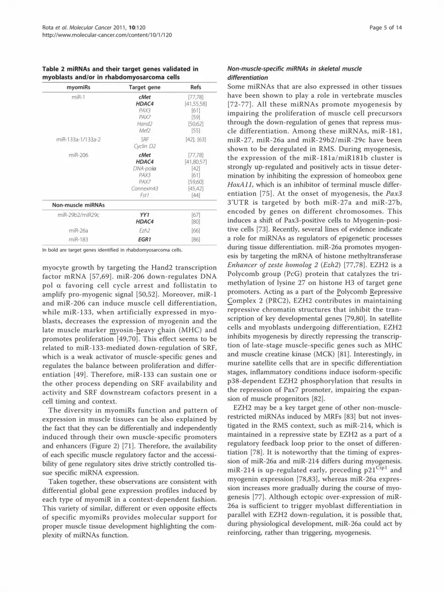
Table 2 miRNAs and their target genes validated inmyoblasts and/or in rhabdomyosarcoma cells
myomiRs Target gene Refs
miR-1 cMetHDAC4PAX3PAX7Hand2Mef2
[77,78][41,55,58]
[61][59]
[50,62][55]
miR-133a-1/133a-2 SRFCyclin D2
[42]; [63]
miR-206 cMetHDAC4DNA-polaPAX3PAX7
Connexin43Fst1
[77,78][41,80,57]
[42][61]
[59,60][45,42][44]
Non-muscle miRNAs
miR-29b2/miR29c YY1HDAC4
[67][80]
miR-26a Ezh2 [66]
miR-183 EGR1 [86]
In bold are target genes identified in rhabdomyosarcoma cells.
Rota et al. Molecular Cancer 2011, 10:120http://www.molecular-cancer.com/content/10/1/120
Page 5 of 14

Recently, the group of Guttridge has shown that themiR-29b2/miR-29c cluster is a target of the PcG tran-scription factor Yin Yang 1 (YY1), which is induced inan NF-kB-dependent manner in the absence of a myo-genic cue [76]. Authors showed that, besides myofibril-lar genes [81,84], YY1 represses miR-29b2/miR-29ctranscription by recruiting EZH2 and HDAC1 on itspromoter (Figure 3). This process results in the expan-sion of undifferentiated muscle precursors. Consistentwith a model of miRNA-dependent suppression of theepigenetic control during myogenesis, in response tomyogenic program activation, miR-29b2/miR-29c beginsto be expressed and inhibits the expression of YY1, thusaccelerating skeletal muscle differentiation [76]. Interest-ingly, although no direct targeting of miR-29b2/miR-29con the 3’UTR of EZH2 was detected, EZH2 levelsdecrease after forced re-expression of miR-29b2/miR-29c. This suggests indirect mechanisms induced by thismiRNA, and possibly other miRNAs acting on epige-netic mediators, to regulate epigenetic pathways as awhole. Moreover, NF-kB loss-of-function experiments inmyoblasts demonstrate that both YY1 and EZH2 areunable to bind the enhancers on miR-29b2/miR-29cpromoters in the absence of an activated NF-kB signal.This observation supports the hypothesis that the NF-kB pathway regulates this YY1-EZH2/miR-29b2/miR-29c network. Therefore, epigenetic molecular networksinvolving feedback regulatory loops with miRNAs mayplay a key role in myogenesis.
Muscle-specific miRNAs in RMSMost studies on the involvement of miRNAs in RMSpathogenesis and their potential therapeutic uses inRMS have been conducted on the myomiR family miR-1/miR-206. We and others have shown that the expres-sion of miR-1 and miR-133a is strikingly decreased inalveolar and embryonal RMS cell lines compared to dif-ferentiated myoblasts and skeletal muscle tissues[85-88]. In particular, two pre-clinical studies reportedthat forced re-expression of miR-206 leads to cell cyclearrest and myogenic differentiation of RMS cells, pre-venting xenografts growth in vivo by targeting themRNA of the oncogenic c-Met receptor [86,87]. ThePonzetto group demonstrated that miR-1 and miR-206are down-regulated in both alveolar and embryonalRMS compared to non-neoplastic skeletal muscle tis-sues, and that they fail to increase in RMS cell lines inresponse to differentiation-inducing treatment [86].Moreover, re-expression of miR-1 or miR-206 throughlentiviral vectors promotes cell differentiation also inalveolar cell lines that are quite resistant to differentia-tive cues, and blocks anchorage-independent growthand invasiveness in vitro. Elegant studies with induciblelentiviral vectors expressing miR-206 at different timesafter RMS xenografts implantation in vivo, clearlydemonstrated that re-expression of miR-206 preventstumor growth [86]. Finally, clusters of hundreds ofgenes up- (muscle lineage) or down-regulated (cellcycle) by miR-206 in RMS were identified, among which
Figure 3 Model for circuits involving Polycomb Group (PcG) proteins and miRNAs during muscle cell differentiation. In blue: indifferentiating myoblasts, miR-29b2/miR-29c, miR-214 and miR-26a are induced by muscle-specific transcription factors, such as MyoD and MEF2,and post-transcriptionally block the expression of YY1 and EZH2 PcG proteins. Together with the inhibition of HDAC4 expression by miR-1, thesephenomena lead to differentiation of myogenic precursor cells. In red: in the absence of a differentiative stimulus, YY1 and EZH2 are highlyexpressed and foster the proliferation (i.e., expansion) of progenitor cells by repressing the expression of miR-29 and miR-214 and down-regulating muscle-specific genes.
Rota et al. Molecular Cancer 2011, 10:120http://www.molecular-cancer.com/content/10/1/120
Page 6 of 14

c-Met was shown to be a miR-206 direct target. ThemiR-206-dependent post-transcriptional inhibition of c-Met expression markedly contributes to the anti-tumoreffects of this miRNA.Similar results are reported in a manuscript published
almost simultaneously to that of Taulli et al. [86] on anembryonal RMS cell line [87]. This study too showed adown-regulation of miR-1 and miR-206 in RMS primarysamples compared to normal muscles, and reported thatforced expression of either miR-1 or miR-206 in theembryonal RMS cell line RD in vitro and in vivo blocksits tumorigenic potential. Consistent with data fromPonzetto’s group, these phenomena occur through miR-206 direct targeting of c-Met mRNA.More recently, Rao et al. [88] showed that miR-1
forced expression in the RD cell line promotes musclegene expression and cell cycle arrest, while miR-133aleads to a decrease of muscle markers expression. Thisis consistent with different roles of miR-1 and miR-133in normal muscle differentiation. However, in contrastto what occurs in healthy myoblasts, both miRNAs inhi-bit cell growth in RMS cell lines. This finding highlights,once more, the importance of cell context in determin-ing the response to miRNAs modulation.The clinical potential of re-expression of miR-1/miR-
206 clusters in RMS is further supported by the obser-vation that these miRNAs directly regulate HDAC4
during differentiation. This is of great importancebecause, among other effects, HDAC4 is responsible forpreventing the expression of cyclin-dependent kinaseinhibitor p21Cip1 that is essential for muscle differentia-tion [89,90]. To date, HDAC inhibitors appear as pro-mising agents for targeted treatment of metastatic RMS[91]. However, re-expression of miR-1/miR-206 clustersis likely to have more complex effects than HDAC4silencing and may be therapeutically more effective [28].
Non-muscle-specific miRNAs in RMSRecently, de-regulation of miR-29 has been reported in asmall cohort of alveolar RMS [92]. A role of the miR-29b2/miR-29c cluster in RMS pathogenesis has beenconfirmed by the recent study of Wang et al. [76]. Pre-viously, these authors showed that an NF-kB-dependentpathway necessary for the expansion of undifferentiatedmyogenic precursors, is aberrantly activated in RMScells [84]. In their latest study, they showed that NF-kBactivation in RMS leads to over-expression of YY1which interacts with EZH2, causing sustained down-reg-ulation of miR-29b2/miR-29c and repression of myogen-esis (Figure 4). Consistent with an anti-myogenic role ofthese two PcG proteins, their levels were found up-regu-lated in tumor tissues from RMS patients compared tonormal adjacent muscle tissues [76]. Interestingly, torepress miR-29b2/miR-29c expression in RMS cells,
Figure 4 Dysregulation of miR-29/YY1 circuit in rhabdomyosarcoma cells. Upper panel, During muscle tissue formation, in normalmyogenic precursor cells miR-29b2/miR-29c targets the 3’UTR of YY1 mRNA inhibiting its expression. Lower panel, Conversely, inrhabdomyosarcoma (RMS) cells NF-kB is up-regulated and YY1 over-produced. The high amount of YY1 in RMS cells is able to recruit EZH2 andHDAC1 on the promoter of miR-29 gene blocking its transcription thus resulting in uncontrolled cell proliferation.
Rota et al. Molecular Cancer 2011, 10:120http://www.molecular-cancer.com/content/10/1/120
Page 7 of 14

YY1 recruits EZH2 to a different site of the miR-29b2/miR-29c promoter than the one used during the expan-sion of normal myoblasts. Ectopic expression of exogen-ous miR-29b2/miR-29c leads to cell cycle arrest anddifferentiation of RMS cell lines, and inhibits RMS xeno-graft growth. Consistent with this observation, miR-29b2/miR-29c levels have been shown to be reduced intumor samples compared to control muscle tissues. Thisstudy was the first to suggest a potential “differentiationtherapy” of RMS through re-expression of a pro-myo-genic miRNA that is involved in the epigenetic controlof differentiation. Along the same lines, our groupshowed that EZH2 expression is increased in tumor tis-sues from RMS patients independently of histologicalsubtype, and correlates with markers of poor prognosis(Abstract # 10-A-4051 AACR 2010). Studies on a largercohort are underway to determine whether the level ofEZH2 expression correlates with the presence of fusionproteins.In addition, we showed concomitant abnormal expres-
sion of miR-26a and EZH2, the former being highlydown-regulated and the latter abnormally expressed inRMS tumor samples and cell lines compared to controls[85]. However, the role of miR-26a in restoring epige-netic processes in RMS needs to be fully elucidated.Interestingly, miR-29b has recently been shown to
directly target HDAC4 during osteoblast differentiationsuggesting that this aspect of miR-29b-dependent regu-lation could be also involved in muscle tissue differen-tiation and possibly in RMS pathogenesis [93]. A furtherrole of miR-29 in epigenetic regulation has been high-lighted by studies on lung cancer showing that thismiRNA targets DNA methyltransferases, leading to aglobal down-regulation of DNA methylation when re-expressed in tumor cells [94]. Notably, an interconnec-tion among miR-29 and miR-206 has been unveiled inliver [95]. These authors showed that miR-206 isrepressed by a YY1/AP1 complex on its promoter, andYY1 down-regulation leads to miR-206 de-repression.This suggests a rationale for future investigations of thisprocess in muscle tissues and RMS [95].A recent publication describes high levels of miR-183
in RMS cell lines and primary tumors [96]. This miRNAbehaves as an onco-miR in several cancers and it hasnot been previously associated with muscle. The miR-183 pro-tumorigenic role in RMS is supported by theevidence that tumor cells in which this miRNA isknocked-down show reduced cell migration in vitro[96]. This phenomenon is due to the release of thedirect repression of Early growth response 1 (EGR1), aregulator of cell migration, by miR-183. Anti-miR-183treatment stimulates the expression of the tumor sup-pressor gene Phosphatase and tensin homolog (PTEN)
as well that, in turn, fosters EGR1 expression reinforcingthe inhibition of cell migration.Finally, our group has shown that miR-27a is signifi-
cantly down-regulated in RMS tissues and cell lines,especially in the alveolar subtype [85]. This is consistentwith its pro-myogenic role in normal development [73]Taken together, these data underscore the complexity
of miRNA function and regulation in RMS and theircentral role in modulating the transition between a dif-ferentiative versus an activated cell state. Moreover, thedata we reviewed point to the fact that apparentlyminor changes in gene expression, even only in onemiRNA, could affect the delicate balance between phy-siologic and pathologic cell fate programs.
miRNAs as diagnostic and prognostic tools in RMSOne of the first studies on miRNAs expression in theclinical context of RMS focused on amplification of the13q31-32 chromosomal region, which is amplified in afraction of alveolar RMS patients [97] and includes theC13orf25 gene [98]. This gene contains the miR-17-92cluster (miR-17, miR-19a, miR-19b, miR-20a, and miR-92), which is considered an onco-miR cluster in sometumor types and cross-talks with MYC, an oncogeneamplified in about 20% of fusion-positive alveolar RMS.This study shows that miR-17-92 expression did notcorrelate with C13orf25 gene amplification in all RMSsamples, irrespective of their alveolar or embryonal ori-gin, suggesting that mechanisms other than amplifica-tion could be responsible for miRNA over-expression.More recently, the Barr group [99] investigated a
minimal common region of the 13q31 amplicon thatcontains the miR-17-92 cluster gene in alveolar RMS.These authors showed that the 13q31 amplification waspresent in about 23% of alveolar RMS, preferentially inPAX7-FKHR-positive cases compared to PAX3-FKHR-positive and fusion-negative tumors. The majority ofalveolar RMS amplified for 13q31 expressed high levelsof five out of six miRNAs within the miR-17-92 cluster,except for miR-18a. Unexpectedly, also a group oftumors that lack 13q31 amplification showed highexpression of all six miRNAs in the miR-17-92 cluster,although the level of expression was lower than inamplified cases. This finding supports the idea that mul-tiple mechanisms in addition to gene amplification regu-late miR-17-92 expression in RMS, as previouslyreported in primary tumors and cell lines [98,100].Moreover, it suggests that the expression of the entiremiRNA cluster can be controlled by a common regula-tory mechanism. Notably, high levels of the five miRNAsin the 13q31 amplified group of patients, most of whomwere PAX7-FKHR-positive, were directly and indepen-dently correlated to a worse outcome when compared
Rota et al. Molecular Cancer 2011, 10:120http://www.molecular-cancer.com/content/10/1/120
Page 8 of 14

to non-amplified cases. Interestingly, expression levels ofthese miRNAs were inversely correlated with outcomewithin the amplified RMS group. Therefore, althoughfurther studies are needed to identify the molecularbasis for these correlations, collectively these resultsassociate amplification and expression of the miR-17-92cluster with specific subsets of alveolar RMS and couldbe useful as prognostic biomarker in these tumor forms.The clinical relevance of the dysregulation of the miR-
1/miR-206 family has been recently highlighted by astudy on a large cohort of 163 RMS patients [101].Besides confirming that all these miRNAs are down-regulated in RMS samples compared to muscle controls,the study of Missiaglia et al. [101] shows that alveolarRMS specimens positive for fusion proteins PAX3- and/or PAX7-FKHR have higher miR-1 levels compared tofusion gene-negative samples. This is an interesting find-ing, considering that alveolar RMS cells usually expresshigher amounts of myogenic factors than embryonalones and that high level of myogenin expression hasbeen recognized as a biomarker of adverse prognosis inRMS [102,103]. Important from a clinical/translationalstandpoint, these authors identified an inverse correla-tion between the expression of miR-206 and overall sur-vival within both the whole RMS group and the genefusion-negative subgroup of patients, while no correla-tion was observed for gene fusion-positive samples.Additionally, miR-206 was shown to be lower in patientswith advanced stage-disease and metastasis at diagnosis,even though significant correlations were detected onlyfor fusion gene-negative patients. These findings high-light the potential of miR-206 expression as a marker ofprognosis and disease progression, especially in embryo-nal tumors that lack specific biomarkers of aggressive-ness. Consistently with the role of miR-206 in muscledetermination, gene expression analysis showed thatmarkers of differentiation are positively correlated withmiR-206 expression in RMS samples. Interestingly, theexpression of inflammatory molecules was inversely cor-related with that of miR-206 suggesting that miR-206could be down-regulated by inflammatory networks inRMS, as already shown for miR-29b2/miR-29c [76,101].In contrast, miR1 and miR-133 do not show any corre-lation with the survival probability in patients.Recently, the expression levels of a specific miRNA
signature were reported to classify RMS patients into 4subgroups, i.e., PAX3-FKHR, PAX7-FKHR and fusion-negative alveolar RMS and embryonal RMS [45].Although the cohort of patients was small, this result isof particular interest since it suggests that miRNAsexpression could be helpful in classifying RMS discrimi-nating between alveolar fusion-negative and embryonalRMS that are often molecularly indistinguishable withcurrent techniques.
The evidence that miRNAs can be released, via differ-ent mechanisms, in human peripheral blood and theirrelative stability and consistent levels in circulation hassuggested that they can be used as non-invasive biomar-kers [104-107]. Among the myomiRs, miR-206 is themost tissue-representative, as it is expressed almostexclusively by skeletal muscle during development andregeneration and is almost undetectable in adult normalskeletal muscle. On this basis, miR-206 circulating levelshave been investigated in sera of RMS patients andfound to be higher as compared with sera from healthydonors or from pediatric patients with other tumors[108]. Considering that miR-206 levels are inversely cor-related with good prognosis, the possibility to detect itspresence in serum of patients could help in the follow-up of highly aggressive neoplasms. This might open theway to a non-invasive approach to the diagnosis and fol-low-up of RMS, which could facilitate the rapid imple-mentation of aggressive treatment protocols andimprove prognosis [109,110]. A possible drawback maybe related to the expression of muscle-specific miRNAsin extremely rare cases of myogenic tumors of child-hood such as leiomyosarcoma and rhabdomyoma. Con-cerning the use of miRNA markers in clinical practice,it must be considered that miRNAs quantification meth-ods in body fluids are still under development, due inpart to the small amounts of circulating miRNAs, espe-cially in serum vs plasma [111,112]. In addition, thechoice of an endogenous control remains critical sinceno housekeeping miRNAs have been identified so far[104,113,114]. Despite the need for more studies tostandardize the measurement methods [113], resultsreported by Miyachi and co-workers appear promising[108].Finally, miRNAs have been recently hypothesized to
regulate drug responsiveness [115,116]. A direct linkbetween miRNAs and drug responsiveness of RMS cellshas been recently unveiled by a study demonstratingthat down-regulation of miR-485-3p is responsible forthe Nuclear Factor- (NF)-YB-dependent decrease inDNA Topoisomerase II (Top2) in the etoposide-resis-tant RH30/v1 RMS cell line [117]. The transcription fac-tor NF-YB binds the Top2 gene promoter, inhibiting itstranscription and thus reducing the effect of Top2 inhi-bitors. Re-expression of miR-485-3p in RH30/v1 cellsreduces NF-YB levels and restores Top2 expression.These effects are associated with an increase in sensitiv-ity of RMS cells to Top2 inhibitors in vitro. This discov-ery could shed light on one of the mechanisms of drugresistance to Top2 inhibitors in this soft-tissue sarcomaand suggest new therapeutic opportunities and pharma-codynamic biomarkers. However, several technical pro-blems such as the choice of a good control miRNA fornormalization and standardization of procedures
Rota et al. Molecular Cancer 2011, 10:120http://www.molecular-cancer.com/content/10/1/120
Page 9 of 14

[114,118], will need to be solved before miRNAs detec-tion in clinical in samples can have practicalapplications.
Perspectives and conclusionsIn summary, recent studies on miRNAs have shown thatmiRNA expression underlies a complex layer of generegulation events guiding biological processes that arefundamental for tissue-specificity and homeostasis [28].Some miRNAs that participate in skeletal/cardiac mus-cle tissue determination have been identified. It is con-ceivable that, in the future, more miRNAs will bediscovered that are potentially able to re-establish cor-rect differentiation in RMS through the modulation ofdiverse molecular pathways.Although the re-expression of selected miRNAs is a
possible strategy for targeted therapy in RMS, it must benoted that miRNA-based therapy presents several chal-lenges. Selectively targeted, efficient re-expression ofmiRNAs is the primary need for an effective therapy. Todate, viral and non-viral vectors have been used in pre-clinical studies to deliver miRNAs. However, viral vec-tors, though efficient in the expression of cDNAs, canbe limited in their practical applications by immuno-genicity and lack of specificity. Non-viral cationic lipo-some-mediated gene transfer approach could beattractive for miRNA therapy; however, cationic lipo-somes developed so far suffer from low efficiency of celltransduction. Moreover, the instability of miRNAs invivo and the potential immunostimulatory effects ofdouble-stranded RNAs are serious obstacles to therapybased on direct delivery of miRNAs. Recently, severaltypes of nanoparticles have been proposed as an alter-nate, highly efficient vehicle to deliver DNA particles tocancer cells [119] and they have been used in preclinicalstudies for an RNA-interference therapy [120]. Morerecently, two studies have shown that anti-miRNAsmolecules stabilized in complexes with either lysine-containing or vessel-targeted nanoparticles are capableto decrease the expression of a liver-specific miRNA orthat of a pro-angiogenic miRNA when systemicallydelivered in vivo [121,122].Recently, locked nucleic acid (LNA) oligos anti-
miRNA were evaluated in non-human primates withunexpectedly positive results [123,124]. Altogether, theseresults appear encouraging for a possible inhibitoryapproach using anti-miRNAs against onco-miRs.In recent years, “epigenetic” therapies aiming at mod-
ulating gene expression at the transcriptional level haveattracted increasing attention. Such treatments havegiven promising results in clinical trials for some typesof tumors [125-127]. In addition to well-known epige-netic drugs acting as either DNA-demethylating agentsor HDAC inhibitors, researchers are working on a class
of agents that inhibit histone methyltransferases such asEZH2, and do not require cell division to target cancercells [128,129]. Interestingly, histone methyltransferaseinhibitors have been shown to synergize with other epi-genetic agents in preclinical studies [130-132].Since EZH2 negatively regulates the expression of pro-
myogenic miRNAs, such as miR-214 and miR-29b2/miR-29c, histone methyltransferase inhibitors may beable to restore physiological levels of expression forthese miRNAs in RMS. Thus, the use of more tradi-tional pharmacological agents could overcome the deliv-ery problems associated with “gene therapy” approaches.On the other hand, “epigenetic” drugs can affect a vari-ety of molecular networks and their in vivo mechanismof action remains controversial.It is noteworthy that miRNA expression can be regu-
lated by epigenetic modifications per se such as DNAmethylation or histone acethylation [23]. Indeed,approximately 50% of miRNA genomic sequences areassociated with DNA regions subjected to methylation,such as CpG islands, and thus are often methylated incancers resulting in silencing of tumor suppressor miR-NAs [133]. Conversely, hypomethylation of miRNAgenes that can lead to over-expression of oncogenicmiRNAs can contribute to tumorigenesis [134,135].Moreover, the same onco-miR can be hypomethylatedor hypermethylated depending of the specific tumorcontext, suggesting a tissue type-dependent epigeneticregulation [135,136]. Therefore, an epigenetic therapywould have to be carefully studied, since it could inducethe re-expression of oncogenic molecules. This has beenthe case with some HDACs and DNA methylation inhi-bitors that have been recently reported to increase themetastatic capability of xenografted tumor cells in ananimal model of RMS through the de-repression of thepro-metastatic Ezrin gene [137]. Interestingly, the Subra-manian group [96] reports that miR-183 silencing inRMS cells is associated with a lowering of Ezrin levels.This report suggests that, besides the re-expression ofpro-differentiative miRNAs, a concomitant inhibition ofonco-miRs may be valuable in combination with an epi-genetic therapy.The high number of mRNAs targeted by a single
miRNA may represent an advantage compared to speci-fic gene silencing (e.g., siRNA). However, this alsomeans that each miRNA can modulate several mole-cules/pathways with potentially unpredictable sideeffects. Therefore, miRNA expression should be con-trolled with the aim to achieve physiological levelsrather than overexpressing miRNAs. A more detailedunderstanding of molecular events governing myogen-esis is needed for the identification of myogenic func-tional steps and networks in which these small RNAsparticipate. Nonetheless, the potential of a therapy based
Rota et al. Molecular Cancer 2011, 10:120http://www.molecular-cancer.com/content/10/1/120
Page 10 of 14

on re-expression of tumor suppressor miRNAs in RMSis high, considering that miRNA re-expression has beenshown to overcome drug resistance in several types oftumor cells and in RMS cells in vitro [117,138-140]. A“miRNA therapy” may be used in the future in combina-tion with conventional therapy in high-risk RMSpatients with metastatic disease, often refractory to con-ventional therapy. Moreover, miRNA expression profil-ing in tumors, and possibly, their detection in peripheralblood during treatment, could predict the response tochemo- and/or radiotherapy and be useful as a prognos-tic signature for the development of treatmentresistance.
Authors’ informationsRR is a PhD and the Head of the Laboratory of Angio-genesis with experience in mechanisms that regulategene expression and cell growth in pediatric cancers. RCis a PhD working on transcriptional regulation in cancerin the Laboratory of Angiogenesis directed by RR. FL isan MD and Full Professor of Pediatrics and the Head ofthe Oncohematology Department with a long standingexperience in preclinical research and clinical manage-ment of pediatric tumor patients. LM is an MD and theDirector of Cancer Centre and Professor of Medicineand Pharmacology at the University of Mississippi Medi-cal Center in Jackson, MS, who has a long experience inpreclinical and clinical research targeting developmentaland cell fate pathways in solid tumors, particularlybreast cancer. AG is an MD and Full Professor inPathology with long lasting experience in the study ofgene expression and cell cycle regulation in cancer.
AcknowledgementsThe present work was supported by grants from Ministero della Sanità Italia(Ricerca Corrente), Associazione Italiana per la Ricerca sul Cancro (AIRCProject 10338) and Istituto Superiore di Sanità (ISS Project 70BF/8) to RR andby grants from Ministero della Salute, Italia (Ricerca Corrente) and AIRC(Special Project 5 × mille) to FL.
Author details1Department of Oncohematology, Ospedale Pediatrico Bambino Gesù, IRCCS,Roma, Italy. 2Sbarro Institute for Cancer Research and Molecular Medicineand Center of Biotechnology, Temple University, Philadelphia, PA, USA.3Department of Human Pathology and Oncology, Università di Siena, Siena,Italy. 4Cancer Institute, University of Mississippi Medical Center, Jackson, MS,USA. 5Dipartimento di Scienze Pediatriche, Università di Pavia, Pavia, Italy.
Authors’ contributionsRR selected the literature, wrote the manuscript and reviewed the finalversion. RC contributed to the conception of the manuscript and to criticaldiscussion. FL, AG and LM contributed to the discussion on clinicalimplications and reviewed the manuscript. All authors read and approvedthe final manuscript.
Competing interestsThe authors declare that they have no competing interests
Received: 4 April 2011 Accepted: 24 September 2011Published: 24 September 2011
References1. Lee RC, Feinbaum RL, Ambros V: The C. elegans heterochronic gene lin-4
encodes small RNAs with antisense complementarity to lin-14. Cell 1993,75:843-854.
2. Wightman B, Ha I, Ruvkun G: Posttranscriptional regulation of theheterochronic gene lin-14 by lin-4 mediates temporal pattern formationin C. elegans. Cell 1993, 75:855-862.
3. Reinhart BJ, Slack FJ, Basson M, Pasquinelli AE, Bettinger JC, Rougvie AE,Horvitz HR, Ruvkun G: The 21-nucleotide let-7 RNA regulatesdevelopmental timing in Caenorhabditis elegans. Nature 2000,403:901-906.
4. Elbashir SM, Harborth J, Lendeckel W, Yalcin A, Weber K, Tuschl T: Duplexesof 21-nucleotide RNAs mediate RNA interference in cultured mammaliancells. Nature 2001, 411:494-498.
5. Lewis BP, Burge CB, Bartel DP: Conserved seed pairing, often flanked byadenosines, indicates that thousands of human genes are microRNAtargets. Cell 2005, 120:15-20.
6. Lee Y, Kim M, Han J, Yeom KH, Lee S, Baek SH, Kim VN: MicroRNA genesare transcribed by RNA polymerase II. Embo J 2004, 23:4051-4060.
7. Borchert GM, Lanier W, Davidson BL: RNA polymerase III transcribeshuman microRNAs. Nat Struct Mol Biol 2006, 13:1097-1101.
8. Winter J, Jung S, Keller S, Gregory RI, Diederichs S: Many roads to maturity:microRNA biogenesis pathways and their regulation. Nat Cell Biol 2009,11:228-234.
9. Martinez J, Patkaniowska A, Urlaub H, Luhrmann R, Tuschl T: Single-stranded antisense siRNAs guide target RNA cleavage in RNAi. Cell 2002,110:563-574.
10. Leuschner PJ, Ameres SL, Kueng S, Martinez J: Cleavage of the siRNApassenger strand during RISC assembly in human cells. EMBO Rep 2006,7:314-320.
11. Hutvagner G, Mlynarova L, Nap JP: Detailed characterization of theposttranscriptional gene-silencing-related small RNA in a GUS gene-silenced tobacco. Rna 2000, 6:1445-1454.
12. Mallanna SK, Rizzino A: Emerging roles of microRNAs in the control ofembryonic stem cells and the generation of induced pluripotent stemcells. Dev Biol 2010, 344:16-25.
13. Wang Y, Russell I, Chen C: MicroRNA and stem cell regulation. Curr OpinMol Ther 2009, 11:292-298.
14. Butcher J, Abdou H, Morin K, Liu Y: Micromanaging oligodendrocytedifferentiation by noncoding RNA: toward a better understanding of thelineage commitment process. J Neurosci 2009, 29:5365-5366.
15. Gangaraju VK, Lin H: MicroRNAs: key regulators of stem cells. Nat Rev MolCell Biol 2009, 10:116-125.
16. Laurent LC: MicroRNAs in embryonic stem cells and early embryonicdevelopment. J Cell Mol Med 2008, 12:2181-2188.
17. Chhabra R, Dubey R, Saini N: Cooperative and individualistic functions ofthe microRNAs in the miR-23a~27a~24-2 cluster and its implication inhuman diseases. Mol Cancer 2010, 9:232.
18. Di Leva G, Croce CM: Roles of small RNAs in tumor formation. Trends MolMed 2010, 16:257-267.
19. Tili E, Croce CM, Michaille JJ: miR-155: on the crosstalk betweeninflammation and cancer. Int Rev Immunol 2009, 28:264-284.
20. Calin GA, Dumitru CD, Shimizu M, Bichi R, Zupo S, Noch E, Aldler H,Rattan S, Keating M, Rai K, et al: Frequent deletions and down-regulationof micro- RNA genes miR15 and miR16 at 13q14 in chronic lymphocyticleukemia. Proc Natl Acad Sci USA 2002, 99:15524-15529.
21. Calin GA, Croce CM: MicroRNA signatures in human cancers. Nat RevCancer 2006, 6:857-866.
22. Garzon R, Fabbri M, Cimmino A, Calin GA, Croce CM: MicroRNA expressionand function in cancer. Trends Mol Med 2006, 12:580-587.
23. Iorio MV, Piovan C, Croce CM: Interplay between microRNAs and theepigenetic machinery: an intricate network. Biochim Biophys Acta 2010,1799:694-701.
24. Ferretti E, De Smaele E, Miele E, Laneve P, Po A, Pelloni M, Paganelli A, DiMarcotullio L, Caffarelli E, Screpanti I, et al: Concerted microRNA control ofHedgehog signalling in cerebellar neuronal progenitor and tumour cells.Embo J 2008, 27:2616-2627.
25. Kota J, Chivukula RR, O’Donnell KA, Wentzel EA, Montgomery CL,Hwang HW, Chang TC, Vivekanandan P, Torbenson M, Clark KR, et al:Therapeutic microRNA delivery suppresses tumorigenesis in a murineliver cancer model. Cell 2009, 137:1005-1017.
Rota et al. Molecular Cancer 2011, 10:120http://www.molecular-cancer.com/content/10/1/120
Page 11 of 14

26. Huang TH, Esteller M: Chromatin remodeling in mammary glanddifferentiation and breast tumorigenesis. Cold Spring Harb Perspect Biol2010, 2:a004515.
27. Guessous F, Zhang Y, Kofman A, Catania A, Li Y, Schiff D, Purow B,Abounader R: microRNA-34a is tumor suppressive in brain tumors andglioma stem cells. Cell Cycle 2010, 9.
28. Taulli R, Bersani F, Ponzetto C: Micro-orchestrating differentiation incancer. Cell Cycle 2010, 9:918-922.
29. Wiggins JF, Ruffino L, Kelnar K, Omotola M, Patrawala L, Brown D, Bader AG:Development of a lung cancer therapeutic based on the tumorsuppressor microRNA-34. Cancer Res 2010, 70:5923-5930.
30. Mishra PJ, Merlino G: MicroRNA reexpression as differentiation therapy incancer. J Clin Invest 2009, 119:2119-2123.
31. Tapscott SJ, Thayer MJ, Weintraub H: Deficiency in rhabdomyosarcomas ofa factor required for MyoD activity and myogenesis. Science 1993,259:1450-1453.
32. De Giovanni C, Landuzzi L, Nicoletti G, Lollini PL, Nanni P: Molecular andcellular biology of rhabdomyosarcoma. Future Oncol 2009, 5:1449-1475.
33. Loeb DM, Thornton K, Shokek O: Pediatric soft tissue sarcomas. Surg ClinNorth Am 2008, 88:615-627, vii.
34. Kohashi K, Oda Y, Yamamoto H, Tamiya S, Takahira T, Takahashi Y, Tajiri T,Taguchi T, Suita S, Tsuneyoshi M: Alterations of RB1 gene in embryonaland alveolar rhabdomyosarcoma: special reference to utility of pRBimmunoreactivity in differential diagnosis of rhabdomyosarcomasubtype. J Cancer Res Clin Oncol 2008, 134:1097-1103.
35. Crist WM, Anderson JR, Meza JL, Fryer C, Raney RB, Ruymann FB,Breneman J, Qualman SJ, Wiener E, Wharam M, et al: Intergrouprhabdomyosarcoma study-IV: results for patients with nonmetastaticdisease. J Clin Oncol 2001, 19:3091-3102.
36. Sorensen PH, Lynch JC, Qualman SJ, Tirabosco R, Lim JF, Maurer HM,Bridge JA, Crist WM, Triche TJ, Barr FG: PAX3-FKHR and PAX7-FKHR genefusions are prognostic indicators in alveolar rhabdomyosarcoma: areport from the children’s oncology group. J Clin Oncol 2002,20:2672-2679.
37. Wachtel M, Dettling M, Koscielniak E, Stegmaier S, Treuner J, Simon-Klingenstein K, Buhlmann P, Niggli FK, Schafer BW: Gene expressionsignatures identify rhabdomyosarcoma subtypes and detect a novel t(2;2)(q35;p23) translocation fusing PAX3 to NCOA1. Cancer Res 2004,64:5539-5545.
38. Lae M, Ahn EH, Mercado GE, Chuai S, Edgar M, Pawel BR, Olshen A, Barr FG,Ladanyi M: Global gene expression profiling of PAX-FKHR fusion-positivealveolar and PAX-FKHR fusion-negative embryonal rhabdomyosarcomas.J Pathol 2007, 212:143-151.
39. Kelly KM, Womer RB, Sorensen PH, Xiong QB, Barr FG: Common andvariant gene fusions predict distinct clinical phenotypes inrhabdomyosarcoma. J Clin Oncol 1997, 15:1831-1836.
40. Davicioni E, Anderson MJ, Finckenstein FG, Lynch JC, Qualman SJ,Shimada H, Schofield DE, Buckley JD, Meyer WH, Sorensen PH, Triche TJ:Molecular classification of rhabdomyosarcoma–genotypic andphenotypic determinants of diagnosis: a report from the Children’sOncology Group. Am J Pathol 2009, 174:550-564.
41. Williamson D, Missiaglia E, de Reynies A, Pierron G, Thuille B, Palenzuela G,Thway K, Orbach D, Lae M, Freneaux P, et al: Fusion gene-negativealveolar rhabdomyosarcoma is clinically and molecularlyindistinguishable from embryonal rhabdomyosarcoma. J Clin Oncol 2010,28:2151-2158.
42. Wachtel M, Runge T, Leuschner I, Stegmaier S, Koscielniak E, Treuner J,Odermatt B, Behnke S, Niggli FK, Schafer BW: Subtype and prognosticclassification of rhabdomyosarcoma by immunohistochemistry. J ClinOncol 2006, 24:816-822.
43. Davicioni E, Anderson JR, Buckley JD, Meyer WH, Triche TJ: Geneexpression profiling for survival prediction in pediatricrhabdomyosarcomas: a report from the children’s oncology group. J ClinOncol 2010, 28:1240-1246.
44. Barr FG, Qualman SJ, Macris MH, Melnyk N, Lawlor ER, Strzelecki DM,Triche TJ, Bridge JA, Sorensen PH: Genetic heterogeneity in the alveolarrhabdomyosarcoma subset without typical gene fusions. Cancer Res2002, 62:4704-4710.
45. Gougelet A, Perez J, Pissaloux D, Besse A, Duc A, Decouvelaere AV,Ranchere-Vince D, Blay JY, Alberti L: miRNA Profiling: How to Bypass the
Current Difficulties in the Diagnosis and Treatment of Sarcomas.Sarcoma 2011, 2011:460650.
46. O’Rourke JR, Georges SA, Seay HR, Tapscott SJ, McManus MT,Goldhamer DJ, Swanson MS, Harfe BD: Essential role for Dicer duringskeletal muscle development. Dev Biol 2007, 311:359-368.
47. McCarthy JJ: MicroRNA-206: the skeletal muscle-specific myomiR. BiochimBiophys Acta 2008, 1779:682-691.
48. Anderson C, Catoe H, Werner R: MIR-206 regulates connexin43 expressionduring skeletal muscle development. Nucleic Acids Res 2006, 34:5863-5871.
49. Chen JF, Mandel EM, Thomson JM, Wu Q, Callis TE, Hammond SM,Conlon FL, Wang DZ: The role of microRNA-1 and microRNA-133 inskeletal muscle proliferation and differentiation. Nat Genet 2006,38:228-233.
50. Kim HK, Lee YS, Sivaprasad U, Malhotra A, Dutta A: Muscle-specificmicroRNA miR-206 promotes muscle differentiation. J Cell Biol 2006,174:677-687.
51. Lagos-Quintana M, Rauhut R, Yalcin A, Meyer J, Lendeckel W, Tuschl T:Identification of tissue-specific microRNAs from mouse. Curr Biol 2002,12:735-739.
52. Rosenberg MI, Georges SA, Asawachaicharn A, Analau E, Tapscott SJ: MyoDinhibits Fstl1 and Utrn expression by inducing transcription of miR-206.J Cell Biol 2006, 175:77-85.
53. van Rooij E, Sutherland LB, Qi X, Richardson JA, Hill J, Olson EN: Control ofstress-dependent cardiac growth and gene expression by a microRNA.Science 2007, 316:575-579.
54. Lim LP, Lau NC, Garrett-Engele P, Grimson A, Schelter JM, Castle J,Bartel DP, Linsley PS, Johnson JM: Microarray analysis shows that somemicroRNAs downregulate large numbers of target mRNAs. Nature 2005,433:769-773.
55. Ivey KN, Muth A, Arnold J, King FW, Yeh RF, Fish JE, Hsiao EC, Schwartz RJ,Conklin BR, Bernstein HS, Srivastava D: MicroRNA regulation of celllineages in mouse and human embryonic stem cells. Cell Stem Cell 2008,2:219-229.
56. Potthoff MJ, Olson EN, Bassel-Duby R: Skeletal muscle remodeling. CurrOpin Rheumatol 2007, 19:542-549.
57. Zhao Y, Samal E, Srivastava D: Serum response factor regulates a muscle-specific microRNA that targets Hand2 during cardiogenesis. Nature 2005,436:214-220.
58. Liu N, Williams AH, Kim Y, McAnally J, Bezprozvannaya S, Sutherland LB,Richardson JA, Bassel-Duby R, Olson EN: An intragenic MEF2-dependentenhancer directs muscle-specific expression of microRNAs 1 and 133.Proc Natl Acad Sci USA 2007, 104:20844-20849.
59. Rao PK, Kumar RM, Farkhondeh M, Baskerville S, Lodish HF: Myogenicfactors that regulate expression of muscle-specific microRNAs. Proc NatlAcad Sci USA 2006, 103:8721-8726.
60. Sweetman D, Goljanek K, Rathjen T, Oustanina S, Braun T, Dalmay T,Munsterberg A: Specific requirements of MRFs for the expression ofmuscle specific microRNAs, miR-1, miR-206 and miR-133. Dev Biol 2008,321:491-499.
61. Sokol NS, Ambros V: Mesodermally expressed Drosophila microRNA-1 isregulated by Twist and is required in muscles during larval growth.Genes Dev 2005, 19:2343-2354.
62. Simon DJ, Madison JM, Conery AL, Thompson-Peer KL, Soskis M,Ruvkun GB, Kaplan JM, Kim JK: The microRNA miR-1 regulates a MEF-2-dependent retrograde signal at neuromuscular junctions. Cell 2008,133:903-915.
63. Zhao X, Sternsdorf T, Bolger TA, Evans RM, Yao TP: Regulation of MEF2 byhistone deacetylase 4- and SIRT1 deacetylase-mediated lysinemodifications. Mol Cell Biol 2005, 25:8456-8464.
64. Williams AH, Valdez G, Moresi V, Qi X, McAnally J, Elliott JL, Bassel-Duby R,Sanes JR, Olson EN: MicroRNA-206 delays ALS progression and promotesregeneration of neuromuscular synapses in mice. Science 2009,326:1549-1554.
65. Sun Y, Ge Y, Drnevich J, Zhao Y, Band M, Chen J: Mammalian target ofrapamycin regulates miRNA-1 and follistatin in skeletal myogenesis. JCell Biol 2010, 189:1157-1169.
66. Chen JF, Tao Y, Li J, Deng Z, Yan Z, Xiao X, Wang DZ: microRNA-1 andmicroRNA-206 regulate skeletal muscle satellite cell proliferation anddifferentiation by repressing Pax7. J Cell Biol 2010, 190:867-879.
67. Dey BK, Gagan J, Dutta A: miR-206 and -486 induce myoblastdifferentiation by downregulating Pax7. Mol Cell Biol 2010, 31:203-214.
Rota et al. Molecular Cancer 2011, 10:120http://www.molecular-cancer.com/content/10/1/120
Page 12 of 14

68. Hirai H, Verma M, Watanabe S, Tastad C, Asakura Y, Asakura A: MyoDregulates apoptosis of myoblasts through microRNA-mediated down-regulation of Pax3. J Cell Biol 2010, 191:347-365.
69. Zhao Y, Ransom JF, Li A, Vedantham V, von Drehle M, Muth AN,Tsuchihashi T, McManus MT, Schwartz RJ, Srivastava D: Dysregulation ofcardiogenesis, cardiac conduction, and cell cycle in mice lacking miRNA-1-2. Cell 2007, 129:303-317.
70. Liu N, Bezprozvannaya S, Williams AH, Qi X, Richardson JA, Bassel-Duby R,Olson EN: microRNA-133a regulates cardiomyocyte proliferation andsuppresses smooth muscle gene expression in the heart. Genes Dev 2008,22:3242-3254.
71. Williams AH, Liu N, van Rooij E, Olson EN: MicroRNA control of muscledevelopment and disease. Curr Opin Cell Biol 2009, 21:461-469.
72. Sarkar S, Dey BK, Dutta A: MiR-322/424 and -503 are induced duringmuscle differentiation and promote cell cycle quiescence anddifferentiation by down-regulation of Cdc25A. Mol Biol Cell 2010,21:2138-2149.
73. Crist CG, Montarras D, Pallafacchina G, Rocancourt D, Cumano A,Conway SJ, Buckingham M: Muscle stem cell behavior is modified bymicroRNA-27 regulation of Pax3 expression. Proc Natl Acad Sci USA 2009,106:13383-13387.
74. Gagan J, Dey BK, Layer R, Yan Z, Dutta A: MICRORNA-378 targets themyogenic repressor myor during myoblast differentiation. J Biol Chem2011.
75. Naguibneva I, Ameyar-Zazoua M, Polesskaya A, Ait-Si-Ali S, Groisman R,Souidi M, Cuvellier S, Harel-Bellan A: The microRNA miR-181 targets thehomeobox protein Hox-A11 during mammalian myoblast differentiation.Nat Cell Biol 2006, 8:278-284.
76. Wang H, Garzon R, Sun H, Ladner KJ, Singh R, Dahlman J, Cheng A,Hall BM, Qualman SJ, Chandler DS, et al: NF-kappaB-YY1-miR-29 regulatorycircuitry in skeletal myogenesis and rhabdomyosarcoma. Cancer Cell2008, 14:369-381.
77. Wong CF, Tellam RL: MicroRNA-26a targets the histone methyltransferaseEnhancer of Zeste homolog 2 during myogenesis. J Biol Chem 2008,283:9836-9843.
78. Juan AH, Kumar RM, Marx JG, Young RA, Sartorelli V: Mir-214-dependentregulation of the polycomb protein Ezh2 in skeletal muscle andembryonic stem cells. Mol Cell 2009, 36:61-74.
79. Levine SS, King IF, Kingston RE: Division of labor in polycomb grouprepression. Trends Biochem Sci 2004, 29:478-485.
80. Marx J: Developmental biology. Combing over the Polycomb groupproteins. Science 2005, 308:624-626.
81. Caretti G, Di Padova M, Micales B, Lyons GE, Sartorelli V: The PolycombEzh2 methyltransferase regulates muscle gene expression and skeletalmuscle differentiation. Genes Dev 2004, 18:2627-2638.
82. Palacios D, Mozzetta C, Consalvi S, Caretti G, Saccone V, Proserpio V,Marquez VE, Valente S, Mai A, Forcales SV, et al: TNF/p38alpha/polycombsignaling to Pax7 locus in satellite cells links inflammation to theepigenetic control of muscle regeneration. Cell Stem Cell 2010, 7:455-469.
83. Liu J, Luo XJ, Xiong AW, Zhang ZD, Yue S, Zhu MS, Cheng SY: MicroRNA-214 promotes myogenic differentiation by facilitating exit from mitosisvia down-regulation of proto-oncogene N-ras. J Biol Chem 2010,285:26599-26607.
84. Wang H, Hertlein E, Bakkar N, Sun H, Acharyya S, Wang J, Carathers M,Davuluri R, Guttridge DC: NF-kappaB regulation of YY1 inhibits skeletalmyogenesis through transcriptional silencing of myofibrillar genes. MolCell Biol 2007, 27:4374-4387.
85. Ciarapica R, Russo G, Verginelli F, Raimondi L, Donfrancesco A, Rota R,Giordano A: Deregulated expression of miR-26a and Ezh2 inrhabdomyosarcoma. Cell Cycle 2009, 8:172-175.
86. Taulli R, Bersani F, Foglizzo V, Linari A, Vigna E, Ladanyi M, Tuschl T,Ponzetto C: The muscle-specific microRNA miR-206 blocks humanrhabdomyosarcoma growth in xenotransplanted mice by promotingmyogenic differentiation. J Clin Invest 2009, 119:2366-2378.
87. Yan D, Dong Xda E, Chen X, Wang L, Lu C, Wang J, Qu J, Tu L: MicroRNA-1/206 targets c-Met and inhibits rhabdomyosarcoma development. J BiolChem 2009, 284:29596-29604.
88. Rao PK, Missiaglia E, Shields L, Hyde G, Yuan B, Shepherd CJ, Shipley J,Lodish HF: Distinct roles for miR-1 and miR-133a in the proliferation anddifferentiation of rhabdomyosarcoma cells. Faseb J 2010, 24:3427-3437.
89. Mottet D, Pirotte S, Lamour V, Hagedorn M, Javerzat S, Bikfalvi A,Bellahcene A, Verdin E, Castronovo V: HDAC4 represses p21(WAF1/Cip1)expression in human cancer cells through a Sp1-dependent, p53-independent mechanism. Oncogene 2009, 28:243-256.
90. Winbanks CE, Wang B, Beyer C, Koh P, White L, Kantharidis P, Gregorevic P:TGF-{beta} regulates miR-206 and miR-29 to control myogenicdifferentiation through regulation of histone deacetylase 4 (HDAC4). JBiol Chem 2011.
91. Wachtel M, Schafer BW: Targets for cancer therapy in childhoodsarcomas. Cancer Treat Rev 2010, 36:318-327.
92. Subramanian S, Lui WO, Lee CH, Espinosa I, Nielsen TO, Heinrich MC,Corless CL, Fire AZ, van de Rijn M: MicroRNA expression signature ofhuman sarcomas. Oncogene 2008, 27:2015-2026.
93. Li Z, Hassan MQ, Jafferji M, Aqeilan RI, Garzon R, Croce CM, van Wijnen AJ,Stein JL, Stein GS, Lian JB: Biological functions of miR-29b contribute topositive regulation of osteoblast differentiation. J Biol Chem 2009,284:15676-15684.
94. Fabbri M, Garzon R, Cimmino A, Liu Z, Zanesi N, Callegari E, Liu S, Alder H,Costinean S, Fernandez-Cymering C, et al: MicroRNA-29 family revertsaberrant methylation in lung cancer by targeting DNAmethyltransferases 3A and 3B. Proc Natl Acad Sci USA 2007,104:15805-15810.
95. Song G, Wang L: Nuclear receptor SHP activates miR-206 expression viaa cascade dual inhibitory mechanism. PLoS One 2009, 4:e6880.
96. Sarver AL, Li L, Subramanian S: MicroRNA miR-183 functions as anoncogene by targeting the transcription factor EGR1 and promotingtumor cell migration. Cancer Res 2010, 70:9570-9580.
97. Gordon AT, Brinkschmidt C, Anderson J, Coleman N, Dockhorn-Dworniczak B, Pritchard-Jones K, Shipley J: A novel and consistentamplicon at 13q31 associated with alveolar rhabdomyosarcoma. GenesChromosomes Cancer 2000, 28:220-226.
98. Williamson D, Selfe J, Gordon T, Lu YJ, Pritchard-Jones K, Murai K, Jones P,Workman P, Shipley J: Role for amplification and expression of glypican-5in rhabdomyosarcoma. Cancer Res 2007, 67:57-65.
99. Reichek JL, Duan F, Smith LM, Gustafson DM, O’Connor RS, Zhang C,Dunlevy MJ, Gastier-Foster JM, Barr FG: Genomic and Clinical Analysis ofAmplification of the 13q31 Chromosomal Region in AlveolarRhabdomyosarcoma: A Report from the Children’s Oncology Group. ClinCancer Res 2011, 17:1463-1473.
100. Wei JS, Johansson P, Chen QR, Song YK, Durinck S, Wen X, Cheuk AT,Smith MA, Houghton P, Morton C, Khan J: microRNA profiling identifiescancer-specific and prognostic signatures in pediatric malignancies. ClinCancer Res 2009, 15:5560-5568.
101. Missiaglia E, Shepherd CJ, Patel S, Thway K, Pierron G, Pritchard-Jones K,Renard M, Sciot R, Rao P, Oberlin O, et al: MicroRNA-206 expression levelscorrelate with clinical behaviour of rhabdomyosarcomas. Br J Cancer2010, 102:1769-1777.
102. Morotti RA, Nicol KK, Parham DM, Teot LA, Moore J, Hayes J, Meyer W,Qualman SJ: An immunohistochemical algorithm to facilitate diagnosisand subtyping of rhabdomyosarcoma: the Children’s Oncology Groupexperience. Am J Surg Pathol 2006, 30:962-968.
103. Heerema-McKenney A, Wijnaendts LC, Pulliam JF, Lopez-Terrada D,McKenney JK, Zhu S, Montgomery K, Mitchell J, Marinelli RJ, Hart AA, et al:Diffuse myogenin expression by immunohistochemistry is anindependent marker of poor survival in pediatric rhabdomyosarcoma: atissue microarray study of 71 primary tumors including correlation withmolecular phenotype. Am J Surg Pathol 2008, 32:1513-1522.
104. Brase JC, Wuttig D, Kuner R, Sultmann H: Serum microRNAs as non-invasive biomarkers for cancer. Mol Cancer 2010, 9:306.
105. Chen X, Ba Y, Ma L, Cai X, Yin Y, Wang K, Guo J, Zhang Y, Chen J, Guo X,et al: Characterization of microRNAs in serum: a novel class ofbiomarkers for diagnosis of cancer and other diseases. Cell Res 2008,18:997-1006.
106. Gilad S, Meiri E, Yogev Y, Benjamin S, Lebanony D, Yerushalmi N,Benjamin H, Kushnir M, Cholakh H, Melamed N, et al: Serum microRNAsare promising novel biomarkers. PLoS One 2008, 3:e3148.
107. Mitchell PS, Parkin RK, Kroh EM, Fritz BR, Wyman SK, Pogosova-Agadjanyan EL, Peterson A, Noteboom J, O’Briant KC, Allen A, et al:Circulating microRNAs as stable blood-based markers for cancerdetection. Proc Natl Acad Sci USA 2008, 105:10513-10518.
Rota et al. Molecular Cancer 2011, 10:120http://www.molecular-cancer.com/content/10/1/120
Page 13 of 14

108. Miyachi M, Tsuchiya K, Yoshida H, Yagyu S, Kikuchi K, Misawa A, Iehara T,Hosoi H: Circulating muscle-specific microRNA, miR-206, as a potentialdiagnostic marker for rhabdomyosarcoma. Biochem Biophys Res Commun2010, 400:89-93.
109. Hays DM, Lawrence W Jr, Wharam M, Newton W Jr, Ruymann FB,Beltangady M, Maurer HM: Primary reexcision for patients with‘microscopic residual’ tumor following initial excision of sarcomas oftrunk and extremity sites. J Pediatr Surg 1989, 24:5-10.
110. Lawrence W Jr, Hays DM, Heyn R, Beltangady M, Maurer HM: Surgicallessons from the Intergroup Rhabdomyosarcoma Study (IRS) pertainingto extremity tumors. World J Surg 1988, 12:676-684.
111. Gaarz A, Debey-Pascher S, Classen S, Eggle D, Gathof B, Chen J, Fan JB,Voss T, Schultze JL, Staratschek-Jox A: Bead array-based micrornaexpression profiling of peripheral blood and the impact of different RNAisolation approaches. J Mol Diagn 2010, 12:335-344.
112. Heneghan HM, Miller N, Kerin MJ: Circulating miRNA signatures:promising prognostic tools for cancer. J Clin Oncol 2010, 28:e573-574,author reply e575-576.
113. Cortez MA, Bueso-Ramos C, Ferdin J, Lopez-Berestein G, Sood AK, Calin GA:MicroRNAs in body fluids-the mix of hormones and biomarkers. Nat RevClin Oncol 2011.
114. Gee HE, Buffa FM, Camps C, Ramachandran A, Leek R, Taylor M, Patil M,Sheldon H, Betts G, Homer J, et al: The small-nucleolar RNAs commonlyused for microRNA normalisation correlate with tumour pathology andprognosis. Br J Cancer 2011, 104:1168-1177.
115. Mishra PJ, Humeniuk R, Mishra PJ, Longo-Sorbello GS, Banerjee D,Bertino JR: A miR-24 microRNA binding-site polymorphism indihydrofolate reductase gene leads to methotrexate resistance. Proc NatlAcad Sci USA 2007, 104:13513-13518.
116. To KK, Zhan Z, Litman T, Bates SE: Regulation of ABCG2 expression at the3’ untranslated region of its mRNA through modulation of transcriptstability and protein translation by a putative microRNA in the S1 coloncancer cell line. Mol Cell Biol 2008, 28:5147-5161.
117. Chen CF, He X, Arslan AD, Mo YY, Reinhold WC, Pommier Y, Beck WT:Novel Regulation of Nuclear Factor-YB by miR-485-3p Affects theExpression of DNA Topoisomerase II{alpha} and Drug Responsiveness.Mol Pharmacol 2011, 79:735-741.
118. Cortez MA, Calin GA: MicroRNA identification in plasma and serum: anew tool to diagnose and monitor diseases. Expert Opin Biol Ther 2009,9:703-711.
119. Pirollo KF, Rait A, Zhou Q, Hwang SH, Dagata JA, Zon G, Hogrefe RI,Palchik G, Chang EH: Materializing the potential of small interfering RNAvia a tumor-targeting nanodelivery system. Cancer Res 2007,67:2938-2943.
120. Hogrefe RI, Lebedev AV, Zon G, Pirollo KF, Rait A, Zhou Q, Yu W, Chang EH:Chemically modified short interfering hybrids (siHYBRIDS):nanoimmunoliposome delivery in vitro and in vivo for RNAi of HER-2.Nucleosides Nucleotides Nucleic Acids 2006, 25:889-907.
121. Su J, Baigude H, McCarroll J, Rana TM: Silencing microRNA by interferingnanoparticles in mice. Nucleic Acids Res 2011, 39:e38.
122. Anand S, Majeti BK, Acevedo LM, Murphy EA, Mukthavaram R, Scheppke L,Huang M, Shields DJ, Lindquist JN, Lapinski PE, et al: MicroRNA-132-mediated loss of p120RasGAP activates the endothelium to facilitatepathological angiogenesis. Nat Med 2010, 16:909-914.
123. Elmen J, Lindow M, Schutz S, Lawrence M, Petri A, Obad S, Lindholm M,Hedtjarn M, Hansen HF, Berger U, et al: LNA-mediated microRNA silencingin non-human primates. Nature 2008, 452:896-899.
124. Lanford RE, Hildebrandt-Eriksen ES, Petri A, Persson R, Lindow M, Munk ME,Kauppinen S, Orum H: Therapeutic silencing of microRNA-122 in primateswith chronic hepatitis C virus infection. Science 2010, 327:198-201.
125. Vigil CE, Martin-Santos T, Garcia-Manero G: Safety and efficacy ofazacitidine in myelodysplastic syndromes. Drug Des Devel Ther 2010,4:221-229.
126. Candelaria M, Herrera A, Labardini J, Gonzalez-Fierro A, Trejo-Becerril C, Taja-Chayeb L, Perez-Cardenas E, de la Cruz-Hernandez E, Arias-Bofill D, Vidal S,et al: Hydralazine and magnesium valproate as epigenetic treatment formyelodysplastic syndrome. Preliminary results of a phase-II trial. AnnHematol 2010, 90:379-387.
127. Fu S, Hu W, Iyer R, Kavanagh JJ, Coleman RL, Levenback CF, Sood AK,Wolf JK, Gershenson DM, Markman M, et al: Phase 1b-2a study to reverseplatinum resistance through use of a hypomethylating agent,
azacitidine, in patients with platinum-resistant or platinum-refractoryepithelial ovarian cancer. Cancer 2010.
128. Suva ML, Riggi N, Janiszewska M, Radovanovic I, Provero P, Stehle JC,Baumer K, Le Bitoux MA, Marino D, Cironi L, et al: EZH2 is essential forglioblastoma cancer stem cell maintenance. Cancer Res 2009,69:9211-9218.
129. Tan J, Yang X, Zhuang L, Jiang X, Chen W, Lee PL, Karuturi RK, Tan PB,Liu ET, Yu Q: Pharmacologic disruption of Polycomb-repressive complex2-mediated gene repression selectively induces apoptosis in cancercells. Genes Dev 2007, 21:1050-1063.
130. Fiskus W, Wang Y, Sreekumar A, Buckley KM, Shi H, Jillella A, Ustun C, Rao R,Fernandez P, Chen J, et al: Combined epigenetic therapy with the histonemethyltransferase EZH2 inhibitor 3-deazaneplanocin A and the histonedeacetylase inhibitor panobinostat against human AML cells. Blood 2009,114:2733-2743.
131. Hayden A, Johnson PW, Packham G, Crabb SJ: S-adenosylhomocysteinehydrolase inhibition by 3-deazaneplanocin A analogues induces anti-cancer effects in breast cancer cell lines and synergy with both histonedeacetylase and HER2 inhibition. Breast Cancer Res Treat .
132. Kalushkova A, Fryknas M, Lemaire M, Fristedt C, Agarwal P, Eriksson M,Deleu S, Atadja P, Osterborg A, Nilsson K, et al: Polycomb target genes aresilenced in multiple myeloma. PLoS One 2010, 5:e11483.
133. Han L, Witmer PD, Casey E, Valle D, Sukumar S: DNA methylation regulatesMicroRNA expression. Cancer Biol Ther 2007, 6:1284-1288.
134. Brueckner B, Stresemann C, Kuner R, Mund C, Musch T, Meister M,Sultmann H, Lyko F: The human let-7a-3 locus contains an epigeneticallyregulated microRNA gene with oncogenic function. Cancer Res 2007,67:1419-1423.
135. Iorio MV, Visone R, Di Leva G, Donati V, Petrocca F, Casalini P, Taccioli C,Volinia S, Liu CG, Alder H, et al: MicroRNA signatures in human ovariancancer. Cancer Res 2007, 67:8699-8707.
136. Lu L, Katsaros D, de la Longrais IA, Sochirca O, Yu H: Hypermethylation oflet-7a-3 in epithelial ovarian cancer is associated with low insulin-likegrowth factor-II expression and favorable prognosis. Cancer Res 2007,67:10117-10122.
137. Yu Y, Zeng P, Xiong J, Liu Z, Berger SL, Merlino G: Epigenetic drugs canstimulate metastasis through enhanced expression of the pro-metastaticEzrin gene. PLoS One 2010, 5:e12710.
138. Li WQ, Li YM, Tao BB, Lu YC, Hu GH, Liu HM, He J, Xu Y, Yu HY:Downregulation of ABCG2 expression in glioblastoma cancer stem cellswith miRNA-328 may decrease their chemoresistance. Med Sci Monit2010, 16:HY27-30.
139. Mongroo PS, Rustgi AK: The role of the miR-200 family in epithelial-mesenchymal transition. Cancer Biol Ther 2010, 10:219-222.
140. Hummel R, Hussey DJ, Haier J: MicroRNAs: predictors and modifiers ofchemo- and radiotherapy in different tumour types. Eur J Cancer 2010,46:298-311.
doi:10.1186/1476-4598-10-120Cite this article as: Rota et al.: MicroRNAs in rhabdomyosarcoma:pathogenetic implications and translational potentiality. MolecularCancer 2011 10:120.
Submit your next manuscript to BioMed Centraland take full advantage of:
• Convenient online submission
• Thorough peer review
• No space constraints or color figure charges
• Immediate publication on acceptance
• Inclusion in PubMed, CAS, Scopus and Google Scholar
• Research which is freely available for redistribution
Submit your manuscript at www.biomedcentral.com/submit
Rota et al. Molecular Cancer 2011, 10:120http://www.molecular-cancer.com/content/10/1/120
Page 14 of 14


