Embed Size (px)
Citation preview
MILWAUKEE PUBLIC MUSEUM
ContributionsInBIOLOGYandGEOLOGY
~umber 56 March 21, 1984
Paleontology and Geology of the Bridger Formation,,outhern Green River Basin, Southwestern Wyoming.Part 7. Survey of Bridgerian Artiodactyla, including
description of a skull and partial skeleton ofAntiacodon pygmaeus.
Robert M. West
REVIEWERS FOR THIS PUBLICATION:Robert J. Emry, National Museum of Natural HistoryPeter Sheehan, Milwaukee Public MuseumJohn A. Wilson, Balcones Research Center, Austin, Texas
ISBN 0-89326-099-1
© 1984Milwaukee Public MuseumPublished by the order of the Board of Trustees
Paleontology and Geology of the BridgerFormation, Southern Green River Basin,
Southwestern Wyoming. Part 7.Survey of Bridgerian Artiodactyla, includingdescription of a skull and partial skeleton of
Antiacodon pygmaeus.
Robert M. WestCarnegie Museum of Natural History
Pittsburgh, PA 15213
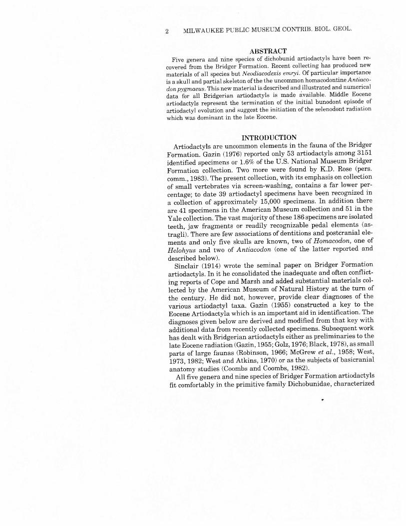
ACKNOWLEDGMENTSI am grateful to Robert J. Emry, U.S. National Museum of Natural His-
tory, Washington, D.C.; Mary R. Dawson, Carnegie Museum of NaturalHistory, Pittsburgh, PA; Richard H. Tedford, American Museum ofNaturalHistory, NewYork, NY; LeoJ. Hickey and Mary Anne Turner, Yale PeabodyMuseum ofNatural History, New Haven, CT; Donald Baird, Princeton Uni-versity, Princeton, NJ; and William D. Turnbull, Field Museum of NaturalHistory, Chicago, IL, for access to and loan of specimens in their respectivecharges. Margery C. Coombs, University of Massachusetts, Amherst, MA,provided pre-publication information on her study of artiodactyl ear regions,and Kenneth D. Rose, The Johns Hopkins University, commented on thepostcranial anatomy ofAntiacodon and allowed me to examine material ofDiacodexis. The drawings ofAntiacodon were prepared by Susan D. Speer-brecher. This research was supported by a series of grants from the NationalScience Foundation.
ABBREVIATIONSCollections are indicated by the following abbreviations:AMNH-American Museum of Natural History, New York, NY;ANSP-Academy of Natural Sciences, Philadelphia, PA;CM-Carnegie Museum of Natural History, Pittsburgh, PA;MPM-Milwaukee Public Museum, Milwaukee, WI;PU-Princeton University, Princeton, NJ;TMM-Texas Memorial Museum, University of Texas, Austin, TX;USNM-U.S. National Museum of Natural History, Washington, D.C.;YPM-Peabody Museum, Yale University, New Haven, CT.
2 MILWAUKEE PUBLIC MUSEUM CONTRIB. BIOL. GEOL.
ABSTRACTFive genera and nine species of dichobunid artiodactyls have been re-
covered from the Bridger Formation. Recent collecting has produced newmaterials of all species but Neodiacodexis emryi. Of particular importanceis a skull and partial skeleton ofthe the uncommon homacodontine Antiaco-don pygmaeus. This new material is described and illustrated and numericaldata for all Bridgerian artiodactyls is made available. Middle Eoceneartiodactyls represent the termination of the initial bunodont episode ofartiodactyl evolution and suggest the initiation of the selenodont radiationwhich was dominant in the late Eocene.
INTRODUCTIONArtiodactyls are uncommon elements in the fauna of the Bridger
Formation. Gazin (1976) reported only 53 artiodactyls among 3151identified specimens or 1.6% of the U.S. National Museum BridgerFormation collection. Two more were found by K.D. Rose (pers.comm., 1983). The present collection, with its emphasis on collectionof small vertebrates via screen-washing, contains a far lower per-centage; to date 39 artiodactyl specimens have been recognized ina collection of approximately 15,000 specimens. In addition thereare 41 specimens in the American Museum collection and 51 in theYale collection. The vast majority ofthese 186 specimens are isolatedteeth, jaw fragments or readily recognizable pedal elements (as-tragli). There are few associations of dentitions and postcranial ele-ments and only five skulls are known, two of Homacodon, one ofHelohyus and two of Antiacodon (one of the latter reported anddescribed below).Sinclair (1914) wrote the seminal paper on Bridger Formation
artiodactyls. In it he consolidated the inadequate and often conflict-ing reports of Cope and Marsh and added substantial materials col-lected by the American Museum of Natural History at the turn ofthe century. He did not, however, provide clear diagnoses of thevarious artiodactyl taxa. Gazin (1955) constructed a key to theEocene Artiodactyla which is an important aid in identification. Thediagnoses given below are derived and modified from that key withadditional data from recently collected specimens. Subsequent workhas dealt with Bridgerian artiodactyls either as preliminaries to thelate Eocene radiation (Gazin, 1955;Golz, 1976;Black, 1978), as smallparts of large faunas (Robinson, 1966; McGrew et al., 1958; West,1973, 1982;West and Atkins, 1970) or as the subjects of basicranialanatomy studies (Coombs and Coombs, 1982).All five genera and nine species ofBridger Formation artiodactyls
fit comfortably in the primitive family Dichobunidae, characterized
WEST: SURVEY OF BRIDGERIAN ARTIODACTYLA 3
by bunodont dentitions, some with upper molar hypocones and somewithout, generally small size and four or five toes. The Dichobunidaeare commonly broken into at least three (Gazin, 1955) and up tofive (Van Valen, 1971) subfamilies. Species thus far known from theBridger Formation are placed in the subfamilies Diacodexinae,Homacodontinae, and Helohyinae.The paucity and low diversity of Bridger Formation artiodactyls
stands in marked contrast with their abundance and rapidly increas-ing diversity in later Eocene rocks (Gazin, 1955; Golz, 1976; Black,1978). The Dichobunidae continue on into the Uintan, apparentlysimultaneously giving rise to several groups of more advancedselenodont artiodactyls. The development of selenodonty now maybe traced back into the late Bridgerian Homacodontinae.This paper surveys the major collections of artiodactyls from the
Bridger Formation, illustrates most of all the species, and givessimplified dental morphometric data. The combined collections areneither large enough nor possess consistent enough stratigraphicdata for any detailed stratigraphic analysis. Further, as is clear fromthe tables ofdental measurements, individual sample sizes are small.In the absence of adequate data on distribution and variation, a fullsystematic revision is not attempted.
Bridger Formation Artiodactyls
Family DichobunidaeSubfamily HomacodontinaeMicrosus cuspidatusAntiacodon pygmaeusAntiacodon venustusHomacodon vagans
Subfamily HelohyinaeHelohyus plicodonHelohyus milleriHelohyus lentus
Subfamily DiacodexinaeN eodiacodexis emryiNeodiacodexis sp.
REPOSITORIES AND LOCALITIESAll the newly collected specimens reported here were recovered
by field parties under my direction between 1970 and 1981. Thosefound between 1970 and 1972 are the property of the AmericanMuseum ofNatural History; those collected subsequent to 1972 are
4 MILWAUKEE PUBLIC MUSEUM CONTRIB. mOL. GEOL.
in the collection of the Milwaukee Public Museum. All localities forthese specimens are designated in the Appendix by MPM numbers;detailed locality data is available in MPM files. Older specimens(those collected prior to my study) are also listed in the appendix;locality data is abbreviated from the various museum cataloguesand is frequently incomplete and/or cryptic. The general geologyand distribution of localities and collecting areas within the BridgerFormation may be found in West (1976) and references therein.
Systematic ReviewOrder Artiodactyla
Family Dichobunidae Gill 1872Subfamily Homacodontinae Peterson 1919
Diagnosis: Small bunodont artiodactyls with conical molar pro-tocones, well-developed hypocones, and the hypoconulid of M, andM2developed from the cingulum posterior to the saddle between thehypoconid and entoconid.
Microsus Leidy 1870Diagnosis: Upper molars lack mesostyle , possess distinct conules,and Ml and M2have prominent hypocones. P4 has a metaconid. Theparaconid on M, is closely appressed to the metaconid and is vestigialor absent on the posterior lower molars. The hypoconulid on M, andM2 is located on the posterior cingulum. The cusps are high andcrescentic. M, length 4.2-4.6mm.
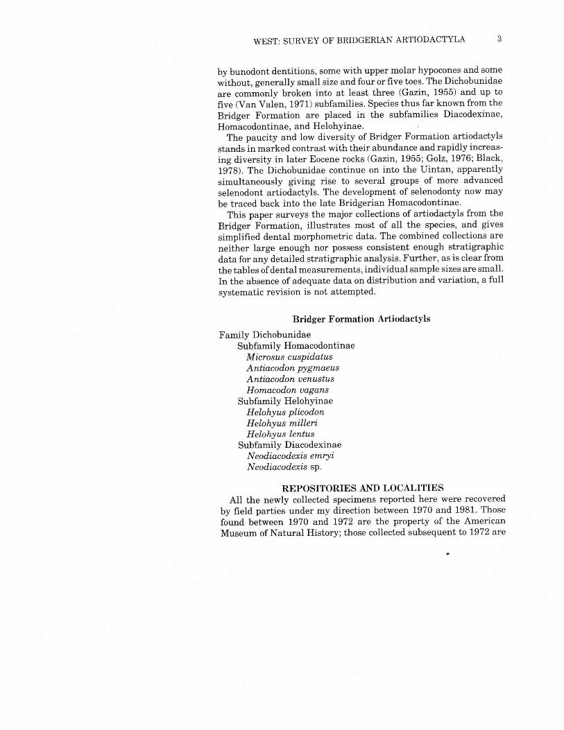
Microsus cuspidatus Leidy 1870Fig. 1 & 2, Table 1
Holotype: ANS 10260, lower molars, Bridger B (Specimen missing[Gillette and Colbert 1976]).Diagnosis: As for the genus.
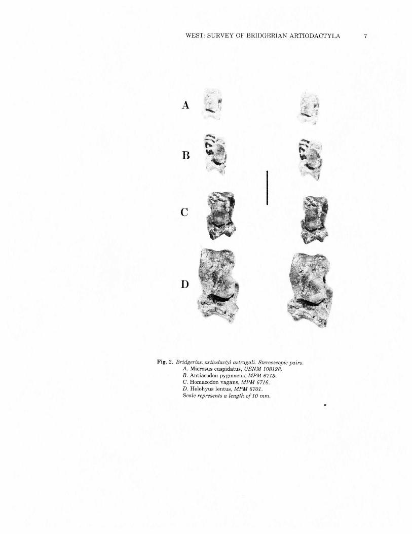
Microsus cuspidatus, which ranges through the entire thicknessof the Bridger Formation, is the smallest Bridgerian artiodactyl.Microsus is a typical homacodontine in the absence of a paraconidon M3 coupled with a large conical M3 heel and a well-developedupper molar hypocone. It is readily differentiated from Antiacodonby the virtual absence of a paraconid on M2and M3and the absenceof upper molar mesostyles. The astragalus (Fig. 2a) shows no appre-ciable angulation, and the deeply grooved calcaneum has a tuberthat is more than one-half the total length of the bone. Althoughthe proximal tibia fragment of MPM 6709 is distorted, there is noindication of the presence of a proximal fibula.
WEST: SURVEY OF BRIDGERIAN ARTIODACTYLA 5
Fig. 1. Microsus cuspidatus. Stereoscopic pairs.A. and B. Maxillae, right Mi _M3, left p4 _M2, USNM 336179. c. Mandible,right partial M2 and M3, MPM 6709. Scales represent a length of 10 mm.
6 MILWAUKEE PUBLIC MUSEUM CONTRIB. BIOL. GEOL.
Antiacodon Marsh 1872Diagnosis: Upper molars with well-developed hypocone on Ml andM2which may be double, a small cusp anterior to the protocone, anda small but distinct mesostyle. Conules are prominent. P4 has ametaconid. Lower molars with metaconid reduced and paraconidand metaconid closely appressed but nonetheless distinct.
Antiacodon pygmaeus (Cope 1872)Figs. 2-14, Tables 2 and 3
Holotype: AMNH 5006, dentary with P4-M1, Bridger BDiagnosis: Small species of Antiacodon, M, length 4.3-4.4mm.Antiacodon pygmaeus is the stratigraphically lower of the two
species here regarded as valid, and is the more abundant.Until recovery of MPM 5896 and 6721 and Rose's recognition in
1983 that USNM 336202 belongs here, Antiacodon pygmaeus hadbeen known only from isolated jaw fragments and teeth. There hadbeen no positive associations of upper and lower dentitions, and nocranial or postcranial remains could be referred to the genus withany certainty.The three specimens mentioned above now allow positive dental
associations and the description of cranial and some postcranialanatomy. Recent studies by Franzen (1981), Rose (1982 and pers.comm.) and Coombs and Coombs (1982) have greatly improved ourknowledge ofthe biology ofdichobunid artiodactyls and provide com-parison for the Antiacodon pygmaeus materials described here. Thefollowing discussion concentrates on MPM 5896 and 6721; elementsofUSNM 336202 are being studied by K.D. Rose (pers. comm., March1983).
Table 1Measurements, in millimeters, of teeth ofMicrosus cuspidatus
N L X N Wa X N Wp X
P4 2 4.0-4.3 4.15 2 2.2-2.5 2.35 4 2.5-2.9 2.78
Ml 4 4.1-4.4 4.28 4 2.3-2.7 2.5 4 2.5-2.9 2.78
M2 5 4.0-4.6 4.3 5 2.4-3.2 2.9 6 2.8-3.4 3.2
M3 4 4.7-5.4 5.05 4 2.8-3.2 3.03 4 2.7-3.2 2.95
p4 1 4.1 1 4.0Ml 1 4.3 1 5.3M2 1 4.8 1 5.8 1 5.4
M3 1 4.2 1 5.2
WEST: SURVEY OF BRIDGERIAN ARTIODACTYLA 7
A !~ ~r r'~.>
,;
B
c
D
Fig. 2. Bridgerian artiodactyl astragali. Stereoscopic pairs.A. Microsus cuspidatus, USNM 108128.B. Antiacodon pygmaeus, MPM 6713,C. Homacodon vagans, MPM 6716.D. Helohyus lentus, MPM 6701.Scale represents a length of 10 mm.
8 MILWAUKEE PUBLIC MUSEUM CONTRIB. BIOL. GEOL.
SKULLFigs. 3-5
MPM 5896 is dorsoventrally crushed to an estimated 50% of itsnatural depth; this estimate is confirmed by the uncrushed skull ofUSNM 336202. While this has resulted in the artificial opening ofsome cranial sutures, most of the bones are readily recognizable andtheir position and relationships comprehensible. The snout has beensubjected to some lateral distortion.The skull of Antiacodon is relatively delicate. It is snub-nosed
with an arcuate cheek tooth row. The snout is not drawn out ornarrowed, as it is in Homacodon (AMNH 12695), but is proportion-ately more like that of early EoceneDiacodexis (AMNH 16141).Thedorsal surface of the skull is relatively planar, without a noticeableinterruption of the dorsal plane in the vicinity of the orbits.Although the crushing of the snout ofMPM 5896 makes interpre-
tation of this area difficult, the nasal bones are narrow, terminatinganteriorly above an apparently shallow nasal incision. Posteriorly,the frontals broaden between and behind the orbits. Medial to theanterior end of the orbits are the origins of the prominent supraor-bital sulci. They are directed antero-posteriorly and flatten and openout at the frontal-nasal suture. Each has a single opening. In con-trast, the supraorbital sulci ofDiacodexis have paired openings, areshorter, and considerably less obvious. The posterolateral part ofthe frontal produces a shelf that overlaps the dorsal border of theorbit.The lacrimal bone is equally divided between its external preorbi-
tal exposure and its internal exposure on the anterior wall of theorbit. The single nasolacrimal canal opens internally in the orbit.The premaxillary and anterior end of the maxillary are missing.
Posteriorly, the maxillary contains, immediately dorsal to the an-terior root ofp3, a small, round infraorbital foramen. It is somewhatsmaller than the equivalent structure in Diacodexis; the infraorbitalcanal is not preserved in either skull ofHomacodon.The zygomatic arch is wide and delicate, with approximately equal
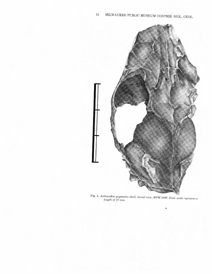
contributions from the jugal and squamosal. There is no postorbitalbar; rather there is a weak dorsal projection from the posterior endof the jugal widely separated from an equally weak ventrolateralprocess from the posterolateral corner of the frontal bone. The otherearly and middle Eocene artiodactyl skulls are inadequately pre-served to allow interpretation of zygomatic morphology.The cranium is bulbous (Fig. 4) with a modest sagittal crest only
along the posterior area of the parietal suture; it merges into aprominent but narrow supraoccipital crest. This supraoccipital crest-
WEST: SURVEY OF BRIDGERIAN ARTIODACTYLA 9
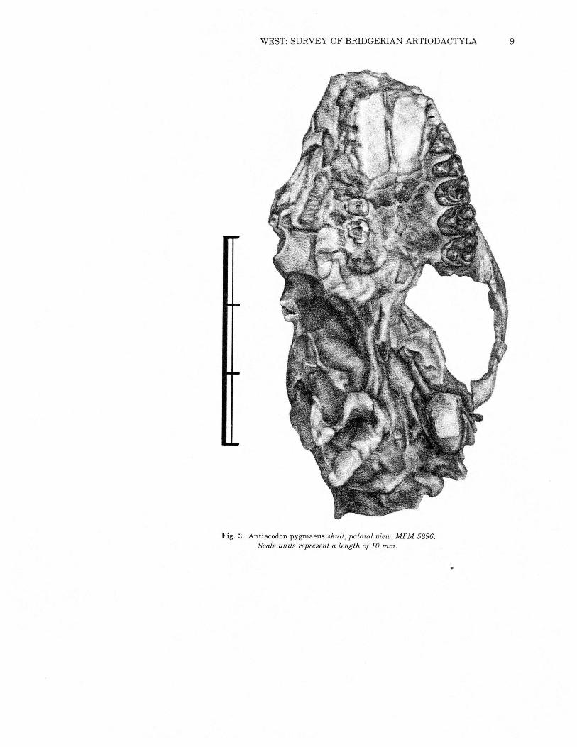
Fig. 3. Antiacodon pygmaeus skull, palatal view, MPM 5896.Scale units represent a length of 10 mm.
10 MILWAUKEE PUBLIC MUSEUM CONTRIB. BIOL. GEOL.
Fig. 4. Antiacodon pygrnaeus skull, dorsal view, MPM 5896. Scale units represent alength of 1 0 mm.
WEST: SURVEY OF BRIDGERIAN ARTIODACTYLA 11
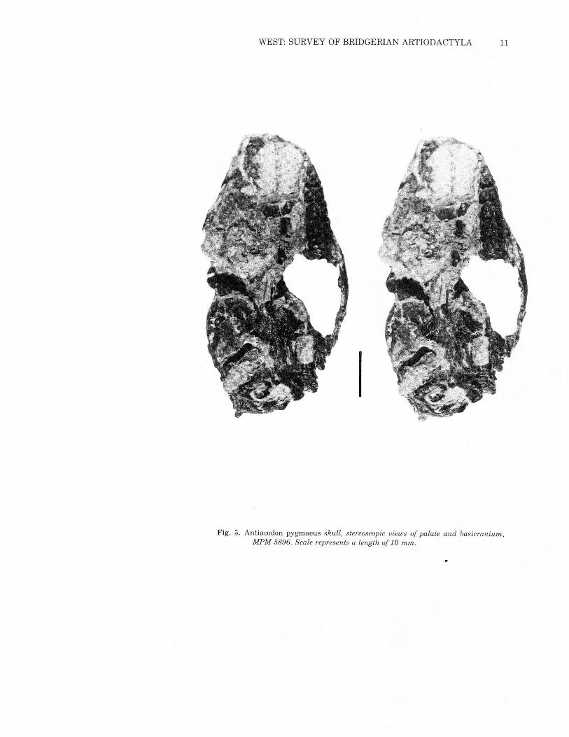
Fig. 5. Antiacodon pygmaeus skull, stereoscopic views of palate and basicranium,MPM 5896. Scale represents a length of 10 mm.
12 MILWAUKEE PUBLIC MUSEUM CONTRIB. BIOL. GEOL.
ing in Antiacodon is considerably more prominent than it is inDiacodexis and the posterior part ofthe Antiacodon braincase showsmore constriction.The glenoid fossa is flat and broad, buttressed posteriorly by a
thin extended postglenoid process and medially by the posterior endof the pterygoid process. Its surface is slightly above the occlusalplane of the upper dentition, as in the other Eocene dichobunids.Details of the Antiacodon palate are not available because of dis-
tortion and poor preservation in MPM 5896 (Figs. 3 and 5).The study of Eocene bunodont artiodactyl basicrania by Coombs
and Coombs (1982) pointed out their general similarity. Antiacodonis a typical bunodont, although it clearly has ossified bullae. Bullaewere not preserved on any of the skulls studied by Coombs andCoombs, although ridges and rugosities on the promontoriasuggested their presence. The retention of a complete bulla on theleft side ofMPM 5896 and parts ofthe bulla on the right side precludecomplete comparison with the basi cranium ofDiacodexis.The Antiacodon basisphenoid is wide medially, and narrows both
posteriorly (where it is pinched between flanges from the basi-occipital), and anteriorly, where it forms the border of the medianlacerate foramen, as in Diacodexis and Homacodon. The contactbetween the lateral edge of the basisphenoid and the medial borderof the alisphenoid is marked by the pterygoid process of the ali-sphenoid. This process is directed antero-medially from the medialedge of the glenoid fossa and posteriorly from the internal border ofthe glenoid fossa. It becomes more prominent anteriorly.The foramen ovale, doubled in Antiacodon, is located in the lateral
wall of the pterygoid process antero-medial to the anterior borderof the glenoid fossa. This opening is single and located slightly moreposteriorly in Diacodexis.A prominent, though delicate, postglenoid process buttresses the
rear of the flattened glenoid fossa. Posteriorly it is penetrated by asmall vertically-directed postglenoid foramen. The opening of thepostglenoid foramen is obscured by the antero-external part of thetympanic tube, a feature not noted on other Eocene bunodont ar-tiodactyls (Coombs and Coombs, 1982).The external auditory meatus is roofed by the squamosal and
anteriorly, ventrally, and posteriorly enclosed in a cylindrical tube,preserved on the right side only, which extends onto the lateralsurface of the bulla. This tube flares moderately laterally, and isquite unlike structures recorded in the external auditory meatus ofother Eocene artiodactyls. Where the tube is missing on the left side,a minor depression between the external auditory meatus and the
WEST: SURVEY OF BRIDGERIAN ARTIODACTYLA 13
middle ear cavity may be regarded as a semilunar depression. Pos-terior to the external auditory meatus, a prominent ventrally-directedprocess is formed of the mastoid process of the petrosal fused withthe lateral part of the exoccipital. The process is composed primarilyof the mastoid, which is roughly triangular in shape and has asubstantial external ventral exposure. The mastoid is grooved onits external surface, but no such pattern can be seen on the ventro-medial side as in Diacodexis. No mastoid foramen is evident and astylomastoid foramen was not found in MPM 5896.The promontorium, globular and bulbous, unlike that of either
Diacodexis or Homacodon, is exposed on both sides. On the left, themedial part of the promontorium is visible medial to the bulla. Theposterior lacerate foramen lies posterior to the promontorium andis connected with the median lacerate foramen by a groove alongthe medial edge of the promontorium, as in Diacodexis, although,because of the shape of the promontorium in Antiacodon, the grooveis much more restricted. Posterior to the posterior lacerate foramenare paired condylar foramina, and an opening possibly referable tothe hypoglossal foramen is near the internal base ofthe paraoccipitalprocess.The bulla, well preserved on the left side, is bulbous and extended
laterally by the prominent tympanic tube. It covers the medial partof the promontorium, though there is no prominent ridge or rugosearea on the promontorium marking the contact area as is presumedin Diacodexis (Coombs and Coombs, 1982). The presence of the bullaon the left side, and bone destruction in the middle ear on the right,preclude detailed study of the middle ear anatomy ofAntiacodon.In summary, the organization of the basicranium of Antiacodon
is generally similar to that of other Eocene bunodont artiodactylsstudied by Coombs and Coombs (1982). They present extensive com-parisons ofDiacodexis, Gobiohyus, ?Helohyus, and Homacodon witha great variety of North American, European, and Asian taxa andconcluded that all four are at essentially the same stage of organiza-tion. All have external mastoid exposure, a conservative primitivecharacter lost in most advanced artiodactyls. None ofthe specimensexamined by Coombs and Coombs retain a bulla but MPM 5896suggests it was present and is merely a postmortem loss in each.There are minor differences among the taxa, but most appear to
have neither functional or phylogenetic significance. Coombs andCoombs (1982) were able, however, to suggest that Diacodexis andHomacodon are part of a group separate from Gobiohyus and?Helohyus (the Helohyidae of their usage, Helohyinae of this paper).Antiacodon has more in common with the Diacodexis-Homacodon
14 MILWAUKEE PUBLIC MUSEUM CONTRIB. BIOL. GEOL.
group, confirming the unity of the Homacodontinae and suggestinga close affinity with the Diacodexinae.
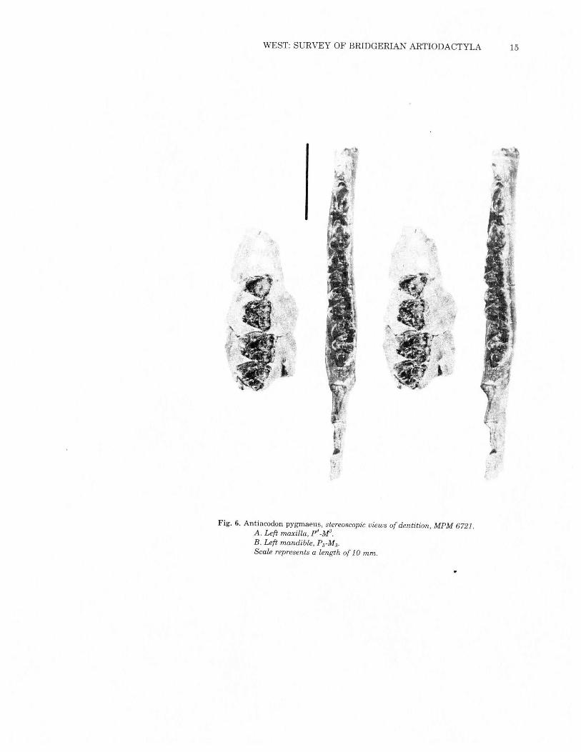
MANDIBLEMPM 6721 preserves both mandibles {Figs. 6 & 7). The left one,
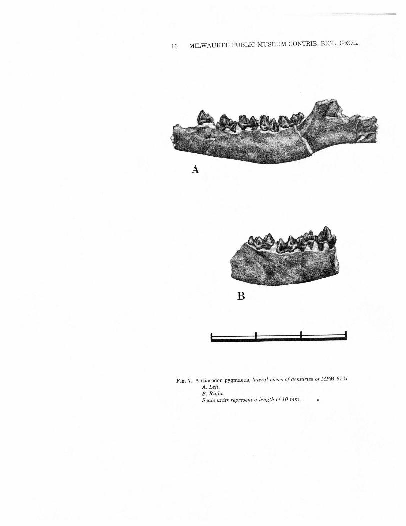
more complete, retains P3-M3and a pair of alveoli for P2.This speci-men is compatible with the features described by Burke (1968) froma Powder Wash locality specimen (CM 10930). In particular it showsdouble-rooted P2 and P3 and mental foramina beneath both P2 andP3. MPM 6721 is preserved somewhat farther posteriorly than CM10930 and confirms the presence of a well excavated massetericfossa, and a gently rising ascending ramus. It also suggests a verylong, straight angular process. The coronoid height and position ofthe condyle cannot be determined from this specimen.
DENTITIONFigs. 3, 5-7
p3_M3are present in both MPM 5281 and MPM 6721 (Fig. 6a). p3(Figs. 3 and 5) is prominently triangular and three-rooted, withanterior, posterior and internal cingula. The paracone is high, almostconnate with the lower, elongate metacone. A low prominent para-style rises from the anterior cingulum; the posterior cingulum isequally prominent but is not developed into a cusp. The protoconeis lower than the other two primary cusps and is quite separate fromthem.p4 is three-rooted, but shorter anteroposteriorly and broader labio-
lingually than p3.The cingulum is weak externally, and modest onthe other sides. The high triangular protocone is flanked anteriorlyby a low parastyle. There is neither a metacone nor a posterior style.The protocone is low with a low crest going antero-externally to theparastyle, and a low median crest extends through the central basinto the internal base of the paracone.Ml and M2are very similar. Each has a strong anterior cingulum,
with a prominent parastyle and minor mesostyle, slightly betterdeveloped on M2.The paracone and metacone are subequal, aligned,with anterior and posterior crests to the anterior and posterior styles.The protocone, with an anterior cingulum on its internal side, islower than the metacone. A well-developed posterior cingulum hasa paired hypocone as an outgrowth on its posterior end. The conulesare strong. The paraconule is anterior of the paracone-protoconeline, with an anterior-external crest extending to just inside theparastyle and a low crest to the center of the protocone. Themetaconule is just posterior to the metacone-protocone line, with a
WEST: SURVEY OF BRIDGERIAN ARTIODACTYLA 15
,
Fig. 6. Antiacodon pygmaeus, stereoscopic views of dentition, MPM 6721 ..A. Left maxilla, P"_M3.B. Left mandible, P3-M3.
Scale represents a length of 10 mm.
16 MILWAUKEE PUBLIC MUSEUM CONTRIB. mOL. GEOL.
B
, I
Fig. 7. Antiacodon pygmaeus, lateral views of dentaries of MPM 6721.A. Left.B. Right.Scale units represent a length of 10 mm.

WEST: SURVEY OF BRIDGERIAN ARTIODACTYLA 17
Table 2Measurements, in millimeters, of teeth ofAntiacodon pygmaeus
N L X N Wa X N Wp XP4 2 4.8-5.0 4.9 2 2.6-3.1 2.85Ml 8 4.3-4.5 4.4 8 2.6-2.9 2.8 8 2.9-3.3 3.11M2 6 4.1-4.8 4.38 6 3.1-3.4 3.22 6 3.3-3.7 3.48M3 3 5.2-5.7 5.47 3 3.3-3.4 3.33 3 3.1-3.2 3.13
p4 2 3.7-3.8 3.75 2 4.5 4.5Ml 2 4.1-4.6 4.35 2 4.8-4.9 4.85 2 4.6-4.7 4.65M2 2 4.4-4.8 4.6 2 5.5-5.7 5.6 2 5.1-5.2 5.15M3 2 4.2-4.5 4.35 2 4.8-5.0 4.9
similar crest to the posterior cingulum, but there is no connectionwith the protocone.The subtriangular M3lacks a hypocone and the paracone is much
larger than the metacone. The parastyle and mesostyle are betterdeveloped than in Ml_M2.The protocone is approximately equal tothe metacone, and the cingulum does not carry around the internalface of the tooth.P2 (not illustrated) is a narrow triangle with a high central cusp
and a lowposterior one. There is a posterior stylid and no cingulum.P3 (Fig. 6b and 7a) is similar to P2,but shows an incipient talonid.
There is a modest metaconid on the posterior flank of the high pro-toconid. The anterior and posterior margins are broken.The P4 ofMPM 6721 is just erupting (Fig. 6b and 7a). The square
trigonid has a high triangular protoconid which sends a sharp crestanterior to the paraconid. There then is an extra anterior cusp onthe antero-internal corner of the tooth. A postero-internal crest ex-tends from the protoconid to the lowmetaconid, and a postero-exter-nal crest extends from the protoconid to the talonid cusp. A ridgeat the posterior edge of the tooth makes up the posterior talonid.M, and M2 are virtually identical (Figs. 6b and 7). The high
trigonid, paraconid and protoconid are approximately equal, and themetaconid is smaller. The metaconid and paraconid have crests tothe protoconid, closing the basin. There is a strong anterior cingulum.The hypoconid is large with a cristid obliqua to the posterior-internalcorner of the metaconid. The entoconid is isolated on the postero-lingual corner of the tooth. The median hypoconulid is separate fromthe posterior cingulum with a lingually-directed ramp and a lowcrest to the hypoconid.
18 MILWAUKEE PUBLIC MUSEUM CONTRIB. BIOL. GEOL.
The trigonid ofM, (Fig. 6b) is similar to that of'M, and M2, thoughthe basin is not quite so thoroughly closed. The cristid obliqua ex-tends to the midpoint between the metaconid and the protoconid.The large heel (hypoconulid) has a low irregular crest to thehypoconid and a large rounded posterior cusp.
Postcranial elementsMPM 6721 includes significant. parts of the hind limb skeleton of
an immature Antiacodon. Elements preserved include both femora(neither complete), both tibiae (left complete), both calcanei, leftastragalus, two terminal phalanges, three metatarsals III or IV (onemissing the epiphysis), five phalanges (twocomplete), onemetatarsalII or V (incomplete proximally), two fragmentary innominates, onepatella, six caudal vertebrae, one lumbar vertebra and nine thoracicvertebrae. Epiphyses on many of these bones are not fused becauseof the animal's age, as noted above by the early eruptive conditionof P4. Many of these elements are duplicated by USNM 336202.These skeletal remains of Antiacodon have been compared with
the Eocene bunodont artiodactyls Diacodexis (Rose, 1982 and pers.comm.) and Messelobunodon (Franzen, 1981), the Oligocene hyper-tragulid Leptomeryx (FMNH UC390), and modern Tragulus (MPMMammalogy 543). Where comparable elements are available, Anti-acodon shows a high degree of similarity with Diacodexis, less withMesselobunodon, and still less with Leptomeryx and Tragulus. All,however, are the same basic type of small, long-legged artiodactyl.Most of the available vertebrae are isolated, although one group
of three thoracics remains in contact in the matrix. Thus it is notpossible to determine the total number, but it is likely that Antiaco-don had 18 to 20 trunk vertebrae as in living artiodactyls. Mes-selobunodon (Franzen 1981) has 19. The tail length is assumed tobe similar to that ofDiacodexis and Messelobunodon, although onlysix caudals are present.Fragments of both innominate bones have been preserved, al-
though all that is available on each side is the acetabulum andnearby parts of the ilium. The ilium shows indications of dorsalflaring as in Diacodexis and some tragulids, but is not preserved asfar dorsally as the sacral articulation. The ilium has a broad dorso-laterally facing gluteal surface and a much narrower ventrally-fac-ing iliac surface. There is a well-developed tuberosity on the acetabu-lar border of the ilium, as in Diacodexis, but Antiacodon also has adeep pit between the anterior border of the acetabulum and thattuberosity. This pit contrasts with the smoother surface ofDiacodexis. Both taxa have a deep, circular acetabulum.
WEST: SURVEY OF BRIDGERIAN ARTIODACTYLA 19
B
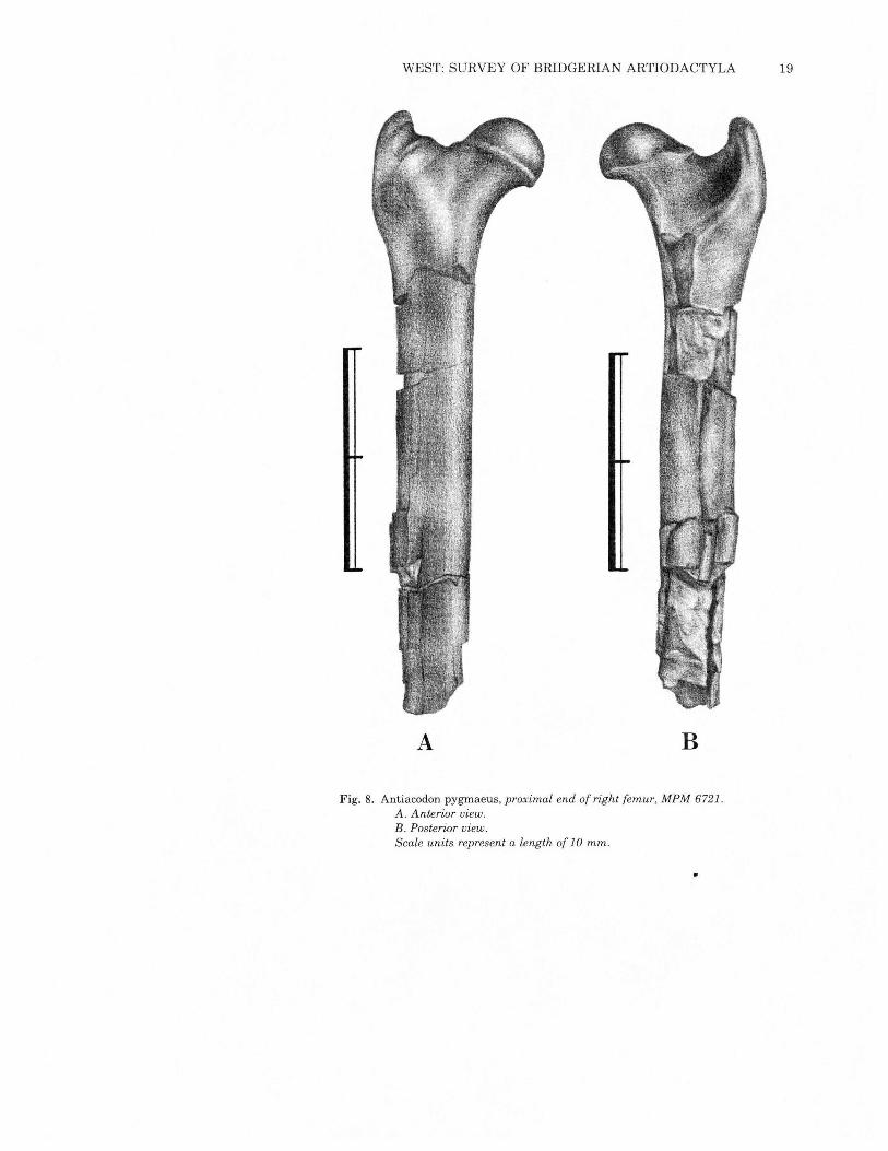
Fig. 8. Antiacodon pygmaeus, proximal end of right femur, MPM 6721.A. Anterior view.B. Posterior view.Scale units represent a length of 10 mm.
A
20 MILWAUKEE PUBLIC MUSEUM CONTRIB. BIOL. GEOL.
A B
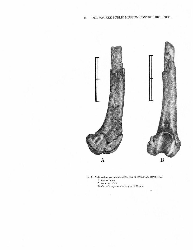
Fig. 9. Antiacodon pygmaeus, distal end of left femur, MPM 6721.A. Lateral view.B. Anterior view.Scale units represent a length of 10 mm.
WEST: SURVEY OF BRIDGERIAN ARTIODACTYLA 21
A
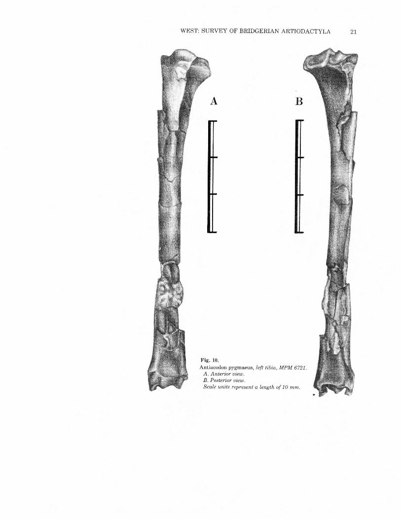
Fig. 10.Antiacodon pygrnaeus, left tibia, MPM 6721.A. Anterior view.B. Posterior view.Scale units represent a length of 10 mm.
B
22 MILWAUKEE PUBLIC MUSEUM CONTRIB. BIOL. GEOL.
A
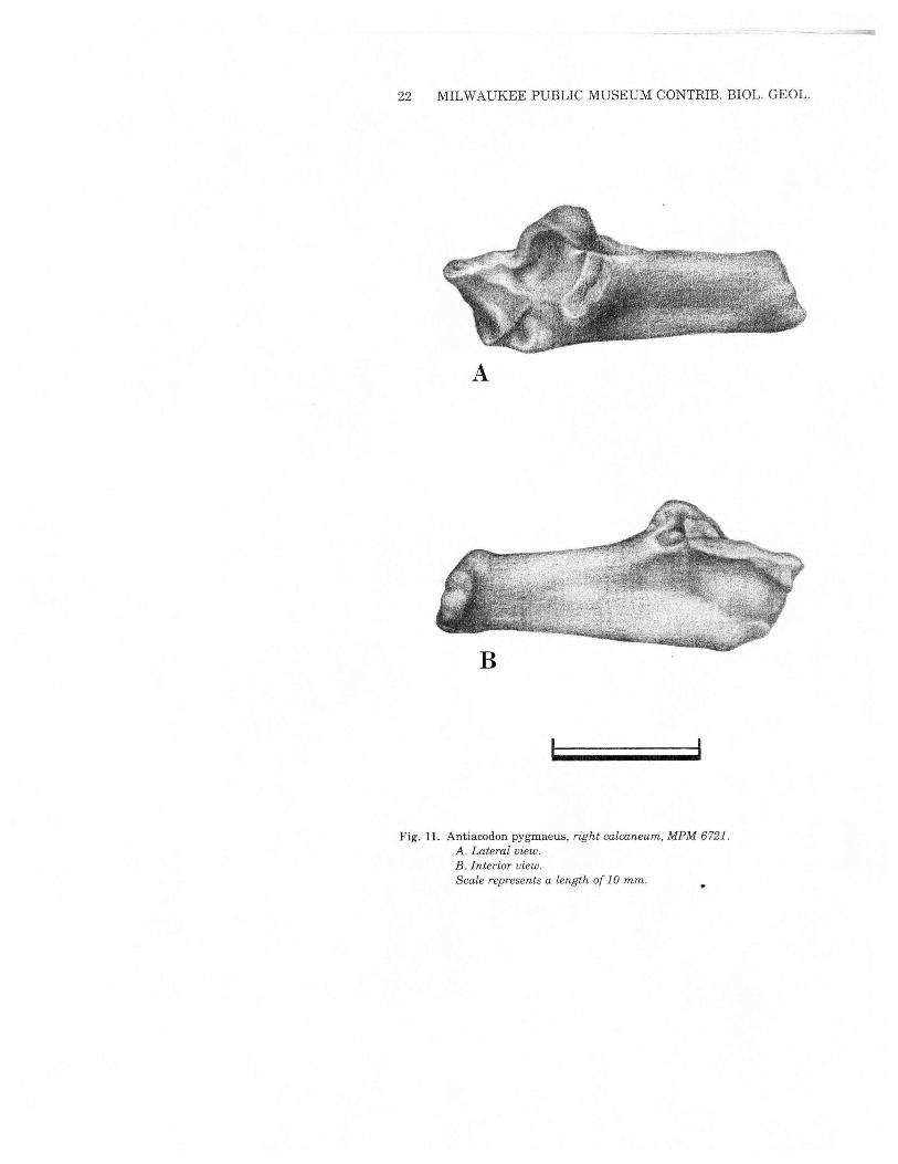
Fig. 11. Antiacodon pygmaeus, right calcaneum, MPM 6721.A. Lateral view.B. Interior view.Scale represents a length of 10 mm.
WEST: SURVEY OF BRIDGERIAN ARTIODACTYLA 23
A
B
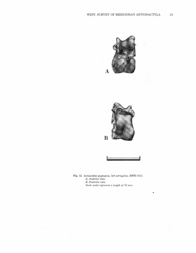
Fig. 12. Antiacodon pygmaeus, left astragalus, MPM 6721.A. Anterior view.B. Posterior view.Scale units represent a length of 10 mm.
24 MILWAUKEE PUBLIC MUSEUM CONTRIB. BIOL. GEOL.
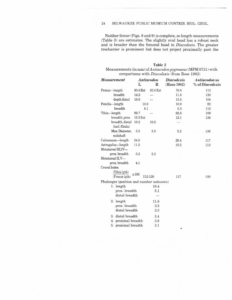
Neither femur (Figs. 8 and 9) is complete, so length measurements(Table 3) are estimates. The slightly oval head has a robust neckand is broader than the femoral head in Diacodexis. The greatertrochanter is prominent but does not project proximally past the
Table 3Measurements (in mm) ofAntiacodon pygmaeus (MPM6721)with
comparisons with Diacodexis (from Rose 1982)Measurement Antiacodon Diacodexis Antiacodon as
L R (Rose 1982) % ofDiacodexisFemur-length 80.0Est 85.0Est 70.8 113
breadth 14.2 11.8 120depth distal 16.0 15.4 104
Patella-length 10.0 10.9 92breadth 6.1 5.3 115
Tibia-length 89.7 82.6 109breadth, prox. 15.5Est 12.1 128breadth, distal 10.2 10.5(inc!.fibula)
Max.Diameter, 5.5 5.5 5.2 106midshaft
Calcaneum-length 24.0 20.4 117Astragalus-length 11.5 10.2 113Metatarsal III ,IV-
prox.breadth 5.3 5.3MetatarsaIII,V-
prox.breadth 4.1Crural Index
(Tibia lgth) x 100(Femur lgth) 112-120 117 100
Phalanges (position and number unknown)1. length 16.4
prox. breadth 5.1distal breadth
2. length 11.8prox. breadth 3.5distal breadth 2.3
3. distal breadth 3.44. proximal breadth 3.85. proximal breadth 3.1
WEST: SURVEY OF BRIDGERIAN ARTIODACTYLA 25
head of the femur. There is a deeply excavated trochanteric fossawhich extends farther distally than in Diacodexis. The lessertrochanter is broken on both specimens, but apparently had a tri-angular base and was of substantial size. A third trochanter is notpresent; a small one occurs onDiacodexis. The shaft is slightly curvedposteriorly. The distal end of the femur is relatively broad and mas-sive. A well-developed extensor fossa is located at the lower end ofthe lateral margin of the patellar groove. Several of the proportionaldistinctions from Diacodexis are likely to be ontogenetic, as MPM6721is a young animal with incompletely formed bones. The patellargroove is slightly shallower and broader than in Diacodexis and hasa primary distal exposure; the patella is robust. The tibia (Fig. 10)is elongate and slender, longer than the femur giving a crural indexof 112 to 120 (Table 3). It is virtually identical to that ofDiacodexis.The fibula is synostosed distally. Although the bone is broken im-mediately proximal to the area offusion, its impression can be tracedabout 60% of the length of the tibia. There is no indication of thepresence of the proximal end of the fibula, unlike the situation inDiacodexis. The fibula of Antiacodon is a proximally incompletesplint, and thus is advanced over the condition shown byDiacodexis.The calcaneum (Fig. 11) of Antiacodon is virtually identical to
that ofBunophorus illustrated by Guthrie (1968). The tuber is halfthe length of the entire bone, and it has a deeply grooved lateralside. The calcaneum of Diacodexis has a deeper plantar groove.The astragalus (Figs. 2 and 12) is straighter than that of
Diacodexis, and has a sharper tibial trochlea. The sustentacular facetis somewhat larger than in Diacodexis, as is the distal astragalar facet(Schaeffer 1947).In the tarsal area, the cuboid is not fused with the navicular, a .
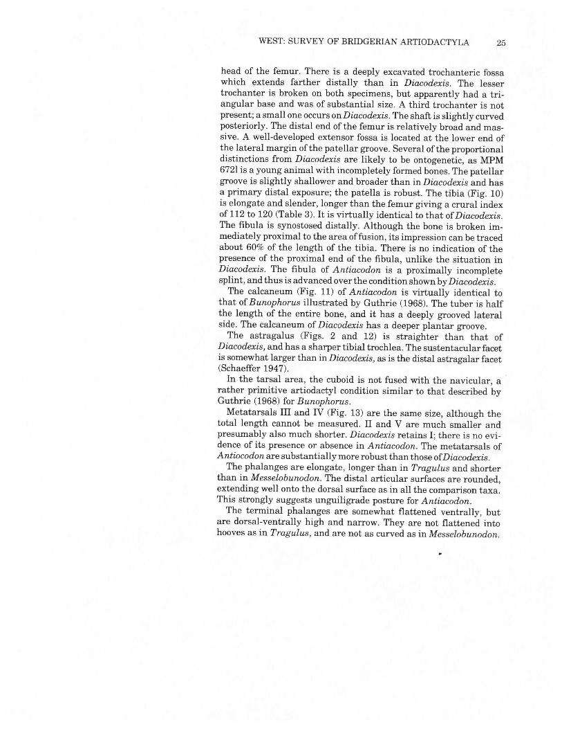
rather primitive artiodactyl condition similar to that described byGuthrie (1968) for Bunophorus.Metatarsals III and IV (Fig. 13) are the same size, although the
total length cannot be measured. II and V are much smaller andpresumably also much shorter. Diacodexis retains I; there is no evi-dence of its presence or absence in Antiacodon. The metatarsals ofAntiocodon are substantially more robust than those ofDiacodexis.The phalanges are elongate, longer than in Tragulus and shorter
than in Messelobunodon. The distal articular surfaces are rounded,extending well onto the dorsal surface as in all the comparison taxa.This strongly suggests unguiligrade posture for Antiacodon.The terminal phalanges are somewhat flattened ventrally, but
are dorsal-ventrally high and narrow. They are not flattened intohooves as in Tragulus, and are not as curved as in Messelobunodon.
26 MILWAUKEE PUBLIC MUSEUM CONTRIB. BIOL. GEOL.
A
B
,Fig. 13. Antiacodon pygmaeus, distal end of left metatarsal IV, MPM 6721.
A. Ventral view.B. Dorsal view.Scale units represent a length of 10 mm.
~
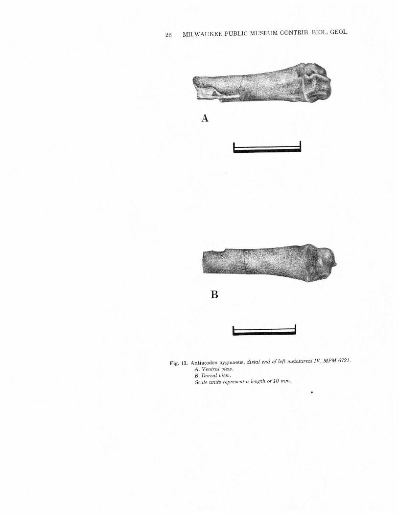
,Fig. 14. Reconstruction of the skeleton of Antiacodon pygmaeus. The stippled areas
are represented by MPM 6721 and 5896; the remainder of the skeleton isbased on Messelobunodon (Franzen 1981) and Diacodexis (Rose 1982).
~trlen>-?ene~-<trl~o>-rj
bj~tl
~~:;z»~•....•otl»o~~r-»
tv-J
28 MILWAUKEE PUBLIC MUSEUM CONTRIB. BIOL. GEOL.
Both specimens show minor lateral grooving.Apart from the massiveness of the patella, the incomplete fibula,
and more robust metatarsals, the skeletal parts of Antiacodon thatcan be compared directly with Diacodexis (Rose, 1982) indicate ahigh degree of similarity (Table 3).Therefore in Fig. 14 Antiacodonis restored as a lithe, long-legged creature with highly developedcursorial locomotion. In fact, it possibly was more advanced in thisrespect than Diacodexis, based on the relatively smaller lateral toesand reduction of the fibula.
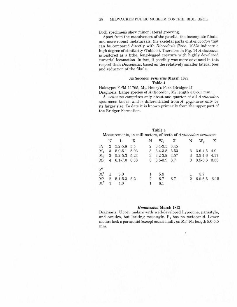
Antiacodon venustus Marsh 1872Table 4
Holotype: YPM 11765, M3,Henry's Fork (Bridger D)Diagnosis: Large species of Antiacodon, M, length 5.0-5.1mm.A. venustus comprises only about one quarter of all Antiacodon
specimens known and is differentiated from A. pygmaeus only byits larger size. To date it is known primarily from the upper part ofthe Bridger Formation.
Table 4Measurements, in millimeters, of teeth of Antiacodon venustus
N L X N Wa X N Wp XP4 2 5.2-5.8 5.5 2 3.4-3.5 3.45Ml 3 5.0-5.1 5.03 3 3.4-3.8 3.53 3 3.6-4.3 4.0M2 3 5.2-5.3 5.23 3 3.2-3.9 3.57 3 3.5-4.6 4.17M3 4 6.1-7.0 6.33 3 3.5-3.9 3.7 3 3.5-3.6 3.53p4
Ml 1 5.0 1 5.8 1 5.7M2 2 5.1-5.3 5.2 2 6.7 6.7 2 6.0-6.3 6.15M3 1 4.0 1 6.1
Homacodon Marsh 1872Diagnosis: Upper molars with well-developed hypocone, parastyle,and conules, but lacking mesostyle. P4 has no metaconid. Lowermolars lack a paraconid (except occasionally onM3)'Mj length 5.0-5.5mm.
WEST: SURVEY OF BRIDGERIAN ARTIODACTYLA 29
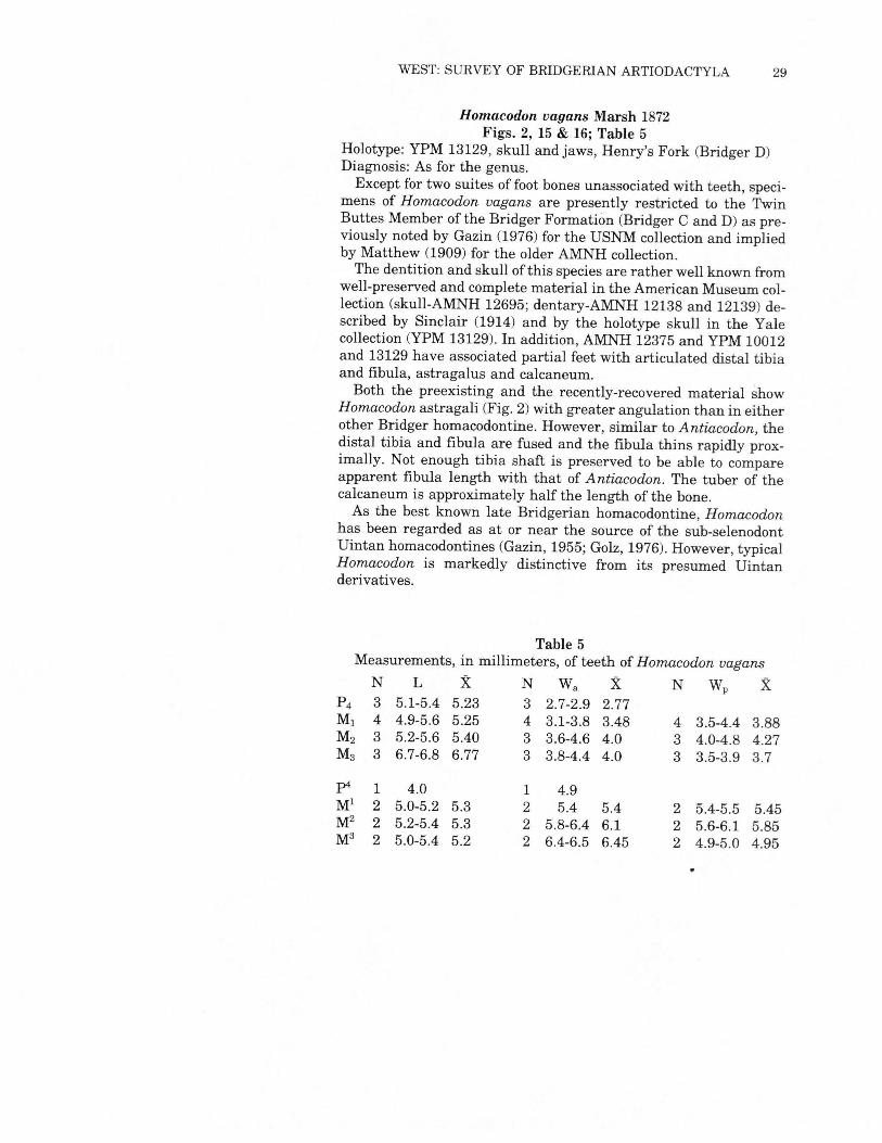
Homacodon vagans Marsh 1872Figs. 2, 15& 16; Table 5
Holotype: YPM 13129, skull and jaws, Henry's Fork (Bridger D)Diagnosis: As for the genus.Except for two suites of foot bones unassociated with teeth, speci-
mens of Homacodon vagans are presently restricted to the TwinButtes Member of the Bridger Formation (Bridger C and D) as pre-viously noted by Gazin (1976) for the USNM collection and impliedby Matthew (1909) for the older AMNH collection.The dentition and skull of this species are rather well known from
well-preserved and complete material in the American Museum col-lection (skull-AMNH 12695; dentary-AMNH 12138 and 12139) de-scribed by Sinclair (1914) and by the holotype skull in the Yalecollection (YPM 13129). In addition, AMNH 12375 and YPM 10012and 13129 have associated partial feet with articulated distal tibiaand fibula, astragalus and calcaneum.Both the preexisting and the recently-recovered material show
Homacodon astragali (Fig. 2) with greater angulation than in eitherother Bridger homacodontine. However, similar to Antiacodon, thedistal tibia and fibula are fused and the fibula thins rapidly prox-imally. Not enough tibia shaft is preserved to be able to compareapparent fibula length with that of Antiacodon. The tuber of thecalcaneum is approximately half the length of the bone.As the best known late Bridgerian homacodontine, Homacodon
has been regarded as at or near the source of the sub-selenodontUintan homacodontines (Gazin, 1955; Golz, 1976). However, typicalHomacodon is markedly distinctive from its presumed Uintanderivatives.
Table 5Measurements, in millimeters, of teeth ofHomacodon vagans
N L X N Wa X N Wp XP4 3 5.1-5.4 5.23 3 2.7-2.9 2.77Ml 4 4.9-5.6 5.25 4 3.1-3.8 3.48 4 3.5-4.4 3.88M2 3 5.2-5.6 5.40 3 3.6-4.6 4.0 3 4.0-4.8 4.27M3 3 6.7-6.8 6.77 3 3.8-4.4 4.0 3 3.5-3.9 3.7
p4 1 4.0 1 4.9Ml 2 5.0-5.2 5.3 2 5.4 5.4 2 5.4-5.5 5.45M2 2 5.2-5.4 5.3 2 5.8-6.4 6.1 2 5.6-6.1 5.85M3 2 5.0-5.4 5.2 2 6.4-6.5 6.45 2 4.9-5.0 4.95
30 MILWAUKEE PUBLIC MUSEUM CONTRIB. BIOL. GEOL.
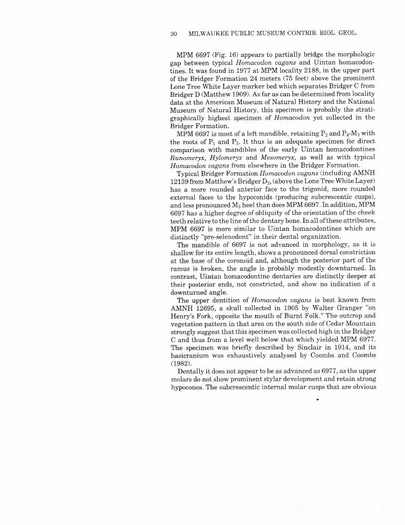
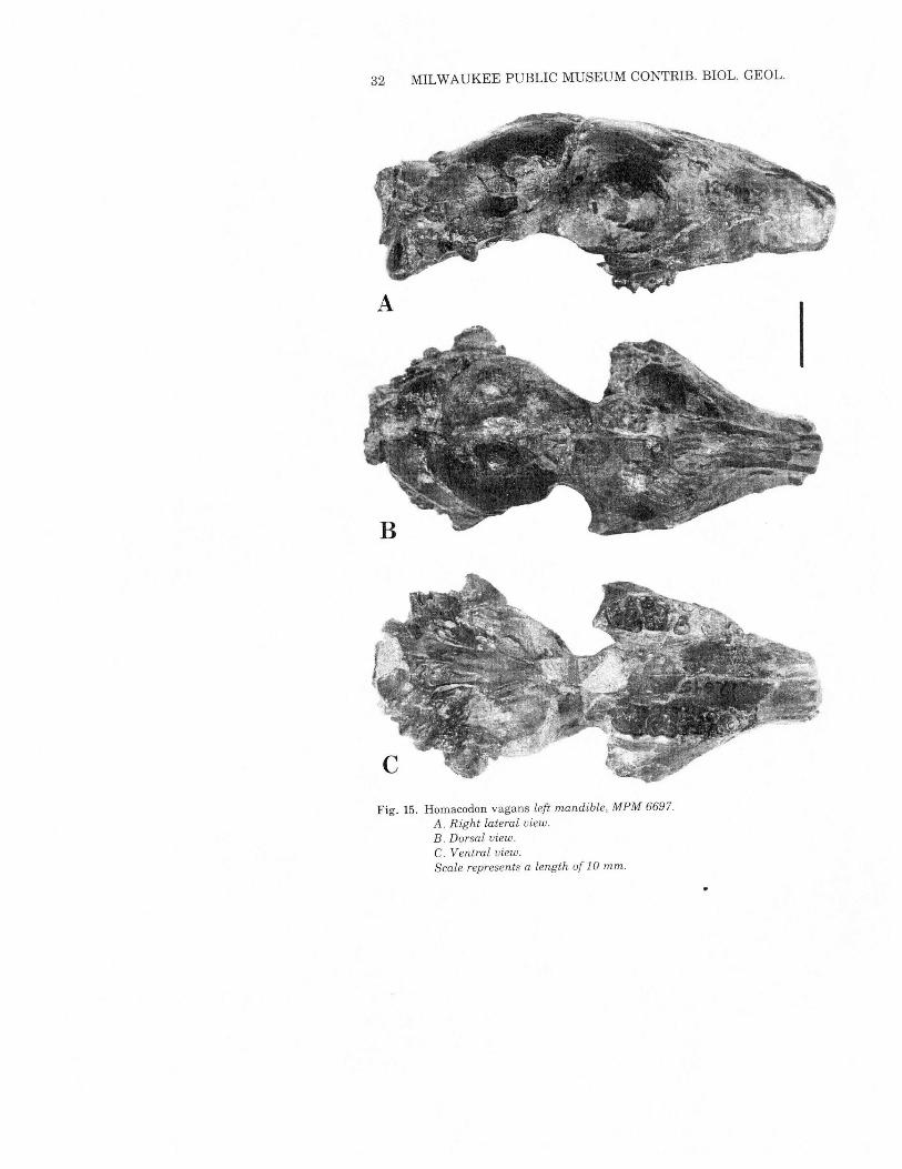
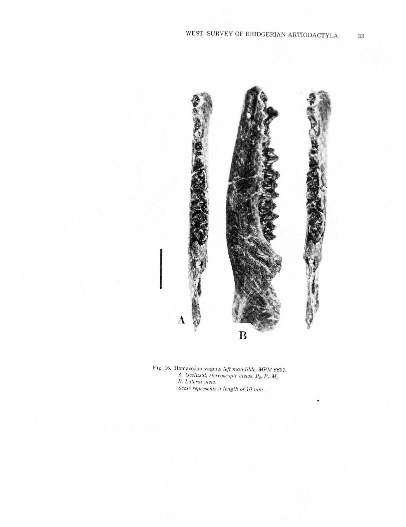
MPM 6697 (Fig. 16) appears to partially bridge the morphologicgap between typical Homacodon vagans and Uintan homacodon-tines. It was found in 1977 at MPM locality 2188, in the upper partof the Bridger Formation 24 meters (75 feet) above the prominentLone Tree White Layer marker bed which separates Bridger C fromBridger D (Matthew 1909). As far as can be determined from localitydata at the American Museum ofNatural History and the NationalMuseum of Natural History, this specimen is probably the strati-graphically highest specimen of Homacodon yet collected in theBridger Formation.MPM 6697 is most of a left mandible, retaining P2 and P4-M3with
the roots of Pi and P3. It thus is an adequate specimen for directcomparison with mandibles of the early Uintan homacodontinesBunomeryx, Hylomeryx and Mesomeryx, as well as with typicalHomacodon vagans from elsewhere in the Bridger Formation.Typical Bridger Formation Homacodon vagans (including AMNH
12139 fromMatthew's Bridger D2, (above the Lone Tree White Layer)has a more rounded anterior face to the trigonid, more roundedexternal faces to the hypoconids (producing subcrescentic cusps),and less pronounced M3heel than doesMPM6697. In addition, MPM6697 has a higher degree of obliquity ofthe orientation ofthe cheekteeth relative to the line ofthe dentary bone. In all ofthese attributes,MPM 6697 is more similar to Uintan homacodontines which aredistinctly "pre-selenodont" in their dental organization.The mandible of 6697 is not advanced in morphology, as it is
shallow for its entire length, shows a pronounced dorsal constrictionat the base of the coronoid and, although the posterior part of theramus is broken, the angle is probably modestly downturned. Incontrast, Uintan homacodontine dentaries are distinctly deeper attheir posterior ends, not constricted, and show no indication of adownturned angle.The upper dentition of Homacodon vagans is best known from
AMNH 12695, a skull collected in 1905 by Walter Granger "onHenry's Fork, opposite the mouth of Burnt Folk." The outcrop andvegetation pattern in that area on the south side ofCedar Mountainstrongly suggest that this specimen was collected high in the BridgerC and thus from a level well below that which yielded MPM 6977.The specimen was briefly described by Sinclair in 1914, and itsbasicranium was exhaustively analysed by Coombs and Coombs(1982).Dentally it does not appear to be as advanced as 6977, as the upper
molars do not show prominent stylar development and retain stronghypocones. The subcrescentic internal molar cusps that are obvious
WEST: SURVEY OF BRIDGERIAN ARTIODACTYLA 31
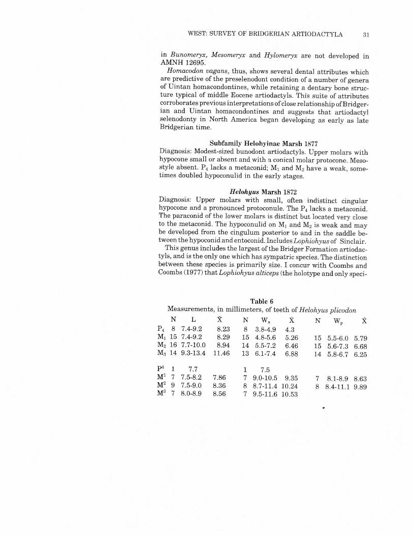
in Bunomeryx, Mesomeryx and Hylomeryx are not developed inAMNH 12695.Homacodon vagans, thus, shows several dental attributes which
are predictive of the preselenodont condition of a number of generaof Uintan homacondontines, while retaining a dentary bone struc-ture typical of middle Eocene artiodactyls. This suite of attributescorroborates previous interpretations ofclose relationship ofBridger-ian and Uintan homacondontines and suggests that artiodactylselenodonty in North America began developing as early as lateBridgerian time.
Subfamily Helohyinae Marsh 1877Diagnosis: Modest-sized bunodont artiodactyls. Upper molars withhypocone small or absent and with a conical molar protocone. Meso-style absent. P4 lacks a metaconid; M, and M2have a weak, some-times doubled hypoconulid in the early stages.
Helohyus Marsh 1872Diagnosis: Upper molars with small, often indistinct cingularhypocone and a pronounced protoconule. The P4 lacks a metaconid.The paraconid of the lower molars is distinct but located very closeto the metaconid. The hypoconulid on M, and M2 is weak and maybe developed from the cingulum posterior to and in the saddle be-tween the hypoconid and entoconid. Includes Lophiohyus of Sinclair.This genus includes the largest of the Bridger Formation artiodac-
tyls, and is the only one which has sympatric species. The distinctionbetween these species is primarily size. I concur with Coombs andCoombs (1977) that Lophiohyus alticeps (the holotype and only speci-
Table 6Measurements, in millimeters, of teeth ofHelohyus plicodonN L X N Wa X N Wp X
P4 8 7.4-9.2 8.23 8 3.8-4.9 4.3Ml 15 7.4-9.2 8.29 15 4.8-5.6 5.26 15 5.5-6.0 5.79M2 16 7.7-10.0 8.94 14 5.5-7.2 6.46 15 5.6-7.3 6.68M3 14 9.3-13.4 11.46 13 6.1-7.4 6.88 14 5.8-6.7 6.25
pi 1 7.7 1 7.5Ml 7 7.5-8.2 7.86 7 9.0-10.5 9.35 7 8.1-8.9 8.63M2 9 7.5-9.0 8.36 8 8.7-11.4 10.24 8 8.4-11.1 9.89M3 7 8.0-8.9 8.56 7 9.5-11.6 10.53
32 MILWAUKEE PUBLIC MUSEUM CONTRIB. BIOL. GEOL.
Fig. 15. Homacodon vagans left mandible, MPM 6697.A. Right lateral view.B. Dorsal view.C. Ventral view.Scale represents a length of 10 mm.
WEST: SURVEY OF BRIDGERIAN ARTIODACTYLA 33
IA
Fig. 16. Homacodon vagans left mandible, MPM 6697.A. Occlusal, stereoscopic views, P2, PrM3.
B. Lateral view.Scale represents a length of 10 mm.
34 MILWAUKEE PUBLIC MUSEUM CONTRIB. BIOL. GEOL.
men of which cannot be located in the American Museum) shouldbe included within Helohyus. In size it is close to H. lentus, althoughthe paraconid reduction and the presence of an external cingulumon the lower molars noted by Sinclair (1914, p. 279) may precludeits inclusion in that species. No material in the present collectionappears referable to "alticeps," so precise determination of its af-finities is not possible.The cranial anatomy ofHelohyus is not as well known as that of
either Antiacodon or Homacodon. Apart from the missing holotypeof Lophiohyus, only a basicranium (AMNH 13079), thoroughly de-scribed by Coombs and Coombs (1982), tentatively is assigned toHelohyus. Materials in the present collection allow the initial asign-ment of postcranial materials to Helohyus on the basis of size.
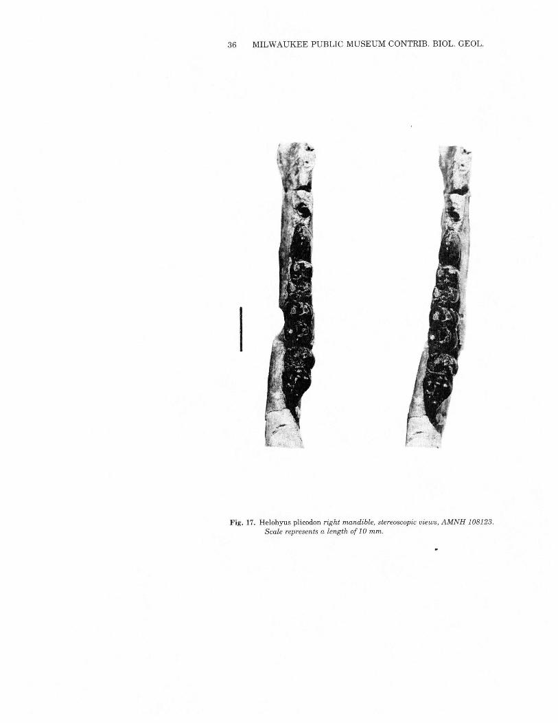
Helohyus plicodon Marsh 1872Figs. 17& 18, Table 6
Holotype: YPM 11893, M3, Bridger.Diagnosis: Small species ofHelohyus; Mj length 7.4-9.1 mm. IncludesH. validus of Marsh.The species occurs through most of the thickness of the Bridger
Formation. Although the available material is insufficient to serveas a statistical base, there seems to be a modest size increase throughthe Bridgerian, accompanied by gradual reduction in the size of themolar paraconid. Comparison of AMNH 12694 with H. plicodon(MPM 6807) from Loc. 2767 in the Bridger B suggests thatH. validuscan be synonymized with H. plicodon.Several specimens include distal tibiae, which have a structure
noticeably different from those ofhomacodontines. The area of distalfibula contact is clearly marked by a prominent triangular rugosity,but the fibula itself is not present. This suggests that either thefibula was reduced relative to its condition in the Homacodontinaeor that it was so weakly fused that the two bones readily separated.Most of the tibia shaft is preserved in MPM 6706, and there is nosuggestion of tibia-fibula contact proximal to the scar on the distaltibia. A similar condition is suggested by MPM 6708, although notnearly so much of the tibia remains.The calcaneum tuber in Helohyus plicodon makes up well over
50% of the length of the bone, while the astragalus is virtuallystraight.These postcranial characters taken together suggest that H. plico-
don was still more cursorially specialized than were the species ofhomacodontines mentioned above, although there is no informationon relative leg lengths.
WEST: SURVEY OF BRIDGERIAN ARTIODACTYLA 35
Helohyus milleri Sinclair 1914Table 7
Holotype: AMNH 12151, dentary and maxilla, C5 (Henry's Forkopposite mouth of Burnt Fork).Diagnosis: Medium-sized species ofHelohyus. Length of M, 9.6-11.1mm.As pointed out by Sinclair in his description of the species, lower
teeth differ from those of H. plicodon only in their slightly largersize. The new material extends the range of the species down intothe Blacks Fork Member.
Table 7Measurements, in millimeters, of teeth ofHelohyus milleriN L X N Wa X N Wp X
P4 3 9.2-10.1 9.6 3 3.6-4.8 4.27Ml 3 9.6-11.1 10.23 3 5.7-7.1 6.43 3 6.2-7.6 6.97M2 3 10.7-11.5 11.20 3 6.5-8.2 7.57 3 7.3-8.2 7.83p4 1 7.8 1 8.0Ml 1 10.6M2 2 10.6-11.0 10.8 2 12.6-14.8 13.7 2 12.0-13.7 12.85M3 2 9.4-11.5 10.45 2 12.2-15.0 13.6
Helohyus lentus (Marsh) 1871Figs. 2 and 19; Table 8
Holotype: YPM 11892, M3,upper Bridger.Diagnosis: Larger species of Helohyus. Average M, length is 12.1mm.This, the largest Bridger Formation artiodactyl, is found only in
the upper part of the Twin Buttes Member, Bridger D. Materialfound during the present investigation, a single lower molar, doesnot contribute to understanding ofH. lentus.The holotype (YPM 11892) and three other specimens (USNM
17711, AMNH 12150 and 108122) are lower teeth or dentary frag-ments. Sinclair (1914) illustrated a maxillary fragment with Ml andM2CPU10084) and suggested that it may be referable to H. lentus.That specimen occludes effectively with USNM 17711, confirmingSinclair's suggestion based solely on isolated M3s.The teeth of PU10084 are typically helohyine in the avsence of a hypocone and the
36 MILWAUKEE PUBLIC MUSEUM CONTRIB. BIOL. GEOL.
I
;; <\.
Fig. 17. Helohyus plicodon right mandible, stereoscopic views, AMNH 108123.Scale represents a length of 10 mm.
WEST: SURVEY OF BRIDGERIAN ARTIODACTYLA 37
B
A
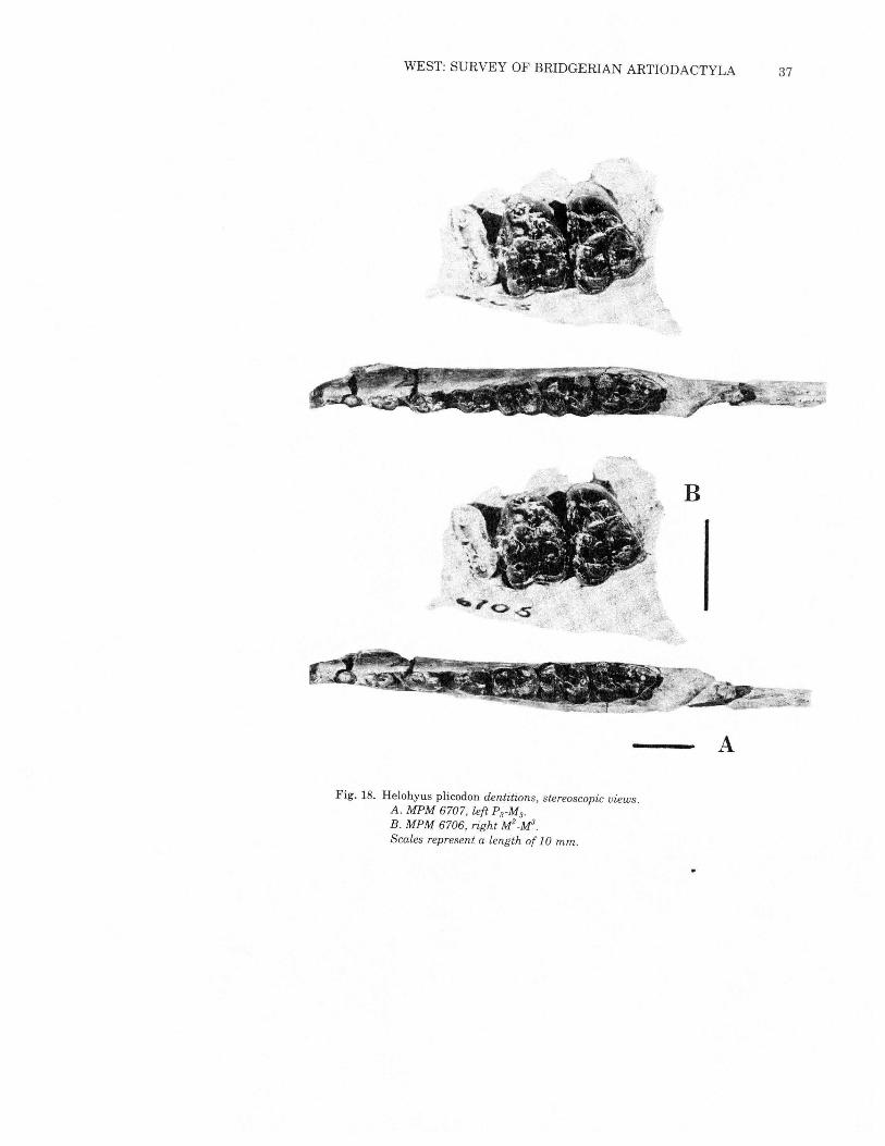
Fig. 18. Helohyus plicodon dentitions, stereoscopic views.A. MPM 6707, left PrM3.
B. MPM 6706, right M2·MaScales represent a length of 10 mm.
38 MILWAUKEE PUBLIC MUSEUM CONTRIB. BIOL. GEOL.
B
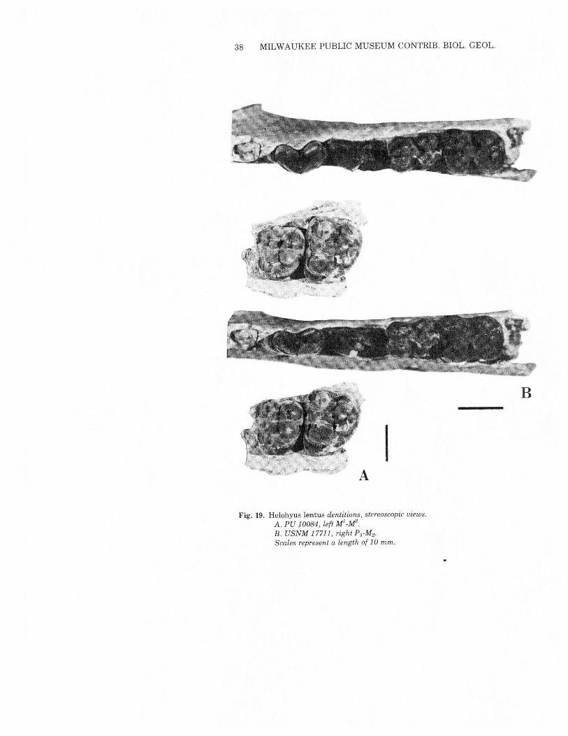
IFig. 19. Helohyus lentus dentitions, stereoscopic views.
A. PU 10084, left Ml_M2.B. USNM 17711, right PrM2·
Scales represent a length of 10 mm.
WEST: SURVEY OF BRIDGERIAN ARTIODACTYLA 39
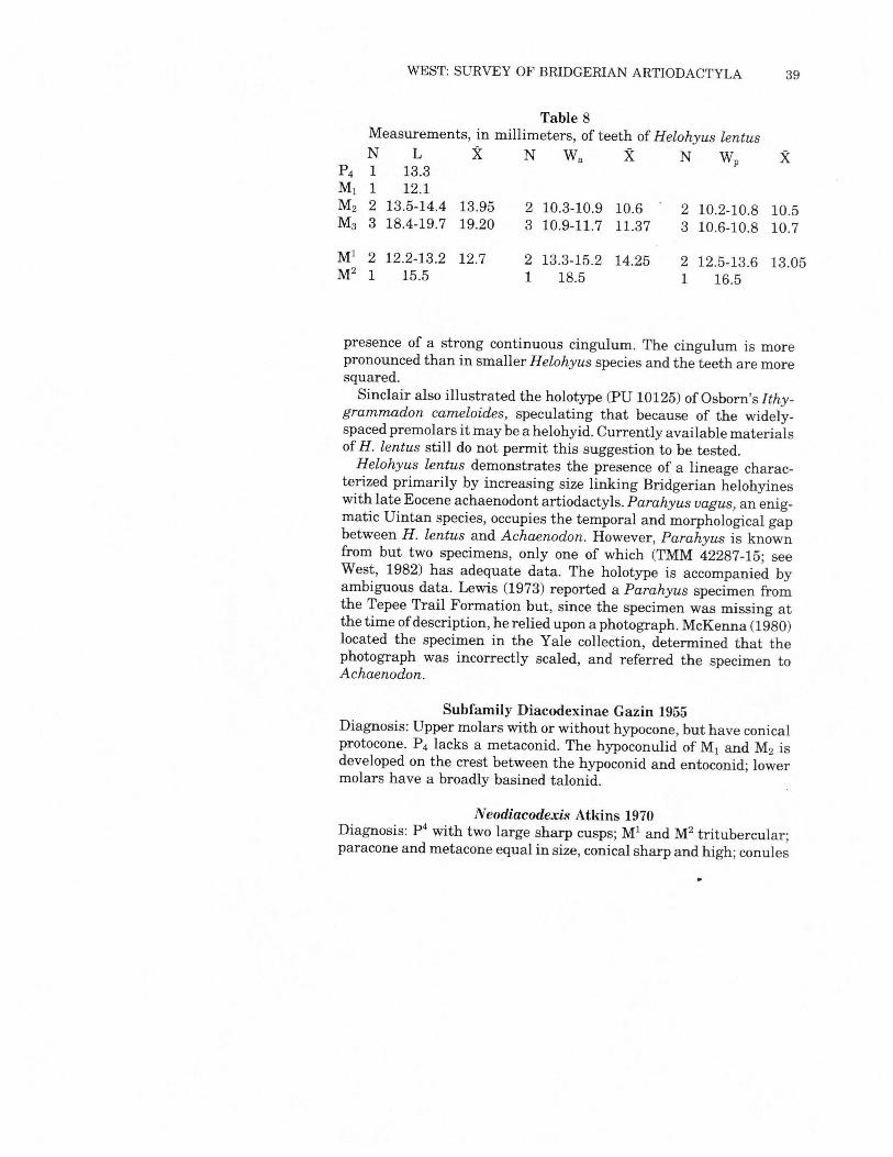
Table 8Measurements, in millimeters, of teeth ofHelohyus lentusN L X N Wa X N Wp X
P4 1 13.3Ml 1 12.1M2 2 13.5-14.4 13.95 2 10.3-10.9 10.6 2 10.2-10.8 10.5M3 3 18.4-19.7 19.20 3 10.9-11.7 11.37 3 10.6-10.8 10.7
Ml 2 12.2-13.2 12.7 2 13.3-15.2 14.25 2 12.5-13.6 13.05M2 1 15.5 1 18.5 1 16.5
presence of a strong continuous cingulum. The cingulum is morepronounced than in smaller Helohyus species and the teeth are moresquared.Sinclair also illustrated the holotype (PU 10125) of Osborn's Ithy-
grammadon cameloides, speculating that because of the widely-spaced premolars it may be a helohyid. Currently available materialsofH. lentus still do not permit this suggestion to be tested.Helohyus lentus demonstrates the presence of a lineage charac-
terized primarily by increasing size linking Bridgerian helohyineswith late Eocene achaenodont artiodactyls. Parahyus vagus, an enig-matic Uintan species, occupies the temporal and morphological gapbetween H. lentus and Achaenodon. However, Parahyus is knownfrom but two specimens, only one of which (TMM 42287-15; seeWest, 1982) has adequate data. The holotype is accompanied byambiguous data. Lewis (1973) reported a Parahyus specimen fromthe Tepee Trail Formation but, since the specimen was missing atthe time ofdescription, he relied upon a photograph. McKenna (1980)located the specimen in the Yale collection, determined that thephotograph was incorrectly scaled, and referred the specimen toAchaenodon.
Subfamily Diacodexinae Gazin 1955Diagnosis: Upper molars with or without hypocone, but have conicalprotocone. P4 lacks a metaconid. The hypoconulid of M, and M2 isdeveloped on the crest between the hypoconid and entoconid; lowermolars have a broadly basined talonid.
Neodiacodexis Atkins 1970Diagnosis: p4 with two large sharp cusps; Ml and M2 tritubercular;paracone and metacone equal in size, conical sharp and high; conules
40 MILWAUKEE PUBLIC MUSEUM CONTRIB. BIOL. GEOL.
well developed and relatively independent of major cusps; meta-conule not hypertrophied; mesostyle present.
Neodiacodexis emryi Atkins 1970Holotype: AMNH 56054, P4_M2,upper Bridger (Tabernacle Buttelocality 5). .Diagnosis: As for genus.No additional material referable to N. emryi was found during
the course of this study, either in the field or in preexisting collec-tions. It remains based upon a single maxillary fragment with p4_M2recovered from late Bridgerian deposits at Tabernacle Butte (Westand Atkins, 1970).
cf. Neodiacodexis sp.Two isolated upper molars (CM 13232 and CM 13418) from the
early Bridgerian Powder Wash locality (Green River Formation ofnortheastern Utah) are tentatively referred to Neodiacodexis. Bothhave rounded triangular outlines, lack a hypocone,have well-developedconules, subequal paracones and metacones, and conical protocones.The structure of the "wings" from the conules to the external cusps,varying mesostyle development, and differential structure of theinternal cingular area preclude allocation ofthese teeth to N. emryi.Nonetheless the similarities are great enough to justify the queriedgeneric assignment.
WEST: SURVEY OF BRIDGERIAN ARTIODACTYLA 41
DiscussionBridgerian artiodactyls quite clearly represent three distinct
lineages within the Dichobunidae. Two, the Diacodexinae and theHomacodontinae have likely ancestral forms in older North Amer-ican strata, while the Helohyinae appear for the first time in theNorth American middle Eocene.The helohyines are the most generalized of the three. Their lower
premolars are simple and trenchant, lacking any lateral cusps, thelower molars retain all three trigonid cusps, and the upper molarslack hypocones. The subfamily seems to continue onward into thelate Eocene with a bifurcation into the achaenodonts on the onehand and smaller forms such as Apriculus on the other.Coombs and Coombs (1977) used Helohyidae at the family level
and placed them in the Anthracotherioidea. This followed their de-termination that all the close relatives ofHelohyus (Gobiohyus, In-dohyus, Raoella, Kunmunella and Bunodentus) are from southernor eastern Asia. I prefer retention of the subfamily rank, but therecognition ofAsian affinities ofHelohyus may be helpful in explain-ing its sudden occurrence in North America in the middle Eocenewithout apparent local ancestors. Coombs and Coombs' (1982) studyof artiodactyl basicrania lent modest support to this contention, asHelohyus and Gobiohyus have somewhat more features in commonthan either shares with Homacodon or Diacodexis.The Diacodexinae are commonly regarded as the "stem" subfamily
of the Artiodactyla, as Diacodexis is the oldest known representativeofthe order. Bymiddle Eocene they became extremely rare, althoughthey did persist into the late Eocene as Tapochoerus (West and At-kins, 1970). Like the helohyines, they lack hypocone development,but their teeth never became as inflated and bulbous as Helohyus.Despite the primitive dentition, appendicular skeletal parts ofDiacodexis (Rose,1982)show a pronounced cursorial specialization.The Diacodexinae probably gave rise to the Homacodontinae in
the latter part of the early Eocene, as late Wasatchian Hexacodusis more clearly aligned with the homacodontines, with its rectangu-lar upper molar outline. Middle Eocene homacodontines (Micros us,Antiacodon and Homacodon) are readily recognizable by the pre-sence of a hypocone and well defined independent conules. The twosmaller taxa, Microsus and Antiacodon, differ primarily in the degreeof development of the paraconid and metaconid on the posteriormolars and in the presence of a small mesostyle in Antiacodon.Homacodon is somewhat larger and has simpler premolars than dothe others. The postcranial elements ofAntiacodon described in thispaper show it to be, like Diacodexis, highly specialized as a cursorialanimal.
42 MILWAUKEE PUBLIC MUSEUM CONTRIB. BIOL. GEOL.
Literature CitedBlack, C. C. 1978. Paleontology and geology of the Badwater Creek area, Central
Wyoming. Part 14. The artiodactyls. Annals Carnegie Mus., 47(10):223-259.Burke, J. J. 1969. An antiacodont from the Green River Eocene of Utah. Kirtlandia,
no.5:7p.Coombs, M. C. and W. P. Coombs. 1982. Anatomy of the ear region of four Eocene
artiodactyls: Gobiohyus, ?Helohyus, Diacodexis and Homacodon. J. Vert. Paleo.,2(2):219-236.
Coombs, W. P., Jr. and M. C. Coombs. 1977. The origin of anthracotheres. N. Jb.Geo!. Palaont, Mh., 10:584-599.
Franzen, J. L. 1981. Das erste skelett eines Dichobuniden (Mammalia, Artiodactyla),geborgen aus mitteleoziinen Olschiefern der "Grube Messel" bei Darmstadt(Deutschland, S-Hessen). Senckenbergiana lethaea, 61(3/6):299-353.
Gazin, C. L. 1955. A review of the upper Eocene Artiodactyla of North America.Smithsonian Misc. ColI., 128(8):96p.
Gazin, C. L. 1976. Mammalian faunal zones of the Bridger middle Eocene. Smiths.Cont. Paleobiology No. 26:25p.
Golz, D. J. 1976. Eocene Artiodactyla of Southern California. Nat!. Hist. Mus. LosAngeles County, Science Bul!. no. 26:85p.
Guthrie, D. A. 1968. The tarsus ofearly Eocene artiodactyls. Jour. Mamma!. 49(2):297-302.
Matthew, W. D. 1909. The Carnivora and Insectivora of the Bridger Basin, middleEocene. Memo.Amer. Mus. Nat. Hist. 9(6):289-567.
McGrew, P.O., J. E. Berman, M. K. Hecht, J. M. Hummel, G. G. Simpson and A. E.Wood. 1959. The geology and paleontology of the Elk Mountain and TabernacleButte area, Wyoming. Bull. Amer. Mus. Nat. Hist., 117(3):117-176.
McKenna, M. C. 1980. Late Cretaceous and early Tertiary vertebrate paleontologicalreconnaissance, Togwotee Pass area, northwestern Wyoming. pp. 321-343, inJacobs, L. L., ed., Aspects of Vertebrate History. Museum ofNorthern Arizona Press,Flagstaff.
Robinson, P. 1966. Fossil Mammalia of the Huerfano Formation, Eocene of Colorado.Peabody Mus. Bull., 21:85p.
Rose, K. D. 1982. Skeleton ofDiacodexis, oldest known artiodacty!. Science, 216:621-623.
Schaeffer, B. 1947. Notes on the origin and function of the artiodactyl tarsus. Amer.Mus. Novit. no. 1356:24p.
Sinclair, W. J. 1914. A revision of the bunodont Artiodactyla of the middle and lowerEocene of North America. Bul!. Amer. Mus. Nat. Hist., 33(32):267-295.
Van Valen, L. 1971. Toward the origin of artiodactyls. Evolution, 25(3):523-529.West, R. M. 1973. Geology and mammalian paleontology of the New Fork-Big Sandy
area, Sublette County, Wyoming. Fieldiana Geology, 29:193p.West, R. M. 1976. Paleontology and Geology of the Bridger Formation, southern
Green River Basin, southwestern Wyoming. Part 1. History of field work andgeological setting. Milw. Public Mus., Cont. Bio!. Geo!., no. 7:12p.
West, R. M. 1982. Fossil mammals from the Lower Buck Hill Group, Eocene ofTrans-Pecos Taxas: Marsupicarnivora, Primates, Taeniodonta, Condylarthra,bunodont Artiodactyla, and Dinocerata. The Pearce-Sellards Series, no. 35:20p.
West, R. M. and E. G. Atkins. 1970. Additional middle Eocene (Bridgerian) mammalsfrom Tabernacle Butte, Sublette County, Wyoming. Amer. Mus. Nov. 2404: 26p.
WEST: SURVEY OF BRIDGERIAN ARTIODACTYLA 43
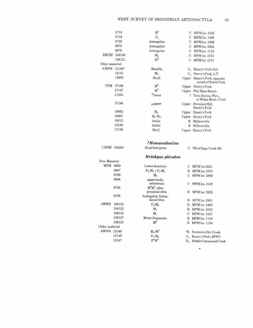
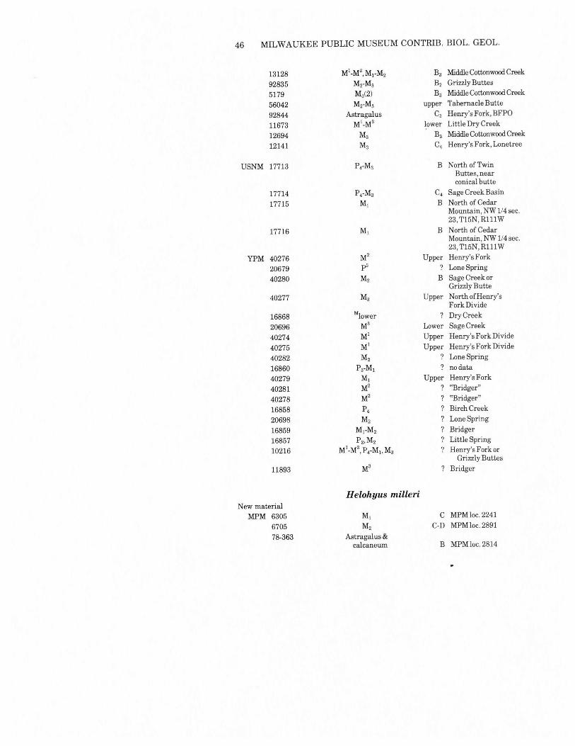
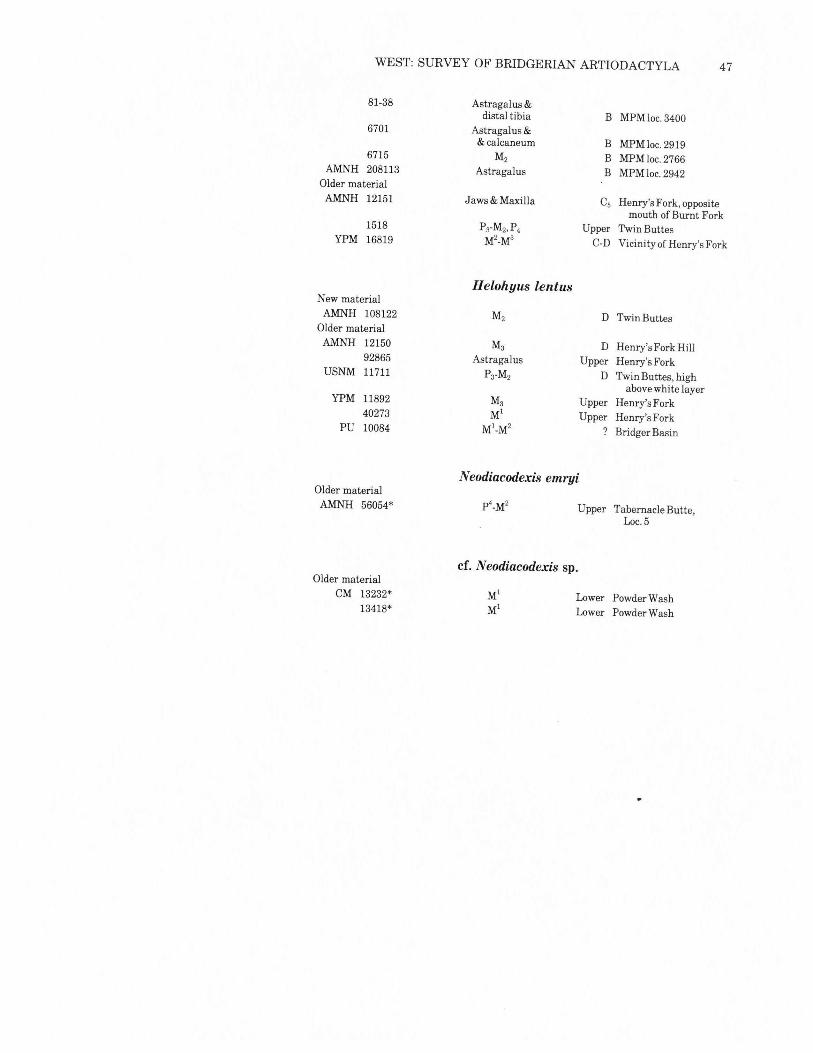
AppendixBelow are complete listings of the fossil materials used in this
study, arranged by species. The first part of each list is the newlycollectedmaterial; following that is older material found in museumcollections, listed by museum acronym. Alongside each museumnumber is a brief description of the nature ofthe material and thestratigraphic level and locality (if known). The asterisks indicatespecimens not measured during this study.
New MaterialMPM 6722
67116709
AMNH 108128108129
Older MaterialAMNH 12146*
12145*USNM 336179
336178336177
YPM 37194371903719337191
New MaterialMPM 5896
6721
Older MaterialAMNH 5006*
5007*12697*11427*11426*11974*13127*
13126*
Microsus cuspidatus
M2 BridgerD MPM loc. 2233astragalus C MPM loc.2406x, astragalus, B MPM loc.2801
calcaneum, proximaltibia, distal femur.
astragalus C MPM loc. 1126M2 C MPMloc.1126
Misc. Cs Henry's Fork,BFPOMI-Ms Cs Henry's Fork Hillp4_MS B South ofChurch ButtesP4-MS B Opposite MillersvilleP4-MS C Dead CowButtesMlower ? Point SpringMs Upper Henry's Fork
Mlower Point GulchMl Upper Henry's Fork
Antiacodon pygmaeus
Skull and lower molarsDentaries, maxilla, par-tial hind limb skeleton.
B MPMloc.2934B MPM loco3395
MI-M2 Lower Cottonwood CreekMI-M2 Lower Cottonwood Creekdentary B Grizzly ButtesP4,M2-Ms Lower Grizzly Buttes WestMrM2 Lower Grizzly Buttes WestM2-MS C4 Henry's Fork, Lone TreeMs ?B Mouth ofSummers
Dry CreekP4-M2' B2 Little Dry Creek
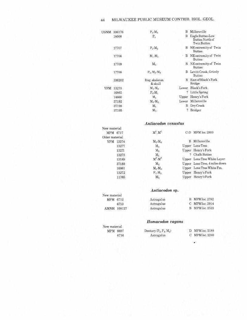
44 MILWAUKEE PUBLIC MUSEUM CONTRIB. BIOL. GEOL.
USNM 336176 P4-Ma B Millersville
18000 P4 B Eagle Buttes-LowButtes North ofTwin Buttes
17707 P4-M2 B NE extremity of TwinButtes
17706 M]"Ma B NE extremity of TwinButtes
17709 Ma B NE extremity of TwinButtes
17708 P.,M2-Ma B Levitt Creek, GrizzlyButtes
336202 frag. skeleton B East of Black's Fork& skull Bridge
YPM 13275 M]"M2 Lower Black's Fork
16865 P4-M\ ? Little Spring
14660 MJ Upper Henry's Fork
37192 M]"M2 Lower Mill ersvill e
37198 M\ B Dry Creek
37195 M2 ? Bridger
Antiacodon venustusNew material
MPM 6717 MJ,M2 C-D MPMloc.2900
Older materialYPM 13274 MJ-Ma B Millersville
13277 Ma Upper LoneTree
13271 Ma Upper Henry's Fork
13273 M\ ? Chalk Buttes
13189 M2_Ma Upper Lone Tree White Layer
37188 M2 Upper Lone Tree, 4 miles down
16861 MJ-Ma Upper Lone Tree White Fm.
13272 P4-M2 Upper Henry's Fork
11765 Ma Upper Henry's Fork
Antiacodon sp.New material
MPM 6712 Astragalus B MPMloc.2782
6713 Astragalus C MPMloc.2914
AMNH 108127 Astragalus B MPM loco2522
Homacodon vagansNew material
MPM 66976716
Dentary (P 2,P 4,Ma)Astragalus
D MPMloc.2188C MPM loco2240
WEST: SURVEY OF BRIDGERIAN ARTIODACTYLA 45
6710 MJ C MPM loco31336718 P4 C MPMloc.10986720 Astragalus C MPMloc.10986975 Astragalus C MPM loco29246976 Astragalus C MPM loco 1102
AMNH 108130 MJ .C MPMloc.2151108131 M2 C MPM loco2151
Older materialAMNH 12140' Maxilla C5 Henry's Fork Hill
12141 Ma C4 Henry's Fork, L.T.12695 Skull Upper Henry's Fork, opposite
mouth of Burnt Fork.YPM 37199 M2 Upper Henry's Fork
37197 Ma Upper Phil Mass Ranch37200 Mlower ? Twin Buttes, Wyo.,
or White River, Utah37196 Mupper Upper Streaked Hill,
Henry's Fork16862 Ma Upper Henry's Fork16867 MrM2 Upper Henry's Fork10012 Ankle B Millersville13129 Ankle B Millersville13129 Skull Upper Henry's Fork
?H omacodontineUSNM 336201 Hind limb parts C NEofSageCreekMt.
H elohyus plicodonNew Material
MPM 6699 Lower dentition C MPMloc22356807 P3-M3 +P4-Ma B MPMloc.27676700 MJ C MPM loco29096698 upper teeth,
calcaneum C MPMloc.31386706 M2M3, tibia
proximal ulna B MPM loco28206708 Astragalus, femur,
distal tibia B MPM loco2801AMNH 108123 P4-Ma D MPM loco2403
108125 M3 B MPM loco2522108124 MJ C MPMloc.2187108137 Molar fragments B MPM loco 1139108133 M2 B MPM loco 1139
Older materialAMNH 12148 MrM3 ?B Summers Dry Creek
12149 P2-M2 C2 Henry's Fork, BFPO12147 p4M3 B3 Middle Cottonwood Creek
46 MILWAUKEE PUBLIC MUSEUM CONTRIB. BIOL. GEOL.
13128 Ml_M2,Ml-M2 B3 Middle Cottonwood Creek
92835 M2-M3 B2 Grizzly Buttes
5179 M3(2) B3 Middle Cottonwood Creek
56042 M2-M3 upper Tabernacle Butte
92844 Astragalus C3 Henry's Fork, BFPO
11673 Ml_M3 lower Little Dry Creek
12694 M3 B3 Middle Cottonwood Creek
12141 M3 C4 Henry's Fork, Lonetree
USNM 17713 P4-M3 B North of TwinButtes, nearconical butte
17714 P4-M3 C4 Sage Creek Basin
17715 Ml B North of CedarMountain, NW 114 sec.23, T15N, R111 W
17716 Ml B North of CedarMountain, NW 114 sec.23,T15N,R111W
YPM 40276 M2 Upper Henry's Fork
20679 p3 ? Lone Spring
40280 M2 B Sage Creek orGrizzly Butte
40277 M3 Upper North of Henry'sFork Divide
16868 Mlower ? Dry Creek
20696 M3 Lower Sage Creek
40274 Ml Upper Henry's Fork Divide
40275 Ml Upper Henry's Fork Divide
40282 M2 Lone Spring
16860 P3-M1 ? no data
40279 Ml Upper Henry's Fork
40281 M2 ? "Bridger"
40278 M2 ? "Bridger"
16858 P4 ? Birch Creek
20698 M2 ? Lone Spring
16859 MrM2 ? Bridger
16857 P2,M2 ? Little Spring
10216 Ml_M3,P4-Ml>M3 ? Henry's Fork orGrizzly Buttes
11893 M3 ? Bridger
Helohyus milleriNew material
MPM 6305 Ml C MPMloc.2241
6705 M2 C-D MPMloc.2891
78-363 Astragalus &calcaneum B MPMloc.2814
WEST: SURVEY OF BRIDGERIAN ARTIODACTYLA 47
81-38 Astragalus &distal tibia B MPM loco3400
Astragalus && calcaneum B MPMloc.2919
M2 B MPM loco2766Astragalus B MPM loco2942
Jaws & Maxilla Cs Henry's Fork, oppositemouth of Burnt Fork
P3-M2,P4 Upper Twin ButtesM2_M3 C-D Vicinity of Henry's Fork
6701
6715AMNH 208113
Older materialAMNH 12151
1518YPM 16819
YPM 1189240273
PU 10084
Helohyus lentus
M2 D Twin Buttes
M3 D Henry's Fork HillAstragalus Upper Henry's Fork
P3-M2 D Twin Buttes, highabove white layer
M3 Upper Henry's ForkMl Upper Henry's Fork
Ml_M2 ? Bridger Basin
New materialAMNH 108122
Older materialAMNH 12150
92865USNM 11711
Older materialAMNH 56054*
Neodiacodexis emryi
Upper Tabernacle Butte,Loc.5
Older materialCM 13232*
13418*
cf. Neodiacodexis sp.
Lower Powder WashLower Powder Wash


