Embed Size (px)
Citation preview

2014;20:4314-4325. Published OnlineFirst June 19, 2014.Clin Cancer Res Paul Cottu, Ivan Bièche, Franck Assayag, et al. Breast Cancer XenograftsTumor-Specific Molecular Changes in Patient-Derived Luminal Acquired Resistance to Endocrine Treatments Is Associated with
Updated version
10.1158/1078-0432.CCR-13-3230doi:
Access the most recent version of this article at:
Material
Supplementary
http://clincancerres.aacrjournals.org/content/suppl/2014/06/23/1078-0432.CCR-13-3230.DC1.html
Access the most recent supplemental material at:
Cited Articles
http://clincancerres.aacrjournals.org/content/20/16/4314.full.html#ref-list-1
This article cites by 50 articles, 15 of which you can access for free at:
E-mail alerts related to this article or journal.Sign up to receive free email-alerts
Subscriptions
Reprints and
To order reprints of this article or to subscribe to the journal, contact the AACR Publications Department at
Permissions
To request permission to re-use all or part of this article, contact the AACR Publications Department at
on August 19, 2014. © 2014 American Association for Cancer Research. clincancerres.aacrjournals.org Downloaded from
Published OnlineFirst June 19, 2014; DOI: 10.1158/1078-0432.CCR-13-3230
on August 19, 2014. © 2014 American Association for Cancer Research. clincancerres.aacrjournals.org Downloaded from
Published OnlineFirst June 19, 2014; DOI: 10.1158/1078-0432.CCR-13-3230

Cancer Therapy: Preclinical
Acquired Resistance to Endocrine Treatments Is Associatedwith Tumor-Specific Molecular Changes in Patient-DerivedLuminal Breast Cancer Xenografts
Paul Cottu1,3, Ivan Bi�eche2, Franck Assayag3, Rania El Botty3, Sophie Chateau-Joubert9, Aur�elie Thuleau3,Thomas Bagarre3, Benoit Albaud4, Audrey Rapinat4, David Gentien4, Pierre de la Grange5, Vonick Sibut6,Sophie Vacher2, Rana Hatem2, Jean-Luc Servely7,9, Jean-Jacques Fontaine9, Didier Decaudin1,3,Jean-Yves Pierga1, Sergio Roman-Roman8, and Elisabetta Marangoni3
AbstractPurpose: Patients with luminal breast cancer (LBC) often become endocrine resistant over time. We
investigated the molecular changes associated with acquired hormonoresistances in patient-derived
xenografts of LBC.
ExperimentalDesign:TwoLBCxenografts (HBCx22 andHBCx34)were treatedwithdifferent endocrine
treatments (ET) to obtain xenografts with acquired resistances to tamoxifen (TamR) and ovariectomy
(OvaR). PI3K pathway activation was analyzed by Western blot analysis and IHC and responses to ET
combined to everolimus were investigated in vivo. Gene expression analyses were performed by RT-PCR and
Affymetrix arrays.
Results: HBCx22 TamR xenograft was cross-resistant to several hormonotherapies, whereas HBCx22
OvaR and HBCx34 TamR exhibited a treatment-specific resistance profile. PI3K pathway was similarly
activated in parental and resistant xenografts but the addition of everolimus did not restore the response to
tamoxifen in TamR xenografts. In contrast, the combination of fulvestrant and everolimus induced tumor
regression in vivo in HBCx34 TamR, where we found a cross-talk between the estrogen receptor (ER) and
PI3K pathways. Expression of several ER-controlled genes and ER coregulators was significantly changed in
bothTamRandOvaR tumors, indicating impairedER transcriptional activity. Expression changes associated
with hormonoresistance were both tumor and treatment specific and were enriched for genes involved in
cell growth, cell death, and cell survival.
Conclusions: PDXmodels of LBC with acquired resistance to endocrine therapies show a great diversity
of resistance phenotype, associated with specific deregulations of ER-mediated gene transcription. These
models offer a tool for developing anticancer therapies and to investigate the dynamics of resistance
emerging during pharmacologic interventions. Clin Cancer Res; 20(16); 4314–25. �2014 AACR.
IntroductionAt least 70% of breast cancers are classified as estrogen
receptor positive (ERþ), commonly called luminal breast
cancers (LBC). Interfering with the ER pathway with anti-estrogens (e.g., tamoxifen) or estrogen deprivation (e.g.,aromatase inhibitors or ovariectomy), decreases mortalityfrom ERþ breast cancer. However, endocrine treatments(ET) efficacy is limited by intrinsic and acquired resistance(1). The main mechanisms of intrinsic resistance to tamox-ifen are lack of expression of ERa and failure to converttamoxifen to its activemetabolite, while acquired resistanceto ET has been associated with several mechanisms (2).These include deregulation of ER-associated transcriptionfactors and coactivators, activation of receptor tyrosinekinase signaling, aberrant expression of cell-cycle regula-tors, increased binding with the activating protein-1 (AP-1)transcription complex, and activation of the stress-activatedprotein kinase/JNK pathway (1, 2).
The majority of the information on these potentialmechanisms has been derived from breast cancer celllines selected for adaptation to exposure to antiestrogens
Authors' Affiliations: Departments of 1Medical Oncology and 2Genetics,Hospital, Institut Curie; 3Laboratory of Preclinical Investigation, Transla-tional Research Department; 4Affymetrix Platform, Translational ResearchDepartment; 5Genosplice; 6Bioinformatics Unit, Inserm U900 Mines Par-isTech; 7INRA, Phase Department; 8Translational Research Department,Institut Curie, Paris, France; and 9Pathology Department, National Veter-inary School of Alfort, Maisons Alfort, France
Note: Supplementary data for this article are available at Clinical CancerResearch Online (http://clincancerres.aacrjournals.org/).
Corresponding Author: Elisabetta Marangoni, Laboratory of PreclinicalInvestigation, Translational Research Department, Institut Curie, 26 rued'Ulm, Paris 75005, France. Phone: 33153197422; Fax: 33153194130;E-mail: [email protected]
doi: 10.1158/1078-0432.CCR-13-3230
�2014 American Association for Cancer Research.
ClinicalCancer
Research
Clin Cancer Res; 20(16) August 15, 20144314
on August 19, 2014. © 2014 American Association for Cancer Research. clincancerres.aacrjournals.org Downloaded from
Published OnlineFirst June 19, 2014; DOI: 10.1158/1078-0432.CCR-13-3230

or long-term estrogen deprivation. However, such modelsidentify mechanisms that can induce tamoxifen resistancein vitro rather than those that actually mediate resistancein patients with breast cancer, and data obtained fromwell-controlled experimental conditions may widely dif-fer from what happens in a real tumor (3). In vivo modelsusing cell line-derived xenografts have added some valueto the biologic analyses of ET resistance, and have been ofgreat help in testing and developing new drugs in thisparticular setting. It is nonetheless well known that cellline-derived xenografts do not adequately reflect breastcancer heterogeneity or morphology in vivo and have poorpredictive value with regard to the clinical setting (4).Within this context, our group has established patient-derived xenografts (PDX) models of primary breast can-cer of all subtypes of and we have shown, as well as othergroups, that breast cancer PDX faithfully recapitulatethe morphologic and biologic features of the parentaltumors (5, 6).The analysis of dynamic changes associated with tumor
relapse in paired samples before and after resistance wouldbe of great interest from a therapeutic point of view. How-ever, paired primary and metastatic samples are not easilyobtained in the current clinical practice, and biopsies frommetastasis often yield poor tumor contents. In this report,we describe the establishment of four PDX models withacquired resistance to different ET, derived from two pre-viously described ERþ breast cancer PDXs. We show thatacquired resistance was not associated with ER loss or ESR1mutations and that the PI3K pathway activation status wassimilarly activated in sensitive and resistant models. Weevaluated the everolimus efficacy in hormono resistantxenografts in combination with various ET modalities andfound that each xenograft displayed a specific pattern ofresponse to these agents. Gene expression analyses showedtranscriptomic reprogramming which was tumor specificand treatment specific.
Expression changes associated with hormonoresistancewere both tumor and treatment specific and were enrichedfor genes involved in cell growth, cell death, and cellsurvival.
Materials and MethodsEstablishment of xenografts models resistant to ET
HBCx22 and HBCx34 PDX models have been estab-lished from untreated early-stage LBC as previouslydescribed (7). Both tumor models responded to ET (7).Luminal B status has been established on both patients’tumors and derived xenografts, and assessed on the basisof low PR/high Ki67 expression (7). To establish hor-mono-resistant models from these xenografts, tumor-bearing mice were treated during 6 to 8 months withdifferent ET, including tamoxifen, fulvestrant, and ovari-ectomy and letrozole combination. At tumor escape,resistant tumors were re-engrafted in Swiss nude micefor three serial passages and treated with the therapyunder which resistance had emerged (Fig. 1A). Resistantxenografts were established when tumors had successfullyunderwent these three passages, and exhibited a resis-tance phenotype defined by a tumor growth patternsimilar between the control group and the treated group.
In vivo efficacy studiesIn vivo efficacy studies with ET were performed in female
Swiss nude mice as previously described (7) in accordancewith the French Ethical Committee. Everolimus was pro-vided by Novartis Pharma and was administered orally at adose of 2.5 mg/kg 3 � week. Optimal tumor growthinhibition (TGI) of treated tumors versus controls wascalculated as the ratio of the mean relative tumor volume(RTV) in treated group to themeanRTV in the control groupat the same time. Statistical significance of TGI was calcu-lated by the paired Student t test by comparing the indi-vidual RTVs in the treated and control groups. Kaplan–Meier survival analysis and log-rank tests were used todetermine and compare the progression-free survival prob-ability between the different treatments arms for theHBCx34 TamR xenograft.
Morphologic and IHC analyses of tumorsXenografted tumors were fixed in 10% neutral buf-
fered formalin, paraffin embedded, and hematoxylin–eosin-saffron (H&E) stained. Outgrowths were analyzedby IHC for expression of biomarkers: ERa, ProgesteroneReceptor (PR), and Ki67 rabbit monoclonal antibodieswere purchased from Clinisciences. Phospho-S6, P-mTOR, P-AKT, insulin-like growth factor-I receptor(IGF-IR), and PTEN rabbit antibodies were purchasedfrom Cell Signaling Technology (Ozyme). Tissue micro-arrays (TMA) were built from the in vivo efficacy studies aspreviously described (8). Three xenografts from eachtreatment group and two tissue cores per tumor wereincluded in the TMA. Tumor sampling from treatedxenografts was performed 24 hours after the last
Translational RelevanceAcquired resistance to endocrine therapy occurs with
high frequency in patients with luminal breast cancer(LBC). We report here the establishment of four newpatient-derived xenografts of LBC with acquired resis-tance in vivo to tamoxifen and estrogen deprivation.Gene expression profiling and in vivo drug-responsestudies showed a great diversity of endocrine resistancephenotypes, associated with tumor-specific deregula-tions of estrogen receptor (ER)-mediated gene transcrip-tion. One tamoxifen-resistant xenograft showed across-talk between the ER and PI3 kinase pathways anda high response to the fulvestrant-everolimus combi-nation. These models offer a clinically relevant tool toevaluate anticancer therapies in the context of endocrineresistance and to investigate the dynamics of acquiredresistances.
Resistance to Endocrine Treatment in Luminal Breast Cancer Xenografts
www.aacrjournals.org Clin Cancer Res; 20(16) August 15, 2014 4315
on August 19, 2014. © 2014 American Association for Cancer Research. clincancerres.aacrjournals.org Downloaded from
Published OnlineFirst June 19, 2014; DOI: 10.1158/1078-0432.CCR-13-3230
treatment. For Ki67 quantification, 10� pictures wereautomatically analyzed with the help of AxioVision4.6.3 Zeiss software measurement tool (Carl Zeiss S.A.S.). The percentage of Ki67-positive cells was quantifiedby the ratio of Ki67-positive nuclei on total number ofnuclei in one field. The Dunnett’s multiple comparisonstest was used for multiple comparisons. PhosphorylatedS6 IHC scores were defined as follow: þþmore than 50%of P-S6þ cells,þ between 10% and 50% of P-S6 þcells,�between 1 and 10%, and � less than 1%.
Western blot analysisProteins were extracted as described previously (9).
Lysates were resolved on 4% to 12% TGX gels (Bio-Rad),transferred into nitrocellulose membranes (Bio-Rad) andimmunoblotted with rabbit antibodies against GAPDH,AKT, P-AKT, S6, or P-S6 (Cell Signaling Technology). Afterwashes, membranes were incubated with the appropriate
secondary antibodies horseradish peroxidase-conjugatedaffinity-purified goat anti–rabbit (Jackson ImmunoRe-search Laboratories, Inc., Interchim).
Real-time RT-PCRRNA extraction and qRT-PCR were performed as previ-
ously described (10, 11). For gene normalization, we usedthe human TATA box-binding protein (TBP, Genbankaccession NM_003194). Detailed protocols for cDNA syn-thesis, PCR amplifications, and normalizations have beendescribed elsewhere (12).
Mutation screeningMutations of PIK3CA (exons 9 and 20), PIK3R1 (exons
11–15), AKT1 (exon 4), and ERS1 (exons 5, 7, and 8encoding the recently underlined somatic mutationsE380Q, V392I, S463P, P535H, L536R, Y537C/N/S,D538G and R555C; refs. 13) were detected by sequencing
05
10152025303540
0 30 60 90 120 150 180
RT
V
Days after ovarectomy
HBCx22: Tumor escape after ovariectomy(individual curves)
Ovariectomy
A BHBCx22 HBCx22 TamR HBCx22 OvaR
ER
H&
E
HBCx34 HBCx34 TamR HBCx34 OvaR
ER
H&
E
C
ParentalHBCx22
HBCx22TamR
HBCx22OvaR
ParentalHBCx34
HBCx34TamR
HBCx34OvaR
P-AKT P-S6P-mTORPTEN
ParentalHBCx22
HBCx22TamR
HBCx22OvaR
P-AKT
AKT
P-S6
S6
GAPDH
ParentalHBCx34
HBCx34TamR
HBCx34OvaR
P-AKT
AKT
P-S6
S6
GAPDH
D
0
5
10
15
20
0 30 60 90 120 150 180 210 240 270
RT
V
Days after start of treatment
HBCx22: Tumor escape under tamoxifen treatment (individual curves)
+Tam
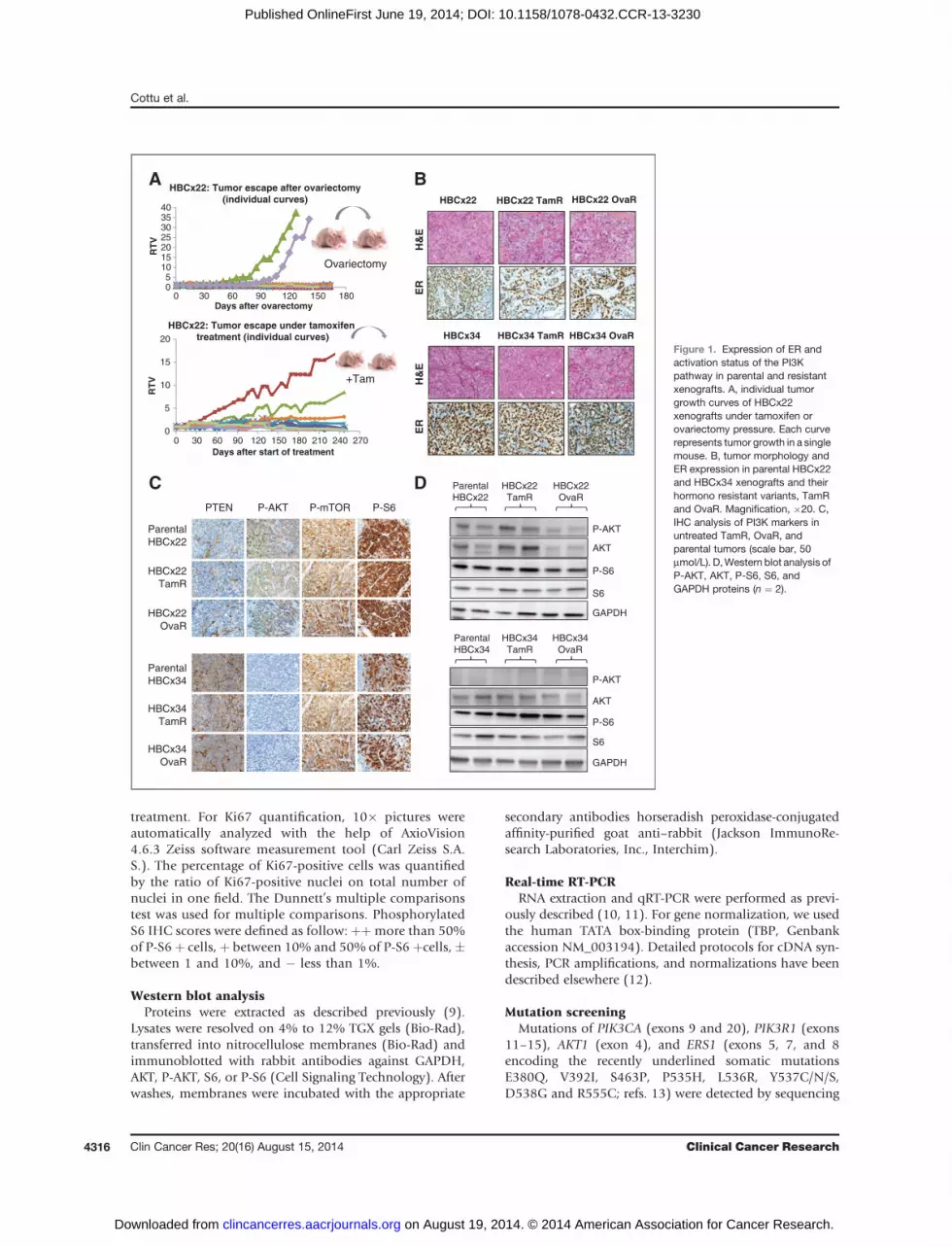
Figure 1. Expression of ER andactivation status of the PI3Kpathway in parental and resistantxenografts. A, individual tumorgrowth curves of HBCx22xenografts under tamoxifen orovariectomy pressure. Each curverepresents tumor growth in a singlemouse. B, tumor morphology andER expression in parental HBCx22and HBCx34 xenografts and theirhormono resistant variants, TamRand OvaR. Magnification, �20. C,IHC analysis of PI3K markers inuntreated TamR, OvaR, andparental tumors (scale bar, 50mmol/L). D,Western blot analysis ofP-AKT, AKT, P-S6, S6, andGAPDH proteins (n ¼ 2).
Cottu et al.
Clin Cancer Res; 20(16) August 15, 2014 Clinical Cancer Research4316
on August 19, 2014. © 2014 American Association for Cancer Research. clincancerres.aacrjournals.org Downloaded from
Published OnlineFirst June 19, 2014; DOI: 10.1158/1078-0432.CCR-13-3230

of cDNA fragments obtained by RT-PCR amplification.Details of the primers and PCR conditions are availableon request. The amplified products were sequenced withthe BigDye Terminator Kit on an ABI Prism 3130 auto-matic DNA sequencer (Applied Biosystems) with detec-tion sensitivity of 5% mutated cells, and the sequenceswere compared with the corresponding cDNA referencesequences (PIK3CA NM_006218, PIK3R1 NM_181523,AKT1 NM_005163, ESR1 NM_000125).
Microarray data analysisGeneChip Human 1.1 ST arrays were hybridized accord-
ing to Affymetrix recommendations using the AmbionWT Expression Kit protocol (Life Technologies) and Affy-metrix labeling and hybridization kits. Arrays were normal-ized according to the GC-RMA normalization procedure(14). Analyses of array datasets were made using EASANA(GenoSplice technology), normalization, backgroundcorrections, and gene annotations were performed as pre-viously described (15). Only genes expressed in at least onecompared condition were analyzed. We performed anunpaired Student t test to compare gene intensities in thedifferent biologic replicates. Genes were considered signif-icantly regulated when fold-change was�1.5 and P� 0.05.The DAVID (Database for Annotation, Visualization andIntegrated Discovery) Gene Ontology website was usedto test the significance of enrichment in specific gene onto-logy annotations. The identification of biologic functionsand the upstream regulator analyses associated with geneexpression datasets were conducted by Ingenuity PathwayAnalysis (Ingenuity Systems) according to their standardprocedures. Raw data files are available from Gene Expres-sion Omnibus under accession number GSE55561.
ResultsER and PI3K pathway status is conserved betweensensitive and resistant modelsTo generate in vivo models of endocrine resistance, we
used two previously described PDX models of hormonosensitive ERþbreast cancer,HBCx22 andHBCx34 (7). Fromeach model, we established a tamoxifen-resistant andovariectomy-resistant xenograft (named HBCx22 TamR,HBCx22 OvaR, HBCx34 TamR, and HBCx34 OvaR), asdescribed in the Materials and Methods section (two exam-ples are shown in Fig. 1A). To compare tumor morphologyand ERa expression between sensitive and resistant tumors,we performed H&E and IHC analyses. Tumor morphologywas conserved between parental and resistant tumors(Fig. 1B) and IHC analysis showed ER expression wasmaintained (Fig. 1B). In addition, we sequenced the ESR1gene hot spots recently found to be mutated in metastasesfrom patients with advanced ERþ breast cancer (13). ESR1status was found to be wild-type in resistant and respondertumors from both HBCx22 and HBCx34 xenografts (datanot shown). As the PI3K/AKT pathway activation has beendescribed as a mechanism of resistance to ET (16, 17), weanalyzed the phosphorylation of AKT, mTOR, and S6 and
the expression of the tumor suppressor PTEN by IHC andWestern blot analysis. The profile of the three HBCx22tumors was similar, showing no PTEN expression and asimilar level of expression of P-AKT, P-mTOR, and P-S6in the sensitive and in OvaR and TamR tumors (Fig. 1Cand D). We also assessed the mutational profile ofPI3KCA, PI3KR1 and AKT1 genes by sequencing analysis.The HBCx22 tumors presented a 24-pb in-frame deletionin the exon 13 of PIK3R1 (c.1704_1727del), previouslyshown to activate the PI3K pathway (ref. 18; data notshown). The IHC andWestern blot analyses of the HBCx34xenografts showed PTEN expression and absence of AKTphosphorylation. S6 and mTOR proteins where phosphor-ylated at similar levels in both parental and resistant tumors(Fig. 1C and D). The status of PI3KCA, PIK3R1, and AKT1genes was found to be wild-type in both parental andresistant xenografts. Overall, these results show that, inthese models, hormonoresistance is not associated with ERloss, ERS1 hotspots mutations, or changes in the activationprofile of the PI3K pathway.
Resistance profile is tumor and treatment specific andin vivo mTOR targeting by everolimus alone arreststumor growth in endocrine-resistant tumors
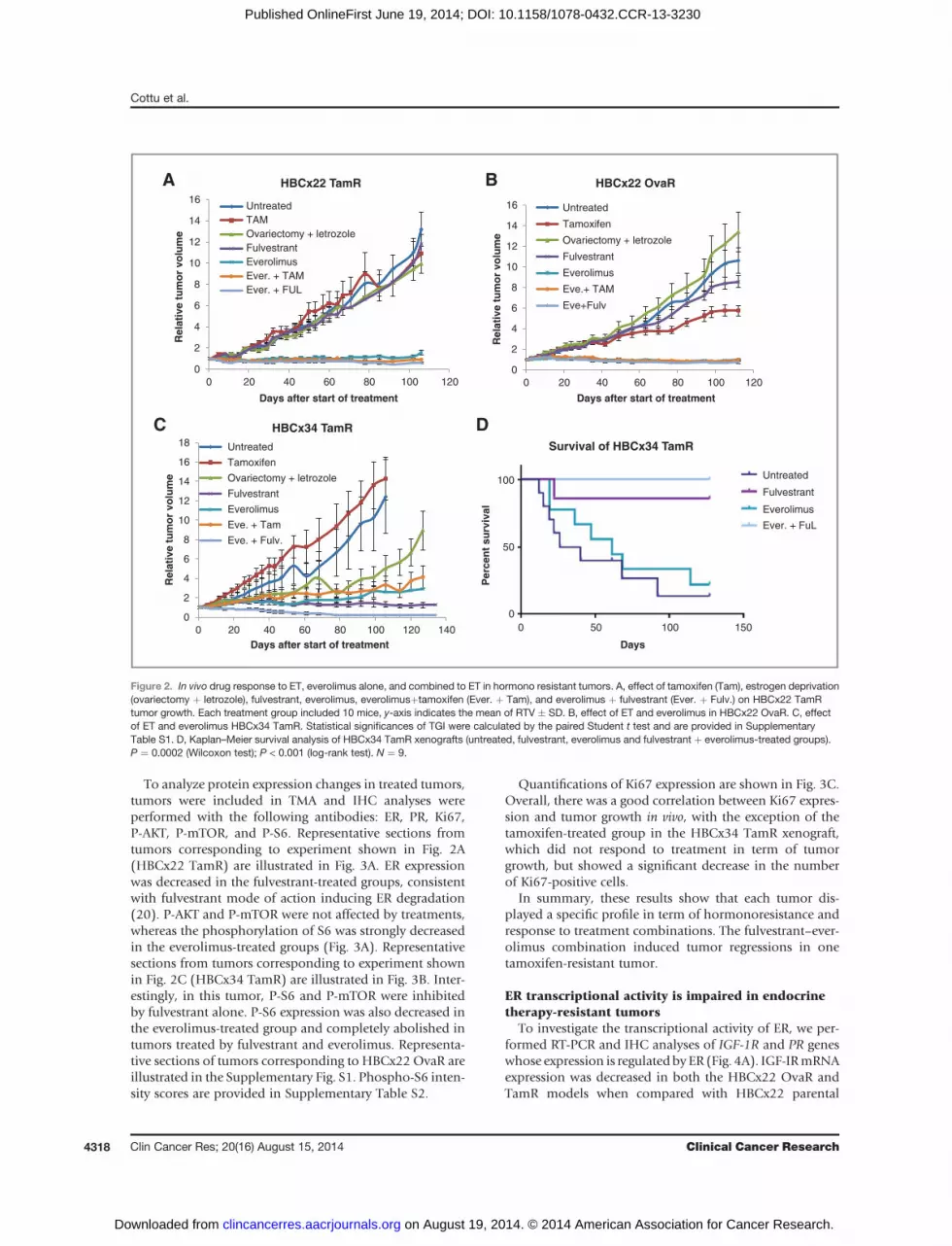
The addition of the mTOR inhibitor everolimus toET has been successful in the treatment of advancedERþ breast cancer (19), but it is unclear whether thiscombination therapy is synergistic. To further refine theresistance profile of the resistant xenografts, we analyzedthe response to ET and everolimus, alone and in combi-nation, in the HBCx22 TamR and OvaR models and theHBCx34 TamR model (Fig. 2A–C, respectively). HBCx22TamR displayed a lack of response to all ET tested(Fig. 2A), whereas HBCx22 OvaR showed a moderateresponse to tamoxifen (TGI ¼ 46%; Fig. 2B). The effect ofeverolimus, alone or combined with ET, in HBCx22TamR and OvaR was identical, with durable tumor sta-bilization (TGI ¼ 90%). In contrast, in HBCx34 TamR,resistance to tamoxifen was not associated with resistanceto other ET treatments (Fig. 2C). Treatment by everolimusalone or combined to tamoxifen resulted in long-termtumor stabilization, whereas combination of everolimuswith fulvestrant resulted in tumor regressions (Fig. 2C).Statistical significances of TGI are provided in Supple-mentary Table S1.
A Kaplan–Meier analysis of tumor progression demon-strated that control and everolimus arms had a significantlyshorter progression-free survival than the fulvestrant andfulvestrant-everolimus arms (log-rank test, P ¼ 0.0002;Fig. 2D). Moreover, the fulvestrant-everolimus combina-tion yielded a significantly higher rate of completeresponses than fulvestrant alone (55.5% vs. 0, P ¼ 0.034by the Fisher exact test) and this benefit remained significantat 127 days (log-rank test, P ¼ 0.024). Although synergismcannot be readily confirmed without dose response in vivostudies, these results strongly underline the high potencyof the fulvestrant-everolimus combination in this particularsetting.
Resistance to Endocrine Treatment in Luminal Breast Cancer Xenografts
www.aacrjournals.org Clin Cancer Res; 20(16) August 15, 2014 4317
on August 19, 2014. © 2014 American Association for Cancer Research. clincancerres.aacrjournals.org Downloaded from
Published OnlineFirst June 19, 2014; DOI: 10.1158/1078-0432.CCR-13-3230
To analyze protein expression changes in treated tumors,tumors were included in TMA and IHC analyses wereperformed with the following antibodies: ER, PR, Ki67,P-AKT, P-mTOR, and P-S6. Representative sections fromtumors corresponding to experiment shown in Fig. 2A(HBCx22 TamR) are illustrated in Fig. 3A. ER expressionwas decreased in the fulvestrant-treated groups, consistentwith fulvestrant mode of action inducing ER degradation(20). P-AKT and P-mTOR were not affected by treatments,whereas the phosphorylation of S6 was strongly decreasedin the everolimus-treated groups (Fig. 3A). Representativesections from tumors corresponding to experiment shownin Fig. 2C (HBCx34 TamR) are illustrated in Fig. 3B. Inter-estingly, in this tumor, P-S6 and P-mTOR were inhibitedby fulvestrant alone. P-S6 expression was also decreased inthe everolimus-treated group and completely abolished intumors treated by fulvestrant and everolimus. Representa-tive sections of tumors corresponding to HBCx22 OvaR areillustrated in the Supplementary Fig. S1. Phospho-S6 inten-sity scores are provided in Supplementary Table S2.
Quantifications of Ki67 expression are shown in Fig. 3C.Overall, there was a good correlation between Ki67 expres-sion and tumor growth in vivo, with the exception of thetamoxifen-treated group in the HBCx34 TamR xenograft,which did not respond to treatment in term of tumorgrowth, but showed a significant decrease in the numberof Ki67-positive cells.
In summary, these results show that each tumor dis-played a specific profile in term of hormonoresistance andresponse to treatment combinations. The fulvestrant–ever-olimus combination induced tumor regressions in onetamoxifen-resistant tumor.
ER transcriptional activity is impaired in endocrinetherapy-resistant tumors
To investigate the transcriptional activity of ER, we per-formed RT-PCR and IHC analyses of IGF-1R and PR geneswhose expression is regulated by ER (Fig. 4A). IGF-IRmRNAexpression was decreased in both the HBCx22 OvaR andTamR models when compared with HBCx22 parental
0
2
4
6
8
10
12
14
16
0 20 40 60 80 100 120
Rel
ativ
e tu
mo
r vo
lum
e
Days after start of treatment
HBCx22 OvaR
Untreated
Tamoxifen
Ovariectomy + letrozole
Fulvestrant
Everolimus
Eve.+ TAM
Eve+Fulv
0
2
4
6
8
10
12
14
16
0 20 40 60 80 100 120
Rel
ativ
e tu
mo
r vo
lum
e
Days after start of treatment
HBCx22 TamR
UntreatedTAMOvariectomy + letrozoleFulvestrantEverolimusEver. + TAMEver. + FUL
0
2
4
6
8
10
12
14
16
18
100
50
0
0 20 40 60 80 100 120 140 0 50 100
Days
150
Rel
ativ
e tu
mo
r vo
lum
e
Per
cen
t su
rviv
al
Days after start of treatment
HBCx34 TamR
Survival of HBCx34 TamRUntreated
UntreatedTamoxifen
Ovariectomy + letrozole
Fulvestrant
Everolimus
Eve. + Tam
Eve. + Fulv.
Fulvestrant
Everolimus
Ever. + FuL
A B
C D
Figure 2. In vivo drug response to ET, everolimus alone, and combined to ET in hormono resistant tumors. A, effect of tamoxifen (Tam), estrogen deprivation(ovariectomy þ letrozole), fulvestrant, everolimus, everolimusþtamoxifen (Ever. þ Tam), and everolimus þ fulvestrant (Ever. þ Fulv.) on HBCx22 TamRtumor growth. Each treatment group included 10 mice, y-axis indicates the mean of RTV � SD. B, effect of ET and everolimus in HBCx22 OvaR. C, effectof ET and everolimus HBCx34 TamR. Statistical significances of TGI were calculated by the paired Student t test and are provided in SupplementaryTable S1. D, Kaplan–Meier survival analysis of HBCx34 TamR xenografts (untreated, fulvestrant, everolimus and fulvestrant þ everolimus-treated groups).P ¼ 0.0002 (Wilcoxon test); P < 0.001 (log-rank test). N ¼ 9.
Cottu et al.
Clin Cancer Res; 20(16) August 15, 2014 Clinical Cancer Research4318
on August 19, 2014. © 2014 American Association for Cancer Research. clincancerres.aacrjournals.org Downloaded from
Published OnlineFirst June 19, 2014; DOI: 10.1158/1078-0432.CCR-13-3230

tumor. This pattern was also observed to a lesser extent inthe HBCx34 OvaR tumor, but not in the HBCx34 TamRtumor. The PR gene mRNA expression was significantlydecreased in theHBCx22 TamRmodel and in bothHBCx34OvaR and TamR xenografts. IHC analyses confirmed verylow levels of PR protein expression and a decrease in theIGF-IR protein expression in the HBCx22 resistant tumorsbut not in the HBCx34 models, where IGF-IR expressionwas higher. We also observed a decreased expression ofMYB in the four resistantmodels (data not shown).We nextanalyzed the gene expression level of the ER coregulatorsFOXA1,GATA3, PBX1, andGREB1 (Fig. 4B). In theHBCx22model, FOXA1 and PBX1 genes were repressed in the OvaRbut not in the TamR tumor. GATA3 showed no significantvariations. In contrast, the HBCx34 xenograft showed anexpression increase of these three genes in the TamR tumors.
Finally, the expression of GREB1, an ER regulatory factor,was strongly decreased in HBCx22 TamR, HBCx34 TamR,and HBCx34 OvaR (Fig. 4C).
Overall, these results suggest that ER transcriptionalactivity might be impaired in hormono resistant tumors.
Acquired resistance to hormonotherapies is associatedwith specific gene expression changes involvingmultiple biologic processes
To identify dynamic gene expression changes associatedwith endocrine resistance, sensitive and resistant tumorswere profiled with gene expression array. The complete listsof genes differentially expressed in HBCx22 TamR, OvaRand HBCx34 TamR, OvaR tumors compared with parentalxenografts are reported in Supplementary Tables S3–S6,respectively. The Venn diagrams generated with the 4-gene
ER PR Ki67 P-AKT P-mTOR P-S6A B
C
ER PR Ki67 P-AKT P-mTOR P-S6
Ctr
Tam.
Fulv.
Ovar.+Letr.
Ever.
Ever.+Tam.
Ever.+Fulv.
% o
f K
i67+
cel
ls
% o
f K
i67+
cel
ls
% o
f K
i67+
cel
lsns ns ns nsns50
40
30
20
10
0
50
40
30
20
10
0
100
80
60
40
20
0
HBCx22 OvaRHBCx22 TamR HBCx34 TamR
Untre
ated
Tamox
ifen
Ovarie
ctom
y + L
etr.
Fulves
trant
Evero
limus
Ever.
+ TAM
Ever.
+ Fulv
.
Untre
ated
Tamox
ifen
Ovarie
ctom
y + L
etr.
Fulves
trant
Evero
limus
Ever.
+ TAM
Ever.
+ Fulv
.
Untre
ated
Tamox
ifen
Ovarie
ctom
y + L
etr.
Fulves
trant
Evero
limus
Ever.
+ TAM
Ever.
+ FuL
Figure 3. IHC analysis of treated tumors (scale bar¼ 50 mmol/L). A, representative sections of TMA obtained from therapeutic experiments on HBCx22 TamRand (B) representative sections of TMA obtained from therapeutic experiments on HBCx34 TamR. Ctr., control; Tam., tamoxifen; Fulv., fulvestrant; Ova.,ovariectomy; Letr., letrozole; Ever., everolimus. C, percentage of Ki-67–positive cells determined from IHC staining in TMAs from experiments shown inFig. 2, n ¼ 6, mean � SD. The Dunnett's multiple comparisons test was used to determine statistical significance of Ki-67 expression differences in treatedversus untreated tumors. ns: P > 0.05; �, P � 0.05; ��, P � 0.01; ���, P � 0.001; ����, P � 0.0001.
Resistance to Endocrine Treatment in Luminal Breast Cancer Xenografts
www.aacrjournals.org Clin Cancer Res; 20(16) August 15, 2014 4319
on August 19, 2014. © 2014 American Association for Cancer Research. clincancerres.aacrjournals.org Downloaded from
Published OnlineFirst June 19, 2014; DOI: 10.1158/1078-0432.CCR-13-3230

lists showed that genes deregulated in TamR and OvaRtumors were only partially overlapping in both HBCx22and HBCx34 xenografts, indicating treatment-specificgene deregulations (Fig. 4D). In addition, TamR and OvaRsignatures were tumor specific, with only 210 and 107 genescommonly deregulated, respectively (Fig. 4D).
The biologic processes (BP) corresponding to genesspecifically deregulated in TamR and OvaR tumors arerepresented in Supplementary Tables S3–S6. In HBCx22TamR tumors, the top biologic processes were response towounding and response to hormone stimulus, includingestrogen and steroid hormones, and processes associatedwith wound healing and inflammation, regulation of cellproliferation, and response to TGF-b signaling. In
HBCx34 TamR, the top biologic processes were relatedto vitamin and nucleotide biosynthesis and regulation ofphosphate metabolism (Supplementary Table S5). Thetop biologic processes of HBCx22 OvaR were nitrogencompound and nucleotide metabolism and antigen pro-cessing, while the HBCx34 OvaR top biologic processesincluded cell adhesion, response to vitamin, and intra-cellular signaling cascades (Supplementary Table S6). Toidentify upstream regulators of the gene expressionchanges, we performed an Upstream Regulator Analysiswith the Ingenuity Software. Several transcription factorswere predicted to be activated in the HBCx22 Tam-Rtumors, including NFKB1, ETS1, Ap1, SP1, STAT3, andCEBPB (Supplementary Table S7). In the HBCx34 TamR
A
B
D
C
HBCx22
OvaR
HBCx22 OvaR HBCx22 OvaRHBCx34 OvaR HBCx34 OvaR
HBCx22
Tam
R
HBCx22 TamR
1,027 299 368 1,116 210 1,211 1,030 391 579 560 107 863
HBCx22 TamR HBCx34 TamR HBCx34 TamR
HBCx22
HBCx22
HBCx34
OvaR
HBCx34
Tam
R
HBCx34
HBCx34
HBCx22
OvaR
HBCx22
Tam
R
HBCx22
HBCx34
OvaR
HBCx34
Tam
R
HBCx34
IGFIR
HBCx22
No
rmal
ized
exp
ress
ion
val
ue
No
rmal
ized
exp
ress
ion
val
ue
No
rmal
ized
exp
ress
ion
val
ue
FOXA1PBX1
GATA3
FOXA1PBX1
GATA3
HBCx34 GREB1
IGFIR PR PR1,000
800
600
400
200
0
600
400
200
0
Parental
TamR
OvaR
4,000
3,000
2,000
1,000
0
4,000
3,000
2,000
1,000
0
6,000
4,000
2,000
0
80
60
40
20
0
IGFIR
/TB
P
IGFIR
/TB
P
PR
/TB
P
20
15
10
5
0
PR
/TB
P
Figure4. Expression analysesof hormono resistant tumors.A, IGF-IR andPRgene andprotein expression in untreatedTamRandOvaR tumors comparedwithparental xenografts determinedbyRT-PCRand IHCanalyses.n¼5,mean�SD.Statistical significance ismeasuredbyStudent t test. �,P�0.05; ��,P�0.01;���, P � 0.001; ����, P � 0.0001. B, expression of FOXA1, GATA3, PBX1, and (C) GREB1 genes determined by RT-PCR (n ¼ 5), in HBCx22 and HBCx34parental, TamR, and OvaR tumors. n¼ 5, mean � SD. D, Venn diagrams representing overlapping genes between resistance modalities and tumor models.
Cottu et al.
Clin Cancer Res; 20(16) August 15, 2014 Clinical Cancer Research4320
on August 19, 2014. © 2014 American Association for Cancer Research. clincancerres.aacrjournals.org Downloaded from
Published OnlineFirst June 19, 2014; DOI: 10.1158/1078-0432.CCR-13-3230

gene dataset, the upstream regulators BACH1 and FOXA1were predicted to be activated.Overall, these results indicate that ET resistance is associ-
ated with tumor-specific and treatment-specific gene expres-sion signatures, involving multiple biologic processes.
Hormonoresistance is associated with expressionmodifications of genes related to cell cycle, apoptosis,and ERBB receptorsWe next analyzed the molecular and cellular functions
of the gene signatures with the Ingenuity Software. The listsof top five molecular and cellular functions with theirrespective scores are shown in Table 1. "Cellular growthand proliferation" were in the top five lists of alteredfunction in both TamR and OvaR tumors and "cell deathand survival" were the most altered functions of OvaRtumors. To further validate these findings, we performedan RT-PCR expression analysis of several genes includedin these categories and significantly modulated in at leastone gene expression dataset. The expression of ERBB4gene was upregulated in three out of four tumors, and theHBCx34 TamR showed a concomitant upregulation ofERBB2,ERBB3, andERBB4genes (Fig.5). Several genesasso-ciated with cellular proliferation were changed, the geneexpression ofMKI67,WEE1, andCDC25Bwere increased in
the HBCx34 TamR. Finally, we found expression changes ingenes associated with cell death and cell survival functions,such as BCLXL, BCL2, and BBC3 (PUMA). In summary, themolecular changes occurring in hormono resistant impairthe expression of several genes associated with cell prolif-eration, cell death and survival, including some targetablegenes such as WEE1 and ERBB3.
DiscussionWe report here the analysis of four original hormono
resistantmodels, obtained from twoERþbreast cancer PDX,which have been rendered resistant to multiple modalitiesof ET, thus mimicking common clinical settings (21).Multiple resistances emerged from a single tumor, whenchallenged with different treatments (tamoxifen and estro-gen deprivation). The resistance phenotype was specific ofboth the original tumor and the treatment modality underwhich resistance appeared. HBCx22 TamR tumor exhibiteda general resistance to ET, whereas HBCx22 OvaR retainedan intermediate sensitivity to tamoxifen. Conversely, resis-tance to tamoxifen was not associated with a universalendocrine resistant phenotype, as suggested by the differentdrug–response profiles of HBCx22 TamR and HBCx34TamR. This is consistent with clinical observations where
Table 1. Top molecular and cellular functions identified by Ingenuity Pathway Analysis (IPA)
Category P # Molecules
HBCx22 TamRCellular movement 6.87E�11–1.09E�02 192Cellular growth and proliferation 9.61E�09–1.11E�02 301Lipid metabolism 2.34E�07–1.08E�02 112Small-molecule biochemistry 2.34E�07–1.08E�02 138Cell-to-cell signaling and interaction 2.62E�07–1.1E�02 140
HBCx22 OvaRCell death and survival 2E�05–3.41E�02 120Cellular growth and proliferation 5.71E�04–3.41E�02 142Cellular assembly and organization 1.16E�03–3.41E�02 30DNA replication, recombination, and repair 1.16E�03–3.41E�02 21Molecular transport 1.16E�03–3.41E�02 28
HBCx34 TamRCellular movement 2.75E�06–1.91E�02 173Cellular development 6.9E�06–1.83E�02 217Cellular growth and proliferation 6.9E�06–1.81E�02 281Cell death and survival 2.92E�05–1.79E�02 274Drug metabolism 3.32E�05–1.64E�02 15
HBCx34 OvaRCell death and survival 8.51E�08–3.3E�02 209Cellular development 2.15E�06–3.3E�02 197Cellular growth and proliferation 2.15E�06–3.3E�02 210Cell cycle 9.02E�06–3.26E�02 87Cellular compromise 2.41E�05–2.4E�02 15
NOTE: P values are calculated by the Fisher exact test.
Resistance to Endocrine Treatment in Luminal Breast Cancer Xenografts
www.aacrjournals.org Clin Cancer Res; 20(16) August 15, 2014 4321
on August 19, 2014. © 2014 American Association for Cancer Research. clincancerres.aacrjournals.org Downloaded from
Published OnlineFirst June 19, 2014; DOI: 10.1158/1078-0432.CCR-13-3230

patients resistant to a given class of drug may still benefitfrom alternate ET (21, 22), while ER expression is generallyhighly conserved in distant metastases (23, 24). Li andcolleagues recently identified ESR1mutations inmetastasesof advanced ERþ breast cancer (13). The fact that we did notfind suchmutations in TamRandOvaR tumors, suggests theexistence of other mechanisms of resistance in these PDX. Apotential mechanism of endocrine resistance is aberrantsignaling through the PI3K signaling pathway (17, 25).Preclinical studies have shown that breast cancer cellswith upregulated AKT signaling are resistant to ET, butsensitivity may be restored by treatment with mTOR inhi-bitors (26, 27). However, no clinical study has demonstrat-ed that secondary resistance to ET is associated with
increased PI3K signaling in patients, and synergy betweenET and everolimus has not been demonstrated in patient’stumors. The recent clinical studies showing that everolimusis beneficial to patients displaying acquired ET resistance(19) did not evaluate the efficacy of everolimus alone,which could spare the patient’s additional ET-related sideeffects. The finding that phosphorylation of mTOR and S6was constitutively high in both parental and hormonoresistant tumors, suggests that activation of the AKT/mTORpathway is not the predominant mechanism of endocrineresistance in these tumors. This is in line with recentgenomic studies showing that tumors harboring PI3KCAmutations were not resistant to tamoxifen and had a goodclinical prognosis (28, 29). Our results show that mTOR
HBCx22
OvaR
HBCx22
Tam
R
HBCx22
HBCx34
OvaR
HBCx34
Tam
R
HBCx34
HBCx22
OvaR
HBCx22
Tam
R
HBCx22
HBCx34
OvaR
HBCx34
Tam
R
HBCx34
HBCx22
OvaR
HBCx22
Tam
R
HBCx22
HBCx34
OvaR
HBCx34
Tam
R
HBCx34
HBCx22
OvaR
HBCx22
Tam
R
HBCx22
HBCx34
OvaR
HBCx34
Tam
R
HBCx34
HBCx22
OvaR
HBCx22
Tam
R
HBCx22
HBCx34
OvaR
HBCx34
Tam
R
HBCx34
HBCx22
OvaR
HBCx22
Tam
R
HBCx22
HBCx34
OvaR
HBCx34
Tam
R
HBCx34
HBCx22
OvaR
HBCx22
Tam
R
HBCx22
HBCx34
OvaR
HBCx34
Tam
R
HBCx34
HBCx22
OvaR
HBCx22
Tam
R
HBCx22
HBCx34
OvaR
HBCx34
Tam
R
HBCx34
HBCx22
OvaR
HBCx22
Tam
R
HBCx22
HBCx34
OvaR
HBCx34
Tam
R
HBCx34
HBCx22
OvaR
HBCx22
Tam
R
HBCx22
HBCx34
OvaR
HBCx34
Tam
R
HBCx34
HBCx22
OvaR
HBCx22
Tam
R
HBCx22
HBCx34
OvaR
HBCx34
Tam
R
HBCx34
HBCx22
OvaR
HBCx22
Tam
R
HBCx22
HBCx34
OvaR
HBCx34
Tam
R
HBCx34
ERBB2 ERBB3 ERBB4 EREG
Ki67
Ki6
7/T
BP
WEE1
WE
E1/
TB
P
CDC25B
CD
C25
B/T
BP
CDK6
CD
K6/
TB
P
CDKN1A BCLXL BCL2 BBC3
CD
KN
1A/T
BP
BC
LX
L/T
BP
BC
L2/
TB
P
BB
C3/
TB
P
4,000
3,000
2,000
1,000
0
400
300
200
100
0
1,500
1,000
500
0
1,500
1,000
500
0
1,500
1,000
500
0
250
200
150
100
50
0
250
200
150
100
50
0
2,000
1,500
1,000
500
0
1,000
800
600
400
200
0
1,000
800
600
400
200
0
1,000
800
600
400
200
0
80
60
40
20
0
ER
BB
2/T
BP
ER
BB
3/T
BP
ER
BB
4/T
BP
ER
G/T
BP
Figure 5. RT-PCR analysis of selected genes related to ERBB receptors, cell growth, and cell death functions. n ¼ 5, mean � SD. Statistical significanceis measured by the Student t test. �, P � 0.05; ��, P � 0.01; ���, P � 0.001; ����, P � 0.0001.
Cottu et al.
Clin Cancer Res; 20(16) August 15, 2014 Clinical Cancer Research4322
on August 19, 2014. © 2014 American Association for Cancer Research. clincancerres.aacrjournals.org Downloaded from
Published OnlineFirst June 19, 2014; DOI: 10.1158/1078-0432.CCR-13-3230

targeting has antiproliferation effect per sewithout restoringtamoxifen sensitivity in TamR tumors. A lack of synergybetween tamoxifen and everolimus has been also reportedin theMCF-7 cell line (30). However, HBCx34 TamR tumorstill responded to fulvestrant and combination with ever-olimus resulted in amarked tumor regression. Interestingly,the IHC analysis of treated tumors showed that fulvestrantalone induced a marked decrease of P-S6. As fulvestrantdirectly induces ER degradation (20), decrease of P-S6 infulvestrant-treated tumors indicates that there is a cross-talkbetween the ER and PI3K/AKT pathways in this tumor.Preclinical in vitro data also suggest a synergism betweenfulvestrant and the PI3K pathway inhibition in cells with anactivated PI3K pathway which has also retained ER expres-sion (17). Results obtained with the HBCx34 TamR xeno-graft confirm these observations. Fulvestrant has also beenused in advanced ET-resistant patients with some efficacy(31). Combinations studies are only beginning in the clinicand our data bring a further preclinical rationale.The expression analysis of ER-dependent genes indicat-
ed that ER transcriptional activity is impaired in TamR andOvaR tumors, despite persistence of ER expression. Loss ofPR and other ER target genes expression has been consis-tently associated with loss of sensitivity to tamoxifen inclinical cohorts (32). Disrupting ER signaling in resistanttumors has also been found in vitro and associated with anincrease in promoter DNA-methylation of these genes(33). Epigenetic silencing of ER-responsive genes is con-served on the long term, and most probably involves nega-tive regulators of proliferation. Epigenetic modificationsand alterations of chromatin may contribute to ET resis-tance by altering ERa transcriptional activity and inducing aswitch from classical ERa signaling to other signaling path-ways through estrogen-responsive elements (34). In thisprocess, several pioneer factors can dynamically modulatechromatin openness in breast cancer cells (35). Among theknown pioneer factors contributing to the estrogenresponse, FOXA1, GATA3, and PBX1 seem to play a rolein ET resistance (36–38). In the HBCx34 TamR tumor,FOXA1 and GATA3 expression was increased, supportingthe possibility of an epigenetic origin of the generalized ETresistance in this tumor. It has been demonstrated thatFOXA1 actively contributes to ERa recruitment on thechromatin in metastatic breast tumors, suggesting thatFOXA1 may promote ET resistance in breast cancer (39).PBX1 expression was increased in both HBCx34 TamR andOvaR tumors in comparison with sensitive counterparts.PBX1 expression has been shown to be higher in ERa-positive breast cancer that performs poorly over time andits target genes define an expression signature predictive ofresistance to endocrine therapies (36). In contrast withFOXA1 and PBX1, GREB1 expression was stronglydecreased in both HBCx22 TamR and HBCx34 TamRtumors.GREB1 is a chromatin-bound ER coactivator, essen-tial for ER-mediated transcription and expressed in tamox-ifen-responding tumors (40). In the HBCx22 tumor, ETresistance was not associated with increased expression ofFOXA1, PBX1, and GATA3. The analysis of HBCx22 TamR
gene expression dataset suggests activation of several tran-scription factors, includingNFKB1, ETS1, AP-1, SP1, STAT3,and CEBPB. Interestingly all of them have been associatedwith endocrine resistance. AP-1, SP1, or NFKB can interactwith ERa to activate transcription of additional genes lack-ing estrogen response elements (41). NF-kB activation hasbeen associated with tamoxifen resistance in the clinic (42)and ETS transcription factors recruit nuclear receptorcoactivators to estrogen-responsive genes, thus leading tohormone-independent growth and resistance to hormone-based therapies (43). Differentially expressed genes werefound to be very partially overlapping between tumors andtreatments, suggesting that transcriptional reprogrammingis both tumor and treatment specific. These observations areconsistent with a previous work showing the existence ofgene expression signatures specific to tamoxifen resistanceand letrozole resistance in MCF-7 cells, mainly involvingER-responsive genes (44). These data have been confirmedon MCF7 xenografts studies which also suggested specificgene expression profiles according to the resistance context,involving ER target genes as well as FOX family genes (3).Gene expression data obtained from clinical series oftamoxifen-treated patients confirmed a potential poor pre-dictive value of a high expression of CXCL or SERPIN genesfrom which, we also identified in the TamR tumors (45). Aparallel dynamic assessment was also performed in a smallseries of 15 letrozole-resistant patients, again pointing outthe role of ER-responsive and proliferation genes in ETresistance, with striking individual variations (46). Amongthe genes differentially regulated in hormono resistanttumors, we found an increased expression of ERBB3 andERBB4, whose expression has been associated with hormo-noresistance (47). Additional in vivo experiments will benecessary to determine whether targeting ERBB receptorscould restore tamoxifen response or increase the antitumoractivity of fulvestrant in the HBCx34 TamR xenograft. Anumber of genes involved in cell growth, cell death, andcell-cycle control were modulated in hormono resistanttumors; these biologic functions have been frequently asso-ciated with endocrine resistance both in vitro and in vivo(2, 48, 49). In patients, cell cycle, cell growth, and cellsurvival signatures are independent predictors of outcomein tamoxifen-treated patients (50).Our data provide furtherevidence that acquisition of endocrine resistance is associ-ated with deregulations of cell survival and cell deathfunctions in patient-derived samples.
In summary, our data indicate that molecular changesassociated with acquired ET resistance are both tumor andtreatment specific, and that alternative therapeutic modal-ities in hormono resistant patient should be considered onan individual clinical and biologic basis. As the dataobtained here have been generated from only two tumors,further validations usingmore PDX and clinical cohorts areneeded to validate our results. We believe that thesemodelswill help enriching the biologic data associated with hor-monoresistance in human breast cancer, and may serve aspredictive tools to evaluate the efficacy of new drugs anddrug combinations in the context of endocrine resistance.
Resistance to Endocrine Treatment in Luminal Breast Cancer Xenografts
www.aacrjournals.org Clin Cancer Res; 20(16) August 15, 2014 4323
on August 19, 2014. © 2014 American Association for Cancer Research. clincancerres.aacrjournals.org Downloaded from
Published OnlineFirst June 19, 2014; DOI: 10.1158/1078-0432.CCR-13-3230

Disclosure of Potential Conflicts of InterestNo potential conflicts of interest were disclosed.
Authors' ContributionsConception and design: P. Cottu, F. Assayag, S. Roman-Roman,E. MarangoniDevelopment of methodology: P. Cottu, I. Bi�eche, F. Assayag, R. El Botty,A. Thuleau, T. Bagarre, S. Vacher, R. Hatem, E. MarangoniAcquisitionofdata (provided animals, acquired andmanagedpatients,provided facilities, etc.): P. Cottu, I. Bi�eche, F. Assayag, S. Chateau-Joubert,A. Thuleau, T. Bagarre, B. Albaud, A. Rapinat, D. Gentien, S. Vacher,R. Hatem, J.L. Servely, J.-J. Fontaine, D. DecaudinAnalysis and interpretation of data (e.g., statistical analysis, biostatis-tics, computational analysis): P. Cottu, I. Bi�eche, F. Assayag, S. Chateau-Joubert, A. Thuleau, T. Bagarre, P. de la Grange, V. Sibut, S. Vacher, R. Hatem,J.L. Servely, S. Roman-Roman, E. MarangoniWriting, review, and/or revision of the manuscript: P. Cottu, I. Bi�eche,F. Assayag, R. El Botty, S. Chateau-Joubert, J.-J. Fontaine, D. Decaudin,S. Roman-Roman, E. Marangoni
Administrative, technical, or material support (i.e., reporting or orga-nizing data, constructing databases): P. Cottu, F. Assayag, A. Thuleau,T. Bagarre, D. Decaudin, J.-Y. PiergaStudy supervision: P. Cottu, F. Assayag, J.-Y. Pierga, S. Roman-Roman,E. Marangoni
AcknowledgmentsThe authors thank Novartis for providing everolimus, and Marie-
Eglantine Dujaric and Lisa Belin from Institut Curie Biostatistics Depart-ment for their help in the statistical analyses.
Grant SupportThis work was also supported by Institut Curie – Institut Carnot Grant
#2012-010.The costs of publication of this article were defrayed in part by the pay-
ment of page charges. This article must therefore be hereby marked advertise-ment in accordance with 18 U.S.C. Section 1734 solely to indicate this fact.
ReceivedNovember 26, 2013; revisedMay5, 2014; acceptedMay29, 2014;published OnlineFirst June 19, 2014.
References1. RingA,DowsettM.Mechanisms of tamoxifen resistance. Endocr Relat
Cancer 2004;11:643–58.2. Musgrove EA, Sutherland RL. Biological determinants of endocrine
resistance in breast cancer. Nat Rev Cancer 2009;9:631–43.3. Creighton CJ, Massarweh S, Huang S, Tsimelzon A, Hilsenbeck SG,
Osborne CK, et al. Development of resistance to targeted therapiestransforms the clinically associatedmolecular profile subtype of breasttumor xenografts. Cancer Res 2008;68:7493–501.
4. Voskoglou-Nomikos T, Pater JL, Seymour L. Clinical predictive valueof the in vitro cell line, human xenograft, andmouse allograft preclinicalcancer models. Clin Cancer Res 2003;9:4227–39.
5. Marangoni E, Vincent-Salomon A, Auger N, Degeorges A, AssayagF, de Cremoux P, et al. A new model of patient tumor-derived breastcancer xenografts for preclinical assays. Clin Cancer Res 2007;13:3989–98.
6. Landis MD, Lehmann BD, Pietenpol JA, Chang JC. Patient-derivedbreast tumor xenografts facilitating personalized cancer therapy.Breast Cancer Res 2013;15:201.
7. Cottu P, Marangoni E, Assayag F, de Cremoux P, Vincent-Salomon A,Guyader C, et al. Modeling of response to endocrine therapy in a panelof human luminal breast cancer xenografts. Breast Cancer Res Treat2012;133:595–606.
8. Romanelli A, Clark A, Assayag F, Chateau-Joubert S, Poupon MF,Servely JL, et al. Inhibiting aurora kinases reduces tumor growth andsuppresses tumor recurrence after chemotherapy in patient-derivedtriple-negative breast cancer xenografts. Mol Cancer Ther 2012;11:2693–703.
9. Marty B, Maire V, Gravier E, Rigaill G, Vincent-Salomon A, Kappler M,et al. Frequent PTEN genomic alterations and activated phosphati-dylinositol 3-kinase pathway in basal-like breast cancer cells. BreastCancer Res 2008;10:R101.
10. Reyal F,GuyaderC,DecraeneC, LucchesiC, AugerN,AssayagF, et al.Molecular profiling of patient-derived breast cancer xenografts. BreastCancer Res 2012;14:R11.
11. Bieche I, Parfait B, Le Doussal V, Olivi M, Rio MC, Lidereau R, et al.Identification of CGA as a novel estrogen receptor-responsive gene inbreast cancer: an outstanding candidate marker to predict theresponse to endocrine therapy. Cancer Res 2001;61:1652–8.
12. Tozlu S, Girault I, Vacher S, Vendrell J, Andrieu C, Spyratos F, et al.Identification of novel genes that co-cluster with estrogen receptoralpha in breast tumor biopsy specimens, using a large-scale real-timereverse transcription-PCR approach. Endocr Relat Cancer 2006;13:1109–20.
13. Li S, Shen D, Shao J, Crowder R, Liu W, Prat A, et al. Endocrine-therapy-resistant ESR1 variants revealed by genomic char-acterization of breast-cancer-derived xenografts. Cell Rep 2013;4:1116–30.
14. Wu Z, Irizarry RA, Gentleman R, Martinez-Murillo F, Spencer F. Amodel-based background adjustment for oligonucleotide expressionarrays. J Am Stat Assoc 2004;99:909–17.
15. GandouraS,Weiss E,RautouPE, FasseuM,Gustot T, Lemoine F, et al.Gene- and exon-expression profiling reveals an extensive LPS-induced response in immune cells in patients with cirrhosis. J Hepatol2013;58:936–48.
16. Schiff R,MassarwehSA, Shou J, Bharwani L,MohsinSK,OsborneCK.Cross-talk between estrogen receptor and growth factor pathways asa molecular target for overcoming endocrine resistance. Clin CancerRes 2004;10:331S–6S.
17. SanchezCG,MaCX,CrowderRJ,Guintoli T, PhommalyC,GaoF, et al.Preclinical modeling of combined phosphatidylinositol-3-kinase inhi-bition with endocrine therapy for estrogen receptor-positive breastcancer. Breast Cancer Res 2011;13:R21.
18. Cheung LW, Hennessy BT, Li J, Yu S, Myers AP, Djordjevic B, et al.High frequency of PIK3R1 and PIK3R2 mutations in endometrialcancer elucidates a novel mechanism for regulation of PTEN proteinstability. Cancer Discov 2011;1:170–85.
19. Baselga J, CamponeM, Piccart M, Burris HA III, RugoHS, Sahmoud T,et al. Everolimus in postmenopausal hormone-receptor-positiveadvanced breast cancer. N Engl J Med 2012;366:520–9.
20. Howell A. Fulvestrant ('Faslodex'): current and future role in breastcancer management. Crit Rev Oncol Hematol 2006;57:265–73.
21. Pritchard KI, Gelmon KA, Rayson D, Provencher L, Webster M,McLeod D, et al. Endocrine therapy for postmenopausal women withhormone receptor-positive her2-negative advanced breast cancerafter progression or recurrence on nonsteroidal aromatase inhibitortherapy: aCanadian consensus statement. CurrOncol 2013;20:48–61.
22. Lonning PE. Clinico-pharmacological aspects of different hormonetreatments. Eur J Cancer 2000;36 Suppl 4:S81–2.
23. Calcagno F, Nguyen T, Dobi E, Villanueva C, Curtit E, Kim S, et al.Safety and efficacy of cabazitaxel in the docetaxel-treated patientswith hormone-refractory prostate cancer. Clin Med Insights Oncol2013;7:1–12.
24. Hoefnagel LD, van de VijverMJ, van Slooten HJ,Wesseling P,Wessel-ing J, Westenend PJ, et al. Receptor conversion in distant breastcancer metastases. Breast Cancer Res 2010;12:R75.
25. Osborne CK, Schiff R. Mechanisms of endocrine resistance in breastcancer. Annu Rev Med 2011;62:233–47.
26. deGraffenried LA, Friedrichs WE, Russell DH, Donzis EJ, MiddletonAK, Silva JM, et al. Inhibition of mTOR activity restores tamoxifenresponse in breast cancer cells with aberrant Akt Activity. Clin CancerRes 2004;10:8059–67.
27. BeeramM, Tan QT, Tekmal RR, Russell D, Middleton A, DeGraffenriedLA. Akt-induced endocrine therapy resistance is reversed by inhibitionof mTOR signaling. Ann Oncol 2007;18:1323–8.
Cottu et al.
Clin Cancer Res; 20(16) August 15, 2014 Clinical Cancer Research4324
on August 19, 2014. © 2014 American Association for Cancer Research. clincancerres.aacrjournals.org Downloaded from
Published OnlineFirst June 19, 2014; DOI: 10.1158/1078-0432.CCR-13-3230

28. Stephens PJ, Tarpey PS, Davies H, Van Loo P, Greenman C, WedgeDC, et al. The landscape of cancer genes and mutational processes inbreast cancer. Nature 2012;486:400–4.
29. Ellis MJ, Ding L, Shen D, Luo J, Suman VJ, Wallis JW, et al. Whole-genome analysis informs breast cancer response to aromatase inhi-bition. Nature 2012;486:353–60.
30. Martin LA, Pancholi S, Farmer I, Guest S, Ribas R, Weigel MT, et al.Effectiveness and molecular interactions of the clinically activemTORC1 inhibitor everolimus in combination with tamoxifen or letro-zole in vitro and in vivo. Breast Cancer Res 2012;14:R132.
31. Di Leo A, Jerusalem G, Petruzelka L, Torres R, Bondarenko IN,Khasanov R, et al. Results of the CONFIRM phase III trial comparingfulvestrant 250mgwith fulvestrant 500mg in postmenopausal womenwith estrogen receptor-positive advanced breast cancer. J Clin Oncol2010;28:4594–600.
32. Ignatiadis M, Sotiriou C. Luminal breast cancer: from biology totreatment. Nat Rev Clin Oncol 2013;10:494–506.
33. StoneA,Valdes-MoraF,Gee JM,FarrowL,McClellandRA, FieglH, et al.Tamoxifen-induced epigenetic silencing of oestrogen-regulated genesin anti-hormone resistant breast cancer. PLoS ONE 2012;7:e40466.
34. Magnani L, Stoeck A, Zhang X, Lanczky A, Mirabella AC, Wang TL,et al. Genome-wide reprogrammingof the chromatin landscapeunder-lies endocrine therapy resistance in breast cancer. Proc Natl Acad SciU S A 2013;110:E1490–9.
35. Jozwik KM, Carroll JS. Pioneer factors in hormone-dependent can-cers. Nat Rev Cancer 2012;12:381–5.
36. Magnani L, Ballantyne EB, Zhang X, Lupien M. PBX1 genomic pioneerfunction drives ERalpha signaling underlying progression in breastcancer. PLoS Genet 2011;7:e1002368.
37. Mehta RJ, Jain RK, Leung S, Choo J, Nielsen T, Huntsman D, et al.FOXA1 is an independent prognostic marker for ER-positive breastcancer. Breast Cancer Res Treat 2012;131:881–90.
38. Theodorou V, Stark R, Menon S, Carroll JS. GATA3 acts upstream ofFOXA1 in mediating ESR1 binding by shaping enhancer accessibility.Genome Res 2013;23:12–22.
39. Ross-InnesCS,StarkR, Teschendorff AE,HolmesKA, Ali HR,DunningMJ, et al. Differential oestrogen receptor binding is associated withclinical outcome in breast cancer. Nature 2012;481:389–93.
40. MohammedH,D'SantosC,Serandour AA, Ali HR,BrownGD,AtkinsA,et al. Endogenous purification reveals GREB1 as a key estrogenreceptor regulatory factor. Cell Rep 2013;3:342–9.
41. Hervouet E, Cartron PF, Jouvenot M, Delage-Mourroux R. Epigeneticregulation of estrogen signaling in breast cancer. Epigenetics2013;8:237–45.
42. Zhou Y, Yau C, Gray JW, Chew K, Dairkee SH, Moore DH, et al.Enhanced NF kappa B and AP-1 transcriptional activity associ-ated with antiestrogen resistant breast cancer. BMC Cancer 2007;7:59.
43. Kalet BT, Anglin SR, Handschy A,O'Donoghue LE, HalseyC, Chubb L,et al. Transcription factor Ets1 cooperates with estrogen receptoralpha to stimulate estradiol-dependent growth in breast cancer cellsand tumors. PLoS ONE 2013;8:e68815.
44. Masri S, Phung S,Wang X,Wu X, Yuan YC,Wagman L, et al. Genome-wide analysis of aromatase inhibitor-resistant, tamoxifen-resistant,and long-term estrogen-deprived cells reveals a role for estrogenreceptor. Cancer Res 2008;68:4910–8.
45. Mihaly Z,KormosM, LanczkyA,DankM,Budczies J, SzaszMA, et al. Ameta-analysis of gene expression-based biomarkers predicting out-come after tamoxifen treatment in breast cancer. Breast Cancer ResTreat 2013;140:219–32.
46. Miller WR, Larionov A. Changes in expression of oestrogen regulatedand proliferation genes with neoadjuvant treatment highlight hetero-geneity of clinical resistance to the aromatase inhibitor, letrozole.Breast Cancer Res 2010;12:R52.
47. Hutcheson IR, Goddard L, Barrow D, McClelland RA, Francies HE,Knowlden JM, et al. Fulvestrant-induced expression of ErbB3and ErbB4 receptors sensitizes oestrogen receptor-positivebreast cancer cells to heregulin beta1. Breast Cancer Res 2011;13:R29.
48. BeckerM, Sommer A, Kratzschmar JR, Seidel H, Pohlenz HD, FichtnerI. Distinct gene expression patterns in a tamoxifen-sensitive humanmammary carcinoma xenograft and its tamoxifen-resistant sublineMaCa 3366/TAM. Mol Cancer Ther 2005;4:151–68.
49. Roberts CG, Millar EK, O'Toole SA, McNeil CM, Lehrbach GM, PineseM, et al. Identification of PUMA as an estrogen target gene thatmediates the apoptotic response to tamoxifen in human breast cancercells and predicts patient outcome and tamoxifen responsiveness inbreast cancer. Oncogene 2011;30:3186–97.
50. Musgrove EA, Sergio CM, Loi S, Inman CK, Anderson LR, AllesMC, et al. Identification of functional networks of estrogen- andc-Myc-responsive genes and their relationship to responseto tamoxifen therapy in breast cancer. PLoS ONE 2008;3:e2987.
www.aacrjournals.org Clin Cancer Res; 20(16) August 15, 2014 4325
Resistance to Endocrine Treatment in Luminal Breast Cancer Xenografts
on August 19, 2014. © 2014 American Association for Cancer Research. clincancerres.aacrjournals.org Downloaded from
Published OnlineFirst June 19, 2014; DOI: 10.1158/1078-0432.CCR-13-3230


