Embed Size (px)
Citation preview

Modeling of the Mechanical Function of the HumanGastroesophageal Junction Using an Anatomically-RealisticThree-Dimensional Model
R. Yassi1, L. K. Cheng1, V. Rajagopal1, M. P. Nash1,2, J. A. Windsor3, and A. J. Pullan1,2,4
1 Auckland Bioengineering Institute, The University of Auckland, New Zealand 2 Department ofEngineering Science, The University of Auckland, New Zealand 3 Department of Surgery, TheUniversity of Auckland, New Zealand 4 Department of Surgery, Vanderbilt University, Nashville,United States of America
AbstractThe aim of this study was to combine the anatomy and physiology of the human gastroesophagealjunction (the junction between the esophagus and the stomach) into a unified computer model. Athree-dimensional computer model of the gastroesophageal junction was created using cross-sectional images from a human cadaver. The governing equations of finite deformation elasticitywere incorporated into the three-dimensional model. The model was used to predict the intraluminalpressure values (pressure inside the junction) due to the muscle contraction of the gastroesophagealjunction and the effects of the surrounding structures. The intraluminal pressure results obtained fromthe three-dimensional model were consistent with experimental values available in the literature. Themodel was also used to examine the independent roles of each muscle layer (circular and longitudinal)of the gastroesophageal junction by contracting them separately. Results showed that the intraluminalpressure values predicted by the model were primarily due to the contraction of the circular musclelayer. If the circular muscle layer was quiescent, the contraction of the longitudinal muscle layerresulted in an expansion of the junction.
In conclusion, the model provided reliable predictions of the intraluminal pressure values during thecontraction of a normal gastroesophageal junction. The model also provided a framework to examinethe role of each muscle layer during the contraction of the gastroesophageal junction.
KeywordsGEJ; mechanical behavior; muscle contraction; finite elasticity
1 IntroductionThe gastroesophageal junction (GEJ) is a sophisticated region of the gastrointestinal tract wherethe narrow esophageal tube adjoins the stomach. In normal situations, the GEJ controls the
Corresponding Author: Name: Dr. Rita Yassi, Phone: +64 9 373 7599 ext: 84250, Fax: +64 9 367 7157, e-mail: [email protected],Address: Auckland Bioengineering Institute, Private Bag 92019, Auckland, New Zealand.Publisher's Disclaimer: This is a PDF file of an unedited manuscript that has been accepted for publication. As a service to our customerswe are providing this early version of the manuscript. The manuscript will undergo copyediting, typesetting, and review of the resultingproof before it is published in its final citable form. Please note that during the production process errors may be discovered which couldaffect the content, and all legal disclaimers that apply to the journal pertain.
NIH Public AccessAuthor ManuscriptJ Biomech. Author manuscript; available in PMC 2010 August 7.
Published in final edited form as:J Biomech. 2009 August 7; 42(11): 1604–1609. doi:10.1016/j.jbiomech.2009.04.041.
NIH
-PA Author Manuscript
NIH
-PA Author Manuscript
NIH
-PA Author Manuscript

entry of food into the stomach, but does not allow the passive return of food due to the presenceof a high-pressure zone at the GEJ. There is a lack of accord between the anatomical andfunctional descriptions of the GEJ by anatomists, physiologists, gastroenterologists,radiologists and surgeons (Castell, 1995; Christensen, 1987; Gray et al., 1979; Liebermann-Meffert and Brauer, 1995). While physiologists refer to the GEJ as a sphincter due to thepresence of a high-pressure zone, anatomists dispute the use of the word sphincter due to thelack of thickening in the muscle layer (Bombeck et al., 1966; Castell, 1975; Castell, 1995;Christensen, 1987; Gray et al., 1979; Liebermann-Meffert and Brauer, 1995). Some studies,however, have shown a slight increase in the circular muscle layer around the GEJ (Bombecket al., 1966; Liebermann-Meffert and Brauer, 1995; Mosher, 1930). This lack of understandingof the microstructure of the GEJ has limited the understanding of the correlation between theanatomy and physiology of the GEJ.
During esophageal manometry, a catheter is inserted transnasally into the patient to providemeasurements of the intraluminal pressure of the GEJ during swallowing (Castell and Richter,2004; Gregersen, 2003; Tytgat, 1991). This is the standard method for evaluating GEJ function.The recorded intraluminal pressure profile of the GEJ is typically divided into three mainstages. The first is known as the resting or basal pressure, which is recorded during the restingstate of the GEJ and has been found to vary between 10–40 mmHg (1.33–5.33 kPa)(Christensen, 1987; Kahrilas, 1997; Patti et al., 1997; Richter et al., 1987; Shafik et al., 2006;Stein et al., 1995). When a swallow commences, the GEJ relaxes and the pressure drops to therelaxation pressure, which ranges between 0–12 mmHg (0–1.6 kPa) (Shi et al., 1998). Thepeak pressure is the maximum pressure caused by the contraction of the GEJ to empty the foodinto the stomach and can range between 50–80 mmHg (6.67–10.67 kPa) (Christensen, 1987;Mittal and Balaban, 1997; Sugarbaker et al., 1993; Tytgat, 1991). As the esophagus passesthrough the diaphragm, the GEJ is encircled by muscular portions of the diaphragm known asthe crural pillars, which contribute to the total measured intraluminal pressure of the GEJ(Delattre et al., 2000). If the GEJ fails to relax or contract, the pressure recorded duringmanometry can detect such abnormalities. However, the contributions of the surroundinganatomical structures and the physiological functions related to the GEJ are difficult to assessusing the current diagnostic methods.
The purpose of this study was to construct an anatomically- and physiologically-realisticcomputer model of the GEJ to aid in the understanding of how the anatomy and physiology ofthe GEJ are integrated in health and disease states, and to investigate the contribution ofsurrounding structures towards the functionality of this region. To this end, an anatomically-realistic three-dimensional (3D) model of the GEJ was constructed from cross-sectional imagesof the region. The governing equations of finite deformation elasticity were incorporated intothe model and solved using the finite element method to investigate normal physiologicalbehavior of human GEJ. The model was then used to investigate the contribution of the differentmuscle layers of the GEJ (longitudinal and circular), and the contribution of the crura to thebehavior of the GEJ.
2 Materials and Methods2.1 Constructing a three-dimensional model of the GEJ
To obtain a realistic geometric representation of the GEJ, a 3D model was constructed usingthe two-dimensional (2D) cross-sectional photographic images obtained from the VisibleHuman Project1 (Spitzer et al., 1996). The inner and outer boundaries of the esophageal walland the crura were manually segmented from the images (spaced 2 mm apart). These segmented
1http://www.nlm.nih.gov/research/visible/visible_human.html
Yassi et al. Page 2
J Biomech. Author manuscript; available in PMC 2010 August 7.
NIH
-PA Author Manuscript
NIH
-PA Author Manuscript
NIH
-PA Author Manuscript

data were aligned, assembled and then used to construct 3D meshes of the esophagus and thecrura using the methods outlined by Bradley et al. (1997). The surfaces of the meshes wererepresented using smoothly continuous cubic Hermite basis functions. The wall of theesophageal model was divided into two layers of equal thickness (Castell, 1995;Christensen,1987;Liebermann-Meffert and Brauer, 1995) to represent the outer longitudinal muscle (LM)layer and the inner circular muscle (CM) layer (Castell and Richter, 2004) with appropriatefiber orientations.
In a healthy human, the GEJ is closed at rest, at the onset of swallowing the GEJ relaxes andopens to allow the passage of food to the stomach. The diameter of the GEJ was visuallyexamined on the images obtained from the Visible Human Project and it was found that thelumen of the GEJ was in an open state. Therefore, the visible human data presented in thisstudy was assumed to provide the relaxed or the stress- free reference state. The pressure ofthe GEJ in the relaxed state was assumed to be 0 mmHg.
2.2 Constitutive relationsA number of experiments have been carried out to obtain the stress-strain relationship of theesophagus at the zero-stress state in animals (Fan et al., 2004; Gregersen et al., 2000; Gregersenet al., 1999; Lu and Gregersen, 2001; Zhao et al., 2007) and in humans (Takeda et al., 2002).In addition, recent work has been carried out to describe the mechanical properties of theesophageal wall (Egorov et al., 2002; Takeda et al., 2004; Vanags et al., 2003) using differentloading conditions and methods. However, to the best of our knowledge, a constitutive relationhas not yet been developed to describe the mechanical behavior of the human GEJ.
Initial work on modeling the passive mechanical behavior of the esophagus in 2D has beencarried out using animal data (Liao et al., 2003; Yang et al., 2004; Yang et al., 2006). Theconstitutive relations developed in these studies neglected the variations of stress and strainthroughout the wall, as well as the muscle’s resistance to shear. To model the 3D physiologicalbehavior of the muscular layers of the GEJ, we used the transversely isotropic constitutiverelation proposed by Guccione, et al. (1991) to model cardiac ventricular muscle. The Guccionerelation is capable of modeling the mechanical behavior of a tissue in 3D and also includes theresistance to shear. The strain-energy density function of this constitutive relation takes anexponential form as follows:
(1)
(2)
The properties of the smooth muscle (longitudinal and circular) in the GEJ were described bythe five material parameters, C, c1..c4, given in Equations (1) and (2). Trial simulations werecarried out to examine the effect each material parameter had on the intraluminal pressureduring muscle contraction of the GEJ. Using the conclusions drawn from these simulations,and by fitting the intraluminal pressure values predicted by the model to pressure valuesavailable from literature, the values of the parameters C, c1, c2, c3, and c4 were set to 1, 5, 195,185, and 0.1 respectively.
2.3 Modeling active contraction of the esophagusTo model the active contraction of the muscle fibers in the GEJ, a technique previouslydeveloped to model the active contractile behavior of cardiac cells was used (Hunter et al.,
Yassi et al. Page 3
J Biomech. Author manuscript; available in PMC 2010 August 7.
NIH
-PA Author Manuscript
NIH
-PA Author Manuscript
NIH
-PA Author Manuscript

1997; Malvern, 1969; Nash, 1998; Nash and Hunter, 2000). In this study, we assumed that themuscle fibers only generated active force in the direction of the fibers’ main axis and that thetransverse and shear strains had no effect on the active tension generated by the fibers. Tomodel the active contraction in the muscle fibers, an extra term was added to the 3D passivestress tensor in Equation (3):
(3)
where J = det F. The extra term altered the T11 component of the Cauchy stress tensor to includethe active tension generated by the fiber, T. Further details can be obtained in Appendix A orfrom Hunter et al. (1998).
2.4 Modeling the intraluminal pressureThe intraluminal pressure recorded during manometry is due to the contraction of the GEJagainst the manometer. A second mesh, referred to as the ‘cavity’ mesh, was created inside theGEJ lumen to calculate the intraluminal pressure values. Geometrically, the nodes of the outerwall of the cavity were identical to the nodes of the internal wall of the GEJ as illustrated witha cylindrical model in Figure 1. This method has previously been used to represent the bloodin a model of cardiac mechanics (Nash, 1998). The cavity was assumed to be incompressibleand all the elements of the cavity mesh have the same hydrostatic pressure value. This pressurewas an additional solution degree of freedom (DOF), which was calculated using a single globalconstraint that equated the current volume of the cavity to the initial volume prior to contraction(Appendix B). As the mesh of the GEJ actively contracted, the size of the cavity mesh wasexpected to reduce in size. However, the cavity mesh was restricted to be of constant volumeand therefore this leads to an increase in the cavity pressure, which was applied to the innerGEJ surface. To allow some movement in the radial direction of the GEJ mesh, one node ofthe cavity mesh (not shared with the GEJ mesh) was constrained to move approximately alongthe long axis of the GEJ. This additional constraint permitted contraction of the GEJ lumenwhile obeying the volume constraint of the cavity mesh. The displacement of the ‘free node’in the axial direction was an additional solution DOF. The displacement of the free node wasregulated to control the degree of cavity contraction, and hence the magnitude of the cavitypressure. To ensure the correct physiological movement of the GEJ towards the mouth(Winans, 1972), the displacements of the distal nodes connected to the stomach were fixed inall directions.
2.5 Mesh refinement and solution convergenceA portion of the 3D model of the esophagus was selected and used to model muscle contractionof the GEJ. The length of the GEJ has been found experimentally to range between 20–50 mm(Christensen, 1987; Kahrilas, 1997; Patti et al., 1997; Weinstock and Clouse, 1987; Winans,1972). Some studies have shown that the proximal 20 mm of the GEJ is wrapped by the crura(Castell and Richter, 2004; Mittal and Balaban, 1997). The location of the crura in relation tothe GEJ found in the literature was used to identify the GEJ in the 3D model. Therefore, thefirst mesh considered for simulations consisted of 80 tri-cubic Hermite elements, surroundedby the crura, representing a 34 mm long GEJ (blue region in Figure 2), 16 elements representingthe lower esophagus and 32 elements representing the top part of the stomach (6305 totalgeometric degrees of freedom). The intraluminal pressure of the GEJ mesh was calculated asthe global h-refinement was increased by sequentially dividing each element in half in eachdirection while maintaining regular element geometry. At each step a convergence analysis ofthe intraluminal pressure was performed.
Yassi et al. Page 4
J Biomech. Author manuscript; available in PMC 2010 August 7.
NIH
-PA Author Manuscript
NIH
-PA Author Manuscript
NIH
-PA Author Manuscript

An increased level of contraction was obtained with incremental increases of the Caactnparameter, which is a spatially constant non-dimensional parameter used to represent the levelof activation. The peak pressure was obtained when the Caactn parameter reached the value of1. At each loading step, convergence was achieved using 5–10 full Newton-Raphson iterations.Convergence was reached when the norm of residuals reached the tolerance of 1×10⁻⁸.Simulations of the GEJ model were carried out using a single 1.9 GHz processor of an IBMpSeries P595 computer (IBM, United States). Numerical simulations were all carried out usingthe CMISS2 software package.
2.6 Investigating the contribution of the cruraBoth the GEJ and the crura contribute towards the intraluminal pressure of the GEJ measuredduring manometry. To investigate the contribution of the crura towards the physiologicalbehavior of the GEJ, two simulations were carried out. In the first simulation, the muscle layersof the GEJ were actively contracted. In the second simulation, the GEJ was actively contractedwhilst an external passive pressure of 37.5 mmHg (5 kPa) was applied to each of the elementsof the LM layer surrounded by the crura. This load was estimated to account for the 44%contribution of the crura towards the final recorded pressure of the GEJ (Shafik et al., 2006).The external pressure was applied using incremental load steps each of 1.5 mmHg (0.2 kPa).Since this is a quasi-static problem, the external pressure and the Caactn parameter wereincremented simultaneously.
During manometry recordings, the resting pressure is usually reached at approximately 45%of the time taken to reach the peak pressure. In this study, we assumed that the increase in theCaactn parameter varied linearly with respect to the time taken to reach the peak pressure.Therefore, the resting pressure was obtained when the Caactn parameter reached a value of0.45. In both cases, with and without crural contribution, the Caactn parameter was increasedin increments of 0.05 to ensure Newton convergence for nonlinear finite deformation elasticitymechanics. Intraluminal pressures of the GEJ due to muscle contraction with and without acrural contribution were calculated.
2.7 Examining the role of different muscle layersStudies have speculated that the contribution of the GEJ to the intraluminal pressure is mainlydue to the contraction of the innermost CM layer (Brasseur et al., 2007). In this study, we aimedto investigate the role of the CM layer towards the intraluminal pressure of the GEJ. The 3Dmodel of the GEJ provided the flexibility to examine the respective role of each muscle layerin a way that is not feasible to perform experimentally on human subjects in-vivo. Within theGEJ model, each muscle layer was individually contracted and the effect on the intraluminalpressure was calculated.
3 Results3.1 Mesh refinement and solution convergence
As shown in Figure 3, the change in the intraluminal pressure decreased with an increase inthe number of DOF. The mesh with 19877 DOF was chosen in this study to model thephysiological behavior of the GEJ as the mesh predicted converged pressure values. The meshconsisted of a total of 672 nodes, 416 tri-cubic Hermite elements. Thirty two elementsrepresented the lower esophagus, 320 elements the GEJ and 64 elements represented the topregion of the stomach. The CPU time for each simulation was approximately 2 days.
2http://www.cmiss.org/
Yassi et al. Page 5
J Biomech. Author manuscript; available in PMC 2010 August 7.
NIH
-PA Author Manuscript
NIH
-PA Author Manuscript
NIH
-PA Author Manuscript

3.2 Contribution of the cruraIntraluminal pressures of the GEJ due to muscle contraction with and without a cruralcontribution were calculated. Table 1 lists the pressure of the 3D model due to the contractionof the GEJ with and without the contribution of the crura. Results from the 3D model showthat approximately 53% of the intraluminal pressure was due to the GEJ.
3.3 The role of muscle contractionResults obtained from examining the role of each muscle layer are listed in Table 2 and showthat the pressure values obtained from the 3D model are mainly due to the contraction of theCM layer. If the CM layer was quiescent, the contraction of the LM layer resulted in anexpansion of the GEJ. This expansion is illustrated by the negative intraluminal pressure duringthe contraction of only the LM. However, with the presence of both layers, the contraction ofthe CM layer dominated the effect of the LM layer and resulted in an overall increase inpressure.
4 DiscussionA mathematical model that provides a realistic representation of a human GEJ would bebeneficial to perform numerical studies in order to provide a better understanding of thefunctionality of the GEJ both in health and disease states. In order to accurately model thebehavior of the different layers that contribute to the functionality of the GEJ, a more detailedunderstanding of the correlation between the physiology and anatomy is required. Suchinformation includes the tissue properties of the different layers of the human GEJ, and aconstitutive relation to model the behavior of these tissues, which is presently lacking. Withthe limited available data in the literature, the focus of our study was to construct a computermodel that provides a prediction of the intraluminal pressure and can be validated with pressurerecordings from manometry.
The intraluminal pressure results obtained from the 3D model constructed in this study showeda 53% contribution of the GEJ to the overall pressure, which was similar to the predicted valueof 56% from experimental data available in the literature (Shafik et al., 2006). Results showedthat the 3D model presented in this study, was capable of predicting realistic intraluminalpressure values when modeling the contraction of a normal GEJ.
To examine the role of each muscle layer, the muscle layers were each contracted individuallywhile the other layer was quiescent. Results showed that the intraluminal pressure valuespredicted by the 3D model are mainly due to the contraction of the CM layer, and are consistentwith the predictions of experimental data found in the literature (Edmundowicz and Clouse,1991; Nicosia et al., 2001; Pouderoux et al., 1997). Results also showed that while the CM wasquiescent, the contraction of the LM layer resulted in an expansion of the GEJ. Similar behaviorhas previously been reported by Nicosia, et al. (2001) during a 2.5 s period where thecontraction of the LM layer preceded the contraction of the CM layer. The benefit of using the3D model was the ability to show the role of the LM layer over a prolonged period of time andduring quiescence of the CM layer.
One of the limitations with the model presented in this study was the uniform pressuremagnitude in the circumferential and longitudinal directions due to the use of the cavity mesh.However, Stein et al., (1995) and Liu et al., (1997) have shown that the pressure recorded fromthe GEJ is axially asymmetrical due to the axial asymmetry of the muscles thickness of theGEJ. The ability to allow variation of pressure throughout the cavity mesh may result in a morerealistic pressure profile that could be directly compared with experimental measurements.
Yassi et al. Page 6
J Biomech. Author manuscript; available in PMC 2010 August 7.
NIH
-PA Author Manuscript
NIH
-PA Author Manuscript
NIH
-PA Author Manuscript
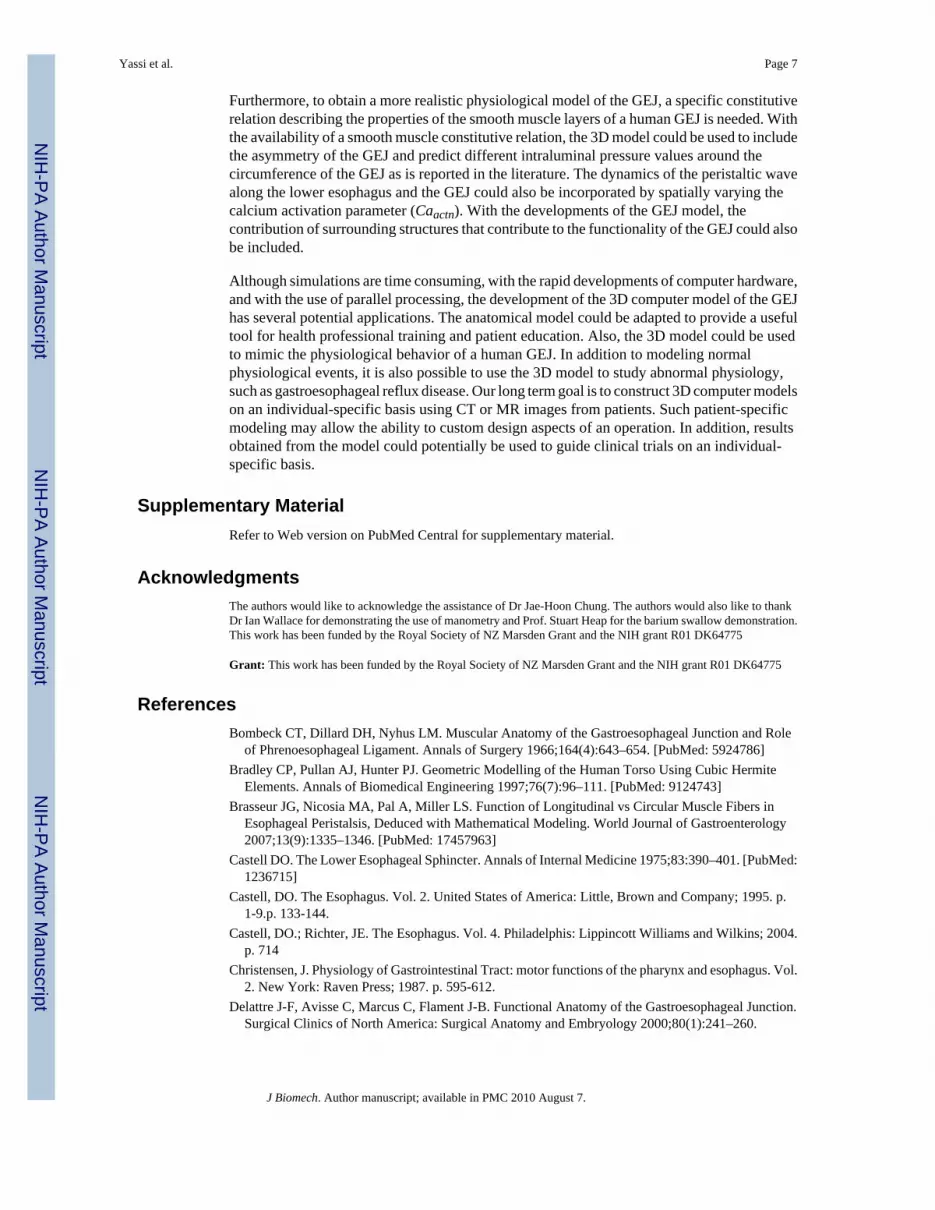
Furthermore, to obtain a more realistic physiological model of the GEJ, a specific constitutiverelation describing the properties of the smooth muscle layers of a human GEJ is needed. Withthe availability of a smooth muscle constitutive relation, the 3D model could be used to includethe asymmetry of the GEJ and predict different intraluminal pressure values around thecircumference of the GEJ as is reported in the literature. The dynamics of the peristaltic wavealong the lower esophagus and the GEJ could also be incorporated by spatially varying thecalcium activation parameter (Caactn). With the developments of the GEJ model, thecontribution of surrounding structures that contribute to the functionality of the GEJ could alsobe included.
Although simulations are time consuming, with the rapid developments of computer hardware,and with the use of parallel processing, the development of the 3D computer model of the GEJhas several potential applications. The anatomical model could be adapted to provide a usefultool for health professional training and patient education. Also, the 3D model could be usedto mimic the physiological behavior of a human GEJ. In addition to modeling normalphysiological events, it is also possible to use the 3D model to study abnormal physiology,such as gastroesophageal reflux disease. Our long term goal is to construct 3D computer modelson an individual-specific basis using CT or MR images from patients. Such patient-specificmodeling may allow the ability to custom design aspects of an operation. In addition, resultsobtained from the model could potentially be used to guide clinical trials on an individual-specific basis.
Supplementary MaterialRefer to Web version on PubMed Central for supplementary material.
AcknowledgmentsThe authors would like to acknowledge the assistance of Dr Jae-Hoon Chung. The authors would also like to thankDr Ian Wallace for demonstrating the use of manometry and Prof. Stuart Heap for the barium swallow demonstration.This work has been funded by the Royal Society of NZ Marsden Grant and the NIH grant R01 DK64775
Grant: This work has been funded by the Royal Society of NZ Marsden Grant and the NIH grant R01 DK64775
ReferencesBombeck CT, Dillard DH, Nyhus LM. Muscular Anatomy of the Gastroesophageal Junction and Role
of Phrenoesophageal Ligament. Annals of Surgery 1966;164(4):643–654. [PubMed: 5924786]Bradley CP, Pullan AJ, Hunter PJ. Geometric Modelling of the Human Torso Using Cubic Hermite
Elements. Annals of Biomedical Engineering 1997;76(7):96–111. [PubMed: 9124743]Brasseur JG, Nicosia MA, Pal A, Miller LS. Function of Longitudinal vs Circular Muscle Fibers in
Esophageal Peristalsis, Deduced with Mathematical Modeling. World Journal of Gastroenterology2007;13(9):1335–1346. [PubMed: 17457963]
Castell DO. The Lower Esophageal Sphincter. Annals of Internal Medicine 1975;83:390–401. [PubMed:1236715]
Castell, DO. The Esophagus. Vol. 2. United States of America: Little, Brown and Company; 1995. p.1-9.p. 133-144.
Castell, DO.; Richter, JE. The Esophagus. Vol. 4. Philadelphis: Lippincott Williams and Wilkins; 2004.p. 714
Christensen, J. Physiology of Gastrointestinal Tract: motor functions of the pharynx and esophagus. Vol.2. New York: Raven Press; 1987. p. 595-612.
Delattre J-F, Avisse C, Marcus C, Flament J-B. Functional Anatomy of the Gastroesophageal Junction.Surgical Clinics of North America: Surgical Anatomy and Embryology 2000;80(1):241–260.
Yassi et al. Page 7
J Biomech. Author manuscript; available in PMC 2010 August 7.
NIH
-PA Author Manuscript
NIH
-PA Author Manuscript
NIH
-PA Author Manuscript

Edmundowicz SA, Clouse RE. Shortening of the Esophagus in Response to Swallowing. AmericanJournal of Physiology: Gastrointestinal and Liver Physiology 1991;260(23):G512–G516.
Egorov VI, Schastlivtsev IV, Prut EV, Baranov AO, Turusov RA. Mechanical Properties of the HumanGastrointestinal Tract. Journal of Biomechanics 2002;35:1417–1425. [PubMed: 12231288]
Fan Y, Gregersen H, Kassab GS. A Two-layered Mechanical Model of the Rat Esophagus: experimentand theory. BioMedical Engineering OnLine 2004;3(40):1–9. [PubMed: 14746653]
Gray SW, Rowe JS, Skandalakis JE. Surgical Anatomy of the Gastroesophageal Junction. The AmericanSurgeon 1979;45(9):575–587. [PubMed: 507565]
Gregersen, H. Biomechanics of the Gastrointestinal Tract: new perspectives in motility research anddiagnostics. London: Springer; 2003. p. 268
Gregersen H, Kassab GS, Fung YC. The Zero-Stress State of the Gastrointestinal Tract: biomechanicaland functional implications. Digestive Diseases and Sciences 2000;45(12):2271–2281. [PubMed:11258545]
Gregersen H, Lee TC, Chien S, Skalak R, Fung YC. Strain Distribution in the Layered Wall of theEsophagus. Journal of Biomechanical Engineering 1999;121:442–448. [PubMed: 10529910]
Guccione JM, McCulloch AD, Waldman LK. Passive Material Properties of Intact VentricularMyocardium Determined From a Cylindrical Model. Journal of Biomechanical Engineering1991;113:42–55. [PubMed: 2020175]
Hunter PJ, McCulloch A, ter-Keurs HEDJ. Modelling the Mechanical Properties of Cardiac Muscle.Progress in Biophysics and Molecular Biology 1998;69(2):289–331. [PubMed: 9785944]
Hunter, PJ.; Nash, MP.; Sands, GB. Computational electromechanics of the heart. In: Panfilov, AV.;Holden, AV., editors. Computational Biology of the Heart. West Sussex UK: John Wiley and SonsLtd; 1997. p. 345-407.
Kahrilas PJ. Anatomy and Physiology of the Gastroesophageal Junction. Gastroenterology Clinics ofNorth America: The Columnar-Lined Esophagus 1997;26(3):467–486.
Liao D, Fan Y, Zeng Y, Gregersen H. Stress Distribution in the Layered Wall of the Rat Oesophagus.Medical Engineering and Physics 2003;25:731–738. [PubMed: 14519345]
Liebermann-Meffert, D.; Brauer, RB. Surgery of the Esophagus, Stomach, and Small Intestine: surgicalanatomy of the distal esophagus and cardia. Vol. 5. Boston: Little, Brown and Company; 1995. p.32-44.
Liu J, Parashar VK, Mittal RK. Asymmetry of Lower Esophageal Sphincter Pressure: is it related to themuscle thickness or its shape. American Journal of Physiology: Gastrointestinal and Liver Physiology1997;272(35):G1509–G1517.
Lu X, Gregersen H. Regional Distribution of Axial Strain and Circumferential Residual Strain in theLayered Rabbit Oesophagus. Journal of Biomechanics 2001;34:225–233. [PubMed: 11165287]
Malvern, LE. Introduction to the Mechanics of a Continuous Medium. New Jersey: Prentice-Hall Inc;1969.
Mittal RK, Balaban DH. The Esophagogastric Junction. The New England Journal of Medicine 1997;336(13):924–932. [PubMed: 9070474]
Mosher HP. The Lower end of the Oesophagus at Birth and in the Adult. The Journal of Laryngologyand Otology 1930;45(3):161–180.
Nash, MP. PhD Thesis. Auckland: The University of Auckland; 1998. Mechanics and Material Propertiesof the Heart using an Anatomically Accurate Mathematical Model; p. 246
Nash MP, Hunter PJ. Computational mechanics of the heart: From tissue structure to ventricular function.Journal of Elasticity 2000;61(1–3):113–141.
Nicosia MA, Brasseur JG, Liu J-B, Miller LS. Local Longitudinal Muscle Shortening of the HumanEsophagus from High-Frequency Ultrasonography. American Journal of Physiology:Gastrointestinal and Liver Physiology 2001;281:G1022–G1033. [PubMed: 11557523]
Patti MG, Gantert W, Way LW. Surgery of the Esophagus: Anatomy and Physiology. Surgical Clinicsof North America: Surgery of the Esophagus 1997;77(5):959–970.
Pouderoux P, Lin S, Kahrilas PJ. Timing, Propagation, Coordination, and Effect of EsophagealShortening During Peristalsis. Gastroenterology 1997;112:1147–1154. [PubMed: 9097997]
Yassi et al. Page 8
J Biomech. Author manuscript; available in PMC 2010 August 7.
NIH
-PA Author Manuscript
NIH
-PA Author Manuscript
NIH
-PA Author Manuscript

Richter JE, Wu WC, Johns DN, Blackwell JN, Nelson JL, Castell JA, Castell DO. Esophageal Manometryin 95 Healthy Adult Volunteers: variability of pressures with age and frequency of “abnormal”contractions. Digestive Diseases and Sciences 1987;32(6):583–592. [PubMed: 3568945]
Shafik A, Shafik I, El-Sibai O, Mostafa RM. The Effect of Esophageal and Gastric Distension on theCrural Diaphragm. World Journal of Surgery 2006;30:199–204. [PubMed: 16425081]
Shi G, Ergun GA, Manka M, Kahrilas PJ. Lower Esophageal Sphincter Relaxation Characteristics Usinga Sleeve Sensor in Clinical Manometry. The American Journal of Gastroenterology 1998;93(12):2373–2379. [PubMed: 9860395]
Spitzer V, Ackerman MJ, Scherzinger AL, Whitlock D. The visible human male: a technical report.Journal of the American Medical Informatics Association 1996;3(2):118–130. [PubMed: 8653448]
Stein HJ, Liebermann-Meffert D, Demeester TR, Siewert JR. Three-dimensional Pressure Image andMuscular Structure of the Human Lower Esophageal Sphincter. Surgery 1995;117(6):692–698.[PubMed: 7778032]
Sugarbaker DJ, Kearney DJ, Richards WG. Esophageal Physiology and Pathophysiology. SurgicalClinics of North America: Motility Disorders of the Gastrointestinal Tract 1993;73(6):1101–1118.
Takeda T, Kassab GS, Liu J, Puckett JL, Mittal RR, Mittal RK. A Novel Ultrasound Technique to Studythe Biomechanics of the Human Esophagus in Vivo. American Journal of Physiology:Gastrointestinal and Liver Physiology 2002;282:785–793.
Takeda T, Nabae T, Kassab GS, Liu J, Mittal RK. Oesophageal Wall Stretch: the stimulus for distensioninduced oesophageal sensation. Neurogastroenterol and Motility 2004;16:721–728.
Tytgat, GNJ. Gastro-Oesophageal Reflux and Gastric Stasis: pathophysiology, diagnosis and therapy.Auckland: Adis International Ltd; 1991. p. 144
Vanags I, Petersons A, Ose V, Ozolanta I, Kasyanov V, Laizans J, Vjaters E, Gardovskis J, Vanags A.Biomedical Properties of Oesophagus Wall Under Loading. Journal of Biomechanics 2003;36:1387–1390. [PubMed: 12893048]
Weinstock LB, Clouse RE. Esophageal Physiology: normal and abnormal motor function. The AmericanJournal of Gastroenterology 1987;82(5):399–405. [PubMed: 3578219]
Winans CS. Alteration of Lower Esophageal Sphincter Characteristics with Respiration and ProximalEsophageal Balloon Distension. Gastroenterology 1972;62(3):380–388. [PubMed: 5011529]
Yang J, Liao D, Zhao J, Gregersen H. Shear Modulus of Elasticity of the Esophagus. Annals of BiomedicalEngineering 2004;32(9):1223–1230. [PubMed: 15493510]
Yang J, Zhao J, Liao D, Gregersen H. Biomechanical Properties of the Layered Oesophagus and itsRemodelling in Experimental Type-1 Diabetes. Journal of Biomechanics 2006;39:894–904.[PubMed: 16488228]
Zhao J, Chen X, Yang J, Liao D, Gregersen H. Opening Angle and Residual Strain in a Three-layeredModel of Pig Oesophagus. Journal of Biomechanics 2007;40(14):3187–3192. [PubMed: 17517416]
Yassi et al. Page 9
J Biomech. Author manuscript; available in PMC 2010 August 7.
NIH
-PA Author Manuscript
NIH
-PA Author Manuscript
NIH
-PA Author Manuscript

Figure 1.A longitudinal cross-sectional view of a cylindrical model and a cavity mesh (lighter shade).The cavity mesh is visible through a transparent section of the cylindrical model. Thedisplacements of the nodes of the cavity mesh were fixed in all directions with the exceptionof one node on the axis, which was constrained to move along the axial direction.
Yassi et al. Page 10
J Biomech. Author manuscript; available in PMC 2010 August 7.
NIH
-PA Author Manuscript
NIH
-PA Author Manuscript
NIH
-PA Author Manuscript
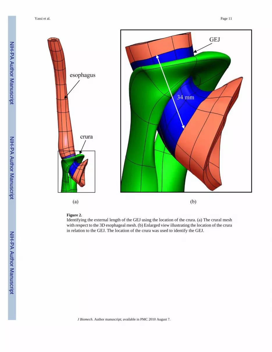
Figure 2.Identifying the external length of the GEJ using the location of the crura. (a) The crural meshwith respect to the 3D esophageal mesh. (b) Enlarged view illustrating the location of the crurain relation to the GEJ. The location of the crura was used to identify the GEJ.
Yassi et al. Page 11
J Biomech. Author manuscript; available in PMC 2010 August 7.
NIH
-PA Author Manuscript
NIH
-PA Author Manuscript
NIH
-PA Author Manuscript

Figure 3.The variation of intraluminal pressure as a function of the number of degrees of freedom ofthe GEJ computer model. The figure shows that the pressure value was converged when themesh had 19877 degrees of freedom or more.
Yassi et al. Page 12
J Biomech. Author manuscript; available in PMC 2010 August 7.
NIH
-PA Author Manuscript
NIH
-PA Author Manuscript
NIH
-PA Author Manuscript

NIH
-PA Author Manuscript
NIH
-PA Author Manuscript
NIH
-PA Author Manuscript
Yassi et al. Page 13
Table 1
The intraluminal pressure values predicted by the model during the contraction of the GEJ with and without thecontribution of the crura.
With crural contribution GEJ onlyBasal pressure mmHg (kPa) 22 (2.95) 12 (1.54)
Experimental basal pressure mmHg (kPa) 10–40 (1.33–5.33)3 6–22 (0.74 – 2.98)4Peak pressure mmHg (kPa) 52 (6.96) 28 (3.78)
Experimental peak pressure mmHg (kPa) 50–80 (6.67–10.67)5 28–45 (3.74–5.98)3Values were taken from the following references (Christensen, 1987; Kahrilas, 1997; Patti et al., 1997; Richter et al., 1987; Shafik et al., 2006; Stein et
al., 1995).
4Values have been calculated by subtracting the 44% contribution of the crura from the values with crural contribution.
5Values were taken from the following references (Christensen, 1987; Mittal and Balaban, 1997; Sugarbaker et al., 1993; Tytgat, 1991).
J Biomech. Author manuscript; available in PMC 2010 August 7.

NIH
-PA Author Manuscript
NIH
-PA Author Manuscript
NIH
-PA Author Manuscript
Yassi et al. Page 14
Table 2
The intraluminal pressure values predicted by the model with the contraction of each individual muscle layer,and both layers of the GEJ. CM (circular muscle), LM (longitudinal muscle),.
LM only CM only Both layersBasal pressure mmHg (kPa)−4 (−0.49) 15 (2.04) 12 (1.54)Peak pressure mmHg (kPa) −7 (−0.97) 38 (5.11) 28 (3.78)
J Biomech. Author manuscript; available in PMC 2010 August 7.


