Embed Size (px)
Citation preview
QAPL 2008
Modelling intracellular fate of FGF receptors withBioAmbients
S. van Bakel2 I. Khan M.G. Vigliotti 1 ,2
Department of Computing, Imperial College,180 Queen’s Gate, London SW7 2BZ, UK
J.K. Heath3
CRUK Growth Factor Group,School of Bioscience,University of Birmingham, Egbaston, B15 2TT UK
Abstract
In this paper we consider a model for different sorting of receptors of Fibroblast Growth Factor via the endocytotic pathway.In order to accurately model the relocation in the differentcompartments of the cell by the ligand-receptor complex, weusethe stochastic version of Bioambients. The stochastic simulation is carried out using BAM (BioAmbient Machine), whichisa Java implementation of BioAmbients via Gillespie’s Algorithm.Our model and the associated results of the simulation shed light on different mechanisms that influence the spatial distribu-tion of the different components in the pathway.
Keywords: Stochastic Process algebra, system biology,FGFpathway, stochastic simulation.
1 Introduction
In recent years it has been recognised that simulation of biochemical reactions using stochas-tic process algebra is adequate for modelling and analysingbiological systems [6,21,4][2,19,14]. The process-algebraic approach -different from mathematical techniques basedon sets of differential equations- forces a rigourous description of the interaction of biolog-ical components. This way of modelling yields further insight into the dynamics of biolog-ical phenomena and, at the same time, allows to derive quantities of interest. It has beenshown that results derived ‘in silico’, like experiments using process algebra, are consistentwith both results derived from real experiments and with theOrdinary Differential Equa-tions (ODEs) approach [2,20,11,10,13,3]. The latter models well the average behaviour oflarge quantities of molecules over time, but does not give aninsight on the evolution of
1 Supported by EPSRC grant SPARTACOS (EP /D047587/1).2 Email: [email protected], [email protected] Email: [email protected]
This paper is electronically published inElectronic Notes in Theoretical Computer Science
URL: www.elsevier.nl/locate/entcs
van Bakel Khan Heat Vigliotti
components to which those quantities refer [25]. Modelling biological complex systems inprocess algebra presents several advantages: models can becompositionally built, offeringthe opportunity to compose parts of the model that are developed at different times by dif-ferent people; models can be easily manipulated by simply changing some components andevaluating the impact of those changes over the behaviour ofthe whole model. Needlessto say, for models built in the process algebraic way,in silico experiments can be repeated-i.e. several runs of the same model can be performed- with different parameters allowinga simple and effective sensitivity analysis.
In recent years, many known stochastic process algebrae have been used to model bio-chemical reactions: PEPA [12], stochasticπ-calculus [18], BioAmbients [21], to name afew. Process calculi have been successfully applied to represent biochemical pathways. Itis useful to think of pathways as protocols, where the participants are the chemicals andthe rules according to which the protocol evolves are prescribed by the chemical reactionspresent in nature; pathways describe the way in which cells communicate. Similar to pro-tocols in computer science, pathways are very complex and present concurrent behaviour,which makes it virtually impossible to understand the temporal evolution of the whole sys-tem by simply analysing each chemical reaction in isolation. Classic process algebra isespecially suited to model biochemical pathways because itnaturally describes the causaldependencies among events and the concurrent behaviour among different competing com-ponents. The ultimate goal of understanding the causal dependencies among chemicalreactions in pathways is to help to develop new therapies by targeting specific components.
In recent years, it has become clear that reactions in pathways can vary according tothe location of components [16,9,8,23]. A typical example of this kind of pathway are thereceptor mediated endocytotic pathways [15]. Among the best known pathways of thiskind we mention the Epidermal Growth Factor (EGF) pathway and the Fibroblast GrowthFactor (FGF) pathway [15]. To model these pathways there is a need to explicitly repre-sent different compartments of the cell to finely describe the relocation of components.The work we present in this paper describes the endocytotic pathway of theFGF [9,8] viaBioAmbients. Endocytosis is a common communication mechanism in eukaryote cells.It is a mechanism by which the cell membrane envaginates to form a membrane limitedvesicle. Vesicles relocate in different compartments inside the cell. Eukaryotic cells con-tinually engage endocytosis to supply the cell with nutrients. There are different causes toendocytosis, however, if initiated by external proteins binding to receptors located on thecell, we speak ofreceptor meditated endocytosis. The extra-cellular protein that initiatesthe endocytosis is called aligand. The route taken by the vesicle in the receptor meditatedendocytosis is well documented in the literature [15]. The vesicle containing the complexligand-receptor moves to thesorting endosome and then to thelate endosome. At this pointthe fate of receptors varies: either they are degraded into the lysosome, or they reach themembrane via therecycling endosome. Receptors are inactive unbound, yet the bindingwith the ligand activates a chemical signal which in turn could be considered the cause ofcell’s activity such as stimulation to divide, to migrate orto differentiate into a differentcell type. Over-stimulation of such signal is deemed to be responsible for several diseasessuch as cancer. Thus it is important to understand very well the causes of the activation anddeactivation of the of the receptor’s signal.
It is believed that signalling of receptors stops in the lysosome as the receptor degrades.However, in recent years new insight has been gained in theFGF endocytotic pathway
2
van Bakel Khan Heat Vigliotti
[9,8,23]. It is important to recall thatFGF hormones are a family of twenty proteins thatshare similar structures. Similarly, the four differentFGF receptors are known to share sim-ilar structures. Very little is known about the different roles of the four receptors as theyrelocate inside the cell. In recent studies [9,8] it was shown that the distribution of the fourreceptors inside the lysosome and the recycling endosome varies quite dramatically. In thispaper we model the different fate of receptors in the endocytotic pathway of theFGF. Thekey point of the paper is the relocation of the complex (FGF:FGFR) in different parts of thecell. To this aim we use the stochastic version of BioAmbients, because it naturally repre-sents both compartments and their movements; other processcaluculi such asπ-calculusor PEPA do not have primitives in their language to directly model compartments. We willshow that our model in BioAmbient faithfully reproduces theresults present in [9,8], andwill shed some light on causes for the sorting of the different receptors in the cell. In shortthe contributions of the paper are:
• Theoretical development in BioAmbients of theFGF endocytotic pathway which focusesmostly on relocation and sorting of receptors. We simulate ’in silico’ some of the experi-ments produced in [8]. The results of the simulations are proved consistent withthe datafrom experiments. This is the first time that such a model has been proposed, to the bestof our knowledge.
• We tested the model in two ways:(i) We run an ’in silico experiment’ of the global model with the four receptors. This
shows that, for a limited period of time, the sorting of the receptors is consistent withthe experiments carried out in isolation.
(ii) We modify the model in such a way that bothFGF andFGFRs are randomly created.Each receptor is created at a different rate. The model showsthat in the very long runmost ofFGFR4s end in the cell membrane. Because the rates for the creation of FGFR4are not known, our model may not be realistic, however it sheds light on the fact thatwith a specific set of rates it is possible have an over-production of FGFR4s. Furthersensitivity analysis is necessary to document different realistic scenarios.
The results of the simulation are obtained by using BAM (BioAmbient Machine) whichis a tool developed at Imperial College London [17]. The tool has been implemented in Java1.5 and simulates the stochastic behaviour of the BioAmbients via Gillespie’s algorithm [7].Our work sets the basis to understand the complex dynamics ofthe recycling ofFGFRs inthe endocytotic pathway. Such understanding could help in developing new therapies forthe diseases caused by the over-stimulation ofFGF receptors.
The BioAmbient model is clearly not complete, but it describes the main dynamics ofthe different sorting of theFGFRs. It can be made more accurate in the future by com-positionally adding new components. Our work is of course anabstraction with respectto reality in the sense of [22]. In simple words, we do not aim to faithfully represent allthe details of the biological system, but we focus on the issue of activation/deactivation ofsignalling in different compartments and on relocation of components. We also do not aimto model accurately a set of chemical reactions involved in the endocytotic pathway. In thisrespect our work is orthogonal to [10,13,11] which consider in great details the chemicalreactions involved in the early stages of binding of theFGF with theFGFR.
The rest of the paper is organised as follows: in Section2 we review the syntax and thesemantics of Bioambients. We define the stochastic semantics along the lines of [12,24].
3
van Bakel Khan Heat Vigliotti
In Section3 we review theFGF endocytotic pathway as described in the literature (seealso [8]) and we present a model in Bioambients. In Section4 we discuss the resultsobtained by running different simulations and we discuss the prediction capacity of ourmodel. Conclusions follow. We include in the paper an appendix that describes in detailswhat experiments we have simulated [8] and the associated quantitative information.
2 BioAmbients
BioAmbients [21] are a dialect of the Ambient Calculus [5] suitable to model membraneand compartments in biology. In the original paper [21], the calculus was presented with astandard operational semantics expressed in terms of rewriting rules, and the implementa-tion in Prolog using Gillespie’s algorithm.
In this section, we introduce both syntax and the stochasticoperational semantics ofBioAmbients, leaving out the formal description of derivation of the underpinning Contin-uous Time Markov Chain, which is standard [12,1,18,24]. We slightly modify the syntaxof the calculus by using explicit recursion as opposed to replication and by introducing thedelay operatorτδ.
We shall assume the existence of a set of names or channelsN , and let the meta-variablesn,m, z, s, . . . range over this set.
Definition 2.1 The set of processes of BioAmbients is given by the followingsyntax:
P,Q ::= 0 | P | Q | (new n) P | [P ] | A〈x∼〉 |∑
i∈I Mi.Pi
M,N ::= entern | exitn | accept n | expeln | τr |
merge+ n | merge− n | $n(x) | $n〈m〉
$ ::= s2s | local | p2c | c2p
We assume that each name has a unique rate associated to it, and that there is an envi-ronmentρ : N → R that formally keeps track of the rate associated to names.
We informally explain the syntax of the calculus. In BioAmbients there are differentprimitives for communication. First of all, communicationhappens on a channeln bysending on a name -or channel-m. $n(x) stands for the input, and$n〈m〉 stands foroutput. There are three ways of communicating: channels in the same ambient performlocal communication, localn(y) for the input on channeln andlocaln〈m〉 for the output ofm on channeln. Inputs and outputs located in sibling ambients respectively performsiblingcommunication; s2sn(y) stands for such input ands2sn〈m〉 stands for output. Finally,parent to child communication happens when inputsp2cn(y) and outputsp2cn〈m〉 arelocated in parent-child ambients respectively (or vice-versa forc2pn(y) andc2pn〈m〉).The capabilities such asexitn or entern give the ambient the power to become active;entern/accept n allow an ambient to move into a sibling,exitn/expeln allow a childambient to leave the parent, whilemerge+ n andmerge− n fuse two sibling ambientsinto a single ambient.
As far as processes are concerned,Nil represents the inactive process;Local sum∑
i∈I Mi.Pi represents the standard choice. Given a set of indexesI and a permutationpon it, we write
∑
p(i)∈I Mp(i).Pp(i) to represent a reordering of the terms of the summation.We reserve the lettersG,C to represent summation as in
∑
i∈I Mi.Pp(i) = Mj .Pj + G
whereG =∑
i∈I i6=j Mi.Pi. In general, inputs are binding operators on the arguments.
4
van Bakel Khan Heat Vigliotti
P | 0 ≡ P
P | Q ≡ Q | P
(P | Q) | R ≡ P | (Q | R)
(new n) 0 ≡ 0
(new m) (new n) P ≡ (new n) (new m) P
(new n) (P | Q) ≡ P | (new n) Q if n /∈ fn(P )
(new m) [P ] ≡ [(new m) P ]∑
i∈I Mi.Pi ≡∑
p(i)∈I Mp(i).Pp(i)
A〈m∼ 〉 ≡ P{m∼/x∼} if A(x∼) = P
Fig. 1. Structural congruence
This means that in the processlocaln(y).P the namey is bound inP , and not accessi-ble from outsideP . A similar argument applies to the other inputs in the communicationprimitives. The processτr.P represents the delay for an amount of time that is exponen-tially distributed with rater. Ambient [P ] represents a compartment with an active processP . Parallel composition P | Q means thatP andQ are running in parallel.Restriction(new a) P of the namea makes that name private and unique toP : the namea becomesbound inP . Recursion A〈x∼〉 models infinite behaviour by assuming the existence of a set
of equations of the formA(x∼)df= P such that{x∼} ⊂ fn(P ), wherefn(P ) stands for the
usual free names ofP . The definition offn(P ) is standard taking into account that the onlybinding operators are inputs and restriction. We writeP{y/m} to mean the substitutionof every occurrence of the namey by m in P . Similarly we writeP{A/Q} to mean thesubstitution of every occurrence of the processA by Q in P .
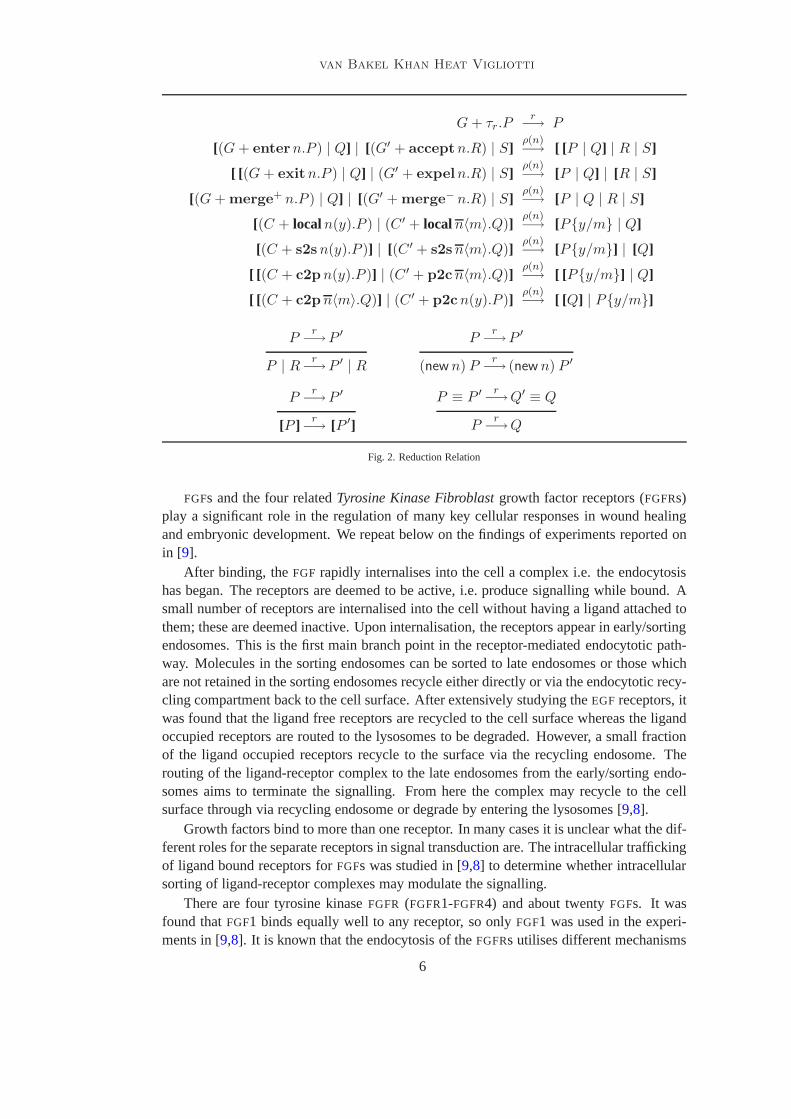
Formally, steps of computation are represented by areduction relation which is definedin Figure2. The reduction relation specifies how terms evolve syntactically, and the rateyielded by the environmentρ is sufficient information to specify how the process evolvesover time as well. We assume that each transition involving the name is exponentiallydistributed with parameter given byρ(n) (for the namen involved in the transition). Thatmeans that we can associate to each term a random variable over any interval∆x for x ∈ R
and regard the evolution of terms as a stochastic process.The underpinning model turns out to be a Continuous Time Markov Chain. We omit in
this paper the formal description on derivation of Continuous Time Markov Chains givena stochastic process algebra since it is standard [12,1,18,24]. The definition of reductionrelation involvesstructural congruence, ≡. This is the smallest congruence defined inFigure1.
3 Modelling the intracellular trafficking of FGFRs BioAmbients
Cells (in complex organisms) communicate with neighbouring cells and their environmentvia receptors situated in the membrane. Cell receptors are classified into families, basedupon similarity in structure, ligand binding and the biological response they induce [9].Receptor Tyrosine Kinases (RTK) are receptors located on the surface of the cell. Theiractivity is induced by the corresponding signallingRTK protein generally located outsidethe cell.
5
van Bakel Khan Heat Vigliotti
G + τr.Pr
−→ P
[(G + entern.P ) | Q] | [(G′ + acceptn.R) | S]ρ(n)−→ [ [P | Q] | R | S]
[ [(G + exitn.P ) | Q] | (G′ + expeln.R) | S]ρ(n)−→ [P | Q] | [R | S]
[(G + merge+ n.P ) | Q] | [(G′ + merge− n.R) | S]ρ(n)−→ [P | Q | R | S]
[(C + localn(y).P ) | (C ′ + localn〈m〉.Q)]ρ(n)−→ [P{y/m} | Q]
[(C + s2sn(y).P )] | [(C ′ + s2sn〈m〉.Q)]ρ(n)−→ [P{y/m}] | [Q]
[ [(C + c2pn(y).P )] | (C ′ + p2cn〈m〉.Q)]ρ(n)−→ [ [P{y/m}] | Q]
[ [(C + c2pn〈m〉.Q)] | (C ′ + p2cn(y).P )]ρ(n)−→ [ [Q] | P{y/m}]
Pr
−→P ′
P | Rr
−→P ′ | R
Pr
−→P ′
(new n) Pr
−→ (new n) P ′
Pr
−→P ′
[P ] r−→ [P ′]
P ≡ P ′ r−→Q′ ≡ Q
Pr
−→Q
Fig. 2. Reduction Relation
FGFs and the four relatedTyrosine Kinase Fibroblast growth factor receptors (FGFRs)play a significant role in the regulation of many key cellularresponses in wound healingand embryonic development. We repeat below on the findings ofexperiments reported onin [9].
After binding, theFGF rapidly internalises into the cell a complex i.e. the endocytosishas began. The receptors are deemed to be active, i.e. produce signalling while bound. Asmall number of receptors are internalised into the cell without having a ligand attached tothem; these are deemed inactive. Upon internalisation, thereceptors appear in early/sortingendosomes. This is the first main branch point in the receptor-mediated endocytotic path-way. Molecules in the sorting endosomes can be sorted to lateendosomes or those whichare not retained in the sorting endosomes recycle either directly or via the endocytotic recy-cling compartment back to the cell surface. After extensively studying theEGF receptors, itwas found that the ligand free receptors are recycled to the cell surface whereas the ligandoccupied receptors are routed to the lysosomes to be degraded. However, a small fractionof the ligand occupied receptors recycle to the surface via the recycling endosome. Therouting of the ligand-receptor complex to the late endosomes from the early/sorting endo-somes aims to terminate the signalling. From here the complex may recycle to the cellsurface through via recycling endosome or degrade by entering the lysosomes [9,8].
Growth factors bind to more than one receptor. In many cases it is unclear what the dif-ferent roles for the separate receptors in signal transduction are. The intracellular traffickingof ligand bound receptors forFGFs was studied in [9,8] to determine whether intracellularsorting of ligand-receptor complexes may modulate the signalling.
There are four tyrosine kinaseFGFR (FGFR1-FGFR4) and about twentyFGFs. It wasfound thatFGF1 binds equally well to any receptor, so onlyFGF1 was used in the experi-ments in [9,8]. It is known that the endocytosis of theFGFRs utilises different mechanisms
6
van Bakel Khan Heat Vigliotti
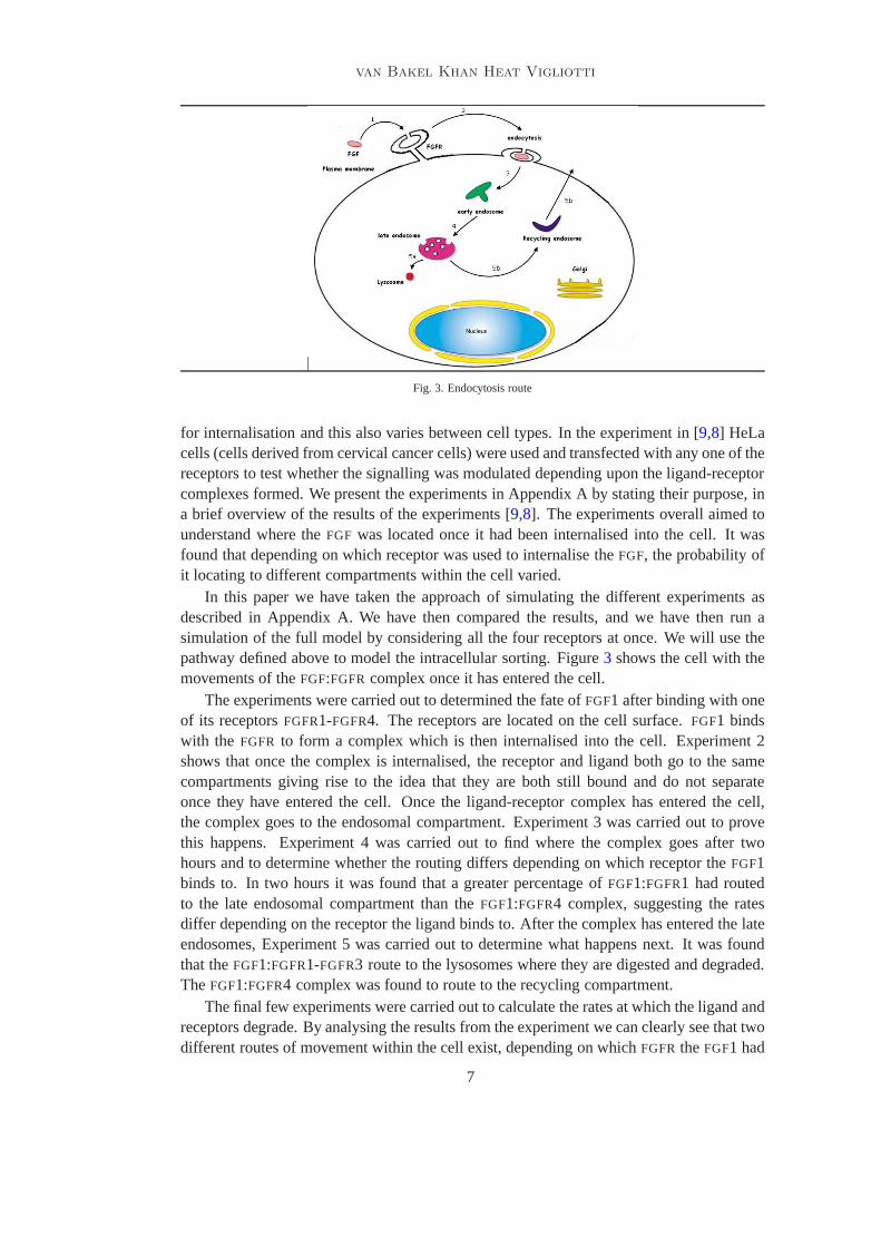
Fig. 3. Endocytosis route
for internalisation and this also varies between cell types. In the experiment in [9,8] HeLacells (cells derived from cervical cancer cells) were used and transfected with any one of thereceptors to test whether the signalling was modulated depending upon the ligand-receptorcomplexes formed. We present the experiments in Appendix A by stating their purpose, ina brief overview of the results of the experiments [9,8]. The experiments overall aimed tounderstand where theFGF was located once it had been internalised into the cell. It wasfound that depending on which receptor was used to internalise theFGF, the probability ofit locating to different compartments within the cell varied.
In this paper we have taken the approach of simulating the different experiments asdescribed in Appendix A. We have then compared the results, and we have then run asimulation of the full model by considering all the four receptors at once. We will use thepathway defined above to model the intracellular sorting. Figure3 shows the cell with themovements of theFGF:FGFR complex once it has entered the cell.
The experiments were carried out to determined the fate ofFGF1 after binding with oneof its receptorsFGFR1-FGFR4. The receptors are located on the cell surface.FGF1 bindswith the FGFR to form a complex which is then internalised into the cell. Experiment 2shows that once the complex is internalised, the receptor and ligand both go to the samecompartments giving rise to the idea that they are both stillbound and do not separateonce they have entered the cell. Once the ligand-receptor complex has entered the cell,the complex goes to the endosomal compartment. Experiment 3was carried out to provethis happens. Experiment 4 was carried out to find where the complex goes after twohours and to determine whether the routing differs depending on which receptor theFGF1binds to. In two hours it was found that a greater percentage of FGF1:FGFR1 had routedto the late endosomal compartment than theFGF1:FGFR4 complex, suggesting the ratesdiffer depending on the receptor the ligand binds to. After the complex has entered the lateendosomes, Experiment 5 was carried out to determine what happens next. It was foundthat theFGF1:FGFR1-FGFR3 route to the lysosomes where they are digested and degraded.TheFGF1:FGFR4 complex was found to route to the recycling compartment.
The final few experiments were carried out to calculate the rates at which the ligand andreceptors degrade. By analysing the results from the experiment we can clearly see that twodifferent routes of movement within the cell exist, depending on whichFGFR theFGF1 had
7
van Bakel Khan Heat Vigliotti
bound with. From the experiments it has been found thatFGF1 internalised byFGFR1-FGFR3 is generally routed to the lysosomes for degradation and that FGF1 internalised byFGFR4 is mainly routed to the recycling compartment.
From the experiments, we have obtained two extremely important pieces of informa-tion:
(i) The movement of theFGF:FGFRcomplex inside the different compartments in the celldepends on whichFGFR receptor it has bound with.
(ii) Rate information detailing the different rates of degradation and recycling depends onwhich receptor theFGF bound with.
In our model we aim to obtain a high-level view of the movements of the complex, asopposed to the finer details of the chemical reactions. That is, we abstract away fromrepresentation of chemical reactions and we just concentrate of localisation of the complex.
The model we are going to implement in BioAmbient is the following:
(i) FGF is found outside cells andFGFRs are located on the cell’s surface. TheFGF
binds with theFGFR and forms a complex;
(ii) The FGF:FGFR complex enters the cell through endocytosis;
(iii) The FGF:FGFR complex moves to the early/sorting endosomal compartment;
(iv) The FGF:FGFR complex is routed to the late endosomes;
(v) From this point there is a choice as to what may happen nextdepending on whichFGFR was used to internalise theFGF1:(a) FGFR1-FGFR3: The complex is routed to the lysosomes where it degrades;(b) FGFR4: The complex moves to the endosomal recycling compartmentto be
recycled back to the cell surface.
To formally model the intracellular sorting pathway we needto make several assump-tions.
(i) For eachFGF protein oneFGFR receptor is required to allow them to bind.
(ii) When theFGF:FGFR complex enters the sorting endosome, we assume they remainbound. It is known from the literature that the complex decomposes in the earlyendosome, and follows the route of recycling. We do not modelthe unbinding directly,but we model theFGFRmoving in other compartments of the cell.
(iii) When the FGFR complex enters the lysosomes, we assume they are completelyde-graded and digested and the ligand and receptor are both destroyed. We do not modeldirectly the degradation of the receptor.
(iv) When theFGFR enters the recycling compartment it returns to the cell surface, readyto be reused.
(v) We do not model the endocytosis of inactive receptors.
A recent paper [23] has shown that ifFGF:FGFR does not decompose in the sortingendosome then the complex recycles back to the cell surface directly. We leave for futurework to compositionally increment our model to take into account this new finding, andto explicitly model the biological switch that causes the unbinding of theFGF:FGFR. Themodel presented in this paper is the starting point to model the behaviour ofFGFRs, whose
8
van Bakel Khan Heat Vigliotti
over-production is deemed to play a key in role in the development of cancer.In BioAmbients,
• we model the compartment such as early endosome, late endosome, lysosome, recyclingendosome;
• we do not explicitly model the cell membrane or the perimeterof the cell;• we model theFGFRas a compartment because it is natural to think of it as movinginside
and outside of different compartments;• we model the binding with the receptor as communication;• we do not model the degradation of either theFGFor theFGFRdirectly, we simply model
the different routes among the different complexes.
The core implementation of the of endocytosis ofFGF in BioAmbients is shown inFigure4. The ligandFGFsends a message to ambientFGFR, which, after having received it,can move to the early endosomeEN, then to the late endosomeLE and then either theFGFR
ends in the ambient of the lysosmeLYSO, where nothing further happens or it enters therecycling compartmentRECYCLE where it is routed out again ready to be used. It must benoted that in this implementation we assume that each compartment allows a finite numberof vesicles to enter or exit. The core model assumes that there are about one-thousand andthree hundred receptors and one thousand ligands. We run four different experiments bychanging the rate of the channellyso and lendo. This is meant to simulate the differentexperiments in [8]. Lastly, we run the full model with the four different receptors at thesame time. The core model presented in Figure4 is easy to manipulate and we have run afew ‘experiments’ which results are reported in Section4.
4 Results
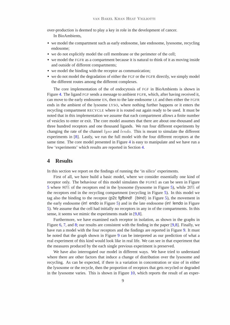
In this section we report on the findings of running the ‘in silico’ experiments.First of all, we have build a basic model, where we consider essentially one kind of
receptor only. The behaviour of this model simulates theFGFR1 as can be seen in Figure5 where80% of the receptors end in the lysosome (lysosome in Figure5), while 20% ofthe receptors end in the recycling compartment (recycling in Figure5). In this model wetag also the binding to the receptor (p2c fgfbind! {bind} in Figure5), the movement inthe early endosome (in! endo in Figure5) and in the late endosome (in! lendo in Figure5). We assume that the cell had initially no receptors in any inof the compartments. In thissense, it seems we mimic the experiments made in [9,8].
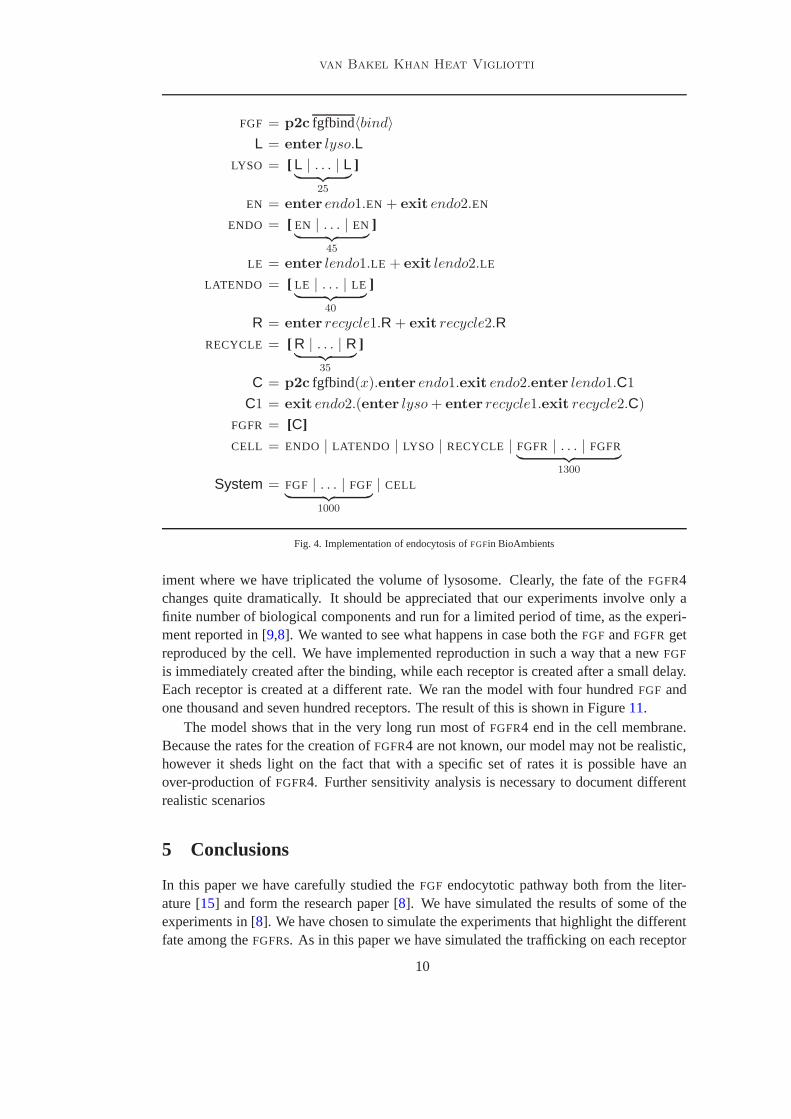
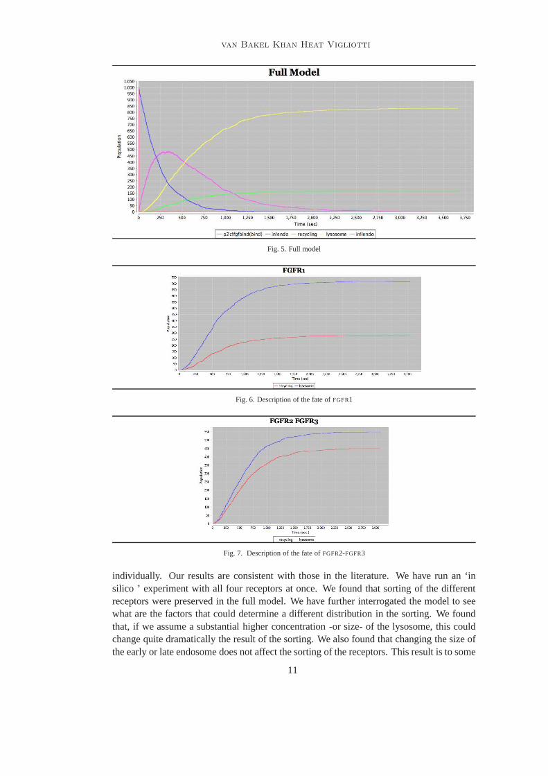
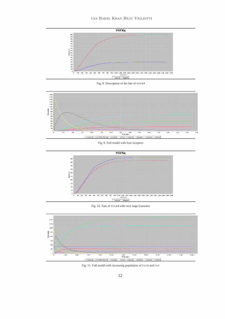
Furthermore, we have examined each receptor in isolation, as shown in the graphs inFigure6, 7, and8; our results are consistent with the finding in the paper [9,8]. Finally, wehave run a model with the four receptors and the findings are reported in Figure9. It mustbe noted that the graph shown in Figure9 can be interpreted as our prediction of what areal experiment of this kind would look like in real life. We can see in that experiment thatthe measures produced by the each single previous experiment is preserved.
We have also interrogated our model in different ways. We have tried to understandwhere there are other factors that induce a change of distribution over the lysosome andrecycling. As can be expected, if there is a variation in concentration or size of in eitherthe lysosome or the recycle, then the proportion of receptors that gets recycled or degradedin the lysosome varies. This is shown in Figure10, which reports the result of an exper-
9
van Bakel Khan Heat Vigliotti
FGF = p2c fgfbind〈bind〉
L = enter lyso.LLYSO = [ L | . . . | L
︸ ︷︷ ︸
25
]
EN = enter endo1.EN + exit endo2.EN
ENDO = [ EN | . . . | EN︸ ︷︷ ︸
45
]
LE = enter lendo1.LE + exit lendo2.LE
LATENDO = [ LE | . . . | LE︸ ︷︷ ︸
40
]
R = enter recycle1.R + exit recycle2.R
RECYCLE = [ R | . . . | R︸ ︷︷ ︸
35
]
C = p2c fgfbind(x).enter endo1.exit endo2.enter lendo1.C1
C1 = exit endo2.(enter lyso + enter recycle1.exit recycle2.C)
FGFR = [C]
CELL = ENDO | LATENDO | LYSO | RECYCLE | FGFR | . . . | FGFR︸ ︷︷ ︸
1300
System = FGF | . . . | FGF︸ ︷︷ ︸
1000
| CELL
Fig. 4. Implementation of endocytosis ofFGFin BioAmbients
iment where we have triplicated the volume of lysosome. Clearly, the fate of theFGFR4changes quite dramatically. It should be appreciated that our experiments involve only afinite number of biological components and run for a limited period of time, as the experi-ment reported in [9,8]. We wanted to see what happens in case both theFGF andFGFR getreproduced by the cell. We have implemented reproduction insuch a way that a newFGF
is immediately created after the binding, while each receptor is created after a small delay.Each receptor is created at a different rate. We ran the modelwith four hundredFGF andone thousand and seven hundred receptors. The result of thisis shown in Figure11.
The model shows that in the very long run most ofFGFR4 end in the cell membrane.Because the rates for the creation ofFGFR4 are not known, our model may not be realistic,however it sheds light on the fact that with a specific set of rates it is possible have anover-production ofFGFR4. Further sensitivity analysis is necessary to document differentrealistic scenarios
5 Conclusions
In this paper we have carefully studied theFGF endocytotic pathway both from the liter-ature [15] and form the research paper [8]. We have simulated the results of some of theexperiments in [8]. We have chosen to simulate the experiments that highlightthe differentfate among theFGFRs. As in this paper we have simulated the trafficking on each receptor
10
van Bakel Khan Heat Vigliotti
Fig. 5. Full model
Fig. 6. Description of the fate ofFGFR1
Fig. 7. Description of the fate ofFGFR2-FGFR3
individually. Our results are consistent with those in the literature. We have run an ‘insilico ’ experiment with all four receptors at once. We foundthat sorting of the differentreceptors were preserved in the full model. We have further interrogated the model to seewhat are the factors that could determine a different distribution in the sorting. We foundthat, if we assume a substantial higher concentration -or size- of the lysosome, this couldchange quite dramatically the result of the sorting. We alsofound that changing the size ofthe early or late endosome does not affect the sorting of the receptors. This result is to some
11
van Bakel Khan Heat Vigliotti
Fig. 8. Description of the fate ofFGFR4
Fig. 9. Full model with four receptors
Fig. 10. Fate ofFGFR4 with very large lysosome
Fig. 11. Full model with increasing population ofFGFRandFGF
12
van Bakel Khan Heat Vigliotti
extend to be expected. By looking at the Markov Chain generated by our specifications,it can be seen that changing the size of the lysosome has an impact on the probability ofbeing routed in this compartment. By simply looking at the results in the experiments asdescribed in [8] this could have been both difficult to guess or expensive to verify with anexperiment. In the literature there has already been an verysuccessful attempt to modelthe FGF signalling pathways i.e. the early stages ofFGF signal propagation and internal-isation using probabilistic model checking PRISM [10]. That work concentrated mostlyon modelling chemical reactions and in this respect their purpose is orthogonal to ours. Itremains to be seen whether our model could be refined in the long term, in order to modelboth compartments and chemical reactions, in which case thework carried out in [11] usingπ-calculus could be directly integrated with ours.
Acknowledgements
We gratefully acknowledge M. Kwiatkowska, G. Norman, and D.Parker for very valuablecontributions to the initial discussions on the topic of thepresent article. We gratefully ac-knowledge Vinod Muganthan and Andrew Phillips for sharing the enterprise of developingBAM [ 17]. We finally thank Luca Cardelli for very useful discussionson the topics of thisarticle and on system biology in general.
References
[1] C. Baier, B. Haverkort, H. Hermanns, and J.-P. Katoen. Model-checking algorithms for continuous-time Markov chains.IEEE Transactions on Software Engineering, 29(6):524–541, 2003.
[2] Muffy Calder, Stephen Gilmore, and Jane Hillston. Modelling the influence of RKIP on the ERK signalling pathwayusing the stochastic process algebra PEPA. In Anna Ingolfsdottir and Hanne Riis Nielson, editors,Proceedings of theBioConcur Workshop on Concurrent Models in Molecular Biology, London, England, August 2004.
[3] Muffy Calder, Stephen Gilmore, and Jane Hillston. Automatically deriving ODEs from process algebra models ofsignalling pathways. In Gordon Plotkin, editor,Proceedings of Computational Methods in Systems Biology (CMSB2005), pages 204–215, Edinburgh, Scotland, 2005.
[4] L. Cardelli. Abstract machine for system biology.Transactions on Computational Systems Biology., 3737:145–168,2005.
[5] L. Cardelli and A.D. Gordon. Mobile ambients.Theoretical Computer Science, 240(1):177–213, 2000.
[6] Vincent Danos and Cosimo Laneve. Formal molecular biology. Theor. Comput. Sci., 325(1):69–110, 2004.
[7] Daniel T. Gillespie. Exact stochastic simulation of coupled chemical reactions.Journal of Physical Chemistry,81(25):2340–2361, 1977.
[8] E. M. Haugsten, V. Sørensen, A. Brech, S. Olsnes, and J. Wesche. Different intracellular trafficking of fgf1 endocytosedby the four homologous fgf receptors.Journal of Cell Science, 118:3869–3881, 2005.
[9] Ellen Margrethe Haugsten. Intracellular trafficking offgf1 endocytosed by its four tyrosine kinase receptors. Master’sthesis, DEPARTMENT OF MOLECULAR BIOSCIENCES, University of Oslo, 2004.
[10] J. Heath, M. Kwiatkowska, G. Norman, D. Parker, and O. Tymchyshyn. Probabilistic model checking of complexbiological pathways. In C. Priami, editor,Proc. Computational Methods in Systems Biology (CMSB’06), volume 4210of Lecture Notes in Bioinformatics, pages 32–47. Springer Verlag, 2006.
[11] J. Heath, M. Kwiatkowska, G. Norman, D. Parker, and O. Tymchyshyn. Probabilistic model checking of complexbiological pathways.Theoretical Computer Science, 2007. To appear.
[12] J. Hillston. A Compositional Approach to Perfomance Modelling. PhD thesis, Department of Computer Science,Edinburgh, 1994.
[13] Marta Kwiatkowska, Gethin Norman, David Parker, Oksana Tymchyshyn, John Heath, and Eamonn Gaffney.Simulation and verification for computational modelling ofsignalling pathways. InWSC ’06: Proceedings of the38th conference on Winter simulation, pages 1666–1674. Winter Simulation Conference, 2006.
[14] Paola Lecca and Corrado Priami. Cell cycle control in eukaryotes: A biospi model.Electron. Notes Theor. Comput.Sci., 180(3):51–63, 2007.
[15] H. Lodish, A. Berk, P. Matsudaira, C. A. Kaiser, M. Krieger, M.P. Scott, S. L. Zipursky, and J. Darnell.Molecular CellBiology. W.H. Freeman and Company, 2003.
[16] M. Miaczynska, L. Pelkmans, and M. Zerial. Not just a sink: endosomes in control of signal transduction.Curr. Opin.Cell., 16:400–4006, 2004.
13
van Bakel Khan Heat Vigliotti
[17] Vinod A. Muganthan, A. Phillips, and M.G. Vigliotti. BioAmbient Machine. InACSD ’08: Proceedings of 8thInternational Conference on Application of Concurrency to System Design 2008, To appear.
[18] C. Priami. Stochasticπ-Calculus.The Computer Journal, 38(7):579–589, 1995.
[19] Corrado Priami, Aviv Regev, Ehud Shapiro, and William Silverman. Application of a stochastic name-passing calculusto representation and simulation of molecular processes.Inf. Process. Lett., 80(1):25–31, 2001.
[20] Andrew Phillips Ralf Blossey, Luca Cardelli. A compositional approach to the stochastic dynamics of gene networks.Transactions on Computational Systems Biology IV, 3939:99–122, 2006.
[21] Aviv Regev, Ekaterina M. Panina, William Silverman, Luca Cardelli, and Ehud Y. Shapiro. Bioambients: an abstractionfor biological compartments.Theoretical Computer Science, 325(1):141–167, 2004.
[22] Aviv Regev and Ehud Y. Shapiro. Cellular abstractions:Cells as computation.Nature, 419(343), 2002.
[23] E. Sandilands, S. Akbarzadeh, A. Vecchione, D.G. McEwan, M.C. Frame, and J.K.Heath. Src kinase modulates theactivation, transport and signalling dynamincs of fibroblast growth factor receptors.Embo Reports, 2007.
[24] M. Vigliotti and P. Harrison. Stochastic mobile ambients. In A. Di Pierro and H. Wiklicky, editors,Proc. 4th Int.Workshop Quantitative Aspects of Programming Languages (QAPL 2006), volume 164 (issue 3) ofElectronic Notes inTheoretical Computer Science, pages 169–186. Elsevier, 2006.
[25] Darren J. Wilkinson.Stochastic Modelling for System Biology. Chapman & Hall, 2006.
Appendix A
5.1 Experimental Data
Experiment 2
Purpose: Determine whereFGF locates to once it has bound withFGFR.
Result: The double staining in the experiment showed that both ligand and receptor endedup in the same compartment showing that they remain bound as acomplex once theyhave become internalised into the cell.
Experiment 3
Purpose: This experiment was carried out to follow the endocytotic pathway and identifywhich compartmentFGF1 locates to after internalisation.
Result: This experiment shows that whenFGF1 bound with any one of the fourFGFR1-FGFR4 receptors they all went to the early/sorting endosomal compartment after inter-nalisation.
Experiment 4
Purpose: This experiment was carried out to find out where theFGF:FGFRcomplex movesto after entering the early/sorting endosomes.
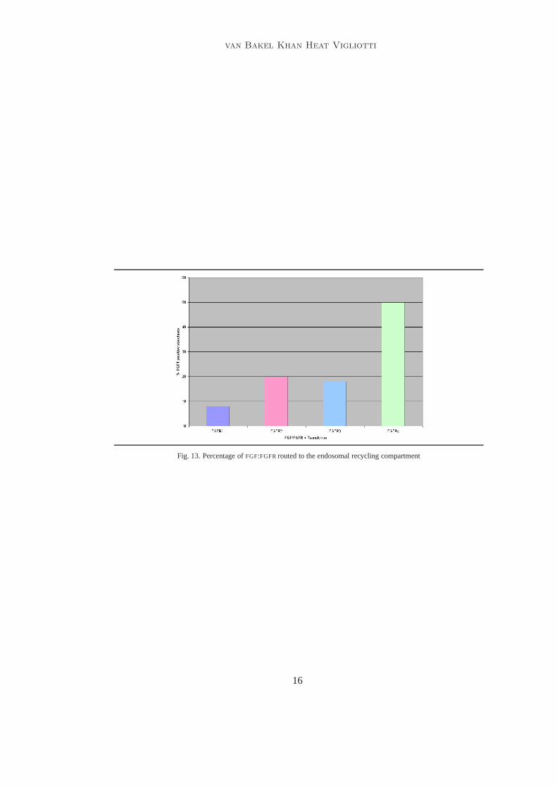
Result: After two hours in the presence it was shown that the major part of FGF1 inter-nalised withFGFR1-FGFR3 went to late endosomes. Figure13 shows the different per-centages which were routed to late endosomes for the different receptors after 2 hours.It shows that 90%FGF1-FGFR1 wereLAMP-1 positive whereas only 45% wereLAMP-1positive forFGFR4. In the case ofFGFR2 andFGFR3 about 70% wereLAMP-1 positive.
Experiment 5
Purpose: This experiment was carried out to to determine where theFGF1:FGFR4 complexlocalised to after late endosomes.
14
van Bakel Khan Heat Vigliotti
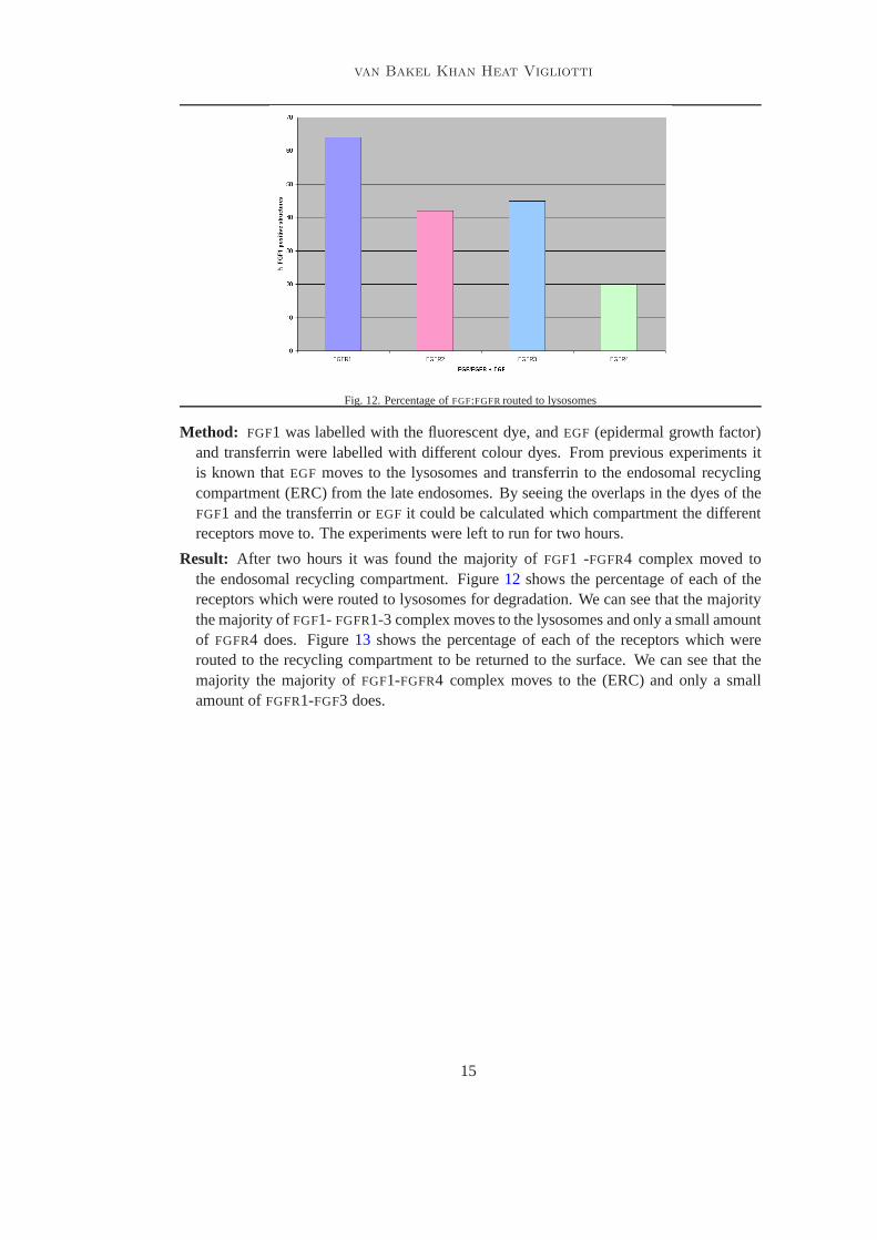
Fig. 12. Percentage ofFGF:FGFRrouted to lysosomes
Method: FGF1 was labelled with the fluorescent dye, andEGF (epidermal growth factor)and transferrin were labelled with different colour dyes. From previous experiments itis known thatEGF moves to the lysosomes and transferrin to the endosomal recyclingcompartment (ERC) from the late endosomes. By seeing the overlaps in the dyes of theFGF1 and the transferrin orEGF it could be calculated which compartment the differentreceptors move to. The experiments were left to run for two hours.
Result: After two hours it was found the majority ofFGF1 -FGFR4 complex moved tothe endosomal recycling compartment. Figure12 shows the percentage of each of thereceptors which were routed to lysosomes for degradation. We can see that the majoritythe majority ofFGF1- FGFR1-3 complex moves to the lysosomes and only a small amountof FGFR4 does. Figure13 shows the percentage of each of the receptors which wererouted to the recycling compartment to be returned to the surface. We can see that themajority the majority ofFGF1-FGFR4 complex moves to the (ERC) and only a smallamount ofFGFR1-FGF3 does.
15
van Bakel Khan Heat Vigliotti
Fig. 13. Percentage ofFGF:FGFRrouted to the endosomal recycling compartment
16


![FIF [Fibroblast Growth Factor - 2 (FGF-2)-Interacting-Factor], a Nuclear Putatively Antiapoptotic Factor, Interacts Specifically with FGF-2](https://img.pdfslide.net/doc/110x75/636296899b985d7ef50271c1/fif-fibroblast-growth-factor-2-fgf-2-interacting-factor-a-nuclear-putatively.jpg)


