Embed Size (px)
Citation preview
© 2022 by the author. This is an open access article distributed under the conditions of the Creative Commons by Attribution License, which permits unrestricted use, distribution, and reproduction in any mediumor format, provided the original work is correctly cited.
Open Access
Recent Progress in Materials
Review
Modifications of Glass Ionomer Cements Using Nanotechnology: Recent Advances
Dimitrios Dionysopoulos *, Olga Gerasimidou, Constantinos Papadopoulos
Department of Operative Dentistry, Faculty of Dentistry, School of Health Sciences, Aristotle
University of Thessaloniki, Thessaloniki 54124, Greece; E-Mails: [email protected];
[email protected]; [email protected]
* Correspondence: DimitriosDionysopoulos; E-Mail: [email protected]
Academic Editor: Eugeniusz Sajewicz
Special Issue: Advanced Dental Materials
Recent Progress in Materials
2022, volume 4, issue 2
doi:10.21926/rpm.2202011
Received: May 01, 2022
Accepted: May 24, 2022
Published: June 01, 2022
Abstract
Glass ionomer cements (GICs) are dental materials that were invented by Wilson & Kent in
1972. They can chemically bond to enamel and dentin and can exhibit anti-cariogenic activity
that allows the release and uptake of fluoride ions. They also possess the ability to render
color. The setting reaction of GICs is a neutralization reaction that results in the formation of
polycarboxylate salts. The most important GIC modification process involves the addition of
resin components, resulting in the development of resin-modified glass ionomer cements
(RMGICs), which contain self-and photo-curing systems. Modification of conventional GICs
and RMGICs can be achieved by the incorporation of nano-sized fillers into the materials.
Conventional GICs and RMGICs can also be modified by introducing nano-sized bioceramics
to the glass powder. It has been previously reported that the incorporation of nano-sized
particles helps improve the mechanical properties of conventional GICs. Conversely, the
commercially available nano-filled RMGICs do not hold any significant advantage over
conventional RMGICs as far as the mechanical and adhesive properties are concerned. Glass
carbomer is a novel glass ionomer material, and the bioactivity of which is better than the
bioactivity of the conventional GICs. However, it is more brittle and less strong than the
Recent Progress in Materials 2022; 4(2), doi:10.21926/rpm.2202011
Page 2/17
modern conventional GICs. Additionally, clinical techniques that can be used to transfer
external energy on the surface of a GIC have also been used for modification. These
techniques can be used to reduce the duration of the initial setting stage and improve the
rate of the setting reactions, resulting in faster development of the mechanical properties.
Premature failure of the restorations can be avoided under these conditions. The lack of
long-term clinical studies limits the use of nano-modified glass ionomers and glass
carbomers in daily clinical practice. More randomized clinical trials are required to justify the
use of these modern modified materials.
Keywords
Bioactive materials; glass ionomer cements; modifications; nanofillers; nanotechnology
1. Introduction
Glass ionomer cements (GICs) are dental materials that were invented by Wilson & Kent in
1972 [1]. They can chemically bond to enamel and dentin. The origin of the anti-cariogenic activity
of the materials can be attributed to the release and uptake of fluoride ions. The materials can be
used to render color effectively [2-4]. Glass ionomers exist in powder and liquid form. The powder
consists of fluoro-alumino-calcium-sodium-silicate glass particles, usually in the form of SiO-AlO2-
CaF2-AlPO4-Na3AlF6. Some formulations contain strontium (Sr) and barium (Ba) compounds that
improve radiopacity and remineralization properties. The liquid is an aqueous solution of various
polycarboxylic acids, which are primarily polyalkenoate acids. The low viscosity of the acids
facilitates the handling of cements [5-7]. Tartaric acid is also added to the liquid to improve the
ease of handling and increase working time [8].
The setting reaction associated with GICs is a neutralization reaction, resulting in the formation
of polycarboxylate salts [9]. Initially, the hydrated protons (Η3Ο+) of the polyacid react with the
basic particles in glass. This promotes the movement of Na+, Ca2+, and Sr2+ ions, and later Al3+ ions,
from glass to the polyacid solution [10-12]. These ions react with the polyacid molecules and form
ionic bonds. Thus, the formed insoluble polysalt network causes the cement to harden [10]. Water
is an essential component of glass ionomers as it is the solvent of polymeric acid. It contributes to
the release of protons. It functions as the matrix for the setting reaction and is also a structural
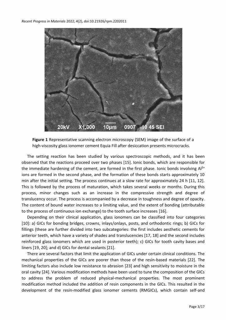
component of cement [13]. The loss of water from freshly hardened cement creates an unsightly
chalk surface due to the formation of microcracks (Figure 1). To prevent dehydration, it is
important to coat the surface of the cement with a hydrophobic material such as Vaseline or a
suitable resin varnish [14].
Recent Progress in Materials 2022; 4(2), doi:10.21926/rpm.2202011
Page 3/17
Figure 1 Representative scanning electron microscopy (SEM) image of the surface of a
high-viscosity glass ionomer cement Equia Fill after desiccation presents microcracks.
The setting reaction has been studied by various spectroscopic methods, and it has been
observed that the reactions proceed over two phases [15]. Ionic bonds, which are responsible for
the immediate hardening of the cement, are formed in the first phase. Ionic bonds involving Al3+
ions are formed in the second phase, and the formation of these bonds starts approximately 10
min after the initial setting. The process continues at a slow rate for approximately 24 h [11, 12].
This is followed by the process of maturation, which takes several weeks or months. During this
process, minor changes such as an increase in the compressive strength and degree of
translucency occur. The process is accompanied by a decrease in toughness and degree of opacity.
The content of bound water increases to a limiting value, and the extent of bonding (attributable
to the process of continuous ion exchange) to the tooth surface increases [16].
Depending on their clinical application, glass ionomers can be classified into four categories
[10]: a) GICs for bonding bridges, crowns, inlays/onlays, posts, and orthodontic rings; b) GICs for
fillings (these are further divided into two subcategories: the first includes aesthetic cements for
anterior teeth, which have a variety of shades and translucencies [17, 18] and the second includes
reinforced glass ionomers which are used in posterior teeth); c) GICs for tooth cavity bases and
liners [19, 20]; and d) GICs for dental sealants [21].
There are several factors that limit the application of GICs under certain clinical conditions. The
mechanical properties of the GICs are poorer than those of the resin-based materials [22]. The
limiting factors also include low resistance to abrasion [23] and high sensitivity to moisture in the
oral cavity [24]. Various modification methods have been used to tune the composition of the GICs
to address the problem of reduced physical-mechanical properties. The most prominent
modification method included the addition of resin components in the GICs. This resulted in the
development of the resin-modified glass ionomer cements (RMGICs), which contain self-and
Recent Progress in Materials 2022; 4(2), doi:10.21926/rpm.2202011
Page 4/17
photo-curing systems [25, 26]. Zinc [27, 28], stainless steel [29], strontium oxide [30], silicon
particles [31], bioactive apatite [32], a mixture of bioactive apatite and strontium [33, 34],
synthetic fibers [35] polyacids containing N-vinylpyrrolidone [36], and amino acid monomers [37]
have been used over the years as additives to improve the mechanical properties of the materials.
In recent years nanotechnology has been used to produce new composite resins. It has been
reported that the method can be used to effectively improve the mechanical properties of the
materials [38, 39]. Similar efforts have been made to improve the physical and mechanical
properties of both conventional and resin-modified GICs. Glass Carbomer, a modified GIC,contains
modified glass particles, nano-hydroxyapatite, and silicone oil [40, 41].
Most recent versions of GICs typically consist of powders that contain some of the polymeric
acids in dried form, resulting in the formation of a low-viscosity acid solution. The freshly mixed
cement prepared under these conditions contains high amounts of acid that promote the rapid
setting of the material. The process also imparts good strength. These types of materials are
labeled as “high-viscosity” GICs, a term typically applied to materials characterized by high
powder/liquid ratios of at least 3.6:1 [16]. In 2015, a novel GIC material known as the Equia Forte
(GC Inc., Kyoto, Japan) was introduced for application in high-load-bearing areas in posterior teeth.
It is a glass hybrid restorative material containing a multifunctional monomer and reinforced with
ultrafine, highly reactive glass particles [42, 43]. The time frame for the development of glass
ionomer materials is presented chronologically in Table 1.
Table 1 Developments of glass ionomer materials.
Date Glass ionomer developments
1972 The invention of conventional GICs
1977 Metal (silver)-reinforced GICs
1980 RMGICs self-polymerized
1990 RMGICs photo-polymerized
1991 RMGICs (photo+self)-polymerized
2003 Glass Carbomer
2007 Nano-modified RMGICs
2008 Nano-modified conventional GICs
2008 High-viscosity conventional GICs
2015 Hybrid restorative GICs
Therefore, the purpose of this literature review is to present the current data for the latest
modifications of GICs, emphasizing the modifications involving nanotechnology.
2. Resin-Modified Glass Ionomer Cements (RMGICs)
RMGICs were introduced in the field of dentistry in the late 1980s. These are hybrid materials
with combined properties of conventional GICs and composite resins. The components present in
the powder are almost the same as the components present in conventional GICs. The liquid
methacrylate monomers and a photoinitiator system are also present in the system [44]. The
monomer is typically 2-hydroxyethyl methacrylate (HEMA), and the photo initiator is usually
camphorquinone (CQ)[45].
Recent Progress in Materials 2022; 4(2), doi:10.21926/rpm.2202011
Page 5/17
Two different chemical reactions occur during the process of setting of RMGICs. The acid-base
reaction is initiated immediately after the process of powder/liquid mixing. The polymerization of
the methacrylate monomers is stimulated using a dental light-curing unit such as light-emitting
diode (LED) devices. The properties of the material can potentially degrade due to the
simultaneous progress of the two antagonistic reactions [46]. The method of mixing and the light-
curing should be conducted following the instructions provided by the manufacturer to avoid
deleterious effects on the structure of the cement [46].
Post photopolymerization, the material is exposed to conditions of a fast initial hardening
process to form the polymer network. However, the acid-base reaction continues after light-curing
and is completed within 10-12 min of mixing [4]. Unlike conventional GICs, moisture-protecting
substances need not be used immediately after application, and this can be attributed to the
formation of the polymer network. They also show greater resistance to compression, diametrical
tensile strength, degree of bending, and modulus of elasticity than the conventional GICs [22, 47].
They present lower water sorption ability, a lesser degree of solubility, and higher translucency
than conventional GICs. These improve the aesthetic performance of the materials. The process of
polymerization shrinkage during setting limits the application of RMGICs. The extent of fluoride
release recorded for the RMGICs is lower than that recorded for the conventional GICs. This can be
attributed to the low solubility (attributable to the less hydrophilic nature) of the material and the
release of unreacted monomers to the surrounding tissues. Fluoride is released in two phases in
conventional GICs. A large amount of fluoride is released during the first phase (burst effect). This
is followed by the steady release of a small amount of fluoride ions during the second phase. The
second phase is longer than the first phase [48, 49]. Small amounts of Na+, Al+3, PO4-3, and Ca+2
ions are also released during the process. They exhibit buffer properties and increase the pH of
the oral fluids in an acidic environment [50, 51].
In terms of biocompatibility, RMGICs lag behind conventional GICs because they release the
monomer HEMA, especially during the first 24 h. It can penetrate dentinal tubules and is
considered potentially cytotoxic to pulpal cells [52-54]. It has been previously reported that low
cytotoxicity (determined by conducting MTT assays) values were recorded for all the tested
materials (conventional GICs, RMGICs and resin composites) and low extraction times were
involved, indicating minimal cytotoxicity of the materials (less than 30% inhibition). One RMGIC
presented significantly higher cytotoxicity compared to the other materials [54]. RMGICs should
always be light-cured for at least the manufacturers’ recommended time at thicknesses no greater
than the maximum recommended value to minimize HEMA release [52]. Efforts have been made
to modify RMGICs with nanoparticles and bioceramic particles to address the persisting issues [55,
56].
3. Modifications of GICs using Nanotechnology
Nanotechnology involves the production of functional materials and structures whose
dimensions are in the range of 1-100 nm. The materials are produced using various physical and
chemical methods [57, 58]. The introduction of nanotechnology in restorative dentistry led to the
development of nano-filler particles. The physical, chemical, and biological properties of a particle
in the nanoscale dimension are different from the properties of the particles at the
atomic/molecular level. The properties of nanomaterials are also different from the properties of
Recent Progress in Materials 2022; 4(2), doi:10.21926/rpm.2202011
Page 6/17
the bulk material [59]. Tooth tissues are composed of nanoscale structural units [60]. Hence,
synthetic nanoparticles of similar nature are needed to mimic the properties of natural teeth. Two
primary approaches in the field of nanotechnology are used for developing small or improved
materials. These methods are also used to form complex assemblies from small components. The
first approach involves the solid-state processing of materials. The processing methods include the
processes such as milling, machining, and lithography. Approaches such as chemical vapor
deposition, monolithic processing, wet etching, and plasma etching are used to fabricate
functional structures at micro and nano levels [61]. The second approach includes the fabrication
of materials via edifice-up particles by harvesting atomic elements [62]. The methodis based on
highly organized chemical synthesis methods and the growth of materials [63].
In the field of dentistry, nanotechnology has been used to modify the surface of bone implants
[64], improve the mechanical properties of resin composites [38, 65, 66], and prevent caries [67].
Efforts have also been made to improve the mechanical properties of both conventional and resin-
modified GICs. The aim is also to significantly improve physical properties and address issues
associated with wear resistance, hardness, and elasticity. Researchers have also aimed to achieve
patient satisfaction in terms of aesthetic appearance. They have attempted to improve the
translucency and polishing ability by incorporating nanoparticles into GICs [68].
3.1 Modifications of Conventional GICs with Nanoparticles
Hydroxyapatite and fluorapatite exhibit significantly high chemical affinity toward bone and
dental tissues and for this reason they have been used in the field of implantology [69] and
prevention of caries [57, 67]. Nano-hydroxyapatite crystals have been found to contribute
significantly to the remineralization of enamel [70, 71]. It has been reported that the addition of
nano-hydroxyapatite to resin composites significantly improves their mechanical properties [72].
Nano-hydroxyapatite or nano-fluorapatite, when used as additives in conventional GIC powder,
significantly improves the compressive strength, tensile strength, and flexural strength of the
materials [73]. Glass ionomers containing nano-fluorapatite have better mechanical and adhesive
properties than those containing nano-hydroxyapatite. This can be potentially attributed to the
low solubility of nano-fluorapatite [36, 73]. The improved mechanical properties of GICs that
contain nano-apatite are apparent due to the formation of ionic bonds between polyacrylic acid
and apatite crystals [36]. It has been claimed that when GIC powder containing nano-
hydroxyapatite is used with a liquid containing a mixture of polyacrylic acid, itaconic acid, and
polymers of N-vinylpyrrolidone (instead of the commonly used polyacrylic copolymer), the
mechanical properties of GICs are further improved. This may be explained by the formation of
strong chemical bonds between N-vinylpyrrolidone and apatite crystals [36]. Nano-apatite-
containing GICs exhibit a better degree of adhesion (with dental tissues) than the other materials.
This can be attributed to the ability of apatite crystals to form strong ionic bonds with Ca ions in
dental tissues [74]. The small nano-apatite particles significantly increase the surface area of the
particles and improve the ability of the particles to penetrate demineralized enamel and dentin.
This results in an increased adhesion to the tooth surface [75].
Apart from nano-apatite, other types of nanoparticles have also been used to improve the
mechanical properties of GICs. More specifically, in some GIC products, nanoparticles (3-5 wt %) of
titanium dioxide (TiO2) have been added to the powder to improve the mechanical properties and
Recent Progress in Materials 2022; 4(2), doi:10.21926/rpm.2202011
Page 7/17
increase the antimicrobial activity of the cement [76, 77]. Although TiO2 is considered to exhibit
toxic effects, results from in vitro studies did not reveal that the toxicity of the nano-TiO2-
containing GIC was higher than the toxicity of the conventional ones [78, 79]. GICs can also be
modified by adding a mixture (4 vol %) of nano-hydroxyapatite and zirconia (ZrO2) particles to the
powdered samples. This also results in improved mechanical behavior. This improvement may be
attributed to the uniform distribution of the glass and the mixture of nano-apatite and zirconia
particles within the matrix of the cement [33]. Fractures in the cement structure were also
observed. The origin of the fractures was attributed to the weak bond between the glass and the
zirconia. Therefore, the content of the mixture of hydroxyapatite and zirconia should not exceed 4
vol% [33].
3.2 Modifications of RMGICs with Nanoparticles
The property of chemical adhesion is considered to be the most important property of GICs.
GICs can effectively adhere to dental tissues. RMGICs bond to dentin both micromechanically (the
resin penetrates the collagen network) and chemically (by forming a chemical bond between the
carboxylic acid groups and the Ca ions of the crystals of hydroxyapatite of partially demineralized
enamel and dentin through ionic reactions) [9, 80]. Nano-RMGICs show a similar bonding
mechanism. It has been observed that minimal penetration of the resin into the dentin occurs in
these cases, indicating that the strength and efficiency of bonding depend more on the ionic bond
than on the micromechanical retention. This is similar to the case of conventional GICs [55]. The
commercially available RMGIC Ketac N100/Ketac Nano (3M ESPE) contains nano-agglomerated
silicon particles and is accompanied by a primer (Ketac Nano Primer), which is applied to the tooth
surface before placement of the cement. However, relevant studies have shown that there is no
significant difference in bond strength to dentin between nano-reinforced and conventional
RMGICs [55].
It has been demonstrated that the use of 37% orthophosphoric acid before the application of
the cement can increase the shear bond strength of nano-RMGICs due to the removal of the
smear layer and increase in the surface energy [56, 81, 82]. However, high molecular-weight
carboxylic polymers (MW = 8000-15000) present in RMGICs cannot penetrate demineralized
dentin. This leaves the dentin collagen network unprotected and exposed to the risk of hydrolytic
degradation [83, 84]. Thus, excessive demineralization of dentin should be avoided when RMGICs
are used [74, 85] as the polyalkenoic polymers cannot penetrate the collagen of the dentin [86].
Therefore, it is suggested that weak acidic primer or aqueous solutions of polyacrylic acid (usually
25%) should be used for pretreatment of the tooth surface to improve the adhesion ability of
nano-RMGICs. A recent clinical study has compared a nano-RMGIC with a conventional RMGIC and
a composite resin. It was found that after 1 year there were no differences among the materials in
terms of retention of the restorations. However, the nano-RMGICs exhibited higher discoloration
and lower marginal integrity than the conventional ones [87]. In another in vitro study, it was
reported that the marginal integrity of nano-RMGIC restorations was lower than that of the
conventional ones. This presumably is explained by the lower bond strength that present the
nano-RMGIC especially to and enamel [88].
Modifications in the shape and size of the fillers can affect the mechanical properties of a glass
ionomer material in the same way as happens in resin composites [22, 89]. RMGICs are more
Recent Progress in Materials 2022; 4(2), doi:10.21926/rpm.2202011
Page 8/17
resistant to bending, changes in tensile bond strength, and solubility compared to conventional
GICs. This can be primarily attributed to the formation of chemical bonds between the glass
particles and the organic matrix [22, 47]. Traditional RMGICs have been found to be more resilient
to bending and fatigue than commercially available nano-RMGICs [89, 90]. Additionally, nano-
RMGICs exhibit inferior mechanical behavior under acidic conditions [89] than conventional
GICs.This can potentially influence the longevity of the nano-RMGIC restorations in the oral cavity.
As mentioned before, the addition of nano-hydroxyapatite, nano-fluorapatite, and nano-
fluorohydroxyapatite particles can improve the surface properties of the RMCIGs. The
improvement can be attributed to the increase in the content of the inorganic phase at the
surface [91]. On the other hand, the use of nano-hydroxyapatite particles significantly extends the
setting time of the material up to 800 s. This is significantly higher than the time range outlined in
the ISO specifications (90-480 s) [92]. Although the exact mechanism of this increase in setting
time is yet to be well understood, it appears as if the nanoparticles interfere with the process of
polymerization of the monomers [93]. The abrasion caused by toothbrushing on the surface of
nano-RMGICs has been found to induce less surface loss compared to conventional RMGICs.
Nevertheless, under clinical conditions where microbial and chemical activities are observed, no
statistically significant differences were found between the two types of RMGICs in terms of
surface roughness and hardness [94]. Hence, despite the modifications brought about by the
addition of nanoparticles, the surface roughness and hardness of the RMGICs were poorer than
those of the composite resins, due to their lower abrasion resistance and their higher solubility
[94-96].
The release of fluoride ions from GICs and RMGICs is one of their major advantages, as fluoride
is well documented to reduce demineralization, increase remineralization, inhibit the growth of
bacterial and inhibit their adhesion to tooth surfaces [97]. Because fluoride is not involved in the
setting process of GIC, it can be released in large amounts by an ion exchange mechanism without
affecting the structure of the cement. GICs also have the ability to uptake fluoride from the oral
fluids that function as fluoride storage, which can be re-released preventing demineralization of
the tooth tissues [98]. However, it has not been clinically confirmed whether the amount of
fluoride released by GICs is sufficient to prevent the formation of caries [99, 100]. It has been
reported that the amount of fluoride release achieved using nano-RMGIC is similar to the amount
of fluoride release achieved using conventional RMGICs. However, the amount is lower than that
released by conventional GICs [49, 89]. Notwithstanding nano-RMGICs release more fluoride in an
acidic environment (pH = 4), the total amount of fluoride they released after 84 days was
comparable to the total amount of fluoride released by conventional RMGICs [89]. Results from in
vitro studies revealed that nano-RMGICs significantly reduce the development of secondary caries.
Unfortunately, there are no long-term clinical trials to confirm these results in vivo.
4. Glass Carbomer
Glass ionomer cements are considered to be bioactive materials as they release biologically
active ions (F, Na, Si, and P) in the oral environment, which can beneficially interact with dental
tissues. They also present buffer properties under acidic conditions and reduce the pH of the
environment. The glass carbomer is a novel glass ionomer cement, which was claimed to be more
bioactive than conventional GICs [10]. Despite the fact that the name “glass carbomer” is the
Recent Progress in Materials 2022; 4(2), doi:10.21926/rpm.2202011
Page 9/17
brand name of a product of the GCP Dental company, it has been accepted to be used in literature
[10, 40, 41]. Its setting reaction is an acid-base reaction that occurs between the aqueous solution
of the polymeric acid and the basic glass containing certain components which are not usually
present in other GICs [10]. In particular, these components are (a) glass beads of cement powder
that have been treated with strong acids (HCl), (b) silicone oil containing polydimethylsiloxane,
and (c) nano-hydroxyapatite and nano-fluorapatite fillers.
The treatment of glass carbomer particles with strong acids results in a significant reduction in
the calcium content on the surface of the particles, which is confined to the interior of the
particles [101]. The glass used in the glass carbomer contains strontium, large amounts of silicon,
and a small amount of calcium. Compared to the other GICs, it contains a relatively larger amount
of silicon but a similar amount of aluminum, phosphorus, and fluoride [102]. The treatment of the
powder particles with the strong acids results in a reduction in the rate of the reaction between
the polyacrylic acid and the copolymer of acrylic-maleic acid. The silicone oil which is added to the
powder is absorbed on the surface of the glass particles, preventing their direct reaction with the
polymeric acids. Thus, the glass carbomer can be easily mixed in a large powder-liquid ratio, and
the high rates of the setting reaction (initiated by the contact between the two components) can
be avoided. The rate of the setting reaction lowers following the mixing of the materials. However,
it can be accelerated by irradiating the system with a light-curing unit (LCU) for at least 60 s
following the manufacturer’s recommendation [40]. The LCU emits heat resulting in an increase in
the temperature of the cement. This catalyzes the setting reactions and accelerates the process of
hardening of the cement. This method has been proposed in various previous studies conducted
on conventional GICs [103-105].
As noted previously, the amount of silica in the glass carbomer is higher than the amount of
silica present in other GICs and hydroxyapatite fillers. This results in the production of a brittle
cement [10]. To address this problem, the manufacturer added silicone oil that bonded with the
structure of the material via hydrogen bonds. Under these conditions, the cement becomes more
resilient.
Two parallel chemical reactions take place during the setting of the glass carbomer. The first
reaction occurs between glass and polyacid, while the second reaction occurs between nano-
hydroxyapatite particles and polyacid. Both are acid-base reactions and occur following the
process of hardening the cement. Then a polysalt matrix is formed, which encompasses glass
particles and hydroxyapatite. The polysalt matrix is similar to the matrix of other GICs except for
the fact that it contains polydimethylsiloxane oil [102]. To date, few long-term clinical studies on
glass carbomer restorations have been published [106-108]. Therefore, there is no evidence of the
effectiveness of the material in oral environment.
5. Thermocuring of Glass Ionomer Cements
As mentioned previously, the mechanical and physical properties of GICs, such as sensitivity to
moisture, initial fracture toughness, and resistance to wear, are poorer than those of the resin-
based restorative materials, which can be primarily attributed to the slow progress of their setting
reaction [109, 110]. Due to these drawbacks of the conventional GICs, it has been suggested to
accelerate their initial setting reaction providing external energy, such as radiant heat, by utilizing
dental light-curing units (LED or diode lasers) usually for 60-120 s [104, 111, 112] or kinetic energy
Recent Progress in Materials 2022; 4(2), doi:10.21926/rpm.2202011
Page 10/17
by using dental ultrasonic scalers, usually for 55 s [113-115]. The delivery of external energy on the
surface of a GIC reduces the duration of the initial stage of the setting process. This also improves
the rate of the setting reactions leading to a faster development of the mechanical properties
avoiding premature failure of their restorations [116]. More specifically, heat transfer from the
light sources to the GIC surface increases ion mobility at the initial stage of the setting process and
reduces the viscosity of the material resulting in the enhanced reactivity of the calcium ions in the
glass and carboxylate groups of the polyalkenoic acids. This process also helps to improve and
accelerate the setting reaction [112].
Various studies have reported that the transfer of external energy to the surface of GICs can
increase surface microhardness [117], resistance to abrasion [118], adhesion to tooth tissues [119],
and compressive strength [111], while it can reduce water sorption and solubility [120]. On the
other hand, it decreases fluoride release from the surface of the material [103-105]. This method
in combination with the development of GICs with novel composition is very promising for the
improvement of the clinical behavior of the tooth restorations of GICs, especially in areas
characterized by high mastication forces, such as the occlusal surfaces of the posterior teeth.
6. Conclusions
In conclusion, modification of conventional GICs with nanoparticles improves their mechanical
properties as the structure of the material is reinforced. Under these conditions, these materials
become more stable and insoluble and the bond strength with the dental tissues improves.
Commercially available RMGICs modified with nanoparticles do not show any significant benefit
over conventional RMGICs in terms of mechanical properties and bond strength to tooth tissues.
Glass carbomer is considered more bioactive than conventional GICs. The former, is apparently
more fragile and less resilient than the latter. These new modified GICs require clinical
documentation for their effectiveness, which can be achieved through conduction of more long-
term clinical trials.
Author Contributions
Dimitrios Dionysopoulos: Conceptualization, collection of the data, writing the manuscript; Olga
Gerasimidou: Data curation, writing the manuscript; Constantinos Papadopoulos: writing-review
and editing the manuscript.
Competing Interests
The authors have declared that no competing interests exist.
References
1. Wilson AD, Kent BE. The glass-ionomer cement, a new translucent dental filling material. J
Appl Chem Biotechnol. 1971; 21: 313.
2. Tyas MJ, Burrow MF. Adhesive restorative materials: A review. Aust Dent J. 2004; 49: 112-121.
3. Dionysopoulos D. The effect of fluoride-releasing restorative materials on inhibition of
secondary caries formation. Fluoride. 2014; 47: 258-265.
Recent Progress in Materials 2022; 4(2), doi:10.21926/rpm.2202011
Page 11/17
4. Forss H, Jokinen J, Spets-Happonen S, SeppäL, Luoma H. Fluoride and mutans streptococci in
plaque grown on glass ionomer and composite. Caries Res. 1991; 25:454-458.
5. Prosser HJ, Powis DR, Wilson AD. Glass-ionomer cements of improved flexural strength. J Dent
Res. 1986; 65: 146-148.
6. Smith DC. Development of glass-ionomer cement systems. Biomaterials. 1998; 19: 467-478.
7. Guggenberger R, May R, Stefan KP. New trends in glass-ionomer chemistry. Biomaterials. 1998;
19: 479-483.
8. Mccabe JF, Walls A. Applied dental materials. Hoboken: John Wiley & Sons; 2013.
9. Anusavice KJ, Shen C, Rawls HR. Phillips' science of dental materials. St Louis: Elsevier Health
Sciences; 2012.
10. Sidhu SK, Nicholson JW. A review of glass-ionomer cement for clinical dentistry. J
FunctBiomater. 2016; 7: 16.
11. Pires R, Nunes TG, Abrahams I, Hawkes GE, Morais CM, Fernandez C. Stray-field imaging and
multinuclear magnetic resonance spectroscopy studies on the setting of a commercial glass-
ionomer cement. J Mater Sci Mater Med. 2004; 15: 201-208.
12. Zainuddin N, Karpukhina N, Hill RG, Law RV. A long-term study on the setting reaction of glass
ionomer cements by 27Al MAS-NMR spectroscopy. Dent Mater. 2009; 25: 290-295.
13. Nicholson JW. Chemistry of glass-ionomer cements: A review. Biomaterials. 1998; 6: 485-494.
14. Earl MSA, Mount GJ, Hume WR. The effect of varnishes and other surface treatments on
water movement across the glass-ionomer cement surface. II. Aust Dent J. 1989; 34: 326-329.
15. Crisp S, Pringuer MA, Wardleworth D, Wilson AD. Reactions in glass-ionomer cements: II. An
infrared spectroscopic study. J Dent Res. 1974; 53: 1414-1419.
16. Nicholson JW. Maturation processes in glass-ionomer dental cements. Acta BiomaterOdontol
Scand. 2018; 4: 63-71.
17. Van Dijken JW. 3-Year clinical evaluation of a compomer, a resin-modified glass ionomer and a
resin composite in class III restorations. Am J Dent. 1996; 9: 195-198.
18. Abdalla AI, Alhadainy HA, García-Godoy F. Clinical evaluation of glass ionomers and
compomers in Class V carious lesions. Am J Dent. 1997; 10: 18-20.
19. McLean JW, Powis DR, Prosser HJ, Wilson AD. The use of glass-ionomer cements in bonding
composite resins to dentine. Br Dent J. 1985; 158: 410-414.
20. Andersson-Wenckert IE, Van Dijken JW, Kieri C. Durability of extensive Class II open-sandwich
restorations with a resin-modified glass ionomer cement after 6 years. Am J Dent. 2004; 17:
43-50.
21. Dionysopoulos D, Sfeikos T, Tolidis K. Fluoride release and recharging ability of new dental
sealants. Eur Arch Paediatr Dent. 2016; 17: 45-51.
22. Xie D, Brantley WA, Culbertson BM, Wang G. Mechanical properties and microstructures of
glass-ionomer cements. Dent Mater. 2000; 16: 129-138.
23. Peutzfeldt A, García-Godoy F, Asmussen E. Surface hardness and wear of glass ionomers and
compomers. Am J Dent. 1997; 10: 15-17.
24. Um CM, Ø ilo G. The effect of early water contact on glass-ionomer cements. Quintessence Int.
1992; 23: 209-214.
25. Wilson AD. Resin-modified glass-ionomer cements. Int J Prosthodont. 1990; 3: 425-429.
Recent Progress in Materials 2022; 4(2), doi:10.21926/rpm.2202011
Page 12/17
26. Soncini JA, Maserejian NN, Trachtenberg F, Tavares M, Hayes C. The longevity of amalgam
versus compomer/composite restorations in posterior primary and permanent teeth: Findings
from the New England children's amalgam trial. J Am Dent Assoc. 2007; 138: 763-772.
27. Zoergiebel J, Ilie N. Evaluation of a conventional glass ionomer cement with new zinc,
formulation: Effect of coating, aging and storage agents. Clin Oral Investig. 2013; 17: 619-626.
28. Boyd D, Towler MR. The processing, mechanical properties and bioactivity of zinc based glass
ionomer cements. J Mater Sci Mater Med. 2005; 16: 843-850.
29. Kerby RE, Bleiholder RF. Physical properties of stainless-steel and silver-reinforced glass-
ionomer cements. J Dent Res. 1991; 70: 1358-1361.
30. Deb S, Nicholson JW. The effect of strontium oxide in glass-ionomer cements. J Mater Sci
Mater Med. 1999; 10: 471-474.
31. Tjandrawinata R, Irie M, Yoshida Y, Suzuki K. Effect of adding spherical silica filler on physico-
mechanical properties of resin modified glass-ionomer cement. Dent Mater J. 2004; 23: 146-
154.
32. Moshaverinia A, Roohpour N, Chee WWL, Schricker SR. A review of powder modifications in
conventional glass-ionomer dental cements. J Mater Chem. 2011; 21: 1319-1328.
33. Gu YW, Yap AU, Cheang P, Khor KA. Zirconia-glass ionomer cement—A potential substitute for
miracle mix. Scr Mater. 2005; 52: 113-116.
34. Gu YW, Yap AU, Cheang P, Khor KA. Effects of incorporation of HA/ZrO2 into glass ionomer
cement (GIC). Biomaterials. 2005; 26: 713-720.
35. Lohbauer U, Walker J, Nikolaenko S, Werner J, Clare A, Petschelt A, et al. Reactive fibre
reinforced glass ionomer cements. Biomaterials. 2003; 24: 2901-2907.
36. Moshaverinia A, Ansari S, Movasaghi Z, Billington RW, Darr JA, Rehman IU. Modification of
conventional glass-ionomer cements with N-vinylpyrrolidone containing polyacids, nano-
hydroxy and fluoroapatite to improve mechanical properties. Dent Mater. 2008; 24:1381-
1390.
37. Kao EC, Culbertson BM, Xie D. Preparation of glass ionomer cement using N-acryloyl-
substituted amino acid monomers—Evaluation of physical properties. Dent Mater. 1996; 12:
44-51.
38. Curtis AR, Palin WM, Fleming GJP, Shortall ACC, Marquis PM. The mechanical properties of
nanofilled resin-based composites: The impact of dry and wet cyclic pre-loading on bi-axial
flexure strength. Dent Mater. 2009; 25: 188-197.
39. Xia Y, Zhang F, Xie H, Gu N. Nanoparticle-reinforced resin-based dental composites. J Dent.
2008; 36: 450-455.
40. Zainuddin N, Karpukhina N, Hill RG, Law RV. Characterisation of remineralising Glass
Carbomer®, ionomer cement by MAS-NMR spectroscopy. Dent Mater. 2012; 28: 1051-1058.
41. Cehreli SB, Tirali RE, Yalcinkava Z, Cehreli ZC. Microleakage of newly developed glass carbomer
cement in primary teeth. Eur J Dent. 2013; 7: 15-21.
42. Ong JEX, Yap AU, Hong JY, Eweis AH, Yahya NA. Viscoelastic properties of contemporary bulk-
fill restoratives: A dynamic-mechanical analysis. Oper Dent. 2018; 43: 307-314.
43. Furhmann D, Murchison D. Whipple S, Vandewalle K. Properties of new glass ionomer
restorative systems marketed for stress-bearing areas. Oper Dent. 2020; 45: 104-110.
44. Dionysopoulos D, Gerasimidou O. Wear of contemporary dental composite resin restorations:
A literature review. Restor Dent Endod. 2021; 46: e18.
Recent Progress in Materials 2022; 4(2), doi:10.21926/rpm.2202011
Page 13/17
45. Mitra SB. Adhesion to dentin and physical properties of a light-cured glass-ionomer liner/base.
J Dent Res. 1991; 70: 72-74.
46. Yelamanchili A, Darvell BW. Network competition in a resin-modified glass-ionomer cement.
Dent Mater. 2008; 24: 1065-1069.
47. Mathis RS, Ferracane JL. Properties of a glass-ionomer/resin-composite hybrid material. Dent
Mater. 1989; 5: 355-358.
48. De Witte AM, De Maeyer EA, Verbeeck RM, Martens LC. Fluoride release profiles of mature
restorative glass ionomer cements after fluoride application. Biomaterials. 2000; 21: 475-482.
49. Dionysopoulos D, Koliniotou-Koumpia E, Helvatzoglou-Antoniades M, Kotsanos N. Fluoride
release and recharge ability of contemporary fluoride-containing restorative materials and
dental adhesives. Dent Mater J. 2013; 3: 296-304.
50. Forss H. Release of fluoride and other elements from light-cured glass ionomers in neutral and
acidic conditions. J Dent Res. 1993; 72: 1257-1262.
51. Czarnecka B, Nicholson JW. Ion release by resin-modified glass-ionomer cements into water
and lactic acid solutions. J Dent. 2006; 34: 539-543.
52. Palmer G, Anstice HM, Pearson GJ. The effect of curing regime on the release of
hydroxethylmethacylate (HEMA) from resin-modified glass-ionomer cements. J Dent. 1999; 27:
303-311.
53. Hamid A, Hume WR. Diffusion of resin monomers through human carious dentin in vitro.
Endod Dent Traumatol. 1997; 13: 1-5. DOI: 10.1111/j.1600-9657.1997.tb00001.x.
54. Kan KC, Messer LB, Messer HH. Variability in cytotoxicity and fluoride release of resin-
modified glass-ionomer cements. J Dent Res. 1997; 76: 1502-1507.
55. Coutinho E, Cardoso MV, De Munck J, Neves AA, Van Landuyt KL, Poitevin A, et al. Bonding
effectiveness and interfacial characterization of a nano-filled resin-modified glass-ionomer.
Dent Matter. 2009; 25: 1347-1357.
56. El-Askary F, Nassif M. Bonding nano-filled resin-modified glass ionomer to dentin using
different self-etch adhesives. Oper Dent. 2011; 36: 413-421.
57. Hannig M, Hannig C. Nanomaterials in preventive dentistry. Nat Nanotechnol. 2010; 5: 565-
569.
58. Najeeb S, Khurshid Z, Matinlirtna JP, Siddiqui F, Nassani MZ, Baroudi K. Nanomodified peek
dental implants: Bioactive composites and surface modification—A review. Int J Dent. 2015;
2015: 381759.
59. Roco MC. Nanotechnology: Convergence with modern biology and medicine.
CurrOpinBiotechnol. 2003; 14: 337-346.
60. Zafar MS, Ahmed N. Nano-mechanical evaluation of dental hard tissues using indentation
technique. World Appl Sci J. 2013; 28: 1393-1399.
61. Zhang L, Webster TJ. Nanotechnology and nanomaterials: Promises for improved tissue
regeneration. Nano Today. 2009; 4: 66-80.
62. Baker Jr JR. Nanotechnology and medicine. J Oral Maxillofac Surg. 2007; 65: 27.
63. Wickson F. Narratives of nature and nanotechnology. Nat Nanotechnol. 2008; 3: 313-315.
64. Le Guéhennec L, Soueidan A, Layrolle P, Amouriq Y. Surface treatments of titanium dental
implants for rapid osseointegration. Dent Mater. 2007; 23: 844-854.
65. Terry DA. Direct applications of a nanocomposite resin system: Part 1—The evolution of
contemporary composite materials. PractProcedAesthet Dent. 2004; 16: 417-432.
Recent Progress in Materials 2022; 4(2), doi:10.21926/rpm.2202011
Page 14/17
66. Chen MH. Update on dental nanocomposites. J Dent Res. 2010; 89: 549-560.
67. Hannig M, Hannig C. Nanotechnology and its role in caries therapy. Adv Dent Res. 2012; 24:
53-57.
68. Khurshid Z, Zafar M, Qasim S, Shahab S, Nassem M, Abureqaiba A. Advances in
nanotechnology for restorative dentistry. Materials. 2015; 8: 717-731.
69. Javed F, Vohra F, Zafar S, Almas K. Significance of osteogenic surface coatings on implants to
enhance osseointegration under osteoporotic-like conditions. Implant Dent. 2014; 23: 679-
686.
70. Huang SB, Gao SS, Yu HY. Effect of nano-hydroxyapatite concentration on remineralization of
initial enamel lesion in vitro. Biomed Mater. 2009; 4: 034104.
71. Huang S, Gao S, Cheng L, Yu H. Remineralization potential of nano-hydroxyapatite on initial
enamel lesions: An in vitro study. Caries Res. 2011; 45: 460-468.
72. Zakir M, Al Kheraif AAA, Asif M, Wong FSL, Rehman IU. A comparison of the mechanical
properties of a modified silorane based dental composite with those of commercially available
composite material. Dent Mater. 2013; 29: e53-e59.
73. Moshaverinia A, Ansari S, Moshaverinia M, Roohpour N, Darr JA, Rehman L. Effects of
incorporation of hydroxyapatite and fluoroapatitenanobioceramics into conventional glass
ionomer cements (GIC). Acta Biomater. 2008; 4: 432-440.
74. Lucas ME, Arita K, Nishino M. Toughness, bonding and fluoride-release properties of
hydroxyapatite-added glass ionomer cement. Biomaterials. 2003; 24: 3787-3794.
75. Lee JJ, Lee YK, Choi BJ, Lee JH, Choi HJ, Son HK, et al. Physical properties of resin-reinforced
glass ionomer cement modified with micro and nano-hydroxyapatite. J NanosciNanotechnol.
2010; 10: 5270-5276.
76. Elsaka SE, Hamouda IM, Swain MV. Titanium dioxide nanoparticles addition to a conventional
glass-ionomer restorative: Influence on physical and antibacterial properties. J Dent. 2011; 39:
589-598.
77. Garcia-Contreras R, Scougall-Vilchis RJ, Contreras-Bulnes R, Sakagami H, Morales-Luckie RA,
Nakajima H. Mechanical, antibacterial and bond strength properties of nano-titanium-
enriched glass ionomer cement. J Appl Oral Sci. 2015; 23: 321-328.
78. Garcia-Contreras R, ScougauVilchis RJ, Contreras-Bulnes R, Kanda Y, Nakajima H, Sakagami H.
Effects of TiO2nanoglass ionomer cements against normal and cancer oral cells. In Vivo. 2014;
28: 895-907.
79. Hall S, Bradley T, Moore JT, Kuykindall T, Minella L. Acute and chronic toxicity of nano-scale
TiO2 particles to freshwater fish, cladocerans, and green algae, and effects of organic and
inorganic substrate on TiO2 toxicity. Nanotoxicology. 2009; 3: 91-97.
80. Lin A, McIntyre NS, Davidson RD. Studies on the adhesion of glass-ionomer cements to dentin.
J Dent Res. 1992; 71: 1836-1841.
81. Imbery TA, Namboodiri A, Duncan A, Amos R, Best AM, Moon PC. Evaluating dentin surface
treatments for resin-modified glass ionomer restorative materials. Oper Dent. 2013; 38: 429-
438.
82. Hamama HH, Burrow MF, Yiu C. Effect of dentine conditioning on adhesion of resin-modified
glass ionomer adhesives. Aust Dent J. 2014; 59: 193-200.
Recent Progress in Materials 2022; 4(2), doi:10.21926/rpm.2202011
Page 15/17
83. Sauro S, Watson TF, Thompson L, Toledano M, Nucci C, Banerjee A. Influence of air-abrasion
executed with polyacrylic acid-Bioglass 45S5 on the bonding performance of a resin-modified
glass ionomer cement. Eur J Oral Sci. 2012; 120: 168-177.
84. Sidhu SK, Schmalz G. The biocompatibility of glass-ionomer cement materials. A status report
for the American Journal of Dentistry. Am J Dent. 2001; 24: 387-396.
85. Hoshika S, DeMunck J, Sano H, Sidhu SK, Van Meerbeek B. Effect of conditioning and aging on
the bond strength and interfacial morphology of glass-ionomer cement bonded to dentin. J
Adhes Dent. 2015; 17: 141-146.
86. Takahashi M, Nakajima M, Tagami J, Scheffel DL, Carvalho RM, Mazzoni A, et al. The
importance of size-exclusion characteristics of type I collagen in bonding to dentin matrices.
Acta Biomater. 2013; 9: 9522-9528.
87. Perdigão J, Dutra-Corrêa M, Saraceni SH, Ciaramicoli MT, Kiyan VH. Randomized clinical trial of
two resin-modified glass ionomer materials: 1-year results. Oper Dent. 2012; 37: 591-601.
88. El Wakeel AM, Elkassas DW, Yousry MM. Bonding of contemporary glass ionomer cements to
different tooth substrates; microshear bond strength and scanning electron microscope study.
Eur J Dent. 2015; 9: 176-182.
89. Moreau JL, Xu HH. Fluoride releasing restorative materials: Effects of pH on mechanical
properties and ion release. Dent Mater. 2010; 26: e227-e235.
90. Pameijer CH, García-Godoy F, Morrow BR, Jefferies SR. Flexural strength and flexural fatigue
properties of resin-modified glass ionomers. J Clin Dent. 2015; 26: 23-27.
91. Lin J, Zhu J, Gu X, Wen W, Li Q, Fischer-Brandies H, et al. Effects of incorporation of nano-
fluorapatite or nano-fluorohydroxyapatite on a resin-modified glass ionomer cement. Acta
Biomater. 2011; 7: 1346-1353.
92. Moraes RR, Gonçalves LS, Lancellotti AC, Consani S, Correr-Sobrinho L, Sinhoreti MA.
Nanohybrid resin composites: Nanofiller loaded materials or traditional microhybrid resins?
Oper Dent. 2009; 34: 551-557.
93. Najeeb S, Khurshid Z, Zafar MS, Khan AS, Zohaib S, Martí JM, et al. Modifications in glass
ionomer cements: Nano-sized filler and bioactivity nanoceramics. Int J Mol Sci. 2016; 17: 1134.
94. De Paula AB, Fucio SB, Ambrosano GM, Alonso RC, Sardi JC, Puppin-Rontani RM.
Biodegradation and abrasive wear of nano restorative materials. Oper Dent. 2011; 36: 670-
677.
95. de Fúcio SB, de Paula AB, de Carvalho FG, Feitosa VP, Ambrosano GM, Puppin-Rontani RM.
Biomechanical degradation of the nano-filled resin-modified glass-ionomer surface. Am J Dent.
2012; 25:315-320.
96. de Paula AB, de Fúcio SB, Alonso RC, Ambrosano GM, Puppin-Rontani RM. Influence of
chemical degradation on the surface properties of nano restorative materials. Oper Dent.
2014; 39: e109-e117.
97. Wiegand A, Buchalla W, Attin T. Review on fluoride-releasing restorative materials—Fluoride
release and uptake characteristics, antibacterial activity and influence on caries formation.
Dent Mater. 2007; 23: 343-362.
98. Dionysopoulos D, Koliniotou-Koumpia E, Kotsanos N. The effect of low-concentration fluoride
solutions on fluoride recharge ability of contemporary dental restoratives and adhesives.
Fluoride. 2015; 48: 351-363.
99. Ullah R, Zafar MS. Oral and dental delivery of fluoride: A review. Fluoride. 2015; 48: 195-204.
Recent Progress in Materials 2022; 4(2), doi:10.21926/rpm.2202011
Page 16/17
100. Zafar MS, Ahmed N. Therapeutic roles of fluoride released from restorative dental materials.
Fluoride. 2015; 8: 184-194.
101. Dionysopoulos D, Kotiniotou-Koumpia E, Heivatzoglou-Antoniades M, Kotsanos N. In vitro
inhibition of enamel demineralization by fluoride-releasing restorative materials and dental
adhesives. Oral Health Prev Dent. 2016; 14: 371-380.
102. Van Den Bosch W, Van Duinen RN. Self hardening glass carbomer composition.
2004;US20060217455A1. Available from:
https://patents.google.com/patent/US20060217455A1/en.
103. Tolidis K, Dionysopoulos D, Gerasimou P, Sfeikos T. Effect of radiant heat and ultrasound on
fluoride release and surface hardness of glass ionomer cements. J Appl BiomaterFunct Mater.
2016; 14: 463-469.
104. Dionysopoulos D, Tolidis K, Strakas D, Gerasimou P, Sfeikos T, Gutknecht N. Effect of radiant
heat on conventional glass ionomer cements during setting by using a blue light diode laser
system (445 nm). Lasers Med Sci. 2017; 32: 703-709.
105. Dionysopoulos D, Tolidis K, Gerasimou P, Sfeikos T. Effect of three clinical curing treatments
on fluoride release and surface hardness of glass-ionomer cements. Int J Periodontics
Restorative Dent. 2017; 37: e197-e203.
106. Gorseta K, Glavina D, Borzabadi-Farahani A, Van Duinen RN, Skrinjaric I, Hill RG, et al. One-
year clinical evaluation of a Glass Carbomer fissure sealant, a preliminary study. Eur J
ProsthodontRestor Dent. 2014; 22: 67-71.
107. Chen X, Du MQ, Fan MW, Mulder J, Huysrnans MC, Frencken JE. Caries-preventive effect of
sealants produced with altered glass-ionomer materials, after 2 years. Dent Mater. 2012; 28:
554-560.
108. Hu X, Zhang W, Fan M, Mulder J, Frencken JE. Frequency of remnants of sealants left behind in
pit and fissures of occlusal surfaces after 2 and 3 years. Clin Oral Investig. 2017; 21: 143-149.
109. De Gee AJ, Van Duinen RN, Werner A, Davidson CL. Early and long-term wear of conventional
and resin-modified glass ionomers. J Dent Res. 1996; 75: 1613-1619.
110. Yap AU, Cheang PH, Chay PL. Mechanical properties of two restorative reinforced glass-
ionomer cements. J Oral Rehabil. 2002; 29:682-688.
111. Kleverlaan CJ, Van Duinen RNB, Feilzer AJ. Mechanical properties of glass ionomer cements
affected by curing methods. Dent Mater. 2004; 20: 45-50.
112. Gavic L, Gorseta K, Glavina D, Czarnecka B, Nicholson JW. Heat transfer properties and
thermal cure of glass-ionomer dental cements. J Mater Sci Mater Med. 2015; 26: 249.
113. Talal A, Tanner KE, Billington R, Pearson GJ. Effect of ultrasound on the setting characteristics
of glass ionomer cements studied by Fourier transform infrared spectroscopy. J Mater Sci
Mater Med. 2009; 20: 405-411.
114. Dehurtevent M, Deveaux E, Hornez JC, Robberecht L, Tabary N, Chai F. Influence of heat and
ultrasonic treatments on the setting and maturation of a glass-ionomer cement. Am J Dent.
2015; 28: 105-110.
115. Thanjal NK, Billington RW, Shahid S, Luo J, Hill RG, Pearson GJ. Kinetics of fluoride ion release
from dental restorative glass ionomer cements: The influence of ultrasound, radiant heat and
glass composition. J Mater Sci Mater Med. 2010; 21: 589-595.
Recent Progress in Materials 2022; 4(2), doi:10.21926/rpm.2202011
Page 17/17
116. Towler MR, Bushby AJ, Billington RW, Hill RG. A preliminary comparison of the mechanical
properties of chemically cured and ultrasonically cured glass ionomer cements, using nano-
indentation techniques. Biomaterials. 2001; 22: 1401-1406.
117. O’Brien T, Shoja-Assadi F, Lea SC, Burke FJ, Palin WM. Extrinsic energy sources affect hardness
through depth during set of a glass-ionomer cement. J Dent. 2010; 38: 490-495.
118. Dionysopoulos D, Tolidis K, Sfeikos T, Karanasiou C, Parisi X. Evaluation of the surface
microhardness and abrasion resistance of two dental glass-ionomer cement materials after
radiant heat treatment. AdvMaterSciEng. 2017. DOI: 10.1155/2017/5824562.
119. Algera TJ, Kleverlaan CJ, De Gee AJ, Prahl-Andersen B, Feilzer AJ. The influence of accelerating
the setting rate by ultrasound or heat on the bond strength of glass ionomers used as
orthodontic bracket cements. Eur J Orthod. 2005; 27: 472-476.
120. Dionysopoulos D, Tolidis K, Strakas D, Gerasimou P, Sfeikos T, Gutknecht N. Effects of blue
diode laser (445 nm) and LED (430-480 nm) radiant heat treatments on dental glass ionomer
restoratives. Opt Laser Tech. 2018; 99: 249-255.
Enjoy Recent Progress in Materialsby:
1. Submitting a manuscript
2. Joining in volunteer reviewer bank
3. Joining Editorial Board
4. Guest editing a special issue
For more details, please visit:
http://www.lidsen.com/journals/rpm
Recent Progress in Materials


