Embed Size (px)
Citation preview
Protein Expression and PuriWcation 41 (2005) 235–240
www.elsevier.com/locate/yprep
Molecular cloning, expression, and puriWcation of SARS-CoV nsp13
Zheng Fan a,1, Kunpeng Peng a,1, Xinyu Tan a, Bin Yin a, Xiuhua Dong a, Feichan Qiu b, Yan Shen a, Heng Wang a, Jiangang Yuan a, Boqin Qiang a, Xiaozhong Peng a,¤
a National Laboratory of Medical Molecular Biology, Institute of Basic Medical Sciences, Chinese Academy of Medical Sciences and Peking Union Medical College, National Human Genome Center, Beijing 100005, China
b Department of Nuclear Medicine, Peking Union Medical College Hospital, Chinese Academy of Medical Sciences and Peking Union Medical College, Beijing 100730, China
Received 28 April 2004, and in revised form 29 July 2004Available online 11 April 2005
Abstract
The SARS-nsp13 protein was identiWed as an mRNA cap1 methyltransferase. In this study, the nsp13 gene was cloned from theSARS-CoV PUMC02 strain viral RNA by RT-PCR, and inserted into the expression plasmid pET30a(+). The recombinant plasmidpET30a(+)-nsp13 was conWrmed by restriction enzymes and sequencing analysis, and transformed into Escherichia coli BL21(DE3).The His-tag-fused protein was expressed by induction of 0.5 mM IPTG and puriWed by a single Ni2+ aYnity chromatography. Theprotein was validated by western blot and MS analysis. A large quantity of the nsp13 protein obtained with this method may be use-ful for further study of its structure and function. 2005 Elsevier Inc. All rights reserved.
Keywords: Severe acute respiratory syndrome (SARS); nsp13 (SARS-CoV methyltransferase); Molecular cloning; Expression; PuriWcation; Massspectrometric identiWcation
In the spring of 2003, a severe epidemic diseasebroke out in China and quickly spread to more than 30countries. It led to over 8000 infected cases and asmany as 774 deaths [1]. This disease was designated assevere acute respiratory syndrome or SARS by theWorld Health Organization (WHO) and the pathogenwas identiWed as a novel coronavirus, named SARS-associated coronavirus (SARS-CoV) [2–4]. The SARS-CoV is a single-stranded plus-sense RNA virus with agenome of about 30 kb in length that has a 5� cap struc-ture and a 3� polyadenylation tail. Upon infection of anappropriate host cell, the 5�-most open-reading frame(ORF) of the viral genome is translated into a largepolyprotein that is cleaved by proteases to releaseseveral nonstructural proteins (nsps), including nsp2
* Corresponding author. Fax: +86 10 65240529.E-mail address: [email protected] (X. Peng).
1 These two authors contributed equally to this work.
1046-5928/$ - see front matter 2005 Elsevier Inc. All rights reserved.doi:10.1016/j.pep.2004.08.003
(3C-like proteinase), nsp9 (RNA-dependent RNApolymerase, RdRp), nsp10 (NTPase/HEL), and so on[5,6]. Among these nsps, the nsp13 protein has beenpredicted as an mRNA cap 1 methyltransferase by 3Djury system [7].
Methylated 5�-terminal cap structures have beendescribed in most eukaryotic and many viral mRNAs. Themethylated cap structure is essential for eYcient initiationof translation and mRNA stability [8]. In all cap structures,including the “minimal” cap 0 (m7G(5�) ppp(5�)N), an N7-methylguanosine (m7G) is attached through a 5�–5� tri-phosphate bridge to the penultimate nucleoside. In somemolecules, additional 2�-O-ribose methylations are foundat the penultimate and the antepenultimate nucleosides,forming the cap 1 (m7G(5�)ppp(5�)Nm) and cap 2(m7G(5�)ppp(5�)NmpNm) structures, respectively [9,10].The enzyme that catalyzes the cap 1 structure is a (nucleo-side-2�-O-)-methyltransferase (2�-O-MTase), which usuallyutilizes S-adenosyl-L-methionine (AdoMet) as the methyl
236 Z. Fan et al. / Protein Expression and PuriWcation 41 (2005) 235–240
donor. The SARS-nsp13 protein belongs to the ancientfamily of AdoMet-dependent ribose 2�-O-MTase. Theenzymatic role of the protein was conWrmed by the pres-ence of the conserved tetrad of residues K–D–K–E essen-tial for mRNA cap 1 formation [7].
Viruses adopt diVerent capping strategies based ontheir replication cycles and hosts, but most of them havethe same cap structure as cellular mRNA. Viruses withcytoplamic replication cycle have to synthesize their 5�-terminal cap structures with their own methyltransfe-rases. The order of methyl transfer reactions is variable,in some viruses the cap 0 structure is necessary for cap 1methylation [10], and in other viruses the cap 1 is a pre-ferred substrate for the cap 0 MTase [11,12]. So viralcap-forming enzymes (cap 0 and/or cap 1) are potentialtargets for antiviral drugs that would interfere with cap-ping of pathogen mRNAs but spare the host cappingenzymes. Exploring the structure and function of theSARS-CoV nsp13 protein will be extremely useful forthe development of anti-SARS agents.
In this study, we cloned the nsp13 gene by RT-PCRfrom SARS-CoV PUMC02 strain genomic RNA, andconstructed a recombinant expression vectorpET30a(+)-nsp13. The His-tag-fused SARS-nsp13 pro-tein was then expressed in Escherichia coli, puriWedthrough a single step aYnity chromatography, andWnally identiWed by both western blot and mass spec-trometry. A large quantity of the nsp13 protein can beobtained with this method.
Materials and methods
Reagents
Restriction enzymes were purchased from NewEngland Biolabs (Berverly, MA). The expand reversetranscriptase and modiWed bovine trypsin (sequencinggrade) were obtained from Roche (Mannheim, Ger-many). Pfu DNA polymerase and T4 DNA ligase werefrom Shanghai Sangon Co (Shanghai, China). Trizolreagent was from Invitrogen (Carlsbad, CA). The plas-mid extraction kit and DNA agarose gel cleanupkit were obtained from BioDev (Beijing, China). Ni2+
-NTA resin was purchased from Qiagen (Valencia,CA). Nitrocellulose membrane was purchased fromAmersham Life Science (Newington, NH), and His-tagmonoclonal antibody was from Novagen (Darmstadt,Germany).
Construction of nsp13 gene in the expression vector pET30a(+)
All cloning techniques including PCR, restrictiondigestion, ligation, and E. coli transformation were per-formed as previously published [13].
SARS-CoV (PUMC02 strain) RNA was extractedwith Trizol reagent according to the manufacturer’sinstruction. The reverse transcription was performed bythe expand reverse transcriptase with the primerTTGTT AACAA GAATA TCACT TGAAA CCAC,which is proximately downstream of the nsp13 gene. TheSARS nsp13 cDNA was subsequently ampliWed byPCR, using the following primers, nsp13 forward, 5�-CGGGA TCCGC AAGTC AAGCG TGGCA AC-3�,and nsp13 reverse 5�-CCGCT CGAGT TAGTT GTTAA CAAGA ATATC AC-3�, the sequences underlinedare the recognition sites of the restriction enzymesBamHI and XhoI. After digestion with BamHI and XhoI,the PCR product was puriWed with gel cleanup kit andinserted between the BamHI and XhoI sites of the vectorpET30a(+). The recombinant vector pET30a(+)-nsp13was identiWed by restriction digestion and the SARS-nsp13 insert was veriWed by sequencing. E. coli DH5�was used for ampliWcation of the recombinant plasmidand E. coli BL21(DE3) was transformed for inducedexpression of the His-tagged SARS-nsp13 protein.
Expression and puriWcation of SARS-nsp13 protein
A single colony of E. coli BL21(DE3) cells trans-formed with the recombinant expression plasmidpET30a(+)-nsp13 was inoculated in a tube containing2 ml of LB medium with 50 �g/ml kanamycin, and cul-tured overnight at 37 °C in a shaking incubator. The cul-ture was transferred to a 1–L Xask containing 200 ml ofthe same medium. The Xask was shaken at 37 °C untilthe OD600 of the culture was about 0.6–0.8. The expres-sion of SARS-nsp13 protein was induced by the additionof 0.5 mM of isopropyl �-D-thiogalactoside (IPTG).After induction for 5 h at 25 °C, the cells were harvestedby centrifugation at 4000g, 4 °C for 30 min. The superna-tant was discarded and the cell pellet was washed, fro-zen, resuspended in PBS (pH 8.0), and then disrupted bysonication. The lysed cells were centrifuged at 12,000g,4 °C for 30 min. The supernatant was Wltered through a0.45 �m syringe Wlter. Ni2+ chelating resin (200 �l) wasequilibrated with 5 ml of sterile deionized water, 2 ml of50 mM NiSO4, and 5 ml of PBS. The supernatant waspassed through the column, followed by washing the col-umn with 5 ml of PBS and 5 ml of Wash BuVer (20 mMimidazole in PBS), respectively. The protein of interestwas then eluted with 1–2 ml of elution buVer (200 mMimidazole in PBS). The eluted fractions were analyzed by12% SDS–PAGE.
Immunoblot analysis of SARS-nsp13 protein by His-tag monoclonal antibody
The protein sample was resolved on 12% SDS–PAGEgel and transferred onto nitrocellulose membrane usingthe Bio-Rad trans-blot apparatus. The membrane was
Z. Fan et al. / Protein Expression and PuriWcation 41 (2005) 235–240 237
incubated for 2 h at room temperature in BuVer I (5%skim milk, 0.02% Tween-20), His-tag monoclonal anti-body was then added at a dilution of 1:1000, and themembrane was incubated at 4 °C overnight. Threewashes were followed by addition of alkaline phospha-tase-conjugated horse anti-mouse IgG at a dilution of1:1000 in BuVer II (5% skim milk, 150 mM NaCl, 50 mMTris–Cl, and pH 7.5), the membrane was incubated for2 h at room temperature. After three washes with PBS,the membrane was immerged into BuVer III (100 mMNaCl, 5 mM MgCl2, and 100 mM Tris–Cl, pH 9.5)containing alkaline phosphatase substrate of 5-bromo-4-chloro-3-indoxyl phosphate (BCIP) and nitroblue tetra-zolium (NBT). The blot was allowed to develop and thereaction was stopped by washing the membrane in dis-tilled water.
Mass spectrometric identiWcation of SARS-nsp13 protein
Excised gel band of interest was in-gel digested withmodiWed bovine trypsin [14]. Excess Coomassie brilliantblue stain was removed by washing twice with 1 ml of100 mM NH4HCO3, 50% acetonitrile for 1 h at roomtemperature. Then clear gel pieces were vacuum driedcompletely using a SpeedVac concentrator (Savant). Thedried gel pieces were re-hydrated by adding 10 �l ofdigestion buVer (50 mM NH4HCO3) containing 0.5 �g oftrypsin. After allowing 30 min for gel pieces to com-pletely absorb the solution, an additional 200 �l of diges-tion buVer without trypsin was added to fully immersethe gel pieces. Digestion continued for about 16 h at37 °C. The digestion buVer, now containing extractedpeptides, was carefully removed and transferred into aclean microfuge tube. The gel pieces were re-extractedtwice by adding 100 �l of 0.1% TFA in 50% acetonitrileand incubating for 30 min at room temperature. Thepooled extracts were vacuum concentrated to reduce thevolume to less than 10 �l. A volume of 0.5 �l of matrixsolution (�-cyano-4-hydroxycinnamic acid saturated in50% v/v acetonitrile/0.5% v/v TFA) was mixed with thesame volume of the extract, then applied onto stainlesssteel target. Mass spectra were recorded in positive ionreXection mode of a matrix assisted laser desorption ion-ization-time of Xight (MALDI-ToF) Voyager DE PRO(Applied Biosystems). Peptide masses obtained weresearched against NCBInr database without any specieslimit using the Mascot search engine available online(www.matrixscience.com).
Results and discussion
Construction of the expression vector pET30a(+)-nsp13
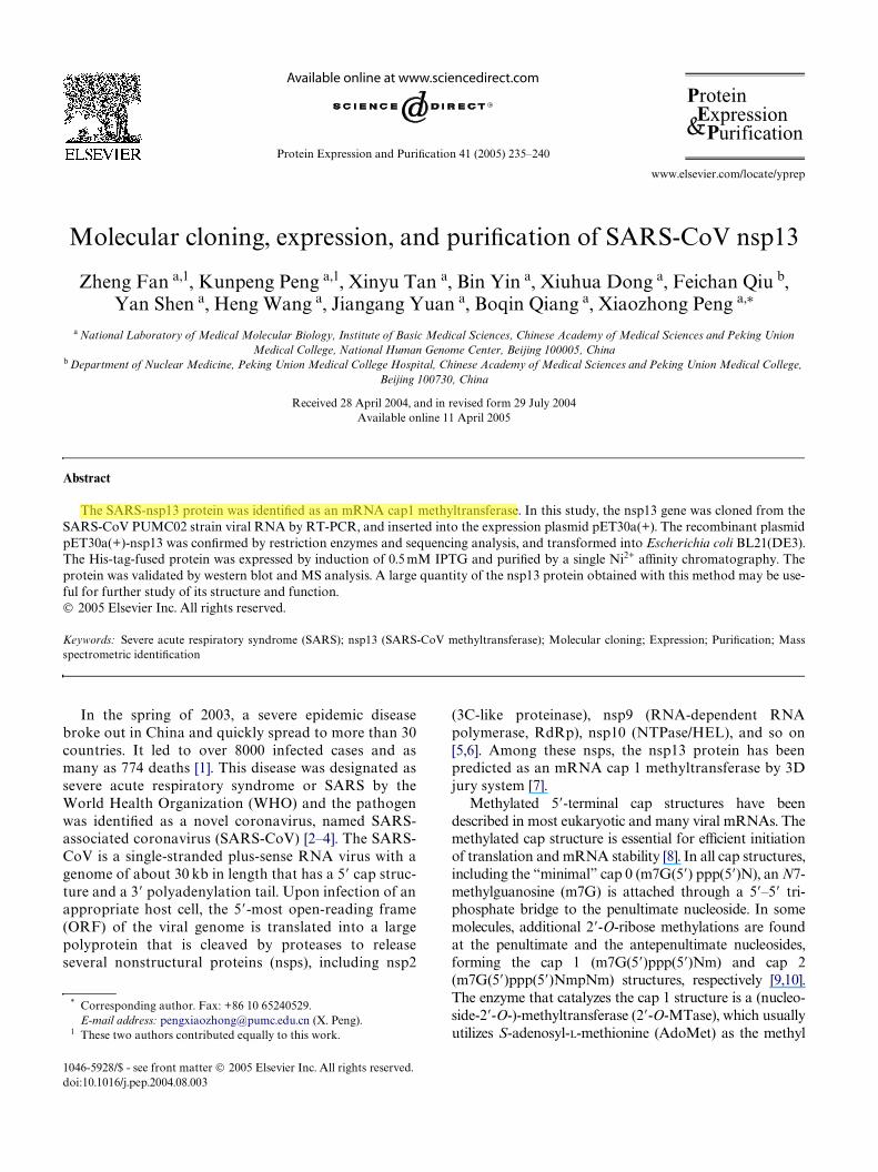
The SARS-nsp13 gene was ampliWed by RT-PCR(Fig. 1A), the ampliWed product was about 900 bp,
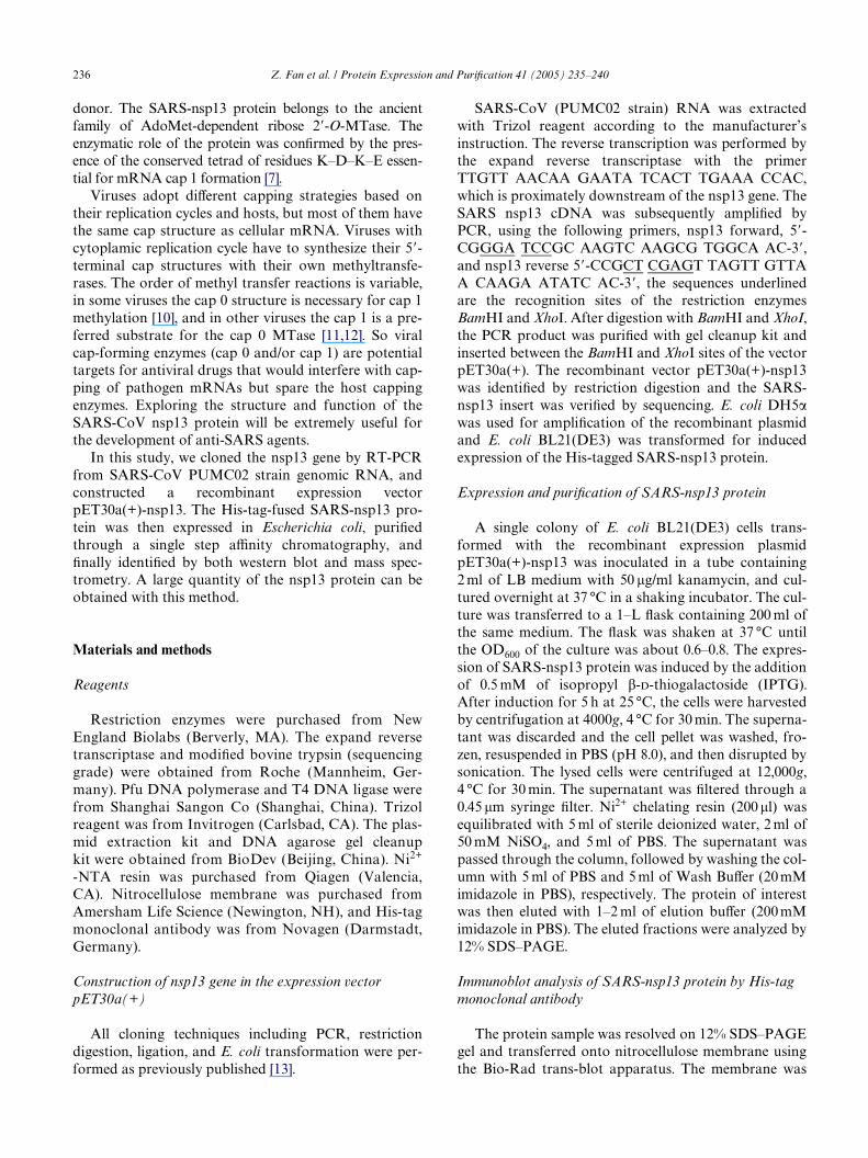
which was accordant to the theoretical length of nsp13gene. The recombinant plasmid was digested by therestriction enzymes BamHI and XhoI. Agarose gel elec-trophoresis of the digest revealed a DNA band atabout 900 bp (Fig. 1B). The inserted fragment was veri-Wed by sequencing, the result was shown in Fig. 2,which was identical to the published SARS-nsp13 genesequence in NCBI and in the correct reading framewith the His-tag in the vector.
Expression and puriWcation of SARS-nsp13 protein
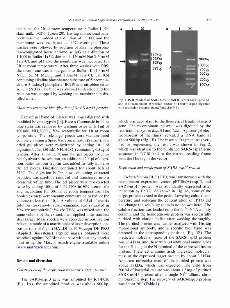
Escherichia coli BL21(DE3) was transformed with therecombinant expression vector pET30a(+)-nsp13, andSARS-nsp13 protein was abundantly expressed afterinduction by IPTG . As shown in Fig. 3A, some of thetarget protein existed in the pellet. Lowering culture tem-perature and reducing the concentration of IPTG didnot change the solubility (data is not shown here). Thesoluble fraction was loaded onto the Ni2+ -NTA aYnitycolumn, and the homogeneous protein was successfullypuriWed with elution buVer after washing thoroughly.The puriWed protein was further analyzed with His-tagmonoclonal antibody, and a speciWc blot band wasdetected at the corresponding position (Fig. 3B). Thepredicted molecular mass of the SARS-nsp13 proteinwas 33.4 kDa, and there were 30 additional amino acidsfor the His-tag in the N-terminal of the expressed fusionprotein. These extra amino acids increased molecularmass of the expressed target protein by about 3.3 kDa.Apparent molecular mass of the puriWed protein wasabout 37 kDa, which was expected. The yield from200 ml of bacterial culture was about 1.3 mg of puriWedSARS-nsp13 protein after a single Ni2+ aYnity chro-matographic step. The recovery of SARS-nsp13 proteinwas about 26% (Table 1).
Fig. 1. PCR product of SARS-CoV PUMC02 strain-nsp13 gene (A)and the recombinant expression vector pET30a(+)-nsp13 digestionwith restriction enzymes BamHI and XhoI (B).
238 Z. Fan et al. / Protein Expression and PuriWcation 41 (2005) 235–240
Table 1
MALDI-ToF MS identiWcation of SARS-nsp13 protein
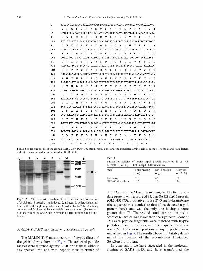
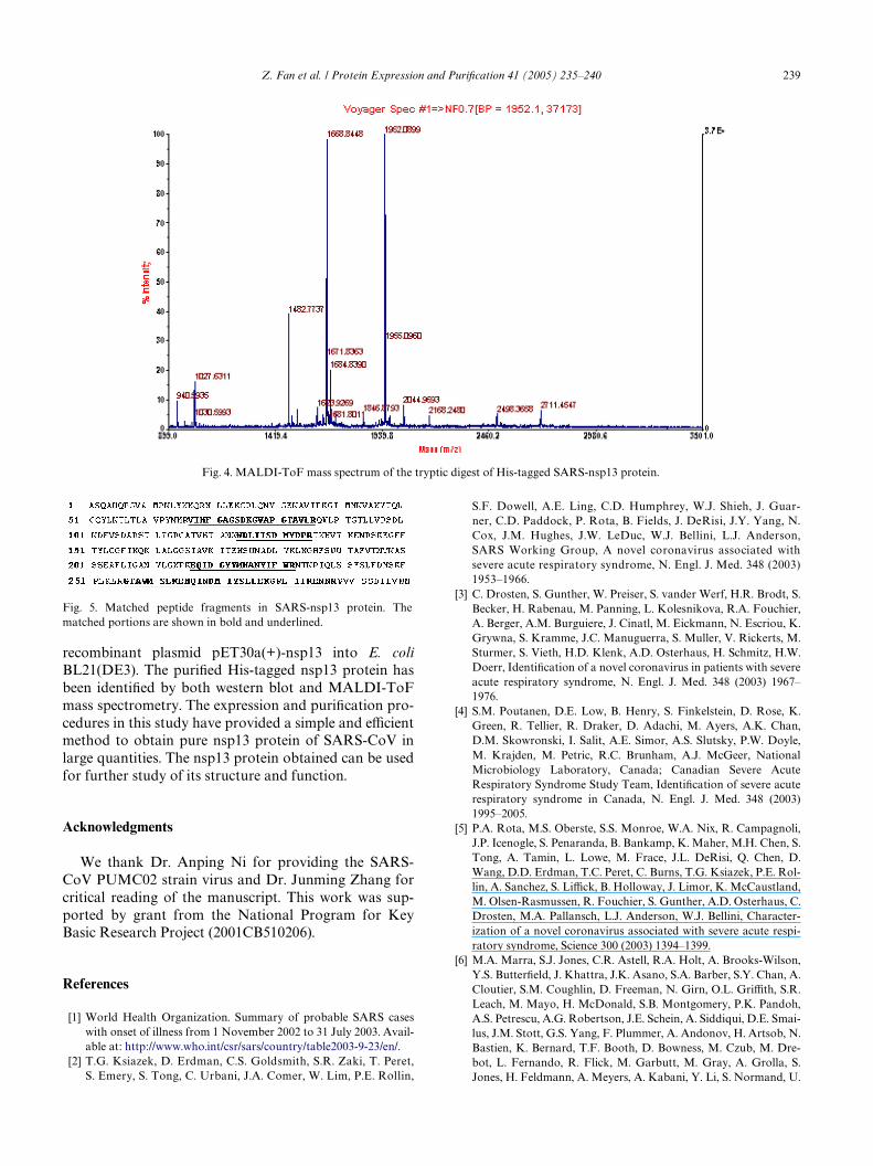
The MALDI-ToF mass spectrum of tryptic digest ofthe gel band was shown in Fig. 4. The achieved peptidemasses were searched against NCBInr database withoutany species limit and with peptide mass tolerance of
Fig. 3. (A) 12% SDS–PAGE analysis of the expression and puriWcationof SARS-nsp13 protein. 1, uninduced; 2, induced; 3, pellet; 4, superna-tant; 5, Xow-through; 6, puriWed nsp13 protein by Ni2+-NTA aYnitycolumn; and M, Low molecular weight protein marker. (B) Westernblot analysis of the SARS-nsp13 protein by His-tag monoclonal anti-body.
§0.1 Da using the Mascot search engine. The Wrst candi-date protein, with a score of 94, was SARS nsp16 protein(GI:30133975), a putative ribose 2�-O-methyltransferase(the sequence was identical to that of the detected nsp13protein here), and was the only one having a scoregreater than 75. The second candidate protein had ascore of 67, which was lower than the signiWcant score of75. Seven peptide fragments were matched with trypticpeptides of nsp13 protein, and the sequence coveragewas 26%. The covered portions in nsp13 protein wereunderlined in Fig. 5. The results above indubitably deter-mined the identity of the recombinant His-taggedSARS-nsp13 protein.
In conclusion, we have succeeded in the molecularcloning of SARS-nsp13, and have transformed the
PuriWcation scheme of SARS-nsp13 protein expressed in E. coliBL21(DE3) with pET30a(+)-nsp13 (200 ml culture)
Step Total protein (mg)
nsp13 protein(mg)
Recovery nsp13 (%)
Extraction 67.8 4.9 100Ni2+-aYnity column 1.3 1.3 26
Fig. 2. Sequencing result of the cloned SARS-CoV PUMC02 strain-nsp13 gene and the translated amino acid sequence. The bold and italic lettersindicate the conserved tetrad of residues K–D–K–E.
Z. Fan et al. / Protein Expression and PuriWcation 41 (2005) 235–240 239
S.F. Dowell, A.E. Ling, C.D. Humphrey, W.J. Shieh, J. Guar-
recombinant plasmid pET30a(+)-nsp13 into E. coliBL21(DE3). The puriWed His-tagged nsp13 protein hasbeen identiWed by both western blot and MALDI-ToFmass spectrometry. The expression and puriWcation pro-cedures in this study have provided a simple and eYcientmethod to obtain pure nsp13 protein of SARS-CoV inlarge quantities. The nsp13 protein obtained can be usedfor further study of its structure and function.
Acknowledgments
We thank Dr. Anping Ni for providing the SARS-CoV PUMC02 strain virus and Dr. Junming Zhang forcritical reading of the manuscript. This work was sup-ported by grant from the National Program for KeyBasic Research Project (2001CB510206).
References
[1] World Health Organization. Summary of probable SARS caseswith onset of illness from 1 November 2002 to 31 July 2003. Avail-able at: http://www.who.int/csr/sars/country/table2003-9-23/en/.
[2] T.G. Ksiazek, D. Erdman, C.S. Goldsmith, S.R. Zaki, T. Peret,S. Emery, S. Tong, C. Urbani, J.A. Comer, W. Lim, P.E. Rollin,
Fig. 5. Matched peptide fragments in SARS-nsp13 protein. Thematched portions are shown in bold and underlined.
ner, C.D. Paddock, P. Rota, B. Fields, J. DeRisi, J.Y. Yang, N.Cox, J.M. Hughes, J.W. LeDuc, W.J. Bellini, L.J. Anderson,SARS Working Group, A novel coronavirus associated withsevere acute respiratory syndrome, N. Engl. J. Med. 348 (2003)1953–1966.
[3] C. Drosten, S. Gunther, W. Preiser, S. vander Werf, H.R. Brodt, S.Becker, H. Rabenau, M. Panning, L. Kolesnikova, R.A. Fouchier,A. Berger, A.M. Burguiere, J. Cinatl, M. Eickmann, N. Escriou, K.Grywna, S. Kramme, J.C. Manuguerra, S. Muller, V. Rickerts, M.Sturmer, S. Vieth, H.D. Klenk, A.D. Osterhaus, H. Schmitz, H.W.Doerr, IdentiWcation of a novel coronavirus in patients with severeacute respiratory syndrome, N. Engl. J. Med. 348 (2003) 1967–1976.
[4] S.M. Poutanen, D.E. Low, B. Henry, S. Finkelstein, D. Rose, K.Green, R. Tellier, R. Draker, D. Adachi, M. Ayers, A.K. Chan,D.M. Skowronski, I. Salit, A.E. Simor, A.S. Slutsky, P.W. Doyle,M. Krajden, M. Petric, R.C. Brunham, A.J. McGeer, NationalMicrobiology Laboratory, Canada; Canadian Severe AcuteRespiratory Syndrome Study Team, IdentiWcation of severe acuterespiratory syndrome in Canada, N. Engl. J. Med. 348 (2003)1995–2005.
[5] P.A. Rota, M.S. Oberste, S.S. Monroe, W.A. Nix, R. Campagnoli,J.P. Icenogle, S. Penaranda, B. Bankamp, K. Maher, M.H. Chen, S.Tong, A. Tamin, L. Lowe, M. Frace, J.L. DeRisi, Q. Chen, D.Wang, D.D. Erdman, T.C. Peret, C. Burns, T.G. Ksiazek, P.E. Rol-lin, A. Sanchez, S. LiYck, B. Holloway, J. Limor, K. McCaustland,M. Olsen-Rasmussen, R. Fouchier, S. Gunther, A.D. Osterhaus, C.Drosten, M.A. Pallansch, L.J. Anderson, W.J. Bellini, Character-ization of a novel coronavirus associated with severe acute respi-ratory syndrome, Science 300 (2003) 1394–1399.
[6] M.A. Marra, S.J. Jones, C.R. Astell, R.A. Holt, A. Brooks-Wilson,Y.S. ButterWeld, J. Khattra, J.K. Asano, S.A. Barber, S.Y. Chan, A.Cloutier, S.M. Coughlin, D. Freeman, N. Girn, O.L. GriYth, S.R.Leach, M. Mayo, H. McDonald, S.B. Montgomery, P.K. Pandoh,A.S. Petrescu, A.G. Robertson, J.E. Schein, A. Siddiqui, D.E. Smai-lus, J.M. Stott, G.S. Yang, F. Plummer, A. Andonov, H. Artsob, N.Bastien, K. Bernard, T.F. Booth, D. Bowness, M. Czub, M. Dre-bot, L. Fernando, R. Flick, M. Garbutt, M. Gray, A. Grolla, S.Jones, H. Feldmann, A. Meyers, A. Kabani, Y. Li, S. Normand, U.
Fig. 4. MALDI-ToF mass spectrum of the tryptic digest of His-tagged SARS-nsp13 protein.
240 Z. Fan et al. / Protein Expression and PuriWcation 41 (2005) 235–240
Stroher, G.A. Tipples, S. Tyler, R. Vogrig, D. Ward, B. Watson,R.C. Brunham, M. Krajden, M. Petric, D.M. Skowronski, C.Upton, R.L. Roper, The Genome sequence of the SARS-associ-ated coronavirus, Science 300 (2003) 1399–1404.
[7] M. von Grotthuss, L.S. Wyrwicz, L. Rychlewski, mRNA cap-1methyltransferase in the SARS genome, Cell 113 (2003) 701–702.
[8] S. Shuman, Structure, mechanism, and evolution of the mRNA cap-ping apparatus, Prog. Nucleic Acid Res. Mol. Biol. 66 (2001) 1–40.
[9] M. Bisaillon, G. Lemay, Viral and cellular enzymes involved insynthesis of mRNA cap structure, Virology 236 (1997) 1–7.
[10] Y. Furuichi, A.J. Shatkin, Viral and cellular mRNA capping: pastand prospects, Adv. Virus Res. 55 (2000) 135–184.
[11] D. Testa, A.K. Banerjee, Two methyltransferase activities in thepuriWed virions of vesicular stomatitis virus, J. Virol. 24 (1977)786–793.
[12] D.C. Hammond, J.A. Lesnaw, Functional analysis of hypomethy-lation variants of the New Jersey serotype of vesicular stomatitisvirus, Virology 160 (1987) 330–335.
[13] J. Sambrook, D.W. Russell, Molecular Cloning: A LaboratoryManual, third ed., Cold Spring Harbor Laboratory Press, ColdSpring Harbor, NY, 2001.
[14] A. Shevchenko, M. Wilm, O. Vorm, M. Mann, Mass spectrometricsequencing of proteins silver-stained polyacrylamide gels, Anal.Chem. 68 (1996) 850–858.


