Embed Size (px)
Citation preview
REVIEW
Molecular mechanisms underlying the regulation andfunctional plasticity of FOXP3þ regulatory T cells
Y Gao1, F Lin1, J Su1,2, Z Gao1,3, Y Li1, J Yang1, Z Deng1, B Liu2, A Tsun1 and B Li1
1Unit of Molecular Immunology, Key Laboratory of Molecular Virology and Immunology, Institut Pasteur of Shanghai, ShanghaiInstitutes for Biological Sciences, Chinese Academy of Sciences, Shanghai, China; 2Department of Surgery, Shanghai Public HealthCenter, Fudan University, Shanghai, China and 3School of Life Sciences, Fudan University, Shanghai, China
CD4þ CD25þ regulatory T (Treg) cells engage in the maintenance of immunological self-tolerance and homeostasis by limitingaberrant or excessive inflammation. The transcription factor forkhead box P3 (FOXP3) is critical for the development andfunction of Treg cells. The differentiation of the Treg cell lineage is not terminal, as developmental and functional plasticityoccur through the sensing of inflammatory signals in the periphery. Here, we review the recent progress in our understanding ofthe molecular mechanisms underlying the regulation and functional plasticity of CD4þ CD25þ FOXP3þ Treg cells, through theperturbation of FOXP3 and its complex at a transcriptional, translational and post-translational level.Genes and Immunity advance online publication, 3 November 2011; doi:10.1038/gene.2011.77
Keywords: Treg; FOXP3; miRNA; T cell plasticity; instability
Forkhead box P3þ (FOXP3 in human, Foxp3 in mice;hereafter referred to as FOXP3 unless specified) regula-tory T (Treg) cells have an important role during immunehomeostasis through the maintenance of immune toler-ance and prevention of inflammatory disease.1 Thetranscription factor FOXP3 is essential for the develop-ment and function of Treg cells.2 The loss of expressionand mutations of FOXP3 lead to the development ofchronic autoimmunity and is presented as the scurfyphenotype in mice3 and X-linked autoimmunity–allergicdysregulation and immunodysregulation, polyendocri-nopathy and enteropathy, X-linked syndrome in hu-mans.4 Although Treg cells are essential for the toleranceof commensal microbiota in the gut,5 an excessive Tregcell response may facilitate tumor growth and chronicinfection by limiting anti-tumor or anti-pathogenicimmune responses, respectively.6,7 Thus, Treg cell func-tion must be tightly controlled to heighten or dampeninflammation according to the desired response. Recentstudies have also identified FOXP3 expression inepithelial cells, multipotent mesenchymal stromal cells,invariant natural killer T cells and macrophages,8–10 butCD4þ CD25þ T lymphocytes remain the most character-ized cell type.
CD4þ helper T (Th) cells are classified into severalsubsets according to the expression of various lineage-
specific transcription factors and cytokines, such as T boxexpressed in T cells (T-bet) and interferon (IFN)-g in Th1cells; GATA-binding protein-3 (GATA-3) and interleukin(IL)-4 in Th2 cells; retinoic acid (RA) receptor-relatedorphan receptor-gt (RORgt) and IL-17 in Th17 cells; andFOXP3 and IL-10 in Treg cells.11 There are two majorsubtypes of Treg cells according to their differentiationorigin; natural Treg (nTreg) and induced Treg (iTreg) cellsdifferentiate and adopt FOXP3 expression in the thymusand periphery, respectively. Naı̈ve T cells differentiateinto iTreg cells in the presence of IL-2, transforminggrowth factor (TGF)-b and stimulation through the T-cellreceptor (TcR).12–15 TGF-b is also required for IL-6-dependent expression of the transcription factor RORgtand inhibition of FOXP3 expression during the genera-tion of Th17 cells from naı̈ve T cells.16–20 However, theexpression of FOXP3 and RORgt are not always mutuallyexclusive, as Foxp3þ T cells in the gut have been shownto co-express high levels of RORgt and concurrentlysecrete IL-17.21–23 Human CD4þ CD25� T cells can alsotransiently express FOXP3 after TcR stimulation withoutthe requirement of other extrinsic signals, but whether ornot they are suppressive during this period is still underdebate.24–30
The instability and plasticity of Treg cells
A number of recent studies have shown that Treg cellsharbor the propensity to reprogram into proinflamma-tory cells.31–39 For instance, Th1-like FOXP3þ Treg cellshave been identified in human subjects with multiplesclerosis.40 The evolutionary advantage of this processcould be explained during inflammation, whereby thesuppressive nature of Treg cells should be limitedfor protective and proinflammatory responses to ensue.
Received 27 July 2011; revised 22 September 2011; accepted 26September 2011
Correspondence: Dr A Tsun or Dr B Li, Unit of MolecularImmunology, Key Laboratory of Molecular Virology and Immunol-ogy, Institut Pasteur of Shanghai, Shanghai Institutes for BiologicalSciences, Chinese Academy of Sciences, 37 Jianguo Middle Road,Shanghai, 200025, China.E-mail: [email protected] or [email protected]
Genes and Immunity (2011), 1–13& 2011 Macmillan Publishers Limited All rights reserved 1466-4879/11
www.nature.com/gene
As Foxp3 expression is required for the maintenance ofTreg cell identity and function,41 recent investigationshave looked into the in vivo role of Foxp3 expressionfor the reprogramming of Treg cells.32,33 Rudensky andcolleagues32 used GFP–Cre knock-in mice driven off theFoxp3 promoter, crossed with R26-YFP mice to fluores-cently track all the cells that express or had previouslyexpressed Foxp3 in vivo. This system utilized a GFP–Cremutated estrogen receptor fusion protein so that theFoxp3 driven Cre could be induced for its translocationinto the nucleus after treatment with tamoxifen. Here,they found that Foxp3 expression in Treg cells was ratherstable (only B4% cells were YFPþ and GFP�) whenincubated under a long-term physiological setting inmice, with or without viral challenge. This observationshould interest those who are investigating the use of exvivo expanded nTreg and iTreg cells for the treatment ofautoimmune disease.42–44 These studies suggest that theuse of ex vivo expanded and infused Treg cells canprovide long-term protective effects in humans. How-ever, other studies have indicated that under a physio-logical setting Treg cells may not be terminallydifferentiated, and that the instability of Treg cellfunction is driven by the local cytokine milieu and theloss of Foxp3 expression in Treg cells.
In contrast to the above, Bluestone and collea-gues127,131–133 used a bacterial artificial chromosomeFoxp3–GFP–Cre system with R26-YFP, in which theyobserved the downregulation of Foxp3 expression in asignificant proportion of YFPþ T cells (B15% cells wereYFPþ and GFP�); these T cells exhibited an activated-memory T-cell phenotype, produced inflammatory cyto-kines and were dubbed ‘ex-Foxp3’ T cells. When these‘ex-Foxp3’ T cells were transferred into non-obesediabetic Rag2�/� mice, they rapidly developed diabetes.Moreover, only a small percentage of YFPþ cells (0.5%)were observed after the transfer of YFP� T cells intorecipient mice, which suggests that the ‘ex-Foxp3’ T-cellpopulation was largely derived from nTreg cells.33
Therefore, the depletion of Foxp3 in Treg cells may haveresulted in the conversion of Treg cells into autoaggres-sive lymphocytes (‘ex-Treg’ cells), a trait that was notobserved in the B4% of the ‘ex-Foxp3’ T cells in thestudy using tamoxifen-treated mice.32 It should be notedthat the loss of Foxp3 in this system could be attributedto the transient expressers of Foxp3 rather than thecommitted immunoregulators. In the bacterial artificialchromosomes system, Cre is driven from birth andinduces YFP expression in all the cells that haveexpressed Foxp3 at some point, whereas the tamoxifenstudy could have labeled a high Foxp3-expressing andmore stable proportion of the overall Foxp3þ population.This supports the notion that a proportion of Foxp3þ
Treg cells can convert into ‘ex-Treg’ cells due to theirnatural heterogeneity, as not all FOXP3þ Treg cells arefully committed to the Treg cell lineage.45 In any case, thepopulations that were monitored in these two reportswere intrinsically different, and may account for thedifferences in their findings. Another study that focusedon characterizing the progenitors of CD4þ follicular Bhelper T cells in the Peyer’s patches has demonstratedthat CD4þ Foxp3þ T cells can lose Foxp3 expression toconvert into CD4þ T-follicular helper cells because of thesurrounding environmental signals;36 the loss of Foxp3expression in Treg cells may also lead to the expression
of IL-4 to potentiate the differentiation of naı̈ve T cellsinto the Th2 lineage.46 Although the controversy sur-rounding the functional relevance of ‘ex-Foxp3’ or ‘ex-Treg’ cells still remains, it is clear that some degree ofFoxp3 depletion in Foxp3þ Treg cells can occur.
Foxp3þ Treg cells may lose Foxp3 expression afterthey are transferred into lymphopenic- or lymphocyte-null mice,34,35,45 and in vitro studies have shown that Tregcells can readily lose Foxp3 expression in culture.47,48
Although the use of rather artificial experimentalconditions may not be fully translatable to the physio-logical occurrence of these ‘ex-Treg’ cells, it is abundantlyclear that Foxp3þ Treg cells can lose the expression ofFoxp3, which in turn leads to the subsequent disruptionin their suppressive ability. IL-2 can induce the upregu-lation of FOXP3 expression in Treg cells,49 but what arethe signals that cause the initial loss of Foxp3 expressionduring the reprogramming of Treg cells? Proinflamma-tory cytokines, such as IL-1(ref. 50) and IL-6 (below), havebeen suggested to be the signals that are required forTreg cell conversion into non-suppressor cells, with IL-6as one center of focus because of its shared function acrossthe Treg–Th17 differentiation axis. An early study by Pasareand Medzhitov51 showed that IL-6 expression, inducedby Toll-like receptor stimulation, led to the loss of Tregcell suppressive function. It was shown more recentlythat IL-6 can convert Foxp3þ Tregs into Th17-like cells tosecrete IL-17.52 But does IL-6 affect both nTreg and iTregcells, and how does this relate to Foxp3 expression?Zheng et al.53 found that IL-6 signals affected Foxp3expression and function in nTreg but not iTreg cells dueto the downregulation of the IL-6 receptor (IL-6R) afterIL-2 and TGF-b treatment (used to convert naive T cellsinto iTreg cells). However, the treatment of nTreg cellswith IL-2 and TGF-b, but not IL-2 alone, also induces thedownregulation of the IL-6R in nTreg cells to renderthem insensitive to IL-6 stimulation. More recently, anactive derivation of vitamin A, all-trans RA, has alsobeen shown to protect nTreg cells from their conversioninto Th17 cells54,55 due to the downregulation of the IL-6R.55,56 However, Dong and colleagues57 found that iTregcells were susceptible to IL-6-induced Foxp3 depletion inTreg cells even after TGF-b treatment. It is unclear as towhy these two reports differ in their characterization ofhow iTreg cells convert into Th17 cells—it may be due tothe different systems used, but what seems clear is thatthe expression of IL-6R on Treg cells has a role in theirsensitivity to reprogramming via IL-6R signaling. Wehave recently developed a system to test the directsignals required for IL-6-mediated downregulation ofFOXP3 expression at the protein level. Here, we foundthat when FOXP3 is overexpressed in FOXP3� cells,FOXP3 could be directed for degradation via IL-6 signals(unpublished data). Thus, the mechanism of ‘ex-Foxp3’T-cell generation via proinflammatory cytokines, such asIL-6, may not only occur at the transcriptional level (seebelow) but also at the protein level.
The heterogeneity of Treg function could also beattributed to the transcriptional changes mediated bytranscription factors, found downstream of the signalsprovided by cytokines. Signal transducer and activator oftranscription 3 (Stat3) is a transcription factor requiredfor Th17 differentiation. Various reports have observedthat Treg cells can co-express Foxp3 and Th markers tocontribute toward proinflammatory31,58 and anti-inflam-
FOXP3þ regulatory T cell regulation and plasticityY Gao et al
2
Genes and Immunity
matory responses (see below). Phosphorylated Stat3interacts with Foxp3, and the expression of Stat3 in Tregcells is essential for the suppression of fatal colitis.59
Stat3-deficient Treg cells can inhibit T-cell proliferationin vitro, but fail to suppress Th17 cell differentiation andTh17-induced colitis;59 this mechanism requires intact IL-10 signaling in Treg cells.60 Thus, Stat3 expression in Tregcells is essential for Treg cell-mediated suppression ofTh17 cell-mediated inflammation, and this occursthrough the simultaneous expression of classical Tregand Th markers. Others have shown that IL-6-, IL-1- andIL-23-mediated negative regulation of Foxp3 expressionis Stat3 dependent.57,61 How Stat3 may also control‘ex-Treg’ cell conversion to produce IL-17, while alsofacilitating Th17-directed suppression, remains to beinvestigated; however, it may be possible that this switchfrom a regulatory to inflammatory cell type is attributedto the synergistic signals provided by other proinflam-matory cytokines.
The redifferentiation of Treg cells into Th-like cells, otherthan Th17 cells, has also been reported. In response to IFN-g, Foxp3þ Treg cells can upregulate the expression of T-bet, which promotes the expression of the chemokinereceptor CXCR3 and the accumulation of T-betþ Treg cellsat the sites of Th1 cell-mediated inflammation.62 Therefore,the expression of T-bet was found to be required for thehomeostasis and function of Treg cells during type 1inflammation.62 IFN regulatory factor 4 is a transcriptionfactor responsible for the expression of IL-4 in Th2 cells.The co-expression of Foxp3 and IFN regulatory factor 4 inTreg cells endows them with the ability to suppress Th2responses.63 The overlapping expression of T-bet and IFNregulatory factor 4 with Foxp3 may be necessary for therapid response of Treg cells to suppress excessiveinflammation by Th cells. Finally, Bcl6, a transcriptionfactor required for the generation of T-follicular helpercells and the expression of CXCR5, has been found to beresponsible for the generation of CXCR5þ Treg cells,which can inhibit germinal center reactions to regulatehumoral immunity.64,65 Considering the above, it wouldseem inappropriate that these reprogrammed cells benamed ‘ex-Treg’ cells, as their further differentiationdetermines the target for suppression, but this name maybe fitting for those Treg cells that have lost their ability tosuppress immune responses.
The expression or loss of expression of Foxp3 inFoxp3þ Treg cells may coincide with the expression ofTh-specific transcription factors or cytokines, but itremains unclear as to how these populations differ.A recent investigation has shed some light on thisdichotomy by demonstrating how Foxp3þ IFN-gþ T cellsretain regulatory functions, but are able to differen-tiate further into single positive IFN-gþ T cells;66 thismechanism could be attributed to the increased activityof Stat1, which was observed in miR-146a knockout Tregcells (see below).67 Therefore, there may be a threshold ofexpression and/or activation of Th-specific transcriptionfactors in Foxp3þ Treg cells that allow them to convertfrom a Th-specific suppressor to a contributor ofproinflammatory responses. However, the exact mechan-isms and co-stimulatory signals that are required for thisconversion have yet to be characterized. Moreover, itmay be possible that Foxp3þ T cells that co-express Thmarkers or are fully converted into Th cells may arisefrom specific pools of Treg cells that are more or less
committed toward the Treg lineage, respectively.45
Further research must be undertaken to resolve thecontribution of ‘ex-Foxp3’ or ‘ex-Treg’ cells as physiolo-gical regulators of immune homeostasis. The animalstrain, type/magnitude of stimuli, genetic system usedto track Foxp3 expression and tissue localization may allhave a role in determining whether Treg cells reprogramin such a manner and the functional phenotype intowhich these Foxp3þ Treg cells convert.
The remainder of this review aims to summarize whatis known about the mechanisms that control FOXP3expression and function in the context of Treg cellplasticity and instability, by describing how FOXP3, andits complex, are regulated at a genomic, transcriptionaland post-translational level (Figure 1). How is FOXP3expression controlled downstream of proinflammatorysignals? Do any posttranslational modifications ofFOXP3 affect FOXP3 or Treg cell stability and function?Understanding these mechanisms in the context ofFOXP3-dependent Treg cell plasticity and instabilityallows for the direct targeting of the molecules at theheart of immune tolerance.
Epigenetic and transcriptional regulationof FOXP3
The FOXP3 gene is comprised of 11 exons, and itspromoter is located �6221 bp upstream of the translationstart site. The 50-untranslated region is interrupted by a6000-bp intron, which contains a splice donor site at the50-end and splice acceptor site at the 30-end, 22 bpupstream of the translation start site. The FOXP3 genepromoter is highly conserved between humans, mice andrats.68 Mantel et al.68 used a reporter assay to analyze thehuman FOXP3 functional promoter and found that theactivation responsive element of the FOXP3 promoter islocated within the �511 to �307 region. The basalpromoter contains six nuclear factors of activated T cells(NFAT) and activator protein-1 (AP-1) binding sites,which positively regulates the transactivation of theFOXP3 promoter after the triggering of the TcR.68 Theregulatory regions of Foxp3 also contains three conservednoncoding DNA sequence elements (CNS1, 2 and 3),69
also known as enhancers.70,71 Using a mouse knockoutstrategy deficient in these CNS regions, it was found thatCNS1 controls peripheral, but not thymic, induction ofFoxp3 expression; CNS2 controls the heritable mainte-nance of Foxp3 expression; and c-Rel binds to CNS3 andfacilitates Foxp3 induction during thymic and peripheralTreg cell differentiation.69
The epigenetic state of the Foxp3 locus determines thetranscriptional activity of the Foxp3 gene. Huehn andcolleagues72,73 found that an enhancer region (CNS2) ofthe Foxp3 gene contained CpG motifs (also called the‘Treg-specific demethylation region’ (TSDR)), which werefully demethylated in nTreg cells, partially demethy-lated in TGF-b-polarized iTreg cells and methylatedin naı̈ve and Th cells. These findings suggest that theepigenetic modification of the Foxp3 gene is critical forthe stable expression and suppressive function of Tregcells. These observations have also been confirmed incord blood Treg cells74 and peripheral Treg cells75 inhumans, where the latter transient expressers of FOXP3bear a partially methylated TSDR.75 Consistent with this
FOXP3þ regulatory T cell regulation and plasticityY Gao et al
3
Genes and Immunity
notion, a TcR response element was found in the firstintron of the Foxp3 gene that is located within this CNSregion76 and also Est-1 binding sites, which are importantfor positively regulating Foxp3 transcription.77,78 TheTSDR also overlaps with the binding site for thetranscription factor cyclic AMP (cAMP) response ele-ment-binding, where an increase in TSDR methylationnegatively correlates with cAMP response element-binding and Foxp3 expression.70 Moreover, the treatmentof Treg cells using 50-azacytidine, a DNA methyltransfer-ase inhibitor, leads to the rapid passive demethylation ofDNA in this region to increase Foxp3 expression, evenin the absence of TGF-b.70 Naı̈ve T cells that are treatedwith DNA methyltransferase inhibitors and TGF-b canconvert into Treg cells, with high Foxp3 expressionand stable suppressive function.79 Conversely, IL-6 canreduce Foxp3 expression and increase TSDR methylationin nTreg cells.79 More recently, the SUMO E3 ligase PIAS1has been shown to recruit DNA methylases andheterochromatin protein 1 to reduce Foxp3 promoteraccessibility.80 The elucidation of how Treg cell instabilityrelates to the known properties of epigenetic regulationof the Foxp3 locus could therefore provide importantinsights into the reversibility of ‘ex-Foxp3’ T cells.Additionally, any future therapies involving the use ofthe ex vivo expanded and infused Treg cells in hu-mans42,43 may require further testing of their epigeneticstate to ensure that the most stable Treg cells are utilized.
The binding of transcription factors to the promoterregions of Foxp3 serve to augment or inhibit thetranscription of this gene. Even though the epigeneticmodifications detailed above offer us an indication of thebasal stability and conformation of the Foxp3 gene, the
transient changes in expression and activity of transcrip-tion factors ultimately determine the outcome of genetranscription. Smad3 and NFAT can activate a Foxp3enhancer (CNS1).71 The binding of both of these factorsincreases the acetylation of histones (such as histone 4) inboth nTreg and iTreg cells stimulated by TGF-b plus TcRactivation.71 More recently, Smad3 phosphorylation hasbeen shown to be inhibited by S1P1 signaling via theactivation of the mTOR pathway to inhibit Treg celldifferentiation and function.81,82 The inhibition of themTOR pathway is now commonly used to selectivelyexpand Treg cells over Teff cells;42,43 thus, this model ofmTOR and Smad3 cross-talk can explain at least oneaspect of how they cooperate to affect Treg cell function.Another molecule that upregulates Treg cell function isRA; RA induces the binding of the RA receptor andretinoid X receptor to a site at CNS1, which leads to localhistone acetylation at the Smad3 binding site. Therefore,RA augments the TGF-b-dependent pathway and Tregcell induction by increasing the accessibility of Smad3 tothe Foxp3 locus.83 More recently, the inhibitor of DNAbinding 3 was identified as a regulator of TGF-b-dependent Foxp3 expression. Inhibitor of DNA binding 3can relieve the inhibition of Foxp3 expression by reducingthe binding of the transcription factor GATA-3 (seebelow) to the Foxp3 promoter, to allow for the enrichmentand increased binding of transcription factor 3 (E2A).Furthermore, it was shown that inhibitor of DNAbinding 3�/� T cells were more prone to differen-tiate into Th17 cells but not into Foxp3þ Treg cells.84
TGF-b signaling may also interact with another setof transcription factors, FOXO1/FOXO3a, to positivelyregulate Foxp3 expression by binding to its promoter,85–87
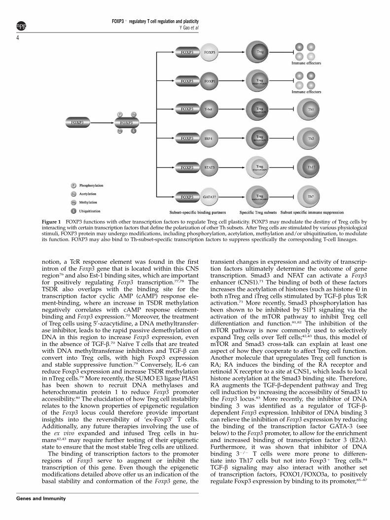
Figure 1 FOXP3 functions with other transcription factors to regulate Treg cell plasticity. FOXP3 may modulate the destiny of Treg cells byinteracting with certain transcription factors that define the polarization of other Th subsets. After Treg cells are stimulated by various physiologicalstimuli, FOXP3 protein may undergo modifications, including phosphorylation, acetylation, methylation and/or ubiquitination, to modulateits function. FOXP3 may also bind to Th-subset-specific transcription factors to suppress specifically the corresponding T-cell lineages.
FOXP3þ regulatory T cell regulation and plasticityY Gao et al
4
Genes and Immunity
but the exact pathways by which they meet remain tobe characterized. Itch, an E3 ubiquitin ligase, has beenfound to ubiquitinate the transcription factor TGF-b-inducible early gene 1 in a TGF-b-dependent and non-proteolytic-dependent pathway at the Foxp3 promoter.88
These reports support the role of TGF-b as an importantpositive regulator of Foxp3 expression and shows howthis pathway signals down to the gene level in Treg cells.
GATA-3 is a crucial transcription factor for thedevelopment and function of Th2 cells. FOXP3 expres-sion cannot be induced in mature Th2 cells and IL-4-producing T cells by TcR stimulation and TGF-btreatment.89 The block in Foxp3 expression may be dueto Stat6-mediated repression of Foxp3 transcription at itspromoter region.90 Luciferase reporter assays havesupported the finding that GATA-3 can bind to thepromoter of Foxp3, in order to mediate its transcriptionalinhibition, and thus inhibit the differentiation of GATA-3þ T cells into iTreg cells.89 However, in nTreg cells, thelow expression of Foxp3 seems to account for a degree ofGATA-3 upregulation by some unclear intrinsic mechan-ism that favors nTreg-to-Th2 conversion.46 A recentstudy by the same group analyzed the effect of GATA-3 depletion in Treg cells and found that these micedeveloped inflammatory disorder.91 Here, GATA-3-defi-cient Treg cells expressed reduced amounts of Foxp3 andwere enhanced in the ability to produce inflammatorycytokines. GATA-3 was also found to interact with theCNS2 region of the Foxp3 gene to promote its expression.Therefore, the role of GATA-3 to inhibit or increase Foxp3expression could be determined by its interaction withTh2- or Treg cell-specific molecules, and seem to differbetween iTreg and nTreg cells.
Runt-related transcription factor 1 (RUNX1)/acutemyeloid leukemia 1 and RUNX3/acute myeloid leuke-mia 2 are also specifically involved in the process of T-cell lineage commitment.92 The mRNA of RUNX1 andRUNX3, as well as FOXP3, can be upregulated by thestimulation of naı̈ve T cells using anti-CD3/CD28antibodies plus TGF-b treatment.93 RUNX1 and RUNX3can bind to the Foxp3 promoter in the presence of TGF-bto enhance the expression of Foxp3. The inactivation ofthe gene encoding for RUNX cofactor–core-bindingfactor-b in mice and the knockdown of RUNX1 andRUNX3 in human T cells reduces the expression ofFOXP3 and the suppressive function of Treg cells.Another report also found that the RUNX–core-bindingfactor-b complexes can control the expression of Foxp3,and the evidence suggests that this complex acts at theCNS2 region.69,94 Thus, RUNX transcription factors arerequired for positively regulating FOXP3 expression andthe function of Treg cells.93
The nuclear factor (NF-kB) signaling pathway is a keyregulator of Foxp3 expression;69,95,96 c-Rel, an NF-kBfamily member, can bind to the Foxp3 enhancer region(CNS3) and control the development of Treg cells bypromoting the formation of a Foxp3-specific enhanceo-some. c-Rel-deficient mice have up to a 90% reduction ofTreg cells compared with wild-type mice, and arecompromised in Treg cell differentiation.69,95,96 Morerecent studies have shown how the cooperative expres-sion of c-Rel and JunB significantly enhances Foxp3promoter activity,97 and the role of RelA has also beenobserved to be important for iTreg cell differentiation in aCD28 signal-dependent manner.98
IL-2 and its receptor IL-2R are crucial for the expansionand survival of Foxp3þ Treg cells in vivo.99–101 Whenmice are deficient in the IL-2R-b gene, the number of Tregcells reduces remarkably, indicating that the signalingpathway downstream of IL-2 affects the differentiation ofTreg cells.99 Additionally, limiting the availability of IL-2to Treg cells increases their propensity to convert intoproinflammatory cells via the signals provided byinflammatory cytokines such as IL-12.31 IL-2 can activateStat5, which binds to the promoter of the Foxp3 gene topromote Treg cell differentiation by regulating theexpression of Foxp3.102,103 Conversely, IL-6 stimulationleads to the binding of Stat3 to the CNS2 region to inhibittranscription, thus controlling Treg/Th17 polariza-tion.49,104 The combination of IL-6 and TGF-b alsoinduces the expression of the Th17 transcription factorRORgt, which can bind to the Foxp3 promoter to inhibitits transcription.105
Other pathways which are involved in the directregulation of the Foxp3 gene include the aryl hydro-carbon receptor, which can induce Foxp3 expression inTreg cells by binding to the conserved aryl hydrocarbonreceptor-binding sites in the Foxp3 promoter.106 Thenotch pathway has also been implicated in negativelycontrolling Foxp3 expression107 through Hes1, which caninteract with the promoter region of the Foxp3 gene108
and also by signaling through the protein kinase C andcanonical NF-kB pathways.109 Finally, Bcl11b has alsobeen shown to increase the suppressive nature of Tregcells, and was found to regulate the genes that encodeFoxp3 and IL-10.110
Histone modifications allow for the control of geneaccessibility and may also regulate the transcription ofFoxp3. The trimethylation of histone H3 lysine 4(H3K4me3) is a permissive mark that facilitates thetranscription of target genes, whereas the trimethylationof histone H3 lysine 27 (H3K27me3) inactivates them.Wei et al.111 generated genome-wide H3K4me3 andH3K27me3 maps in naive, Th1, Th2, Th17, iTreg andnTreg cells, and found that the plasticity of Th cells wasmore flexible than that previously envisioned. Forexample, H3K27me3 was detected at the Il4 gene innaı̈ve, Th1 and Th17 cells, whereas nTreg cells had littleor no repressive marks in the Il4 gene. This indicates thatTreg cells have a higher propensity for their inductioninto IL-4-secreting cells and, as mentioned above, havethe increased plasticity to convert into Th2 cells.46,111
More importantly, the authors found no evidence ofsignificant H3K27me3 marks in association with theFoxp3 promoter in all of the tested T-cell populations,which suggests that Foxp3 may be expressed morewidely and transiently than previously thought.
As the current evidence shows, there are manytranscription factors that can function positively ornegatively at the Foxp3 gene to regulate Foxp3 expres-sion. However, an extra level of regulation is exertedupon the Foxp3 gene through epigenetic means at theTSDR. Similar to Foxp3, transcription factors may confereither upregulatory or inhibitory effects on gene tran-scription dependent on the timing of their recruitmentand the accumulation of other transcription modulators.In this sense, there is still a long way before we will fullyunderstand the kinetics of how these transcriptionfactors interact in vivo as multiple signals are receivedby Treg cells at any given time. The expression and
FOXP3þ regulatory T cell regulation and plasticityY Gao et al
5
Genes and Immunity
degree of activation of Th-specific lineage markers mayalso determine whether Treg cells further differentiateinto lineage-specific suppressor cells or reprogram intoproinflammatory/non-suppressive ‘ex-Treg’ cells.
Regulation of microRNA expression inFOXP3þ Treg cells
The discovery and recent drive for the identification ofmicroRNAs (miRNAs)112–117 that are involved in geneexpression regulation has added a whole new dimensionto our understanding of biological regulatory systems.miRNAs are small (B22 nucleotides) noncoding RNAmolecules that can target partially complementarysequences primarily at the 30-untranslated region ofmRNAs, leading to their degradation or the preventionof translation.118,119 This ultimately results in the down-regulation of protein expression. So far, more than 700miRNAs have been identified in the human genome,whereby each miRNA has the ability to target anddownregulate multiple mRNAs to create an overwhelm-ing complex gene regulatory network.118,119
Unsurprisingly, miRNAs were found to be expressedin the hematopoietic system,120 where they have beenshown to have a broad role in regulating immunity—extending to both the innate and adaptive arms of theimmune system.121–124 In lymphocytes, miRNAs havebeen shown to determine their differentiation andfunction.125 In this respect, the disruption of the RNaseIIIDicer—an endonuclease that facilitates miRNA matura-tion—in T and B cells causes a dramatic reduction in thenumbers of thymocytes due to the increase in cellapoptosis.126–129 However, although Dicer is involved inthe maturation of thymocytes, it does not affect CD4/CD8 lineage commitment.126
The disruption of Dicer specifically in the CD4 lineageusing CD4–Cre Dicerfl/fl mice results in normal thymo-cyte numbers, a general reduction in T-cell numbers inthe periphery, the induction of a Th1 bias127,130 and asubstantial reduction of Treg cell numbers during theirdifferentiation in the thymus.127 IL-2 production isalso highly dampened.130 These peripheral conventionalT cells are less able to express Foxp3 during in vitrostimulation in the presence of TGF-b, and the instabilityof Treg cells in this system leads to a late onset ofcolitis.127 A more recent investigation into the disrup-tion of Drosha (another RNAseIII enzyme related tomiRNA biogenesis) in the CD4 T cell population hasrevealed a similar phenotype to the Dicer modeldescribed above.131
Subsequent studies that utilized Foxp3–Cre mice toeliminate miRNA specifically in Treg cells via Dicer131–133
and Drosha131 have shown no disruption in theirdevelopment but a reduction in their suppressive ability,as indicated by the presentation of fatal early-onsetlymphoproliferative autoimmune syndrome.131–133 More-over, these mice display a similar phenotype to those thatlack Foxp3 and/or are depleted of Treg cells. A milderand later onset of disease progression seen in the CD4–Cre-based disruption of miRNA127,130 may be attributedto the concurrent disruption of T-effector cell function. Inthe periphery, Rudensky and colleagues132 observed areduced number of Treg cells under non-inflammatoryconditions; however, Treg cells could be activated to
proliferate under inflammatory conditions but to theloss of suppressive capacity by the downregulation ofTreg cell-specific effector molecules, such as cytotoxicT-lymphocyte antigen-4 (CTLA-4) and IL-10. Bluestoneand colleagues127,131–133 found that peripheral Treg cellsdeficient in miRNA may adopt Th1-, Th2- and Treg celllike effector profiles, with a reduction of Foxp3 levels inTreg cells to confer this instability. These studies show adefined requirement of miRNAs for the differentiationand suppressive function of Treg cells, with thepossibility that miRNA expression profiles may beindicative of Treg cell instability.
The Treg miRNA signature has been explored inmice127 and humans134 to reveal Treg-specific miRNAcandidates for further functional analysis. In human Tregcells, miR-31 has been shown to bind to the 30-untranslated region of FOXP3 mRNA for its down-regulation, and miR-21 can positively—albeit indir-ectly—regulate FOXP3 expression.134 The miR-31-lowand miR-21-high signatures have also been shown invalproate-treated T cells in which the inhibition ofhistone deacetylases can induce FOXP3 expressionthrough the induction of the transcription factor Ets-1;77,135 however, this miRNA signature was shown not tooccur as a consequence of FOXP3 expression.135 T-cellresponses in miR-155-deficient mice are biased towardTh2 differentiation but not Th17 and Th1 effectorresponses,124,136,137 and are protected against EAE, thusreflecting a role for miR-155 in the immune system.Rudensky and colleagues132 have recently shown thatmiR-155-deficient Treg cells have impaired proliferation,and along with other independent investigators foundmiR-155 to be highly expressed in Foxp3þ T cells andthat Foxp3 can directly upregulate the expression of miR-155.127,138–140 Vigorito and colleagues141 have also inves-tigated the role of miR-155 in Treg cells and found thatmice deficient in miR-155 have reduced numbers of Tregcells in the spleen and thymus, but their suppressivecapacity, the expression of Treg markers and peripheralsurvival rate remained intact. Suppressor of cytokinesignaling-1 protein expression is regulated by miR-155,which determines Treg cell responsiveness to IL-2signaling.140 The repression of suppressor of cytokinesignaling-1 allows for Treg cell competitiveness in envi-ronments that are limited in the availability of IL-2.140
A recent study using a Treg cell line HOZOT identi-fied FOXO3a as a target for miR-155.142 As Foxp3 isproposed to upregulate miR-155 expression by inducingits resident gene, and FOXO3a is a positive regulator ofFoxp3 expression, this could provide a negative feedbackof Foxp3 expression through miRNAs. Other studieshave found that miR-142-3p can regulate adenyl cyclase9 mRNA and the subsequent production of cAMP inTreg cells.143 cAMP is a determinant of Treg cellsuppressor function.144,145 Foxp3 mediates the down-regulation of miR-142-3p expression, which has yet to becharacterized as a direct or indirect process, to disruptthe maintenance of cAMP production.143 Lu et al.67
recently identified that mir-146a expression is crucialfor Treg cell function, where miR-146a-mediated down-regulation of Stat1 is required for Treg-mediated sup-pression of the Th1 response.67 In this model, theexpression level of Stat1 is crucial for Treg function,such that low Stat1 expression in Treg cells render themunable to mitigate Th1 responses, but excessive Stat1
FOXP3þ regulatory T cell regulation and plasticityY Gao et al
6
Genes and Immunity
activation due to the loss of mir-146a expressionresults in Treg cell instability and their reprogramminginto Th1-like IFNg-secreting cells.67 Furthermore, a recentanalysis of miR-146a has revealed its major role in thedampening of inflammation and anti-tumor responsesin mice.146
Treg cells present a miRNA signature that is uniquecompared with conventional T cells; however, T cellswith an induced overexpression of Foxp3 (and duringtheir early activation) may also upregulate the expressionTreg cell-specific miRNAs.127 During autoimmune dis-ease, an alteration in the miRNA signature of Treg cellshas been identified between healthy and diseasedpatients. In multiple sclerosis, 23 miRNAs were foundto be differentially expressed between diseased andhealthy human subjects by array studies, where miR-106b, miR-19b and miR-25 were upregulated inCD4þCD25hi CD127low Treg cells of multiple sclerosispatients.147 During diabetes, an increased miR-510 anddecreased miR-342 and miR-191 expressions were foundin Treg cells of diseased patients compared with healthycontrols.148 In a mouse model of systemic lupuserythematosus, Divekar et al.149 had an unexpectedobservation of a decrease in Dicer but an increase inmiR-155 expression in Treg cells from diseased miceprone to systemic lupus erythematosus, which suggeststhe existence of a Dicer-independent miR-155 processingmechanism that precedes the onset of disease. Here, theyfound that 12 miRNAs increased and 54 reduced in Tregcells from diseased patients compared with healthydonor controls.149 Our recent miRNA microarray studieshave also identified similar patterns of miRNA expres-sion in a Treg cell line as the studies above (unpublisheddata). These investigations bring a correlative expressionof miRNA in diseased conditions, but whether or notmiRNA profile changes arise as a contributor or as aconsequence of inflammation require further investiga-tion.
The discovery of Dicer/Drosha-independent miRNAprocessing by Argonaute 2 adds further complexity tothe global role of miRNA in the immune system; this hasyet to be investigated in Treg cells.150–152 Combined withthe invention of next-generation sequencing methods forthe analysis of miRNA expression in the immunesystem,153,154 there remains a lot to be discovered beforewe can fully understand the role of different miRNAs inTreg cell function. Future studies of Treg cell miRNAsignatures under different settings, such as cancer,infection and autoimmune disease, may provide cluesinto the intrinsic stability of Treg cells that lead to thedevelopment of disease. Other miRNAs that are im-portant for Th cell function may also have a role in Tregcells when subjected to proinflammatory differentiationcues. Recent efforts have pursued to therapeuticallytarget miRNAs in the immune system. Thus, thetargeting of miRNAs specifically in Treg cells may alsoallow for the treatment of human diseases.
Dynamic regulation of the FOXP3 complex
FOXP3 is a key transcription factor required for thesuppressive function of Treg cells. Upon the induction ofFOXP3 expression in Treg cells, a number of inflamma-tory cytokines, such as IL-2 and IFN-g, are down-
regulated, whereas IL-10, CTLA-4, glucocorticoid-induced tumor necrosis factor receptor (TNFR)-relatedprotein (GITR) and CD25 are upregulated.99 Recentstudies have suggested that FOXP3 does not functionas a single molecule, but by forming a large supra-molecular complex.155,156 Li et al.155 detected an endo-genous FOXP3 complex of more than 500 kD in size inhuman T cells, and found that FOXP3 may exist ashomodimers or homotetramers. We propose that FOXP3may determine the function and plasticity of Treg cellsby interacting with different binding partners, and thatthe regulation of the FOXP3 complex is highly dynamic(Figure 1).
FOXP3 protein contains a proline-rich region, zinc-finger domain, leucine-zipper domain and a forkheaddomain. In humans, FOXP3 is expressed as two isoforms;one is the full-length form representing an ortholog tomurine Foxp3 and the other is a smaller form lackingexon 2 (amino acids 72–106 of the full-length form),which only exists in humans. Human T cells over-expressing FOXP3Dexon2 have an intermediate prolif-erative response to TcR stimulation and producemarginally more IL-2 than cells expressing only thefull-length protein.2 However, the distinct physiologicalfunction of FOXP3Dexon2 remains unclear. Ziegler andcolleagues157 have revealed that the RA receptor-RORacan interact with exon 2 of FOXP3 for the inhibition ofRORa-mediated transcriptional activation. Moreover,FOXP3 may bind to the AF2 domain of RORa todownregulate the expression of IL-17, IL-22 and CXCR3,which shows that the high expression of FOXP3 in T cellscan inhibit the expression of proinflammatory cytokinesand subject them to differentiate into Treg cells.157
However, the reverse effects of RORa on FOXP3 requiresfurther investigation at the protein level. Additionally,Foxp3 can to be processed by convertases into a shorterform by its cleavage at the N and/or C terminals, whichare functionally distinct from one another.158,159
NFAT binds cooperatively to composite DNA with AP-1 to regulate the expression of IL2, IL4 and IFN-g. NFAT isactivated by calcium and calcineurin, whereas AP-1 isinduced by the protein kinase C/Ras signal path-ways.160,161 Wu et al.162 found that the FKH domain ofFOXP3 could bind to NFAT at the same DNA region ofthe NFAT–AP-1 complex. They then proposed thatFOXP3 could compete with the NFAT–AP-1 complex torepress the transcription of NFAT-AP-1 target genes.162
In light of these findings, NFAT therefore displaysbifunctional-like properties by binding to various tran-scription factors, and may affect the plasticity of T cellsby acting like a switch in response to different stimuli.Recently, the structure of the NFAT1–FOXP3–DNA complexwas solved.163 These investigators found that the FKHdomain of FOXP3 can form stable domain-swappeddimers in solution in the presence and absence of DNA.The interface of the domain-swapped dimer is importantfor the suppressive function of FOXP3, whereby themutations at this interface eliminate FOXP3-mediatedsuppressive function.163 As FOXP3 was earlier shown toform dimers via its leucine-zipper domain,155,164,165 itsdimerization therefore occurs at both the leucine-zipperand FKH domain. However, the question remains as tohow the FKH domain is regulated by its interactionpartners to allow for FOXP3-mediated suppression orinduction of its target genes.
FOXP3þ regulatory T cell regulation and plasticityY Gao et al
7
Genes and Immunity
The transcription factor RUNX1 can upregulate theexpression of IL-2 and downregulate cell surfacemolecules, such as CD25, CTLA-4 and in particularGITR.166 RUNX1 is expressed in both Teff and Treg cells,and in Foxp3þ Treg cells RUNX1 can interact physicallywith Foxp3. Foxp3-mediated upregulation of Treg cell-associated molecules, such as CD25, CTLA-4 and GITR,relies on its interaction with RUNX1.167 Therefore,RUNX1 functions differently in Teff and Treg cells. It istherefore conceivable that FOXP3 could determine thedifferential fate of T cells by binding to a number ofkey transcription factors during different inflammatorysettings.
Eos is a zinc-finger transcription factor that belongs tothe Ikaros family, and a recently identified functionalcomponent of the Foxp3 complex that acts specifically topromote Foxp3-mediated target gene repression.168 Eos ishighly expressed in Treg cells, especially in activatedTreg cells (CD4þ CD25hi CD62Llo) and can bind to theproline-rich domain of Foxp3. The knockdown of Eosreverses Foxp3-mediated suppression of IL-2 expression,but has little effect on CD25, CTLA-4 and GITRexpression.168 Therefore, Eos is necessary for genesilencing but not for the expression of Foxp3-activatedgenes. The mechanism by which Eos is involved inFoxp3-dependent gene silencing may be through its co-repressors such as C-terminal binding protein 1, whichaffects histone modification and promoter methylationinvolved in selective gene silencing.168
Finally, FOXP3 has also been found to directly interactwith c-Rel through its N-terminal region to repress theNF-kB pathway in mature Treg cells.169 It is highly likelythat FOXP3 has many other binding partners. We arecurrently investigating the role of various interactionpartners of FOXP3 that were elucidated via the purifica-tion of the FOXP3 complex and by high-affinity in vitroprotein–protein binding studies. Dissecting the interac-tion motifs of FOXP3 with its binding partners couldallow for the targeting of these protein–protein interac-tion interfaces to block particular FOXP3-mediatedpathways.
Post-translational modification and stabi-lity of FOXP3
Post-translational modifications are essential for theexpression, location, stability and function of manyfunctional proteins, including histones, transcriptionfactors and other cellular proteins.170–172 The differentmodes of protein post-translational modifications includeacetylation, methylation, phosphorylation, ubiquitination,neddylation and sumoylation. These modifications maycross-talk and regulate one another synergistically;173
currently, data is lacking as to the exact modifications thatFoxp3 could undergo. The upstream signals that may leadto these modifications and the resultant downstreamfunctions must also be dissected to fully understand thesemechanisms in the context of Treg cell instability andplasticity. In addition, the post-translational modificationof FOXP3 may affect a plethora of pathways, including thedynamic regulation of the FOXP3 complex and FOXP3stability.
FOXP3 was recently identified as an acetylated proteinin human primary CD4þ CD25þ Treg cells.174 Both the
histone acetyltransferase TIP60 and histone deacetylaseHDAC7 can be recruited to the proline-rich domain ofFOXP3, and are required for FOXP3-mediated suppres-sion of IL-2 expression.174 HDAC9 may also interact withFOXP3 in resting Treg cells, which can be disrupted byTcR stimulation and reversed by the pretreatment of Tregcells using the protein deacetylation inhibitor trichostatinA.175 This suggests that the interactions between thecomponents of the FOXP3 complex are highly dynamicand dependent on the circumstantial stimuli. The histoneacetyltransferase p300 was recently identified for itsability to acetylate and stabilize Foxp3 protein.176 Thisprocess can be reversed by the histone deacetylase SIRT1,which has been shown to colocalize with Foxp3 tomediate this process.176–178 Impaired proteasome-mediated Foxp3 degradation through the reduction ofFoxp3 ubiquitination, combined with its hyperacetyla-tion, can increase the suppressive function of Treg cells176
and affect target gene occupancy of Foxp3;175,179 however,this could be perturbed under the influence of Treg cell-destabilizing cytokines such as IL-6.180 We have recentlyidentified a stress-signal-activated E3 ubiquitin ligase,named STUB1, that can interact with FOXP3 to promoteits polyubiquitination and degradation in vitro and invivo, whereby the overexpression of STUB1 in Treg cellsimpairs their suppressive function (unpublished data).This process is linked to the heat-shock response of Tregcells, which may indicate the role of heat and/or thecombination of proinflammatory signals to downregu-late FOXP3 expression in Treg cells and allow for theirconversion into proinflammatory cells (unpublisheddata).
Post-translational modification is a truly efficient anddynamic process. Our knowledge in this research field inthe context of FOXP3 is still very limited. Modificationssuch as phosphorylation, sumoylation and neddylationhave not been reported but are currently being investi-gated in our lab, and the distinct sites of acetylationand ubiquitination, regulation of different HATs andHDACs, and their corresponding upstream signalingpathways remain unclear. How these post-translationalchanges in FOXP3 regulate its target gene expressionand its own stability would be of high interest to theTreg cell instability field as a means of finding new drugtargets to therapeutically manipulate Treg cells to suitour requirements.
Conclusions
FOXP3, as a master regulatory transcription factor, has acentral role in immune regulation mediated by FOXP3þ
Treg cells. Its transcription, expression, modification andfunction all require strict and precise regulation, includ-ing extracellular stimulation, intracellular signaling,transcriptional and translational regulation, post-transla-tional modification and its interaction with other enzy-matic and non-enzymatic nuclear cofactors. All thesephenomena could affect the plasticity toward thedevelopment and function of Treg cells within the localtissue microenvironment. Understanding these molecu-lar mechanisms in the context of the flexibility andplasticity of FOXP3þ Treg cells would therefore provideus with clues on how to design new and novel tools forthe therapeutic modulation of Treg cell function to treat
FOXP3þ regulatory T cell regulation and plasticityY Gao et al
8
Genes and Immunity
immune diseases, such as autoimmunity, infectiousdisease, allergy and cancer.
Conflict of interest
The authors declare no conflict of interest.
Acknowledgements
The research is supported by the National Science Foun-dation of China (NSFC) 30972702, SMCST09JC1416100;Shanghai Pasteur Foundation; Shanghai ‘Rising Star’program 10QA1407900; China-Germany PPP program;Novo Nordisk-Chinese Academy of Sciences Founda-tion; and the Chinese Academy of Sciences (CAS)network lab program. BL is a recipient of CAS ‘100-talent’ program. AT is a recipient of CAS ‘InternationalYoung Scientist Fellowship’ and supported by NSFC31050110129. We gratefully acknowledge the support ofthe Sanofi-Aventis-Shanghai Institutes for BiologicalSciences scholarship program. We apologize to anyauthors whose publications in the field have not beenfully cited due to space limitations. We thank membersin the Institut Pasteur of Shanghai for their criticalcomments and helpful discussions.
References
1 Hori S, Nomura T, Sakaguchi S. Control of regulatory T celldevelopment by the transcription factor Foxp3. Science 2003;299: 1057–1061.
2 Ziegler SF. FOXP3: of mice and men. Annu Rev Immunol 2006;24: 209–226.
3 Brunkow ME, Jeffery EW, Hjerrild KA, Paeper B, Clark LB,Yasayko SA et al. Disruption of a new forkhead/winged-helix protein, scurfin, results in the fatal lymphoproliferativedisorder of the scurfy mouse. Nat Genet 2001; 27: 68–73.
4 Bennett CL, Christie J, Ramsdell F, Brunkow ME, FergusonPJ, Whitesell L et al. The immune dysregulation, polyendo-crinopathy, enteropathy, X-linked syndrome (IPEX) is causedby mutations of FOXP3. Nat Genet 2001; 27: 20–21.
5 Littman DR, Rudensky A. Th17 and regulatory T cells inmediating and restraining inflammation. Cell 2010; 140:845–858.
6 Zou W. Regulatory T cells, tumour immunity and immu-notherapy. Nat Rev Immunol 2006; 6: 295–307.
7 Workman CJ, Szymczak-Workman AL, Collison LW, PillaiMR, Vignali DA. The development and function of regula-tory T cells. Cell Mol Life Sci 2009; 66: 2603–2622.
8 Manrique SZ, Correa MA, Hoelzinger DB, Dominguez AL,Mirza N, Lin HH et al. Foxp3-positive macrophages displayimmunosuppressive properties and promote tumor growth.J Exp Med 2011; 208: 1485–1499.
9 Sundin M, D’Arcy P, Johansson CC, Barrett AJ, Lonnies H,Sundberg B et al. Multipotent mesenchymal stromal cellsexpress FoxP3: a marker for the immunosuppressive capa-city? J Immunother 2011; 34: 336–342.
10 Monteiro M, Almeida CF, Caridade M, Ribot JC, Duarte J,Agua-Doce A et al. Identification of regulatory Foxp3+invariant NKT cells induced by TGF-beta. J Immunol 2010;185: 2157–2163.
11 Campbell DJ, Koch MA. Phenotypical and functionalspecialization of FOXP3+ regulatory T cells. Nat Rev 2011;11: 119–130.
12 Zheng SG, Wang JH, Gray JD, Soucier H, Horwitz DA.Natural and induced CD4+CD25+ cells educate CD4+CD25�
cells to develop suppressive activity: the role of IL-2, TGF-beta, and IL-10. J Immunol 2004; 172: 5213–5221.
13 McKarns SC, Kaminski NE. TGF-beta 1 differentiallyregulates IL-2 expression and [3H]-thymidine incorporationin CD3 epsilon mAb- and CD28 mAb-activated splenocytesand thymocytes. Immunopharmacology 2000; 48: 101–115.
14 Zheng SG, Gray JD, Ohtsuka K, Yamagiwa S, Horwitz DA.Generation ex vivo of TGF-beta-producing regulatory T cellsfrom CD4+CD25� precursors. J Immunol 2002; 169: 4183–4189.
15 Chen W, Jin W, Hardegen N, Lei KJ, Li L, Marinos N et al.Conversion of peripheral CD4+CD25� naive T cells toCD4+CD25+ regulatory T cells by TGF-beta induction oftranscription factor Foxp3. J Exp Med 2003; 198: 1875–1886.
16 Ivanov II, McKenzie BS, Zhou L, Tadokoro CE, Lepelley A,Lafaille JJ et al. The orphan nuclear receptor RORgammatdirects the differentiation program of proinflammatoryIL-17+ T helper cells. Cell 2006; 126: 1121–1133.
17 Mangan PR, Harrington LE, O’Quinn DB, Helms WS,Bullard DC, Elson CO et al. Transforming growth factor-betainduces development of the T(H)17 lineage. Nature 2006; 441:231–234.
18 Zhou L, Ivanov II, Spolski R, Min R, Shenderov K, Egawa Tet al. IL-6 programs T(H)-17 cell differentiation by promotingsequential engagement of the IL-21 and IL-23 pathways. NatImmunol 2007; 8: 967–974.
19 Veldhoen M, Hocking RJ, Atkins CJ, Locksley RM, StockingerB. TGFbeta in the context of an inflammatory cytokine milieusupports de novo differentiation of IL-17-producing T cells.Immunity 2006; 24: 179–189.
20 Bettelli E, Carrier Y, Gao W, Korn T, Strom TB, Oukka M et al.Reciprocal developmental pathways for the generation ofpathogenic effector TH17 and regulatory T cells. Nature 2006;441: 235–238.
21 Voo KS, Wang YH, Santori FR, Boggiano C, Arima K, Bover Let al. Identification of IL-17-producing FOXP3+ regulatory Tcells in humans. Proc Natl Acad Sci USA 2009; 106: 4793–4798.
22 Hovhannisyan Z, Treatman J, Littman DR, Mayer L.Characterization of interleukin-17-producing regulatory T cellsin inflamed intestinal mucosa from patients with inflammatorybowel diseases. Gastroenterology 2011; 140: 957–965.
23 Zhou L, Lopes JE, Chong MM, Ivanov II, Min R, Victora GDet al. TGF-beta-induced Foxp3 inhibits T(H)17 cell differ-entiation by antagonizing RORgammat function. Nature 2008;453: 236–240.
24 Wang J, Ioan-Facsinay A, van der Voort EI, Huizinga TW,Toes RE. Transient expression of FOXP3 in human activatednonregulatory CD4+ T cells. Eur J Immunol 2007; 37: 129–138.
25 Allan SE, Passerini L, Bacchetta R, Crellin N, Dai M, OrbanPC et al. The role of 2 FOXP3 isoforms in the generation ofhuman CD4+ Tregs. J Clin Invest 2005; 115: 3276–3284.
26 Allan SE, Crome SQ, Crellin NK, Passerini L, Steiner TS,Bacchetta R et al. Activation-induced FOXP3 in human Teffector cells does not suppress proliferation or cytokineproduction. Int Immunol 2007; 19: 345–354.
27 Walker MR, Kasprowicz DJ, Gersuk VH, Benard A, VanLandeghen M, Buckner JH et al. Induction of FoxP3 andacquisition of T regulatory activity by stimulated humanCD4+CD25� T cells. J Clin Invest 2003; 112: 1437–1443.
28 Gavin MA, Torgerson TR, Houston E, DeRoos P, Ho WY,Stray-Pedersen A et al. Single-cell analysis of normal andFOXP3-mutant human T cells: FOXP3 expression withoutregulatory T cell development. Proc Natl Acad Sci USA 2006;103: 6659–6664.
29 Tran DQ, Ramsey H, Shevach EM. Induction of FOXP3expression in naive human CD4+FOXP3T cells by T-cellreceptor stimulation is transforming growth factor-betadependent but does not confer a regulatory phenotype. Blood2007; 110: 2983–2990.
30 Pillai V, Ortega SB, Wang CK, Karandikar NJ. Transientregulatory T-cells: a state attained by all activated humanT-cells. Clin Immunol 2007; 123: 18–29.
FOXP3þ regulatory T cell regulation and plasticityY Gao et al
9
Genes and Immunity
31 Oldenhove G, Bouladoux N, Wohlfert EA, Hall JA, Chou D,Dos Santos L et al. Decrease of Foxp3+ Treg cell number andacquisition of effector cell phenotype during lethal infection.Immunity 2009; 31: 772–786.
32 Rubtsov YP, Niec RE, Josefowicz S, Li L, Darce J, Mathis Det al. Stability of the regulatory T cell lineage in vivo. Science2010; 329: 1667–1671.
33 Zhou X, Bailey-Bucktrout SL, Jeker LT, Penaranda C,Martinez-Llordella M, Ashby M et al. Instability of thetranscription factor Foxp3 leads to the generation ofpathogenic memory T cells in vivo. Nat Immunol 2009; 10:1000–1007.
34 Duarte JH, Zelenay S, Bergman ML, Martins AC, DemengeotJ. Natural Treg cells spontaneously differentiate into patho-genic helper cells in lymphopenic conditions. Eur J Immunol2009; 39: 948–955.
35 Addey C, White M, Dou L, Coe D, Dyson J, Chai JG.Functional plasticity of antigen-specific regulatory T cells incontext of tumor. J Immunol 2011; 186: 4557–4564.
36 Tsuji M, Komatsu N, Kawamoto S, Suzuki K, Kanagawa O,Honjo T et al. Preferential generation of follicular B helper Tcells from Foxp3+ T cells in gut Peyer’s patches. Science 2009;323: 1488–1492.
37 O’Connor RA, Leech MD, Suffner J, Hammerling GJ,Anderton SM. Myelin-reactive, TGF-beta-induced regulatoryT cells can be programmed to develop Th1-like effectorfunction but remain less proinflammatory than myelin-reactive Th1 effectors and can suppress pathogenic T cellclonal expansion in vivo. J Immunol 2010; 185: 7235–7243.
38 Esposito M, Ruffini F, Bergami A, Garzetti L, Borsellino G,Battistini L et al. IL-17- and IFN-gamma-secreting Foxp3+ Tcells infiltrate the target tissue in experimental autoimmu-nity. J Immunol 2010; 185: 7467–7473.
39 Deknuydt F, Bioley G, Valmori D, Ayyoub M. IL-1beta andIL-2 convert human Treg into T(H)17 cells. Clin Immunol2009; 131: 298–307.
40 Dominguez-Villar M, Baecher-Allan CM, Hafler DA. Identi-fication of T helper type 1-like, Foxp3+ regulatory T cells inhuman autoimmune disease. Nat Med 2011; 17: 673–675.
41 Williams LM, Rudensky AY. Maintenance of the Foxp3-dependent developmental program in mature regulatory Tcells requires continued expression of Foxp3. Nat Immunol2007; 8: 277–284.
42 Hippen KL, Merkel SC, Schirm DK, Nelson C, Tennis NC,Riley JL et al. Generation and large-scale expansion of humaninducible regulatory T cells that suppress graft-versus-hostdisease. Am J Transplant 2011; 11: 1148–1157.
43 Hippen KL, Merkel SC, Schirm DK, Sieben CM, Sumstad D,Kadidlo DM et al. Massive ex vivo expansion of humannatural regulatory T cells (T(regs)) with minimal loss ofin vivo functional activity. Sci Transl Med 2011; 3: 83ra41.
44 Brunstein CG, Miller JS, Cao Q, McKenna DH, Hippen KL,Curtsinger J et al. Infusion of ex vivo expanded T regulatorycells in adults transplanted with umbilical cord blood: safetyprofile and detection kinetics. Blood 2011; 117: 1061–1070.
45 Komatsu N, Mariotti-Ferrandiz ME, Wang Y, Malissen B,Waldmann H, Hori S. Heterogeneity of natural Foxp3+ Tcells: a committed regulatory T-cell lineage and an uncom-mitted minor population retaining plasticity. Proc Natl AcadSci USA 2009; 106: 1903–1908.
46 Wang Y, Souabni A, Flavell RA, Wan YY. An intrinsicmechanism predisposes Foxp3-expressing regulatory T cellsto Th2 conversion in vivo. J Immunol 2010; 185: 5983–5992.
47 Hoffmann P, Boeld TJ, Eder R, Huehn J, Floess S, WieczorekG et al. Loss of FOXP3 expression in natural humanCD4+CD25+ regulatory T cells upon repetitive in vitrostimulation. Eur J Immunol 2009; 39: 1088–1097.
48 Koenen HJ, Smeets RL, Vink PM, van Rijssen E, Boots AM,Joosten I. Human CD25highFoxp3pos regulatory T cellsdifferentiate into IL-17-producing cells. Blood 2008; 112:2340–2352.
49 Zorn E, Nelson EA, Mohseni M, Porcheray F, Kim H, Litsa Det al. IL-2 regulates FOXP3 expression in human CD4+CD25+regulatory T cells through a STAT-dependent mechanism andinduces the expansion of these cells in vivo. Blood 2006; 108:1571–1579.
50 Li L, Kim J, Boussiotis VA. IL-1beta-mediated signalspreferentially drive conversion of regulatory T cells but notconventional T cells into IL-17-producing cells. J Immunol2010; 185: 4148–4153.
51 Pasare C, Medzhitov R. Toll pathway-dependent blockade ofCD4+CD25+ T cell-mediated suppression by dendritic cells.Science 2003; 299: 1033–1036.
52 Xu L, Kitani A, Fuss I, Strober W. Cutting edge: regulatoryT cells induce CD4+CD25-Foxp3- T cells or are self-inducedto become Th17 cells in the absence of exogenous TGF-beta.J Immunol 2007; 178: 6725–6729.
53 Zheng SG, Wang J, Horwitz DA. Cutting edge:Foxp3+CD4+CD25+ regulatory T cells induced by IL-2 andTGF-beta are resistant to Th17 conversion by IL-6. J Immunol2008; 180: 7112–7116.
54 Lu L, Zhou X, Wang J, Zheng SG, Horwitz DA. Character-ization of protective human CD4CD25 FOXP3 regulatoryT cells generated with IL-2, TGF-beta and retinoic acid.PLoS One 2010; 5: e15150.
55 Zhou X, Kong N, Wang J, Fan H, Zou H, Horwitz D et al.Cutting edge: all-trans retinoic acid sustains the stability andfunction of natural regulatory T cells in an inflammatorymilieu. J Immunol 2010; 185: 2675–2679.
56 Xiao S, Jin H, Korn T, Liu SM, Oukka M, Lim B et al. Retinoicacid increases Foxp3+ regulatory T cells and inhibitsdevelopment of Th17 cells by enhancing TGF-beta-drivenSmad3 signaling and inhibiting IL-6 and IL-23 receptorexpression. J Immunol 2008; 181: 2277–2284.
57 Yang XO, Nurieva R, Martinez GJ, Kang HS, Chung Y,Pappu BP et al. Molecular antagonism and plasticity ofregulatory and inflammatory T cell programs. Immunity 2008;29: 44–56.
58 Sharma MD, Hou DY, Baban B, Koni PA, He Y, Chandler PRet al. Reprogrammed foxp3(+) regulatory T cells provideessential help to support cross-presentation and CD8(+) T cellpriming in naive mice. Immunity 2010; 33: 942–954.
59 Chaudhry A, Rudra D, Treuting P, Samstein RM, Liang Y, KasA et al. CD4+ regulatory T cells control TH17 responses in aStat3-dependent manner. Science 2009; 326: 986–991.
60 Chaudhry A, Samstein RM, Treuting P, Liang Y, Pils MC,Heinrich JM et al. Interleukin-10 signaling in regulatory Tcells is required for suppression of Th17 cell-mediatedinflammation. Immunity 2011; 34: 566–578.
61 Kimura A, Naka T, Kishimoto T. IL-6-dependent and-independent pathways in the development of interleukin17-producing T helper cells. Proc Natl Acad Sci USA 2007;104: 12099–12104.
62 Koch MA, Tucker-Heard G, Perdue NR, Killebrew JR, UrdahlKB, Campbell DJ. The transcription factor T-bet controlsregulatory T cell homeostasis and function during type 1inflammation. Nat Immunol 2009; 10: 595–602.
63 Zheng Y, Chaudhry A, Kas A, deRoos P, Kim JM, Chu TTet al. Regulatory T-cell suppressor program co-opts transcrip-tion factor IRF4 to control T(H)2 responses. Nature 2009; 458:351–356.
64 Chung Y, Tanaka S, Chu F, Nurieva RI, Martinez GJ,Rawal S et al. Follicular regulatory T cells expressing Foxp3and Bcl-6 suppress germinal center reactions. Nat Med 2011;17: 983–988.
65 Linterman MA, Pierson W, Lee SK, Kallies A, Kawamoto S,Rayner TF et al. Foxp3(+) follicular regulatory T cells controlthe germinal center response. Nat Med 2011; 17: 975–982.
66 Feng T, Cao AT, Weaver CT, Elson CO, Cong Y. Interleukin-12converts Foxp3+ regulatory T cells to interferon-gamma-producing Foxp3+ T cells that inhibit colitis. Gastroenterology2011; 140: 2031–2043.
FOXP3þ regulatory T cell regulation and plasticityY Gao et al
10
Genes and Immunity
67 Lu LF, Boldin MP, Chaudhry A, Lin LL, Taganov KD,Hanada T et al. Function of miR-146a in controlling Tregcell-mediated regulation of Th1 responses. Cell 2010; 142:914–929.
68 Mantel PY, Ouaked N, Ruckert B, Karagiannidis C,Welz R, Blaser K et al. Molecular mechanisms underlyingFOXP3 induction in human T cells. J Immunol 2006; 176:3593–3602.
69 Zheng Y, Josefowicz S, Chaudhry A, Peng XP, Forbush K,Rudensky AY. Role of conserved non-coding DNA elementsin the Foxp3 gene in regulatory T-cell fate. Nature 2010; 463:808–812.
70 Kim HP, Leonard WJ. CREB/ATF-dependent T cell receptor-induced FoxP3 gene expression: a role for DNA methylation.J Exp Med 2007; 204: 1543–1551.
71 Tone Y, Furuuchi K, Kojima Y, Tykocinski ML, Greene MI,Tone M. Smad3 and NFAT cooperate to induce Foxp3expression through its enhancer. Nat Immunol 2008; 9:194–202.
72 Floess S, Freyer J, Siewert C, Baron U, Olek S, Polansky J et al.Epigenetic control of the foxp3 locus in regulatory T cells.PLoS Biol 2007; 5: e38.
73 Baron U, Floess S, Wieczorek G, Baumann K, Grutzkau A,Dong J et al. DNA demethylation in the human FOXP3 locusdiscriminates regulatory T cells from activated FOXP3(+)conventional T cells. Eur J Immunol 2007; 37: 2378–2389.
74 Liu J, Lluis A, Illi S, Layland L, Olek S, von Mutius E et al. Tregulatory cells in cord blood—FOXP3 demethylation asreliable quantitative marker. PLoS One 2010; 5: e13267.
75 Janson PC, Winerdal ME, Marits P, Thorn M, Ohlsson R,Winqvist O. FOXP3 promoter demethylation reveals thecommitted Treg population in humans. PLoS One 2008; 3:e1612.
76 Polansky JK, Kretschmer K, Freyer J, Floess S, Garbe A, BaronU et al. DNA methylation controls Foxp3 gene expression.Eur J Immunol 2008; 38: 1654–1663.
77 Mouly E, Chemin K, Nguyen HV, Chopin M, Mesnard L,Leite-de-Moraes M et al. The Ets-1 transcription factorcontrols the development and function of natural regulatoryT cells. J Exp Med 2010; 207: 2113–2125.
78 Polansky JK, Schreiber L, Thelemann C, Ludwig L, KrugerM, Baumgrass R et al. Methylation matters: binding of Ets-1to the demethylated Foxp3 gene contributes to the stabiliza-tion of Foxp3 expression in regulatory T cells. J Mol Med (Berl)2010; 88: 1029–1040.
79 Lal G, Zhang N, van der Touw W, Ding Y, Ju W, Bottinger EPet al. Epigenetic regulation of Foxp3 expression in regu-latory T cells by DNA methylation. J Immunol 2009; 182:259–273.
80 Liu B, Tahk S, Yee KM, Fan G, Shuai K. The ligase PIAS1restricts natural regulatory T cell differentiation by epigeneticrepression. Science 2010; 330: 521–525.
81 Liu G, Yang K, Burns S, Shrestha S, Chi H. The S1P(1)-mTORaxis directs the reciprocal differentiation of T(H)1 and T(reg)cells. Nat Immunol 2010; 11: 1047–1056.
82 Liu G, Burns S, Huang G, Boyd K, Proia RL, Flavell RA et al.The receptor S1P1 overrides regulatory T cell-mediatedimmune suppression through Akt-mTOR. Nat Immunol2009; 10: 769–777.
83 Xu L, Kitani A, Stuelten C, McGrady G, Fuss I, Strober W.Positive and negative transcriptional regulation of the Foxp3gene is mediated by access and binding of the Smad3 proteinto enhancer I. Immunity 2010; 33: 313–325.
84 Maruyama T, Li J, Vaque JP, Konkel JE, Wang W, Zhang Bet al. Control of the differentiation of regulatory T cells andT(H)17 cells by the DNA-binding inhibitor Id3. Nat Immunol2010; 12: 86–95.
85 Harada Y, Elly C, Ying G, Paik JH, DePinho RA, Liu YC.Transcription factors Foxo3a and Foxo1 couple the E3 ligaseCbl-b to the induction of Foxp3 expression in inducedregulatory T cells. J Exp Med 2010; 207: 1381–1391.
86 Ouyang W, Beckett O, Ma Q, Paik JH, DePinho RA, Li MO.Foxo proteins cooperatively control the differentiation ofFoxp3+ regulatory T cells. Nat Immunol 2010; 11: 618–627.
87 Kerdiles YM, Stone EL, Beisner DR, McGargill MA, Ch’en IL,Stockmann C et al. Foxo transcription factors controlregulatory T cell development and function. Immunity 2010;33: 890–904.
88 Peng DJ, Zeng M, Muromoto R, Matsuda T, Shimoda K,Subramaniam M et al. Noncanonical K27-linked polyubiqui-tination of TIEG1 regulates Foxp3 expression and tumorgrowth. J Immunol 2011; 186: 5638–5647.
89 Mantel PY, Kuipers H, Boyman O, Rhyner C, Ouaked N,Ruckert B et al. GATA3-driven Th2 responses inhibitTGF-beta1-induced FOXP3 expression and the formationof regulatory T cells. PLoS Biol 2007; 5: e329.
90 Takaki H, Ichiyama K, Koga K, Chinen T, Takaesu G,Sugiyama Y et al. STAT6 Inhibits TGF-beta1-mediated Foxp3induction through direct binding to the Foxp3 promoter,which is reverted by retinoic acid receptor. J Biol Chem 2008;283: 14955–14962.
91 Wang Y, Su MA, Wan YY. An Essential Role of theTranscription Factor GATA-3 for the Function of RegulatoryT Cells. Immunity 2011; 35: 337–348.
92 Egawa T, Tillman RE, Naoe Y, Taniuchi I, Littman DR. Therole of the Runx transcription factors in thymocyte differ-entiation and in homeostasis of naive T cells. J Exp Med 2007;204: 1945–1957.
93 Klunker S, Chong MM, Mantel PY, Palomares O, Bassin C,Ziegler M et al. Transcription factors RUNX1 and RUNX3 inthe induction and suppressive function of Foxp3+ inducibleregulatory T cells. J Exp Med 2009; 206: 2701–2715.
94 Rudra D, Egawa T, Chong MM, Treuting P, Littman DR,Rudensky AY. Runx-CBFbeta complexes control expressionof the transcription factor Foxp3 in regulatory T cells. NatImmunol 2009; 10: 1170–1177.
95 Ruan Q, Kameswaran V, Tone Y, Li L, Liou HC, Greene MIet al. Development of Foxp3(+) regulatory t cells is driven bythe c-Rel enhanceosome. Immunity 2009; 31: 932–940.
96 Long M, Park SG, Strickland I, Hayden MS, Ghosh S. Nuclearfactor-kappaB modulates regulatory T cell development bydirectly regulating expression of Foxp3 transcription factor.Immunity 2009; 31: 921–931.
97 Son JS, Sahoo A, Chae CS, Hwang JS, Park ZY, Im SH. JunBand c-Rel cooperatively enhance Foxp3 expression duringinduced regulatory T cell differentiation. Biochem Biophys ResCommun 2011; 407: 141–147.
98 Soligo M, Camperio C, Caristi S, Scotta C, Del Porto P,Costanzo A et al. CD28 costimulation regulates FOXP3 in aRelA/NF-kappaB-dependent mechanism. Eur J Immunol2011; 41: 503–513.
99 Sakaguchi S, Yamaguchi T, Nomura T, Ono M. Regulatory Tcells and immune tolerance. Cell 2008; 133: 775–787.
100 Zheng SG, Wang J, Wang P, Gray JD, Horwitz DA. IL-2 isessential for TGF-beta to convert naive CD4+CD25� cells toCD25+Foxp3+ regulatory T cells and for expansion of thesecells. J Immunol 2007; 178: 2018–2027.
101 Chen Q, Kim YC, Laurence A, Punkosdy GA, Shevach EM.IL-2 controls the stability of Foxp3 expression in TGF-beta-induced Foxp3+ T cells in vivo. J Immunol 2011; 186:6329–6337.
102 Yao Z, Kanno Y, Kerenyi M, Stephens G, Durant L, WatfordWT et al. Nonredundant roles for Stat5a/b in directlyregulating Foxp3. Blood 2007; 109: 4368–4375.
103 Burchill MA, Yang J, Vogtenhuber C, Blazar BR, Farrar MA.IL-2 receptor beta-dependent STAT5 activation is required forthe development of Foxp3+ regulatory T cells. J Immunol2007; 178: 280–290.
104 Yang XP, Ghoreschi K, Steward-Tharp SM, Rodriguez-Canales J, Zhu J, Grainger JR et al. Opposing regulation ofthe locus encoding IL-17 through direct, reciprocal actions ofSTAT3 and STAT5. Nat Immunol 2011; 12: 247–254.
FOXP3þ regulatory T cell regulation and plasticityY Gao et al
11
Genes and Immunity
105 Burgler S, Mantel PY, Bassin C, Ouaked N, Akdis CA,Schmidt-Weber CB. RORC2 is involved in T cell polarizationthrough interaction with the FOXP3 promoter. J Immunol2010; 184: 6161–6169.
106 Quintana FJ, Basso AS, Iglesias AH, Korn T, Farez MF,Bettelli E et al. Control of T(reg) and T(H)17 cell differentia-tion by the aryl hydrocarbon receptor. Nature 2008; 453:65–71.
107 Samon JB, Champhekar A, Minter LM, Telfer JC, Miele L,Fauq A et al. Notch1 and TGFbeta1 cooperatively regulateFoxp3 expression and the maintenance of peripheral reg-ulatory T cells. Blood 2008; 112: 1813–1821.
108 Ou-Yang HF, Zhang HW, Wu CG, Zhang P, Zhang J, Li JCet al. Notch signaling regulates the FOXP3 promoter throughRBP-J- and Hes1-dependent mechanisms. Mol Cell Biochem2009; 320: 109–114.
109 Barbarulo A, Grazioli P, Campese AF, Bellavia D, Di Mario G,Pelullo M et al. Notch3 and canonical NF-kappaB signalingpathways cooperatively regulate Foxp3 transcription. JImmunol 2011; 186: 6199–6206.
110 Vanvalkenburgh J, Albu DI, Bapanpally C, Casanova S,Califano D, Jones DM et al. Critical role of Bcl11b insuppressor function of T regulatory cells and prevention ofinflammatory bowel disease. J Exp Med 2011; 208: 2069–2081.
111 Wei G, Wei L, Zhu J, Zang C, Hu-Li J, Yao Z et al. Globalmapping of H3K4me3 and H3K27me3 reveals specificity andplasticity in lineage fate determination of differentiatingCD4+ T cells. Immunity 2009; 30: 155–167.
112 Lau NC, Lim LP, Weinstein EG, Bartel DP. An abundant classof tiny RNAs with probable regulatory roles in Caenorhabditiselegans. Science 2001; 294: 858–862.
113 Lee RC, Ambros V. An extensive class of small RNAs inCaenorhabditis elegans. Science 2001; 294: 862–864.
114 Lagos-Quintana M, Rauhut R, Lendeckel W, Tuschl T.Identification of novel genes coding for small expressedRNAs. Science 2001; 294: 853–858.
115 Wightman B, Ha I, Ruvkun G. Posttranscriptional regulationof the heterochronic gene lin-14 by lin-4 mediates temporalpattern formation in C. elegans. Cell 1993; 75: 855–862.
116 Lee RC, Feinbaum RL, Ambros V. The C. elegans hetero-chronic gene lin-4 encodes small RNAs with antisensecomplementarity to lin-14. Cell 1993; 75: 843–854.
117 Fire A, Xu S, Montgomery MK, Kostas SA, Driver SE,Mello CC. Potent and specific genetic interference bydouble-stranded RNA in Caenorhabditis elegans. Nature 1998;391: 806–811.
118 Bartel DP. MicroRNAs: target recognition and regulatoryfunctions. Cell 2009; 136: 215–233.
119 Guo H, Ingolia NT, Weissman JS, Bartel DP. MammalianmicroRNAs predominantly act to decrease target mRNAlevels. Nature 2010; 466: 835–840.
120 Chen CZ, Li L, Lodish HF, Bartel DP. MicroRNAs modu-late hematopoietic lineage differentiation. Science 2004; 303:83–86.
121 Navarro F, Lieberman J. Small RNAs guide hemato-poietic cell differentiation and function. J Immunol 2010;184: 5939–5947.
122 O’Neill LA, Sheedy FJ, McCoy CE. MicroRNAs: the fine-tuners of Toll-like receptor signalling. Nat Rev 2011; 11: 163–175.
123 O’Connell RM, Kahn D, Gibson WS, Round JL, Scholz RL,Chaudhuri AA et al. MicroRNA-155 promotes autoimmuneinflammation by enhancing inflammatory T cell develop-ment. Immunity 2010; 33: 607–619.
124 O’Connell RM, Rao DS, Chaudhuri AA, Baltimore D.Physiological and pathological roles for microRNAs in theimmune system. Nat Rev 2010; 10: 111–122.
125 Belver L, Papavasiliou FN, Ramiro AR. MicroRNA control oflymphocyte differentiation and function. Curr Opin Immunol2011; 23: 368–373.
126 Cobb BS, Nesterova TB, Thompson E, Hertweck A, O’ConnorE, Godwin J et al. T cell lineage choice and differentiation in
the absence of the RNase III enzyme Dicer. J Exp Med 2005;201: 1367–1373.
127 Cobb BS, Hertweck A, Smith J, O’Connor E, Graf D, Cook Tet al. A role for Dicer in immune regulation. J Exp Med 2006;203: 2519–2527.
128 Koralov SB, Muljo SA, Galler GR, Krek A, Chakraborty T,Kanellopoulou C et al. Dicer ablation affects antibodydiversity and cell survival in the B lymphocyte lineage. Cell2008; 132: 860–874.
129 Ventura A, Young AG, Winslow MM, Lintault L, Meissner A,Erkeland SJ et al. Targeted deletion reveals essential andoverlapping functions of the miR-17 through 92 family ofmiRNA clusters. Cell 2008; 132: 875–886.
130 Muljo SA, Ansel KM, Kanellopoulou C, Livingston DM, RaoA, Rajewsky K. Aberrant T cell differentiation in the absenceof Dicer. J Exp Med 2005; 202: 261–269.
131 Chong MM, Rasmussen JP, Rudensky AY, Littman DR.The RNAseIII enzyme Drosha is critical in T cells forpreventing lethal inflammatory disease. J Exp Med 2008;205: 2005–2017.
132 Liston A, Lu LF, O’Carroll D, Tarakhovsky A, Rudensky AY.Dicer-dependent microRNA pathway safeguards regulatoryT cell function. J Exp Med 2008; 205: 1993–2004.
133 Zhou X, Jeker LT, Fife BT, Zhu S, Anderson MS, McManusMT et al. Selective miRNA disruption in T reg cells leads touncontrolled autoimmunity. J Exp Med 2008; 205: 1983–1991.
134 Rouas R, Fayyad-Kazan H, El Zein N, Lewalle P, Rothe F,Simion A et al. Human natural Treg microRNA signature:role of microRNA-31 and microRNA-21 in FOXP3 expres-sion. Eur J Immunol 2009; 39: 1608–1618.
135 Fayyad-Kazan H, Rouas R, Merimi M, El Zein N, Lewalle P,Jebbawi F et al. Valproate treatment of human cord bloodCD4-positive effector T cells confers on them the molecularprofile (microRNA signature and FOXP3 expression) ofnatural regulatory CD4-positive cells through inhibition ofhistone deacetylase. J Biol Chem 2010; 285: 20481–20491.
136 Rodriguez A, Vigorito E, Clare S, Warren MV, Couttet P,Soond DR et al. Requirement of bic/microRNA-155 fornormal immune function. Science 2007; 316: 608–611.
137 Thai TH, Calado DP, Casola S, Ansel KM, Xiao C, Xue Y et al.Regulation of the germinal center response by microRNA-155. Science 2007; 316: 604–608.
138 Marson A, Kretschmer K, Frampton GM, Jacobsen ES,Polansky JK, MacIsaac KD et al. Foxp3 occupancy andregulation of key target genes during T-cell stimulation.Nature 2007; 445: 931–935.
139 Zheng Y, Josefowicz SZ, Kas A, Chu TT, Gavin MA,Rudensky AY. Genome-wide analysis of Foxp3 target genesin developing and mature regulatory T cells. Nature 2007; 445:936–940.
140 Lu LF, Thai TH, Calado DP, Chaudhry A, Kubo M, Tanaka Ket al. Foxp3-dependent microRNA155 confers competitivefitness to regulatory T cells by targeting SOCS1 protein.Immunity 2009; 30: 80–91.
141 Kohlhaas S, Garden OA, Scudamore C, Turner M,Okkenhaug K, Vigorito E. Cutting edge: the Foxp3 targetmiR-155 contributes to the development of regulatory T cells.J Immunol 2009; 182: 2578–2582.
142 Yamamoto M, Kondo E, Takeuchi M, Harashima A, Otani T,Tsuji-Takayama K et al. miR-155, a modulator of FOXO3aprotein expression, is underexpressed and cannot be upre-gulated by stimulation of HOZOT, a line of multifunctionaltreg. PLoS One 2011; 6: e16841.
143 Huang B, Zhao J, Lei Z, Shen S, Li D, Shen GX et al. miR-142-3p restricts cAMP production in CD4+CD25� T cells andCD4+CD25+ TREG cells by targeting AC9 mRNA. EMBORep 2009; 10: 180–185.
144 Bopp T, Becker C, Klein M, Klein-Hessling S, Palmetshofer A,Serfling E et al. Cyclic adenosine monophosphate is a keycomponent of regulatory T cell-mediated suppression. J ExpMed 2007; 204: 1303–1310.
FOXP3þ regulatory T cell regulation and plasticityY Gao et al
12
Genes and Immunity
145 Bopp T, Dehzad N, Reuter S, Klein M, Ullrich N, Stassen Met al. Inhibition of cAMP degradation improves regulatory Tcell-mediated suppression. J Immunol 2009; 182: 4017–4024.
146 Boldin MP, Taganov KD, Rao DS, Yang L, Zhao JL, KalwaniM et al. miR-146a is a significant brake on autoimmunity,myeloproliferation, and cancer in mice. J Exp Med 2011; 208:1189–1201.
147 De Santis G, Ferracin M, Biondani A, Caniatti L, RosariaTola M, Castellazzi M et al. Altered miRNA expres-sion in T regulatory cells in course of multiple sclerosis.J Neuroimmunol 2010; 226: 165–171.
148 Hezova R, Slaby O, Faltejskova P, Mikulkova Z, Buresova I,Raja KR et al. microRNA-342, microRNA-191 and microRNA-510 are differentially expressed in T regulatory cells of type 1diabetic patients. Cell Immunol 2009; 260: 70–74.
149 Divekar AA, Dubey S, Gangalum PR, Singh RR. Dicerinsufficiency and microRNA-155 overexpression in lupusregulatory T cells: an apparent paradox in the setting of aninflammatory milieu. J Immunol 2010; 186: 924–930.
150 Cheloufi S, Dos Santos CO, Chong MM, Hannon GJ. A dicer-independent miRNA biogenesis pathway that requires Agocatalysis. Nature 2010; 465: 584–589.
151 Cifuentes D, Xue H, Taylor DW, Patnode H, Mishima Y,Cheloufi S et al. A novel miRNA processing pathwayindependent of Dicer requires Argonaute2 catalytic activity.Science 2010; 328: 1694–1698.
152 Yang JS, Maurin T, Robine N, Rasmussen KD, Jeffrey KL,Chandwani R et al. Conserved vertebrate mir-451 provides aplatform for Dicer-independent, Ago2-mediated microRNAbiogenesis. Proc Natl Acad Sci USA 2010; 107: 15163–15168.
153 Kuchen S, Resch W, Yamane A, Kuo N, Li Z, Chakraborty Tet al. Regulation of microRNA expression and abundanceduring lymphopoiesis. Immunity 2010; 32: 828–839.
154 Yamane A, Resch W, Kuo N, Kuchen S, Li Z, Sun HW et al.Deep-sequencing identification of the genomic targets of thecytidine deaminase AID and its cofactor RPA in B lympho-cytes. Nat Immunol 2010; 12: 62–69.
155 Li B, Samanta A, Song X, Iacono KT, Brennan P, Chatila TAet al. FOXP3 is a homo-oligomer and a component of asupramolecular regulatory complex disabled in the humanXLAAD/IPEX autoimmune disease. Int Immunol 2007; 19:825–835.
156 Zhou Z, Song X, Li B, Greene MI. FOXP3 and its partners:structural and biochemical insights into the regulation ofFOXP3 activity. Immunol Res 2008; 42: 19–28.
157 Du J, Huang C, Zhou B, Ziegler SF. Isoform-specificinhibition of ROR alpha-mediated transcriptional activationby human FOXP3. J Immunol 2008; 180: 4785–4792.
158 de Zoeten EF, Lee I, Wang L, Chen C, Ge G, Wells AD et al.Foxp3 processing by proprotein convertases and control ofregulatory T cell function. J Biol Chem 2009; 284: 5709–5716.
159 Hancock WW, Ozkaynak E. Three distinct domains con-tribute to nuclear transport of murine Foxp3. PLoS One 2009;4: e7890.
160 Macian F, Lopez-Rodriguez C, Rao A. Partners in transcrip-tion: NFAT and AP-1. Oncogene 2001; 20: 2476–2489.
161 Rao A, Luo C, Hogan PG. Transcription factors of the NFATfamily: regulation and function. Annu Rev Immunol 1997; 15:707–747.
162 Wu Y, Borde M, Heissmeyer V, Feuerer M, Lapan AD, StroudJC et al. FOXP3 controls regulatory T cell function throughcooperation with NFAT. Cell 2006; 126: 375–387.
163 Bandukwala HS, Wu Y, Feurer M, Chen Y, Barbosa B, GhoshS et al. Structure of a domain-swapped FOXP3 dimer onDNA and its function in regulatory T cells. Immunity 2011; 34:479–491.
164 Lopes JE, Torgerson TR, Schubert LA, Anover SD, OcheltreeEL, Ochs HD et al. Analysis of FOXP3 reveals multipledomains required for its function as a transcriptionalrepressor. J Immunol 2006; 177: 3133–3142.
165 Chae WJ, Henegariu O, Lee SK, Bothwell AL. The mutantleucine-zipper domain impairs both dimerization and sup-pressive function of Foxp3 in T cells. Proc Natl Acad Sci USA2006; 103: 9631–9636.
166 Djuretic IM, Cruz-Guilloty F, Rao A. Regulation of geneexpression in peripheral T cells by Runx transcription factors.Adv Immunol 2009; 104: 1–23.
167 Ono M, Yaguchi H, Ohkura N, Kitabayashi I, Nagamura Y,Nomura T et al. Foxp3 controls regulatory T-cell function byinteracting with AML1/Runx1. Nature 2007; 446: 685–689.
168 Pan F, Yu H, Dang EV, Barbi J, Pan X, Grosso JF et al. Eosmediates Foxp3-dependent gene silencing in CD4+ regula-tory T cells. Science 2009; 325: 1142–1146.
169 Loizou L, Andersen KG, Betz AG. Foxp3 interacts withc-Rel to mediate NF-kappaB repression. PLoS One 2011; 6:e18670.
170 Tang Y, Zhao W, Chen Y, Zhao Y, Gu W. Acetylation isindispensable for p53 activation. Cell 2008; 133: 612–626.
171 Sykes SM, Mellert HS, Holbert MA, Li K, Marmorstein R,Lane WS et al. Acetylation of the p53 DNA-bindingdomain regulates apoptosis induction. Mol Cell 2006; 24:841–851.
172 Xia ZP, Sun L, Chen X, Pineda G, Jiang X, Adhikari A et al.Direct activation of protein kinases by unanchored poly-ubiquitin chains. Nature 2009; 461: 114–119.
173 Bode AM, Dong Z. Post-translational modification of p53 intumorigenesis. Nat Rev Cancer 2004; 4: 793–805.
174 Li B, Samanta A, Song X, Iacono KT, Bembas K, Tao R et al.FOXP3 interactions with histone acetyltransferase and classII histone deacetylases are required for repression. Proc NatlAcad Sci USA 2007; 104: 4571–4576.
175 Tao R, de Zoeten EF, Ozkaynak E, Chen C, Wang L,Porrett PM et al. Deacetylase inhibition promotes thegeneration and function of regulatory T cells. Nat Med 2007;13: 1299–1307.
176 van Loosdregt J, Vercoulen Y, Guichelaar T, Gent YY,Beekman JM, van Beekum O et al. Regulation of Tregfunctionality by acetylation-mediated Foxp3 protein stabili-zation. Blood 2009; 115: 965–974.
177 Beier UH, Wang L, Bhatti TR, Liu Y, Han R, Ge G et al.Sirtuin-1 targeting promotes Foxp3+ T-regulatory cell func-tion and prolongs allograft survival. Mol Cell Biol 2011; 31:1022–1029.
178 van Loosdregt J, Brunen D, Fleskens V, Pals CE, Lam EW,Coffer PJ. Rapid temporal control of Foxp3 protein degrada-tion by sirtuin-1. PLoS One 2011; 6: e19047.
179 Chen C, Rowell EA, Thomas RM, Hancock WW, Wells AD.Transcriptional regulation by Foxp3 is associated with directpromoter occupancy and modulation of histone acetylation.J Biol Chem 2006; 281: 36828–36834.
180 Samanta A, Li B, Song X, Bembas K, Zhang G, Katsumata Met al. TGF-beta and IL-6 signals modulate chromatin bindingand promoter occupancy by acetylated FOXP3. Proc NatlAcad Sci USA 2008; 105: 14023–14027.
FOXP3þ regulatory T cell regulation and plasticityY Gao et al
13
Genes and Immunity


