Embed Size (px)
Citation preview
E-Mail [email protected]
Review Article
Sex Dev 2015;9:269–278 DOI: 10.1159/000442059
Molecular Pathology of Cryptorchidism-Induced Infertility
Maria José Docampo Faruk Hadziselimovic
Institute for Cryptorchidism Research, Kindermedizinisches Zentrum, Liestal , Switzerland
etiology of cryptorchidism. Familial cases have been de-scribed, and a family history of undescended testes repre-sents a risk factor [Foresta et al., 2008]. Syndromic crypt-orchidism, usually accompanied by other genital and/or extragenital features, often results from single gene ab-normalities. In the case of isolated cryptorchidism, how-ever, the evidence is weaker due to the multifactorial na-ture of the trait, its heterogeneous manifestation and the existence of potential gene-environment interactions. In experimental animal models, deletion of genes involved in testicular descent, such as Insl3 and its receptor Rxfp2 , Hoxa10 , Hoxa11, or Ar causes cryptorchidism. However, mutations in their human orthologs cannot explain crypt-orchidism since they are infrequent and also present in the normal population [Foresta et al., 2008]. Moreover, isolated cryptorchidism was found not to be commonly associated with defects in genes involved in hypogonado-tropic hypogonadism, such as FGRF1 , GNRHR , PROK2 , TAC3 , or TACR3 [Laitinen et al., 2011]. A summary of gene mutations associated with isolated congenital crypt-orchidism is shown in table 1 [Tannour-Louet et al., 2010].
The incidence of azoospermia in unilateral cryptorchi-dism is as high as 13%, regardless of treatment, and in-creases to 89% in untreated bilateral cryptorchidism
Key Words
Cryptorchidism · EGR4 · Infertility · Molecular pathophysiology · Piwi pathway · Transposon
Abstract
Cryptorchidism is the most common cause of non-obstruc-tive azoospermia in man. In contrast to the general belief that temperature-dependent effects on the undescended gonad damage cryptorchid testes before sexual maturation is complete, molecular pathology strongly supports the the-ory that impaired mini-puberty is responsible for azoosper-mia and infertility in cryptorchidism. Molecular biological observations favor LH deficiency, with EGR4 as a master reg-ulatory gene in Leydig cell dysgenesis, as the reason for im-paired mini-puberty, and recent evidence supports the idea that infertility in cryptorchidism is a consequence of altera-tions in the Piwi pathway. © 2015 The Author(s)
Published by S. Karger AG, Basel
Cryptorchidism is considered as a disease with com-
plex etiology, in which hormonal, genetic, anatomical, and environmental factors are involved. Some evidence is consistent with a genetic component playing a role in the
Accepted: September 2, 2015 by M. Schmid Published online: December 8, 2015
Prof. Dr. Faruk Hadziselimovic Institute for Cryptorchidism Research Kindermedizinisches Zentrum Bahnhofplatz 11, CH–4410 Liestal (Switzerland) E-Mail praxis @ kindertagesklinik.ch
© 2015 The Author(s)Published by S. Karger AG, Basel1661–5425/15/0095–0269$39.50/0
www.karger.com/sxd Th is article is licensed under the Creative Commons Attribution-NonCommercial-NoDerivatives 4.0 International License (CC BY-NC-ND) (http://www.karger.com/Services/OpenAccessLicense). Usage and distribution for commercial purposes as well as any dis-tribution of modifi ed material requires written permission.
Docampo/Hadziselimovic
Sex Dev 2015;9:269–278DOI: 10.1159/000442059
270
[Hadziselimovic and Herzog, 2001]. Thus, cryptorchi-dism is the most common cause of non-obstructive azo-ospermia in man [Fedder et al., 2004]. In 2005, impaired mini-puberty was proposed to be responsible for azo-ospermia and adult infertility in cases of cryptorchidism [Hadziselimovic et al., 2004, 2005].
The Physiological Meaning of Mini-Puberty
Eleven years ago, we found that the potential for male fertility is established in infancy during a period of 30 to 90 postnatal days, a period we designated as mini-puber-ty [Hadziselimovic et al., 2004, 2005]. Due to a transient increase in gonadotropins and testosterone during mini-puberty, gonocytes differentiate into Ad spermatogonia, which establish male germ cell memory and male-specif-ic DNA methylation pathways [Hadziselimovic et al., 2015].
The question as to whether impaired testosterone se-cretion is a result of defective mini-puberty is controver-sial. Two studies indicated that cryptorchid boys may have a mild primary testicular dysfunction. Pierik et al. [2009] found testosterone and free-androgen deficiency in cryptorchid infants, indicating disturbed testicular function evident early after birth. Scandinavian results
support the hypothesis that cryptorchidism is associated with a primary testicular disorder, which could be a cause or consequence of cryptorchidism. Hormonal malfunc-tion in 3-month-old boys is reflected in low inhibin B production in a Finnish cohort and a high gonadotropin level in Finnish and Danish cohorts [Suomi et al., 2006].
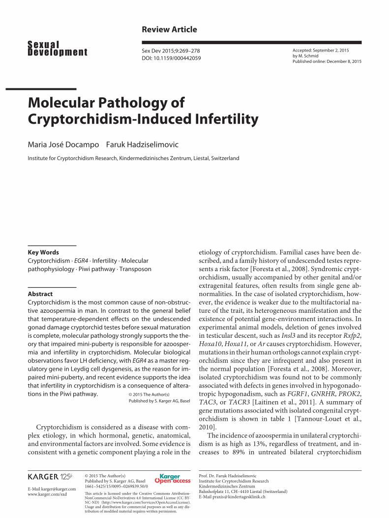
However, cryptorchid boys have low or even undetect-able levels of luteinizing hormone (LH) and testosterone surges [Gendrel et al., 1977], atrophic Leydig cells, and abrogated differentiation of gonocytes into Ad spermato-gonia [Hadziselimovic et al., 1986] ( fig. 1 ). The vast ma-jority of data available support the conclusion that in many children with undescended testes the response of Leydig cells to human chorionic gonadotropin (hCG) is diminished as compared to normal boys [for review, see Jockenhovel and Swerdloff, 1989]. Nevertheless, some cryptorchid boys show a normal response, with a higher incidence among unilateral cryptorchidism. Except for a blunted testosterone response to hCG, there is no evi-dence of altered steroidogenesis in cryptorchid testes pri-or to puberty [Jockenhovel and Swerdloff, 1989]. Pre-treatment of cryptorchid boys with hCG cancelled out the differences in their response to a stimulation test as com-pared to a control population [Gendrel et al., 1977]. Thus, the cause of the lower testosterone response seems to be at the pituitary or hypothalamic level, and may be a result of insufficient Leydig cell stimulation. Numerous LH-RH tests have demonstrated a lower LH response by gonado-tropin-releasing hormone [Job et al., 1974; Gendrel et al., 1977; Jacobelli et al., 1979; Mazzi et al., 1979; Bollerslev et al., 1986; Hamza et al., 2001].
During the last 40 years, histology has highly contrib-uted to better understand the etiology of cryptorchidism. In 1975, we proposed pronounced Leydig cell atrophy starting in early infancy as evidence to support endocri-nopathy as an etiological factor in cryptorchidism [Hadziselimovic et al., 1975]. Development of Ad sper-matogonia from gonocytes, which occurs during the first months of life, has been shown to be testosterone-depen-dent and is disturbed in cryptorchid boys [Hadziselimo-vic et al., 2005; Zivkovic et al., 2007]. Semi-thin section analysis of the contralateral descended testis in unilateral cryptorchidism confirmed several studies from the late 1960s that suggested cryptorchidism is a bilateral disease [Salle et al., 1968; Hedinger, 1971; Huff et al., 2001]. Cryptorchid boys with severely impaired mini-puberty and typical testicular histology will develop azoospermia or infertility irrespective of early and successful surgery [Hadziselimovic and Herzog, 2001; Hadziselimovic and Hoecht, 2008]. The high azoospermia risk (HAZR) group
Table 1. Single gene mutations associated with cryptorchidism
Gene Name
SOS1 son of sevenless homolog 1 (Drosophila)HOXD13 homeobox D13RAF1 Raf-1 proto-oncogene, serine/threonine kinaseSPATA12 spermatogenesis associated 12SOX2 SRY (sex-determining region Y)-box 2ESR1 estrogen receptor 1HOXA10 homeobox A10FGFR1 fibroblast growth factor receptor 1NR5A1 nuclear receptor subfamily 5, group A, member 1ZNF215 zinc finger protein 215ZNF214 zinc finger protein 214KRAS Kirsten rat sarcoma viral oncogene homologPTPN11 protein tyrosine phosphatase, non-receptor type 11RXFP2 relaxin/insulin-like family peptide receptor 2PWCR small nuclear ribonucleoprotein polypeptide NCYP19A1 cytochrome P450, family 19, subfamily A, polypeptide 1INSL3 insulin-like 3PROKR2 prokineticin receptor 2KAL1 anosmin 1ARX aristaless-related homeoboxAR androgen receptor
Adapted from Tannour-Louet et al. [2010].
Molecular Pathology of Cryptorchidism-Induced Infertility
Sex Dev 2015;9:269–278DOI: 10.1159/000442059
271
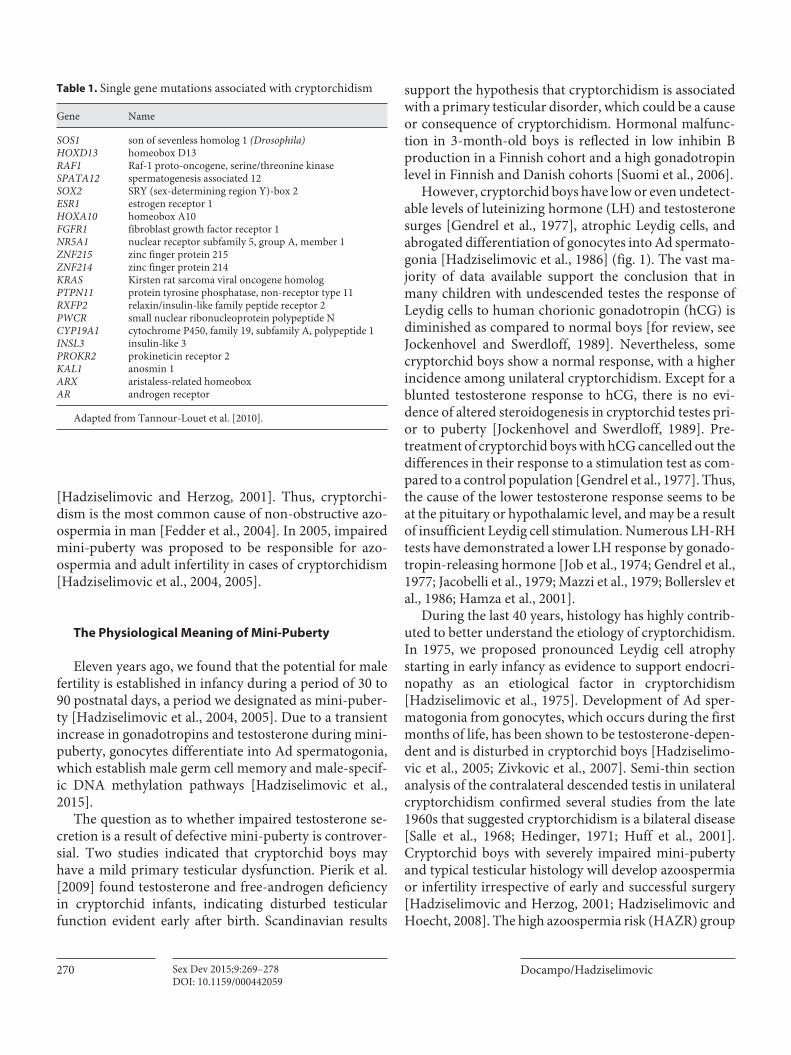
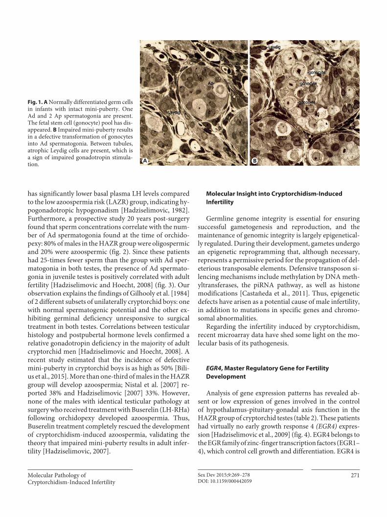
has significantly lower basal plasma LH levels compared to the low azoospermia risk (LAZR) group, indicating hy-pogonadotropic hypogonadism [Hadziselimovic, 1982]. Furthermore, a prospective study 20 years post-surgery found that sperm concentrations correlate with the num-ber of Ad spermatogonia found at the time of orchido-pexy: 80% of males in the HAZR group were oligospermic and 20% were azoospermic ( fig. 2 ). Since these patients had 25-times fewer sperm than the group with Ad sper-matogonia in both testes, the presence of Ad spermato-gonia in juvenile testes is positively correlated with adult fertility [Hadziselimovic and Hoecht, 2008] ( fig. 3 ). Our observation explains the findings of Gilhooly et al. [1984] of 2 different subsets of unilaterally cryptorchid boys: one with normal spermatogenic potential and the other ex-hibiting germinal deficiency unresponsive to surgical treatment in both testes. Correlations between testicular histology and postpubertal hormone levels confirmed a relative gonadotropin deficiency in the majority of adult cryptorchid men [Hadziselimovic and Hoecht, 2008]. A recent study estimated that the incidence of defective mini-puberty in cryptorchid boys is as high as 50% [Bili-us et al., 2015]. More than one-third of males in the HAZR group will develop azoospermia; Nistal et al. [2007] re-ported 38% and Hadziselimovic [2007] 33%. However, none of the males with identical testicular pathology at surgery who received treatment with Buserelin (LH-RHa) following orchidopexy developed azoospermia. Thus, Buserelin treatment completely rescued the development of cryptorchidism-induced azoospermia, validating the theory that impaired mini-puberty results in adult infer-tility [Hadziselimovic, 2007].
Molecular Insight into Cryptorchidism-Induced
Infertility
Germline genome integrity is essential for ensuring successful gametogenesis and reproduction, and the maintenance of genomic integrity is largely epigenetical-ly regulated. During their development, gametes undergo an epigenetic reprogramming that, although necessary, represents a permissive period for the propagation of del-eterious transposable elements. Defensive transposon si-lencing mechanisms include methylation by DNA meth-yltransferases, the piRNA pathway, as well as histone modifications [Castañeda et al., 2011]. Thus, epigenetic defects have arisen as a potential cause of male infertility, in addition to mutations in specific genes and chromo-somal abnormalities.
Regarding the infertility induced by cryptorchidism, recent microarray data have shed some light on the mo-lecular basis of its pathogenesis.
EGR4 , Master Regulatory Gene for Fertility
Development
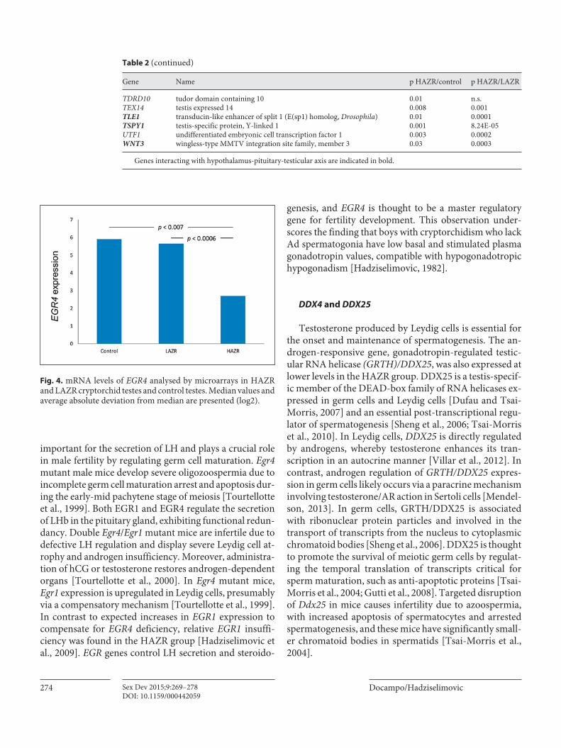
Analysis of gene expression patterns has revealed ab-sent or low expression of genes involved in the controlof hypothalamus-pituitary-gonadal axis function in the HAZR group of cryptorchid testes ( table 2 ). These patients had virtually no early growth response 4 (EGR4) expres-sion [Hadziselimovic et al., 2009] ( fig. 4 ). EGR4 belongs to the EGR family of zinc-finger transcription factors (EGR1–4), which control cell growth and differentiation. EGR4 is
A B
Fig. 1. A Normally differentiated germ cells in infants with intact mini-puberty. One Ad and 2 Ap spermatogonia are present. The fetal stem cell (gonocyte) pool has dis-appeared. B Impaired mini-puberty results in a defective transformation of gonocytes into Ad spermatogonia. Between tubules, atrophic Leydig cells are present, which isa sign of impaired gonadotropin stimula-tion.
Docampo/Hadziselimovic
Sex Dev 2015;9:269–278DOI: 10.1159/000442059
272
A C
B D
Fig. 2. Summarized results from Hadziselimovic and Hoecht [2008], prospective study. A Numbers of Ad spermatogonia vary among the HAZR (Ad-negative/both testes) and LAZR (Ad-posi-tive/both testes) groups. The lowest germ cell count was observed in the HAZR group and the highest was observed in the LAZR and control group. B Sperm count, after 20 years of the follow-up study. In the HAZR group the mean sperm count was 25 times less
than that in the LAZR group. C FSH levels were minimally in-creased in the HAZR group (normal range in adult males: 2–8 IU/l). D The LAZR patients with the healthiest histology have LH levels in the hypogonadotropic range, while the HAZR group has normal LH values despite more severe testicular pathology, indi-cating LH deficiency (normal range in adult males: 4.8–10.8 IU/l).
Fig. 3. Schematic model showing a domi-nant role of Ad spermatogonia in predict-ing fertility outcome and the importance of plasma FSH levels (++/ − − strong correla-tion; +/ − significant correlation). AdCDT (scrotal testis) is the best predictor of future fertility. AdUDT (undescended testis) is a decisive factor for supporting an FSH neg-ative feedback mechanism. GCTUDT (to-tal germ cell count in undescended testis) and GCTCDT (total germ cell count in scrotal testis) have no direct influence ei-ther on the sperm count or on the plasma FSH level.
Molecular Pathology of Cryptorchidism-Induced Infertility
Sex Dev 2015;9:269–278DOI: 10.1159/000442059
273
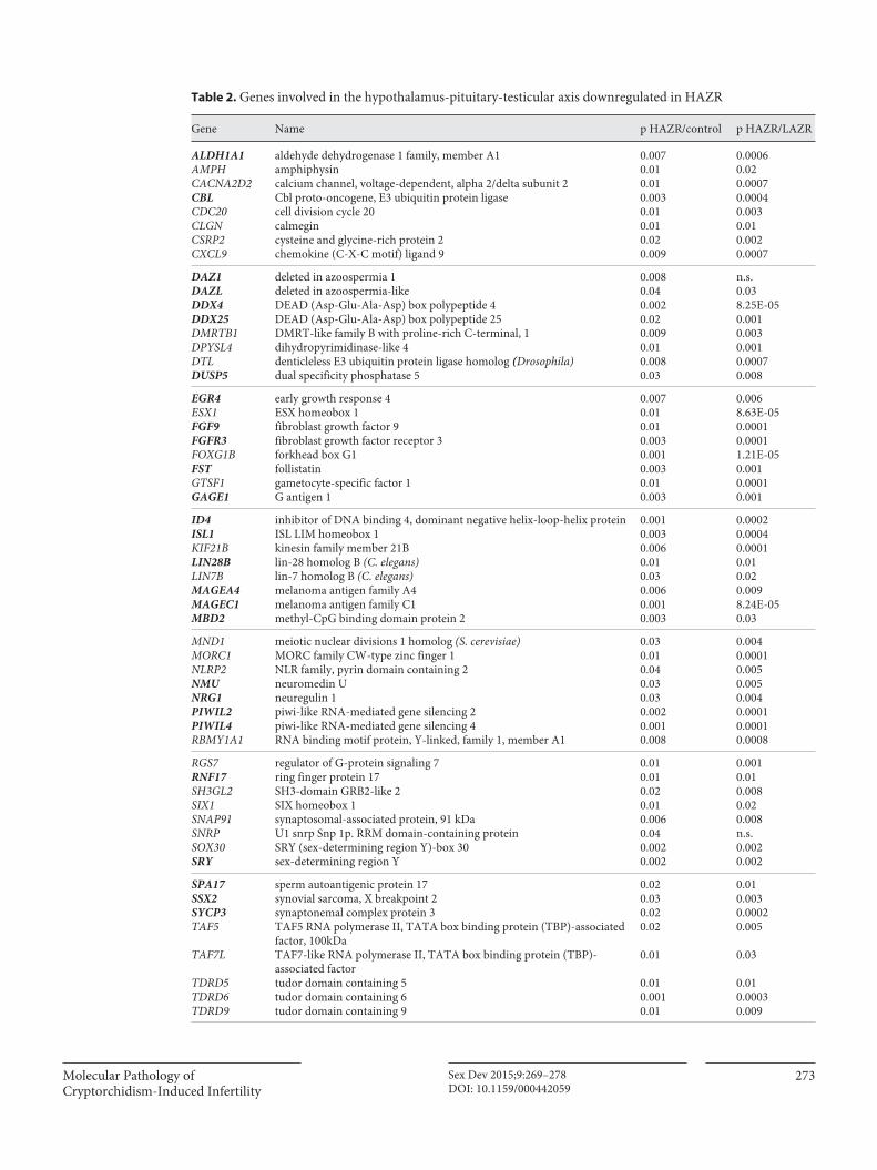
Gene Name p HAZR/control p HAZR/LAZR
ALDH1A1 aldehyde dehydrogenase 1 family, member A1 0.007 0.0006AMPH amphiphysin 0.01 0.02CACNA2D2 calcium channel, voltage-dependent, alpha 2/delta subunit 2 0.01 0.0007CBL Cbl proto-oncogene, E3 ubiquitin protein ligase 0.003 0.0004CDC20 cell division cycle 20 0.01 0.003CLGN calmegin 0.01 0.01CSRP2 cysteine and glycine-rich protein 2 0.02 0.002CXCL9 chemokine (C-X-C motif) ligand 9 0.009 0.0007
DAZ1 deleted in azoospermia 1 0.008 n.s.DAZL deleted in azoospermia-like 0.04 0.03DDX4 DEAD (Asp-Glu-Ala-Asp) box polypeptide 4 0.002 8.25E-05DDX25 DEAD (Asp-Glu-Ala-Asp) box polypeptide 25 0.02 0.001DMRTB1 DMRT-like family B with proline-rich C-terminal, 1 0.009 0.003DPYSL4 dihydropyrimidinase-like 4 0.01 0.001DTL denticleless E3 ubiquitin protein ligase homolog (Drosophila) 0.008 0.0007DUSP5 dual specificity phosphatase 5 0.03 0.008
EGR4 early growth response 4 0.007 0.006ESX1 ESX homeobox 1 0.01 8.63E-05FGF9 fibroblast growth factor 9 0.01 0.0001FGFR3 fibroblast growth factor receptor 3 0.003 0.0001FOXG1B forkhead box G1 0.001 1.21E-05FST follistatin 0.003 0.001GTSF1 gametocyte-specific factor 1 0.01 0.0001GAGE1 G antigen 1 0.003 0.001
ID4 inhibitor of DNA binding 4, dominant negative helix-loop-helix protein 0.001 0.0002ISL1 ISL LIM homeobox 1 0.003 0.0004KIF21B kinesin family member 21B 0.006 0.0001LIN28B lin-28 homolog B (C. elegans) 0.01 0.01LIN7B lin-7 homolog B (C. elegans) 0.03 0.02MAGEA4 melanoma antigen family A4 0.006 0.009MAGEC1 melanoma antigen family C1 0.001 8.24E-05MBD2 methyl-CpG binding domain protein 2 0.003 0.03
MND1 meiotic nuclear divisions 1 homolog (S. cerevisiae) 0.03 0.004MORC1 MORC family CW-type zinc finger 1 0.01 0.0001NLRP2 NLR family, pyrin domain containing 2 0.04 0.005NMU neuromedin U 0.03 0.005NRG1 neuregulin 1 0.03 0.004PIWIL2 piwi-like RNA-mediated gene silencing 2 0.002 0.0001PIWIL4 piwi-like RNA-mediated gene silencing 4 0.001 0.0001RBMY1A1 RNA binding motif protein, Y-linked, family 1, member A1 0.008 0.0008
RGS7 regulator of G-protein signaling 7 0.01 0.001RNF17 ring finger protein 17 0.01 0.01SH3GL2 SH3-domain GRB2-like 2 0.02 0.008SIX1 SIX homeobox 1 0.01 0.02SNAP91 synaptosomal-associated protein, 91 kDa 0.006 0.008SNRP U1 snrp Snp 1p. RRM domain-containing protein 0.04 n.s.SOX30 SRY (sex-determining region Y)-box 30 0.002 0.002SRY sex-determining region Y 0.002 0.002
SPA17 sperm autoantigenic protein 17 0.02 0.01SSX2 synovial sarcoma, X breakpoint 2 0.03 0.003SYCP3 synaptonemal complex protein 3 0.02 0.0002TAF5 TAF5 RNA polymerase II, TATA box binding protein (TBP)-associated
factor, 100kDa0.02 0.005
TAF7L TAF7-like RNA polymerase II, TATA box binding protein (TBP)-associated factor
0.01 0.03
TDRD5 tudor domain containing 5 0.01 0.01TDRD6 tudor domain containing 6 0.001 0.0003TDRD9 tudor domain containing 9 0.01 0.009
Table 2. Genes involved in the hypothalamus-pituitary-testicular axis downregulated in HAZR
Docampo/Hadziselimovic
Sex Dev 2015;9:269–278DOI: 10.1159/000442059
274
important for the secretion of LH and plays a crucial role in male fertility by regulating germ cell maturation. Egr4 mutant male mice develop severe oligozoospermia due to incomplete germ cell maturation arrest and apoptosis dur-ing the early-mid pachytene stage of meiosis [Tourtellotte et al., 1999]. Both EGR1 and EGR4 regulate the secretion of LHb in the pituitary gland, exhibiting functional redun-dancy. Double Egr4 / Egr1 mutant mice are infertile due to defective LH regulation and display severe Leydig cell at-rophy and androgen insufficiency. Moreover, administra-tion of hCG or testosterone restores androgen-dependent organs [Tourtellotte et al., 2000]. In Egr4 mutant mice, Egr1 expression is upregulated in Leydig cells, presumably via a compensatory mechanism [Tourtellotte et al., 1999]. In contrast to expected increases in EGR1 expression to compensate for EGR4 deficiency, relative EGR1 insuffi-ciency was found in the HAZR group [Hadziselimovic et al., 2009]. EGR genes control LH secretion and steroido-
genesis, and EGR4 is thought to be a master regulatory gene for fertility development. This observation under-scores the finding that boys with cryptorchidism who lack Ad spermatogonia have low basal and stimulated plasma gonadotropin values, compatible with hypogonadotropic hypogonadism [Hadziselimovic, 1982].
DDX4 and DDX25
Testosterone produced by Leydig cells is essential for the onset and maintenance of spermatogenesis. The an-drogen-responsive gene, gonadotropin-regulated testic-ular RNA helicase (GRTH)/DDX25 , was also expressed at lower levels in the HAZR group. DDX25 is a testis-specif-ic member of the DEAD-box family of RNA helicases ex-pressed in germ cells and Leydig cells [Dufau and Tsai-Morris, 2007] and an essential post-transcriptional regu-lator of spermatogenesis [Sheng et al., 2006; Tsai-Morris et al., 2010]. In Leydig cells, DDX25 is directly regulated by androgens, whereby testosterone enhances its tran-scription in an autocrine manner [Villar et al., 2012]. In contrast, androgen regulation of GRTH/DDX25 expres-sion in germ cells likely occurs via a paracrine mechanism involving testosterone/AR action in Sertoli cells [Mendel-son, 2013]. In germ cells, GRTH/DDX25 is associated with ribonuclear protein particles and involved in the transport of transcripts from the nucleus to cytoplasmic chromatoid bodies [Sheng et al., 2006]. DDX25 is thought to promote the survival of meiotic germ cells by regulat-ing the temporal translation of transcripts critical for sperm maturation, such as anti-apoptotic proteins [Tsai-Morris et al., 2004; Gutti et al., 2008]. Targeted disruption of Ddx25 in mice causes infertility due to azoospermia, with increased apoptosis of spermatocytes and arrested spermatogenesis, and these mice have significantly small-er chromatoid bodies in spermatids [Tsai-Morris et al., 2004].
Gene Name p HAZR/control p HAZR/LAZR
TDRD10 tudor domain containing 10 0.01 n.s.TEX14 testis expressed 14 0.008 0.001TLE1 transducin-like enhancer of split 1 (E(sp1) homolog, Drosophila) 0.01 0.0001TSPY1 testis-specific protein, Y-linked 1 0.001 8.24E-05UTF1 undifferentiated embryonic cell transcription factor 1 0.003 0.0002WNT3 wingless-type MMTV integration site family, member 3 0.03 0.0003
Genes interacting with hypothalamus-pituitary-testicular axis are indicated in bold.
Table 2 (continued)
Fig. 4. mRNA levels of EGR4 analysed by microarrays in HAZR and LAZR cryptorchid testes and control testes. Median values and average absolute deviation from median are presented (log2).
Molecular Pathology of Cryptorchidism-Induced Infertility
Sex Dev 2015;9:269–278DOI: 10.1159/000442059
275
DDX4 (VASA) is a gonadotropin-dependent gene un-derexpressed in the HAZR as compared to the LAZR group and control testes. Interestingly, sterile mice with Vasa (Ddx4) deficiency have malformed processing bod-ies (P-bodies) with an ultrastructural appearance identi-cal to that observed in our previous study [Hadziselimo-vic, 1977; Kuramochi-Miyagawa et al., 2010].
Germ Cell Development Genes
In addition to alterations in the hypothalamus-pitu-itary-testicular axis, RNA profiling analysis revealed a gene expression signature that distinguishes HAZR pa-tients from control and LAZR patients [Hadziselimovic et al., 2011]. Concomitant with the reduction in germ cells, genes involved in germ cell development and fertil-ity and spermatogonial stem cell self-renewal show lower or even undetectable expression in HAZR testes. Some of the genes for which low levels of mRNA are found in tes-tes lacking Ad spermatogonia are involved in meiotic and post-meiotic stages of adult spermatogenesis. If juvenile Ad spermatogonia express these genes in normal prepu-bertal testes, some of the molecular events that initiate the testicular expression program at the onset of puberty and maintain it during adulthood occur very early in prepu-bertal testes and are likely altered in the HAZR group of cryptorchid patients. This idea is further corroborated by the fact that genes proposed as biomarkers of adult hu-man spermatogonia (FGFR3 , UTF1 , CBL , and SNAP91) [Von Kopylow et al., 2010] are present in LAZR, but miss-ing in HAZR testes [Hadziselimovic et al., 2011]. Several genes transcribed mainly in Sertoli cells, such as DUSP5 and DMRTB1 , appeared to be expressed at lower levels in testes lacking Ad spermatogonia. This finding supports the idea that the proliferation of Sertoli cells is also im-paired after defective mini-puberty, and that fertility po-tential is established during this period through both the maturation of Ad spermatogonia and proliferation of Sertoli cells [Zivkovic and Hadziselimovic, 2009], under-scoring the need for a normal testicular environment in spermatogenesis.
Other genes for which no mRNA is detected in the HAZR group included genes whose deletion is classically involved in azoospermia: DAZ1 , DAZL , RBMY1A1 , SYCP3 , and CDC20 [Hadziselimovic et al., 2011]. Deleted in azoospermia (DAZ) family proteins and mammalian RNA-binding motif protein Y chromosome (RBMY) are encoded by the male-specific region of the Y chromo-some. DAZ and DAZ-like protein (DAZL) are expressed
in germ cells and are essential for normal spermatogen-esis [Yen, 2004]. DAZ members are necessary for late-stage meiosis and the development of haploid spermatids, whereas DAZL functions primarily in primordial germ cell formation [Kee et al., 2009]. Azoospermia associated with decreased DAZ may go along with reduced levels of SYCP3 [Reynolds et al., 2007]. Although they cause azo-ospermia, microdeletions of the long arm of the Y chro-mosome do not seem to be associated with cryptorchi-dism [Fedder et al., 2004; Vutyavanich et al., 2007]. Fur-thermore, in azoospermic men with AZFc deletion and no DAZ gene expression, EGR4 is upregulated [Gatta et al., 2010]. Thus, a lack of both EGR4 and DAZ1 mRNAs in the HAZR subset of cryptorchid patients suggests that DAZ/DAZL acts downstream of EGR4 function, rein-forcing the notion that cryptorchidism-induced azo-ospermia is predominantly the result of an endocrine condition rather than being caused directly by mutations in these genes.
Role of Transposon-Silencing Genes and P-Bodies
Epigenetic reprogramming, which includes the era-sure and resetting of DNA methylation, occurs in pri-mordial germ cells and is important for preventing the passage of DNA methylation defects from one generation to the next, restoring the developmental potency of the germline, and preparing the genome for the establish-ment of male-specific DNA methylation patterns in im-printed genes [Castañeda et al., 2011; Bortvin, 2013]. De novo methylation of germ cells begins in prenatal life and is completed by the onset of meiosis [Oakes et al., 2007]. Nearly 40% of the mammalian genome are constituted by endogenous transposable elements (transposons), which are deleterious and cause genomic instability and cell death. In order to prevent their activity, the promoters of the genes are constitutively repressed by hypermethyl-ation [Smith and Meissner, 2013]. However, this repres-sion is lost in male germ cells during the developmental window of epigenetic reprogramming, requiring a robust defense system comprised of the P-element-induced wimpy testis (Piwi)-interacting RNA (piRNA) pathway and de novo DNA methylation machinery [Aravin et al., 2008; Kuramochi-Miyagawa et al., 2008]. piRNAs consti-tute a unique mode of epigenetic regulation, as they are a type of small non-coding RNA prominently expressed in the germline that constitute an innate defense system against the activity of genetic mobile elements. piRNAs are bound by Piwi-like proteins (Piwil1, Piwil2 and Pi-
Docampo/Hadziselimovic
Sex Dev 2015;9:269–278DOI: 10.1159/000442059
276
wil4), which belong to the Argonaute family of proteins [Siomi et al., 2010]. piRNA-Piwi protein complexes allow selective silencing of transposons by both post-transcrip-tional and transcriptional mechanisms, such as the deg-radation of transposon RNA or DNA methylation of the transposon (i.e. epigenetic silencing). Since they maintain genomic stability in the germline, piRNAs are indispens-able for fertility [Aravin et al., 2007]. The 3 members of the Piwi protein family in the mouse are required for spermatogenesis [Deng and Lin, 2002; Kuramochi-Miya-gawa et al., 2004; Carmell et al., 2007]. Inactivation of Miwi2 (Piwil4) or Mili (Piwil2) leads to loss of DNA methylation, derepression of transposons, defects in spermatogenesis, and sterility [Aravin and Hannon, 2008].
Members of the piRNA pathway need other proteins to function, including Maelstrom (MAEL), DEAD box polypeptide 4 (DDX4; mammalian VASA), Moloney leu-kemia virus 10-like 1 (MOV10L1), GASZ, and members of the Tudor family of proteins [Aravin et al., 2009]. Tu-dor proteins regulate the biological functions of Piwi pro-teins by acting as a scaffold to organize the components of the piRNA pathway [Siomi et al., 2010]. Mice deficient in each of the genes essential for transposon silencing are sterile [Soper et al., 2008; Shoji et al., 2009; Frost et al., 2010; Kuramochi-Miyagawa et al., 2010]. This intricate genome defense machinery is localized in specialized cy-toplasmic granules or nuage, which are considered to be germline analogs of somatic P-bodies [Aravin et al., 2009]. P-bodies contain aggregates of specific mRNAs and proteins and are sites of RNA degradation and silenc-ing [Eulalio et al., 2007; Castañeda et al., 2011]. P-bodies exhibit a complex mechanism for regulating the organi-zation and function of Piwi proteins and piRNAs in transposon-silencing pathways. This P-body mechanism includes a methylation-directed protein-protein interac-tion mediated by germline Tudor domain proteins and Piwi proteins [Chen et al., 2009].
RNA profiling data are consistent with the notion that cryptorchidism-induced azoospermia is due to germ cell death stemming from defects in the epigenetic regulatory pathways essential for genome stability and spermato-genesis. Results have shown alterations in mRNA levels for genes involved in the piRNA-Piwi pathway in the HAZR group: RNA levels for 2 members of the Piwi pro-tein family, as well as other related genes important for transposon silencing, are decreased as compared to LAZR patients (PIWIL2 , PIWIL4 , DDX4 , MAEL , MOV10L1 , 7 members of the Tudor family of proteins, and GTSF1) [Hadziselimovic et al., 2012, 2015]. Thus, males with
cryptorchidism and impaired mini-puberty show altera-tions in Piwi-pathway gene expression that appear to re-sult in the deregulation of LINE1 transposons [Hadzise-limovic et al., 2015], which ultimately causes apoptosis of germ cells. Notably, morphological changes consistent with altered function of P-bodies in the HAZR group were found at the ultrastructural level [Hadziselimovic et al., 2015]. Recently, testosterone was shown to influence testicular functions by regulating the piRNA-pathway [Kang et al., 2014]. This finding is consistent with the ob-servation that gonadotropin and testosterone insufficien-cy during mini-puberty induces alterations in the Piwi-piRNA pathway.
Microrchidia-1 (MORC1) is another gene involved in male fertility. Morc1 -deficient male mice are sterile and develop micro-testes due to germ cell apoptosis prior to meiosis [Watson et al., 1998]. MORC1 was recently shown to silence transposable elements, including LINEs, in the mouse male germline by facilitating DNA meth-ylation in a piRNA-independent manner [Pastor et al., 2014]. Interestingly, MORC1 signals are also decreased in HAZR cryptorchid testes [Hadziselimovic et al., 2011]. Thus, the failure of different transposon repression mech-anisms and subsequent genomic instability are likely in-volved in the development of azoospermia in cryptorchid patients. Multiple mechanisms would not be surprising, as L1 has been shown to be silenced in at least 3 different ways, including piRNA-mediated and piRNA-indepen-dent methylation [Di Giacomo et al., 2013].
Novel Implications for Treatment
Successful scrotal relocation of the testis reduces, but does not prevent, infertility in certain individuals [Kolon et al., 2014]. Since abnormal mini-puberty is responsible for adult-onset infertility in cryptorchidism, post-surgi-cal hormonal treatment is highly recommended in HAZR cryptorchid boys who underwent successful early orchi-dopexy but remain at risk to become infertile as adults.
Concluding Remarks
Whole-genome expression analysis strongly supports the theory that impaired mini-puberty is responsible for azoospermia and adult infertility in cryptorchidism. Mul-tiple differences in gene expression between HAZR and LAZR groups underscore the importance of an intact hy-pothalamus-pituitary-testicular axis in fertility develop-
Molecular Pathology of Cryptorchidism-Induced Infertility
Sex Dev 2015;9:269–278DOI: 10.1159/000442059
277
ment. Molecular biological observations support LH de-ficiency, with EGR4 as a master gene in Leydig cell dys-genesis, as the reason for impaired mini-puberty.
In contrast to the general belief that temperature-de-pendent effects on cryptorchid gonads damage unde-scended testes before sexual maturation is complete, re-cent evidence is consistent with the idea that infertility in cryptorchidism is a consequence of alterations in the Piwi
pathway and transposon derepression. Thus, abnormal germ cell development in cryptorchidism is preceded by a hormone imbalance and perturbation in germ cell-spe-cific gene expression during mini-puberty. In addition, intact function of P-bodies during mini-puberty contrib-utes to the establishment of germ cell memory and male-specific DNA methylation pathways.
References
Aravin AA, Hannon GJ: Small RNA silencing pathways in germ and stem cells. Cold Spring Harb Symp Quant Biol 73: 283–290 (2008).
Aravin AA, Hannon GJ, Brennecke J: The Piwi-piRNA pathway provides an adaptive defense in the transposon arms race. Science 318: 761–764 (2007).
Aravin AA, Sachidanandam R, Bourc’his D, Schaefer C, Pezic D, et al: A piRNA pathway primed by individual transposons is linked to de novo DNA methylation in mice. Mol Cell 31: 785–799 (2008).
Aravin AA, Van Der Heijden GW, Castaneda J, Vagin VV, Hannon GJ, Bortvin A: Cytoplas-mic compartmentalization of the fetal piRNA pathway in mice. PLoS Genet 5:e1000764 (2009).
Bilius V, Verkauskas G, Dasevicius D, Kazlauskas V, Malcius D, Hadziselimovic F: Incidence of high infertility risk among unilateral cryptor-chid boys. Urol Int 95: 142–145 (2015).
Bollerslev J, Rohl H, Krag Sorensen E, Bennet P: Gonadotropin and androgen levels in pa-tients operated upon for cryptorchidism. Dan Med Bull 33: 336–338 (1986).
Bortvin A: PIWI-interacting RNAs (piRNAs) – a mouse testis perspective. Biochemistry (Mosc) 78: 592–602 (2013).
Carmell MA, Girard A, van de Kant HJG, Bourc’his D, Bestor TH, et al: MIWI2 is es-sential for spermatogenesis and repression of transposons in the mouse male germline. Dev Cell 12: 503–514 (2007).
Castañeda J, Genzor P, Bortvin A: piRNAs, trans-poson silencing, and germline genome integ-rity. Mutat Res 714: 95–104 (2011).
Chen C, Jin J, James DA, Adams-Cioaba MA, Park JG, et al: Mouse Piwi interactome identi-fies binding mechanism of Tdrkh Tudor do-main to arginine methylated Miwi. Proc Natl Acad Sci USA 106: 20336–20341 (2009).
Deng W, Lin H: miwi , a murine homolog of piwi , encodes a cytoplasmic protein essential for spermatogenesis. Dev Cell 2: 819–830 (2002).
Di Giacomo M, Comazzetto S, Saini H, DeFazio S, Carrieri C, et al: Multiple epigenetic mech-anisms and the piRNA pathway enforce LINE1 silencing during adult spermatogene-sis. Mol Cell 50: 601–608 (2013).
Dufau ML, Tsai-Morris CH: Gonadotropin-reg-ulated testicular helicase (GRTH/DDX25):an essential regulator of spermatogenesis. Trends Endocrinol Metab 18: 314–320 (2007).
Eulalio A, Behm-Ansmant I, Izaurralde E: P bod-ies: at the crossroads of post-transcriptional pathways. Nat Rev Mol Cell Biol 8: 9–22 (2007).
Fedder J, Crüger D, Oestergaard B, Petersen GB: Etiology of azoospermia in 100 consecutive nonvasectomized men. Fertil Steril 82: 1463–1465 (2004).
Foresta C, Zuccarello D, Garolla A, Ferlin A: Role of hormones, genes, and environment in hu-man cryptorchidism. Endocr Rev 29: 560–580 (2008).
Frost RJA, Hamra FK, Richardson JA, Qi X, Bas-sel-Duby R, Olson EN: MOV10L1 is neces-sary for protection of spermatocytes against retrotransposons by Piwi-interacting RNAs. Proc Natl Acad Sci USA 107: 11847–11852 (2010).
Gatta V, Raicu F, Ferlin A, Antonucci I, Scioletti AP, et al: Testis transcriptome analysis in male infertility: new insight on the pathogen-esis of oligo-azoospermia in cases with and without AZFc microdeletion. BMC Genom-ics 11: 401 (2010).
Gendrel D, Roger M, Chaussain JL, Canlorbe P, Job JC: Correlation of pituitary and testicular responses to stimulation tests in cryptorchid children. Acta Endocrinol (Copenh) 86: 641–650 (1977).
Gilhooly PE, Meyers F, Lattimer JK: Fertility prospects for children with cryptorchidism. Am J Dis Child 138: 940–943 (1984).
Gutti RK, Tsai-Morris CH, Dufau ML: Gonado-tropin-regulated testicular helicase (DDX25), an essential regulator of spermatogenesis, prevents testicular germ cell apoptosis. J Biol Chem 283: 17055–17064 (2008).
Hadziselimovic F: Cryptorchidism. Ultrastruc-ture of normal and cryptorchid testis develop-ment. Adv Anat Embryol Cell Biol 53: 3–71 (1977).
Hadziselimovic F: Pathogenesis and treatment of undescended testes. Eur J Pediatr 139: 255–265 (1982).
Hadziselimovic F: Early successful orchidopexy does not prevent from developing azoosper-mia. Int Braz J Urol 32: 570–573 (2007).
Hadziselimovic F, Herzog B: The importance of both an early orchidopexy and germ cell mat-uration for fertility. Lancet 358: 1156–1157 (2001).
Hadziselimovic F, Hoecht B: Testicular histology related to fertility outcome and postpubertal hormone status in cryptorchidism. Klin Pädi-atr 220: 302–307 (2008).
Hadziselimovic F, Herzog B, Seguchi H: Surgical correction of cryptorchism at 2 years: electron microscopic and morphometric investiga-tions. J Pediatr Surg 10: 19–26 (1975).
Hadziselimović F, Thommen L, Girard J, Herzog B: The significance of postnatal gonadotropin surge for testicular development in normal and cryptorchid testes. J Urol 136: 274–276 (1986).
Hadziselimovic F, Emmons LR, Buser MW: A di-minished postnatal surge of Ad spermatogo-nia in cryptorchid infants is additional evi-dence for hypogonadotropic hypogonadism. Swiss Med Wkly 134: 381–384 (2004).
Hadziselimovic F, Zivkovic D, Bica DTG, Em-mons LR: The importance of mini-puberty for fertility in cryptorchidism. J Urol 174: 1536–1539; discussion 1538–1539 (2005).
Hadziselimovic F, Hadziselimovic NO, Demou-gin P, Krey G, Hoecht B, Oakeley EJ: EGR4 is a master gene responsible for fertility in crypt-orchidism. Sex Dev 3: 253–263 (2009).
Hadziselimovic F, Hadziselimovic NO, Demou-gin P, Oakeley EJ: Testicular gene expression in cryptorchid boys at risk of azoospermia. Sex Dev 5: 49–59 (2011).
Hadziselimovic F, Hadziselimovic NO, Demou-gin P, Krey G, Oakeley EJ: Deficient expres-sion of genes involved in the endogenous de-fense system against transposons in cryptor-chid boys with impaired mini-puberty. Sex Dev 5: 287–293 (2012).
Hadziselimovic F, Hadziselimovic NO, Demou-gin P, Krey G, Oakeley EJ: Piwi-pathway al-teration induces LINE-1 transposon dere-pression and infertility development in crypt-orchidism. Sex Dev 9: 98–104 (2015).
Hamza AF, Elrahim M, Elnagar O, Maaty SA, Bassiouny IE, Jehannin B: Testicular descent: when to interfere? Eur J Pediatr Surg 11: 173–176 (2001).
Docampo/Hadziselimovic
Sex Dev 2015;9:269–278DOI: 10.1159/000442059
278
Hedinger C: Diagnostic and prognostic value of testis biopsy. Schweiz Med Wochenschr 101: 1084–1089 (1971).
Huff DS, Fenig DM, Canning DA, Carr MC,Zderic SA, Snyder HM: Abnormal germ cell development in cryptorchidism. Horm Res 55: 11–17 (2001).
Jacobelli A, Agostino A, Vecci E, Simeoni A, Fer-rantelli M: Studies on the pituitary-testicular axis in boys with cryptorchidism, in Bierich JR, Giarola A (eds): Cryptorchidism, pp 261–268 (Academic Press, London 1979).
Job JC, Garnier PE, Chaussain JL, Toublanc JE, Canlorbe P: Effect of synthetic luteinizing hormone-releasing hormone on the release of gonadotropins in hypophysogonadal disor-ders of children and adolescents. IV. Unde-scended testes. J Pediatr 84: 371–374 (1974).
Jockenhovel F, Swerdloff RS: Alterations in the steroidogenic capacity of Leydig cells in cryptorchid testis, in Abney TO, Keel BA (eds): The Cryptorchid Testis, pp 35–54 (CRC Press, Inc, Boca Raton 1989).
Kang HJ, Moon MJ, Lee HY, Han SW: Testoster-one alters testis function through regulation of piRNA expression in rats. Mol Biol Rep 41: 6729–6735 (2014).
Kee K, Angeles VT, Flores M, Nguyen HN, Reijo Pera RA: Human DAZL , DAZ and BOULE genes modulate primordial germ cell and haploid gamete formation. Nature 462: 222–225 (2009).
Kolon TF, Herndon CDA, Baker LA, Baskin LS, Baxter CG, et al: Evaluation and treatment of cryptorchidism: AUA guideline. J Urol 192: 337–345 (2014).
Kuramochi-Miyagawa S, Kimura T, Ijiri TW, Isobe T, Asada N, et al: Mili , a mammalian member of piwi family gene, is essential for spermatogenesis. Development 131: 839–849 (2004).
Kuramochi-Miyagawa S, Watanabe T, Gotoh K, Totoki Y, Toyoda A, et al: DNA methylation of retrotransposon genes is regulated by Piwi family members MILI and MIWI2 in murine fetal testes. Genes Dev 22: 908–917 (2008).
Kuramochi-Miyagawa S, Watanabe T, Gotoh K, Takamatsu K, Chuma S, et al: MVH in piRNA processing and gene silencing of retrotrans-posons. Genes Dev 24: 887–892 (2010).
Laitinen EM, Tommiska J, Virtanen HE, Oeh-landt H, Koivu R, et al: Isolated cryptorchi-dism: no evidence for involvement of genes underlying isolated hypogonadotropic hypo-gonadism. Mol Cell Endocrinol 341: 35–38 (2011).
Mazzi C, Riva LP, Morandi G, Mainini E, Scarsi G, Salaroli A: A study of cryptorchid subjects. Evaluation of the hypophyseal-testicular axis in the prepubertal period, in Bierich JR, Gia-rola A (eds): Cryptorchidism, pp 269–276 (Academic Press, London 1979).
Mendelson CR: GRTH: a key to understanding androgen-mediated germ cell signaling. En-docrinology 154: 1967–1969 (2013).
Nistal M, Paniagua R, Riestra ML, Reyes-Múgica M, Cajaiba MM: Bilateral prepubertal testicu-lar biopsies predict significance of cryptorchi-dism-associated mixed testicular atrophy, and allow assessment of fertility. Am J Surg Pathol 31: 1269–1276 (2007).
Oakes CC, La Salle S, Smiraglia DJ, Robaire B, Trasler JM: Developmental acquisition of ge-nome-wide DNA methylation occurs prior to meiosis in male germ cells. Dev Biol 307: 368–379 (2007).
Pastor WA, Stroud H, Nee K, Liu W, Pezic D, et al: MORC1 represses transposable elements in the mouse male germline. Nat Commun 5: 5795 (2014).
Pierik FH, Deddens JA, Burdorf A, de Muinck Keizer-Schrama SM, de Jong FH, Weber RFA: The hypothalamus-pituitary-testis axis in boys during the first six months of life: a com-parison of cryptorchidism and hypospadias cases with controls. Int J Androl 32: 453–461 (2009).
Reynolds N, Collier B, Bingham V, Gray NK, Cooke HJ: Translation of the synaptonemal complex component Sycp3 is enhanced in vivo by the germ cell specific regulator Dazl. RNA 13: 974–981 (2007).
Salle B, Hedinger C, Nicole R: Significance of tes-ticular biopsies in cryptorchidism in children. Acta Endocrinol (Copenh) 58: 67–76 (1968).
Sheng Y, Tsai-Morris CH, Gutti R, Maeda Y, Du-fau ML: Gonadotropin-regulated testicular RNA helicase (GRTH/Ddx25) is a transport protein involved in gene-specific mRNA ex-port and protein translation during sper-matogenesis. J Biol Chem 281: 35048–35056 (2006).
Shoji M, Tanaka T, Hosokawa M, Reuter M, Stark A, et al: The TDRD9-MIWI2 complex is es-sential for piRNA-mediated retrotransposon silencing in the mouse male germline. Dev Cell 17: 775–787 (2009).
Siomi MC, Mannen T, Siomi H: How does the royal family of Tudor rule the PIWI-interact-ing RNA pathway? Genes Dev 24: 636–646 (2010).
Smith ZD, Meissner A: DNA methylation: roles in mammalian development. Nat Rev Genet 14: 204–220 (2013).
Soper SFC, van der Heijden GW, Hardiman TC, Goodheart M, Martin SL, et al: Mouse Mael-strom, a component of nuage, is essential for spermatogenesis and transposon repression in meiosis. Dev Cell 15: 285–297 (2008).
Suomi AM, Main KM, Kaleva M, Schmidt IM, Chellakooty M, et al: Hormonal changes in 3-month-old cryptorchid boys. J Clin Endo-crinol Metab 91: 953–958 (2006).
Tannour-Louet M, Han S, Corbett ST, Louet JF, Yatsenko S, et al: Identification of de novo copy number variants associated with human disorders of sexual development. PLoS One 5:e15392 (2010).
Tourtellotte WG, Nagarajan R, Auyeung A, Mueller C, Milbrandt J: Infertility associated with incomplete spermatogenic arrest and ol-igozoospermia in Egr4 -deficient mice. Devel-opment 126: 5061–5071 (1999).
Tourtellotte WG, Nagarajan R, Bartke A, Mil-brandt J: Functional compensation by Egr4 in Egr1 -dependent luteinizing hormone regula-tion and Leydig cell steroidogenesis. Mol Cell Biol 20: 5261–5268 (2000).
Tsai-Morris CH, Sheng Y, Lee E, Lei KJ, Dufau ML: Gonadotropin-regulated testicular RNA helicase (GRTH/Ddx25) is essential for sper-matid development and completion of sper-matogenesis. Proc Natl Acad Sci USA 101: 6373–6378 (2004).
Tsai-Morris CH, Sheng Y, Gutti R, Li J, Pickel J, Dufau ML: Gonadotropin-regulated testicu-lar RNA helicase (GRTH/DDX25) gene: cell-specific expression and transcriptional regu-lation by androgen in transgenic mouse testis. J Cell Biochem 109: 1142–1147 (2010).
Villar J, Tsai-Morris CH, Dai L, Dufau ML: An-drogen-induced activation of gonadotropin-regulated testicular RNA helicase (GRTH/Ddx25) transcription: essential role of a non-classical androgen response element half-site. Mol Cell Biol 32: 1566–1580 (2012).
Von Kopylow K, Kirchhoff C, Jezek D, Schulze W, Feig C, et al: Screening for biomarkers of sper-matogonia within the human testis: a whole genome approach. Hum Reprod 25: 1104–1112 (2010).
Vutyavanich T, Piromlertamorn W, Sirirungsi W, Sirisukkasem S: Frequency of Y chromo-some microdeletions and chromosomal ab-normalities in infertile Thai men with oligo-zoospermia and azoospermia. Asian J Androl 9: 68–75 (2007).
Watson ML, Zinn AR, Inoue N, Hess KD, Cobb J, et al: Identification of morc (microrchidia) , a mutation that results in arrest of spermato-genesis at an early meiotic stage in the mouse. Proc Natl Acad Sci USA 95: 14361–14366 (1998).
Yen PH: Putative biological functions of the DAZ family. Int J Androl 27: 125–129 (2004).
Zivkovic D, Hadziselimovic F: Development of Sertoli cells during mini-puberty in normal and cryptorchid testes. Urol Int 82: 89–91 (2009).
Zivkovic D, Bica DTG, Hadziselimovic F: Rela-tionship between adult dark spermatogonia and secretory capacity of Leydig cells in cryptorchidism. BJU Int 100: 1147–1149 (2007).


