Embed Size (px)
Citation preview

Biochem. J. (1994) 298, 69-77 (Printed in Great Britain)
Molybdenum(VI) salts convert the xanthine oxidoreductase apoprotein intothe active enzyme in mouse L929 fibroblastic cells*Francesco FALCIANI,t Mineko TERAO,t Stefano GOLDWURM,t Anna RONCHI,t Anna GATTI,t Claudio MINOIA,tMarco Li CALZI,t Mario SALMONA,t Giovanni CAZZANIGAt and Enrico GARATTINlt§tMolecular Biology Unit, Laboratory of Enzymology, Centro Daniela e Catullo Borgomainerio, Istituto di Ricerche Farmacologiche 'Mario Negri', via Eritrea 62,20157 Milano, and tLaboratorio di Igiene Industriale, Fondazione Clinica del Lavoro, IRCCS, via Alzaia 29, 27100 Pavia, Italy
The mouse L929 fibroblastic cell line presents low, but detectable,levels of the mRNA encoding xanthine oxidoreductase underbasal conditions, and it responds to type I and type II interferonsby inducing the expression of the transcript [Falciani, Ghezzi,Terao, Cazzaniga, and Garattini (1992) Biochem. J. 285,1001-1008]. This cell line, however, does not show any detectableamount of xanthine oxidoreductase enzymic activity, eitherbefore or after treatment with the cytokines. Molybdenum(VI)salts, in the millimolar range, are capable of activating xanthineoxidoreductase in L929 cells both under basal conditions andafter treatment with interferon-cc. The increase is observed inmouse L929 as well as in clones derived from it, but not in manyother human and mouse cell lines. The induction observed inL929 cells is post-translational in nature and it is insensitive tocycloheximide, indicating that the molybdenum ion converts a
INTRODUCTION
The xanthine oxidoreductase (XOR) enzymic system catalysesthe oxidation of hypoxanthine to xanthine and xanthine to uricacid. XOR is a dimeric protein which consists of two identicalmonomeric subunits of 150 kDa each (Rajagopalan and Handler,1967; Carpani et al., 1990) and it binds 1 molecule of FAD pereach subunit (Rajagopalan and Handler, 1967). It is one of themammalian flavoproteins that require a molybdopteridin co-factor ('molybdenum cofactor') for their activity (Bray, 1975,1982, 1988; Johnson, 1980). The structure of the molybdenumcofactor which is covalently bound to the protein is known(Johnson and Rajagopalan, 1982; Johnson et al., 1980a,b, 1984;Kramer et al., 1987; Gardlik and Rajagopalan, 1990) andresembles that of many other molybdenum containing oxido-reductases present in various eukaryotic and prokaryotic organ-isms (Rajagopalan and Johnson, 1992). The enzyme is theproduct of a single gene (Amaya et al., 1990) and it is present incells as two interconvertible forms, xanthine dehydrogenase(XD) and xanthine oxidase (XO) (Della Corte and Stirpe, 1968,1972). The former uses NADI as the acceptor of the reducingequivalents produced during the oxidation of the substrates,whereas the latter transfers the electrons to molecular oxygen(Nishino et al., 1989; Saito and Nishino, 1989). XO is implicatedas a possible mediator of tissue damage in many pathologicalconditions (Parks and Granger, 1983; McCord, 1985; Terada etal., 1991; de Groot and Littauer, 1988) because of its ability toproduce superoxide anions as a product of the catalysed reactions(Fridovich, 1970).
pool of inactive xanthine oxidoreductase apoenzyme into itsholoenzymic form. When grown in the absence of sodiummolybdate, the L929 cell line has undetectable intracellular levelsof the molybdenum cofactor, since the cell extracts are unable tocomplement the nitrate reductase defect of the nit-i mutant ofNeurospora crassa. L929 cells grown in the presence of millimolarconcentrations of sodium molybdate, however, become com-petent to complement the nit-i defect. L929 cells accumulatemolybdenum ion inside the intracellular compartment as effici-ently as TEnd cells, a mouse endothelial cell line that expressesxanthine oxidoreductase activity both under basal conditionsand after treatment with interferon-y, suggesting that L929 cellshave a defect in one or more of the metabolic steps leading to thesynthesis of the molybdenum cofactor.
Recently we cloned the cDNA coding for the mouse XOR(Terao et al., 1992) and we demonstrated that the expression ofthe gene is under the control of interferons (IFNs) and IFN-inducing agents both in vivo (Terao et al., 1992) and in vitro(Falciani et al., 1992). The expression of the XOR transcript wasstudied in detail in L929 fibroblastic cells (Falciani et al., 1992).This cell line is, in fact, widely used in the assay of the antiviralactivity ofIFNs and it is thus a biologically relevant experimentalmodel. During the course of studies aimed at clarifying themolecular mechanisms underlying the induction of this enzymeby type I and type II IFNs, we noticed that L929 cells respondto the cytokines with a strong induction of XOR mRNA thatdoes not result in a parallel increase in XD or XO enzymicactivities. In L929 cells, the two enzymic activities are undetect-able both under basal conditions and after treatment with IFN-a and -y (Falciani et al., 1992).
In the present study we demonstrate that L929 cells contain a
pool ofXOR inactive protein that is induced after treatment withIFNs. Furthermore, we observe that high concentrations ofmolybdenum salts are required to activate the enzyme. Theactivating effect of molybdenum salts is observed in L929, butnot in two cell lines where XD and XO activities are expressedunder basal conditions and after treatment with IFNs respect-ively. The data suggest that L929 cells are impaired in theproduction of the molybdenum cofactor that is essential for theactivity of XOR. These results may have important implicationsin the use of L929 cells as an experimental model to study thepleiotropic activity of IFNs.
Abbreviations used: XOR, xanthine oxidoreductase; XD, xanthine dehydrogenase; XO, xanthine oxidase; IFN, interferon; A.T.C.C., American TypeCulture Collection; 1 x SSC, 0.15 M NaCI/0.015 M sodium citrate, pH 7.0.
* This work is dedicated to the memory of Nella Zambetti-Garattini, a wonderful and understanding mother.§ To whom reprint requests and correspondence should be sent.
Biochem. J. (1994) 298, 69-77 (Printed in Great Britain) 69

70 F. Falciani and others
MATERIALS AND METHODS
Cell lines and reagentsL929 is a mouse fibroblastic cell line obtained from the AmericanType Culture Collection (A.T.C.C.), Rockville, MD, U.S.A.These cells were routinely passaged in RPMI 1640 containing10% (v/v) fetal-calf serum. The NIH3T3 mouse fibroblasts(A.T.C.C.), the TEnd mouse endothelial cells transformed by thepolyoma-virus large T antigen (from Dr. Erwin Wagner, Instituteof Molecular Pathology, Vienna, Austria), the COL026 mouse
colon adenocarcinoma (A.T.C.C.), the L1210 mouse lymphocyticleukaemia (A.T.C.C.) and the F9 mouse teratocarcinoma (fromDr. B. Terrana, Sclavo Laboratories, Siena, Italy) were grown inDulbecco's modified Eagle's medium containing 10% fetal-calfserum. Unless otherwise stated, for the experiments involvingtreatments with sodium molybdate, cells were allowed to reachconfluency before addition of the salt to the growth medium.Under these experimental conditions, sodium molybdate doesnot have significant effects on the viability of cells. If cells are
treated for 24 h, 50 mM is the lowest concentration at whichsodium molybdate shows signs of toxicity, producing a cytostaticeffect and reducing the viability to 800%. For the experimentsinvolving IFNs, cells were always treated at 500% of theirconfluency.Recombinant human IFN-a A/D (BglII) (6.4 x 10⁷ units/mg)
was a gift from Dr. M. Brunda (Hoffmann-La Roche, Nutley,NJ, U.S.A.). Mouse IFN-y (108 units/mg) was from HollandBiotechnology (Leiden, The Netherlands). Sodium molybdate,ammonium molybdate and cycloheximide were from Sigma (St.Louis, MO, U.S.A.). All the other reagents were of the highestpurity available.
Measurement of XOR and XO enzymic activitiesCell monolayers from a 25 cm2 dish were washed twice with0.9% NaCl, harvested using a 'rubber policeman', and pelletedby centrifugation at 1500 g for 10 min. Cells (3 x 10⁶) were
resuspended in 60 1l ofhomogenization buffer (0.05 M Tris/HCl,pH 7.8) and disrupted by sonication using a Branson sonifier atits maximum setting, twice, for 5 s at 4 'C. The total homogenate(2-10,1) was used for XOR and XO assays, using [8-14C]hypoxanthine (Amersham, Little Chalfont, Bucks., U.K.) as
substrate according to the procedure of Reiners et al. (1987).XOR and XO activities were normalized for the content ofprotein in the sample. Proteins were measured by the method ofBradford (1976), with BSA as a standard. The assay was
conducted under conditions of linearity relative to the substrateand to the protein content. A unit of activity is defined as theamount of enzyme capable of transforming 1 nmol of substrateinto xanthine and uric acid in 1 min at 37 'C. The detection limitof this enzymic assay is 5 pmol ofhypoxanthine transformed intoxanthine and uric acid.
Gel electrophoresis of XOR and Western-blottng analysisGel electrophoresis of cell extracts prepared as described in theprevious section, transfer of separated proteins to nitrocellulosefilters (Stratagene, La Jolla, CA, U.S.A.) and incubation withprimary and secondary antibody were carried out according tostandard protocols (Maniatis et al., 1989). The primary antibodywas kindly given by Dr. T. Nishino (Department ofBiochemistry,Yokohama City University, Yokohama, Japan) and it was raisedin rabbits against purified rat xanthine dehydrogenase. Pre-liminary experiments using a 98% pure preparation of mouse.
demonstrated that this antibody specifically recognizes the pro-tein as predicted on the basis of the high level of structuralsimilarity between the rat and the mouse homologues (Amaya etal., 1990; Terao et al., 1992). The secondary antibody was a goatanti-rabbit immunoglobulin fraction linked to horseradish per-oxidase (Bio-Rad, Richmond, CA, U.S.A.). Immunoreactiveprotein bands were revealed by using either radioactive proteinG (Amersham) or by a chemiluminescence-based procedureusing the ECL detection kit (Amersham) according to theinstructions of the manufacturer. Membranes were exposed toKodak X-Omat X-ray films, and quantification of the XORprotein band was performed with a RAS 3000 video imagingsystem (Amersham). XOR was revealed in SDS/polyacrylamidegels run under non-reducing conditions by active staining ac-cording to the procedure of Waud and Rajagopalan (1976).
Northern-blottng analysisTotal RNA was prepared from L929, NIH3T3 and TEnd cellsaccording to a modification of the guanidium isothiocyanate/CsCl method (Rambaldi et al., 1987). RNA (20 gzg) was thenfractionated on a 1.2 %-agarose gel with 6% formaldehyde andblotted on to synthetic nylon membranes (GeneScreen Plus;New England Nuclear, Boston, MA, U.S.A.). These membraneswere hybridized with XDgtl, a 1.8 kb Eco RI fragment of mouseliver XD cDNA (Terao et al., 1992) and they were subsequentlyhybridized with mouse a-actin cDNA (Minty et al., 1981).The probes were labelled to a specific radioactivity of(1-2) x 109 c.p.m./,ug by using hexanucleotide primers and[32P]dCTP (Feinberg and Vogelstein, 1983). Hybridization wasperformed at 60 °C overnight in a solution containing 1 M NaCl,1 % (w/v) SDS, 10% (w/v) dextran sulphate (Sigma), 100 ,ug ofsalmon sperm DNA/ml (Boehringer, Mannheim, Germany) and(1-2) x 106 c.p.m. of labelled probe/ml. The membranes werewashed twice with 2 x SSC/1% SDS (I x SSC is 0.15 MNaCl/0.015 M sodium citrate, pH 7.0) for 30 min at 65 'C andwith 0.1 x SSC for 30 min at room temperature. The membraneswere dried and exposed to Kodak X-Omat X-ray films with twointensifying screens (du Pont Cronex; du Pont de Nemours, BadHomburg, Germany) at -70 'C.
Determination of total intracellular molybdenumCells were seeded in 10 cm2 Falcon six-well clusters (BecktonDickinson, Lincoln Park, NJ, U.S.A.) and allowed to reachconfluency. After incubation under appropriate experimentalconditions and aspiration of the growth medium, cells werescraped off with a rubber policeman in 750 ,ul of 2% HNO3.Wells were washed with another 750 ,1 of HNO3 and the twoextracts were combined and centrifuged at 15000 g in a Microfuge(Beckman, Palo Alto, CA, U.S.A.). The determination of 98Mowas carried out after 100-fold dilution of the extracts in distilledwater. All measurements were performed using a Perkin-ElmerSciex Elan 5000 ICP mass spectrometer (Perkin-Elmer SciexInstruments, Toronto, Canada). The detection limit for themethod is 0.005 ,ug/litre, and recovery of Mo standards variedbetween 96.7 and 100.2 %.
Complementatlon of nitrate reductase enzymic activity In extractsof the nitl1 mutants of Neurospora crassa by cell fractionsobtained from L929, NIH3T3 and TEnd cellsnit-i and wild-type Neurospora crassa were obtained from theFungal Genetics Stock Center (Kansas City, KA, U.S.A.). Strainsof N. crassa were maintained on 2% (w/v) Bactoagar stopes andgrown in liquid culture by following in detail the procedure ofXOR isolated as previously described (Carpani et al., 1990)
Molybdenum and xanthine oxidoreductase
0L-
0u
XOR 28 S
z
XOR
Actin
M(kDa)
205116-
80
Actin
N H3T3
o z0s.:..s..: 0
_- U LA_.
50-
L. LiL929 NIH3T3
_ ~~Actin
TEnd
M(kDa)205-116-
80-
50-
-ac0C-
-5 -5C C0 Z aC) U. uj
.
Jr ....
A..
I 111
F9 TEnd COLO-26
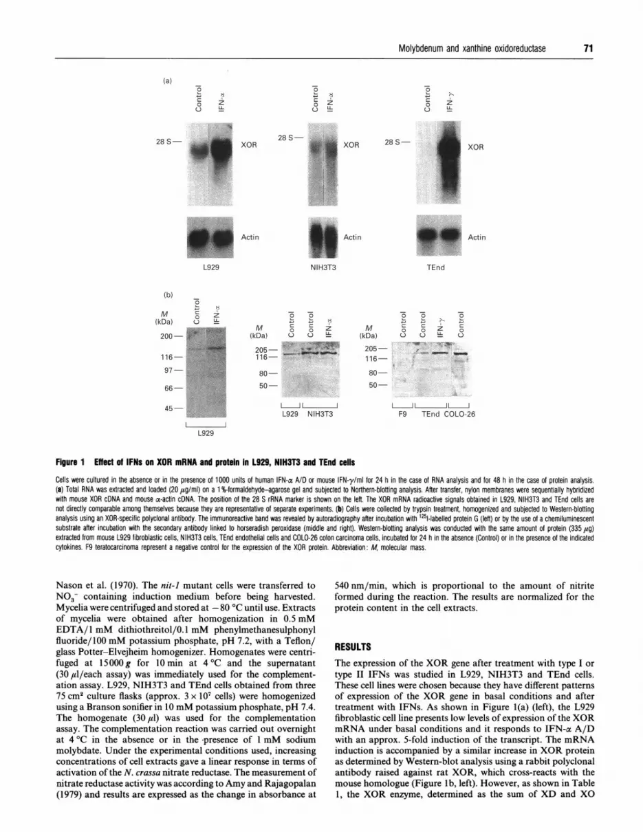
Figure 1 Effect of IFNs on XOR mRNA and protein In L929, NIH3T3 and TEnd cells
Cells were cultured in the absence or in the presence of 1000 units of human IFN-a A/D or mouse IFN-y/ml for 24 h in the case of RNA analysis and for 48 h in the case of protein analysis.(a) Total RNA was extracted and loaded (20 ,ug/ml) on a 1 %-formaldehyde-agarose gel and subjected to Northern-blotting analysis. After transfer, nylon membranes were sequentially hybridizedwith mouse XOR cDNA and mouse ac-actin cDNA. The position of the 28 S rRNA marker is shown on the left. The XOR mRNA radioactive signals obtained in L929, NIH3T3 and TEnd cells arenot directly comparable among themselves because they are representative of separate experiments. (b) Cells were collected by trypsin treatment, homogenized and subjected to Western-blottinganalysis using an XOR-specific polyclonal antibody. The immunoreactive band was revealed by autoradiography after incubation with 1251-labelled protein G (left) or by the use of a chemiluminescentsubstrate after incubation with the secondary antibody linked to horseradish peroxidase (middle and right). Western-blotting analysis was conducted with the same amount of protein (335 ,ug)extracted from mouse L929 fibroblastic cells, NIH3T3 cells, TEnd endothelial cells and COLO-26 colon carcinoma cells, incubated for 24 h in the absence (Control) or in the presence of the indicatedcytokines. F9 teratocarcinoma represent a negative control for the expression of the XOR protein. Abbreviation: M, molecular mass.
Nason et al. (1970). The nit-i mutant cells were transferred toNO3- containing induction medium before being harvested.Mycelia were centrifuged and stored at -80 °C until use. Extractsof mycelia were obtained after homogenization in 0.5 mMEDTA/ 1 mM dithiothreitol/0. 1 mM phenylmethanesulphonylfluoride/l00 mM potassium phosphate, pH 7.2, with a Teflon/glass Potter-Elvejheim homogenizer. Homogenates were centri-fuged at 15000g for 10 min at 4 °C and the supernatant(30 ,ul/each assay) was immediately used for the complement-ation assay. L929, NIH3T3 and TEnd cells obtained from three75 cm2 culture flasks (approx. 3 x 10⁷ cells) were homogenizedusing a Branson sonifier in 10 mM potassium phosphate, pH 7.4.The homogenate (30,1) was used for the complementationassay. The complementation reaction was carried out overnightat 4 °C in the absence or in the -presence of 1 mM sodiummolybdate. Under the experimental conditions used, increasingconcentrations of cell extracts gave a linear response in terms ofactivation of the N. crassa nitrate reductase. The measurement ofnitrate reductase activity was according to Amy and Rajagopalan(1979) and results are expressed as the change in absorbance at
540 nm/min, which is proportional to the amount of nitriteformed during the reaction. The results are normalized for theprotein content in the cell extracts.
RESULTSThe expression of the XOR gene after treatment with type I or
type II IFNs was studied in L929, NIH3T3 and TEnd cells.These cell lines were chosen because they have different patternsof expression of the XOR gene in basal conditions and aftertreatment with IFNs. As shown in Figure l(a) (left), the L929fibroblastic cell line presents low levels of expression of the XORmRNA under basal conditions and it responds to IFN-ac A/Dwith an approx. 5-fold induction of the transcript. The mRNAinduction is accompanied by a similar increase in XOR proteinas determined by Western-blot analysis using a rabbit polyclonalantibody raised against rat XOR, which cross-reacts with themouse homologue (Figure Ib, left). However, as shown in Table1, the XOR enzyme, determined as the sum of XD and XO
-5
0u
(a)
28 S-
zLL
0L-
0u
zLL
71
XOR28 S-
L929
0L-
0U
zL1
(b)
M(kDa)
200-
116-
97
66-
45
l IL929

72 F. Falciani and others
Table 1 Effect of IFN-ce (AID) and IFN-y on the levels of XOR enzymicactivityCells were cultured in the absence or in the presence of 1000 units of human IFN-a A/D ormouse IFN-y/ml for 48 h. Cells were collected by scraping with a rubber policeman andhomogenized as described in the Materials and methods section; 10 ,l of the homogenate wasincubated with [14C]hypoxanthine for 10 min in the presence of NAD+. XOR total activity isdetermined as the sum of XD and XO enzymic activities after quantification of the amount of[14C]xanthine and uric acid formed; 0.01 uniUmg of protein is the limit of detection of theenzymic assay. Abbreviation: N.D., not done. Each experimental value is the mean + S.D. fromthree separate culture dishes.
XOR activity (units/mg of protein)
Treatment L929 NIH3T3 TEnd COL026
NoneIFN-aIFN-y
< 0.01 18.83 + 1.94< 0.01 18.24 + 2.31< 0.01 17.63 + 1.32
0.12+ 0.010.11 + 0.021.98 + 0.10
11.71 + 0.23N.D.N.D.
M(kDa)
200-116-
80-
-a
0u
-5
a C)
0
0u
-a
00
Z U
50-
L929 3T3 L929 3T3
Figure 2 Induction of a single protein with XO activity by sodium molybdate
L929 and NIH3T3 fibroblastic cells were grown for 24 h in the absence (control) or in thepresence (Mo) of 10 mM sodium molybdate. Cells were harvested by scraping them off thewells, homogenized, and the resulting extracts containing 300 ,ug of protein were subjected to7.5% SDS/PAGE under non-reducing conditions. Gels were stained with NitroBlue Tetrazoliumin the presence of hypoxanthine for the active staining of xanthine (left panel) and withCoomassie Blue for the detection of proteins. The arrow indicates the position of the band withXO enzymic activity. The positions of molecular-mass (M) standards are indicated on the left.
Table 2 Effect of sodium molybdate on XOR enzymic activity in L929,NIH3T3 and TEnd cells after incubation in the absence and in the presenceof IFNsL929, NIH3T3 and TEnd cells were incubated in the absence or in the presence of 1000 unitsof human IFN-oa A/D or mouse IFN-y/ml for 30 h and subsequently the cells were treated withmedium alone (none), medium containing 10 mM sodium molybdate (Mo), medium containing1000 units of human IFN-ac A/D/ml (IFN-a), medium containing 1000 units of mouse IFN-y/ml(IFN-y) or medium containing the combination of the fwo compounds (either IFN-a+ Mo orIFN-y+ Mo) for another 18 h. Cells were harvested by scraping with a rubber policeman,homogenized, and an appropriate aliquot of the homogenate was used for the assay of total XORenzymic activity determined as the sum of XD and XO activities; 0.01 unit/mg of protein is thelimit of detection of the enzymic assay. Abbreviation: N.D., not done. Each experimental valueis the mean+ S.D. from three separate culture dishes.
XOR enzymic activity (units/mg of protein)
Treatment L929 NIH3T3 TEnd
NoneMoIFN-aIFN-a + MoIFN-yIFN-y + Mo
< 0.010.39 + 0.03
< 0.010.94 + 0.06
< 0.01N.D.
11.11 +1.3411.42 +1.5113.92 + 3.9511.25 + 0.5412.40 + 0.82N.D.
0.10 + 0.010.13 + 0.030.12+ 0.02N.D.0.74+ 0.080.69 + 0.06
activities, is undetectable both under basal conditions and aftertreatment with IFN-a A/D or IFN-y.The inability to measure XOR enzymic activity is not due to
lack of sensitivity of the assay used. In fact, L929 expresses
approximately the same amount of XOR mRNA and immuno-reactive protein as TEnd, a cell line that expresses measurablelevels of XOR activity (see below). Moreover, XOR enzymicactivity in L929 cells is not measurable even if a quantity ofprotein more than ten times higher than that ofTEnd cells is usedfor the assay (results not shown).As shown in Figure 1(a) (middle), Figure 1(b) (middle) and in
Table 1, NIH3T3 fibroblasts express significant amounts ofXOR message, immunoreactive protein and enzymic activityunder basal conditions. Densitometric analysis of the signalcorresponding to the XOR immunoreactive band demonstratesthat NIH3T3 has approx. 10-fold higher levels of the protein
relative to L929 cells grown in basal medium. In NIH3T3 cells,although IFN-a A/D increases the expression of the XORmRNA by 1.5-fold, the respective protein is not significantlyinduced. Similar results were obtained after treatment of L929cells with IFN-y (data not presented).As shown on Figure 1(a) (right), TEnd expresses high levels of
XOR mRNA after treatment for 24 h with IFN-y (but not withIFN-a A/D; results not shown). This cell line expresses XORmRNA also under basal conditions, since a specific signal for thetranscript is observed in the control lane when the Northern blotis exposed for a longer period of time (results not shown). Figurel(b) (right) demonstrates that the induction of the expression ofthe XOR gene is accompanied by an augmentation of therespective protein, even though the response (approx. 8-foldincrease) is lower than expected on the basis of the up-regulationof the mRNA. In TEnd cells, the enzymic activity (Table 1) islow, but readily detectable, and it is stimulated by approx. 20-fold after treatment with IFN-y for 24 h. F9 teratocarcinomaand COL026 adenocarcinoma cell extracts are shown in Figurel(b) (right) as negative and positive controls respectively, todemonstrate the specificity of theXOR band detected in Western-blotting analysis. In fact, F9 cells do not show detectable levelsof XOR enzymic activity before and after treatment with IFN-aA/D or IFN-y (results not shown), while COL026 expressesamounts of the enzyme that are similar to those observed inNIH3T3 (Table 1).
Since XD and XO are known to depend on the presence of amolybdopteridine cofactor for their activity (Rajagopalan andJohnson, 1992), the effect of molybdenum on L929 XOR wasstudied. As shown in Table 2, addition of sodium molybdate tothe culture medium at a concentration of 10 mM is capable ofinducing XOR activity (measured as the sum of XD and XO)both under basal conditions and after treatment for 24 h withIFN-ac A/D. Similar results are obtained when sodium molybdateis replaced by ammonium molybdate (results not shown). Theaction of molybdenum is not limited to the L929 clone routinelypassaged in our laboratory, since it is consistent with anotherL929 clone obtained from a different laboratory (Dr. SantoLandolfo, Istituto di Microbiologia, University of Turin, Turin,Italy), and a virally infected L929 clone (Radaelli et al., 1984)(results not shown). However, it is specific for mouse L929derivative lines, since the salt is not affecting XOR activity in

Molybdenum and xanthine oxidoreductase 73
Table 3 Effect of allopurinol on the Induction of XOR enzymic actilty bysodium molybdate In L929 cellsL929 fibroblastic cells were treated for 24 h with medium alone (None), medium containing theindicated concentrations of allopurinol (Allo), medium containing 10 mM sodium molybdate(Mo) or with medium containing both compounds (Allo + Mo) as indicated. Cells wereharvested by scraping with a rubber policemen, homogenized, and an appropriate aliquot of thehomogenate was used for the assay of both total XOR (XD + XO) and XO enzymic activities;0.01 unit/mg of protein is the limit of detection of the enzymic assay. Each experimental valueis the mean+ S.D. from three separate culture dishes.
Enzymic activity(units/mg of protein)
Treatment XO +XD XO
NoneAllo (0.1 mM)Allo (1 mM)MoAllo (0.1 mM) + MoAllo (1 mM) + Mo
< 0.01<0.01<0.01
0.35+ 0.030.07 + 0.01
< 0.01
<0.01< 0.01< 0.01
0.12 + 0.020.02 + 0.01
< 0.01
NIH3T3 or TEnd cells under any experimental condition (Table2). Other mouse and human cell lines, such as F9 teratocar-cinoma, L1210 lymphocytic leukaemia, COL026 adeno-carcinoma, HEPG2 hepatoma and HUVEC umbilical-vein en-dothelial cells, are not responsive to sodium molybdate treatmentin terms ofXOR activity (results not shown). These cell types donot present XOR enzymic activity in basal conditions, except formouse COL026 adenocarcinoma.
Figure 2 demonstrates that the activation of XOR enzymicactivity by sodium molybdate in L929 cells is accompanied bythe induction of a specific protein band with XO activity,migrating with an apparent molecular mass of 300 kDa onSDS/PAGE under non-reducing conditions. In these exper-imental conditions, purified XOR migrates as an enzymicallyactive dimer (Carpani et al., 1990). A similar band is observed inextracts of NIH3T3 cells grown in the absence of molybdenum.Approximately the same amount of protein was loaded in all thelanes of the gel, as demonstrated by the Coomassie Blue stainingof a similar gel run in parallel (right-hand side of the Figure). Asdemonstrated by Table 3, the induced XOR activity is inhibited
by addition to the culture medium of allopurinol (0.1 and 1 mM),a specific irreversible inhibitor of XD and XO (the level ofinhibition is similar for both total XOR and XO activities). Atthese two concentrations, allopurinol is not toxic as judged bythe morphology, the growth and the viability of L929 cells.Taken together, these data prove that a bona fide XOR enzymeis induced by molybdenum salts.The activation ofXOR enzyme is observed when molybdenum
is added to the cell cultures, but not to the cell homogenates. Infact, if L929 cells are disrupted by sonication and incubated inthe presence of sodium molybdate (50,tM) for 2 h at roomtemperature or for 12 h at 4 °C, XOR is not detectable. Underthese experimental conditions the enzyme is stable, since XORactivity does not change in NIH3T3 cell extracts. In fact ifNIH3T3 extracts are incubated for 2 h at room temperature,the XOR activity observed is 10.7 + 0.8 in the absence and10.5 + 0.6 units/mg of protein in the presence of sodiummolybdate, whereas after 12 h at 4 °C, the values are 10.0 + 0.3and 10.5 + 0.2 respectively. Similar results for L929 and NIH3T3are obtained when sodium molybdate is increased to 5 mM.The time course and the dose response for the induction of
XOR activity in L929 cells are presented in Figure 3. In theseexperiments, total XOR (XD + XO) and XO enzymic activitieswere measured separately. As shown in Figure 3(a), sodiummolybdate at a concentration of 10 mM induces both XO andXOR activity with similar kinetics, and the ratio XO/XOR(approx. 50 %) is not changed at any time point. The kinetics ofinduction of XOR and XO enzymic activities are relatively slow.Maximal increase is observed at 16 h, and the two activitiesremain high until 24 h. At 48 h a decrease in the levels of XORand XO is observed, probably as a consequence of the mildtoxicity produced by prolonged treatment with high concentra-tions of sodium molybdate. Figure 3(b) demonstrates that thecontinuous presence of the molybdenum salt in the medium isrequired to obtain maximal activation of XOR and XO. If cellsare incubated in the presence of 10 mM sodium molybdate for 1,4 and 8 h before change to fresh medium without molybdenumsalt and further incubation up to 24 h, submaximal activation ofthe two enzymes is observed (compare the values obtained atthese time points relative to that obtained after treatment of cellsfor 24 h in the presence of sodium molybdate). Figure 3(c) showsthat maximal induction of XOR and XO is observed afterincubation of cells for 24 h in the presence of 20 mM sodiummolybdate, even though detectable amounts of the two enzymic
0.6z,>
0.
M ly 0.2*u C
-a
o io 40Time (h)
do do 0 10 iO 30Time (h)
0.6
0.4
0.2
0
[Mo] (mM)
Figure 3 Effect of the exposure tme and concentration of sodium molybdate on the Induction of XOR enzymic activity in L929 cells
(a) Cells were treated with 10 mM sodium molybdate for the indicated amount of time. Cells were scraped from culture dishes, homogenized, and an aliquot of the homogenate was used for the
assay of total XOR (0) or XO (O) enzymic activity. (b) Cells were treated with 10 mM sodium molybdate for the indicated amount of time, washed and incubated with fresh medium without
the molybdenum salt for up to 24 h. Cells were harvested and used for the determination of total XOR (-) and XO (O) enzymic activities as in (a). (c) Cells were treated with the indicated amounts
of sodium molybdate for 24 h. Measurement of total XOR (0) and XO ([1) enzymic activities were performed as described in (a). Each experimental value is the mean + S.D. for three separateculture dishes.
(a)0.8 -
(b)0.6 -
0.4-
0.2-
o _c- - I II~ ..
r-
Fs-

74 F. Falciani and others
0
_ 2_
E EEC o0o o. x, E E E coU o Cs -n to u
- XOR
Table 4 Effect of cycloheximide on the levels of XOR enzymic activity inL929 cellsL929 cells were cultured for 30 min in medium alone or in medium containing 20 ,ug ofcycloheximide (CHX)/ml, followed by 6 h in the absence (None) or presence of 10 mM sodiummolybdate (Mo). CHX+ Mo* indicates cells incubated simultaneously for 6 h with thecombination of cycloheximide and Mo. Cells were homogenized, and an aliquot of thehomogenates was used for the determination of total XOR enzymic activity; 0.01 unit/mg ofprotein is the limit of detection of the enzymic assay. Each experimental value is themean+S.D. from three separate culture dishes.
Treatment
-Actin
,IL1210 L929
0
o5 o5 + -o
o o Z 0 z 0 0u C) 2 U- C) 2
I1IL929 3T3
Figure 4 Effects of sodium molybdate on the steady-state levels of XORmRNA and protein
(a) L929 cells were treated for 24 h with the indicated concentrations of sodium molybdate.Total RNA was extracted and loaded (20,g/ml) on a 1% formaldehyde agarose gel andsubjected to Northern-blotting analysis. After transfer, nylon membranes were sequentiallyhybridized with mouse XOR cDNA and mouse a-actin cDNA. The position of the 28 S ribosomalRNA marker is shown on the left. RNA extracted from the mouse L1210 cell line was used asa negative control. (b) L929 cells were treated for 24 h with 1000 units of IFN-a A/D/ml (IFN-a), 2 mM sodium molybdate (Mo) or a combination of the two compounds. 3T3 cells wereincubated in the absence (Control) or in the presence of 2 mM sodium molybdate (Mo). Cellswere homogenized, and an aliquot of the homogenates containing 300 ,g of protein wassubjected to Western-blotting analysis using polyclonal antibodies specific for XOR. Theimmunoreactive protein band was revealed by autoradiography after incubation with 1251-labelledprotein G. Abbreviation: M, molecular mass.
activities are already evident after incubation with 500 ,uM. Nofurther activation of the two enzymes is observed if the con-centration of the molybdenum salt is raised up to 50 mM.As shown in Figure 4(a), Northern-blotting experiments,
performed on total RNA extracted from L929 cells incubated for24 h in the presence of increasing concentrations of sodiummolybdate, demonstrate that the steady-state levels of XORmRNA are not influenced by molybdenum. Thus the metal doesnot induce XOR enzymic activity through an increase in theexpression of the corresponding gene. Furthermore, Figure 4(b)rules out the possibility that the induction of XOR enzyme bymolybdenum is the consequence of increased translation fromthe respective transcript. In fact, the levels of the 150 kDa XOR
NoneMoCHXCHX + Mo*CHX + Mo
XOR enzymic activity(units/mg of protein)
< 0.010.52 + 0.03
< 0.010.40 + 0.090.80 + 0.10
Table 5 Effect of the exposure tme of sodium molybdate in the growthmedium on the intracellular level of molybdenum in L929, NIH3T3 and TEndcellsCells were exposed to 5 mM sodium molybdate for the indicated amount of time. Theintracellular concentration of molybdenum (Mo) was determined. Each experimental value is themean+S.D. from three separate culture dishes.
[Mo] (,ug/106 cells)
Time L929 NIH3T3 TEnd
0 min 0.37 + 0.0210 min 1.07+0.1230 min 1.82 + 0.132 h 1.36 + 0.08
24 h 1.27+0.1148 h 1.54+0.03
0.82 + 0.041.95 + 0.022.67 + 0.182.99+ 0.263.45+ 0.291.76 + 0.09
immunoreactive protein are not changed after incubation ofL929 cells with 2 mM sodium molybdate, a concentration thatpotently activates XOR enzymic activity. Finally, as shown inTable 4, the induction ofXOR enzymic activity is not blocked byincubation of the L929 cells with cycloheximide at concentrationsknown to inhibit protein synthesis. At this concentration, cyclo-heximide has a slight inducing effect on XOR enzymic activity ifcells are preincubated with the protein-synthesis inhibitor for30 min before addition of sodium molybdate. Thus de novo
protein synthesis is not required for the inducing effect triggeredby sodium molybdate.To study further the defect of L929 cells leading to the
production of an inactive form of XOR apoprotein, thetime course and the concentration curve for the intracellularmolybdenum was compared with those observed in NIH3T3 andTEnd cells. As shown in Table 5, after incubation with 5 mMsodium molybdate, the accumulation of the metal is similar inL929 relative to TEnd and NIH3T3 cells. Maximal levels ofmolybdenum are evident after 30 min in the three cell lines.However, NIH3T3 accumulate an approx. 2-fold higher amountof molybdenum relative to the other two cell lines at 24 h.Moreover, the intracellular levels of molybdenum in L929 andTEnd are similar and approx. 50% lower than those observed inNIH3T3, in the absence of sodium molybdate in the growthmedium (0 min). Table 6 demonstrates that the accumulation of
(a)
28S-
18 S-
(b)
M(kDa)
200-
116-97-
66-
45-0.43 + 0.041.23 + 0.031.89 + 0.141.80 + 0.061.91 + 0.132.10+ 0.02

Molybdenum and xanthine oxidoreductase 75
Table 6 Effet of the concentradon of sodium molybdate In the growthmedium on the Intracellular level of molybdenum In L929, NIH3T3 and TEndcellsCeHs were exposed to the indicated concentrations of sodium melybdate for 24 h. Theintracellular concentration of molybdenum (Mo) was determined. Each experimental value is themean +S.D. from three separate culture dishes.
[Mo] (1g/106 cells)Concn.(mM) L929 NIH3T3 TEnd
00.11.0
1050
0.29 + 0.050.30 + 0.022.97 + 1.845.64 +1.36
16.68 + 5.54
1.12 +0.311.09 + 0.346.72 +1.23
15.63 + 6.8549.13 + 4.15
0.37 + 0.030.52 + 0.054.62 + 0.279.25 + 2.63
12.01 + 1.37
Table 7 Effect of sodium molybdate on the capacity of L929- and NIH3T3-cell extracts to restore nItrate reductase enzymic activity of the nit-i mutantof Neurospora crassaL929 and NIH3T3 cells were exposed to medium alone (L929; NIH3T3) or medium containing5 mM sodium molybdate [L929 (Mo); NIH3T3 (Mo)] for 24 h. Cells were harvested,homogenized, and an aliquot of the homogenate containing the same amount of protein(180 1ug) was incubated overnight at 4 OC with extracts of mycelia from nit-1 Neurospora crassain the absence (- Mo) or in the presence (+ Mo) of 1 mM sodium molybdate. Appropriateblanks of nit-i extracts (nit-1) were incubated in parallel with the samples in the presence orin the absence of Mo (- Mo; + Mo). Nitrate reductase activity was measured in reconstitutedextracts, and the results are normalized for the amount of protein present in the L929- andNIH3T3-cell homogenates. Values are expressed as AA540/min per mg of protein in theeukaryotic-cell extract. < 1.0, below the limit of detection of the assay. Each experimental valueis the mean + S.D. for three assays.
Nitrate reductase activity
Extract -Mo + Mo
nit-1L929 + nit-1L929 (Mo) + nit-1NIH3T3 + nit-INIH3T3 (Mo)+nit-1
< 1.0< 1.026.6 + 2.124.6 + 1.710.4 + 0.6
< 1.042.8 + 2.557.8 + 3.047.5 + 1.728.8 + 2.4
molybdenum is similar for all the three lines, even thoughNIH3T3 retains 2-3-fold more molybdenum than L929 or TEndcells. The results are consistent when expressed on the basis ofthe protein content in the various cells. Moreover, the averagevolume of L929 relative to NIH3T3 and TEnd cells is verysimilar, as assessed by direct measurement in a Coulter counter(results not shown).The relative amount of intracellular molybdenum cofactor
which is essential for the activity of XOR was next evaluated.For this purpose, extracts of L929 and NIH3T3 cells grown inthe presence and in the absence of 5 mM sodium molybdate weretested for their ability to complement the nitrate reductase defectobserved in extracts of the nit-i mutant of N. crassa. Thiscomplementation test is widely used to assess the presence ofmolybdenum cofactor in an indirect way (Joh-nson, 1980). In theabsence ofmolybdenum exogenously added to the reconstitutionmixture, the assay allows the determination of the levels of thepreformed molybdenum cofactor present in the mouse cell lines(Ketchurm and Swain, 1973). In this case, as shown in Table 7,extracts obtained from NIH3T3 cells grown either in the absence
or in the presence of sodium molybdate rescue N. crassa nitratereductase activity. By contrast, L929 cells grown in the presenceof molybdenum rescue nitrate reductase activity, whereas thosegrown in the absence of the metal do not. If the complementationassay is carried out in the presence of molybdenum (1 mM)exogenously added to the reconstitution mixture, an enzymicactivity (the converting enzyme) capable of metabolizing pre-cursors of the molybdenum cofactor that accumulate in the cellsof the N crassa nit-I mutants is preferentially measured (Johnsonet al., 1989; Rajagopalan and Johnson, 1992). Under theseexperimental conditions it is evident from Table 7 that both L929cells and NIH3T3, grown either in the absence or in the presenceof sodium molybdate, can rescue nit-i nitrate reductase activity.The reconstitution experiments involving complementation ofthe N. crassa extracts for nitrate reductase activity were per-formed using crude extracts of the micelia. Qualitatively similarresults were obtained if the fungal extract was passed through aSephadex G-25 column in order to obtain a nitrate reductasepreparation free of the low-molecular-mass pterydine precursors(results not shown).
DISCUSSIONThe data presented here demonstrate that mouse L929 fibroblastshave a defect in the assembly of active XOR both under basalconditions and after treatment with type I or type II IFNs. Theenzymic activity is rescued by the addition of millimolar concen-trations of sodium molybdate to the growth medium. Theactivation of XOR by molybdenum salts is specific for mouseL929 fibroblasts, since it is not observed in a panel of othermouse and human cell lines. Our experimental data suggest thatL929 cells represent a mutant for the expression of XOR. Thedeficit, however, cannot be explained by a structural alteration ofthe XOR protein, but rather by impaired synthesis of themolybdenum cofactor that is essential for the activity of theenzyme. As demonstrated by the use of a heterologous comple-mentation test, L929 cells have undetectable levels of molyb-denum cofactor if grown in the absence of sodium molybdate.If the cells are grown in the presence of the molybdenum salt, thecofactor can be measured using the same complementation test.NIH3T3 cells which are fully competent for the expression ofXOR enzyme complement the nit-i nitrate reductase deficit,regardless of whether they are grown in the absence or in thepresence of sodium molybdate.Molybdenum-cofactor biosynthesis and metabolism have been
studied in eukaryotes (Johnson et al., 1989) and more extensivelyin prokaryotes (Stewart, 1988). In eukaryotes, most of theinformation on the biosynthesis of the molybdenum cofactorhave been gathered from studies on fibroblasts obtained fromhuman patients suffering from a rare genetic disease known as'combined deficiency of sulphite oxidase and xanthine oxidase'(Johnson and Wadman, 1989). The symptoms of this disease arepredominantly the consequence of the sulphite oxidase defect,which leads to a severe impairment of the central nervous system(Aukett et al., 1988; Endres et al., 1988). At present, twocomplementation groups, A and B (Johnson et al., 1989), havebeen characterized. Group-A patients are unable to accumulatemolybdopterin precursors as a consequence of unknown meta-bolic defects in this biosynthetic pathway, whereas group-Bpatients are unable to metabolize molybdopterin precursors tomolybdenum cofactor because of a lack of converting enzyme(Johnson et al., 1989). The converting enzyme is a proteincatalysing the transformation of the precursor Z (Rajagopalanand Johnson, 1992) to a pteridin compound which subsequentlyinteracts with molybdenum and produces the molybdenum

76 F. Falciani and others
cofactor. This is exactly the same enzymic activity that is lackingin the nit-] mutants of the N. crassa (Rajagopalan and Johnson,1992) that we used in the present study. In prokaryotes, themetabolism of molybdenum cofactor has been studied both interms of its genetics and biochemistry on several pleiotropicmutants of Escherichia coli selected as chlorate-resistant strains(Stewart, 1988). In bacterial cells, several genes are directlyinvolved in the biosynthetic pathway leading to the formation ofactive molybdenum cofactor. The chlA genes 4 and 5 encode thetwo subunits that constitute the active form of the convertingenzyme, whereas the chlN gene is involved in the activation of thesmall subunit of the complex (Reiss et al., 1987; Pitterle andRajagopalan, 1989; Pitterle et al., 1990; Zurick et al., 1991). ThechiD and chiG gene products are involved in the transport acrossthe membrane and possibly in the insertion of the molybdenumions in the pteridine precursor respectively (Stewart, 1988; Glaserand DeMoss, 1971). In fact, chlD mutants are defective in one ormore factors involved in the transport of molybdenum inside thecell (Stewart, 1988). By contrast, the chiG mutant seems toexpress an altered form of a putative intracellular carrier proteinnecessary to donate molybdenum to compound Z, the immediateprecursor of molybdenum cofactor (Glaser and DeMoss, 1971).L929 cells are certainly not defective in the expression of the
converting enzyme. In fact, extracts of this cell line, grown bothin the absence and in the presence of sodium molybdate, canrescue N. crassa nitrate reductase if the complementation test isperformed in the presence of exogenously added molybd-enum(VI) ions. These complementation conditions are knownto allow the indirect measurement of the converting enzyme(Johnson et al., 1989; Johnson and Rajagopalan, 1992).
Insofar as the XOR enzymic activity is restored by treatmentin high concentrations of sodium molybdate, L929 cells resemblethe chiD and chlG mutants of E. coli as well as the nifQ mutantsofKlebsiellapneumoniae (Imperial et al., 1984). The chiD mutantof E. coli and the nifQ mutant of K. pneumoniae are defective inthe transport of molybdenum inside the cell. Unequivocaldemonstration of a lesion in the transport ofmolybdenum acrossthe cell membrane would require experiments of intracellularaccumulation of the metal using fast kinetics and concentrationsof the metal similar to those observed in the growth mediumwithout supplementation of sodium molybdate. However, twopieces of evidence suggest that the deficit in the expression ofXOR activity in L929 cells is not the result of a defectiveintracellular accumulation of molybdenum. chiD mutant cellscontain approx. 10-20% of the levels of molybdenum present inwild-type E. coli, whereas in the absence of sodium molybdate inthe growth medium, L929 fibroblasts have intracellular levels ofthe metal that are lower than those observed in NIH3T3, butsimilar to those observed in TEnd endothelial cells, which arecompetent for the expression of XOR. Moreover, at concen-tration of Mo between 0.1 and 50 mM, the kinetics and theconcentration profile for the intracellular accumulation ofmolybdenum seem to be similar in the three cell lines. Althoughthe absolute amounts of intracellular molybdenum in NIH3T3are higher than those in L929 and TEnd, clearly this fact alonedoes not explain the observed differences in the expression ofXOR.At present we do not have any direct evidence to explain the
mechanism underlying the defect observed in L929 cells, eventhough it is quite possible that a factor or an enzyme involved inone of the several steps leading to the synthesis of molybdenumcofactor is altered in its structure and it requires high concentra-tions of molybdenum to work. This would account for the factsthat the molybdenum cofactor does not accumulate in the
port across the membrane is normal. It would be tempting tospeculate that the deficit observed in L929 cells is analogous tothat observed in ChlG mutants of E. coli.
Since the molecular defect observed in L929 probably resultsin the synthesis of the molybdenum cofactor, it is anticipatedthat this cell line must be also deficient in the expression ofsulphite oxidase and aldehyde oxidase as well. We are currentlytrying to address this issue, even though the low sensitivity of thespectrophotometric assays used for the determination of thesetwo enzymic activities and the lack of cDNA clones coding forthe two proteins in mammals make these studies difficult toperform in L929 cells.
It would be of considerable interest to know whether the defectin the expression of XOR observed in L929 is a genetic traitacquired by the process of immortalization and stabilization invitro of the cell line or whether the defect was already present inthe original mouse strain. In fact, this cell line has been derivedmore than 50 years ago from normal subcutaneous areolar andadipose tissue of a 100-day-old male C3H/An mouse. Mice fromthe syngeneic strain C3H/HeN do not present a deficit of XO inthe liver (results not shown). By contrast, it is known that thismouse strain contains approximately one-tenth of the amount ofaldehyde oxidase present in C57BL/6 mice (Holmes et al., 1981;Huff and Chaykin, 1967). Unfortunately, this evidence does notallow us to draw any conclusion, especially in view of the factthat the situation in the whole animal is certainly differentfrom that observed in cell culture. Moreover, it is to beremembered that laboratory animals are fed with semi-syntheticdiets which contain molybdenum ions, and the daily requirementfor this metal is extremely low (Rajagopalan, 1988).
In conclusion, in spite of the fact that the mechanism under-lying the defect ofXOR expression in L929 is still not completelyunderstood, the findings presented here have a potential andgeneral interest at least in two areas of investigation.
First of all, the correction of the XOR defect in L929 cells bymolybdenum salts might have some clinical relevance for theaetiology and the treatment of the combined deficiency ofmolybdoenzyme activities. So far, all the patients suffering fromthis disease appear to be deficient in molydopterin rather thanmolybdenum itself (Rajagopalan, 1988). Although administra-tion of molybdenum proved ineffective to correct the deficit inone reported case, this need not be true in all instances. To thebest of our knowledge, the effect of molybdenum salts on thedeficit ofXOR and sulphite oxidase activity observed in primaryfibroblast obtained from these patients has never been testedsystematically. It is thus possible that a subgroup of group-A-type fibroblasts might be responsive to molybdenum challenge ifthe underlying defect is similar to that observed in mouse L929cells. For these reasons, it would be interesting to test the effectof high concentrations of the metal on XOR or sulphite oxidaseactivities in group-A fibroblasts. It would be also worthwhileperforming cell-fusion experiments between human group-A andgroup-B mutants and mouse L929 cells to investigate the relativecomplementation pattern.
Secondly, our findings are of concern to those investigatorsinvolved in the study of the biological effects produced by IFNs.In fact, although it is yet to be established that XOR proteinplays any role in the immunomodulatory, antiviral or antipro-liferative activities triggered by these cytokines, it is clear that therelative gene is regulated by both IFN-a and -y without de novosynthesis of intermediary transcription factors (Terao et al.,1992; Falciani et al., 1992). As such the XOR gene should beconsidered as an IFN-responsive one. Since L929 cells are largelyused for the study of both the antiviral and the antiproliferativeactivity of IFNs, it should be emphasized that this cell line isintracellular compartment, even though the molybdenum trans-

Molybdenum and xanthine oxidoreductase 77
defective in the expression of at least one of the possible genesinvolved in the pleiotropic response to these cytokines.
This work was supported in part by Grants from the Consiglio Nazionale delleRicerche (CNR), Progetto Finalizzato 'Chimica Fine', 'Ingegneria Genetica', ProgettoFinalizzato 'Biotecnologie e Biostrumentazione' and from the Associazione per laRicerca Contro il Cancro (AIRC). We would like to thank Dr. Takeshi Nishino(Department of Biochemistry, Yokohama School of Medicine) for supplying us withthe polyclonal antibodies against rat XD. We are also grateful to Dr. A. Caruso andDr. A. Colombo (Farmitalia Carlo Erba, Nerviano, Italy) for the growth of theNeurospora crassa strains. F. F. is a recipient of a fellowship from the FondazioneAttilia Pofferi.
REFERENCESAmaya, Y., Yamazaki, K., Sato, M., Noda, K., Nishino, T. and Nishino, T. (1990) J. Biol.
Chem. 265, 14170-14175Amy, N. K. and Rajagopalan, K. V. (1979) J. Bacteriol. 140, 114-124Aukett, A., Bennett, M. J. and Hosking, G. P. (1988) Dev. Med. Child. Neurol. 30, 531-535Bradford, M. M. (1976) Anal. Biochem. 72, 248-254Bray, R. C. (1975) Enzymes 3rd edn. 12, 299-419Bray, R. C. (1982) in Flavin and Flavoproteins (Massey, V. and Williams, C. H., eds.),
pp. 775-785, Elsevier, New YorkBray, R. C. (1988) 0. Rev. Biophys. 21, 299-329Carpani, G., Racchi, M., Ghezzi, P., Terao, M. and Garattini, E. (1990) Arch. Biochem.
Biophys. 279, 237-241de Groot, H. and Littauer, A. (1988) Biochem. Biophys. Res. Commun. 155, 278-282Della Corte, E. and Stirpe, F. (1968) Biochem. J. 108, 349-351Della Corte, E. and Stirpe, F. (1972) Biochem. J. 126, 739-745Endres, W., Shih, Y. S., Gunther, R., Ibel, H., Duran, M. and Wadman, S. K. (1988) Eur. J.
Pediatr. 148, 246-249Falciani, F., Ghezzi, P., Terao, M., Cazzaniga, G. and Garattini, E. (1992) Biochem. J. 285,
1001-1008Feinberg, A. P. and Vogelstein, B. (1983) Anal. Biochem. 132, 6-13Fridovich, I. (1970) J. Biol. Chem. 245, 4053-4057Gardlik, S. and Rajagopalan, K. V. (1990) J. Biol. Chem. 265, 13047-13054Glaser, J. H. and DeMoss, J. A. (1971) J. Bacteriol. 108, 854-860Holmes, R. S., Leijten, L. R. and Duley, J. A. (1981) Anim. Blood Groups Biochem. Genet.
12, 193-199Huff, S. D. and Chaykin, S. (1967) J. Biol. Chem. 242, 1265-1270Imperial, J., Ugalde, R. A., Shah, V. K. and Brill, W. J. (1984) J. Bacteriol. 158, 187-194Johnson, J. L. (1980) in Molybdenum and Molybdenum-Containing Enzymes (Coughlan,
M. P., ed.), pp. 345-383, Pergamon Press, Oxford
Received 14 April 1993/13 August 1993; accepted 1 September 1993
Johnson, J. L. and Rajagopalan, K. V. (1982) Proc. Natl. Acad. Sci. U.S.A. 79, 6856-6860Johnson, J. L., and Wadman, S. K. (1989) in The Metabolic Basis of Inherited Disease
(Scriver, C. R., Baudet, A. L., Sly, W. S. and Valle, D., eds.), 6th edn., vol. 1,pp. 1463-1475, McGraw-Hill Book Co., New York
Johnson, J. L., Hainline, B. E. and Rajagopalan, K. V. (1980a) J. Biol. Chem. 255,1783-1786
Johnson, J. L., Waud, W. R., Rajagopalan, K. V., Duran, M., Beemer, F. A. and Wadman,S. K. (1980b) Proc. Natl. Acad. Sci. U.S.A. 77, 3715-3719
Johnson, J. L., Hainline, B. E., Rajagopalan, K. V. and Arison, B. H. (1984) J. Biol. Chem.259, 5414-5422
Johnson, J. L., Wuebbens, M. M., Mandell, R. and Shih, V. E. (1989) J. Clin. Invest. 83,897-903
Ketchum, P. A. and Swarin, R. S. (1973) Biochem. Biophys. Res. Commun. 52, 1450-1456Kramer, S. P., Johnson, J. L., Ribeiro, A. A., Millington, D. S. and Rajagopalan, K. V. (1987)
J. Biol. Chem. 262, 16357-16363Maniatis, T., Fritsch, E. F. and Sambrook, J. (1989) Molecular Cloning: A Laboratory
Manual, Cold Spring Harbor Laboratory Press, Cold Spring Harbor, NYMcCord, J. M. (1985) N. Engl. J. Med. 312, 159-163Minty, A. J., Caravatti, M., Robert, B., Cohen, A., Daubas, P., Weydert, A., Gros, F. and
Buckingham, M. E. (1981) J. Biol. Chem. 256, 1008-1014Nason, A., Antoine, A. D., Ketchum, P. A., Frazier, W. A., IlIl and Lee, D. K. (1970) Proc.
Natl. Acad. Sci. U.S.A. 65,137-144Nishino, T., Nishino, T., Schopfer, L. M. and Massey, V. (1989) J. Biol. Chem. 264,
2518-2527Parks, D. A. and Granger, D. N. (1983) Am. J. Physiol. 245, G285-G289Pitterle, D. M. and Rajagopalan, K. V. (1989) J. Bacteriol. 171, 3373-3378Pitterle, D. M., Johnson, J. L. and Rajagopalan, K. V. (1990) FASEB J. 4, A1957Radaelli, A., Righi, M., Liboi, E. and De Giuli Morghen, C. (1984) J. Gen. Virol. 65,
295-307Rajagopalan, K. V. (1988) Annu. Rev. Nutr. 8, 401-427Rajagopalan, K. V. and Handler, P. (1967) J. Biol. Chem. 242, 4097-4107Rajagopalan, K. V. and Johnson, J. L. (1992) J. Biol. Chem. 267, 10199-10202Rambaldi, A., Young, D. C. and Griffin, J. D. (1987) Blood 69, 1409-1413Reiners, J. J., Jr., Pence, B. C., Barcus, M. C. S. and Cantu', A. R. (1987) Cancer Res. 47,
1775-1 779Reiss, J., Kleinhofs, A. and Klingmuller, W. (1987) Mol. Gen. Genet. 206, 352-355Saito, T. and Nishino, T. (1989) J. Biol. Chem. 264, 10015-10022Stewart, V. (1988) Microbiol. Rev. 52 (2), 190-232Terada, L. S., Rubinstein, J. D., Lesnefsky, E. J., Horwitz, L. D., Leff, J. A. and Repine, J. E.
(1991) Am. J. Physiol. 260, H805-H810Terao, M., Cazzaniga, G., Ghezzi, P., Bianchi, M., Falciani, F., Perani, P. and Garattini, E.
(1992) Biochem. J. 283, 863-870Waud, W. R. and Rajagopalan, K. V. (1976) Arch. Biochem. Biophys. 172, 354-364Zurick, T. R., Pitterle, D. M. and Rajagopalan, K. V. (1991) FASEB J. 5, A470


