Embed Size (px)
Citation preview

Morphological, molecular and biogeographic evidence support two new speciesin the Uroptychus naso complex (Crustacea: Decapoda: Chirostylidae)
Gary C.B. Poore a,⇑, Nikos Andreakis b
a Museum Victoria, GPO Box 666, Melbourne, Vic. 3000, Australiab Australian Institute of Marine Science, PMB No. 3, Townsville, QLD 4810, Australia
a r t i c l e i n f o
Article history:Received 19 November 2010Revised 28 March 2011Accepted 30 March 2011Available online 22 April 2011
Keywords:ChirostylidaeTaxonomyPhylogeographyCryptic speciesAustraliaWallace’s line
a b s t r a c t
The tropical to subtropical squat lobster Uroptychus naso Van Dam, 1933 (Chirostylidae) is a widely dis-tributed species originally described from Indonesia, subsequently reported from the Philippines, Taiwan,Japan and it has recently been discovered on the continental slope of north-western Australia. Popula-tions of U. naso occur along the Indo-Pacific Ocean continental margin crossing the recently proposedmarine analog of Wallace’s line, responsible for past population fragmentation and ancient speciation.Sequence data from mitochondrial (COI, 16S) and nuclear (H3) DNA regions were used to assess genea-logical relationships among geographically disjoint populations of the species throughout its known dis-tribution range. Several mitochondrial lineages, corresponding to geographically isolated populations andthree cryptic species were encountered, namely, U. naso sensu stricto and two new species, Uroptychuscyrano and Uroptychus pinocchio spp. nov. U. pinocchio is encountered only in Japan, Taiwan and the Phil-ippines; U. cyrano is confined to north-western Australia; and U. naso consists of three genetically distinctpopulations distributed on both sides of the marine Wallace’s line. Fossil-calibrated divergence timeapproximations indicated a most recent common ancestor (MRCA) for U. naso and U. cyrano from earlyEocene whilst northern and southern populations of the former have been separated probably sincethe Miocene. These patterns may represent a standard distribution trend for several other deep-sea inver-tebrate species with similar geographical ranges.
� 2011 Elsevier Inc. All rights reserved.
1. Introduction
Molecular phylogenetics and systematics have greatly im-proved our understanding of marine diversity thereby allowingfor a broader perception of morphological character evolutionand delineation of species, the latter essential for biodiversity esti-mates. This is most important in marine invertebrates with so-called wide geographical distribution ranges but which prove tocomprise morphologically cryptic species or species complexes(Caputi et al., 2007; Knowlton, 1986; Machordom and Macpherson,2004; Seidel et al., 2009).
Squat lobsters of the families Chirostylidae, Galatheidae, Muni-didae and Munidopsidae (Baba et al., 2008; Taylor et al., 2010) arefound in all oceans, typically associated with coral assemblages indeep-sea waters from continental slopes to abyssal depths. Speciesencountered in the Southwest Pacific (SWP) for instance, arehypothesised to be the result of a relatively old explosive radiationevent followed by morphological stasis (Machordom andMacpherson, 2004). This hypothesis is now being tested by alarge-scale comparative phylogeographic survey that incorporates
recent collections of squat lobsters from north-western Australia(NWA) combined with previously described taxa of SWP origin(Australia’s Marine Biodiversity Hub – http://www.marinehub.org/index.php/site/home/). The Chirostylidae comprise seven gen-era and 192 described species. Of these, Uroptychus represents themost species-rich genus with 124 described species (Baba et al.,2008) and perhaps 70 more yet to be described (K. Baba, pers.comm.). Amongst these species, individuals of Uroptychus nasoVan Dam, 1933 are distinctive with a particularly long broadrostrum, several prominent lateral carapace spines and tuberculatedorsal carapace. The species was described from the Kei Islands(Indonesia) and subsequently populations were reported through-out the western Pacific (WP) archipelago in Indonesia, thePhilippines, Taiwan and Japan (Baba et al., 2008 and referencestherein). Recently collected material from the continental slopeof NWA was tentatively identified as belonging to this species.
The distribution range of U. naso overlaps the contact zonebetween the Indian and the Pacific Oceans and is typical of manymarine species exhibiting geographically associated geneticdiscontinuities (de Bruyn et al., 2004; Lourie and Vincent, 2004;Williams et al., 2002). The collision between Asian andAustralasian plates during the Miocene and the separation of theIndian and the Pacific basins during the Pleistocene represent a
1055-7903/$ - see front matter � 2011 Elsevier Inc. All rights reserved.doi:10.1016/j.ympev.2011.03.032
⇑ Corresponding author.E-mail address: [email protected] (G.C.B. Poore).
Molecular Phylogenetics and Evolution 60 (2011) 152–169
Contents lists available at ScienceDirect
Molecular Phylogenetics and Evolution
journal homepage: www.elsevier .com/locate /ympev

major biogeographical barrier between populations and sister taxain the Indian and Pacific Oceans. On the other hand, the Indo-Pacific break (IPB) is nearly perpendicular to Wallace’s line. Thelatter is an alternative biogeographical boundary, hypothesised tobe responsible for the separation of terrestrial flora and fauna ofeastern Asian and Australasian origins (Barber et al., 2000, 2002;Lourie and Vincent, 2004; Porter, 1989; Williams et al., 2002).
Preliminary molecular analyses from NWA specimens identifiedas U. naso indicated that two distinct mitochondrial lineages occursympatrically. This result raised the question of the identity oridentities of this species throughout its distribution range. Giventhe reported distribution of U. naso, we regard this species as anexcellent model system to test the impact of the IPB and the mar-ine Wallace’s line.
Our material from NW Australia was supplemented by freshmaterial from throughout the species’ range in the NW Pacificand compared with most of the specimens previously reportedand now in museums. Importantly, the morphology of allspecimens was compared with that of the type specimens.Mitochondrial and nuclear coding regions together with ribosomalgenes have been valuable tools in inferring phylogenies, recon-structing genealogies, corroborating morphological systematicsand uncovering speciation patterns in decapod crustaceans (e.g.,Cabezas et al., 2008, 2009 for Munididae). The mitochondrialpartial COI and 16S genes were used to infer phylogenetic
relationships between evolutionary significant units (ESUs sensuMoritz, 1994). Additionally, the nuclear Histone 3 (H3) gene mar-ker was employed to assess whether hybridization may have oc-curred among the lineages, due to biparental inheritance andeventual crossing-over during meiosis of the nuclear markers,and to verify if a biological species concept can be applied.
Our objectives are to: (a) investigate the extend of hidden mor-phological and genetic diversity; (b) assess congruence betweenmorphological differentiation and molecular phylogenies; and (c)elucidate phylogeographic relationships among ESUs within thisspecies complex against contrasting biogeographic hypotheses. U.naso and two previously unrecognised new species are described.New species epithets reflect the name of the original species, nasomeaning nose.
2. Materials and methods
2.1. Specimen collection and identification
Ethanol-preserved specimens were collected during a survey in2007 of the macrofauna of the continental slope of northern Wes-tern Australia (a northern extension of the sampling in southernWA reported by Poore et al., 2008) and now housed in MuseumVictoria (NMV). Additional ethanol-preserved specimens were
Table 1Specimens analyzed in this study. #, specimen catalogue number for tissue. Details of station locations are given with material examined in the taxonomic section. Cataloguenumbers 56 and 145 were supplied by C.-W. Lin, NTOU.
# Haplotype Clade Voucher location Voucher registration Sampling site Depth (m) Collection year GenBank Acc numbers COI/16S/H3
Uroptychus naso Van Dam, 1933175 – – ZMA De.101.667 Indonesia, Kei Is, Siboga stn 251 – – –/–/JF715103176 – – ZMA De.101.692 Indonesia, Kei Is, Siboga stn 153 – – –177 – – NTOU Taiwan, Nang fang-Ao – 1995 –/JF715120/JF71509911 1 I NTOU Taiwan, stn CP115 381–440 2001 JF715132/–/–180 1 I NTOU Taiwan, Su-Ao – 1998 JF715138/JF715121/JF71509556 11 I NTOU Taiwan, stn CP216 209–280 2003 JF715133/JF715124/JF715097145 12 I NTOU Taiwan – – JF715134/–/–185 13 II NTOU Philippines, stn CP2343 309–356 2001 JF715140/JF715126/JF715101186 14 II NTOU Philippines, stn CP2343 309–356 2001 JF715141/JF715127/JF715102187 15 II NTOU Philippines, stn CP2343 309–356 2001 JF715139/JF715123/JF71509829A 2 III NMV J60839 Australia, WA, stn 095 206–202 2007 JF715142/JF715129/JF715094219 2 III NMV J60838 Australia, WA, stn 112 202–191 2007 JF715143/JF715128/JF71509229B 3 III NMV J56118 Australia, WA, stn 095 206–202 2007 JF715137/JF715130/JF71509342 9 III NMV J56128 Australia, WA, stn 099 211–205 2007 JF715135/JF715119/JF71509045 10 III NMV J57259 Australia, WA, stn 098 206–187 2007 JF715136/JF715118/JF715091179 – – NTOU Taiwan, Dasi – 1991 –/JF715125/–181 – – NTOU Taiwan, stn CP115 381–440 2001 –182 – – NTOU Taiwan, stn CP114 128–250 2001 –/JF715122/JF715096183 – – NTOU Taiwan, stn CP114 128–250 2001 –/–/JF715100222 – – ZMUC CRU-11333 Indonesia, Kei Is 245 1922 –223 – – ZMUC CRU-11335 Indonesia, Kei Is 268 1922 –/–/JF715104224 – – ZMUC CRU-20206 Indonesia, Kei Is 245 1922 –225 – – ZMUC CRU-20207 Indonesia, Kei Is 270 1922 –
Uroptychus cyrano new species31A 7 IV NMV J57256 Australia, WA, stn 6 210–205 2007 JF715144/JF715109/JF71508231B 5 IV NMV J57256 Australia, WA, stn 6 210–205 2007 JF715146/JF715116/JF715084213 5 IV NMV J57256 Australia, WA, stn 6 210–205 2007 JF715152/JF715110/JF715085214 16 IV NMV J57256 Australia, WA, stn 6 210–205 2007 JF715147/JF715114/JF715078215 17 IV NMV J57256 Australia, WA, stn 6 210–205 2007 JF715148/JF715111/JF71507932 8 V NMV J57262 Australia, WA, stn 142 210–203 2007 JF715155/JF715117/JF71508330A 5 IV NMV J57263 Australia, WA, stn 112 202–191 2007 JF715145/JF715106/JF71508830B 6 V NMV J57263 Australia, WA, stn 112 202–191 2007 JF715153/JF715107/JF715086217 8 V NMV J57263 Australia, WA, stn 112 202–191 2007 JF715154/JF715113/JF715081218 18 IV NMV J57263 Australia, WA, stn 112 202–191 2007 JF715151/JF715115/JF715087216 16 IV NMV J60841 Australia, WA, stn 112 206–202 2007 JF715150/JF715112/JF71508029C 4 IV NMV J60841 Australia, WA, stn 095 206–202 2007 JF715149/JF715108/JF715089
Uroptychus pinocchio new species178 – – NTOU – Taiwan, Aodi – 2000 –/ JF715131/–226 – – ZMUC CRU-20208 Japan, Kyushu 189 1898 –/–/JF715105
G.C.B. Poore, N. Andreakis / Molecular Phylogenetics and Evolution 60 (2011) 152–169 153

borrowed from: the National Taiwan Ocean University (NTOU)collected off Taiwan and the Philippines: the Zoological Museum,University of Copenhagen (ZMUC), collected from the Kei Islands,Indonesia, and Japan; the Muséum nationale d’Histoire naturelle,Paris (MNHN) from Indonesia and the Philippines; and typematerial, Kei Islands, Indonesia, was borrowed from the ZoologicalMuseum, Amsterdam (ZMA). It is probable that some of the mu-seum material was preserved in formalin (see Table 1 for details).These localities cover the published distribution of the species withthe addition of WA. Our material totalled 72 specimens. Onlyselected specimens have been included in the molecular analysis.
2.2. Morphological treatment
The delineation of new species was based first on targetinggenetically distinct ESUs. ESUs are here intended as highly statisti-cally supported assemblages of sequences containing low intra-clade genetic divergence. Only the COI marker was deployed forinitial identification of highly supported clades. The ESUs corre-lated with geographical distribution, common gross morphologyand color patterns of populations (as represented by our limitedsamples) were examined in detail. In this way ESUs respect thephylogenetic species concept and are characterized by distinctevolutionary trajectories although the possibility of interbreedingis not excluded. New taxa are described by modifying a DELTAdatabase (Dallwitz et al., 1999) volunteered by Kareen Schnabel(Schnabel, 2009) and incorporating characters from recent descrip-tions of new species of Uroptychus (Baba and Lin, 2008). By thetraditional standards of chirostylid taxonomy the differencesbetween taxa described here might be considered slight. Dimen-sions are total carapace length including rostrum (tl.), widest car-apace width including branchial spines (cw.) and width betweenantennal spines (aw.). Total lengths and relative length of articlesof the cheliped were measured from photographs and are meansfrom several specimens. The relative lengths of articles of otherpereopods were measured along the extensor margin fromillustrations.
2.3. DNA extraction, PCR amplification and sequencing
Total DNA was extracted from 50–100 mg ethanol- or formalin-preserved abdominal tissue or pereopod of the target specimen fol-lowing the salt-based extraction procedure described by Aljanabiand Martinez (1997) with minor modifications. We are not surewhether specimens from ZMA and ZMUC have been initially pre-served in formalin. Yet, due to the limited amount of tissue, DNAfrom these specimens could not be extracted with an extractionprotocol suitable for formalin-fixed tissues. These specimens weretherefore washed (2 � 30 min) in buffer containing 10 mM PBS,27 mM KCl and 137 mM NaCl prior DNA extraction as aforemen-tioned; PCR reactions were performed in combinations of variableconcentrations of oligos, MgCl2, Bovine Serum Albumin, relaxedannealing temperatures and different TAQ enzymes. Quantityand quality of DNA were examined by means of 1% agarose TAEbuffer gel electrophoresis against known standards.
Partial COI and 16S sequences were PCR-amplified using theprimer pair LCO1490-HCO2198 described by Folmer et al. (1994)and the primer pair 16Sarl–16Sbrh described by Palumbi andBenzie (1991) respectively. The H3 Histone gene was PCR-amplified using the primer pair H3F–H3R published by Colganet al. (1998). Standard PCR reactions were performed in 30 ll ofmedium containing approximately 10 ng DNA, 1.5 mM MgCl2,0.2 mM dNTPs, 1 lM of forward and reverse primers each, 1�PCR reaction buffer and 1.25 units of iTaq DNA polymerase(Scientifix). The amplification cycle for the partial COI markerincluded an initial denaturation at 94 �C for 4 min followed by 35
cycles of 94 �C 1 min, 50 �C 1 min and 72 �C 1.5 min followed bya final extension cycle at 72 �C for 7 min. The partial 16S genewas amplified under the same conditions except for the lowerannealing temperature (45 �C). The Histone 3 was amplified underthe same reaction conditions and 94 �C for 3 min followed by30 cycles of 94 �C 30 s, 55 �C 30 s, and 72 �C 40 s followed by a finalextension cycle at 72 �C for 7 min. Quantity and length of the PCR-products were examined by 1% gel electrophoresis as describedabove. Multiple amplification products were never observed. PCRreactions were sent to Macrogen Inc. (Korea, www.macrogen.com)for purification and direct sequencing on both directions.
Standard protocols for DNA extraction were used on museumspecimens of uncertain preservation history (given the smallamount of tissue available) including the type specimens.
2.4. Sequence alignments and phylogenetic analysis
Nucleotide sequences were assembled using the computersoftware Sequencher 4.9 (Gene Codes). Partial COI and Histone 3sequences were aligned manually in Bioedit v7.0.9 (Hall, 1999).Because many regions of the partial 16S gene are extremely diver-gent and may produce unreliable alignments, sequences wereeither aligned in Bioedit using the ClustalW algorithm (Thompsonet al., 1994) with several gap openings and extension penalties orin MUSCLE (http://www.ebi.ac.uk/Tools/muscle/index.html). Thelatter is known to achieve the highest accuracy scores so farreported (Edgar, 2004). Finally, 16S alignments were refined by eye.
Phylogenetic information was assessed by calculating g1 statis-tics as a measure of the skewness of distribution of three-lengthsamong 10000 random parsimony trees (Hillis and Huelsenbeck,1992) 1992 in PAUP�. The significance of the g1 value was com-pared with critical values (p = 0.01) for four state characters giventhe number of distinct sequences and the number of parsimonyinformative sites. Hierarchical Likelihood Ratio Tests (hLRTs) werecomputed in Modeltest Version 3.7 (Posada and Crandall, 1998) toidentify the best-fitting parameters (substitution model, gammadistribution, proportion of invariable sites, transition–transversionratio) for Bayesian inference (BI) and maximum likelihood (ML)analyses given the alignment. We used the GTR substitution modelwhen the Modeltest output could not be implemented in MrBayes.In these cases model parameters were treated as unknown vari-ables with uniform default priors and were estimated as part ofthe analysis.
Outgroup comparisons of the concatenated mitochondrial data-set were performed against partial COI and 16S sequences of Pagu-rus pollicaris and P. bernhardus (Paguridae). Maximum parsimony(MP) and ML phylogenies were conducted in PAUP� 4.0b10 versionfor Windows (Swofford, 2002). Bayesian inference for posteriorprobability estimates of the nodes coupled with Markov chainMonte Carlo (MCMC) algorithm was implemented in MrBayesv3.1.2 (Huelsenbeck and Ronquist, 2001). MP trees were inferredusing the heuristic search option, 500 random sequence additionsand tree bisection–reconnection (TBR) branch swapping. Charac-ters were unweighted and treated as unordered and gaps weretreated as missing data. BI and ML computations were constrainedwith the best fitting model of evolution identified by Modeltest. MLheuristic searches were run in PAUP� under ten random additionsand TBR branch swapping. BI was conducted for 5000,000 genera-tions of two parallel runs of four chains each, starting from a ran-dom tree and sampling every 1000th generation. The convergenceof the parameter estimates was graphically confirmed by plottingvalues of likelihood against the generation time in Tracer v1.5(Rambaut and Drummond, 2007). Bootstrap support for individualclades in MP an ML was calculated on 1000 replicates using thesame methods, options and constraints as used in the tree-inferences but with all identical sequences removed (Felsenstein,
154 G.C.B. Poore, N. Andreakis / Molecular Phylogenetics and Evolution 60 (2011) 152–169

1985). Genealogical relationships among sequences within cryp-tic species were calculated using the Median Joining algorithm(e = 0, equally weighted characters) implemented in the softwareNetwork v4.5.1.6 (http://www.fluxus-technology.com). Themethod identifies groups of closely related haplotypes and uses‘‘median vectors’’ to connect sequences into a tree or network.Median vectors can be interpreted biologically as extinct individ-uals or haplotypes that have not been sampled yet (Bandelt et al.,1999).
2.5. Divergence times estimates
The concatenated COI-16S-H3 alignment was engaged in BEASTv.1.5.4 (Drummond and Rambaut, 2007) to approximate diver-gence times among cryptic species using an uncorrelated log-normal relaxed clock method which accounts for clade-specificrate heterogeneity (Drummond et al., 2006) under a HKY + I + Gmodel and a speciation Yule process as tree prior. Specimen 178,belonging to Uroptychus pinocchio, was excluded from the compu-tation due to missing data from two out of three markers. Resultsrelate only to the other two species of Uroptychus. The H3 Histonegene was included in the analysis in order to take advantage of theconservative nature of this marker, capable of detecting deep clad-ogenesis events within the U. naso complex (clade ((I, II) III) fromclades (IV, V). Three fossil records were identified as calibrationpoints on the basis of the earliest representative at a particulartaxonomical level for that node. These are: Munida primaeva Seger-berg, 1900 (Galatheidae) of Danian age, 61.7–65.5 MYA (Jakobsenand Collins, 1997); Haumuriaegla glaessneri (Aeglidae) of Haumuri-an age, 66–80 MYA (Feldmann, 1984) and Protaegla miniscula(Aeglidae) of Albian age, 99.6–112 MYA (Feldmann et al., 1998).Another, Pristinaspina gelasina Schweitzer and Feldmann, 2000
Table 2Sequence and alignment statistics. l, alignment length; n, number of new sequences;h, number of haplotypes; c, number of clades; g1, phylogenetic informativeness of thedata; m, evolutionary model selected by Modeltest; v, variable, parsimony uninfor-mative sites; p, parsimony informative sites.
l n h c g1 m v p
COI 669 24 18 4 �0.34 HKY + I 40 16516S 450 26 15 3 �0.77 K81uf + G 61 77H3 346 28 4 3 �0.33 – 4 4COI-16S 1122 – – 3 �0.33 – 104 234COI-16S-H3 1472 – – 4 �0.35 – 104 238
Fig. 1. Median-joining network reconstruction based on 555 bps of the mitochondrial COI marker. Circles represent haplotypes; numbers in circles, haplotype id; circle size isproportional to the frequency of that haplotype; bars across lines connecting haplotypes indicate base changes. C, N and P represent the type localities of the three species, U.cyrano, U. naso and U. pinocchio; histogram, pairwise K2P distances between mitochondrial clades.
G.C.B. Poore, N. Andreakis / Molecular Phylogenetics and Evolution 60 (2011) 152–169 155

(Cenomanian to Maastrichtian age, 65.5–99.6 MYA), once consid-ered the first chirostylid in the fossil record, was not employedin the computations given its recently recognised shared morpho-logical characters with Kiwaidae (Ahyong et al., in press; Schnabeland Ahyong, 2010). Posterior distributions of divergence timeapproximation with 95% intervals of credibility were obtained fol-lowing a MCMC computation of 10,000,000 generations, samplingevery 1000th generation and an initial burnin of 10,000 genera-tions. Convergence of the run and exploration of the results weredone in Tracer v1.5 by plotting values of likelihood against thegeneration time (Rambaut and Drummond, 2007).
3. Results
3.1. Phylogenetic analysis
A total of 24, 26 and 28 new sequences was obtained from theCOI, 16S and H3 DNA regions respectively (see Table 2 for se-quences, alignment length, model selection and summary statis-tics). Only H3 sequences were obtained from the specimens fromZMA and ZMUC. Sequence analysis for these specimens wasrepeated several times to ensure reliability of this result. Lengthdistribution of 10,000 random trees computed for single markerand concatenated alignments were considerably left-skewed indi-cating significant levels of phylogenetic structure in the datasets(Table 2). Four different datasets of putative U. naso sequenceswere analyzed for phylogenetic and phylogeographic inference:(1) a trimmed 555 bp alignment of COI sequences for median join-ing network reconstructions; (2) a concatenated COI-16S align-ment; (3) only the H3 alignment; (4) a concatenated COI-16S-H3alignment with specimens 178, 179 excluded due to missing datafrom COI and H3 genes. Model-constrained BI, ML and MP analyses(not shown), inferred from the COI marker, revealed five highly dif-ferentiated ESUs (clades I–V) characterized by strong bootstrapsupport and low intra-clade sequence divergence (see genealogicalnetwork reconstruction in Fig. 1 and Table 3a for distances). COIESUs were further structured into two major sister clades divergingp = 15.7% from one another: clade ((I, II) III) and clade (IV, V).Topologies obtained from model-constrained BI, ML and MPanalyses of the partial 16S gene (data not shown; see Table 3bfor distances) and the concatenated COI-16S dataset (Fig. 2a) werecongruent. Yet, in both cases, ESU II was less supported thus form-ing a unique cluster within clade I. ESU III was strongly supportedby several substitutions in COI and 16S gene regions. ESU IV wasstatistically weaker alone but formed a single statistically robustcluster with ESU V. The nuclear H3 was less informative, yet able
Table 3K2P distance calculations for the COI and 16S markers. m, K2P mean diversity within-ESUs; d, coefficient of differentiation estimated as the proportion of inter-ESUdiversity.
COI U. naso U. cyrano m d
I II III IV V
I – 0.0161 0.961II 0.024 – 0.0036III 0.056 0.052 – 0.0065IV 0.304 0.328 0.277 – 0.0052V 0.308 0.331 0.283 0.02 – 0.0015
16S I + II III IV + V m d
I + II – 0.00642 0.852III 0.01342 – 0.00182IV + V 0.04214 0.03818 – 0.00247
178
180Uroptychus pinocchio
I180
11
145
177
72/-I
177
179
18296/1.0
Uroptya
56
185
18775/
96/1.0II
tychus n
186
45
42
75/-
III
naso
42
29a
29b
219
95/0.9100/1.0
219
30a
218IV
31b
213
21565/1.0
Uropty
31a
216
214
y chus cy214
29c
32
217V
yrano
217
30b
Pagurus pollicaris100/1.0
65/0.95
Pagurus bernhardus
0.05
29a-29b-42-45I - II - III
29a-29b-42-45177-180-182-183
184-185-186-187 219
b
IV VIV - V29c-30a-30b-31a31b-32-213-214215-216-217-218
T G C C C A G
NW Australia 42 - ESU III
i)
T G C C C A Gc
Philippinesii) Philippines187 - ESU II
ii)
Taiwan180 ESU I
iii)180 - ESU I
VI 175-223-226
Fig. 2. (a) Maximum likelihood phylogeny based on bi-partitioned concatenatedmitochondrial regions; numbers on nodes indicate ML bootstrap support andBayesian posterior probabilities respectively; letters on clades indicate ESUsidentified by the COI marker, (b) median network reconstruction obtained fromthe H3 Histone; clade and specimen numbers are reported in each box; gray linesconnecting circles indicate base changes and (c) selected sites on H3 Histonealignment showing degenerated positions.
156 G.C.B. Poore, N. Andreakis / Molecular Phylogenetics and Evolution 60 (2011) 152–169

to clearly resolve the assemblage of ESUs I–III from the sister clus-ter IV + V by five synapomorphies and cluster of IV + V from a newESU, VI, by one (Fig. 2b). No genetic variability was encountered atthe within-ESU level indicating the low evolutionary speed of theH3 marker. Additionally, all specimens of mitochondrial ESUs Iand II from Taiwan and Philippines were characterized by twounresolved positions at the nuclear marker, when compared toESU III, scored following the degenerated genetic code (Fig. 2c).ESU VI included only museum material from Kei Islands and Japan(2 and 1 sequences respectively) that we assume were formalin-fixed. Of the three markers, only the H3 gene was successfully se-quenced, mainly due to the difficulties in obtaining suitable qualityof DNA for PCR amplification. Given its lower resolution power andin the absence of support from at least one mitochondrial gene re-gion, we consider the sole H3 gene taxonomically unreliable. CladeVI had no morphological support.
3.2. Species differentiation
The re-examination of Western Australian specimens belongingto clades III and IV + V (Fig. 1) revealed two morphologically dis-tinct species. Following comparison with type material we haveconcluded that clades I–III and other material available to us
belong to U. naso. Clade IV + V represents a second species, a Wes-tern Australian endemic, described as new below. Smaller differ-ences, between clades I, II and III, and between IV and V, werenot detected in the morphology and are discussed below. Oneexceptional sequence from a specimen from Aodi, Taiwan (se-quence 178) fell within the U. naso clade I but was morphologicallyquite distinctive. Baba et al. (2009a) remarked on this specimenand its color photo illustrates a color pattern different from othersfrom Taiwan. This specimen and others from the NW Pacific belongto a second new species that can be differentiated morphologicallybut for which we have no reliable molecular data.
3.3. Divergence times estimates
Estimates from a log-normal uncorrelated relaxed molecularclock using two independent calibration fossils, suggested twointervals of major cladogenesis events, responsible for speciationwithin the U. naso species complex. A first event took place in lateCretaceous, approximately 80 MYA (95% credibility interval:66.16–98.03 MYA), separating the new species, U. cyrano (mito-chondrial clades IV and V) from U. naso (mitochondrial clades I–II–III; 95% credibility interval: 30.06–77.01 MYA). Apparently,populations of U. naso colonized Western Australia, Indonesia,
182182187
1851.71 – 29.76
186
56
145
Urop
ades
I-II7.2 – 45.43
145
11
180
15.9 – 58.24
ptychus
Cl a
5 02 – 39 73
1.0
rate
180
177
29a30.06 – 77.01
us nas o
5.02 – 39.73
1.0
Mut
atio
n
42
45
29b4.0 – 50.21
o
Cla
de II
I
0.98
M
29b
219
214
C
216
29c
66.16 – 98.03 Uro
3.48 – 41.63
1.0
31a
215
2135.4 – 49.3 ade
IV
optych
213
31b
30a13 3 71 0776 14 168 8
Cl
hus cyr1 0218
32
217
13.3 – 71.0776.14 – 168.8
de V
rano
1.0
217
30b
Munida distiza
Cla
d0.72 – 32.76
1.0
Munida guttataMunida taenia61.85 – 65.54
20.05 – 58.22102-244
p
1.0
1.0AMunida subrugosaAegla abtao
Aegla alacalufi70.09 – 101.27
outg
roup
BEumunida annulosaEumunida sternomaculata
Aegla alacalufi84.85 – 176.76
o
14.61 – 117.831.0Eumunida funambulus
0.0255075100150 125
MioceneEoceneCretaceousMYA
Munida primaeva — DanianProtaegla miniscula Albian
A—B
Fig. 3. Divergence time estimates of Uroptychus spp. with 95% credibility intervals in major nodes (numbers in gray boxes) based on the concatenated dataset (COI-16S-H3).Specimens 178 and 179 have been excluded from the computation due to missing data. Calibration fossils and nodes are reported as A and B. Color gradient throughout thetopology indicates changes in mutation rate. A time scale in MY before present and a geological time scale is given below.
G.C.B. Poore, N. Andreakis / Molecular Phylogenetics and Evolution 60 (2011) 152–169 157

Philippines, Taiwan and Japan in late Oligocene and diverged intogenetically distinct populations during Miocene. We considerUroptychus cyrano (mitochondrial clades IV and V) to be a Tethysrelict, in this study endemic to Western Australia, although thepresence of this species in Indonesia cannot be excluded. Indepen-dently of the directionality of dispersal, genetically distinct popu-lations of U. naso are here considered as the result of multiplecolonization events followed by geographical isolation that mayhave commenced in early Miocene.
4. Taxonomy
4.1. Uroptychus. naso Van Dam, 1933
See Figs. 4a, 5a, 6 and 7.
4.1.1. SynonymyUroptychus naso Van Dam (1933: p. 23, Figs. 35–37; Van Dam
(1939: 402, one specimen only, see U. pinocchio; Van Dam (1940:
p. 97; Baba (1969: p. 42–45 (part), Fig. 2a; Baba (1988: p. 39; Wuet al. (1998 (1997): p. 81, Fig. 5, color Fig. 12B; Baba (2005: p. 49,228; Baba et al., (2008Baba et al., 2008: p. 37, color fig. 1F; Wang(2008: p. 750; Baba et al. (2009: p. 47–48 (part), Figs. 38 (color), 40.
4.1.2. Material examinedTypes. Indonesia, Kei Is, Kur I. and Taam I., 304 m (Siboga stn 253),ZMA De. 101.692 (syntypes: male, 16.9 mm; male, 13.0 mm);204 m (Siboga stn 251), ZMA De. 101.667 (syntypes: male,18.8 mm, female fragmented).
Other material. Japan. E of southern Kyushu, 32�190N, 128�120E,153–363 m, 1 September 1932, Store Nordiske Telegraf Comp.(det. A.J. Van Dam), ZMUC CRU-20227 (male, 16.0 mm).
Taiwan. Locality listed by place name are fishing ports whereby-catch was sampled. Nanfang-Ao, 9 Nov 1995, NTOU A00976(ovigerous female, 15.2 mm). Su-Ao, 14 May 1998, NTOU A00978(ovigerous female, 13.9 mm). Dasi, 5 June 1991, NTOU A00979(male, 15.4 mm); 19 October 1995, NTOU (male, 15.8 mm). StnCP114, 24�51.030N, 121�58.300E, 128–250 m, 21 November 2001,
Fig. 4. (a) Uroptychus naso Van Dam, 1933, Taiwan, NTOU stn CP115. (b) Uroptychus cyrano new species, Western Australia, stn SS05/2007 006, NMV J57256. (c) Uroptychuspinocchio new species, Philippines, 2008 LUMINWAN stn CP2865 (specimen not seen). (d) Uroptychus pinocchio new species, Taiwan, Aodi, NTOU. Photos a, c and d by T.-Y.Chan; b by K. Gowlett-Holmes. Not to same scale.
158 G.C.B. Poore, N. Andreakis / Molecular Phylogenetics and Evolution 60 (2011) 152–169

NTOU A00981 (ovigerous female, 14.9 mm; male, 10.2 mm). StnCP115, 24�53.870N, 122�02.050E, 381–440 m, 21 May 2001, NTOUA00982 (male, 15.2 mm). Stn CP216, 24�34.710N, 122�04.020E,209–280 m, 27 August 2003, NTOU A00983 (male, �15 mm).
Philippines. Panglao, 09�26.60N, 123�51.30E, 309–356 m, 23 May2005 (stn CP2343), NTOU A00984 (female with small Sacculinaexterna, 17.5 mm; female, 15.2 mm; male, 15.2 mm, 17.9; malewith paired sacculinid externae, 17.9 mm). 14�00.50N, 120�16.50E,189–192 m, 1980 (MUSORSTOM 2 stn CP19), MNHN Ga 6233 (1ovigerous female, 18.1 mm), MNHN Ga 6231 (male, 19.1 mm).14�00N, 120�100E, 170–174 m, (MUSORSTOM 2 stn CP54), MNHNGa 6236 (male, 17.7 mm). 13�590N, 120�18.50E, 186–187 m, 1976(MUSORSTOM stn 35), MNHN Ga 6228 (male, 19.1 mm). 14�10N,120�170E, 215–216 m, 1980 (MUSORSTOM 2 stn CP53), MNHN Ga6235 (three males, max. 15.4 mm). 14�1.70N, 120�160 E, 183–185 m, 1976 (MUSORSTOM stn 3), MNHN Ga 6227 (1 ovigerousfemale).
Indonesia. Kei Is, 5�48.50S, 132�140E, 270 m, sand, 1 May 1922(Th. Mortensen stn 45), ZMUC CRU-20207 (ovigerous female,17.9 mm); 5�37.160S, 132�260E, 245 m, sand, 3 May 1922 (Th.Mortensen stn 49), ZMUC CRU-20206 (male, 18.0 mm), ZMUCCRU-11333 (male, 13.4 mm); ?locality, 268 m, 30 April 1922 (Th.Mortensen stn 44), ZMUC CRU-11335 (female with Sacculina exter-na, 17.0 mm). Kei Is, 5�170N, 132�500E, 315–349 m, 24 October
1991 (KARUBAR stn CP16), MNHN Ga 4204 (male, 13.0 mm). Tan-imbar I., 225–223 m, (KARUBAR stn CP86), MNHN Ga 4196 (fourfemales, three males, max. 15.7 mm). 296–299 m, (KARUBAR stnCP05), MNHN Ga 4171 (male, 13.8 mm). 9�320N 131�20E, 219–217 m, 4 November 1991 (KARUBAR stn CP82), MNHN Ga 4162(three females, two males, max. 12.5 mm).
Australia. WA, near Mermaid Reef, 17�26.10S, 120�26.40E, 206–202 m, 20 Jun 2007 (stn SS05/2007 095), NMV J60847 (male,16.5 mm); NMV J56118 (ovigerous female, 14.0 mm). Off PointLeveque, 15�0.80S, 121�43.10E, 205–211 m, 25 Jun 2007 (stn SS05/2007 099), NMV J56128 (two males, 9.4, 4.5 mm). 14�59.40S,121�39.10E, 206–187 m, 25 June 2007 (stn SS05/2007 098), NMVJ57259 (male, 11.0 mm). 14�58.10S, 121�40.60E, 203–210 m, 2 July2007 (stn SS05/2007 142), NMV J60837 (male, 10.7 mm).14�58.80S, 121�40.20E, 202–191 m, 28 June 2007 (stn SS05/2007112), NMV J60838 (two males, 7.5, 4.7 mm).
4.1.3. DescriptionCarapace moderately convex from side to side, total length (tl.)
1.43 times greatest width (cw.) in males, 1.36 in females (range,1.28–1.52); distance between antennal spines (aw.) 0.49 timesgreatest width (cw.). Rostrum narrowly triangular, with shallowgroove in dorsal midline, with rounded ventral ridge, depressedanteriorly, length 0.4 times total length; dorsal surface with tuber-cles except in midline; lateral margin with 7–9 blunt teeth alongdistal two-thirds. Cervical groove at about midlength of carapace,deep medially (indistinct cervical groove and groove betweenanterior and posterior branchial regions laterally). Carapace cov-ered with conical tubercles, spine-like laterally, strongest on ante-rior branchial region, with scattered setae; frontal margintransverse (with small spine at midpoint); antennal spine sharp,well produced; anterolateral margin irregularly covered with shortspines; branchial margin strongly convex; anterior branchial mar-gin with strong spine, more or less unequally bifid, covered withspinules; posterior branchial margin with 6–7 strong spines,decreasing in length and becoming broader and more closelyspaced posteriorly, obscurely dentate over most posterior fifth.Pterygostomial flap irregularly covered with small tubercles, ante-rior margin with prominent upturned spine (and smallerdenticles).
Sternal plastron (sternites 3–7) one times as long as wide, wid-ening posteriorly; excavated sternum rounded anteriorly, withsharp midventral ridge; sternite three slightly depressed relativeto sternite 4, anterior margin excavate, with U-shaped median si-nus (sinus obscurely dentate), lateral margin rounded anterolater-ally, irregularly denticulate laterally, surface smooth; sternite fouranterior margin 0.64 times as wide as posterior margin, anterolat-erally angled, surface tuberculate.
Abdominal somites smooth (except for transverse granularridge on somite 1); tergites rounded anteriorly and posteriorly.
Telson 0.4 times as long in midline as greatest width; anteriorsection 0.5 times as long as midline length; anterolateral lobes pro-jecting beyond posterolateral lobes, broadly rounded to subtlytruncate, setose; posterior margin shallowly excavate, evenlysetose.
Eyestalk 0.33 times length of rostrum; cornea globular,pigmented.
Antennule article 1 with laterodistal triangular scale; article 3,2.4 times as long as wide. Antenna articles 3–5 with distomesialspines on all articles; article 5, two times as long as article 4;antennal scale reaching to end of lateral margin of article 5, 3.7times as long as wide, lateral margin with small spine or withoutspines.
Maxilliped three merus with 1 distolateral spines, three lateralmarginal spines; carpus with seven spines along extensor margin.
Fig. 5. (a) Uroptychus naso Van Dam (1933), syntype, Siboga stn 253, ZMA. (b)Uroptychus cyrano new species, Western Australia, stn SS05/2007 142, NMV J57262.(c) Uroptychus pinocchio new species, Philippines, MUSORSTOM stn 61, MNHNGa6230.
G.C.B. Poore, N. Andreakis / Molecular Phylogenetics and Evolution 60 (2011) 152–169 159

Pereopod 1 of adult male 2.9 times as long as carapace (max.),female 2.6 times as long as carapace (max.), with scattered setae,especially on fingers; ischium with prominent complex thorn-likeprojection on upper margin; merus with oblique spines, more orless in three rows, along extensor margin, with row of obliquespines in row on upper face, and with numerous oblique spines,more or less in two rows, along flexor margin; carpus 1.1 timesas long as ischium, with numerous oblique spines, more or lessin row, along extensor margin, with row of oblique spines, severalduplicated, on upper face, and with numerous oblique spines, moreor less in 3 rows, along flexor margin; propodus 1.4 times as longas carpus, width at base of fingers 1.9 times distal width of carpusin adult, with scattered prickles over extensor margin and uppersurface and with about 23 spines in two uneven rows along flexormargin, cutting edge of fixed finger denticulate, evenly convex;dactylus 0.38 times as long as total length of propodus, upper mar-gin smooth, setose, with one tooth on cutting edge, cutting edgewith prominent blunt tooth about one-third along, curved anddenticulate beyond.
Pereopods 2–4 diminishing in relative length anterior to poster-ior (85%), with strongly spinose ischium–carpus and with scatteredsetae. Pereopod 2 ischium with two spines distally on extensormargin; merus 0.44 times as long as total length of carapace, 3.8times as long as broad, with row of c. 13 oblique spines alongextensor margin, row of spinules laterally and with three promi-nent spines distally on flexor margin, smaller ones proximally; car-
pus with seven sharp spines along extensor margin; propodus 0.55times as long as merus, 3.2 times as long as wide, with row of ninerobust setae along flexor margin plus pair distally, with clusters oflong setae on extensor margin and with short rows of setae on me-sial and lateral faces; dactylus 0.65 times as long as propodus, withrow of 11 robust setae along flexor margin, plus 2 ungues, moreproximal much stouter than distal. Pereopod 3 ischium with twospines distally on extensor margin; merus 0.84 times length ofpereopod 2 merus, 3.5 times as long as wide, with row of c. 13 ob-lique spines along extensor margin, row of spinules laterally andwith three prominent spines distally on flexor margin, smaller onesproximally; carpus with seven sharp spines along extensor margin;propodus 0.71 times as long as merus, 3.5 times as long as wide,with row of eight robust setae along flexor margin plus pair dis-tally, with clusters of long setae along extensor margin; propodusthree dactylus 0.68 times as long as propodus, with row of eight ornine robust setae along flexor margin, plus 2 ungues, more proxi-mal much stouter than distal. Pereopod 4 ischium with two obso-lete spines on extensor margin; merus 0.67 times length ofpereopod 2 merus, 2.8 times as long as wide, with row of c. 12 ob-lique spines along extensor margin, row of spinules laterally andwith three prominent spines distally on flexor margin, smaller onesproximally; carpus with c. six sharp spines along extensor margin;propodus 0.92 times as long a merus, 3.6 times as long as wide,with row of eight robust setae along flexor margin plus pair dis-tally, with clusters of long setae along extensor margin (longer
5 mm
5 mm
g
ah
d
ce
f
b
Fig. 6. Uroptychus naso Van Dam, 1933. (a and b) Carapace in dorsal and lateral views. (c) Sternal plastron. (d) Telson. (e) Left antennule and antenna, ventral view. (f) Leftmaxilliped 3, lateral view. (g) Right pereopod 1, upper face in situ. (h) Right pleopod 1, lateral view. (a–f) From syntype, ZMA, Siboga stn 253. (h) From male, NMV J60847.Pereopod 1 drawn at 2/3 scale of carapace, sternum and telson.
160 G.C.B. Poore, N. Andreakis / Molecular Phylogenetics and Evolution 60 (2011) 152–169

than on pereopod 3); dactylus 0.7 times as long as propodus, dac-tylus ornamentation as in pereopods 2 and 3.
Male pleopod 2 (gonopod 2) endopod, posterior margin evenlycurved, anterior projection broad, rounded-truncate, setose; exo-pod distally rounded, without distal setae.
Color in life: carapace with pale middorsal stripe running fromgastric region and tapering to abdominal somite 4; pereopods 2–4 orangish with paler transverse bands on ends of meri, carpiand propodi (photo T.Y. Chan).
Maximum total length, male 18.3 mm, female 17.9 mm.Ovum diameter 1.1–1.2 mm.
4.1.4. DistributionSouthern Japan; East China Sea (Baba, 1969); Taiwan (Wu et al.,
1998 (1997)); Philippines; Indonesia (Baba, 1988; Van Dam, 1933,1939, 1940); northern Western Australia. Latitudinal range: 32�N–15�S. Lower shelf to upper continental slope, 128–440 m depth.
4.1.5. RemarksU. naso is distinguished from U. cyrano and U. pinocchio by its
generally orange color with middorsal whitish band on the cara-pace and abdomen. It is the only one of these three species inwhich the setation of the propodus of pereopod 2 is the same aspereopods 3 and 4. The carapace of U. naso is broader than of theother species (1.4–1.5 times as long as wide vs. 1.7–1.9 in the otherspecies) and appears flatter. The rostrum is strongly depressedanteriorly while it is directed horizontally in the others. The cara-pace gives the appearance of having fewer and more widely spacedlateral branchial spines than the other two. This feature is variableand the difference difficult to quantify. The dactylus of the chelipedhas only one tooth on the cutting edge, two in the other species,and is generally smooth on the upper surface.
Within U. naso, individuals belonging to Asian populations(mitochondrial clades I + II) were morphologically identical to indi-viduals from NW Australia (mitochondrial clade III; Figs. 1–3) cor-roborating the 100% of genetic similarity recovered from thenuclear H3 marker. All specimens examined from these cladeswere highly variable in the degree and complexity of carapacespination and no repeatable difference could be detected. Malesfrom Australia were slightly more elongate (mean tl/cw ratio ofsix males: 1.46; tl range, 7.5–16.8 mm) than males from Asia(mean tl/cw ratio of 13 males: 1.42; tl range, 10.2–18.6) but is un-likely to be significant given the different sizes of the individualsconcerned. Differences in color pattern may support the geneticdivergence but color of only one individual, from Taiwan, is known(Fig. 4a). We do not exclude the possibility that clades (I + II) and IIImay represent recently diverging biologically distinct yet morpho-logically cryptic taxa, maintained by geographic isolation. How-ever, we are reluctant to describe another species based onmitochondrial clade III in the absence of (a) fixed morphologicaldifferences, (b) analysis of specimens from intermediate locationsand (c) molecular support from a nuclear marker.
4.2. Uroptychus. cyrano sp. nov.
See Figs. 4b, 5b, 8 and 9.
4.2.1. Material examinedHolotype. Australia, WA, Near Mermaid Reef, 17�26.10S, 120�26.10E,206–202 m, 20 June 2007 (stn SS05/2007 095), NMV J60841 (male14.4 mm).
Paratypes. Australia. Collected with holotype, NMV J60848 (female,11.5 mm). WA, off Point Leveque, 14�58.10S, 121�40.60E, 203–210 m,
5 mm
a
b
c
Fig. 7. Uroptychus naso Van Dam, 1933. (a–c) Right pereopods 2–4 with terminal articles in detail. All from syntype, ZMA, Siboga stn 253.
G.C.B. Poore, N. Andreakis / Molecular Phylogenetics and Evolution 60 (2011) 152–169 161

2 July 2007 (stn SS05/2007 142), NMV J57262 (male, 14.7 mm). OffBarrow I., 20�58.90S, 114�43.40E, 205–210 m, 10 June 2007 (stnSS05/2007 006), from stalked crinoid, NMV J57256 (four males9.5–13.6 mm; two ovigerous females 10.9, 13.1 mm; juvenile7.9 mm; plus free chelipeds). Off Point Leveque, 14�58.70S,121�40.20E, 198 m, 28 June 2007 (stn SS05/2007 112), NMV J57263(five males 7.3–8.6; four females 8.6–14.4 mm).
4.2.2. DescriptionCarapace moderately convex from side to side, total length (tl.)
1.74 times greatest width (cw.) (range, 1.62–1.89); distance be-tween antennal spines (aw.) 0.59 times greatest width (cw.). Ros-trum narrowly triangular, with shallow groove in dorsal midline,with rounded ventral ridge, directed horizontally anteriorly, length0.4 times total length; dorsal surface with tubercles except in mid-line; lateral margin with 6–10 blunt teeth along distal two-thirds.Cervical groove at about midlength of carapace, deep medially(indistinct cervical groove and groove between anterior and poster-ior branchial regions laterally). Carapace covered with low tubercles,most with two or more microtubercles, strongest on anterior andposterior branchial regions and posterior to cervical groove, withtransverse rows of 2–4 setules; frontal margin transverse (withsmall spine at midpoint); antennal spine sharp, well produced;
anterolateral margin irregularly covered with short spines; bran-chial margin strongly convex; anterior branchial margin with twosimilar spines in tandem; posterior branchial margin with ninestrong similar spines, evenly spaced, intermediate spaces U-shaped,last two spines obscurely dentate. Pterygostomial flap irregularlycovered with small tubercles, anterior margin with prominent up-turned spine.
Sternal plastron (sternites 3–7) 1.6 times as long as wide,parallel-sided over most of length; excavated sternum roundedanteriorly, with sharp midventral ridge; sternite three slightlydepressed relative to sternite 4, anterior margin excavate, withU-shaped median sinus (sinus obscurely dentate), lateral marginrounded anterolaterally, irregularly denticulate laterally, surfacesmooth; sternite four anterior margin 0.6 times as wide as poster-ior margin, anterolaterally angled, surface moderately rugose.
Abdominal somites smooth (except for transverse granularridge on somite 1); tergites rounded anteriorly and posteriorly.
Telson 0.4 times as long in midline as greatest width; anteriorsection 0.45 times as long as midline length; anterolateral lobesprojecting beyond posterolateral lobes, broadly rounded to subtlytruncate, setose; posterior margin shallowly excavate, evenly setose.
Eyestalk 0.33 times length of rostrum; cornea globular,pigmented.
5 mm
5 mm
a
d
c
f
g
h
e
b
Fig. 8. Uroptychus cyrano new species. (a and b) Carapace and anterior abdominal somites in dorsal and lateral views; c, sternal plastron. (d) Telson. (e) Left antennule andantenna, ventral view. (f) Left maxilliped 3, lateral view. (g) Left pereopod 1, upper face in situ. (h) Left pleopod 1, lateral view. All from holotye. Pereopod 1 drawn at 2/3 scaleof carapace, sternum and telson.
162 G.C.B. Poore, N. Andreakis / Molecular Phylogenetics and Evolution 60 (2011) 152–169
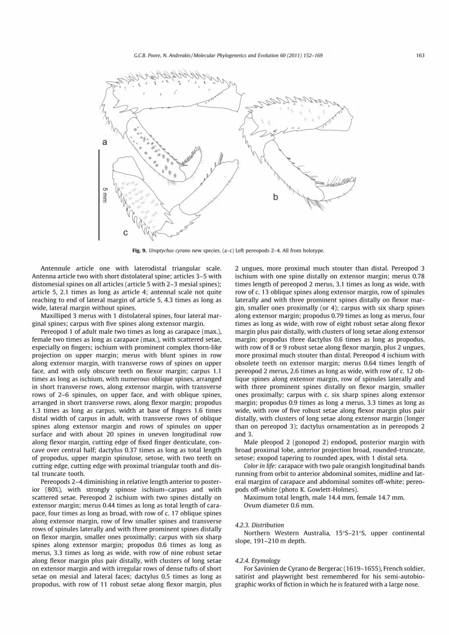
Antennule article one with laterodistal triangular scale.Antenna article two with short distolateral spine; articles 3–5 withdistomesial spines on all articles (article 5 with 2–3 mesial spines);article 5, 2.1 times as long as article 4; antennal scale not quitereaching to end of lateral margin of article 5, 4.3 times as long aswide, lateral margin without spines.
Maxilliped 3 merus with 1 distolateral spines, four lateral mar-ginal spines; carpus with five spines along extensor margin.
Pereopod 1 of adult male two times as long as carapace (max.),female two times as long as carapace (max.), with scattered setae,especially on fingers; ischium with prominent complex thorn-likeprojection on upper margin; merus with blunt spines in rowalong extensor margin, with transverse rows of spines on upperface, and with only obscure teeth on flexor margin; carpus 1.1times as long as ischium, with numerous oblique spines, arrangedin short transverse rows, along extensor margin, with transverserows of 2–6 spinules, on upper face, and with oblique spines,arranged in short transverse rows, along flexor margin; propodus1.3 times as long as carpus, width at base of fingers 1.6 timesdistal width of carpus in adult, with transverse rows of obliquespines along extensor margin and rows of spinules on uppersurface and with about 20 spines in uneven longitudinal rowalong flexor margin, cutting edge of fixed finger denticulate, con-cave over central half; dactylus 0.37 times as long as total lengthof propodus, upper margin spinulose, setose, with two teeth oncutting edge, cutting edge with proximal triangular tooth and dis-tal truncate tooth.
Pereopods 2–4 diminishing in relative length anterior to poster-ior (80%), with strongly spinose ischium–carpus and withscattered setae. Pereopod 2 ischium with two spines distally onextensor margin; merus 0.44 times as long as total length of cara-pace, four times as long as broad, with row of c. 17 oblique spinesalong extensor margin, row of few smaller spines and transverserows of spinules laterally and with three prominent spines distallyon flexor margin, smaller ones proximally; carpus with six sharpspines along extensor margin; propodus 0.6 times as long asmerus, 3.3 times as long as wide, with row of nine robust setaealong flexor margin plus pair distally, with clusters of long setaeon extensor margin and with irregular rows of dense tufts of shortsetae on mesial and lateral faces; dactylus 0.5 times as long aspropodus, with row of 11 robust setae along flexor margin, plus
2 ungues, more proximal much stouter than distal. Pereopod 3ischium with one spine distally on extensor margin; merus 0.78times length of pereopod 2 merus, 3.1 times as long as wide, withrow of c. 13 oblique spines along extensor margin, row of spinuleslaterally and with three prominent spines distally on flexor mar-gin, smaller ones proximally (or 4); carpus with six sharp spinesalong extensor margin; propodus 0.79 times as long as merus, fourtimes as long as wide, with row of eight robust setae along flexormargin plus pair distally, with clusters of long setae along extensormargin; propodus three dactylus 0.6 times as long as propodus,with row of 8 or 9 robust setae along flexor margin, plus 2 ungues,more proximal much stouter than distal. Pereopod 4 ischium withobsolete teeth on extensor margin; merus 0.64 times length ofpereopod 2 merus, 2.6 times as long as wide, with row of c. 12 ob-lique spines along extensor margin, row of spinules laterally andwith three prominent spines distally on flexor margin, smallerones proximally; carpus with c. six sharp spines along extensormargin; propodus 0.9 times as long a merus, 3.3 times as long aswide, with row of five robust setae along flexor margin plus pairdistally, with clusters of long setae along extensor margin (longerthan on pereopod 3); dactylus ornamentation as in pereopods 2and 3.
Male pleopod 2 (gonopod 2) endopod, posterior margin withbroad proximal lobe, anterior projection broad, rounded-truncate,setose; exopod tapering to rounded apex, with 1 distal seta.
Color in life: carapace with two pale orangish longitudinal bandsrunning from orbit to anterior abdominal somites, midline and lat-eral margins of carapace and abdominal somites off-white; pereo-pods off-white (photo K. Gowlett-Holmes).
Maximum total length, male 14.4 mm, female 14.7 mm.Ovum diameter 0.6 mm.
4.2.3. DistributionNorthern Western Australia, 15�S–21�S, upper continental
slope, 191–210 m depth.
4.2.4. EtymologyFor Savinien de Cyrano de Bergerac (1619–1655), French soldier,
satirist and playwright best remembered for his semi-autobio-graphic works of fiction in which he is featured with a large nose.
a
c
b
5 mm
Fig. 9. Uroptychus cyrano new species. (a–c) Left pereopods 2–4. All from holotype.
G.C.B. Poore, N. Andreakis / Molecular Phylogenetics and Evolution 60 (2011) 152–169 163

4.2.5. RemarksU. cyrano shares with U. pinocchio a similar color pattern, gen-
erally whitish with two longitudinal orange bands on the cara-pace and abdomen. The carapace of both is narrower than in U.naso (see above) and is generally more barrel-shaped or convexdorsally (compare lateral views) and lacks the depressed rostrum.The two species also both possess differentiated setation on thepropodus of pereopod 2, rows of tufts of short setae on the me-sial and lateral faces. Both have two teeth on the cutting edge ofthe dactylus of the cheliped and spinules on the upper edge. U.cyrano has nine lateral branchial spines, the intervening spacesnarrowly U-shaped, more evenly spaced than the seven branchialspines in U. pinocchio in which they are more closely spaced pos-teriorly. The lateral teeth of the rostrum of U. cyrano extendalong the distal two-thirds whereas in U. pinocchio they are con-fined to the distal half.
U. cyrano is represented by clades IV and V on Figs. 1–3.
4.3. Uroptychus. pinocchio sp. nov.
Figs. 4c and d, 5c, 10 and 11.
4.3.1. SynonymyUroptychus naso; Van Dam (1939: p. 402–403 (part); Baba
(1969, pp. 42–45 (part), Figs. 1, 2b; Baba et al (2008: 37 (part), col-or Fig. 1F; Baba et al. (2009: p. 47–48 (part), color Fig. 39.
4.3.2. Material examinedHolotype. Philippines, 14�10N, 120�170E, 184–186 m, 1980(MUSORSTOM 2 stn CP2), MNHN Ga6232 (male, 13.2 mm).
Paratypes. Philippines, 14�02.20N, 120�18.10E, 202–184 m, 1976(MUSORSTOM stn 61), MNHN Ga6230 (male, 13.2 mm). 14�00N,120�170E, 170–187 m, 1980 (MUSORSTOM 2 stn CP51), MNHNGa-6234 (one ovigerous female, damaged but complete,14.9 mm; one female with Sacculina externa, 10.9 mm).
Other material.Japan. W coast of Kyushu, 32�100N, 128�200E, 189 m, Schönau col-ln, 1898, ZMUC CRU-20208 (male, 16.5 mm). S of Goto I., 32�250N,128�520E, 225 m, Suenson colln, 10 Nov 1911, ZMUC CRU-20226(ovigerous female, 13.4 mm). 19 km W of Nagasaki, 32�020N,128�450E, 198 m, Suenson colln, 1898, ZMUC CRU-20225 (male,14.2 mm). Hirado Strait, Nagasaki, 32�100N, 128�200E, 189 m, Suen-son colln, 1900, ZMUC CRU-11200 (male, 9.2 mm).
Taiwan. Aodi, 27 Mar 2000, NTOU A00975 (male, 12.7 mm).
5 mm
5 mm
a
b
c
d
i
h
ge
f
c
Fig. 10. Uroptychus pinocchio new species. (a and b) Carapace in dorsal and lateral views. (c) Sternal plastron. (d) Telson. (e) Left antennule and antenna, ventral view. (f)Antennal scale. (g) Left maxilliped 3, lateral view. (h) Left pereopod 1, upper face in situ. (i) Left pleopod 1, lateral view. (a–e and g–i) From holotye. (f) From MNHN Ga-6234.Pereopod 1 drawn at 2/3 scale of carapace, sternum and telson.
164 G.C.B. Poore, N. Andreakis / Molecular Phylogenetics and Evolution 60 (2011) 152–169

4.3.3. DescriptionCarapace moderately convex from side to side, total length (tl.)
1.72 times greatest width (cw.) (ranges 1.55–1.86); distancebetween antennal spines (aw.) 0.57 times greatest width (cw.).Rostrum narrowly triangular, with shallow groove in dorsalmidline, with rounded ventral ridge, directed horizontally anteri-orly, length 0.4 times total length; dorsal surface with tubercles ex-cept in midline; lateral margin with 7–9 blunt teeth along distalhalf. Cervical groove at about midlength of carapace, deep medially(indistinct cervical groove and groove between anterior and pos-terior branchial regions laterally). Carapace covered with lowtubercles, most with 2 or more microtubercles, strongest on ante-rior and posterior branchial regions and posterior to cervicalgroove, with transverse rows of 2–4 setules; frontal margin trans-verse (with small spine at midpoint); antennal spine sharp, wellproduced; anterolateral margin irregularly covered with shortspines; branchial margin strongly convex; anterior branchial mar-gin with two similar spines in tandem (usually with a third appliedto the second); posterior branchial margin with 6–7 strong spines,more prominent and more widely spaced anteriorly, obscurely dif-ferentiated posteriorly, intermediate spinules usually visible, anddentate over most posterior fifth. Pterygostomial flap irregularlycovered with small tubercles, anterior margin with prominent up-turned spine or produced as sharp triangle.
Sternal plastron (sternites 3–7) 1.2 times as long as wide,parallel-sided over most of length; excavated sternum roundedanteriorly, with sharp midventral ridge; sternite 3 slightlydepressed relative to sternite 4, anterior margin excavate, withU-shaped median sinus (gaping), lateral margin rounded anterolat-erally, irregularly denticulate laterally, surface moderately rugose;
sternite 4 anterior margin 0.64 times as wide as posterior margin,anterolaterally angled, surface smooth.
Abdominal somites smooth (except for transverse granularridge on somite 1); tergites rounded anteriorly and posteriorly.
Telson 0.4 times as long in midline as greatest width; anteriorsection 0.45 times as long as midline length; anterolateral lobesprojecting beyond posterolateral lobes, broadly rounded to subtlytruncate, setose; posterior margin shallowly excavate, evenly setose.
Eyestalk 0.26 times length of rostrum; cornea globular, pig-mented.
Antennule article 1 with laterodistal triangular scale; article 3,2.8 times as long as wide. Antenna article 2 with short distolateralspine; articles 3–5 with distomesial spines on all articles; article 5,1.4 times as long as article 4; antennal scale not quite reaching toend of lateral margin of article 5, three times as long as wide, lat-eral margin with small spine or without spines.
Maxilliped 3 merus with one distolateral spines, five lateralmarginal spines; carpus with six spines along extensor margin(irregular).
Pereopod 1 of adult male 2.8 times as long as carapace (max.),female 2.2 times as long as carapace (max.), with scattered setae,especially on fingers; ischium with prominent complex thorn-likeprojection on upper margin; merus with blunt spines in row alongextensor margin, with transverse rows of spines on upper face, andwith only obscure teeth on flexor margin; carpus 1.3 times as longas ischium, with numerous oblique spines, arranged in short trans-verse rows, along extensor margin, with transverse rows of 2–6spinules, on upper face, and with oblique spines, arranged in shorttransverse rows, along flexor margin; propodus 1.3 times as long ascarpus, width at base of fingers 1.6 times distal width of carpus inadult, with transverse rows of oblique spines along extensor mar-
5 mm
a
b
c
Fig. 11. Uroptychus pinocchio new species. (a–c) Left pereopods 2–4. All from holotype.
G.C.B. Poore, N. Andreakis / Molecular Phylogenetics and Evolution 60 (2011) 152–169 165

gin and rows of spinules on upper surface and with about 20 spinesin uneven longitudinal row along flexor margin, cutting edge offixed finger denticulate, concave over central half; dactylus 0.34times as long as total length of propodus, upper margin spinulose,setose, with two teeth on cutting edge, cutting edge with proximalrounded tooth and distal triangular tooth.
Pereopods 2–4 diminishing in relative length anterior to poster-ior (80%), with strongly spinose ischium–carpus and with scatteredsetae. Pereopod 2 ischium with 2 spines distally on extensor mar-gin; merus 0.44 times as long as total length of carapace, 3.7 timesas long as broad, with row of c. 17 oblique spines along extensormargin, row of few smaller spines and transverse rows of spinuleslaterally and with three prominent spines distally on flexor margin,smaller ones proximally; carpus with nine sharp spines alongextensor margin; propodus 0.57 times as long as merus, 3.2 timesas long as wide, with row of eight sets or 1 or 2 robust setae alongflexor margin plus pair distally, with clusters of long setae onextensor margin and with two irregular rows of dense tufts of shortsetae on mesial and lateral faces; dactylus 0.61 times as long aspropodus, with row of 14 robust setae along flexor margin, plus2 unges, more proximal much stouter than distal. Pereopod 3ischium with two spines distally on extensor margin; merus 0.8times length of pereopod 2 merus, 3.2 times as long as wide, withrow of c. 13 oblique spines along extensor margin, row of spinuleslaterally and with three prominent spines distally on flexor margin,smaller ones proximally; carpus with seven sharp spines alongextensor margin; propodus 0.7 times as long as merus, three timesas long as wide, with row of 8 robust setae along flexor margin pluspair distally, with clusters of long setae along extensor margin;propodus 3 dactylus 0.6 times as long as propodus, with row ofeight or nine robust setae along flexor margin, plus 2 ungues, moreproximal much stouter than distal. Pereopod 4 ischium withoutteeth on extensor margin; merus 0.61 times length of pereopod 2merus, 2.3 times as long as wide, with row of c. 12 oblique spinesalong extensor margin, row of spinules laterally and with threeprominent spines distally on flexor margin, smaller ones proxi-mally (obsolete); carpus with c. nine sharp spines along extensormargin; propodus 0.96 times as long a merus, 3.3 times as longas wide, with row of three robust setae along flexor margin pluspair distally, with clusters of long setae along extensor margin(longer than on pereopod 3); dactylus 0.64 times as long as prop-odus, dactylus ornamentation as in pereopods 2 and 3.
Male pleopod 2 (gonopod 2) endopod, posterior margin withbroad proximal lobe, anterior projection narrow, rounded distally,setose; exopod tapering to rounded apex, with three distal setae.
Color in life: carapace with two bright brick-red or orangish lon-gitudinal bands running from orbit to anterior abdominal somites,midline and lateral margins of carapace and abdominal somitesoff-white-yellowish; pereopods brick-red with distal articles ofpereopods 3 and 4 bluish (photos by T.Y. Chan differ slightly).
Maximum total length, male 16.4 mm, female 14.9 mm.Ovum diameter 0.8 mm.
4.3.4. DistributionSW Japan (Baba, 1969; Van Dam, 1939); Taiwan (Baba et al.,
2009a); Philippines. Latitudinal range: 35�N–14�N. Lower shelf,upper continental slope, 153–225 m.
4.3.5. EtymologyFor Pinocchio, a wooden puppet that dreamt of becoming a real
boy in the 1883 novel Le avventure di Pinocchio by Carlo Collodi.Pinocchio’s nose grew longer when he told lies.
4.3.6. RemarksVan Dam (1939) assigned four males and two females from Japan
to U. naso of which we have seen four belonging to U. pinocchio and
only one to U. naso. Baba (1969) commented on differences in thesetation of the propodus of pereopod 2 between females from TosaBay, Japan and a male from the East China Sea. His illustration of malepleopods are more likely from U. pinocchio. The specimens on whichhe commented, originally lodged in the Zoological Laboratory, Fac-ulty of Agriculture, Kyushu University, and transferred to theKitakyushu Museum of Natural History, Kitakyushu, cannot now befound (M. Shimomura, pers. comm.). Baba et al. (2009a) remarkedagain on the setation of pereopod 2 in the specimen from Aodi whichwe have examined. We are able to confirm that the setation is notlocality- or sex-dependent as he questioned but is a real differencebetween U. pinocchio and U. naso. In U. naso, setae are arranged inmore or less transverse rows, especially along the upper marginwhere they are longer than laterally. In U. pinocchio, and in U. cyrano,long setae along the upper margin are few but both lateral and mesialfaces bear dense clusters of short setae in uneven rows. Similar clus-ters occur on the dactylus of pereopod 1. See above for further differ-ences between this species and U. naso and the more similar U. cyrano.
5. Discussion
5.1. Morphological and molecular delineation of species
The combination of mitochondrial and nuclear molecular phy-logenies with morphology indicated that the tropical species com-plex U. naso sensu lato consists of several mutually monophyleticmitochondrial ESUs that we interpret as three biologically distinctspecies. Mitochondrial clades (I + II) and III represent two geneti-cally distinct yet morphologically identical mitochondrial lineages.Specimens belonging to these clades share identical nuclear H3 se-quences and it is concluded that all belong to biological species U.naso sensu stricto. Individuals belonging to mitochondrial clades(I + II) and III are morphologically and genetically distinct fromindividuals of mitochondrial clades IV and V which are describedas U. cyrano sp. nov. A single 16S sequence (#178) obtained fromone specimen from Aodi, Taiwan, differed significantly from se-quences of individuals from clades I + II (mean p distance = 1.4%,2% and 4.4% from individuals of clades (I and II), III and (IV andV) respectively; results from the 16S phylogeny, data not shown)and belongs to a second new species, U. pinocchio sp. nov.
These three species are morphologically distinctive withinUroptychus and would appear to be a monophyletic clade. Untilnow, variation in setation of pereopod 2 (Baba, 1969) and differentcolor patterns (Baba et al., 2009) have not been questioned or dis-missed as specific characters. Here, these differences and othermorphological features support genetic divergence between ESUsrevealed by molecular data.
5.2. Phylogeography of U. naso
Genetic discontinuities within species are often associated withgeographical patterns of distribution and may be associated withspecific differences in morphology (Anker, 2007; Caputi et al.,2007; Rhyne and Lin, 2006). Hidden genetic diversity however, isnot necessarily accompanied by morphological changes due tohomoplasy (Bickford et al., 2007 and references therein). Mito-chondrial clades I and II of U. naso are genetically and geographi-cally distinct from clade III (Fig. 2a and Table 3) but could not beseparated morphologically. Mitochondrial clade I is formed by foursamples from Taiwan whilst haplotypes forming clade II have beenobtained from three individuals in the same sample taken in dee-per water in the Philippines. Individuals belonging to clade III oc-cur in NWA. This clade was strongly supported by differences inCOI and 16S gene regions as well as with resolved vs. unresolvedpositions of the Histone 3 gene compared to clades I + II (Fig. 2b
166 G.C.B. Poore, N. Andreakis / Molecular Phylogenetics and Evolution 60 (2011) 152–169

and c). Because individuals from clades I + II and III do not occur insympatry, we assume the nuclear shared base pairs positions to bethe result of incomplete lineage sorting due to slow convergentevolution of unsorted ancestral polymorphisms rather than ongo-ing hybridization or introgression between hybrids (Comboschet al., 2008; van Oppen et al., 2001). In this study, genetic data havebeen produced from widely geographically separated specimens.Yet, we know the species exists in intermediate regions (i.e. Indo-nesia). Following comparison of all material with the Indonesiansyntypes, clade III is here interpreted as a genetically isolated pop-ulation of U. naso rather than a new, reproductively isolatedspecies.
5.3. Phylogeography of U. cyrano and U. pinocchio
Sister mitochondrial clades IV and V shared the same morpholog-ical features and identical nuclear genotypes but distinct mitochon-drial haplotypes. Individuals belonging to these clades are found insympatry on the WA continental shelf at around 200 m depth be-tween 15�S and 21�S and likely constitute the same biological spe-cies. At the northern end of its range, U. cyrano co-occurs with U.naso mitochondrial clade III but it was not encountered in southernWestern Australia by Poore et al. (2008). The similarities in morphol-ogy of U. cyrano and U. pinocchio suggest that these are sister species.Yet, we have limited material to support or contradict this hypothe-sis. Available material of U. cyrano and of U. pinocchio are geograph-ically widely separated (NWA and the Philippines). Whether orwhere these species occur in sympatry, is unknown.
5.4. Sequencing of difficult templates
Unfortunately, we were unable to produce mitochondrial se-quences from two individuals of U. naso from Indonesia (a syntypeand ZMUC CRU-11335) and one of U. pinocchio from Japan (ZMUCCRU-20208). These specimens were probably formalin-fixed andcould be characterized only by the sole Histone 3 gene. The PCRamplification problems were mainly related to the difficulties inextracting sufficient quality and quantity of DNA. Additionally,the scarce amount of material prevented us from using the DNAextraction protocols suitable for formalin-preserved tissues thatrequire large amount (g) of starting tissue (e.g., Klanten et al.,2003). In a preliminary cladogram based on the H3 Histone se-quences, these specimens clustered in a sister clade to U. cyrano.However, given the conservative nature of the marker and the dif-ficulties encountered in amplification we must discount this result.Overall, Histone genes exhibit an acceptable level of resolutionpower for phylogenetic inference in crustaceans (Malay and Pau-lay, 2010). In addition, histones are represented by short DNA se-quences, highly repeated in eukaryotic genomes and organizedinto clusters (Maxson et al., 1983). For these reasons these genesmay represent an easy-to-amplify yet informative marker at thespecies level, suitable for difficult templates such as DNA obtainedfrom tissues previously preserved in formalin.
5.5. Phylogenetic inference
The DNA regions used in this study are known to be suitable ininferring phylogenetic and phylogeographic patterns at the inter-and intra-specific levels with the mitochondrial markers beingthe most appropriate for cryptic species identification because ofthe fast mutation rate, quick lineage-specific substitution fixationand small effective population size (e.g., Cabezas et al., 2008,2009; Machordom and Macpherson, 2004; Malay and Paulay,2010; Seidel et al., 2009). In our study, intra- and inter-specific ge-netic divergence was similar in magnitude to the ones reported inmunidid species using the same mitochondrial marker systems
(Machordom and Macpherson, 2004) and in species of the hermitcrab genus Calcinus (Diogenidae) using the Histone 3 gene (Malayand Paulay, 2010). H3 was successful in testing hypotheses of an-cient hybridization, introgression or incomplete lineage sortingamong undiscovered cryptic lineages by observing incongruencein the topology between mitochondrial and nuclear tree topologies(Sota, 2002; Sota and Vogler, 2001; Ting, 2007). We did notencounter topological incongruence between markers in our study.The sequence information gathered from the Histone 3 gene there-fore helped to suggest in which cases a biological species conceptcould be assumed.
5.6. Historical biogeography and divergence time estimates
Molecular phylogenies and approximations of divergence datesamong cryptic species and mitochondrial clades are consistentwith a Tethyan MRCA for U. naso and U. cyrano from Palaeoceneto early Eocene (60–50 MYA), probably of Indo-west Pacific origin.The latter region borders an archipelago of islands comprising Ja-pan in the north, Taiwan, the Philippines, Papua New Guinea andIndonesia separated by shallow seas (Fig. 1). Squat lobsters of theU. naso complex are confined to the 150–400 m depth range nearthe edge of this shallow shelf. This continental margin is continu-ous along the entire WP and Indian Ocean; it represents a majorcenter of evolutionary origin of species that radiate mainly fromthe East Indies (Briggs, 2005) and is a center of biodiversity fordecapods and brachyurans (Feldmann and Schweitzer, 2006; Nget al., 2008). It is therefore not surprising that genetically distinctpopulations of Uroptychus, or other squat lobster species, occurthroughout this region or at least within the range of 35�N to 15�S.
Sharp genetic breaks have often been described among popula-tions of terrestrial plant and animal species separated by Wallace’sline into Asian and Australian partitions (Schulte et al., 2003 andreferences therein). On the other hand, similar phylogeographicpatterns have been observed for marine invertebrate species dis-tributed on either side of Huxley’s line, a modification of Wallace’sline (de Bruyn et al., 2004; Huxley, 1868; Lourie and Vincent,2004). Furthermore, an invisible marine equivalent of Wallace’sline, localized along the Java and Flores Seas, has been recentlyhypothesised as responsible for low connectivity and populationfragmentation of corals within a few 100 km (Barber et al., 2000,2002). Our phylogenies, inferred from individuals collected alongthe western Pacific side of Huxley’s line indicate: (a) the presenceof two genetically distinct cryptic species, U. naso and U. cyrano sp.nov. (mean COI p distance = 15.7%) and b) a second major geneticbreak within U. naso (mean COI p distance = 3.25%) due to frag-mentation between NWA (mitochondrial clade III;) and SWP pop-ulations (mitochondrial clades I and II; Fig. 1). These levels ofgenetic divergence are associated with geographically predefinedpopulations and are partitioned across a south to north cline alongHuxley’s line rather than an east to west Indian-Pacific oceanbreak. Estimated divergence dates are consistent with a round ofsecondary cladogenesis events beginning in early Miocene, respon-sible for the geographically associated genetic split among U. nasopopulations (Fig. 3). Speciation and fragmentation of populationswithin U. naso can be therefore attributed either to Wallace’s mar-ine equivalent or to the break between Asian and Australian conti-nental plates. The same may be true for the speciation leading tosister taxa, U. cyrano in WA and U. pinocchio in the NW Pacific.
The collision between the Australian continental land mass(including Papua New Guinea) and Indonesia during Miocene (25MYA) might have minimized the marine corridor between theIndo-Pacific water bodies, leaving the deep water of Timor Troughstill able to separate the Australian and Asian continents (Hall,1997; Poore and O’Hara, 2007). The succeeding early- to mid-Mio-cene was a warm period which facilitated the introduction of
G.C.B. Poore, N. Andreakis / Molecular Phylogenetics and Evolution 60 (2011) 152–169 167

warm-water species into southern Australia. East to west larvaltransport through the region is facilitated today by the SoutheastEquatorial Current yet, genetic isolation of local shallow-waterpopulations has also been reported (Barber et al., 2002). It seemsreasonable to assume that the presence of U. naso mitochondrialclade III on the western Australian continental margin representsa historical event of dispersal and geographical isolation since Mio-cene, maintained by restrictive current flows.
6. Concluding remarks
Despite the considerable debates related to the exact positionand origins of the biogeographic breaks mentioned, it is unques-tionable that the composition and geographical patterns of marineand terrestrial biota are influenced by the complicated geologicalhistory of the zone (plate tectonic history of the East Indies trian-gle, break between ocean bodies during Pleistocene and the colli-sion of the Australian and the Eurasian plates some 25 MY beforepresent; Schulte et al., 2003). Information from other deep-seainvertebrates should reveal whether the aforementioned historicalsignatures account for a standard distribution trend of species, use-ful for biodiversity discovery and estimates, connectivity, conser-vation and sustainable use of marine resources.
Acknowledgments
We are grateful to Nic Bax and Alan Williams, CSIRO, who se-cured funds for the ‘‘Voyages of Discovery’’ research program.We thank CSIRO colleagues Alan Williams and Rudy Kloser fortheir leadership of the surveys, Mark Lewis (Gear Officer) andKaren Gowlett-Holmes (Curator, Marine Invertebrates) for helpwith gear and dealing with samples while on board of FRV SouthernSurveyor. We acknowledge the Commonwealth Department ofSustainability, Environment, Water, Population and Communities,and the CSIRO Wealth from Oceans Flagship for financial supportand the field and laboratory components of the ‘‘Voyages ofDiscovery’’ program. We appreciate the valuable contribution ofAnna McCallum, Museum Victoria, whose preliminary identifica-tions lead to the discovery of these taxa in Australia. For the loanof material we thank: Tin-Yam Chan and Chia-wei Lin, NTOU;Jørgen Olesen, Zoological Museum, University of Copenhagen andDirk Platvoet, Zoological Museum, Amsterdam; Chia-wei Lin,NTOU for two COI sequences. We thank Régis Cleva for his hospi-tality and access to material at the Muséum nationale d’Histoirenaturelle, Paris; Karen Gowlett-Holmes (CMAR) and Tin-Yam Chan(NTOU) for the use of photographs.
This work has been funded through the Commonwealth Envi-ronment Research Facilities (CERF) program, an Australian Govern-ment initiative supporting world class, public good research. TheCERF Marine Biodiversity Hub is a collaborative partnershipbetween the University of Tasmania, CSIRO Wealth from OceansFlagship, Geoscience Australia, Australian Institute of MarineScience and Museum Victoria.
We gratefully acknowledge support for squat lobster researchfrom Continental Margin Ecosystems (COMARGE) (http://www.ifr-emer.fr/comarge/en/index.html), one of fourteen Census of MarineLife (CoML http://www.coml.org/) field projects dedicated to thedescription and understanding of biodiversity patterns on conti-nental margins.
References
Ahyong, S.T., Schnabel, K.E., Macpherson, E., in press. Phylogeny and fossil record ofthe marine squat lobsters. In: Poore, G.C.B., Ahyong, S.T., Taylor, J. (Eds.),Crustacean Issues vol. 20: The Biology of Squat Lobsters. CSIRO Publishing,Melbourne and CRC Press, Boca Raton (Chapter 4).
Aljanabi, S.M., Martinez, I., 1997. Universal and rapid salt-extraction of high qualitygenomic DNA for PCR-based techniques. Nucleic Acids Research 25, 4692–4693.
Anker, A., 2007. Pseudalpheopsis guana gen. nov., sp. nov. (Crustacea: Decapoda), anew alpheid shrimp from the British Virgin Islands, Caribbean Sea. ZoologicalStudies 46, 428–440.
Baba, K., 1969. Chirostylids and galatheids from dredgings and trawlings operatedin the East China Sea by the Japanese Fisheries Research Vessel Kaiyo Maru in1967. OHMU 2, 41–57.
Baba, K., 1988. Chirostylid and galatheid crustaceans (Decapoda: Anomura) of the‘‘Albatross’’ Philippine Expedition, 1907–1910. Researches on Crustacea, SpecialNumber 2, 1–203.
Baba, K., 2005. Deep-sea chirostylid and galatheid crustaceans (Decapoda:Anomura) from the Indo-West Pacific, with a list of species. Galathea Report20, 1–317.
Baba, K., Lin, C.-W., 2008. Five new species of chirostylid crustaceans (Crustacea:Decapoda: Anomura: Chirostylidae) from Taiwan. Zootaxa 1918, 1–24.
Baba, K., Macpherson, E., Poore, G.C.B., Ahyong, S.T., Bermudez, A., Cabezas, P., Lin,C.-W., Nizinski, M., Rodrigues, C., Schnabel, K.E., 2008. Catalogue of squatlobsters of the world (Crustacea: Decapoda: Anomura—families Chirostylidae,Galatheidae and Kiwaidae). Zootaxa 1905, 1–220.
Baba, K., Macpherson, E., Lin, C.-W., Chan, T.-Y., 2009. Crustacean fauna of Taiwan:squat lobsters (Chirostylidae and Galatheidae). National Taiwan OceanUniversity, Keelung.
Bandelt, H.J., Forster, P., Röhl, A., 1999. Median-joining networks for inferringintraspecific phylogenies. Molecular Biology and Evolution 16, 37–48.
Barber, P., Palumbi, S., Erdmann, M., Moosa, M.K., 2000. A marine Wallace’s line?Nature 406, 692–693.
Barber, P., Palumbi, S., Erdmann, M.V., Moosa, K., 2002. Sharp genetic breaks amongpopulations of Haptosquilla pulchella (Stomatopoda) indicate limits to larvaltransport: patterns, causes, and consequences. Molecular Ecology 11, 659–674.
Bickford, D., Lohman, D.J., Sodhi, N.S., Ng, P.K.L., Meier, R., Winker, K., Ingram, K.K.,Das, I., 2007. Cryptic species as a window on diversity and conservation. Trendsin Ecology & Evolution 22, 148–155.
Briggs, J.C., 2005. The marine East Indies: diversity and speciation. Journal ofBiogeography 31, 1975–1985.
de Bruyn, M., Wilson, J.A., Mather, P.B., 2004. Huxley’s line demarcates extensivegenetic divergence between eastern and western forms of the giant freshwaterprawn Macrobrachium rosenbergii. Molecular Biology and Evolution 30,251–257.
Cabezas, P., Macpherson, E., Machordom, A., 2008. A new genus of squat lobster(Decapoda: Anomura: Galatheidae) from the South West Pacific and IndianOcean inferred from morphological and molecular evidence. Journal ofCrustacean Biology 28, 68–75.
Cabezas, P., Macpherson, E., Machordom, A., 2009. Morphological and moleculardescription of new species of squat lobster (Crustacea: Decapoda: Galatheidae)from the Solomon and Fiji Islans (SW Pacific). Zoological Journal of the LinneanSociety 156, 465–493.
Caputi, L., Andreakis, N., Mastrototaro, F., Cirino, P., Vassillo, M., Sordino, P., 2007.Cryptic speciation in a model invertebrate chordate. Proceedings of the NationalAcademy of Sciences of the United States of America 104, 9364–9369.
Colgan, D.J., McLauchlan, A., Wilson, G.D.F., Livingston, S.P., Edgecombe, G.D.,Macaranas, J., Cassis, G., Gray, M.R., 1998. Histone H3 and U2 snRNA DNAsequences and arthropod molecular evolution. Australian Journal of Zoology 46,419–437.
Combosch, D.J., Guzman, H.M., Schuhmacher, H., Vollmer, S.V., 2008. Interspecifichybridization and restricted trans-Pacific gene flow in the Tropical EasternPacific Pocillopora. Molecular Ecology 17, 1304–1312.
Dallwitz, M.J., Paine, T.A., Zurcher, E.J., 1999. User’s guide to the DELTA system. Ageneral system for processing taxonomic descriptions. CSIRO Division ofEntomology, Canberra.
Drummond, A.J., Rambaut, A., 2007. BEAST: Bayesian evolutionary analysis bysampling trees. BMC Evolutionary Biology 7, 214.
Drummond, A.J., Ho, S.Y.W., Phillips, M.J., Rambaut, A., 2006. Relaxed phylogeneticsand dating with confidence. PLoS Biology 4, e88.
Edgar, R.C., 2004. MUSCLE: multiple sequence alignment with high accuracy andhigh throughput. Nucleic Acids Research 32, 1792–1797.
Feldmann, R.M., 1984. Haumuriaegla glaessneri n. gen. and sp. (Decapoda; Anomura;Aeglidae) from Haumurian (Late Cretaceous) rocks near Cheviot, New Zealand.New Zealand Journal of Geology and Geophysics 27, 379–385.
Feldmann, R.M., Schweitzer, C.E., 2006. Paleobiogeography of Southern Hemispheredecapod Crustacea. Journal of Paleontology 80, 83–103.
Feldmann, R.M., Vega, F.J., Applegate, S.P., Bishop, G.A., 1998. Early Cretaceousarthropods from the Tlayúa Formation at Tepexi de Rodríguez, Puebla, Mexico.Journal of Paleontology 72, 79–90.
Felsenstein, J., 1985. Confidence limits on phylogenies: an approach using thebootstrap. Evolution 39, 783–791.
Folmer, O., Black, M., Heoh, W., Lutz, R.A., Vrijenhoek, R., 1994. DNA primers for theamplification of mitochondrial cytochrome C oxidase subunit I from diversemetazoan invertebrates. Molecular Marine Biology and Biotechnology 3, 294–299.
Hall, R., 1997. Cenozoic plate tectonic reconstructions of SE Asia. Geological Societyof London, Special Publications 126, 11–23.
Hall, T.A., 1999. BioEdit: a user-friendly biological sequence alignment editor andanalysis program for Windows 95/98/NT. Nucleic Acids Symposium 41, 95–98.
Hillis, D.M., Huelsenbeck, J.P., 1992. Signal, noise and reliability in molecularphylogenetic analysis. Journal of Heredity 83, 189–195.
168 G.C.B. Poore, N. Andreakis / Molecular Phylogenetics and Evolution 60 (2011) 152–169

Huelsenbeck, J.P., Ronquist, F., 2001. MRBAYES: Bayesian inference of phylogenetictrees. Bioinformatics 17, 754–755.
Huxley, T.H., 1868. On the classification and distribution of the Alectoromorphaeand Heteromorphae. Proceedings of the Zoological Society of London 6, 294–319.
Jakobsen, S.L., Collins, J.S.H., 1997. New Middle Danian species of anomuran andbrachyuran crabs from Fakse, Denmark. Bulletin of the Geological Society ofDenmark 44, 89–100.
Klanten, S., Van Herwerden, L., Howard, C.J., 2003. Acquiring reef fish DNAsequences from formalin-fixed museum specimens. Bulletin of MarineScience 73, 771–776.
Knowlton, N., 1986. Cryptic and sibling species among the Decapod crustacea.Crustacean Biology 6, 263–356.
Lourie, S.A., Vincent, C.J., 2004. A marine fish follows Wallace’s Line: thephylogeography of the three-spot seahorse (Hippocampus trimaculatus,Syngnathidae, Teleostei) in Southeast Asia. Journal of Biogeography 31, 1975–1985.
Machordom, A., Macpherson, E., 2004. Rapid radiation and cryptic speciation ingalatheid crabs of the genus Munida and related genera in the South WestPacific: molecular and morphological evidence. Molecular Phylogenetics andEvolution 33, 259–279.
Malay, M.C.D., Paulay, G., 2010. Peripatric speciation drives diversification anddistributional patterns of reef hermit crabs (Decapoda: Diogenidae: Calcinus).Evolution 64, 634–662.
Maxson, R., Cohn, R., Kedes, L., Mohun, T., 1983. Expression and organization ofhistone genes. Annual Review of Genetics 17, 239–277.
Moritz, C., 1994. Defining ‘‘evolutionarily significant units’’ for conservation. Trendsin Ecology & Evolution 9, 373–375.
Ng, P.K.L., Guinot, D., Davie, P.J.F., 2008. Systema Brachyurorum: part I. Anannotated checklist of extant brachyuran crabs of the world. Raffles Bulletinof Zoology 17, 1–286.
Palumbi, S.R., Benzie, J., 1991. Large mitochondrial DNA differences betweenmorphologically similar penaeid shrimp. Marine Biology and Biotechnology 1,27–34.
Poore, G.C.B., O’Hara, T.D., 2007. Marine biogeography and biodiversity of Australia.In: Connell, S.D., Gillanders, B.M. (Eds.), Marine Ecology. Oxford UniversityPress, pp. 177–198.
Poore, G.C.B., McCallum, A.W., Taylor, J., 2008. Decapod Crustacea of the continentalmargin of southwestern and central Western Australia: preliminaryidentifications of 524 species from FRV Southern Surveyor voyage SS10–2005.Museum Victoria Science Reports 11, 1–106.
Porter, S.C., 1989. Some geological implications of average Quaternary glacialconditions. Quaternary Research 32, 245–261.
Posada, D., Crandall, K.A., 1998. Modeltest: testing the model of DNA substitution.Bioinformatics 14, 817–818.
Rambaut, A., Drummond, A.J., 2007. Tracer v1.4. <http://beast.bio.ed.ac.uk/Tracer>.Rhyne, A.L., Lin, J., 2006. A western Atlantic peppermint shrimp complex:
redescription of Lysmata wurdemanni (Gibbes), description of four newspecies and remarks on L. rathbunae Chace (Crustacea: Decapoda:Hippolytidae). Bulletin of Marine Science 79, 165–204.
Schnabel, K.E., 2009. A review of the New Zealand Chirostylidae (Anomura:Galatheoidea) with description of six new species from the Kermadec Islands.Zoological Journal of the Linnean Society 155, 542–582.
Schnabel, K.E., Ahyong, S.T., 2010. A new classification of the Chirostyloidea(Crustacea: Decapoda: Anomura). Zootaxa 2687, 56–64.
Schulte, J.A., Melville, L., Larson, A., 2003. Molecular evidence of ancient divergenceof lizard taxa on either side of the Wallace’s line. Proceedings of the ZoologicalSociety of London B 270, 597–603.
Schweitzer, C.E., Feldmann, R.M., 2000. First notice of the Chirostylidae (Decapoda)in the fossil record and new Tertiary Galatheidae (Decapoda) from theAmericas. Bulletin of the Mizunami Fossil Museum 27, 147–165.
Seidel, R.A., Lang, B.K., Berg, D.J., 2009. Phylogeographic analysis reveals multiplecryptic species of amphipods (Crustacea: Amphipoda) in Chihuahuan Desertsprings. Biological Conservation 142, 2303–2313.
Sota, T., 2002. Radiation and reticulation: extensive introgressive hybridization inthe carabid beetles Ohomopterus inferred from mitochondrial gene genealogy.Population Ecology 44, 145–156.
Sota, T., Vogler, A.P., 2001. Incongruence of mitochondrial and nuclear gene trees inthe carabid beetles Ohomopterus. Systematic Biology 50, 39–59.
Swofford, D.L., 2002. PAUP�. Phylogenetic Analysis Using Parsimony (�and OtherMethods). Version 4. Sinauer Associates, Sunderland.
Taylor, J., Ahyong, S.T., Andreakis, N., 2010. New records and new species of themunidopsine squat lobsters (Decapoda: Anomura: Galatheidae: Munidopsinae)from Australia. Zootaxa 2642, 1–18.
Thompson, J.D., Higgins, D.G., Gibson, T.J., 1994. CLUSTAL W: improving thesensitivity of progressive multiple sequence alignment through sequenceweighting, position specific gap penalties and weight matrix choice. NucleicAcids Research 22, 4673–4680.
Ting, N., 2007. Mitochondrial relationships and divergence dates of the Africancolobines: evidence of Miocene origins for the living colobus monkeys. Journalof Human Evolution 55, 312–325.
Van Dam, A.J., 1933. Die Chirostylidae der Siboga-Expedition. Decapoda VIII:Galatheidea: Chirostylidae. Siboga-Expéditie, 39a7, 1–46.
Van Dam, A.J., 1939. Ueber einige Uroptychus-Arten des Museums zu Kopenhagen.Bijdragen tot de Dierkunde 27, 392–407.
Van Dam, A.J., 1940. Anomura, gesammelt vom Dampfer Gier in der Java-See.Zoologischer Anzeiger 129, 95–104.
van Oppen, M.J.H., McDonald, B.J., Willis, B., Miller, D.J., 2001. The evolutionaryhistory of the coral genus Acropora (Scleractinia, Cnidaria) based on amitochondrial and a nuclear marker: reticulation, incomplete lineage sorting,or morphological convergence? Molecular Biology and Evolution 18, 1315–1329.
Wang, Y., 2008. Infraorder Anomura MacLeay, 1838. In: Liu, J.-Y. (Ed.),Checklist of Marine Biota of China Seas. Science Press, Qingdao, pp. 748–751.
Williams, S.T., Jara, J., Gomez, E., Knowlton, N., 2002. The marine Indo-West Pacificbreak: contrasting the resolving power of mitochondrial and nuclear genes.Integrative and Comparative Biology 42, 941–952.
Wu, M.F., Chan, T.-Y., Yu, H.P., 1997. On the Chirostylidae and Galatheidae(Crustacea: Decapoda: Galatheidea) of Taiwan. Annual of Taiwan Museum 40,75–153 (1998).
G.C.B. Poore, N. Andreakis / Molecular Phylogenetics and Evolution 60 (2011) 152–169 169


