Embed Size (px)
Citation preview
Morphology of the Epidermis of the Neotropical CatfishPimelodella lateristriga (Lichtenstein, 1823) withEmphasis in Club CellsEduardo Medeiros Damasceno, Juliana Castro Monteiro, Luiz Fernando Duboc, Heidi Dolder,
Karina Mancini*
Departamento de Ciencias Agrarias e Biologicas, Centro Universitario Norte do Espırito Santo, Universidade Federal do Espırito Santo, Sao Mateus, Espırito Santo, Brasil
Abstract
The epidermis of Ostariophysi fish is composed of 4 main cell types: epidermal cells (or filament containing cells), mucouscells, granular cells and club cells. The morphological analysis of the epidermis of the catfish Pimelodella lateristriga revealedthe presence of only two types of cells: epidermal and club cells. The latter were evident in the middle layer of theepidermis, being the largest cells within the epithelium. Few organelles were located in the perinuclear region, while therest of the cytoplasm was filled with a non-vesicular fibrillar substance. Club cells contained two irregular nuclei withevident nucleoli and high compacted peripheral chromatin. Histochemical analysis detected prevalence of protein withinthe cytoplasm other than carbohydrates, which were absent. These characteristics are similar to those described to mostOstariophysi studied so far. On the other hand, the epidermal cells differ from what is found in the literature. The presentstudy described three distinct types, as follows: superficial, abundant and dense cells. Differences among them wererestricted to their cytoplasm and nucleus morphology. Mucous cells were found in all Ostariophysi studied so far, althoughthey were absent in P. lateristriga, along with granular cells, also typical of other catfish epidermis. The preset studycorroborates the observations on club cells’ morphology in Siluriformes specimens, and shows important differences inepidermis composition and cell structure of P. lateristriga regarding the literature data.
Citation: Damasceno EM, Monteiro JC, Duboc LF, Dolder H, Mancini K (2012) Morphology of the Epidermis of the Neotropical Catfish Pimelodella lateristriga(Lichtenstein, 1823) with Emphasis in Club Cells. PLoS ONE 7(11): e50255. doi:10.1371/journal.pone.0050255
Editor: Michael Koval, Emory University School of Medicine, United States of America
Received July 18, 2012; Accepted October 16, 2012; Published November 30, 2012
Copyright: � 2012 Damasceno et al. This is an open-access article distributed under the terms of the Creative Commons Attribution License, which permitsunrestricted use, distribution, and reproduction in any medium, provided the original author and source are credited.
Funding: The authors have no support or funding to report.
Competing Interests: The authors have declared that no competing interests exist.
* E-mail: [email protected]
Introduction
The animal epidermis is a tissue exposed on the body surface,
which is in direct contact with the surrounding environment. It
acts in numerous functions related to the interface organism/
environment, being also involved in the protection mechanisms
against physical, chemical and biological agents, such as patho-
gens. In specimens of the Superorder Ostariophysi, epidermis is
composed of four cells types: epidermal, mucous, granular and
club cells [1–8].
Epidermal cells, also known as filament containing cells, are the
smallest and most numerous cells, being the major epithelium
covering cells, and are found all over the epiderm, from basal to
superficial layers [1], [3]. Mucous and granular cells are
conspicuous round cells with peripheral flattened nucleus, located
on the apical region of the epithelium [1]. They are important
functional constituents of fish epidermis producing a glycoprotein
that, when secreted, lubricates the skin and favors the animal’s
motion inside water.
Club cells are distributed throughout the epidermal layer and
possess cytoplasm filled with material to be secreted and one
centered nucleus [1–4], [7], [9–11]. They are found in different
fish groups associated with distinct functions [12]. Zaccone and
collaborators [13] demonstrated the presence of serotonin in these
cells and suggested a pheromonal function. Other authors
attributed an antipathogenic function to these cells [14–16], or
suggested a phagocytic function [17]. Chondroitin and keratin
were also found in some fish [12], suggesting a healing function,
thus helping on repair of damaged tissue [18].
Among their various functions in most Ostariophysi, the club
cells are related to production, storage and release of the alarm
substance, leading to intra or interspecific alarm reaction in
phylogenetically close species [16], [19–22]. In Ostariophysi, the
alarm reaction is triggered when individuals are threatened or
preyed upon his injured epidermis. This event causes disruption of
the club cells cytoplasmic membrane, resulting in exposition and
releasing of cytoplasmic content into the water, which is detected
by other individuals in the school [16], [21], [23–26].
Despite several studies have already described the club cells in
fishes, their results were based on cell-function relations, charac-
terizing ecological and behavioral studies [10], [21], [27–33].
We choose the genus Pimelodella Eigenmann & Eigenmann 1888
due to the lack of morphological studies of the epidermis of
Neotropical fishes. Pimelodella is one of the most diverse genera of
the Heptapteridae family, with 82 described species until the
conclusion of this work [34], [35]. They are distributed from
southern South America to Panama and Central America [36].
The genus is popularly known as ‘‘mandi-chorao’’ (crying mandi)
because of the sound it makes during his capture. Rhamdia and
Pimelodella are among the most common Heptapteridae in South
PLOS ONE | www.plosone.org 1 November 2012 | Volume 7 | Issue 11 | e50255
America, being endemic to Neotropical regions, however, its
biology is poorly known [37].
Therefore, this study aimed to describe the structure of the skin
of the catfish Pimelodella lateristriga (Lichtenstein, 1823) with
emphasis on club cell morphology.
Materials and Methods
The collections of Pimelodella lateristriga were performed in two
points of Sao Mateus river basin (Espırito Santo state, Brazil)
(PELD 1: 18u39900.80S and 40u05939.90W and PELD 2:
18u39902.20S and 40u07923.40W) during the months of August
and September of 2011. All Brazilian rivers are considered public
area; however, we were authorized directly by all owners to have
access to the collection areas. Catches were made using a trawl
net, under SISBIO license – permanent license to zoological
material sampling – number 19158-1; Prof. Dr. Luiz Fernando
Duboc.
For the identification, the species were fixed in the field with
formalin 10%. This identification was made by Prof. Dr.
Leonardo Ferreira da Silva Ingenito at the lowest taxonomic
level. Surplus copies were listed in the Zoological Collection of the
North Capixaba [CZNC - CEUNES/UFES: number CZNC 72
(PELD 1, 3 ex.) and number CZNC 65 (PELD 2, 10 ex.)].
For light and transmission electron microscopies, the species
were anesthetized with Benzocaine 0,5 g/l before dissection. All
skin fragments were taken from the sacrificed individuals.
MethodsLight Microscopy: P. lateristriga skin fragments with approxi-
mately 1 cm3 were removed from the cranial and caudal regions
of the animals (asterisks, Fig. 1) and fixed with Bouin’s solution for
24 hours at 4uC. The fragments were washed, dehydrated in
ascending ethanol series, clarified in xylene and then routinely
embedded in paraffin (Paraplast). After embedding, the samples
were sectioned (7 mm thick) and stained with Harris hematoxylin.
For cytochemical procedures, the slides were stained with
Mallory’s trichrome, periodic acid-Schiff (PAS) and Bromophenol
Blue. Mallory’s trichrome method was employed to mark
connective tissue areas. To do so, the slides were stained with
Harris hematoxylin, then rinsed in 0.5% Acid Fuchsin aqueous
solution and bathed in a solution of Aniline Blue 0.5%–2%
Orange G - 1% phosphotungstic acid. In the glycoproteins
detecting method (PAS) the slides were washed with 1% periodic
acid, dipped in Schiff reactive, and counter-stained with Harris
hematoxylin. In Bromophenol Blue technique, for proteins
detection, slides were washed with 1% Bromophenol Blue aqueous
solution and rinsed with 0.5% acetic acid.
Transmission Electron Microscopy: skin fragments with ap-
proximately 1 mm3, were removed from the cranial and caudal
regions of the animals. Tissues were fixed by immersion in
Karnovsky solution (2.5% glutaraldehyde, 4% paraformaldehyde
in 0.1M sodium phosphate buffer, pH 7.2) for 24 hours at 4uC.
The material was washed, post-fixed with 1% osmium tetroxide,
dehydrated in increasing series of acetone, infiltrated, and
embedded in epoxy resin. The embedded material was sectioned
and collected on copper grids, thus contrasted with uranyl acetate
and lead citrate solutions.
Scanning Electron Microscopy: skin fragments with approxi-
mately 1 cm3, were fixed by immersion in Karnovsky solution
(glutaraldehyde 2.5%, 4% paraformaldehyde in 0.1 M sodium
phosphate buffer, pH 7.2), washed and bathed in sucrose solutions
(0.5M; 1M 1.5M, 2M, 3M, and 2.5M, in this order) for 24 hours
in each solution at 4uC. The fragments were criofractured in liquid
nitrogen, post-fixed in 1% osmium tetroxide and dehydrated in
increasing series of ethanol. Thus, the samples were critical point
dried and coated with gold/palladium. For ultrastructural
characteristics, the micrographs were analyzed using measurement
tools from the imaging software Adobe Photoshop.
Stereological and morphometric analysis: Micrographs were
taken from different randomly chosen fields at a 4006 magnifi-
cation. The volume density (%) of skin components (epithelial
cells, club cells, connective and muscle tissues) was obtained by the
stereological methods described by [38]. Stereology was employed
using a test-system with 165-test-points over a known area. The
volume densities of the structures were estimated as Vv[struc-
ture] = Pp[structure]/PT, being Pp the number of points that were
superimposed over the structure and PT the total number of test-
points contained in the area surrounded by the frame (grid). The
connective tissue height, nuclear and cellular area (epithelial and
club cells) were measured with a 4006 magnification, using the
software Image Pro-Plus. Fifteen micrographs of the anterior and
posterior regions were used for measurements, being all cells from
both regions measured. The means were compared with the
Mann-Whitney ‘‘U’’ test with significance level of a= 0.05.
Results
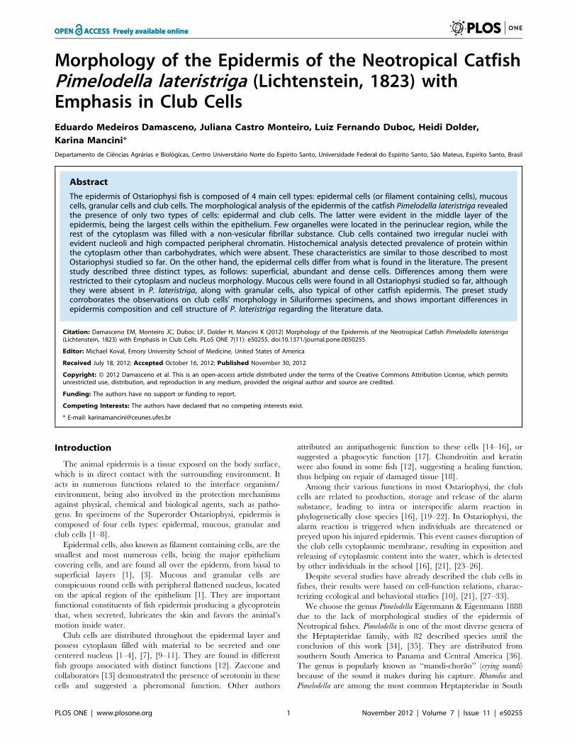
The skin of Pimelodella lateristriga is composed of a stratified
epithelium, which is supported by a thick layer of dense irregular
connective tissue (38.19%) and a wide muscle tissue (32.53%)
(Figure 2A, C). The epithelium is composed of two morpholog-
ically distinct cell types: the epidermal cells (15.91%) and the club
cells (13.31%) (Fig. 2B, C). The epidermal cells are small, with
average area of 1,475.74 mm2, when comparing the conspicuous
club cells that have average area of 2,991.68 mm2. These two cell
types form a heterogeneous stratified epithelium composed of
small flattened cells and large globular cells (Fig. 2B, C). The
number of layers varies according to the disposal and heteroge-
neity of cell types, with generally two layers of club cells containing
epidermal cells interspersed among them (Fig. 2B–C).
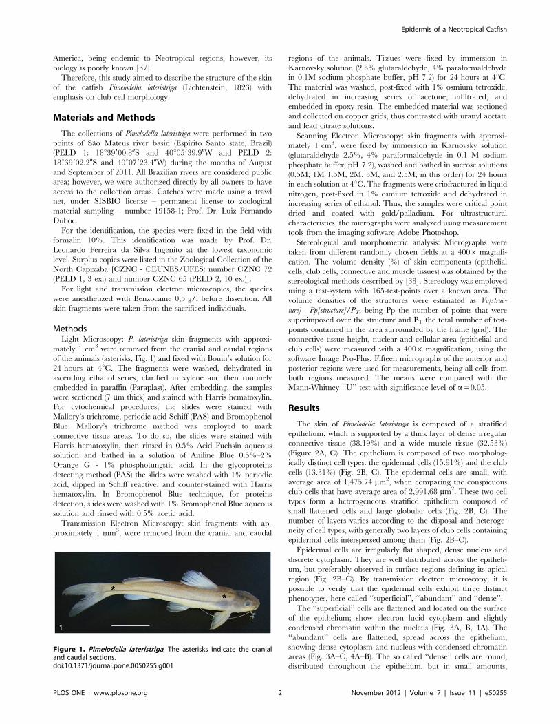
Epidermal cells are irregularly flat shaped, dense nucleus and
discrete cytoplasm. They are well distributed across the epitheli-
um, but preferably observed in surface regions defining its apical
region (Fig. 2B–C). By transmission electron microscopy, it is
possible to verify that the epidermal cells exhibit three distinct
phenotypes, here called ‘‘superficial’’, ‘‘abundant’’ and ‘‘dense’’.
The ‘‘superficial’’ cells are flattened and located on the surface
of the epithelium; show electron lucid cytoplasm and slightly
condensed chromatin within the nucleus (Fig. 3A, B, 4A). The
‘‘abundant’’ cells are flattened, spread across the epithelium,
showing dense cytoplasm and nucleus with condensed chromatin
areas (Fig. 3A–C, 4A–B). The so called ‘‘dense’’ cells are round,
distributed throughout the epithelium, but in small amounts,
Figure 1. Pimelodella lateristriga. The asterisks indicate the cranialand caudal sections.doi:10.1371/journal.pone.0050255.g001
Epidermis of a Neotropical Catfish
PLOS ONE | www.plosone.org 2 November 2012 | Volume 7 | Issue 11 | e50255
showing dense cytoplasm and globular nucleus with condensed
chromatin regions (Fig. 3C, 4A–B)
Either light or electron microscopy assay did not show any
mucous and/or granular cells.
Club cells are arranged in two layers, constituting the largest
extension of the epithelium. Skin fragments of cranial and caudal
portions revealed no differences in the occurrence, density or
morphology of club cells (Table 1). They are found mainly in the
middle region, rarely reaching the apical surface (Fig. 2B–C).
These cells show elongated and globular shapes (Fig. 2B–C, 4A–B,
5A–C, 6A). The nucleus is always central, measuring
1,325.73 mm2 in area (Fig. 5, 6). Two nuclei are found per cell,
very close to one another, with irregular shape and slightly
condensed chromatin, although with peripheral regions of
compression and prominent nucleoli (Fig. 6A–C–E).
The cytoplasm of club cells is rather poor in organelles and rich
in non vesicle secretion (Fig. 6). The few observed organelles
(endoplasmic reticulum, Golgi complexes, polyribosomes and
mitochondria) are located in the perinuclear region (Fig. 6B–C),
while the rest of the cytoplasm is filled with a filamentous
substance (Figure 6F). Therefore, the cytoplasmic content can be
separated into two regions: one light and electron lucid around the
nucleus and other abundant and electron dense, which occupies
nearly the entire cytoplasmic volume (Fig. 6B–C). Large vacuoles
are occasionally displayed in the peripheral cytoplasm (Fig. 6A).
The plasma membrane shows invaginations throughout its length,
making the cell surface irregular and associated with the epidermal
cells (Fig. 6A).
The club cells cytoplasm shows low glycoproteins content, as
determined by the PAS technique (Fig. 7A), while the Bromo-
phenol Blue technique, used for proteins detection, shows positive
reaction (Fig. 7B).
Underneath the epithelium is a layer of loose connective tissue
with fibroblasts and associated melanophores (Fig. 4B). Below the
loose connective tissue, is a thick layer of dense connective tissue
with an average thickness of 1,347.63 mm, highlighted in blue by
Mallory’s trichrome (Fig. 7C). Likewise, below dermis, lays a thick
skeletal muscle tissue layer, as evidenced in red/brown by
Mallory’s trichrome (Fig. 7C).
Discussion
In the present study, we analyzed skin fragments of two distinct
body regions in Pimelodella lateristriga: cranial and caudal. However,
the statistical analyzes revealed no differences regarding the
volume density and area of club cells between those body parts
(Table 1). Furthermore, no morphological differences were
observed.
The epithelium of P. lateristriga, as in all Ostariophysi species
studied so far, is classified as stratified and heterogeneous, showing
different cell types. However, the number of layers and thickness of
the epithelium is quite varied across species. In general, there is a
thick stratified epithelium, as in Siluriformes [4], [7], [11], [39], in
Characiformes [2] and in non-Ostariophysi such as Aguiliformes
[40]. P. lateristriga epithelium is formed only by one or two layers of
club cells lined with small epidermal cells. Club cells are
concentrated in the middle region of the epithelium, thus not
reaching the surface. This location corroborates previous findings,
for both marine and fresh water Siluriformes [4], [7], [31], [41].
The presence of two different cell types within P. lateristriga
epidermis - epidermal and club cells - is not shared by other
Siluriformes, which had their epithelial structure described [2],
Figure 2. Tegument. (A) Light micrograph of the tegument showingan epithelial tissue (ET) supported by a connective tissue (CT) and askeletal muscle tissue (MT). (B) Light micrograph of the stratifiedepithelial tissue composed by epidermal (EC) and club cells (CC). (C)Scanning electron micrograph showing the epithelial tissue withepidermal (EC) and club cells (CC), the connective tissue (CT) and theskeletal muscle tissue (MT). Scale bars: 25 mm (A); 15 mm (B) and 20 mm(C).doi:10.1371/journal.pone.0050255.g002
Figure 3. Transmission electron micrographs of epidermis withemphasis in the epidermal cells. The epidermis is composed bydistinct cell types: club cell (CC), superficial epidermal (SEC), denseepidermal (DEC) and abundant epidermal (AEC). Scale bars: 10 mm (A)and 5 mm (B, C).doi:10.1371/journal.pone.0050255.g003
Epidermis of a Neotropical Catfish
PLOS ONE | www.plosone.org 3 November 2012 | Volume 7 | Issue 11 | e50255
[4], [9], [7], [10], [11], and Cypriniformes [5], [8], [10]. The main
difference was the lack of mucous and granular cells in P.
lateristriga, which are common and abundantly distributed within
the epithelium of other species.
In general, mucous cells are characterized by their large size
(similar to club cells), containing cytoplasmic vesicles filled with
PAS positive secretion; they are located in the apical region of the
epithelium and possess pores in their plasma membrane, from
where the mucous secretion is released. The absence of mucous
cells in P. lateristriga is unexpected and would be related to
environmental or seasonal factors, since the collections occurred in
a short amount of time and in the same season. [10], studying the
effects of steroids on the skin of Phoxinus phoxinus (Linnaeus, 1758)
(Cypriniformes), noted that there is a close relationship between
steroid action and the amount of both mucous and club cells.
Granular and mucous cells are morphologically similar [1].
However, granular cells’ cytoplasm is PAS negative and filled with
electron dense granules. As the mucous cells, its absence within the
epithelium may be related to intrinsic and extrinsic factors.
The epidermal cells found in P. lateristriga were called
‘‘abundant’’, ‘‘superficial’’ and ‘‘dense’’, since there are no
citations in the literature so far, even for other species (there is
only a single set called epidermal cells). The majority of studies
involving ultrastructure of fish skin use high magnification
micrographs, which make it difficult to identify these cell types
[1], [9], [42–44].
Regarding the Ostariophysi, the most striking feature of club
cells is their size, being easily identified by light microscopy.
Although they are considered club cells, (i. e. in the shape of a
club) in P. lateristriga they are irregularly shaped, ranging from
globular to elongate, which is, indeed, a common morphological
variation. In Phoxinus phoxinus (Cypriniformes) the same cell type is
characterized as having a club shape (Phoxinus laevis as a species
cited in [19]; in Astyanax mexicanus (De Filippi, 1853) (Characi-
formes) it is described as oval [44]; and in Siluriformes it varies
from globular to elongated. In the ariid catfish Plicofollis
argyropleuron (Valenciennes 1840) it is described as elongated (Arius
tenuispinis species cited in [4], whereas in Ariopsis felis (Linnaeus
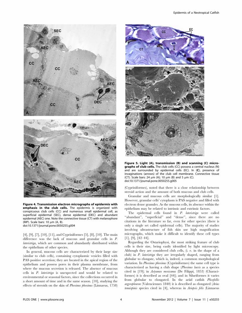
Figure 4. Transmission electron micrographs of epidermis withemphasis in the club cells. The epidermis is organized withconspicuous club cells (CC) and numerous small epidermal cell, assuperficial epidermal (SEC), dense epidermal (DEC) and abundantepidermal (AEC) one. Note the connective tissue (CT) with melanophore(MP). Scale bars: 10 mm (A, B).doi:10.1371/journal.pone.0050255.g004
Figure 5. Light (A), transmission (B) and scanning (C) micro-graphs of club cells. The club cells (CC) possess a central nucleus (N)and are surrounded by epidermal cells (EC). In (C), presence ofinvaginations (arrows) of the club cell membrane. Connective tissue(CT). Scale bars: 24 mm (A); 10 mm (B) and 5 mm (C).doi:10.1371/journal.pone.0050255.g005
Epidermis of a Neotropical Catfish
PLOS ONE | www.plosone.org 4 November 2012 | Volume 7 | Issue 11 | e50255
1766) is globular (species Arius felis as cited in [31]. In the bullhead
Coreobagrus brevicorpus (Mori 1936) this cell type shape varies from
globular to elongated (cited as Pseudobagrus brevicorpus in [7], being
elongated in the African sharptooth catfishes Clarias gariepinus
(Burchell 1822) [3] and Clarias batrachus (Linnaeus 1758) [45], and
globular in Ictalurus punctatus (Rafinesque 1818) [41].
The morphology of club cells in P. lateristriga is quite similar to
that described for other Siluriformes [9], [41], [4] and Cyprini-
formes [5], [8] due to its large size, central location, presence of
two nuclei and negative reaction to the PAS method.
The nuclear morphology of club cells in P. lateristriga is in
agreement with those found in non-Ostariophysi, as the lamprey
species Ichthyomyzon bdellum (Jordan 1885) (cited as Ichthyomyzon
unicuspis [42], in Cypriniformes [5], [8] and other Siluriformes [2],
[4], [7], [9], [45], [46]. The nucleus is described as central,
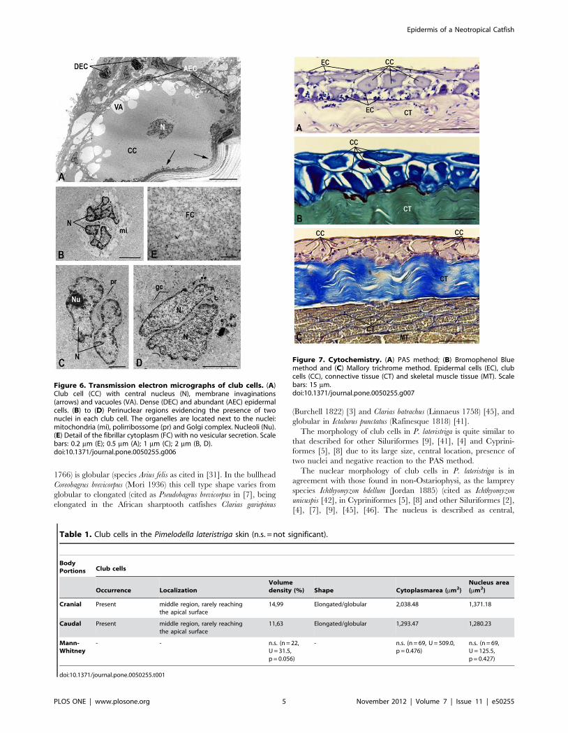
Figure 6. Transmission electron micrographs of club cells. (A)Club cell (CC) with central nucleus (N), membrane invaginations(arrows) and vacuoles (VA). Dense (DEC) and abundant (AEC) epidermalcells. (B) to (D) Perinuclear regions evidencing the presence of twonuclei in each club cell. The organelles are located next to the nuclei:mitochondria (mi), polirribossome (pr) and Golgi complex. Nucleoli (Nu).(E) Detail of the fibrillar cytoplasm (FC) with no vesicular secretion. Scalebars: 0.2 mm (E); 0.5 mm (A); 1 mm (C); 2 mm (B, D).doi:10.1371/journal.pone.0050255.g006
Table 1. Club cells in the Pimelodella lateristriga skin (n.s. = not significant).
BodyPortions Club cells
Occurrence LocalizationVolumedensity (%) Shape Cytoplasmarea (mm2)
Nucleus area(mm2)
Cranial Present middle region, rarely reachingthe apical surface
14,99 Elongated/globular 2,038.48 1,371.18
Caudal Present middle region, rarely reachingthe apical surface
11,63 Elongated/globular 1,293.47 1,280.23
Mann-Whitney
- - n.s. (n = 22,U = 31.5,p = 0.056)
- n.s. (n = 69, U = 509.0,p = 0.476)
n.s. (n = 69,U = 125.5,p = 0.427)
doi:10.1371/journal.pone.0050255.t001
Figure 7. Cytochemistry. (A) PAS method; (B) Bromophenol Bluemethod and (C) Mallory trichrome method. Epidermal cells (EC), clubcells (CC), connective tissue (CT) and skeletal muscle tissue (MT). Scalebars: 15 mm.doi:10.1371/journal.pone.0050255.g007
Epidermis of a Neotropical Catfish
PLOS ONE | www.plosone.org 5 November 2012 | Volume 7 | Issue 11 | e50255
irregularly shaped, with peripheral condensed chromatin and
prominent nucleoli. In addition, club cells’ nuclei in Siluriformes
are usually binucleated, which is a strong indicative of intense cell
activity, although sometimes they appear as mononucleated cells.
However, in Characiformes (e.g. Astyanax mexicanus) the nucleus is
usually single [44].
The cytoplasm of club cells in P. lateristriga is filled with
homogeneously dispersed fibrillar material, similar to that
described in other Siluriformes [1], [9] and Cypriniformes [1],
[43]. However, in non-Ostariophysi such as eels and lampreys
instead of regular fibrillar material, the cytoplasm showed spiral
filaments arranged in bundles and oriented in different planes [1],
[42]. In the peripheral region of the cytoplasm, next to the plasma
membrane, large vacuoles could be seen in P. lateristriga, as well as
demonstrated for Ictalurus punctatus [9], for the arrid Ariopsis felis
[31] and for the non-Ostariophysi, such as eels [47]. Given the
above, it is suggested that the vacuoles are structures typically
found in club cells and may be directly related to the mechanism
of secretion release.
In the perinuclear region, different from the rest of the fibrillar
cytoplasm, are the club cells organelles (endoplasmic reticulum,
Golgi complex, free ribosomes in the form of polyribosomes
aggregates, mitochondria and lysosomes). Such cytoplasmic
composition and organization are observed in most Siluriformes
and Cypriniformes studied so far [1], [9], [43], including P.
lateristriga. [1] described an unusual structure present in the
perinuclear region of club cells in Corydoras aeneus (Gill 1858)
(Siluriformes): an aggregation of smooth surface vesicles with
vesicles concentrically arranged around a central oval area. The
central area was fibrillar and collapsed vesicles were occasionally
found.
The cytoplasmic structure found in Corydoras aeneus was not
observed in other Siluriformes as P. lateristriga and Ictalurus punctatus
[9] or in Cypriniformes [1], [43]. This structure would be present
only in certain Ostariophysi species, indicating a difference
between club cells within the group.
The cytoplasm of club cells in P. lateristriga showed negative
reaction to the PAS method, indicating absence of glycoproteins in
its composition, as detected in other Siluriformes [9], [2], [11] and
Cypriniformes [5], [8]. The cytoplasm of club cells showed
positive reaction to the method of Bromophenol Blue, indicating
high protein content, as observed in other Siluriformes [2], [9],
[48]. The detection of protein and non-detection of glycoproteins
supports the observation of a large amount of ribosomes/
polyribosomes in P. lateristriga in comparison to the low occurrence
of rough endoplasmic reticulum and Golgi complex.
The plasma membrane of club cells in P. lateristriga showed
remarkable invaginations, unlike the observed by [1] in Corydoras
aeneus (Siluriformes) and in Carassius auratus (Cypriniformes). Such
invaginations confer cell adhesion, essential to the epithelium that
is usually submitted to pressure and friction.
This study aimed to describe the morphology of the epidermis
of a fish without focusing on functional aspects. There are few
studies in the literature regarding to electron microscopy. Thus,
the present findings represent a benchmark in the epidermal
ultrastructure of a Neotropical species from the order Siluriformes.
Furthermore, the existing studies that use high magnification
micrographs do not allow the study of organization and
composition of the epidermis. Therefore, further analyses are
necessary, due to differences between morphological and cyto-
chemical/immunocytochemical studies on the epidermis of the
fish species studied so far, especially in electron microscopy in an
attempt to a more precise characterization of the components of
the epidermis in Ostariophysi.
Acknowledgments
We would like to thanks the Laboratorio de Ultraestrutura Celular ‘Carlos
Alberto Redins’ (CCS/UFES) for allowing the use of their microscopy
equipment. We also would like to thank Marcos de Lucca Moreira Gomes
for translating the manuscript.
Author Contributions
Conceived and designed the experiments: ED JCM KM LFD. Performed
the experiments: ED JCM KM. Analyzed the data: ED JCM KM LFD.
Contributed reagents/materials/analysis tools: JCM KM LFD HD. Wrote
the paper: JCM KM LFD.
References
1. Henrikson RC, Matoltsy AG (1968) The fine structure of the teleost epidermis.
III. Club cells and other cell types. J Ultrastruct Res 21: 222–232.
2. Park JY, Kim IS, Kim SY (2003) Structure and histochemistry of the skin of a
torrent catfish, Liobagrus mediadiposalis. Environ Biol Fishes 66: 3–8.
3. Guerra RR, Santos NP, Cecarelli P, Mangetti AJ, Silva JRMC, et al. (2006)
Stratum adiposum. A special structure of the african catfish skin (Clarias
gariepinus, Burchell 1822). Anat Histol Embryol 35: 144–146.
4. Al-Banaw A, Kenngott R, Al-Hassan JM, Mehana N, Sinowatz F (2009)
Histochemical analysis of glycoconjugates in the skin of a catfish (Arius Tenuispinis,
Day). Anat Histol Embryol 39: 42–50.
5. Halbgewachs CF, Marchant TA, Kusch RC, Chivers DP (2009) Epidermal club
cells and the innate immune system of minnows. Biol J Linn Soc 98: 891–897.
6. Barbosa A, Magalhaes EJ, Hoffmann A, Ide LM (2010) Conspecific and
heterospecific alarm substance induces behavioral responses in piau fish Leporinus
piau. Acta Ethol 13:119–126.
7. Park JY, Oh MK, Kang EJ, Kim CH, Beon MS (2010) On the vascularization
and structure of the skin of a Korean bullhead Pseudobagrus brevicorpus (Bagridae,
Teleostei) based on its entire body and appendages. J Appl Ichthyol 26: 64–70.
8. Stabell OB, Vegusdal A (2010) Socializing makes thick-skinned individuals: on
the density of epidermal alarm substance cells in cyprinid fish, the crucian carp
(Carassius carassius). J Comp Physiol A 196: 639–647.
9. Yoakim EG, Grizzle JM (1982) Ultrastructure of alarm substance cells in the
epidermis of the channel catfish, Ictalurus punctatus (Rafinesque). J Fish Biol 20:
213–221.
10. Pfeiffer W,Riegelbauer G, Meier G, Scheibler B (1985) Effect of Hypoxanthine
3(N)-oxide and Hypoxanthine-1(N)-oxide on central nervous excitation of the
black tetra Gymnocorymbus ternetzi (Characidae, Ostariophysi, Pisces) indicated by
dorsal light response. J Chem Ecol 11: 507–523.
11. Lizarazo RJB, Virguez MQ, Ramırez EG, Caicedo DR, Giraldo HH (2008)
Histologıa y morfometrıa de piel del pez Eremophilus mutisii (Trychomecteridae,
Siluriformes). Rev Biol Trop 56: 885–893.
12. Ralphs JR, Benjamin M (1992) Chondroitin and keratan sulphate in the
epidermal club cells of teleosts. J Fish Biol 40: 473–475.
13. Zaccone G, Tagliafierro G, Fasulo S, Contini A, Ainis L, Ricca MB (1990)
Serotonin-like immunoreactivity in the epidermal club cells of teleost fishes.
Histochem 93: 355–357.
14. Suzuki Y, Kaneko T (1986) Demonstration of the mucous hemagglutinin in the
club cells of eel skin. Dev Comp Immunol 10: 509–518.
15. Al-Hassan JM, Thomson M, Ali M, Criddle RS (1987) Toxic and
pharmacologically active secretions from the Arabian Gulf catfish (Arius
thalassinus, Ruppel). Toxicol Toxin Rev 6: 1–43.
16. Smith RJF (1992) Alarm signals in fishes. Rev Fish Biol and Fish 2: 33–63.
17. Lufty RG (1964) Studies on the epidermis of the catfish Synodontis schall. Sch Ain
Shams Sci Bull Cairo 10: 153–163.
18. Iger Y, Abraham M (1990) The process of skin healing in experimentally
wounded carp. J Fish Biol 36: 421–437.
19. Pfeiffer W (1960) Uber die Schreckreaktion bei Fischen und die Herkunft des
Schreckstoffes. Z Vergl Physiol 43: 578–614.
20. Pfeiffer W (1962) The fright reaction of fish. Biol Rev 37: 495–511.
21. Pfeiffer W (1977) The distribution of fright reaction and alarm substance cells in
fishes. Copeia 4: 653–665.
22. Smith RJF (1997) Does one result trump all others? A response to Magurran,
Irving and Henderson. Proc R Soc London, Ser B 264: 445–450.
23. Smith RJF (1976) Seasonal loss of alarm substance cells in North American
cyprinoid fishes and its relation to abrasive spawning behaviour. Canadian J Zool
54, 1172–1182.
Epidermis of a Neotropical Catfish
PLOS ONE | www.plosone.org 6 November 2012 | Volume 7 | Issue 11 | e50255
24. Kats LB, Dill LM (1998) The scent of death: Chemosensory assessment of
predation risk by prey animals. Ecoscience 5:361–394.25. Chivers DP, Smith RJF (1998) Chemical alarm signalling in aquatic predator/
prey systems: A review and prospectus. Ecoscience 5:338–352.
26. Wisenden BD (2000) Scents of danger: The evolution of olfactory ornamentationin chemically mediated predator-prey interactions. In Espmark Y, Amundsen T,
Rosenqvist G, editors. Animal Signals: Signalling and Signal Design in AnimalCommunication. Trondheim: Tapir Academic Press. pp. 365–386.
27. Solomon DJ (1977) A review of chemical communication in freshwater fish.
J Fish Biol 11: 363–376.28. Pfeiffer W, Riegelbauer G (1978). The Effect of the Alarm Substance on the
Central Nervous Excitation of the Black Tetra Gymnocorymbus ternetzi (Char-acidae, Ostariophysi, Pisces) Indicated by Dorsal Light Response. J Comp
Physiol 123: 281–288.29. Lawrence BJ, Smith RJF (1989) Behavioral response of solitary fathead minnow,
Pimephales promelas, to alarm substance. J Chem Ecol 15: 209–219.
30. Wisenden BD, Smith RJ F (1997) The effect of physical condition and shoalmatefamiliarity on proliferation of alarm substance cells in the epidermis of fathead
minnows. J Fish Biol 50:799–808.31. Smith ME (2000) Alarm response of Arius felis to chemical stimuli from injured
conspecifics. J Chem Ecol 26: 1634–1647.
32. Brown GE, Adrian-Jr JC, Shih ML (2001) Behavioral responses of fatheadminnows to hypoxanthine-3-N-oxide at varying concentrations. J Fish Biol 58:
1465–1470.33. Jung JA, Tonn WM (2011) Alarm substances elicit limited population - level
responses in fathead minnow. Ecol Fresh Fish 20: 220–230.34. Bockman FA, Guazzelli GM (2003) Family Heptapteridae (Heptapterids). In:
Reis RE, Kullander SO, Ferraris Jr CJ, editors. Checklist of the freshwater fishes
of South and Central America. Porto Alegre: Edipucrs, p. 406–431.35. Trajano E, Reis RE, Bichuette ME (2004) Pimelodella spelaea: A new cave catfish
from central Brazil, with data on ecology and evolutionary considerations(Siluriformes: Heptapteridae). Copeia 2004: 315–325.
36. Burgess WE (1989) An atlas of freshwater and marine catfishes. A preliminary
survey of the Siluriformes. Neptune City: TFH Publications. 784 p.
37. Malabarba LR, Reis RE, Vari RP, Lucena ZMS, Lucena CAS (1998) Phylogeny
and classification of Neotropical Fishes. Porto Alegre: Edipucrs. 603 p.
38. Weibel ER (1979) Stereological methods. I. Practical methods for biological
morphometry. Academic Press, London.
39. Wisenden BD, Pogatshnik J, Gibson D, Bonacci L, Schumacher A, Willett A
(2008) Sound the alarm: learned association of predation risk with novel auditory
stimuli by fathead minnows (Pimephales promelas) and glowlight tetras (Hemi-
grammus erythrozonus) after single simultaneous pairings with conspecific chemical
alarm cues. Environ Biol Fish 81: 141–147.
40. Nakamura O, Watanabe T, Kamiya H, Muramoto K (2001) Galectin
containing cells in the skin and mucosal tissues in Japanese conger eel, Conger
myriaster: an immunohistochemical study. Develop Compar Immun 25: 431–437.
41. Chapman GB, Johnson EG (1997) An electron microscope study of intrusions
into alarm substance cells of the channel catfish. J Fish Bio 51: 503–514.
42. Downing SW, Novales RR (1971) The fine structure of lamprey epidermis II.
Club cells. J Ultrastruct Res 35: 295–303.
43. Pfeiffer W, Sasse D, Arnold M (1971) Die Schreckstoffzellen von Phoxinus phoxinus
und Morulius chrysophakedion (Cyprinidae, Ostariophysi, Pisces). Z Zellforsch 118:
203–213.
44. Peters N, Schmidt W, Fricke D (1990) Fine Structure of the Club Cells (Alarm
Substance Cells) in the Epidermis of Astynnax mexicanus FILIPPI 1853
(Characinidae, Pisces) and its Cave Forrns ‘‘Anoptichthys’’. Int Revue Ges
Hydrobiol 75: 257–267.
45. Mittal AK, Garg TK (1994) Effect of an anionic detergent – sodium dodecyl
sulphate exposure on club cells in the epidermis of Clarias batrachus. J Fish Biol 44:
857–875.
46. Pfeiffer W (1970) Uber die Schreckstoffzellen der Siluriformes (Ostariophysi,
Pisces). Ant Anz 126: 113–119.
47. Whitear M, Zaccone G (1984) Fine structure and histochemistry of club cells in
the skin of three species of eel. Z Mikrosk Anat Forsch 98: 481–501.
48. Agrawal N, Mittal AK (1992) Structure and histochemistry of the epithelia of lips
and associated structures of a catfish Rita rita. Japan J Ichthyol 39: 93–102.
Epidermis of a Neotropical Catfish
PLOS ONE | www.plosone.org 7 November 2012 | Volume 7 | Issue 11 | e50255