Embed Size (px)
Citation preview
Morphology of the Symbiosis BetweenCorculumcardissa(Mollusca: Bivalvia) and Symbiodinium
corculorum (Dinophyceae)
MARK A. FARMER,1 WILLIAM K. FITT 2,*, AND ROBERT K. TRENCH3
1Department of Cellular Biology, and2Institute of Ecology, University of Georgia, Athens, GA 30602USA,3Department of Ecology, Evolution and Marine Biology, University of California at Santa
Barbara, Santa Barbara, CA 93106 USA
Abstract. Light and transmission electron microscopy oftissues of the symbiotic clamCorculum cardissa(L)showed that a symbiotic dinoflagellate,Symbiodinium cor-culorum(Trench), is found predominantly in the mantle andthe gills. The data suggest that inC. cardissathe algae arelocated in a zooxanthellal tubular system that is associatedwith the hemocoel and is similar to that seen in tridacnine(“giant”) clams. The algae occur within the lumen of thetertiary tubules and are thus separated from the hemolymphby a tissue that is one cell layer thick. Under a lightmicroscope the tertiary tubules appear as rows of symbiontsoriginating from the digestive diverticulum, presumablybranching from the primary tubules that are also seen insymbiotic tridacnine clams. This morphological arrange-ment is discussed with regard to the ontogeny and theevolution of the tubular system within symbiotic bivalves.
Introduction
Several species of marine bivalves in the family Cardi-idae harbor symbiotic dinoflagellates that belong to thegenusSymbiodinium. These bivalves include all of the spe-cies in the subfamily Tridacninae, including the well-knowngenera of larger clams,TridacnaandHippopus, as well asless well-known genera of much smaller clams in the sub-family Fraginae, such asCorculumandFragum(Kawaguti,1950, 1983; Schneider, 1998). For many years, the symbi-otic algae in tridacnines were depicted as being located inthe hemal spaces, whence they were culled by wandering
amoebocytes and digested in the digestive gland; the indi-gestible remains were thought to reside in the kidneys (e.g.,Yonge, 1936, 1953, 1975, 1980; Goreauet al., 1973). How-ever, a system of tubules, arising from one of the divertic-ular ducts of the stomach ofTridacna and ramifyingthrough much of the clam and containing the symbionts,was described by K. Mansour (1946a, b), but forgotten.Finally, 46 years later, the “zooxanthellal tubular system”was redescribed by Nortonet al. (1992), who proposed thatthe primary, secondary, and blind-ended tertiary tubes of thetubular system do not connect with the hemocoel; thereforethe algae are not found in the hemolymph compartment (seeFitt, 1993, for a review).
In the heart cockle,Corculum cardissa, as in the gianttridacnine clams, symbiotic dinoflagellates are located inthe mantle tissue; but unlike the tridacnine,C. cardissahasmany algae located in the gills as well (Kawaguti, 1968).Early electron microscopic images, in both instances, wereinterpreted as indicating that the algae are within the hemalsystem (Kawaguti, 1966, 1968). This interpretation wasconsistent with the author’s observations that blood cells areapparently in contact with the algae inC. cardissaandTridacna (Kawaguti, 1966, 1968).
The occurrence of symbiotic algae in a tubular systemin Tridacna (Mansour, 1946a, b; Nortonet al., 1992)raises the question of whether a similar zooxanthellaltubular system also occurs in the cockleCorculum car-dissa, or any other related species (i.e., Fragumspp.).The goal of this study was to document evidence of atubular system inC. cardissa, and to determine whetherthe tubules would penetrate the gill tissue, a conceptuallydifficult morphology. In the current study, ultrastructuralobservations indicate that a tubular system also exists in
Received 10 June 1999; accepted 14 February 2001.* To whom correspondence should be addressed. E-mail: fitt@
sparrow.ecology.uga.edu
Reference:Biol. Bull. 200: 336–343. (June 2001)
336

Corculum; that the symbioticdinoflagellates occur withinthe lumina of the tubes, which themselves are located withinthe hemocoel; and that the algae within the tertiary tubules areseparated from the hemolymph by a tissue that is mostly onlyone cell layer thick.
Materials and Methods
Corculum cardissa(Linne) was collected from the sandyreef flat at about 0.5 m depth in Belau (Palau), WesternCaroline Islands. Animals were fixed in 6% glutaraldehyde,
Figure 1. Light micrograph of a paraffin-embedded section through the mantle tissue ofTridacna maximashowing rows of symbiotic algae (arrowheads) in tertiary tubules. Iridophores (ir), animal cells with crystallineproteins that refract light, are found in the mantles of all species of clams containing symbiotic algae. Scale bar,20 mm.
Figure 2. Light micrograph of a paraffin-embedded section through the gill tissue ofCorculum cardissashowing rows of symbiotic algae in tertiary tubules. (a) Overview; scale bar5 12mm. (b) Higher magnification,scale bar5 10 mm.
337ALGAL SYMBIOSIS IN THE HEART COCKLE CORCULUM

postfixed in 3% osmium tetraoxide, dehydrated, and embed-ded in Spurr’s medium as previously described (Trenchetal., 1981). The tissues ofTridacnaspp. were fixed, embed-ded, and observed as described in Trenchet al. (1981).Thick sections (1mm) were prepared for examination bylight microscopy on an LKM Ultratome V. These werephotographed with an Olympus Vanox microscope and aPM-10 camera. Ultrathin sections for electron microscopicexamination were prepared on an RMC-6000 ultrami-crotome, stained with uranyl acetate and lead citrate in thestandard manner, and observed and photographed with aPhilips 400 transmission electron microscope (TEM).
Results and Discussion
Light microscopic examination ofCorculum cardissarevealed that, similar to observations made on symbionts
living in mantle tissues ofTridacna (Fig. 1; Mansour,1946a; Fitt and Trench, 1981; Nortonet al., 1992; Nortonand Jones, 1992), algal cells are arranged in rows in both themantle (e.g., Kawaguti, 1968; Figs. 1, 2) and the gills (Fig.2). However light microscope observations could not re-solve the tertiary tubule structure in either genus of clam.
Electron microscopic examination of the gills ofCorcu-lum (Fig. 3) shows that the algae are indeed juxtaposed toanimal blood cells. Kawaguti’s (1968) early descriptionsfrom C. cardissanote that algae are sometimes accompa-nied by “wandering cells,” but he includes no figures. Incontrast, TEM pictures of symbionts inTridacna croceaandT. maximaclearly show nearby animal cells (Kawaguti,1966; Fitt and Trench, 1981). Algal symbionts inC. car-dissaare not in direct contact with the animal’s blood cells,but are separated from the hemolymph and the blood cells
Figure 3. Transmission electron micrograph of a portion of the gill ofCorculum cardissashowing a portionof the tertiary tubule (t), and a blood cell (BC) close to cells of the algaSymbiodinium corculorum(Sc) in thetubule. Scale bar, 1mm.
338 M. A. FARMER ET AL.

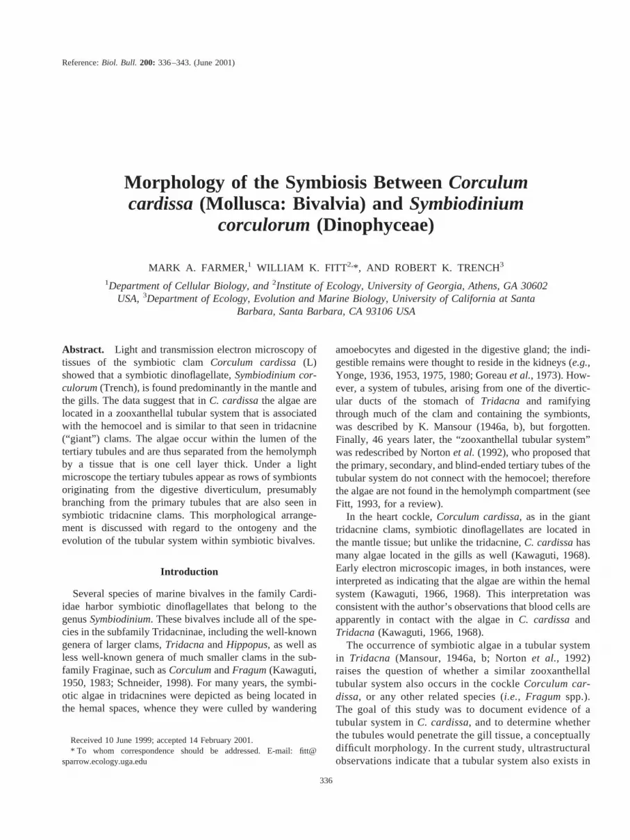
by the cells of the tubules, which at the tertiary level areabout one cell layer thick (Figs. 3–5). Evidence for thiscomes from closer examination of high-magnification TEMimages of the structural relations between the algae, thecells of the tubules, and the blood cells (Fig. 4). First, algaein the lumina of the tubules are often pressed against theinner plasmalemma of the tubular cells; when nuclei of theappressed tubular cells are apparent (as in Fig. 4a), thiscould lead to the interpretation that the algae are intracel-lular (e.g., Kawaguti, 1968). Second, the algae are clearlyseparated from the molluscan blood cells by the cells com-posing the tubules (Fig. 4b). Overlapping cell processesform the tertiary tubules (Fig. 5). Two or more unseparatedadjacent symbionts (Fig. 6) also indicate that the algae arein tubules and not living intracellularly within host cells.
In bivalves that harbor symbiotic dinoflagellates, thestructure of the tubular system, in which the tubules arisefrom the digestive system and are contiguous with it, sug-gests that the morphological and functional relation betweenhost and symbionts in bivalves is directly analogous to thatfound in symbiotic cnidarians (Fitt, 1993). In both cases, thealgae enter the digestive systemvia the mouth. In bivalves,symbiotic dinoflagellates entervia the mouth, and exitviathe anus (Ricard and Salvat, 1977; Trenchet al., 1981;Maruyama and Heslinga, 1997); their entire residence in theclam is in association with the digestive system. In contrast,symbionts in cnidarians enter and exitvia the mouth andeventually take up residence inside of host digestive cells.The location of the symbionts in bivalves and cnidarians isalso analogous with respect to metabolite flux between host
Figure 4. Transmission electron micrograph of the relation between the symbiotic algae, the tubule cells,and the blood cells in gill tissue ofCorculum cardissa. (a) An algal cell closely appressed to a tubule cell. Thealgal cell wall is juxtaposed to the tubule cell plasmalemma, which can be followed around the enclosed cellnucleus, which it encloses. (b) Two algal cells in adjacent tertiary tubules, separated by the cytoplasm of the twotubule cells (tc). A blood cell is close by (bc). Scale bars, 1mm.
339ALGAL SYMBIOSIS IN THE HEART COCKLE CORCULUM

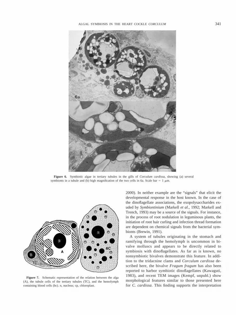
and symbiont (Fittet al., 1985). In bivalves, where the algaeare intercellular, they are separated from the hemolymph(circulating nutrients) by the proximal and distal plasma-lemma of the tubule cells (Fig. 7). Hence, nutrient exchangebetween the algae and the hemolymph of bivalves maypotentially be regulated by the tubule cells. In cnidarians,where the algae are intracellular, the symbionts are sepa-rated from their nutrient source, the gastrovascular system,by two membranes, the host cell plasmalemma and thesymbiosome membrane.
The presence of the tubular system in symbiotic bivalvesis also significant from ontogenetic and evolutionary per-spectives. Studies of algal symbioses in tridacnines (Fitt andTrench, 1981) clearly show that the tubules develop only inthe presence of dinoflagellate symbionts; the algae in thetubules are observed as “rows extending from the region ofthe stomach and digestive gland toward the developingsiphonal tissue” (Fittet al., 1981). Juvenile clams that were
allowed to develop in the absence ofSymbiodiniumdid notshow evidence of this feature. From the report of Nortonetal. (1995), it is also apparent that, when the algae are lostfrom Tridacnaduring events of thermal “stress,” the tubulesatrophy. Whether the tubules are reformed should the sym-biosis recover, or whether lack of recovery of bleachedclams is the result of the inability of the tubular system toregenerate, is unknown.
For marine symbioses, only two other instances havebeen recorded in which the symbionts appear to play asignificant role in the ontogenetic event in the host. Oneexample is the process of strobilation in symbiotic scypho-zoans such asMastigias (Sugiura, 1964) andCassiopeia(Colley and Trench, 1985); these jellyfish produce ephyraeonly in the presence ofSymbiodinium. Another example isthe influence thatVibrio fischeri, a symbiotic luminousbacteria, has on morphogenesis of certain parts of the lightorgan in the squidEprymna scolopes(Claes and Dunlap,
Figure 5. Transmission electron micrograph of an alga in the gill ofCorculum cardissasurrounded byoverlapping processes of tertiary tubule cells (tc). Scale bar, 1mm.
340 M. A. FARMER ET AL.
2000). In neither example are the “signals” that elicit thedevelopmental response in the host known. In the case ofthe dinoflagellate associations, the exopolysaccharides ex-uded bySymbiontinium(Markell et al., 1992; Markell andTrench, 1993) may be a source of the signals. For instance,in the process of root nodulation in leguminous plants, theinitiation of root hair curling and infection thread formationare dependent on chemical signals from the bacterial sym-bionts (Brewin, 1991).
A system of tubules originating in the stomach andramifying through the hemolymph is uncommon in bi-valve molluscs and appears to be directly related tosymbiosis with dinoflagellates. As far as is known, nononsymbiotic bivalves demonstrate this feature. In addi-tion to the tridacnine clams andCorculum cardissade-scribed here, the bivalveFragum fragumhas also beenreported to harbor symbiotic dinoflagellates (Kawaguti,1983), and recent TEM images (Kempf, unpubl.) showmorphological features similar to those presented herefor C. cardissa. This finding supports the interpretation
Figure 6. Symbiotic algae in tertiary tubules in the gills ofCorculum cardissa, showing (a) severalsymbionts in a tubule and (b) high magnification of the two cells in 6a. Scale bar5 1 mm.
Figure 7. Schematic representation of the relation between the alga(A), the tubule cells of the tertiary tubules (TC), and the hemolymphcontaining blood cells (bc). n, nucleus; cp, chloroplast.
341ALGAL SYMBIOSIS IN THE HEART COCKLE CORCULUM

that all algal symbionts in molluscs occur in tubuleextensions of the digestive system. Cladistic analysesbased on morphological characters (Schneider, 1992,1998) and phylogenetic relationships based on analysis ofsmall subunit ribosomal RNA gene sequences (Ma-ruyama et al., 1998) both indicate that the known bi-valves with symbiotic dinoflagellates are closely related,all belonging to Cardiidae. In addition, the availablemolecular genetic evidence (NcNallyet al., 1994) revealsthat the symbiotic algae associated withCorculum andTridacna are also very closely related (LaJeunesse,2000), but not identical. We also suppose that freshwaterbivalves, such asAnodonta, that are symbiotic with thegreen algaChlorella sp. (Pardy, 1980) may demonstratea tubular structure in which to house the algae, as thesesymbionts probably also enter their hosts through thedigestive system.
The only other molluscan group that shows an analogousmorphology is the opisthobranch gastropods: some sacco-glossan opisthobranchs temporarily harbor derived chloro-plasts from feeding (Trench, 1975), and eolid nudibranchsoften maintain dinoflagellates for a short time after feedingon symbiotic cnidarians (Kempf, 1984). Most significantlyin relation to symbiotic bivalves, in eolidacean nudibranchs“branches of the posterior aorta . . . accompany the branchesof the midgut gland (digestive diverticulum) into the cerata. . .” (Hyman, 1967, p. 477), suggesting development oftubules in conjunction with development of blood vessels,or vice versa.
We speculate that bivalves, like their gastropod relatives,possess a suite of genes that encode the expression of thetubular system, but these genes are expressed only afteractivation by some “signal” produced by dinoflagellatesymbionts as they enter the host digestive tract. Thesesituations would be analogous to the production of variousgalls in plants following infection by bacteria, fungi, in-sects, or other parasitic plants (Bidwell, 1979), all of whichproduce chemical signals.
Acknowledgments
We thank Professor Stephen Kempf for providing us withunpublished transmission electron micrographs of tissues ofFragum fragum, and two anonymous reviewers for sugges-tions on improving the text. WKF acknowledges supportfrom NSF and the NOAA National Undersea ResearchProgram (UNCW and CMRC).
Literature Cited
Bidwell, R. G. S. 1979. Plant Physiology. 2nd ed. MacMillan, NewYork.
Brewin, N. J. 1991. Development of the legume root nodule.Annu. Rev.Cell Biol. 7: 191–226.
Claes, M. F., and P. V. Dunlap. 2000. Aposymbiotic culture of thesepiolid squidEuprymna scolopes: role of the symbiotic bacterium
Vibrio fischeri in host animal growth development, and light organmorphogenesis.J. Exp. Zool.286: 280–296.
Colley, N. J., and R. K. Trench. 1985. Cellular events in the re-establishment of a symbiosis between a marine dinoflagellate and acoelenterate.Cell Tissue Res.239: 93–103.
Fitt, W. K. 1993. Nutrition of giant clams. Pp. 31–40 inBiology andMariculture of Giant Clams., W. K. Fitt, ed. ACIAR, Canberra, Aus-tralia.
Fitt, W. K., and R. K. Trench. 1981. Spawning, development, andacquisition of zooxanthellae byTridacna squamosa(Mollusca: Bi-valvia). Biol. Bull. 161: 213–235.
Fitt, W. K., C. R. Risher, and R. K. Trench. 1981. Larval biology oftridacnid clams.Aquaculture39: 181–195.
Fitt, W. K., T. A. V. Rees, and D. Yellowlees. 1985. The relation-ship between pH and the availability of dissolved inorganic nitrogen inthe zooxanthella-giant clam symbiosis.Limnol. Oceanogr.40: 976–982.
Goreau, T. F., N. I. Goreau, and C. M. Yonge. 1973. On the utilizationof photosynthetic products from zooxanthellae and of a dissolvedamino acid inTridacna maxima(Mollusca: Bivalvia).J. Zool. Lond.169: 417–454.
Hyman, L. H. 1967. The Invertebrates. Vol. VI, Mollusca I.McGrawHill, New York.
Kawaguti, S. 1950. Observations on the heart cockle,Corculum cardissa(L), and its associated zooxanthellae.Pac. Sci.4: 43–49.
Kawaguti, S. 1966. Electron microscopy on the mantle of the giant clamwith special reference to zooxanthellae and iridophores.Biol. J.Okayama Univ.12: 81–92.
Kawaguti, S. 1968. Electron microscopy on zooxanthellae in the mantleand gill of the heart shell.Biol. J. Okayama Univ.14: 1–11.
Kawaguti, S. 1983. The third record of an association between bivalvemollusks and zooxanthellae.Proc. Jpn. Acad. Ser. B59: 17–20.
Kempf, S. C. 1984. Symbiosis between the zooxanthellaSymbiodinium(5Gymnodinium) microadriaticum. (Freudenthal) and four species ofnudibranchs.Biol. Bull. 166: 110–126.
LaJeunesse, T. C. 2000. Diversity, distribution and host specificity ofalgal symbionts of the dinoflagellate genusSymbiodinium. Ph.D dis-sertation, University of California Santa Barbara.
Mansour, K. 1946a. Communication between the dorsal edge of themantle and the stomach ofTridacna. Nature (Lond.)157: 844.
Mansour, K. 1946b. Source and fate of the zooxanthellae of the visceralmass ofTridacna elongata. Nature (Lond.)158: 130.
Markell, D. A., and R. K. Trench. 1993. Macromolecules exuded bysymbiotic dinoflagellates in culture: Amino acid and sugar composi-tion. J. Phycol.29: 64–68.
Markell, D. A., R. K. Trench, and R. Iglesias-Prieto. 1992. Macro-molecules associated with the cell walls of symbiotic dinoflagellates.Symbiosis12: 19–31.
Maruyama, T., and G. A. Heslinga. 1997. Fecal discharge of zooxan-thellae in the giant clamTridacna derasa, with reference to theirgrowth rate.Mar. Biol. 127: 473–477.
Maruyama, T., M. Ishikura, S. Yamazaki, and S. Kanai. 1998.Molecular phylogeny of zooxanthellate bivalves.Biol. Bull. 195: 70–77.
McNally, K. L., N. S. Govind, P. E. Thome, and R. K. Trench. 1994.Small subunit ribosomal DNA sequence analyses and a reconstructionof the inferred phylogeny among symbiotic dinoflagellates (Pyrrho-phyta).J. Phycol.30: 316–329.
Norton, J. H., and G. W. Jones. 1992. The giant clam: an anatomicaland histological atlas. Australian Center for InternationalAgricultural Research (ACIAR) Monograph Series, Canberra,142 pp.
Norton, J. H., M. A. Shepherd, H. M. Long, and W. K. Fitt. 1992. The
342 M. A. FARMER ET AL.

zooxanthellal tubular system in the giant clam.Biol. Bull. 183: 503–506.
Norton, J. H., H. C. Prior, B. Baillie, and D. Yellowlees. 1995. Atro-phy of the zooxanthellal tubular system in bleached giant clamsTri-dacna gigas. J. Invertebr. Pathol.66: 307–310.
Pardy, R. L. 1980. Symbiotic algae and14C incorporation in the fresh-water clamAnodonta. Biol. Bull.158: 349–355.
Ricard, M., and B. Salvat. 1977. Faeces ofTridacna maxima(Mollus-ca-Bivalvia), composition and coral reef importance.Proc. Third Int.Coral Reef Symp., Miami:495–502.
Schneider, J. A. 1992. Preliminary cladistic analysis of the bivalvefamily Cardiidae.Am. Malacol. Bull.9: 145–155.
Schneider, J. A. 1998. Phylogeny of the Cardiidae (Bivalvia): Phyloge-netic relationships and morphological evolution within the subfamiliesClinocardiinae, Lymnocardiinae, Fraginae and Tridacninae.Malacolo-gia 40: 321–373.
Sugiura, Y. 1964. On the life history of rhizostome medusae. II. Indis-pensability of zooxanthellae for strobilation inMastigias papua. Em-bryologia 8: 223–233.
Trench, R. K., 1975. Of “leaves that crawl”: functional chloroplasts inanimal cells.Soc. Exp. Biol. Symp.XXIX: 229–265.
Trench, R. K., D. S. Wethey, and J. W. Porter. 1981. Observations onthe symbiosis with zooxanthellae among the tridacnidae (Mollusca,Bivalvia). Biol. Bull. 161: 180–198.
Yonge, C. M. 1936. Mode of life, feeding, digestion and symbiosis withzooxanthellae in the Tridacndae.Scientific Report, Great Barrier ReefExpedition1: 283–331.
Yonge, C. M. 1953. Mantle chambers and water circulation in theTridacnidae.Proc. Zool. Soc. Lond.123: 551–561.
Yonge, C. M. 1975. Giant clams.Sci. Am.232: 96–105.Yonge, C. M. 1980. Functional morphology and evolution in the Tri-
dacnidae.Rec. Aust. Mus.33: 735–777.
343ALGAL SYMBIOSIS IN THE HEART COCKLE CORCULUM


