Embed Size (px)
Citation preview
ZOOLOGIA 27 (3): 483–488, June, 2010doi: 10.1590/S1984-46702010000300024
© 2010 Sociedade Brasileira de Zoologia | www.sbzoologia.org.br | All rights reserved.
The peritrich ciliate Carchesium polypinum (Linnaeus, 1758)Ehrenberg, 1830 is commonly found in freshwater ecosystemsand is a good indicator of poor water quality (WEI et al. 2004).Ciliates of the genus Carchesium live in colonies containing zoo-ids, contractile stalks and free-swimming telotrochs (ZAGON 1971)and have high colonization rates in eutrophic ecosystems(KUSUOKA & WATANABE 1989). They generally do not display hostspecificity and could be found on seaweeds, emerged and sub-mersed macrophytes, and attached to different groups of aquaticinvertebrates (FOISSNER et al 1992, COOK et al. 1998, MAYÉN-ESTRADA
& ALADRO-LUBEL 2002), including the gastropod Pomacea figulina(Spix, 1827) (DIAS et al. 2008). In addition to colonize living hosts,they also attach to inert substrates and have been recorded indiverse lotic systems associated to sediment (KUSUOKA & WATANABE
1989, SOLA et al. 1996, MADONI & BASSANINI 1999, MADONI 2005,MADONI & BRAGHIROLI 2007).
Prosobranch molluscs in the Ampullariidae family arewidely distributed in subtropical and tropical regions, fresh-water ecosystems, preferentially inhabiting still waters in lotic
systems. Molluscs in the genus Pomacea also tolerate organicpollution (THIENGO 1995), and are promising indicators of wa-ter quality (COLER et al. 2005). This condition increases the eco-logical opportunity for colonization by peritrich ciliates whichpresent a high preference for eutrophic environments.
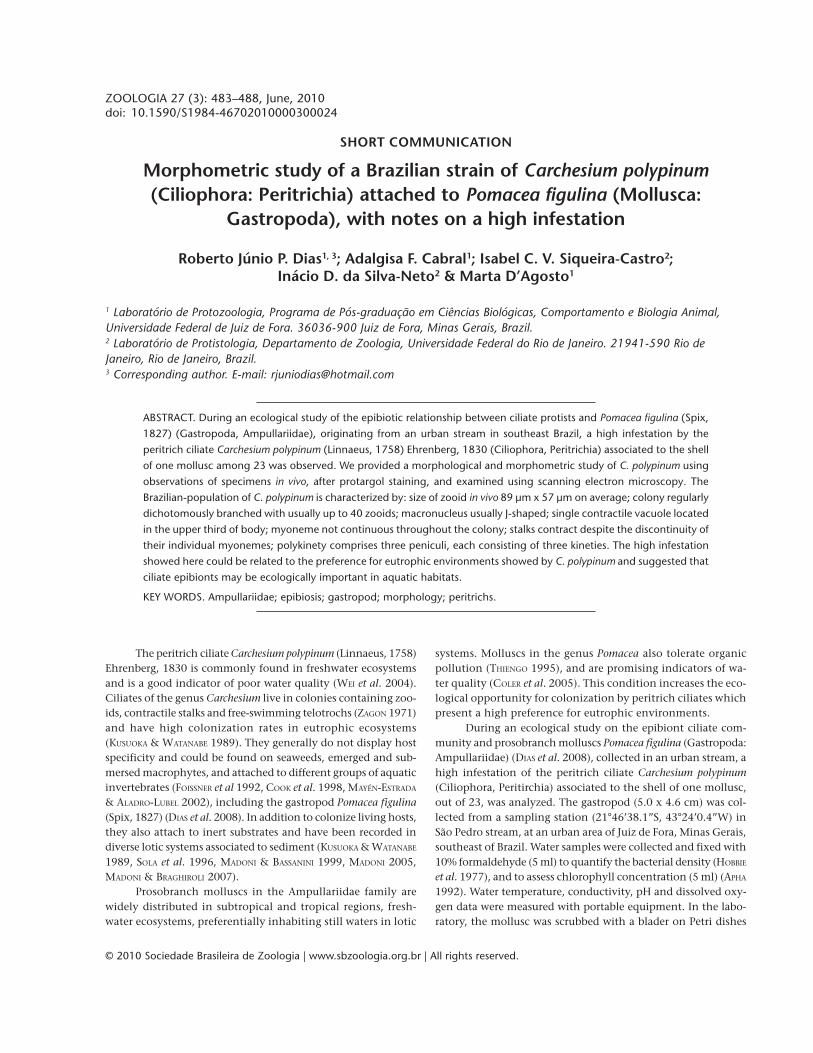
During an ecological study on the epibiont ciliate com-munity and prosobranch molluscs Pomacea figulina (Gastropoda:Ampullariidae) (DIAS et al. 2008), collected in an urban stream, ahigh infestation of the peritrich ciliate Carchesium polypinum(Ciliophora, Peritirchia) associated to the shell of one mollusc,out of 23, was analyzed. The gastropod (5.0 x 4.6 cm) was col-lected from a sampling station (21°46’38.1”S, 43°24’0.4”W) inSão Pedro stream, at an urban area of Juiz de Fora, Minas Gerais,southeast of Brazil. Water samples were collected and fixed with10% formaldehyde (5 ml) to quantify the bacterial density (HOBBIE
et al. 1977), and to assess chlorophyll concentration (5 ml) (APHA
1992). Water temperature, conductivity, pH and dissolved oxy-gen data were measured with portable equipment. In the labo-ratory, the mollusc was scrubbed with a blader on Petri dishes
SHORT COMMUNICATION
Morphometric study of a Brazilian strain of Carchesium polypinum(Ciliophora: Peritrichia) attached to Pomacea figulina (Mollusca:
Gastropoda), with notes on a high infestation
Roberto Júnio P. Dias1, 3; Adalgisa F. Cabral1; Isabel C. V. Siqueira-Castro2;Inácio D. da Silva-Neto2 & Marta D’Agosto1
1 Laboratório de Protozoologia, Programa de Pós-graduação em Ciências Biológicas, Comportamento e Biologia Animal,Universidade Federal de Juiz de Fora. 36036-900 Juiz de Fora, Minas Gerais, Brazil.2 Laboratório de Protistologia, Departamento de Zoologia, Universidade Federal do Rio de Janeiro. 21941-590 Rio deJaneiro, Rio de Janeiro, Brazil.3 Corresponding author. E-mail: [email protected]
ABSTRACT. During an ecological study of the epibiotic relationship between ciliate protists and Pomacea figulina (Spix,
1827) (Gastropoda, Ampullariidae), originating from an urban stream in southeast Brazil, a high infestation by the
peritrich ciliate Carchesium polypinum (Linnaeus, 1758) Ehrenberg, 1830 (Ciliophora, Peritrichia) associated to the shell
of one mollusc among 23 was observed. We provided a morphological and morphometric study of C. polypinum using
observations of specimens in vivo, after protargol staining, and examined using scanning electron microscopy. The
Brazilian-population of C. polypinum is characterized by: size of zooid in vivo 89 µm x 57 µm on average; colony regularly
dichotomously branched with usually up to 40 zooids; macronucleus usually J-shaped; single contractile vacuole located
in the upper third of body; myoneme not continuous throughout the colony; stalks contract despite the discontinuity of
their individual myonemes; polykinety comprises three peniculi, each consisting of three kineties. The high infestation
showed here could be related to the preference for eutrophic environments showed by C. polypinum and suggested that
ciliate epibionts may be ecologically important in aquatic habitats.
KEY WORDS. Ampullariidae; epibiosis; gastropod; morphology; peritrichs.
484 R. J. P. Dias et al.
ZOOLOGIA 27 (3): 483–488, June, 2010
containing previously filtered water collected from the sameplace. The ciliates were observed in vivo through bright field anddifferential interference contrast microscopy, stained using theprotargol technique (DIECKMANN 1995), and prepared for scan-ning electron microscopy (SILVA-NETO 1994). Examinations of invivo and prepared slides were made with an Olympus BX51 brightfield microscopy while the biometric analyses were performedusing Image Pro-Plus 5.0 software. The ciliates were then identi-fied according to ZAGON & SMALL (1970), ESTEBAN & FERNÁNDEZ-GALIANO (1989), and FOISSNER et al. (1992). The mollusc was sentto the Malacology Sector, Instituto Oswaldo Cruz, and identi-fied by Dr Silvana Thiengo.
Morphological analyses revealed that a single species ofa colonial peritrich Carchesium polypinum colonized P. figulinacollected in São Pedro stream (Figs 1-3). The overall morphol-ogy of the Brazilian strain of C. polypinum described in thepresent study is very similar to the species studied by ZAGON
(1970, 1971), ZAGON & SMALL (1970), CURDS et al. (1983), ESTEBAN
& FERNÁNDEZ-GALIANO (1989), FOISSNER et al. (1992), so we consid-ered both conspecific. We provided a morphometric charac-
terization of the species based on 10 characters measured fromliving individuals and nine characters measured from protargol-stained specimens (Tab. I).
The colonies of C. polypinum are branched with invertedbell-shaped zooids arranged on the branches. The colonial as-semblage has a main stalk that is joined to the substratum by afixing disc. The colonies had usually up to 40 zooids. The zooidsare found in symmetrical dichotomous colonies. Colonies rangein length from 700 µm to 2 mm. Zooids in vivo from 76.9 to110.6 µm in length, and between 35.6 and 83.6 µm in width.The size of impregnated zooids varied between 45.0-57.1 µm inlength and 36.4-55.9 µm in width. The length of the zooid de-creased by 42% when stained. Cytoplasm slightly greyish, usu-ally containing several large bronwish food vacuoles (7-20 µmin diameter). A single contractile vacuole located in the upperthird of body. Macronucleus J-shaped that usually descended toa region just aboral to the telotroch band. The macronucleus islarge and occupies a significant proportion of the body volume.The micronucleus was located close to the J-shaped macro-nucleus. The peristomial disc in vivo is 50.1-107.8 µm wide and
Table I. Measurements (µm) of C. polypinum attached to P. figulina from São Pedro stream, southeast Brazil. A total number of 15zooids were measured in vivo and 15 zooids after protargol staining. (Mean) Arithmetic mean, (SD) standard deviation, (CV)coefficient of variation in%.
Mean SD CV Range
In vivo
Total length of the body from epistomial disk to aboral end 88.9 14.7 16.5 76.9 – 110.6
Width of the body below the peristomial disk 57.1 14.2 24.9 35.6 – 83.6
Width of the body at midpoint between oral and aboral ends 37.1 8.2 22.2 25.9 – 51.3
Width of peristomial disk 70.8 17.0 24.0 50.1 – 107.8
Thickness of peristomial disk 8.7 4.0 46.3 5.0 – 20.8
Width of scopula 13.7 3.7 26.6 7.7 – 20.6
Width of basal stalk 12.7 3.7 29.0 7.0 – 19.0
Width of lateral stalk 13.0 3.1 24.1 10.8 – 17.4
Diameter of contractile vacuoles 17.2 3.9 22.6 10.3 – 25.0
Diameter of food vacuoles 13.3 5.4 40.4 6.7 – 26.0
Protargol
Total length of the body from epistomial disk to aboral end 50.9 3.5 6.8 45.0 – 57.2
Width of the body at midpoint between oral and aboral ends 45.3 5.3 11.7 36.4 – 55.9
Distance between trochal band and scopula 12.6 2.1 16.8 9.2 – 17.6
Length of micronucleus 7.0 2.1 29.4 5.0 – 9.1
Width of micronucleus 8.1 0.2 2.3 7.9 – 8.3
Width of macronucleus at midpoint 4.6 1.3 29.2 2.9 – 7.4
Width of myoneme 2.9 1.0 34.8 1.2 – 4.9
Length of peristomial area 31.2 3.3 10.7 22.6 – 38.3
Width of peristomial area 22.2 3.4 15.5 14.9 – 27.2
485Morphometric study of Carchesium polypinum attached to Pomacea figulina
ZOOLOGIA 27 (3): 483–488, June, 2010
1 2
45 6
3
9
10 11 12
87
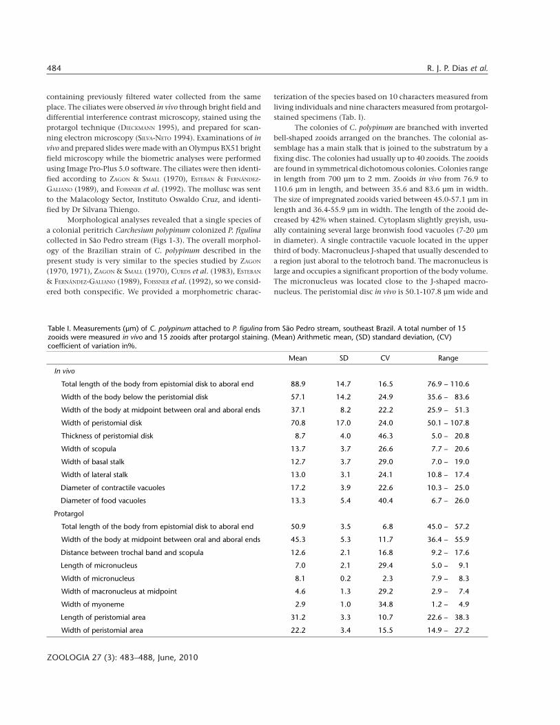
Figures 1-12. Photomicrographic images of C. polypinum attached to the freshwater apple snail P. figulina. (1-3) Colonies of C. polypinumattached to P. figulina (high infestation). (4) Schematic drawing of the colony of C. polypinum. (5) Schematic drawing of two zooidsshowing macronuleus (Ma), contractile vacuoles (CV) and the non-continuous myonemes of the colony (arrow). (6) A diagrammaticdrawing of the buccal morphology based on protargol silver preparations, showing oral polykinetids 1 (P1), oral polykinetids 2 (P2), andoral polykineitids 3 (P3). (7) Colony (in vivo) showing myonemes (My), differential interference contrast microscopy. (8) Detail of thenon-continuous myonemes (arrow), differential interference contrast microscopy. (9) Zooid (in vivo) showing macronuleus (Ma) andfood vacuoles (FV), differential interference contrast microscopy. (10-11) Colony (in vivo) showing macronuleus (Ma), myonemes (My),food vacuoles (FV) and contractile vacuoles (CV), phase contrast and bright field microscopy, respectively. (12) Protargol impregnationshowing macronuleus (Ma) and myonemes (My). Bars: (1-2) 1 cm, (3) 1 mm, (4 and 7) 100 µm, (8-12) 50 µm.
486 R. J. P. Dias et al.
ZOOLOGIA 27 (3): 483–488, June, 2010
5.0-20.8 µm thick. Located on the side opposite to the peris-tome is the lateral stalk, which in non-contracted specimensmeasured 10.8-17.4 µm in width. The lateral stalk surface hadirregular folds as showed by scanning electron microscopy. Themyoneme is not continuous throughout the colony. The stalkscontract despite the discontinuity of their individual myonemes.Myoneme ranges from 1.2-4.9 µm in width, presenting fibersthat extended anteriorly within the zooid, from the scopula tocentral part of the cell body. The myoneme stains heavily withsilver, but the remainder of the stalk is transparent. As observedin other peritrichs, the infraciliature of the zooid of C. polypinumis formed by the oral infraciliature and the aboral ciliary wreath.The aboral ciliary wreath (trochal band) is constituted by a ridgeof kinetosomes placed in two staggered rows surrounding theposterior end of the organism. The distance between the trochalband and the scopula is 9.2-17.6 µm. However, these numbersprobably are smaller than the actual values because contractionis initiated by silver-impregnation. The oral infraciliature is well
developed in this species. The oral apparatus is usual forperitrichs. The haplokinety and polykinety circle about one turnaround the peristomial disc and make a further turn after plung-ing into the infundibulum. The polykinety comprises threepeniculi (oral polykinetids) in the lower half of infundibulum,each consisting of three kineties. The oral polykinety 1 (P1) isparalleled at its point of separation by a second triple row ofkinetossomes. The posterior ends of the three kineties of P1 ter-minate at slightly different levels. The oral polykinetid 2 (P2)appears to begin at a slight angle and from a single point pre-sents a short distance from P1, turns slightly, and can then beclearly seen as three distinct ciliated rows of kinetossomes. P2 isinterposed between oral polykinetids 1 and 3. The oralpolykinetids 3 (P3), presents three short rows of kinetosomes,and appears approximately in the aboral one-third of the in-fundibulum (Figs 4-17, Tab. I).
The Brazilian strain of C. polypinum is similar to the popu-lation described by FOISSNER et al. (1992) in terms of body length
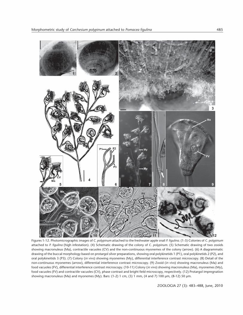
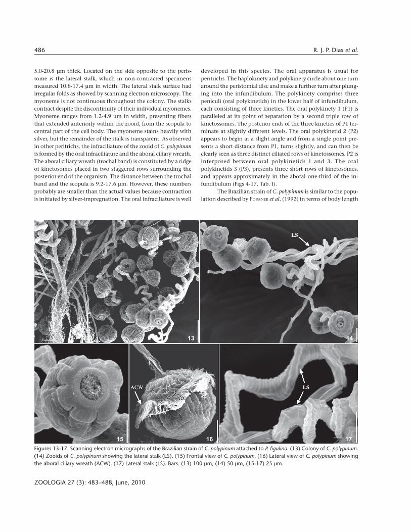
Figures 13-17. Scanning electron micrographs of the Brazilian strain of C. polypinum attached to P. figulina. (13) Colony of C. polypinum.(14) Zooids of C. polypinum showing the lateral stalk (LS). (15) Frontal view of C. polypinum. (16) Lateral view of C. polypinum showingthe aboral ciliary wreath (ACW). (17) Lateral stalk (LS). Bars: (13) 100 µm, (14) 50 µm, (15-17) 25 µm.
13 14
15 16 17
487Morphometric study of Carchesium polypinum attached to Pomacea figulina
ZOOLOGIA 27 (3): 483–488, June, 2010
(77-110 µm vs. 80-140 µm), number and position of contrac-tile vacuoles, shape of macronucleus, number of zooids, andthe oral infraciliature. As demonstrated by recent papers, theinfraciliature revealed with silver impregnation is highly spe-cies-specific, especially the structure of infundibularpolykineties in the oral apparatus, playing an essential role inthe determination of species (CLAMP 1990, JI & SONG 2004, JI etal. 2005). Several published reports describe the morphologyof the colony and zooids of C. polypinum (KAHL 1935, LOM 1964,CURDS 1969, ZAGON 1970, 1971, ZAGON & SMALL 1970, CURDS etal. 1983, ESTEBAN & FERNÁNDEZ-GALIANO 1989, FOISSNER et al. 1992),however, few morphometric characters were included in thesestudies. In the present study, we provide a characterization ofthe species based on new morphometric characters as used byUTZ (2007).
Studies emphasizing genetic variation within-species areneeded to determine whether C. polypinum collected from dif-ferent sites around the world could be considered a single ge-netic unit. For example, GENTEKAKI & LYNN (2009) concludedthat colonies of C. polypinum isolated from different locationsin the Grand River basin in Southwestern Ontario, Canada,probably are not a single morphospecies as previously thought.
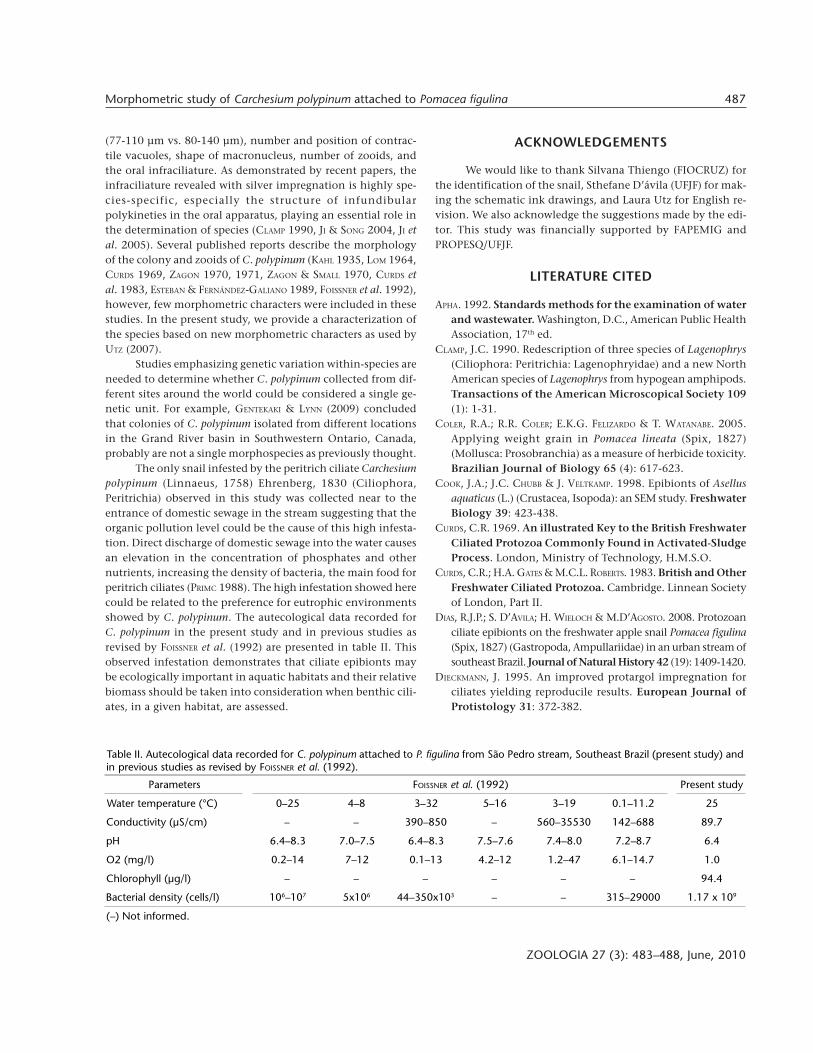
The only snail infested by the peritrich ciliate Carchesiumpolypinum (Linnaeus, 1758) Ehrenberg, 1830 (Ciliophora,Peritrichia) observed in this study was collected near to theentrance of domestic sewage in the stream suggesting that theorganic pollution level could be the cause of this high infesta-tion. Direct discharge of domestic sewage into the water causesan elevation in the concentration of phosphates and othernutrients, increasing the density of bacteria, the main food forperitrich ciliates (PRIMC 1988). The high infestation showed herecould be related to the preference for eutrophic environmentsshowed by C. polypinum. The autecological data recorded forC. polypinum in the present study and in previous studies asrevised by FOISSNER et al. (1992) are presented in table II. Thisobserved infestation demonstrates that ciliate epibionts maybe ecologically important in aquatic habitats and their relativebiomass should be taken into consideration when benthic cili-ates, in a given habitat, are assessed.
ACKNOWLEDGEMENTS
We would like to thank Silvana Thiengo (FIOCRUZ) forthe identification of the snail, Sthefane D’ávila (UFJF) for mak-ing the schematic ink drawings, and Laura Utz for English re-vision. We also acknowledge the suggestions made by the edi-tor. This study was financially supported by FAPEMIG andPROPESQ/UFJF.
LITERATURE CITED
APHA. 1992. Standards methods for the examination of waterand wastewater. Washington, D.C., American Public HealthAssociation, 17th ed.
CLAMP, J.C. 1990. Redescription of three species of Lagenophrys(Ciliophora: Peritrichia: Lagenophryidae) and a new NorthAmerican species of Lagenophrys from hypogean amphipods.Transactions of the American Microscopical Society 109(1): 1-31.
COLER, R.A.; R.R. COLER; E.K.G. FELIZARDO & T. WATANABE. 2005.Applying weight grain in Pomacea lineata (Spix, 1827)(Mollusca: Prosobranchia) as a measure of herbicide toxicity.Brazilian Journal of Biology 65 (4): 617-623.
COOK, J.A.; J.C. CHUBB & J. VELTKAMP. 1998. Epibionts of Asellusaquaticus (L.) (Crustacea, Isopoda): an SEM study. FreshwaterBiology 39: 423-438.
CURDS, C.R. 1969. An illustrated Key to the British FreshwaterCiliated Protozoa Commonly Found in Activated-SludgeProcess. London, Ministry of Technology, H.M.S.O.
CURDS, C.R.; H.A. GATES & M.C.L. ROBERTS. 1983. British and OtherFreshwater Ciliated Protozoa. Cambridge. Linnean Societyof London, Part II.
DIAS, R.J.P.; S. D’AVILA; H. WIELOCH & M.D’AGOSTO. 2008. Protozoanciliate epibionts on the freshwater apple snail Pomacea figulina(Spix, 1827) (Gastropoda, Ampullariidae) in an urban stream ofsoutheast Brazil. Journal of Natural History 42 (19): 1409-1420.
DIECKMANN, J. 1995. An improved protargol impregnation forciliates yielding reproducile results. European Journal ofProtistology 31: 372-382.
Table II. Autecological data recorded for C. polypinum attached to P. figulina from São Pedro stream, Southeast Brazil (present study) andin previous studies as revised by FOISSNER et al. (1992).
Parameters FOISSNER et al. (1992) Present study
Water temperature (°C) 0–25 4–8 3–32 5–16 3–19 0.1–11.2 25
Conductivity (µS/cm) – – 390–850 – 560–35530 142–688 89.7
pH 6.4–8.3 7.0–7.5 6.4–8.3 7.5–7.6 7.4–8.0 7.2–8.7 6.4
O2 (mg/l) 0.2–14 7–12 0.1–13 4.2–12 1.2–47 6.1–14.7 1.0
Chlorophyll (µg/l) – – – – – – 94.4
Bacterial density (cells/l) 106–107 5x10⁶ 44–350x103 – – 315–29000 1.17 x 10⁹
(–) Not informed.
488 R. J. P. Dias et al.
ZOOLOGIA 27 (3): 483–488, June, 2010
ESTEBAN, G. & D. FERNÁNDEZ-GALIANO. 1989. Morphology andmorphogenesis in Carchesium polypinum (Ciliophora:Peritrichida). Transactions of the American MicroscopicalSociety 108 (4): 345-353.
FOISSNER, W.; H. BERGER & F. KOHMANN. 1992. Taxonomische undökologische revision der ciliaten des saprobiensystems –Band II: Peritrichia, Heterotrichida, Odontostomatida.Munich, Informationsberichte des Bayer Landesamtes fürWasserwirtschaft.
GENTEKAKI, E. & D.H. LYNN. 2009. High genetic diversity but nopopulation structure of the peritrichous ciliate Carchesiumpolypinum in the Grand River basin (North America) inferredfrom nuclear and mitochondrial markers. AppliedEnvironmental Microbiology 75 (10): 3187-3195.
HOBBIE J.E.; R.J. DALEY; S. JASPER. 1977. Use of nuclepore filtersfor counting bacteria by fluorescence microscopy. AppliedEnvironmental Microbiology 33: 1225-1228.
JI, D. & W. SONG. 2004. Notes on a new marine peritrichousciliate (Ciliophora: Peritrichida), Zoothamnopsis sinica n. sp.from north China, with reconsideration of Zoothamniummaximum Song, 1986. Acta Protozoologica 43 (1): 61-71.
JI, D.; W. SONG; K.A.S. AL-RASHEID & P. SUN. 2005. Description ofZoothamnium foissneri n. sp. and redescription of Z.duplicatum Kahl, 1933 and Z. mucedo Entz, 1884, threespecies of marine peritrichous ciliates. European Journalof Protistology 41 (1): 45-56.
KAHL, A. 1935. Urtiere oder Protozoa I. Wimpertiere oder Ciliata(Infusoria). 4. Peritricha und Chonotricha, p. 651-886. In:F. DAHL (Ed.). Die Tierwelt Deutschlands. Jena, GustavFischer.
KUSUOKA, Y. & Y. WATANABE. 1989. Distinction of emigration bytelotroch formation and death by predation in peritrichciliates: SEM observation on the remaining stalks ends.Microbiology Ecology 62: 7-12.
LOM, J. 1964. The morphology and morphogenesis of the buccalciliary organelles in some peritrichous ciliates. Archiv fürProtistenkunde 107: 131-162.
MADONI, P. 2005. Ciliated protozoan cominities and saprobicevaluation of water quality in the hilly zone of sometributaries of Po River (northern Italy). Hydrobiologia 541:55-69.
MADONI, P. & S. BARGHIROLI. 2007. Changes in the ciliate assemblagealong a fluvial system related to physical, chemical and
geomorphologic characteristics. European Journal ofProtistology 43: 67-75.
MADONI, P. & N. BASSANINI. 1999. Longitudinal changes in theciliate protozoa communities along a fluvial system pollutedby organic matter. European Journal of Protistology 35:391-402.
MAYÉN-ESTRADA, R. & M.A. ALADRO-LUBEL. 2002. Distribution andPrevalence of 15 species of epibiont petrich ciliates on thecrayfish Camabarellus patzcuarensis Villalobos, 1943 in lakePátzcuaro, Michoacán, México. Crustaceana 74 (11): 1213-1224.
PRIMC, B. 1988. Trophic relationships of ciliated Protozoadeveloped under different saprobic conditions in theperiphyton of the Sava River. Verhandlungen InternationaleVereiningen Limnologie 26:1116-1119.
SILVA-NETO, I.D. 1994. Observations sur é ultrastructure du ciliéheterotriche Licnophora auerbachi Cohn, 1866, epibionte del’etoile de mer. Annales des Sciences Naturelles, Zoologieet Biologie Animale, 2: 49-62
SOLA, A.; J.F. LONGÁS; SERRANO S. & A. GUINEA. 1996. Influence ofenvironmental characteristics on the distribution of ciliatesin the River Henares (Central Spain). Hydrobiologia 32 (4):237-252.
THIENGO, S.C. 1995. Família Pilidae p50-69. In: Tópicos em mala-cologia médica. F.S. Barbosa (Ed.). Rio de Janeiro, EditoraFiocruz.
UTZ, L.R.P. 2007. First record of Epistylis plicatilis (Ciliophora:Peritrichia) attached to Pomacea canaliculata (Mollusca:Gastropoda) in Southern Brazil. Zootaxa 1454: 49-57.
WEI, M.; Y. YUNE; Y. SHEN & X. ZHANG. 2004. Intraespecificphylogeography of Carchesium polypinum (Peritrichia,Ciliophora) from China, inferred from 18s-ITS1-5.8Sribosomal DNA. Life Science 47 (1): 11-17.
ZAGON, I.S. 1970. Carchesium polypinum: cytostructure afterprotargol silver deposition. Transactions of the AmericanMicroscopical Society 89 (3): 450-468.
ZAGON, I.S. 1971. Scanning electron microscope observation onsome life history stages of Carchesium polypinum (Ciliata,Peritrichia). Journal of Protozoology 18 (2): 328-332.
ZAGON, I.S. & E.B. SMALL. 1970. Carchesium polypinum: somaticand buccal structure analysis after protargol staining.Transactions of the American Microscopical Society 89:443-449.
Submitted: 26.II.2009; Accepted: 23.II.2010.Editorial responsibility: Marcus V. Domingues


