Embed Size (px)
Citation preview

Mosaicism of Podocyte Involvement Is Related toPodocyte Injury in Females with Fabry DiseaseMichael Mauer1,2, Emily Glynn3, Einar Svarstad4,5, Camilla Tøndel5,6, Marie-Claire Gubler7,
Michael West8, Alexey Sokolovskiy3, Chester Whitley1, Behzad Najafian3*
1Department of Pediatrics, University of Minnesota, Minneapolis, United States of America, 2Department of Medicine, University of Minnesota, Minneapolis, United
States of America, 3Department of Pathology, University of Washington, Seattle, United States of America, 4Department of Medicine, Haukeland University Hospital,
Bergen, Norway, 5Department of Clinical Medicine, University of Bergen, Bergen, Norway, 6Department of Pediatrics, Haukeland University Hospital, Bergen, Norway,
7U983, Universite Rene Descartes, Hopital Necker-Enfants Malades AP-HP, Paris, France, 8Division of Nephrology, Department of Medicine, Dalhousie University, Halifax,
Nova Scotia, Canada
Abstract
Background: Fabry disease. an X-linked deficiency of a-galactosidase A coded by the GLA gene, leads to intracellularglobotriaosylceramide (GL-3) accumulation. Although less common than in males, chronic kidney disease, occurs in ,15%of females. Recent studies highlight the importance of podocyte injury in Fabry nephropathy development and progression.We hypothesized that the greater the % of podocytes with active wild-type GLA gene (due to X-inactivation of the mutantcopy) the less is the overall podocyte injury.
Methods: Kidney biopsies from 12 treatment-naive females with Fabry disease, ages 15 (8–63), median [range], years werestudied by electron microscopy and compared with 4 treatment-naive male patients.
Results: In females, 51 (13–100)% of podocytes (PC) per glomerulus had no GL-3 inclusions, this consistent with a non-Fabrypodocyte phenotype (NFPC). In PC with GL-3 inclusions [Fabry podocyte phenotype (FPC)], GL-3 volume density perpodocyte was virtually identical in females and males, consistent with little or no cross-correction between FPC and NFPC.%NFPC per glomerulus (%NFPC/glom) correlated with age in females (r = 0.65, p = 0.02), suggesting a survival disadvantagefor FPC over time. Age-adjusted %NFPC/glom was inversely related to foot process width (FPW) (r =20.75, p = 0.007), anindicator of PC injury. GL-3 volume density in FPC in females correlated directly with FPW.
Conclusions: These findings support important relationships between podocyte mosaicism and podocyte injury in femaleFabry patients. Kidney biopsy, by providing information about podocyte mosaicism, may help to stratify females with Fabrydisease for kidney disease risk and to guide treatment decisions.
Citation: Mauer M, Glynn E, Svarstad E, Tøndel C, Gubler M-C, et al. (2014) Mosaicism of Podocyte Involvement Is Related to Podocyte Injury in Females withFabry Disease. PLoS ONE 9(11): e112188. doi:10.1371/journal.pone.0112188
Editor: Stuart E. Dryer, University of Houston, United States of America
Received May 26, 2014; Accepted October 13, 2014; Published November 11, 2014
Copyright: � 2014 Mauer et al. This is an open-access article distributed under the terms of the Creative Commons Attribution License, which permitsunrestricted use, distribution, and reproduction in any medium, provided the original author and source are credited.
Data Availability: The authors confirm that all data underlying the findings are fully available without restriction. All relevant data are within the paper.
Funding: This work was supported by an NIH RDCRN U54 grant (5U54NS065768-04) and an investigator initiated grant from Genzyme, a Sanofi Company. Thefunder provided support in the form of research grants for authors [BN, MM, and MLW], and travel support and honoraria for authors [BN, MM, CT, and MLW] butdid not have any additional role in the study design, data collection and analysis, decision to publish, or preparation of the manuscript. The specific roles of theseauthors are articulated in the ‘author contributions’ section.
Competing Interests: BN is a consultant to Genzyme and Amicus Therapeutics, receives investigator initiated research support from Genzyme and Roche, andhonoraria and travel support from Genzyme. BN is also a member of the Medical Advisory Board of Amicus Therapeutics. MM is a consultant to Genzyme, andreceives investigator initiated research support, honoraria, and travel support from Genzyme. MM is also a member of the Genzyme funded FDA mandated FabryRegistry Board. This interest for MM has been reviewed and managed by the University of Minnesota in accordance with its conflict of interest policies. MM is alsoa consultant to Amicus. CT and ES received travel support and speakers honoraria from Shire and Genzyme. MLW has received research support, honoraria, and/ortravel support from Actelion, Amicus, Excelsior Pharmaceuticals, Genzyme, Glaxo SmithKline, Shire, and Sumitomo Pharma. Behzad Najafian is a PLOS ONEEditorial Board member, and commercial affiliations do not alter the authors’ adherence to PLOS ONE Editorial policies and criteria.
* Email: [email protected]
Introduction
Fabry disease is a storage disease caused by deficiency of the a-galactosidase A (aGal A) enzyme that hydrolyzes the terminal a-galactosyl moieties from glycolipids and glycoproteins. This leads
to the accumulation of its substrates, predominately globotriao-
sylceramide (GL-3) in various cell types and organs, causing a
constellation of complications including skin lesions, strokes,
cardiac arrhythmias and cardiomyopathy, neuropathies and renal
failure. [1] aGal A is encoded by the GLA gene located on the X
chromosome locus Xq21.3-q22. Similar to other X-linked
diseases, the complications are typically less frequent and more
variable in severity in females, [2,3,4] although they can be as
severe as in male patients. [5,6] A significant proportion of female
patients suffer from important complications, including 40% with
clinical renal disease (mainly proteinuria) [7] and about 15% with
serious renal events. [2] Fabry disease is associated with significant
life expectancy reductions in both sexes. [8] It is of great
PLOS ONE | www.plosone.org 1 November 2014 | Volume 9 | Issue 11 | e112188

importance to understand the factors associated with disease
severity in females. Currently, there are no reliable tests to identify
females at greater risk to develop kidney failure, thus justifying
earlier treatment with enzyme replacement therapy (ERT).
Podocytes are terminally differentiated cells with pivotal role in
preserving glomerular structure and function. [9] Recent studies
suggest that GL-3 accumulation in podocytes plays an important
role in the pathophysiology of Fabry nephropathy. [10] These cells
are also much more resistant to ERT than most other kidney cell
types. [11,12] Similar to other ‘‘terminally differentiated cells’’
podocytes do not easily regenerate following injury [13].
Continuous podocyte loss leads to progressive reduction of these
cells in the glomeruli, this eventually reaching critical levels
causing irreversible glomerular scarring [14]. Despite recent
evidence that higher doses of ERT during childhood may result
in partial to almost complete clearance of podocytes from GL-3
inclusions, [15] there is no consensus as to when to initiate ERT,
especially in females, and the relative clinical effectiveness of the
different licensed ERT doses remain unsettled. [16,17,18] We
hypothesized that podocytes, due to random X-inactivation, are
heterogeneously involved by Fabry disease in female patients and
that this heterogeneity could influence podocyte injury. Herein we
describe a method to quantify the % of podocytes with the Fabry
phenotype and report an inverse relationship in females between
age-adjusted % podocytes with no GL-3 inclusions in glomeruli
and foot process width, a sensitive indicator of podocyte injury
[19], supporting a relationship between X-inactivation and
podocyte injury in females with Fabry disease. We also found no
evidence of cross-correction between podocytes without and with
the Fabry phenotype in females with Fabry disease.
Methods
These studies were performed in accordance with principles of
the Declaration of Helsinki and were reviewed and approved by
the Institutional Review Board of the University of Minnesota,
Comite. de Protection des Personnes ‘‘Ile-De-France II.’’ and the
Regional Ethics Committee of Western Norway. Informed
consents approved by the institutional board review committees
were obtained prior to these studies.
Subjects and Clinical ParametersKidney biopsies from 12 ERT-naive females with Fabry disease,
age 15 (8–63) years were studied by electron microscopy for
distribution of podocyte involvement by the Fabry phenotype.
Biopsies were obtained for assessment of the severity of the lesions
of Fabry nephropathy in order to aid clinical decision-making
regarding ERT initiation and/or as a baseline biopsy prior to
ERT initiation. Biopsies from 4 ERT-naive males with Fabry
disease, age 14 (7–18), were studied for comparison; 7/16 patients
presented here were included in our previous publications. [10,15]
The demographic and clinical data of all patients are presented in
Table 1.
9/12 female and 3/4 male patients had results of GLA mutation
analysis in their medical records confirming the diagnosis of Fabry
disease (Table 1). The diagnosis of Fabry disease in the other
patients was based on family history or clinical findings with or
without reduced leukocyte aGal A activity and confirmed by the
kidney biopsy findings. Protein excretion per gram creatinine
(UPCR) was based on urine samples obtained close to the date of
biopsy. GFR was estimated by the plasma clearance of iohexol
where available or by creatinine clearance. Except for one of the
patients where we were not able to find information about the use
of renin-angiotensin system blockers, none of the patients were
receiving these drugs.
Biopsies from 6 healthy living kidney donors, age 37 (16–52)
were used to estimate normal control values for podocyte foot
process with (FPW), as described below.
Biopsy Tissue Preparation and Electron MicroscopyElectron microscopy specimens were fixed in 2.5% glutaralde-
hyde, and embedded in PolyBed. Random glomerular sections
were prepared as previously described. [20] Thin sections were
mounted on formvar coated copper slot grids. Overlapping digital
low magnification (,10,000 x) images of the entire glomerular
profiles were obtained using a JEOL CX100 electron microscope
for the podocyte mosaicism studies. High magnification (,30,000
x) images were obtained according a systematic uniform random
sampling protocol for estimation of fraction of the volume (Vv) of
PC cytoplasm occupied by GL-3 inclusions [Vv(Inc/PC)], Vv of
inclusions/glomerular mesangial cell [Vv(Inc/Mes)], Vv of inclu-
sions/glomerular endothelial cell [Vv(Inc/Endo)], and podocyte
average FPW as previously described [10,20].
Identification of Podocytes with and without the FabryPhenotypeObservers were masked to any of the patient characteristics.
Montages of complete glomerular profiles were prepared from the
above images in Adobe Photoshop software (Adobe Photoshop
CS5 Extended, version 12.0632). Twice digital magnification was
applied to the images. Podocyte nuclei were identified and
glomerular profiles with less than 10 podocyte nuclei (n = 3) were
excluded from these studies. The cytoplasmic profiles surrounding
each podocyte nucleus were carefully examined for presence of
GL-3 inclusions. Podocyte nuclear profiles with cytoplasmic GL-3
inclusions, consistent with Fabry phenotype podocytes (FPC) or
without cytoplasmic GL-3 inclusions, consistent with non-Fabry
phnotype podocytes (NFPC) cytoplasmic GL-3 inclusions were
counted in each glomerulus and %NFPC/glom was calculated.
The maximum number of immediately adjacent podocytes,
including podocytes on the other side of the same capillary loop,
which were NFPC was recorded as an estimate of the size of
podocyte mosaic patches on the section.
Electron Microscopy StereologyBased on the best quality of tissue preservation and images, one
glomerulus per biopsy was arbitrarily selected for detailed GL-3
volume density measurement in podocytes with visible nuclei on
the section. Boundaries of nuclei and cell membranes, excluding
the tertiary foot processes were traced using the magnetic lasso tool
in separate layers in Adobe Photoshop (Figure 1). Similarly, the
most convex points of cytoplasmic GL-3 inclusions were connected
using the magnetic lasso tool to draw a polygon around the GL-3
inclusion aggregates in a separate layer. The tracings were colored
differentially for nuclei, cytoplasm and inclusions (Figure 1). The
observed magnification was calculated from the average of 10
horizontal and 10 vertical random measurements performed on
images from a SPI grating carbon replica #02902-AB (Structure
Probe, Inc., West Chester, PA, USA) with horizontal and vertical
lines 0.463 mm apart obtained at the same magnification as for the
montage images. Subsequently, the measurement tool of the
software was calibrated. The area of cell, nucleus and inclusion
aggregate profiles were obtained separately for each podocyte
profile with a visible nucleus from the Adobe Photoshop
measurement log. The fractional volume of GL-3 inclusions per
each nucleated podocyte profile with GL-3 inclusions [Vv(Inc/
Podocyte Mosaicism in Fabry Females
PLOS ONE | www.plosone.org 2 November 2014 | Volume 9 | Issue 11 | e112188

Table
1.Clin
ical
characteristicsofsubjects.
Case
Sex
Age(year)
UPCR
(mg/g)
UACR
(mg/g)
GFR
(ml/min/1.73m
2)
GLA
Mutation
Mutation
Type
Aangiokerato
ma
Corn
eal
Opacity
1F
840
NA
NA*
NA
NA
++
2F
11
0.02
53
105
c.800T.G
(p.M
267R)
Missense
++
3F
12
29
NA
109
W236X
Nonsense
22
4F
13
0NA
97
Y216D
Missense
2+
5F
13
60
5100
NA
NA
+2
6F
14
62
11
90
c.800T.G
(p.M
267R)
Missense
NA
NA
7F
16
30
12
127
NA
NA
22
8F
34
150
NA
118
R301Q
Missense
+2
9F
34
100
599
R112C
Missense
2+
10
F39
100
NA
78
1270T
Missense
++
11
F39
40
ND
99
N215S
Missense
+2
12
F63
1150
NA
46
C.427G.C
p.Ala143Pro
Missense
+2
13
M7
0NA
183
Y216D
Missense
++
14
M16
92
12
112
c.1212_1214delAAG
Deletion
++
15
M18
251
135
96
c.800T.G(p.
M267R)
Missense
++
16
M23
102
NA
111
NA
NA
NA
NA
Abbreviations:UPCR=urineprotein/creatinineratio;UACR=urinealbumin/creatinineratio;GFR
=glomerularfiltrationrate;NA=datanotavailable;*Serum
creatininewithin
thenorm
alrange;F=female;M=male;ND=Not
detectab
le.
doi:10.1371/journal.pone.0112188.t001
Podocyte Mosaicism in Fabry Females
PLOS ONE | www.plosone.org 3 November 2014 | Volume 9 | Issue 11 | e112188

FPC)] was estimated as follows: VV (Inc=FPC)~Ainclusions|100
Acell{Anucleus
.
The overall average glomerular volume fraction of GL-3
inclusions per podocytes [Vv(Inc/PC)], endothelial cells
[Vv(Inc/Endo)] and mesangial cells [Vv(Inc/Mes)] were estimated
using unbiased stereology methods as previously detailed. [10] For
clarity, we emphasize that Vv(Inc/FPC) is an estimate of GL-3
inclusion density in nucleated podocyte profiles with GL-3
inclusions, while Vv(Inc/PC) is an estimate of the same parameter
in all visible podocyte profiles regardless of GL-3 content and
including the podocytes with and without visible nuclei over 3 (1–
3), median (range), glomeruli per biopsy. Average foot process
width (FPW) was also estimated using unbiased stereology methods
as detailed elsewhere. [10,20,21] In order to compare podocyte
injury in FPC vs. NFPC in female patients with Fabry disease,
FPW was separately estimated in systematically and uniformly
obtained electron micrographs with and without FPC.
Statistical AnalysesStatistica 8.0 (Statsoft, Inc.) was used for statistical analysis.
Comparison between groups was made by student t-test after
confirming homogeneity of variances. Relationships between
variables were evaluated using Pearson correlation. Partial
correlations were performed to control for confounding variables.
Random effects model variance component analysis was per-
formed to estimate % contribution of biopsies (inter-subject) and
glomeruli (intra-subject) variations to total variance of %NFPC per
glomerulus (%NFPC/glom). p,0.05 was considered statistically
significant.
Results
Electron Microscopy Examination of Kidney BiopsiesExamination of electron micrographs from all biopsies allowed
easy and reliable distinction between male and female patients by
the identification in females of podocytes with visible nuclei
without GL-3 cytoplasmic inclusions, termed non-Fabry podocytes
(NFPC) (Figure 1) in contrast with podocytes containing GL-3
inclusions, termed Fabry phenotype podocytes (FPC). However,
mosaicism of the Fabry phenotype was not easily identifiable in the
other glomerular or extra-glomerular cells, perhaps, at least in
part, due to uncertainty about cellular boundaries (e.g., mesangial
or endothelial cells). In 4 female patient biopsies, no GL-3
inclusions were identified in endothelial and/or mesangial cells,
thus, comments about mosaicism could not be made in those cell
types. In 7/12 female patient biopsies, parietal epithelial cell
profiles with no GL-3 inclusions were identified while occasional
parietal epithelial cells with enlarged cytoplasm had abundant GL-
3 inclusions. Characteristic lamellar GL-3 inclusions were not
easily identified in proximal tubular epithelial cells. Distal tubular
epithelial cells showed variable GL-3 inclusions in both males and
females. Thus, this variability could not be accounted for X-
inactivation mosaicism in those cells.
Distribution of GL3 Inclusions among Podocytes in Malesand Females54 (27–87) podocytes per biopsy were examined for presence of
GL-3 inclusions in female patients. 51 (13–100)% of podocyte
profiles per biopsy with visible nuclei and no GL-3 inclusions, were
classified as NFPC in these females. NFPC were distributed as
Figure 1. Mosaicism of podocyte Fabry phenotype in a glomerulus from a female patient with Fabry disease. (A) Montage image of aglomerulus (,3,0006). Podocyte bodies with visible nuclei are colored blue, podocyte nuclei purple, and GL-3 inclusions yellow. The white rectangleis magnified in B. (B) Magnified view of three podocyte profiles without (at the bottom) and three other podocyte profiles with GL-3 inclusions (onthe top). Arrows show GL-3 inclusions in mesangial (M) cells (black) and endothelial (E) cells. P is a podocyte profile with no visible nucleus on thissection.doi:10.1371/journal.pone.0112188.g001
Podocyte Mosaicism in Fabry Females
PLOS ONE | www.plosone.org 4 November 2014 | Volume 9 | Issue 11 | e112188

single cells among FPC or in patches composed of 2–6 podocytes
(Figure 1).
Two biopsies from female patients (one with 3 and another with
2 glomeruli) had no GL-3 inclusions in podocytes with visible
nuclei (cases #9 and #11, Table 1). Case #9 showed rare
podocyte profiles that were filled with GL-3. However, because
these GL-3 containing podocytes had no visible nuclei they were
not included in calculation of %NFPC per glomerulus. This case
had a missense mutation (R112C) with GFR, UPCR and UACR
values all within the normal range (Table 1). Clinical examination
revealed corneal opacities, but no angiokeratoma. Echocardio-
grams showed normal left ventricular size and function with an
estimated ejection fraction of 55–60% and normal right ventric-
ular size and systolic function. Case #11 had distal tubular cells
with GL-3 inclusions. This case had a cardiac variant mutation
(N215S) with normal GFR and UPCR values and no detectable
albumin in the urine (Table 1). The diagnosis of Fabry disease was
made based on known family history. Clinical examination
revealed angiokeratoma, but no corneal opacities. Clinically, she
was asymptomatic. No echocardiograms were available from this
case. None of these two subjects had a history of stroke. In order to
determine whether random sectioning through podocytes may
have obscured the observation of GL-3 inclusions in podocyte
profiles 4 male patients with Fabry disease were similarly studied,
1–3 (median 2.5) glomeruli, containing 18–36 (median 22)
podocyte profiles per glomerulus with visible nuclei were
examined (Table 1). All podocyte profiles from these glomeruli
contained abundant GL-3 inclusions, consistent with FPC, except
for two very small profiles in a 7-year-old boy. The volume
fraction of GL-3 inclusions per FPC [Vv(Inc/FPC)] was nearly
identical in males (0.5660.11) and females (0.5360.13; p = 0.54).
Inter- and Intra-subject Variations of PodocytePhenotype MosaicismBiopsies from 9 female patients with more than one glomerulus
(3 (2–5), median (range)) available for electron microscopy were
used to compare inter- and intra- subject variability of podocytes
with the Fabry the phenotype. The %NFPC/glom in 2–4
glomeruli per biopsy in female patients is shown in Figure 2.
Variance component analysis showed that only 9.6% of total
variance in podocyte Fabry phenotype mosaicism originated from
inter-glomerular (intra-subject) variation, while the vast majority of
variance lay in differences in this parameter among the subjects.
Relationships Between % Podocytes with no GL-3Inclusions and Female Patient Characteristics, RenalFunction and Other Glomerular Structural ParametersValues of %NFPC/glom, Vv(Inc/PC), volume fraction of GL-3
inclusions per mesangial cells [Vv(Inc/Mes)], volume fraction of
inclusions per endothelial cells [Vv(Inc/Endo)], and foot process
width (FPW) are provided in Table 2. The average %NFPC/glom
was calculated in biopsies from female patients with more than one
available glomerulus. There was a direct relationship between age
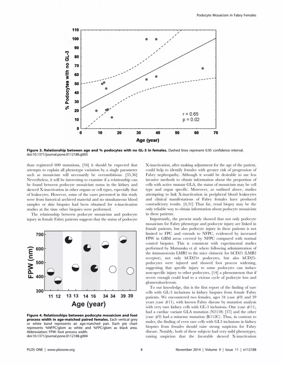
and %NFPC/glom (r = 0.65; p= 0.02, Figure 3). No statistically
significant relationship was found between %NFPC/glom and
urine albumin/creatinine ratio (UACR) (available in 6/12
patients), urine protein creatinine ratio (UPCR) or glomerular
filtration rate (GFR). As expected, %NFPC/glom was inversely
related to Vv(Inc/PC) (r =20.70, p = 0.02) for all podocytes.
Simple linear regression analysis revealed no statistically significant
relationships between %NFPC/glom and foot process width,
Vv(Inc/Endo), Vv(Inc/Mes). However, adjusted for age, signifi-
cant inverse relationships were found between %NFPC/glom and
FPW (r =20.75, p = 0.007), and Vv(Inc/Mes) (r = 0.70, p = 0.02),
but not with Vv(Inc/Endo).
To better visualize the relationships between %NFPC/glom
and FPW and UPCR, 10 female patients were grouped into 5 age-
matched pairs, 3 pairs with identical ages and the 2 other pairs
with ages no more than 2 years apart. Except for one pair, in each
pair, the subject with lower %NFPC/glom had greater FPW
value, suggestive of an inverse relationship between the extent of
podocyte injury and the number of NFPC in age-matched Fabry
females (Figure 4). Even an 8 year old female with 20% NFPC/
glom (case #1) had greater FPW than an 11 year old (case #2)
with 50% NFPC/glom. The one exception was a pair with greater
FPW in a 14 year old girl with 22% NFPC/glom (case #6)
compared to a 16 years old with 13% %NFPC/glom (case #7)
(Figure 4). The relationships between %NFPC/glom and UPCR
among the above pairs paralleled the relationships between
%NFPC/glom and FPW, except for cases #2 and #3 with
almost identical %NFPC/glom (50 and 52%, respectively) and
slightly (,15%) greater UPCR for case#2 (Figure 4). However,
the relationship between %NFPC and UPCR remained not
statistically significant after age adjustment. Also, when adjusted
for age, Vv(Inc/FPC) in females correlated with FPW (r = 0.64,
p = 0.03). In females with Fabry disease, FPW in electron
micrographs where glomerular basement membranes were cov-
ered by FPC (5626160 nm) was not statistically different from
those where glomerular basement membranes were covered by
NFPC (6636283 nm). On the other hand, FPW in either of these
areas were greater than FPW in normal control biopsies
(406634 nm, p= 0.037 for FPC and p=0.048 for NFPC).
Discussion
We recently demonstrated that the fraction of the volume of
podocytes occupied by GL-3 [Vv(Inc/PC)] increases with age in
children with Fabry disease and is directly correlated with FPW
and to proteinuria,9 the latter a strong predictor of renal disease
risk among Fabry disease patients.23 Increased FPW is a common
concomitant of injury in podocytes and is seen in a variety of
conditions known to be associated with injury in these cells.
[21,22], [23] Thus careful examination of this cell in Fabry disease
is important. This is the first study to provide unbiased electron
microscopic morphometric measures of the heterogeneity of
podocyte involvement in females with Fabry disease, almost
certainly as a result of X-inactivation. Gubler et al first describedthe heterogeneous distribution of GL-3 inclusions in podocytes in
3 females with Fabry disease. [24] Valbuena et al also described
variable podocyte GL-3 accumulation in 4 females with Fabry
disease. [25] However, no quantitative assessment of podocyte
GL-3 or statistical analyses was provided in these reports. Similar
to these studies, [24,25] we found that a distinction between Fabry
and non-Fabry phenotype, consistent with cells carrying active
mutant or wild-type GLA, respectively, could be easily made in
podocytes but not in other renal cells of female patients. This could
be due to distinct borders of podocyte cell bodies from their
neighbor cells, or because podocytes are apparently very long lived
[26] and may thus retain GL-3 inclusions for years while other cell
types are more frequently replaced.
Importantly, we documented, through unbiased morphometric
measurements, that the average fraction of podocyte cytoplasm
occupied by GL-3 was virtually identical in male and female
patients, suggesting that FPC do not benefit from the enzymatic
activity of the adjacent NFPC or from the generally higher residual
plasma aGal A in females than on males. [27] [28,29].
Podocyte Mosaicism in Fabry Females
PLOS ONE | www.plosone.org 5 November 2014 | Volume 9 | Issue 11 | e112188

Also important, we found that inter-glomerular variation in
podocyte phenotype mosaicism in a given female Fabry patient’s
renal biopsy is much smaller than inter-patient variation. This
suggests that estimation of podocyte phenotypic mosaicism even in
a single glomerulus is representative of this phenomenon in the
biopsy and validates the study of podocyte phenotype mosaicism in
a few glomeruli. It also suggests that podocyte mosaciasm in
females is established relatively early in embryogenesis. It should
be noted that in these studies we used random profiles of
glomeruli. Although we observed NFPC in patches of up to 5 cells
in these two-dimensional glomerular cross-sections, these patches
are likely larger in three dimensions. Nonetheless we posit that
these patches are not very large, otherwise we might have expected
greater inter-glomerular variability as a result of random
sectioning through glomeruli.
This study is the first to show a quantitative relationship in
females with Fabry disease between the X-inactivation phenom-
enon and podocyte injury manifest as increased FPW. Although
X-inactivation may play a role in the clinical phenotypic
expression of X-linked diseases, a direct link between skewed X-
inactivation and severity of the Fabry phenotype has been
controversial. Dobrovolny et al. reported that the trend line
between the age and the Mainz severity score index (MSSI) [30]
was steeper in 10 Fabry females with vs. 28 without unfavorably
skewed X-inactivation in leukocytes, urinary and salivary cells.
This suggested that random X-inactivation could influence the
severity of the Fabry phenotype in female patients. [31] In
contrast, Maier et al. found that in 46% of the 28 Fabry females
studied, skewed X-inactivation did not correlate with phenotype
severity. [4] Similarly, in a more recent study, while confirming
random X-inactivation in leukocytes in ,82% of 77 female Fabry
patients, there was no relationship between X-inactivation ratios
and age, aGal A activity, MSSI scores, cardiac involvement,
neuropathic pain or proteinuria. [32] We did not find a statistically
significant relationship between % NFPC/glom and UPCR.
However, in regression models including UPCR among the
predictor variables, UPCR accounted for much less of the
variance for rates of GFR decline in women than in men with
Fabry disease. [33] Also, increased UPCR can reflect parameters
other than podocyte injury, such as impaired tubular reabsorption
reabsorption of filtered protein. However, after adjusting for age,
we documented a significant inverse relationship between
%NFPC/glom and FPW, a widely accepted indicator of podocyte
injury. Thus, having more NFPC in glomeruli was associated with
less podocyte injury. However, having a greater proportion of
NFPC in older females with Fabry disease is not necessarily
indicative of less podocyte injury. In fact, the observed increase in
the %NFPC/glom with age in female patients in the current study
is suggestive of progressive FPC loss with aging due to a survival
disadvantage caused by the Fabry phenotype. Thus, glomeruli of
older female patients may have fewer total podocytes than those of
younger females with lesser %NFPC/glom, a hypothesis that
remains to be tested in future studies. On the other hand, our side-
by-side comparisons of age-matched female pairs revealed a robust
inverse relationship between %NFPC/glom and FPW. However,
given that Fabry disease is genetically heterogeneous, with more
Figure 2. Intra- and inter-subject variability of podocyte mosaicism for Fabry phenotype in females. X axis shows case numbers (seeTable 1). Each vertical dashed line represents a biopsy and each circle represents % podocytes with no GL-3 inclusions in one glomerulus.doi:10.1371/journal.pone.0112188.g002
Podocyte Mosaicism in Fabry Females
PLOS ONE | www.plosone.org 6 November 2014 | Volume 9 | Issue 11 | e112188

Table
2.GlomerularStructuralParam
eters.
Case
Sex
Age(year)
%NFPC/glom
Vv(Inc/PC)
Vv(Inc/Mes)
Vv(Inc/Endo)
Vv(Inc/FPC)
FPW*(nm)
1F
820
0.28
0.03
0.08
0.50
524
2F
11
50
0.43
0.02
00.62
426
3F
12
52
0.21
00
0.42
372
4F
13
45
0.20
00.03
0.42
464
5F
13
21
0.26
0.03
0.03
0.68
663
6F
14
22
0.38
0.07
0.02
0.53
602
7F
16
13
0.51
0.03
0.02
0.67
461
8F
34
55
0.43
0.01
0.01
0.70
620
9F
34
100
00
0.01
0427
10
F39
58
0.21
0.01
00.35
654
11
F39
100
0.02
0.01
00
441
12
F63
68
0.16
0.04
0.06
0.48
673
13
M7
8**
0.21
0.01
0.09
0.58
369
14
M16
00.44
0.10
0.37
0.56
551
15
M18
00.50
0.16
0.21
0.61
823
16
M23
00.34
0.56
0.27
0.65
714
Abbreviations:%NFP
C/glom
=%
non-Fab
rypodocytesperglomerulus;Vv(Inc/PC)=
volumefractionofGL3
inclusionsperpodocyte;Vv(Inc/Mes)=vo
lumefractionofGL3
inclusionspermesangialcell;
Vv(Inc/En
do)=
volume
fractionofGL3
inclusionsperendothelialcell;Vv(Inc/FP
C)=
volumefractionofGL3
inclusionsperFabry
podocytes;FP
W=footprocess
width;F=female;M=male;*N
orm
alvaluesforFP
Wobtainedfrom
biopsiesfrom
6healthy
livingdonors
was
406634nm.**Thebiopsy
containedtw
overy
smallpodocyte
profileswithnoGL3
inclusions,most
likely
dueto
random
sectioning.
doi:10.1371/journal.pone.0112188.t002
Podocyte Mosaicism in Fabry Females
PLOS ONE | www.plosone.org 7 November 2014 | Volume 9 | Issue 11 | e112188
than registered 600 mutations, [34] it should be expected that
attempts to explain all phenotype variation by a single parameter
such as mosaicism will necessarily be overambitious. [35,36]
Nevertheless, it will be interesting to examine if a relationship can
be found between podocyte mosaicism status in the kidney and
skewed X-inactivation in other organs or cell types, especially that
of leukocytes. However, some of the cases presented in this study
were from historical archived material and no simultaneous blood
samples or skin biopsies had been obtained for x-inactivation
studies at the time other biopsies were performed.
The relationship between podocyte mosaicism and podocyte
injury in female Fabry patients suggests that the status of podocyte
X-inactivation, after making adjustment for the age of the patient,
could help to identify females with greater risk of progression of
Fabry nephropathy. Although it would be desirable to use less
invasive methods to obtain information about the proportion of
cells with active mutant GLA, the status of mosaicism may be cell
type and organ specific. Moreover, as outlined above, studies
attempting to link X-inactivation in peripheral blood leukocytes
and clinical manifestations of Fabry females have produced
contradictory results. [4,31] Thus far, renal biopsy may be the
only reliable way to obtain information about podocyte mosaicism
in these patients.
Importantly, the present study showed that not only podocyte
mosaicism for Fabry phenotype and podocyte injury are linked in
female patients, but also podocyte injury in these patients is not
limited to FPC and extends to NFPC, evidenced by increased
FPW in GBM areas covered by NFPC compared with normal
control biopsies. This is consistent with experimental studies
performed by Matsusaka et al. where following administration of
the immunotoxin LMB2 to the mice chimeric for hCD25 (LMB2
receptor), not only hCD25+ podocytes, but also hCD25-
podocytes were injured and showed foot process widening,
suggesting that specific injury to some podocytes can induce
non-specific injury to other podocytes, [14] a phenomenon that if
severe enough could lead to a vicious cycle of podocyte loss and
glomerulosclerosis.
To our knowledge, this is the first report of the finding of rare
cells with GL-3 inclusions in kidney biopsies from female Fabry
patients. We encountered two females, ages 34 (case #9) and 39
years (case #11), with known Fabry disease by mutation analysis
with very rare kidney cells with GL-3 inclusions. One (case #11),
had a cardiac variant GLA mutation (N215S) [37] and the other
(case #9) had a missense mutation (R112C). Thus, in contrast to
males, the finding of even rare cells with GL3 inclusions in kidney
biopsies from females should raise strong suspicion for Fabry
disease. Notably, both of these subjects had very mild phenotypes,
raising suspicion that the favorable skewed X-inactivation
Figure 3. Relationship between age and % podocytes with no GL-3 in females. Dashed lines represent 0.95 confidence interval.doi:10.1371/journal.pone.0112188.g003
Figure 4. Relationships between podocyte mosaicism and footprocess width in age-matched paired females. Each vertical greyor white band represents an age-matched pair. Each pie chartrepresents %NFPC/glom as white and %FPC/glom as black area.Abbreviation: FPW: foot process width.doi:10.1371/journal.pone.0112188.g004
Podocyte Mosaicism in Fabry Females
PLOS ONE | www.plosone.org 8 November 2014 | Volume 9 | Issue 11 | e112188

(predominantly affecting the mutant copy of GLA) observed in
kidney biopsies may have also been present in other organs,
especially since R112C mutation has been associated with classical
Fabry disease causing ESRD [38], while case #9 in the present
study had normal renal function and no history of cardiac disease
or strokes. We did not have access to blood samples or other
biopsies from these subjects to do correlative studies with our
findings in kidney biopsies.
This study has some limitations. Although, it is the largest
kidney biopsy study of females with Fabry disease, given the
heterogeneity of Fabry mutations and clinical manifestations,
additional studies are needed to confirm our results. We did not
examine the status of X-inactivation in podocytes directly, rather
we studied their apparent phenotype as to presence/absence of
GL-3 inclusions in cell body profiles as a surrogate for X-
inactivation. However, the easy discrimination between males and
females on biopsies based on podocyte mosaicism supports validity
of this surrogate. We cannot exclude the possibility of overesti-
mation of %NFPC/glom in this study due to missing GL-3
inclusions in a single section through podocyte cell bodies.
However, among 4 biopsies examined from male patients, only
2 of 98 podocyte nucleated profiles, both of very small size, had no
GL-3 inclusions. Thus, we believe the extent of %NFPC/glom
overestimation due to random sectioning is trivial.
In summary, this is the first study showing that mosaicism of
podocytes in females with Fabry disease is related to podocyte
injury. The extent of podocyte mosaicism for the Fabry phenotype
is quite uniform among the glomeruli. The fraction of the cell body
occupied by GL-3 in affected podocytes in females is the same as
in males, indicating the absence of significant cross-correction.
The relative number of podocytes without the Fabry phenotype
increases with age in female patients, suggesting either a
disproportionate loss of Fabry-affected podocytes over time and/
or selection bias. Information about podocyte mosaicism in kidney
biopsies may be useful to identify females with Fabry disease with
increased risk of developing progressive podocyte and nephron
loss. The methodology we introduced is applicable to future
longitudinal biopsy studies to test this hypothesis.
Acknowledgments
Special thanks to Frieda Maiers, Ann Palmer and Paul Murry for their help
with electron microscopy images and histologic measurements, Cathy
Bagne for clinical coordinator assistance, and Kristina Chayet for
secretarial assistance.
Author Contributions
Conceived and designed the experiments: MM BN. Performed the
experiments: MM EG AS BN. Analyzed the data: AS BN. Contributed
reagents/materials/analysis tools: MM ES CT MGMW CW. Contributed
to the writing of the manuscript: MM BN EG ES CT MG AS MW CW.
References
1. Zarate YA, Hopkin RJ (2008) Fabry’s disease. Lancet 372: 1427–1435.
2. Wilcox WR, Oliveira JP, Hopkin RJ, Ortiz A, Banikazemi M, et al. (2008)
Females with Fabry disease frequently have major organ involvement: lessons
from the Fabry Registry. Mol Genet Metab 93: 112–128.
3. MacDermot KD, Holmes A, Miners AH (2001) Anderson-Fabry disease: clinical
manifestations and impact of disease in a cohort of 60 obligate carrier females.
J Med Genet 38: 769–775.
4. Maier EM, Osterrieder S, Whybra C, Ries M, Gal A, et al. (2006) Disease
manifestations and X inactivation in heterozygous females with Fabry disease.
Acta Paediatr Suppl 95: 30–38.
5. Wang RY, Lelis A, Mirocha J, Wilcox WR (2007) Heterozygous Fabry women
are not just carriers, but have a significant burden of disease and impaired
quality of life. Genet Med 9: 34–45.
6. Gibas AL, Klatt R, Johnson J, Clarke JT, Katz J (2008) Disease rarity, carrier
status, and gender: a triple disadvantage for women with Fabry disease. J Genet
Couns 17: 528–537.
7. Deegan PB, Baehner AF, Barba Romero MA, Hughes DA, Kampmann C, et al.
(2006) Natural history of Fabry disease in females in the Fabry Outcome Survey.
J Med Genet 43: 347–352.
8. Mehta A, Ricci R, Widmer U, Dehout F, Garcia de Lorenzo A, et al. (2004)
Fabry disease defined: baseline clinical manifestations of 366 patients in the
Fabry Outcome Survey. Eur J Clin Invest 34: 236–242.
9. Reiser J, Sever S (2013) Podocyte biology and pathogenesis of kidney disease.
Annu Rev Med 64: 357–366.
10. Najafian B, Svarstad E, Bostad L, Gubler MC, Tondel C, et al. (2011)
Progressive podocyte injury and globotriaosylceramide (GL-3) accumulation in
young patients with Fabry disease. Kidney Int 79: 663–670.
11. Thurberg BL, Rennke H, Colvin RB, Dikman S, Gordon RE, et al. (2002)
Globotriaosylceramide accumulation in the Fabry kidney is cleared from
multiple cell types after enzyme replacement therapy. Kidney Int 62: 1933–
1946.
12. Germain DP, Waldek S, Banikazemi M, Bushinsky DA, Charrow J, et al. (2007)
Sustained, long-term renal stabilization after 54 months of agalsidase beta
therapy in patients with Fabry disease. J Am Soc Nephrol 18: 1547–1557.
13. Kriz W, LeHir M (2005) Pathways to nephron loss starting from glomerular
diseases-insights from animal models. Kidney Int 67: 404–419.
14. Matsusaka T, Sandgren E, Shintani A, Kon V, Pastan I, et al. (2011) Podocyte
injury damages other podocytes. J Am Soc Nephrol 22: 1275–1285.
15. Tondel C, Bostad L, Larsen KK, Hirth A, Vikse BE, et al. (2013) Agalsidase
benefits renal histology in young patients with Fabry disease. J Am Soc Nephrol
24: 137–148.
16. Terryn W, Cochat P, Froissart R, Ortiz A, Pirson Y, et al. (2013) Fabry
nephropathy: indications for screening and guidance for diagnosis and treatment
by the European Renal Best Practice. Nephrol Dial Transplant 28: 505–517.
17. Najafian B, Mauer M, Hopkin RJ, Svarstad E (2013) Renal complications of
Fabry disease in children. Pediatr Nephrol 28: 679–687.
18. Warnock DG, Mauer M (2014) Fabry Disease: Dose Matters. J Am Soc
Nephrol.
19. Kriz W, Shirato I, Nagata M, LeHir M, Lemley KV (2013) The podocyte’s
response to stress: the enigma of foot process effacement. Am J Physiol Renal
Physiol 304: F333–347.
20. Najafian B, Mauer M (2011) Quantitating glomerular endothelial fenestration:
an unbiased stereological approach. Am J Nephrol 33 Suppl 1: 34–39.
21. Toyoda M, Najafian B, Kim Y, Caramori ML, Mauer M (2007) Podocyte
detachment and reduced glomerular capillary endothelial fenestration in human
type 1 diabetic nephropathy. Diabetes 56: 2155–2160.
22. Deegens JK, Dijkman HB, Borm GF, Steenbergen EJ, van den Berg JG, et al.
(2008) Podocyte foot process effacement as a diagnostic tool in focal segmental
glomerulosclerosis. Kidney Int 74: 1568–1576.
23. Topham PS, Haydar SA, Kuphal R, Lightfoot JD, Salant DJ (1999)
Complement-mediated injury reversibly disrupts glomerular epithelial cell actin
microfilaments and focal adhesions. Kidney Int 55: 1763–1775.
24. Gubler MC, Lenoir G, Grunfeld JP, Ulmann A, Droz D, et al. (1978) Early renal
changes in hemizygous and heterozygous patients with Fabry’s disease. Kidney
Int 13: 223–235.
25. Valbuena C, Carvalho E, Bustorff M, Ganhao M, Relvas S, et al. (2008) Kidney
biopsy findings in heterozygous Fabry disease females with early nephropathy.
Virchows Arch 453: 329–338.
26. Wolf G, Chen S, Ziyadeh FN (2005) From the periphery of the glomerular
capillary wall toward the center of disease: podocyte injury comes of age in
diabetic nephropathy. Diabetes 54: 1626–1634.
27. Pinto LL, Vieira TA, Giugliani R, Schwartz IV (2010) Expression of the disease
on female carriers of X-linked lysosomal disorders: a brief review. Orphanet J -
Rare Dis 5: 14.
28. Migeon BR (2006) The role of X inactivation and cellular mosaicism in women’s
health and sex-specific diseases. JAMA 295: 1428–1433.
29. Aerts JM, Groener JE, Kuiper S, Donker-Koopman WE, Strijland A, et al.
(2008) Elevated globotriaosylsphingosine is a hallmark of Fabry disease. Proc
Natl Acad Sci U S A 105: 2812–2817.
30. Whybra C, Kampmann C, Krummenauer F, Ries M, Mengel E, et al. (2004)
The Mainz Severity Score Index: a new instrument for quantifying the
Anderson-Fabry disease phenotype, and the response of patients to enzyme
replacement therapy. Clin Genet 65: 299–307.
31. Dobrovolny R, Dvorakova L, Ledvinova J, Magage S, Bultas J, et al. (2005)
Relationship between X-inactivation and clinical involvement in Fabry
heterozygotes. Eleven novel mutations in the alpha-galactosidase A gene in
the Czech and Slovak population. J Mol Med (Berl) 83: 647–654.
32. Elstein D, Schachamorov E, Beeri R, Altarescu G (2012) X-inactivation in Fabry
disease. Gene 505: 266–268.
33. Warnock DG, Ortiz A, Mauer M, Linthorst GE, Oliveira JP, et al. (2012) Renal
outcomes of agalsidase beta treatment for Fabry disease: role of proteinuria and
timing of treatment initiation. Nephrol Dial Transplant 27: 1042–1049.
Podocyte Mosaicism in Fabry Females
PLOS ONE | www.plosone.org 9 November 2014 | Volume 9 | Issue 11 | e112188

34. (2013) The Human Gene Mutation Database at the Institute of Medical
Genetics in Cardiff.35. Ashton-Prolla P, Tong B, Shabbeer J, Astrin KH, Eng CM, et al. (2000) Fabry
disease: twenty-two novel mutations in the alpha-galactosidase A gene and
genotype/phenotype correlations in severely and mildly affected hemizygotesand heterozygotes. J Investig Med 48: 227–235.
36. Eng CM, Ashley GA, Burgert TS, Enriquez AL, D’Souza M, et al. (1997) Fabrydisease: thirty-five mutations in the alpha-galactosidase A gene in patients with
classic and variant phenotypes. Mol Med 3: 174–182.
37. Bekri S, Enica A, Ghafari T, Plaza G, Champenois I, et al. (2005) Fabry disease
in patients with end-stage renal failure: the potential benefits of screening.
Nephron Clin Pract 101: c33–38.
38. Wang C, Wang Y, Zhu F, Xiong J (2013) A Missense Mutation of the alpha-
Galactosidase A Gene in a Chinese Family of Fabry Disease with Renal Failure.
Kidney Blood Press Res 37: 221–228.
Podocyte Mosaicism in Fabry Females
PLOS ONE | www.plosone.org 10 November 2014 | Volume 9 | Issue 11 | e112188


