Embed Size (px)
Citation preview
MUTANTS OFALFALFA MOSAIC VIRUS
MUTANTS OFALFALFA MOSAIC VIRUS
PROEFSCHRIFT
TER VERKRIJGING VAN DE GRAAD VANDOCTOR IN DE WISKUNDE EN NATUUR-WETENSCHAPPEN AAN DE RIJKSUNIVER-SITEIT TE LEIDEN, OP GEZAG VAN DERECTOR MAGNIFICUS DR. A.A.H. KASSE-NAAR, HOOGLERAAR IN DE FACULTEITDER GENEESKUNDE, VOLGENS BESLUITVAN HET COLLEGE VAN DEKANEN TE VER-DEDIGEN OP WOENSDAG 2 MAART 1983 TE
KLOKKE 15.15 UUR
DOOR
JAN ROOSIEN
GEBOREN TE
GASSELTERBOERVEENSCHEMONDIN 1948
1983DUTCH EFFICIENCY BUREAU-PIJNACKER
PROMOTOR:CO-PROMOTOR:REFERENT:
Prof. Dr. P.H. van KnippenbergDr. L. van Vloten-DotingDr. E.M.J. Jaspars
Voor mijn Ouders
Aan Jasper Jeroen
Machteld en Miranda
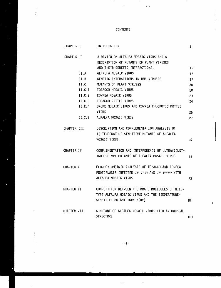
CONTENTS
CHAPTER I INTRODUCTION
CHAPTER II A REVIEW ON ALFALFA MOSAIC VIRUS AND ADESCRIPTION OF MUTANTS OF PLANT VIRUSESAND THEIR GENETIC INTERACTIONS. 13
II.A ALFALFA MOSAIC VIRUS 13II.B GENETIC INTERACTIONS IN RNA VIRUSES 17II.C MUTANTS OF PLANT VIRUSES 20II.C.I TOBACCO MOSAIC VIRUS 20II.C.2 COWPEA MOSAIC VIRUS 23II.C.3 TOBACCO RATTLE VIRUS 24II.C.4 BROME MOSAIC VIRUS AND COWPEA CHLOROTIC MOTTLE
VIRUS 25II.C.5 ALFALFA MOSAIC VIRUS 27
CHAPTER III DESCRIPTION AND COMPLEMENTATION ANALYSIS OF13 TEMPERATURE-SENSITIVE MUTANTS OF ALFALFAMOSAIC VIRUS 37
CHAPTER IV COMPLEMENTATION AND INTERFERENCE OF ULTRAVIOLET-INDUCED Mts MUTANTS OF ALFALFA MOSAIC VIRUS 55
CHAPTER V FLOW CYTOMETRIC ANALYSIS OF TOBACCO AND COWPEAPROTOPLASTS INFECTED IN VIW AND IN VITRO WITHALFALFA MOSAIC VIRUS 73
CHAPTER VI COMPETITION BETWEEN THE RNA 3 MOLECULES OF WILD-TYPE ALFALFA MOSAIC VIRUS AND THE TFMPERATURE-SENSITIVE MUTANT Tbts 7(UV) 87
CHAPTER VII A MUTANT OF ALFALFA MOSAIC VIRUS WITH AN UNUSUALSTRUCTURE 101
-6-
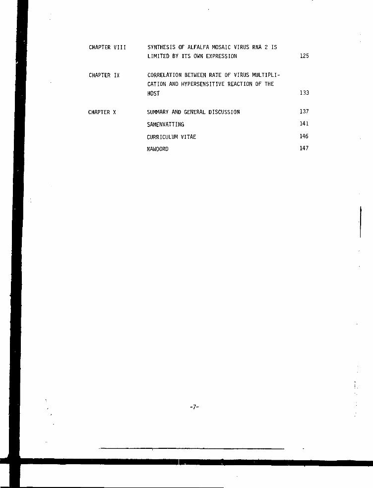
CHAPTER VIII SYNTHESIS OF ALFALFA MOSAIC VIRUS RNA 2 ISLIMITED BY ITS OWN EXPRESSION 125
CHAPTER IX CORRELATION BETWEEN RATE OF VIRUS MULTIPLI-CATION AND HYPERSENSITIVE REACTION OF THEHOST 133
CHAPTER X SUMMARY AND GENERAL DISCUSSION 137
SAMENVATTING 141
CURRICULUM VITAE 146
NAWOORD 147
-7-
CHAPTER I
INTRODUCTION
In the 90 years after the discovery of viruses as an infective agent
(Iwanowski, 1892; Beyerinck, 1898) many organisms belonging to such divers
phyla as bacteria, protozoa, fungi, animals and plants were found to be
affected by them. Viruses consist of a piece of genetic information (DNA
or RNA) wrapped in a protective structure. A lot of viruses are known to
infect plants (Matthews, 1982). Since plant viruses can reduce crop yields
considerably, they are important from an economical point of view. But also
from a scientific point of view they are interesting. Viruses contain a
relatively simple genome compared to the genomes of living organisms, which
allows studying the expression of its genetic information relatively easy.
Knowledge about their multiplication might result in effective protective
measures.
Plant viruses can contain DNA or RNA as genetic information. The latter
kind of viruses can be divided in those containing single or double stran-
ded RNA. The abundant class of the single stranded RNA viruses includes
those with a complete genome carried on one RNA molecule (monopartite),
but also species are found for which the genome consists or two or three
RNA molecules, viruses with bi- or tripartite genomes, respectively. The
plant virus alfalfa mosaic virus (AMV) belongs to this last category. AMV
has been studied in the Plant Virus Group in Leiden since 1964. The three
genomic RNA molecules 1, 2 and 3 are separately encapsidated into bacilli-
form particles with different lengths (B, M and Tb). Two molecules of a
fourth RNA molecule (RNA 4 ) , representing the subgenomic messenger for the
coat protein, are encapsidated into a separate particle (Ta) . Infectivity
requires the presence of the three genomic RNAs and the coat protein or the
subgenomic RNA (Van Vloten-Doting and Jaspars, 1977).
Despite the vast knowledge of the virus structure and genome organiza-
tion (Van Vloten-Doting et at., 1982), insight in the functions involved
in the infection process are scarce. To solve questions such as which pro-
teins are involved in the different steps of virus multiplication and what
is their precise function, mutants are indispensable.
The goal of the research described in this thesis was to isolate tempe-
rature-sensitive (ts) mutants and to use these for a genetic and biochemical
characterization of the infection process. When I started working in the
-9-
Plant Virus Group in 1978 I joined a project which had already started some
years before. Thermosensitive mutants of spontaneous or uv induced origin
were isolated and mapped on the genome by supplementation tests. Complemen-
tation experiments showed RNA 1 and RNA 2 each to carry 2 complementation
groups, while on RNA 3 only one complementation group was found. This work
is presented in chapter 3. In chapter 4 the isolation and characterization
of newly isolated ts mutants located on AMV RNA 2 is described. Complemen-
tation between some of these mutants was observed, but some combinations of
mutants showed interference. The RNA 2 encoded product might be a protein
with two domains acting in a multimeric form. Chapter 5 contains experiments
carried out to follow the infection process in tobacco plants and proto-
plasts with the use of a Fluorescence Activated Cell Sorter. In contrast to
the results with in vivo infected protoplasts no clear correlation between
fluorescence and infectivity could be observed for in vitro infected tobac-
co protoplasts. The content of chapter 6 shows the behaviour of a mutant
with a ts coat protein when mixedly infected with wildtype AMV. No rescue
of the mutant was found which could be the result of a ts mutation in the
mutant RNA 3 itself. The last experimental chapters concentrate on non-ts
mutants isolated along with the ts mutants. Chapter 7 is concerned with a
mutant which exhibits particles with a morphology quite different from nor-
mal AMV. Predominantly spheroidal structures were found as a result of
mutations in RNA 3. In chapter 8 and 9 a symptom mutant is described and
its behaviour compared to wt with respect to infectivity in cowpea proto-
plasts and cowpea and tobacco plants.
Literature on AMV and on genetic interactions and biochemical features
observed in (plant) virus mutants can be found in chapter 2.
Some of the work done with ts mutants, performed in cooperation with
others, is not included in this thesis (Smit et dl.3 1981; Sarachu et at.,
1983). Since most of the chapters consist of papers already published or
submitted for publication, some overlap in introduction and materials and
methods will be found.
-10-
REFERENCES
Bei j e n nek, M.W. (1898). Veral. Gewone. Vergad.Wis Natuurk.Afd.K.Akad.Wet.
Amsterdam 7_, 229-235.
Iwanowski, D. (1892). Buil.Acad.Imp.Sci.St-Petersbourg,(New Ser) 3, 65-70.
Matthews, R.E.F. (1982). Intervirology 17, 28-179.
Sarachu, A.N., Nassuth, A., Roosien, J., Van Vloten-Doting, L. and Bol,
J.F. (1983). Virology 124, in press.
Smit, C.H., Roosien, J., Van Vloten-Doting, L. and Jaspars, E.M.J. (1981).
Virology U £ , 169-173.
Van Vloten-Doting, L. and Jaspars, E.M.J. (1977). Comprehensive Virology
11, 1-53. Edited by H. Fraenkel-Conrat and R.R. Wagner, New York: Ple-
num Press.
Van Vloten-Doting, L., Bol, J.F., Nassuth, A., Roosien, J. and Sarachu,
A.N. (1983). NATO SRI series. Eds L.Dure and O.Ciferri (in press).
-11-
CHAPTER II
A REVIEW ON ALFALFA MOSAIC VIRUS AND A DESCRIPTION OF MUTANTS OF PLANTVIRUSES AND THEIR GENETIC INTERACTION
This chapter deals with literature concerning alfalfa mosaic virus, ge-netic interactions in general and a description of mutants of plant viruses.A complete review of all literature on these topics is beyond the scopeof this thesis and therefore only the most relevant data are discussed.
A. ALFALFA MOSAIC VIRUSThe plantvirus alfalfa mosaic virus (AMV) was first described by Weimer
(1931). AMV research had to await the development of methods of purifica-tion and biochemical analysis however, before significant facts about itsnature could be resolved. Bancroft and Kaesberg (1958, 1960) detected threemajor components upon centrifugational analysis of AMV. They were calledbottom (B), middle (M) and Top (T) component depending on the sedimentationrate. Infectivity seemed to reside on the heaviest component (Bancroft,1961). Particle dimensions measured after shadowing were rather variable.Lenghts up to 55 nm were found. Particle diameter was constant at a valueof 20 nm. Examination of negatively stained viral preparations showed thepresence of four major classes of particles. Besides B and M the top com-ponent seemed to consist of top component b (Tb) and top component a (Ta).The particles had a diameter of 17-18 nm and the lengths were 20-30; 36;48 and 58 nm for Ta, Tb, M and B respectively (Gibbs et al., 1963). Thesevirions were antigenically identical indicating that the same type of pro-tein was present in all particles (Moed and Veldstra, 1968; Van Tol, 1981).Infectivity could not specifically be contributed to one of the components.On the RNA level infectivity seemed to coincide predominantly with thelarge RNA (Gillaspie and Bancroft, 1965). Furhter fractionation of AMV par-ticles established that infectivity of the fast sedimenting species couldbe stimulated by addition of top particles, indicating a cooperative effectbetween different components (Wood and Bancroft, 1965). Experiments carriedout with RNA isolated from Mg separated viral preparations (Kelley andKaesberg, 1962) similarly showed an enhanced infectivity after combining
-13-
the ssparated fractions (Van Vloten-Doting and Jaspars, 1967). It was not
until 1970 when it was shown that the three fast sedimenting particles (B,
M,Tb) were all required for infectivity (Van Vloten-Doting et al., 1970).
These particles contained each one species of RNA, denominated 1, 2 and 3
for B, M and Tb respectively. The Ta particle contained two molecules of
RNA 4 (Bol et al., 1971). Some minor particles containing other RNA mole-
cules were also observed (Bol and Lak-Kaashoek, 1974).
AMV particle structure is thought to be mainly dependent upon interac-
tions of the coat protein and the RNA (Hull, 1969; Kaper and Geelen, 1971).
Based on the molecular weights of the particles, their RNAs and the coat
protein, it could be calculated that the number of coat protein molecules
was 240, 186, 150 and 132 for B, M, Tb and Ta respectively (Heijtink et
al., 1977). Also a particle, present in Ta preparations, has been described
which contained a smaller number of coat protein subunits (Heijtink ana
Jaspars, 1976). The AMV components have a bacilliform shape and the protein
subunits are organized in a hexagonal lattice ip the cilindrical part. At
the particle ends a pentagonal organization of the coat protein subunits
is assumed (Gibbs et al., 1963; Hull et al., 1969a; Mellema, 1975). No
pH-dependent swelling of AMV B and Ta particles was observed (Oostergetel
et al., 1981). For the smallest AMV particle it has recently been found
that the organization of the coat protein molecules is differing from that
in the other particles (Cusack et al., 1981; Oostergetel, 1983). Recon-
struction of AMV particles from purified RNA and protein has not been pos-
sible. Only partial reconstitution has been found (Lebeurier et al., 1969;
Hull, 1970a; Driedonks et al., 1978 ; Oostergetel, 1983). However reversible
dissociation could occur (Bol and Kruseman, 1969; Hull, 1970a; Verhagen et
al., 1976).
AMV RNAs extracted from unfractionated virus preparations are infecti-
ous, but the level of infection is much lower than that of an equal amount
of particles. The three genomic RNAs together are not infectious. They re-
quire the presence of RNA 4 or coat protein for infectivity (Bol et al.,
1971). The fact that AMV particles could be uncoated by their own RNA (Van
Vloten-Doting and Jaspars, 1972; Verhagen et al., 1976) revealed special
sites on all AMV RNA molecules, which interact with the coat protein (Van
Boxtel, 1976). These so called high-affinity sites were situated at the
3' end of the RNA molecules (Houwing and Jaspars, 1978; 1980; Koper-
Zwarthoff et al., 1979; Stoker et al., 1980). Recently it has been found
-14-
that also internally in RNA 4 (Houwing and Jaspars, 1982) and in RNA 1(Zuidema et al., 1983) a site with a high affinity for the coat protein ispresent. The activation of the AMV genome requires the presence of the coatprotein on the high-affinity sites of all the genomic RNA's (Smit and Jas-pars, 1980; Smit et al., 1981).
Concerning the RNA structure, it has been found that at the 5' end a cap7 r t (-1
structure (m G ppp G) is present (Pinck, 1975), but no poly (A) tailswere discovered (Bol et al., 1975). Hybridization studies showed that RNA1, 2 and 3 contained unique information but the information of RNA 4 waspresent in RNA 3 (Bol et al., 1975; Gould and Symons, 1978). A small homo-logy between the RNAs 5 could not be excluded. Sequence analysis showedthat all RNAs have about the same sequence of 150 nuclectides at the 3' end(Pinck and Pinck, 1979; Koper-Zwarthoff et al., 1979; Gunn and Symons,1980). The complete nucleotide sequence of RNA 4 is known (Brederode etal., 1980). Also the sequence at the 5'end of the genomic RNA moleculesand the sequence of the intracistronic region of RNA 3 have been elucidated(Koper-Zwarthoff et al., 1980; Pinck et al., 1981). Recently the completenucleotide sequence of RNA 1 has been unraveled (B.J.C. Cornelissen, per-sonal communication).
Upon translation of the AMV RNAs in in vitvo systems, it was observedthat RNA 4 is the messenger for the coat protein (Van Ravenswaay Claassenet al., 1967; Rutgers, 1977). RNA 3 is a dicistronic messenger from whichonly a 35 k (=kilodalton) protein is translated (Mohier et al., 1975; VanVloten-Doting et al., 1977). RNA 1 and RNA 2 encode products of 115 k and100 k respectively (Van Tol and Van Vloten-Doting, 1979). RNA 1 also indu-ces the synthesis of two smaller products of 58 k and 62 k depending on theamount of RNA used for in vitro translation. These products have the N-terminus in common with the 115 k product. Translation in the presence ofglutamine resulted in read-through of the smaller products and predominant-ly 115 k was found (Van Tol et al., 1980).
The complete aminoacid-sequence of the coat protein of AMV is known (VanBeynum et al., 1977; Collot et al., 1977; Castel et al., 1979). The coatprotein alone is able to assemble into a 30 S particle consisting of 60coat protein subunits organized in a T=l structure (Driedonks et al., 1976;1977). Crystallization of these particles has not been possible unless atryptic digestion was carried out (Fukuyama et al., 1981). This trypticdigestion results in the loss of the 25 N terminal aminoacids, which were
-15-
shown to be essential for the activation of the genomic RNAs by the coatprotein (Bol et al., 1974). These trypsinized molecules also lose theiraffinity for the high affinity sites (D.Zuidema, personal communication).The behaviour of the AMV coat protein in solution can be described as thatof a molecule consisting of a rigid core from which the 36 N terminalaminoacids protrude as a highly mobile random coiled structure (Andreeet al., 1981; Kan et al., 1982).
The study of the infection process in whole plants has been concentratedon different levels. Electronmicroscopy showed different strain dependentaggregation forms of viral particles in the cytoplasm and vacuoles of in-fected plant cells (Hull et al., 1969b; 1970; De Zoeten and Gaard, 1969).Also a strain dependent localization in the cytoplasm or in cytoplasmicinvaginations in chloroplasts was observed (Dingjan-Versteegh et al.,1974). Attempts have been made to isolate an RNA dependent RNA polymerasefrom virus infected plants (Weening and Bol, 1975; Bol et al., 1976a). Al-though RNA synthesizing activity was detected, this was also found inhealthy plants (Clerx-Van Haaster et al., 1978; Linthorst et al., 1930).This enzyme could be made template dependent and addition of AMV RNA 4 re-sulted in the synthesis of RNA products of considerable lenath (Linthorst,1982). Analysis of the protein composition of the RNA dependent RNA poly-merase from infected plants did not reveal proteins ressembling the viralencoded proteins observed in in vitro translation.
From infected plants RNA preparations were obtained which contained theReplicative Forms (RF) of AMV RNA 1, 2 and 3 (Pinck and Hirth, 1972; Mohieret al.3 1974). For RNA 4 also an RF was found (Bol et al., 1975) but thisseemed to be an artifact since experiments carried out with mutants (Smitand Jaspars, 1982) and analysis of viral minus strand RNA (Nassuth and Bol,1982) did not reveal the independent replication of RNA 4.
After fractionation of homogenates from infected tobacco plants no viralcoded proteins could be detected except the coat protein (Van Tol, 1981).However in polyribosomes AMV RNAs were found (Bol et al., 1976b), but uponaddition of a postribosomal supernatant these polyribosomes were not ableto induce synthesis of proteins other than the coat protein. Recently poly-ribosomes, obtained from AMV infected tobacco plants, were found to syn-thesize all viral coded products when added to the rabbit reticulocyte invitro translation system (L.Neeleman, personal communication).
The use of protoplasts has been very successful in elucidating features
-16-
on the multiplication process of many plant viruses (Tabeke, 1977; Nassuth,1982), since in this system a better synchronization of the infection pro-cess is obtained than in whole plants. Infection of tobacco protoplastswith AMV has been reported (Motoyoshi et al.s 1975), but could not be re-peated in our laboratory. However cowpea protoplasts could reproduciblybecome infected with AMV. The infectivity requirements for these proto-plasts were essentially the same as those for whole plants (Alblas and Bol,1977; 1978). Using this protoplast system and exploiting the divided genomeit has been possible to assign the RNA synthesizing capacity of the virusto RNA 1 and RNA 2. However again the coat protein is required (Nassuth etal.j 1981). In the absence of RNA 3 the minus strands of RNA 1 and RNA 2are overproduced, indicating that an RNA 3 encoded product plays a role inthe change from minus to plus strand RNA synthesis (Nassuth and Bol, 1982).The time course of the synthesis of the RNAs shows RNA 3 plus and minusstrand synthesis to be earlier than that of RNA 1 and RNA 2. No minusstrand RNA 4 was detectable and the RNA 4 plus strand synthesis was ratherlate in the infection process. The RNAs are encapsidated efficiently. Be-sides the coat protein no other viral encoded proteins could be detectedin cowpea protoplasts (Nassuth et al.s 1983a). Indications were obtainedthat (a) host encoded product(s) (is) are involved in the infection pro-cess (Nassuth et al., 1983b).
B. GENETIC INTERACTIONS IN RNA VIRUSESThe capacity of a virus to multiply resides in its genome which encodes
a number of proteins functioning in this process. By direct sequencing ofthe nucleotides of the genome and looking for open reading frames someinsight can be obtained in the number of proteins encoded and sequences in-volved in regulatory functions. These studies can be complemented by invitro translation studies in case the genome consists of plus strand RNA.The size and a number of encoded proteins can be found. (Atabekov and Mo-rozov, 1979; Davies and Hull, 1982; Van Vloten-Doting and Neeleman, 1982).Functions of viral coded proteins are difficult to resolve in this wayexcept of course for the structural protein(s) which form(s) an integratepart of the virions. Viruses with a divided genome have the advantage thateach genome part or combinations of genome parts can be used to study the
infection process (Nassuth, 1982). In that way information can be obtained \i
-17-
about the location of the information involved in the multiplication. How-ever no information can be obtained about the number of functions presenton each genome segment nor about the way the viral products are functio-ning. For the elucidation of these questions mutants are required.
The mutation rate of RNA genomes is much higher than that of DNA genomes(Holland et al.a 1982). For that reason spontaneous mutants of RNA virusesare rather easily obtained. The mutation rate can be further increasedusing a variety of mutagens, like nitrous acid (Gierer and Mundry ,- 1958),N-methyl-N'-nitro-N-nitrosoguanidine (MNNG) (Singer and Fraenkel-Conrat,1967), 5 Fluorouracil (Kramer et at., 1964) and uv-irradiation (Shimizuet al., 1982a). In plant viruses with a divided genome, the mutations canbe addressed to a certain component by treating only that component withthe mutagen (De Jager, 1978). The selection of mutants after the mutagenictreatment can fulfill criteria like plaque morphology for animal or bacte-rial viruses, sensitivity to certain substances, and pH or temperature de-pendent growth. For the isolation of mutants of these viruses a replicasystem is generally used. Selection criteria for plant virus mutant arecharacters like the symptom size, alteration of symptom type, time appea-rance after temperature treatment and thermosensitivity of the infectionprocess. The lack of a proper replica system does not allow the handlingof large numbers of infective centers in the selection for mutants.
After the isolation of mutants, their stability has to be checked. Some-times mutants still produce virus under conditions chosen to be restrictivefor their multiplication (leakiness). Also reversion to a wildtype (wt)phenotype is sometimes observed as a result of a back mutation at the samesite as the original mutation or because of a mutation somewhere else inthe genome. This latter type of mutations is called suppressor mutationand examples have been observed for instance in reovirus (Ramig andFields, 1979; Ahmed et at., 1980) and influenza (Scholtissek and Spring,1982). Indications have been obtained that mutations involved in RNA syn-thesis might increase the mutation rate (Pringle et al., 1981). For plantviruses a change from one host to another can induce changes in the virus(Yarwood, 1979; Donis-Keller et al., 1981).
The mapping of mutants on the genome is rather difficult for RNA vi-ruses. Recombination has been only reported for some picorna viruses (Coo-per, 1968; Me Mahon and Slade, 1981; King et al., 1982). However a lot ofRNA viruses contain a divided genome (Matthews, 1982), which allows assign-
-18-
ment of the mutation to a certain genome segment by analysis of artificialmixtures of mutant and wt components (pseudo-recombinants). Another type oftest used in the localization of mutations in plant virus mutants is thesupplementation test (Bancroft and Lane, 1973). The mutant is propagatedunder restrictive conditions in the presence of each of the purified wtcomponents. Multiplication will only take place in the presence of the wtcomponent which substitutes for the mutant defect. The mutation is thenassumed to be located on that component. For monopartite RNA viruses themapping requires data from RNA sequencing and polypeptide analysis.
Since mutations can in principle be situated in every function involvedin the replication process the number of functional groups can be obtainedby mixed infection of pairs of all mutants under conditions where the mu-tants alone do not multiply. A mutant with a mutation in one function willbe able to complement a mutant with a mutation in a different function. Inthis way grouping of mutants in different complementation g> jps is pos-sible. These groups represent often a polypeptide involved in a certainfunction. A prerequisite for these complementation experiments is howeverthat the products responsible for the different functions are diffusable(active in trans). Some proteins or certain regulatory sequences on nucleicacid molecules can represent non-diffusable functions (active in cis) andcan therefore not be complemented. The level of complementation can varyconsiderably between certain pairs of mutants. A low level can indicateintracistronic complementation. In this case both mutants carry defectsin the same cistron, but since the products function as a multimer, stabi-lizing forces between the subunits render the complex functional (Crickand Orgel, 1964). Examples are found among some influenza virus mutants(Heller and Scholtissek, 1980; Thierry and Spring, 1981; Shimizu et al.,1982b; Massicot et al., 1982). Another example of this intracistronic com-plementation finds its origin in proteins made up of two different func-tional domains (Oamieson et al., 1974; Jamieson and Subak-Sharpe, 1974;Spoerel et al., 1979). Mutants with mutations in the same cistron but indifferent domains will still be able to multiply, since each of the mutantproducts contains one functional domain. Sometimes mutants in the samecistron can display negative complementation or interference. Their pro-ducts exert a destabilizing influence on each other or sometimes even onthe wt product, so that the functioning of the products is less in themixed infection than for each of the mutants alone (Sundaram and Finch,1967; Honess, 1981).
-19-
In general complementation studies can be used to establish the nature
of a gene product. It can yield information about the identity of subunits
in oligomeric enzymes, the function of different regions of polypeptide
chains. Even indications can be obtained on the tertiary and quaternary
structure.
C. MUTANTS OF PLANT VIRUSES
This section deals with literature on plant virus mutants and strains
with special emphasis on thermosensitive (is) mutants. No attempts will
be made to give a complete description of all strains and mutants. For that
the reader is referred to reviews of Atabekov (1977) and Lane (1979).
C.1 Tobacco Mosaic Virus
Tobacco mosaic virus (TMV) is a monopartite virus consisting of rigid
rods containing an RNA genome of 6395 nucleotides (Goelet et al., 1982),
encoding upon in vitro translation the synthesis of a 110 k (=kilodalton)
product and a 168 k read-through product. Two subgenomic mRNAs are found
which encode a polypeptide of 30 k and the coat protein, respectively
(Zaitlin, 1979). In infected protoplasts virus induced and/or virus coded
polypeptides of 260, 240, 170, 116.5, 96, 90, 82, 72, 30 and 17,5 k detec-
ted (Huber, 1979).
For TMV nitrous acid is generally used as mutagen. Mutants were selected
by altered symptoms induced in certain host plants (Gierer and Mundry,
1958; Siegel, 1965; Sehgal and Krause, 1968), altered symptom size after
incubation at high temperature (Jockusch, 1964; Peters and Murphy, 1975)
or delayed time appearance of local lesions after shifting infected plants
from restrictive to permissive temperature (Dawson and Jones, 1976).
Numerous TMV strains and mutants were isolated exhibiting defects in
particle production (e.g. Siegel et al., 1962). The coat protein of some
of these strains did not aggregate with TMV RNA, but was found in soluble
form in the cell. Anomalous aggregation of protein in vivo and in vitro
was observed (Siegel et al., 1966; Zaitlin and Ferris, 1964). Dependent on
the degree of aminoacid changes, the defectiveness in this type of mutants
is more or less marked. In some strains the coat protein is found in the
plant in an insoluble form (Siegel et al., 1962; Zaitlin and Keswani, 1964)
or was not even present (Sarkar and Smitamana, 1981). Most amino acid re-
-20-
placements in the nitrous acid induced mutants could be accounted for bythe A -* G and C •+ U conversion, but also other changes were observed (Ata-bekov, 1977).
For TMV numerous mutants have been isolated which were thermoresistant(tr) or thsrmosensitive (ts). The tp strains produced more infectious virusat 35°C than at 20°C (Lebeurier and Hirth, 1966; Lebeurier and Wurtz, 1958)and virion preparations contained much more defective short particles at20°C than at 35°C. However even at 35°C some defective particles were found.This partial defectiveness, even at the optimal growth temperature was alsoobserved for a ts mutant (Kattanis and Battow, 1971a).
Jockusch (1964) first described systematically a number of ts mutantsof TMV. He compared the in vivo behaviour (the amount of infectious virusproduced in mutant infected plants or leaf discs at 32°C and 23°C) withthe in vitro behaviour of the coat protein isolated from these mutants(Jockusch, 1966a). They could be divided in two groups. One group formed tat the non permissive temperature few viral particles. The infectivity ofextracts from infected plants kept at 32°C was low or lacking (Atabekovet at., 1970; Kassanis and Bastow, 1971b). Accumulation of infective RNAunprotected by the coat protein appeared (Jockusch, 1966a). At 32°C theinfection was unable to spread from cell to cell, but after shift down to23°C a rapid accumulation of appreciable amounts of infective mature viralparticles followed. A little soluble coat protein was detectable by sero-logical means in extracts at 32°C. Presumably most coat protein was dena-turated unsoluble material, since normal coat protein synthesis was foundat 32°C (Jockusch, 1968a). Coat protein preparations of this type of mutantsdenaturated in vitro at the non-permissive temperature (Jockusch, 1966b,c).Under these conditions no assembly in rod-like particles with helicalstruc-ture could be found (Jokusch, 1966c). By measuring the denaturation kine-tics, the ts coat protein mutants could be distinguished from their trcounterparts. Some mutants were defective at both temperatures (Jockusch,1966b; Hariharasubramanien et at., 1970). Comparison of the aminoacid se-quence in a number of mutants (Wittman-Liebold et al., 1965) revealed in-fornation about the parts of the coat protein required for a proper func-tioning (Oroszlandand Gilden, 1979). For some of the TMV ts coat proteinmutants it was shown that they could be complemented by a temperatureresistant helper virus. This phenomenon was called phenotypic mixing orgenomic masking, since the mutant RNA was encapsidated by the coat protein
-21-
supplied by the helper virus (Atabekov et at., 1970; Kassanis and Bastow,1971c).
Besides these coat protein mutants also mutants in non-structural geneswere obtained. They constitute mutants of the second group (Jockusch,1966a). A number of these mutants could not spread from the primary infec-ted cell to adjacent cell at the non permissive temperature (Jockusch 1968;Nishigushi et at., 1978;1979; Peters and Murphy, 1975). One of these mu-tants was shown to be a double mutant by means of complementation experi-ments (Taliansky et al., 1982a). In the presence of wt helper virus itcould spread at the non permissive temperature, but no mutant particleswere found. The mutant produced normal coat protein since it could comple-ment a mutant with a ts coat protein (Jockusch, 1966a). The mutant carriesbesides the defect in spreading a ts defect in the RNA molecule itself (Ta-liansky et al., 1982a). The encapsidation apparently starts at the normalorigin of assembly (Lebeurier et al., 1977; Guilley et at., 1979) but alsoat a second site with affinity for the TMV coat protein (Guilley et al.,1975). This results in the formation of RNase sensitive particles (Talian-sky et al., 1982b). Another mutant also defective in spreading at the nonpermissive temperature (Nishigushi et al., 1978; 1979) carried a changed30 k product (Leonard and Zaitlin, 1982). This polypeptide might be invol-ved in the spreading from cell to cell by exerting influence on the numberof plasiiiodesmata (Shalla et at., 1982).. The defect in this mutant couldalso be complemented by a coat protein mutant with a ts defect (Talianskyet at., 1982c) indicating that the structural gene was not involved.
Between a number of mutants isolated by Dawson and Jones (1976) no com-plementation was found, so that no division in functional groups was pos-sible. However they obtained mutants disturbed in the RNA synthesis at 35°C(Dawson and White, 1978; 1979). For one of these mutants no synthesis ofsingle stranded genomic RNA was detectable, although the production of re-plicative form (RF) and replicative intermediate (RI) RNAs was normal. Uponshift down the single-stranded RNA synthesis was resumed, indicating thatthe function was not irreversibly lost. Some differences in the in vitroand in vivo stabilities of the replicase of the mutant as compared to wtwere found, but these effects were small. In another type of mutant the syn-thesis of all RNA at the non permissive temperature stopped. After pro-longed incubation at that temperature, the mutant was not able to resumethe RNA synthesis when shifted down to the permissive temperature. Possibly
-22-
the mutant replicase or a subunit was inactivated and could not be replacedat normal temperature.
Concludingly it can be remarked that for TMV five functionally differentts types have been found. Mutants with a ts defect in the coat protein orin the RNA itself affected the encapsidation of the RNA. Mutations in afunction involved in the spreading of the infection from cell to cell ap-parently were localized in the 30 k polypeptide. Mutants with defects inthe RNA synthesis fell apart in two groups. One group was only affected inthe single strand RNA synthesis, while the other group was defective in thesynthesis of all RNA.
C.2 Cowpea Mosaic VirusCowpea mosaic virus (CPMV) is a bipartite icosahedral virus containing
plus strand RNA molecules of about 2.0 x 10 and 1.4 x 10 molecular weight.These RNA molecules encode 200 k and 105 k proteins respectively, whichare processed in vivo and in vitro in a number of smaller products. Thepolypeptide encoded on the large RNA yields the VPg and 87 k, 58 k, 32 kand 24 k products, while the small RNA encoded polypeptide yields the twoviral coat proteins (23 k and 37 k) and 93 k, 79 k, 60 k, 58 k, 48 k and47 k products (Rottier et al., 1979; 1980a; 1980b; Franssen et at., 1982).
For cowpea mosaic virus mutants were obtained after nitrous acid treat-ment of the virus (De Jager, 1978). Three mutants had altered symptoms onsome bean and cowpea varieties. Two of them produced small amounts ofinfectious virus in Blackeye cowpeas. Using supplementation tests and theconstruction of pseudo-recombinants, it was shown that two of these mutantscarried mutations on the small RNA (M-DNA). In the other mutant the largeRNA (B-RNA) seemed to be changed. Mixed inoculation of the two mutants, lo-calized on the small RNA, did not yield wt symptoms, indicating that com-plementation did not appear (De Jager, 1976). Another nitrous acid inducedsymptom mutant gave small chlorotic lesions in Pinto bean, which react towt with the development of large necrotic lesions. The mutant carried mu-tations on both components. The mutation on M could complement the M mu-tation in one of the aforementioned M mutants, since mixed inoculation ofboth mutants induced local lesions with a wt phenotype, containing onlymutant virus (De Jager and Breekland, 1979). This might indicate that atleast two complementation groups are present on RNA 2 of CPMV. The elec-trophoretic mobility of the mutant virions is changed, which might mean
-23-
that one of its coat proteins is altered. Two spontaneous mutants induced
a systemical infection in Early Red cowpea which reacts hypersensitive to
wt infection. For one of these mutants the symptoms were a systemic mosaic,
while the other induced a systemic necrosis',. In both cases was the B-
component responsible for the mutant character (De Jager, 1978; De Jager
and Wesseling, 1981).
A mutant differing from wt with respect to the symptoms it induced in
cowpea and bean plants was found to be ta. It is presumably a double mu-
tant since a mixture of mutant B component and wt M component yielded lo-
cal lesions of wt size but mutant color, whereas the combination of wt B
component and mutant M component gave rise to local lesions of mutant
size but wt color. Thus the M component seems to govern the local lesion
size and the B component is responsible for the local lesion color. The ts
mutation was located on the M component (De Jager and McLean, 1979). Mu-
tant particles were as stable against heating as wt, indicating that the
coat proteins had not been altered (De Jager et al., 1977). The amount of
bound replicase of this mutant at 30°C was much lower than that of wt. The
activity of the mutant replicase preparation had however the same tempe-
rature dependency as the wt preparation (De Jager et al., 1977). By shift
up experiments it was shown that the mutant induced a defective replicase
or replicase subunit. It is unknown whether this replicase (subunit) is
not synthesized or that there is a defect in the viral induced cytopathic
structures in which the replicase is assumed to act.
One mutant, again selected on the basis of different symptoms, produced
relative higher amount of top component than the wt. This characteristic
was assigned to the M component. At 30°C the relative high amount of mu-
tant top component was not observed (De Jager and Van Kammen, 1970).
The mutants obtained for CPMV map on both RNAs. RNA 2 seems to contain
at least two functional groups. The number of functional groups on RNA 1
is still obscure. Assignment of the different groups to the proteins ob-
served in infected cells has not yet been done.
C.3 Tobaaoo Rattle Virus
Tobacco rattle virus (TRV) (CAM isolate) is a bipartite virus, contai-
ning two RNA molecules of molecular weight 2,5 x 10 and 0,7 x 10 encap-
sidated into long and short rods respectively. The lenght of the short
rods is different for the different isolates (Lane, 1979). Upon in vitro
-24-
translation the small RNA yields a 22 k product, the coat protein. Howeverin another strain also a 31 k product is observed, which contains sometryptic peptides in common with the coat protein (Mayo et al., 1976). InCAM strain infected cells however an RNA molecule, derived from RNA 2, wasobserved which could be translated in a 30 k protein. Apparently for somestrains the small rods contain varying amount of this third RNA species(Bisaro and Siege!, 1980). In vivo the coat protein was detectable andRNA 1 induced the synthesis of 187 k and 142 k polypeptides (Mayo, 1982).
After nitrous acid treatment of the CAM strain of TRV and inoculationin the presence of excess short particles, to prevent the developmentof unstable infective centres (Sanger, 1968), three mutants were obtained,which were unable to form lesions in Chenopodium at 30°C (Robinson, 1973a).As would be expected from the selection procedure, the mutations mappedon the large RNA. Mutant particles were, like wt, stable against thermalinactivation, in accordance with the assumption that the small component,carrying the coat protein gene, was of wt origin. Also serologically andwith respect to their particles dimensions the mutants were indistinguis-hable from wt. In one host only one of the mutants produced small quanti-ties of infectious RNA at 3O°C, while the other was not ts. However in adifferent host both mutants were impaired in the production of infectiousRNA at 30°C (Robinson, 1973b). By following the development of uv resis-tance of potential infective centres, it was found that at 30 C both mu-tants fail to replicate in the initially infected cells (Robinson, 1974).Complementation experiments between these mutants were not carried out.
A symptom mutant of spontaneous origin was shown to produce the sameparticles as the wt CAM strain by electronmicroscopy. The symptom mutationwas located on RNA 2. No immunological difference or deviating trypticpeptides were observed between the coat protein of the mutant and wt. Theseresults could point to the existence of another cistron on the small RNA(Robinson, 1977).
C.4 Bvome Moscde Virus and Cowpea Chlorotia Mottle VirusBrome mosaic virus (BMV) and cowpea chlorotic mottle virus (CCMV) are
closely related icosahedral viruses containing a tripartite genome withRNA molecules of molecular weight 1,1 x 10⁶ (RNA 1); 1.0 x 10⁶ (RNA 2) and0,8 x 106 (RNA 3). Besides the genomic RNAs also an RNA 4 (0,3 x 10⁶) ispresent in virions, representing the messenger for the coat protein. In vi-
-25-
tro translation experiments showed RNA 1, 2, 3 and 4 to encode polypep-
tides of 120 k, 110 k, 35 k and 20 k (coat protein) respectively (Shin
and Kaesberg, 1976). In vivo the same products were observed in infected
protoplasts (Okuno and Furasawa, 1979; Kiberstis et at., 1981).
Three spontaneous BMV and four nitrous acid induced BMV and CCMV mu-
tants were characterized by symptoms on different hosts. By construction
of pseudo-recombinants and supplementation tests it appeared that in both
viruses RNA 2 was involved in local lesion formation on certain host plants.
In BMV however, also RNA 3 played a role in this characteristic. Besides
for both viruses mutations in RNA 3 had influence on the component ratio
and the coat protein (Bancroft and Lane, 1973). Two of the CCMV mutants
carried a ts defect on RNA 1 and RNA 3 respectively. The RNA 3 mutant had
a low specific infectivity, even at the permissive temperature, resulting
from a low amount of RNA 1 in the particles. At 32°C considerable amounts
of uncoated RNA were observed in infected leaves. The coat protein was
much less heat stable than wt coat protein. Deviant aggregation forms
appeared upon self-assembly of the mutant protein (Bancroft et dl., 1972).
Aminoacid sequencing showed the mutant coat protein to have two aminoacid
replacements (Rees and Short, 1982). In cowpea and tobacco protoplasts this
mutant also behaved defective at the non-permissive temperature. No mutant
coat protein was detectable, which might indicate a high turn-over at 32°C.
The mutant could however be rescued by wt because mutant RNA 3 was effi-
ciently encapsidated into wt. coat protein (Dawson et al.3 1975; Dawson and
Watts, 1979). Also some non-ts CCh'V mutants located on RNA 3 were obtained
after nitrous ?cid tretment of the virur. C c t protein isolated from these
mutants displayed an altered aggregation in self-assembly (Bancroft et al.,
1976).
In an attempt to isolate specifically ts mutants of CCMV, plants infected
with MNNG treated virus were incubated two days at 35°C and after transfer
to 25°C the time of local lesion appearance was scored (Dawson, 1978). In
this way a number of ts mutants was obtained. However most mutants origi-
nated from the same lesion and might carry the same ts defect. The mutants
did not produce infectious virus nor infectious RNA at 35°C. Supplementa-
tion experiments indicated that three ts mutants were located on RNA 3 and
ten mapped on RNA 1. As expected complementation was found between RNA 1
and RNA 3 mutants. No interference of these mutants with the wt replication
at 35°C was observed. Some RNA 1 mutants occasionally seemed to complement
-26-
each other but leakiness and reversion might be the reason for that ob-servation. Biochemical analysis of two RNA 1 and RNA 3 ts mutants showedthat the plus strand RNA synthesis decreased after shift-up to 35°C. Inone RNA 1 and one RNA 3 ts mutant the RNA 4 synthesis immediately stoppedat 35 C. However in all mutants the amount of the other RNAs decreasedgradually as did the ts RNA species. Therefore no specific function in RNAsynthesis could be attributed to either one of the RNAs (Dawson, 1981).
A non-ts variant of CCMV has been obtained by passage through beanplants. This strain carried a symptom marker on RNA 3. The coat proteinof this mutant was serologically identical to the original virus. This wasconfirmed by analysis of tryptic peptides. The symptom marker may repre-sent another function on RNA 3 (Wyatt and Kuhn, 1980).
C.5 Alfalfa Mosaic Virus
The main characteristics of this viruses are summarized in the firstsection of this chapter. Among the spontaneous mutants and strains of AMVsymptom differences on different hosts have been found. Local lesion in-duction in bean and cowpea plants seems to reside on RNA 2 (Dingjan-Ver-steegh et al., 1972). From the construction of pseudo-hybrids they obtainedindications that the lesion type on tobacco and the component ratio weredetermined by RNA 3. In different strains there is considerable variationin particle length. Strain VRU produces very long particles, encapsidatingmore RNA species (Hull, 1970b). The mutant coat protein alone can aggregateinto long tubular forms (Driedonks et al., 1978a). Many changes in theaminoacid sequence have been found (Castel et al., 1979). In another strainalso particles longer than the B component were observed. These particlesencapsidate two molecules of RNA 2 (Heijtink, 1974). The partial aminoacid!sequence revealed many differences with other AMV strains (Castel, 1979).Comparison on the RNA level of the sequences at the 5' end of the RNA 4molecules of different strains showed them to be conserved (Swinkels andBol, 1980). This might indicate that they could play a role in the repli-case recognition.
Isolation of mutants which could multiply at high temperature was accom-plished by local lesion passage at 34°C (Franck and Hirth, 1976). Threeisolates were obtained which were much more infective than wt at high tem-perature. At the low temperature the mutants multiplied less than wt. Inall cases RNA 1 carried a mutation conferring temperature resistance to the
-27-
virus. However this mutation was accompanied by an RNA 3 mutation, which
reduced the ability to replicate at low temperature. Apparently both RNA 1
and RNA 3 functions are involved in replication.
Another set of AMV mutants was isolated after MNNG treatment and
scoring for local symptoms on a normally systemic reacting host (Hartmann
et al., 1976) these mutants had different symptoms on tobacco and different
proportions of the nucleoprotein components. Also the temperature optimum
for multiplication was shifted to a higher temperature and the mutant yield
was lower than wt. At higher temperature the mutants did not produce Ta,
while the parent strain did. By constructing pseudo-recombinants it was
shown for two of the mutants that RNA 3 governed this latter characteristic.
For all mutants the altered response in tobacco plants seemed likewise
caused by a mutation in RNA 3. Tryptic fingerprinting of the mutant coat
proteins established many differences with wt.
REFERENCES
Ahmed,R., Chakraborty, P.R., Graham, A.F., Ramig, R.F. and Fields, B.N.
(1980). J. of Virology 34, 383-389.
Alblas, F. and Bol, J.F. (1977). J. of Gen. Virology 36, 175-185.
Alblas, F. and Bol, J.F. (1978). J. of Gen. Virology 41, 653-656.
Andree, P.J., Kan, J.H. and Mellema, J.E. (1981). FEBS Letters U0_, 265-268.
Atabekov, J.G. (1977). In: Comrehensive Virology ( H.Fraenkel-Conrat and
R.R.Wagner, Sds) Plenum Press, New ïork.vo'i 11, pp 143-200.
Atabekov, J.G. and Morozov, S.Yu. (1979). Advan. Virus Res. ^ 5 , 1-91.
Atabekov, J.G., Schaskolskaya, N.D., Atabekova, T.I. and Sacharovskaya, G.N.
(1970). Virology 41, 397-407.
Bancroft, J.B. (1961). Virology 14, 296-297.
Bancroft, J.B. and Kaesberg, P. (1958). Nature London 181., 720-721.
Bancroft, J.B. and Kaesberg, P. (1960). Bioahim. Biophys. Aota 39_, 519-528.
Bancroft, J.B. and Lane, L.C. (1973). J. of Gen. Virology 19^, 381-389.
Bancroft, J.B., McDonald, J.G. and Rees, M.W. (1976). Virology 75, 293-305.
Bancroft, J.B., Rees, M.W., Dawson, J.R.O., McLean, G.D. and Short, M.N.
(1972). J. of Gen. Virology 16, 69-81.
Bisaro, D.M. and Siegel, A. (1980). virology Utf, 194-201.
Bol, J.F. and Kruseman, 0. (1969). Virology ,485-488.
Bol, J.F. and Lak-Kaashoek, M. (1974). Virology 60^, 476-484.
-28-
Bol, J.F., Bakhuizen, CE.G.C. and Rutgers, T. (1976b). Virology 75_, 1-17.
Bol, J.F., Brederode, F.Th., Janze, G.C. and Rauh, D.K. (1975). Virology
65, 1-15.
Bol, J.F., Clerx-Van Haaster, C.H. and Weening, C.J. (1976a). Ann. Miaro-
biol.dnst. Pasteur) 127A, 183-192.
Bol, J.F., Kraal, B. and Brederode, F.Th. (1974). Virology 58, 101-110.
Bol, J.F., Van Vloten-Doting, L. and Jaspars, E.M.J. (1971). Virology 46_,
73-85.
Brederode, F.Th., Koper-Zwarthoff, E.C. and Bol, J.F. (1980). Nual. Aaids
Res. 8, 2213-2223.
Castel, A. (1979). Ph.D. Thesis Rijksuniversiteit Leiden. The Netherlands.
Castel, A., Kraal, B., De Graaf, J.M. and Bosch, L. (1979). Eur. J. Bioahem.
102, 125-138.
Clerx-Van Haaster, C.H. and Bol, J.F. (1978). Virology 9J., 453-463.
Col lot, D., Peter, R., Das, B., Wolff, B. and Duranton, H. (1977). Bioehim.
Biophys. Aeta 492, 267-283.
Cooper, P.D. (1968). Virology 35_, 584-596.
Cooper, P.D. (1977). In: Comprehensive Virology ( H.Fraenkel-Conrat and
R.R.Wagner, Eds) Plenum Press, New York. Vol 9, pp 133-207.
Crick, F.H.C, and Orgel, L.E. (1964). J. Mol. Biol. 8, 161-165.
Cusack, S., Krijgsman, P.C.J., Meliema, J.E. and Oostergetel, G.T. (1980).
In: E.M.B.L. Research Reports 1980, pp 243-246.
Davies, J.W. and Huil, R. (1982). J. of Gen. Virology 61, 1.
Dawson, J.R.O. and Watts, J.W. (1979). J. of Gen. Virology £5, 133-137.
Dawson, J.R.O., Motoyoshi, F., Watts, J.W. and Bancroft, J.B. (1975). J. of
Gen. Virology 29, 99-107.
üawson, W.O. (1978). Virology 90, 112-118.
(1981). Virology _115, 130-136.
and Jones, G.E. (1976). Molea. Gen. Genet. U5, 307-309.
and White, J.L. (1978). Virology 90, 209-213.
and White, J.L. (1979). Virology 93, 104-110.
De Jager, C.P. (1976). Virology 70, 151-163.
De Jager, C.P. (1978). Ph.D. Thesis Landbouwhogeschool Wageningen. The Ne-
therlands.
De Jager, C.P. and Breekland, L. (1979). Virology 99,312-318.
De Jager, C.P. and McLean, L. (1979). Virology 99, 167-169.
üe Jager, C.P. and Van Kammen, A. (1970). Virology 4^, 281-287.
-29-
Dawson,Dawson,Dawson,Dawson.
W.OW.O
W.O
W.O
De Jager, C.P., Zabel, P., Van Der Beek, C.P. and Van Kammen, A. (1977).
Urology 76, 164-172.De Zoeten, G.A. and Gaard, G. (1969). Virology 39, 768-774.Dingjan-Versteegh, A., Van Vloten-Doting, L. and Jaspars, E.M.J. (1972).
Virology 49, 716-722.Dingjan-Versteegh, A., Verkuil, B. and Jaspars, E.M.J. (1974). Neth. J. of
Plant Path. 80, 72-76.Donis-Keller, H., Browning, K.S. and Clark, J.M.jr. (1981). virology 110,
43-54.Driedonks, R.A., Krijgsman, P.C.J. and Mellema, J.E. (1976). Phil. Trans.
E. Soa. hond. B. 276, 131-141.Driedonks, R.A., Krijgsman, P.C.J. and Mellema, J.E. (1977). J. Mol. Biol
113, 123-140.Driedonks, R.A., Krijgsman, P.C.J. and Mellema, J.E. (1978), Euv. J. Bio-
ahem. 82, 405-417.Driedonks, R.A., Krijgsman, P.C.J. and Mellema, J.E. (1978a). J. Mol. Biol.
124, 713-719.Franck, A. and Hir th , L. (1976). Virology _70, 283-291.
Franssen, H., Goldbach, R.W., Broekhuisen, M., Moerman, M. and Van Kammen,
A. (1982). J. of Virology 4U, 8-17.
Fukuyama, K., Abdel-Meguid, S.A. and Rossman, M.G. (1981). J. Mol. Biol.
150, 33-41.
Gibbs, A . J . , Nixon, H.L. and Woods, R.D. (1963). Virology Jl£, 441-449.
Gierer , A. and Mundry, K.W. (1958). Nature London JL82, 1457-1458.
G i l l a s p i e , A.G. and Bancroft , J .B . (1965). Virology Z7_, 391-397.
Goelet, P . , Lomonossoff, G.P. , But le r , P . J .G . , Akam, M.E., Gai t , M.J. and
Karn, J . (1982). PNAS 79, 5818-5822.
Gould, A.R. and Symons, R.H. (1978). Eur. J. Bioahem. 9J., 269-278.
Gui l ley , H., Jonard, G., Kukla, B. and Richards, K.E. (1979). Nual. Aeids
Res. 6 , 1287-1308.
Guil ley, H., Jonard, G. and Richards, K.E. (1975). Eur. J. Bioohem. £ 4 , 135
-144.
Gunn, M.R. and Symons, R.H. (1980). FEBS Letters 109_, 145-150.Hariharasubramanian, V., Zaitlin, M. and Siegel, A. (1970). virology 40,
579-589.Hartmann, D., Mohier, E . , Leroy, C. and Hir th , L. (1976). Virology _74, 470-
480.
-30-
Heller, E. and Scholtissek, C. (1980). J. of Gen. Virology 49_, 133-139.Heijtink, R.A. (1974). Ph.D. Thesis Rijksuniversiteit Leiden. The Nether-
lands.
Hei j t ink , R.A. and J a s p a r s , E.M.J. (1976). Virology 69_, 75-80.
Hei j t ink, R.A., Houwing, C.J. and J a spa r s , E.M.J. (1977). Biochemistry 16,
4684-4693.
Holland, J . , Spindler , K., Horodyski, F . , Grabau, E . , Nichol, S. and Van de
Pol, S. (1982). Science 215, 1577-1585.
Honess, R.W. (1981). J. of Gen. Virology 5_7, 297-306.
Houwing, C.J. and J a spa r s , E.M.J. (1978). Biochemistry J 7 , 2927-2933.
Houwing, C.J. and Ja spa r s , E.M.J. (1980). Biochemistry Vi_, 5255-5260.
Houwing, C.J. and Ja spa r s , E.M.J. (1982). Biochemistry _21, 3408-3414.
Huber, R. (1979). Ph.D. Thesis Landbouwhogeschool Wageningen. The Nether-
lands.
Hull , R. (1969). Advances in Virus Research J15, 365-433.
Hull , R. (1970a). Virology 40, 34-37.
Hull , R. (1970b). Virology 42, 283-292.
Hull , R., H i l l s , G.J. and Markham, R. (1969a). Virology 3T_, 416-428.
Hull , R., H i l l s , G.J. and P l a s k i t t , A. (1969b). J. Ultrastruat. Res. 26,
465-479.
Hull , R., H i l l s , G.J. and P l a s k i t t , A. (1970). Virology 42, 753-772.
Janrieson, A.T. and Subak-Sharpe, J.H. (1974). J. of Gen. Virology 2A_, 481-
492.
Jamieson, A.T. , Gentry, G.A. and Subak-Sharpe, J .H. (1974). J. of Gen. Viro-
logy 2A_, 465-480.
Jockusch, H. (1964). Z. Vererbungsl. 95_, 379-382.
Jockusch, H. (1966a). Z. Vererbungsl. 98, 32C.343.
Jockusch, H. (1966b). Bioahem. Biophys. Res. Comm. 24_, 577-584.
Jockusch, H. (1966c). Z. Vererbungsl. 98, 344-362.
Jockusch, H. (1968). Virology _35, 94-101.
Kan, J . H . , Andree, P . J . , Kouijzer, L.C. and Mellema, J .E . (1982). Eur. J.
Biochem. 126, 29-33
Kaper, J.M. and Geelen, J.L.M.C. (1971). J. Mol. Biol. 56, 277-294.
Kassanis, B. and Bastow, C. (1971a). J. of Gen. Virology JL1, 157-170.
Kassanis, B. and Bastow, C. (1971b). J. of Gen. Virology _U, 171-176.
Kassanis, B. and Bastow, C. (1971c). J. of Gen. virology _10, 95-98.
Kelley, J . J . and Kaesberg, P. (1962). Bioahim. Biophys. Acta 6_1, 865-871.
7
- 3 1 -
K i b e r s t i s , P . A . , L o e s c h - F r i e s , L . S . a n d H a l l , T . C . ( 1 9 8 1 ) . Virology 1 1 2 ,
8 0 4 - 8 0 8 .
K i n g , A . M . Q . , McMahon, D . , S l a d e , W.R. a n d Newman, J . W . I . ( 1 9 8 2 ) . J. of
Virology _ 4 1 , 6 6 - 7 7 .
K o p e r - Z w a r t h o f f , E . C . , B r e d e r o d e , F . T h . , W a l s t r a , P . a n d B o l , J . F . ( 1 9 7 9 ) .
Nual. Aoids Res. ]_, 1887-1900.Koper -Zwar thof f , E . C . , B r e d e r o d e , F . T h . , Veeneman, G . , Van Boom, J . H . and
B o l , J . F . ( 1 9 8 0 ) . Nuol. Aaids Res. 8, 5635-5647 .
Kramer, G . , Wit tmannn, G. and S c h u s t e r , M. ( 1 9 6 4 ) . Z. Naturforsch. J19b, 46 -
5 1 .
Lane, L.C. (1979). In: Nucleic Aoids in Plants.(T.C.Hall and J.W.Davies,
Eds). CRC Press, INC. Vol I I , pp 65-110.
Lebeurier, G. and Hirth, L. (1966). Virology 29, 385-395.Lebeurier , G. and Wurtz, M. (1968). C. R. Hebd. Seances Aaad. Sci. Paris.
267, 871.
Lebeurier , G., Nicolaieff , A. and Richards, K.E. (1977). PMS 74, 149-153.
Lebeurier , G., Wurtz, M. and Hir th , L. (1969). C. R. Hebd. Seances Acad.
Sci. Paris. 268D, 2002.Leonard, D.A. and Zaitlin, M. (1982). Virology JL17, 416-424.Linthorst, H.J.M. (1982). Ph.D. Thesis Rijksuniversiteit Leiden. The Nether-
lands .Linthorst, H.J.M., Bol, J.F. and Jaspars, E.M.J. (1980). J. of Gen. Virolo-
gy 46, 511-515.Massicot, J.G., Van Wyke, K., Chanock, R.M. and Murphy, B.R. (1982). viro-
logy IY7_, 496-500.Matthews, R.E.F. (1982). Intrevirology 1_7, 1-199.Mayo, M.A. (1982). Intervirology _17, 240-246.Mayo, M.A., Fritsch, C. and Hirth, L. (1976). Virology 69_, 408-415.McCahon", D. and Slade, W.R. (1981). J. of Gen. Virology !53, 333-342.Mellema, J.E. (1975). J. Mol. Biol. 94, 643- 648.Moed, J.R. and Veldstra, H. (1968). Virology 36, 459-466.Mohier, E., Hirth, L., Le Muer, M.A. and Gerlinger, P. (1975). virology 68,
349-359.Mohier, E., Pinck, L. and Hirth, L. (1974). Virology 5(3, 9-15.Motoyoshi, F., Hull, R. and Flack, I.H. (1975). J. of Gen. virology Z]_, 263-
266.Nassuth, A. (1982). Ph.D. Thesis Rijksuniversiteit Leiden. The Netherlands.
-32-
Nassuth, A. and Bol, J . F . (1982). Virology _123, in p re s s .
Nassuth, A., Alblas , F. and Bol, J . F . (1981). J. of Gen. Virology 5_3, 207-
214.
Nassuth, A., Alb las , F . , Van Der Geest, A.J.M, and Bol, J . F . (1983b). Viro-
logy , in p re s s .
Nassuth, A., Ten Bruggencate, G. and Bol, J . F . (1983a). Virology Jj!4_, in
p res s .
Nishiguchi, M., Motoyoshi, F. and Oshima, N. (1978). J. of Gen. Virology
39, 53-61.
Nishiguchi, M., Motoyoshi, F. and Oshima, N. (1979). J. of Gen. Virology
46, 497-500.
Okuno, T. and Furasawa, I . (1979). Virology 99_, 218-225.
Oostergetel, G.T. (1983). Ph.D. Thesis Rijksuniversiteit Leiden. The Nether-
lands .
Oosterge te l , G.T., Krijgsman, P . C . J . , Mellema, J . E . , Cusack, S. and Mi l l e r ,
A. (1981). Urology 109, 206-210.
P e t e r s , D.L. and Murphy, T.M. (1975). Virology 65_, 595-600.
Pinck, L. (1975). FEBS Letters 59_, 24-28.
Pinck, L. and Hi r th , L. (1972). Virology 49, 413-425.
Pinck, L. and Pinck, M. (1979). FEBS Letters 107, 61-65.
Pinck, M., F r i t s c h , C , Ravelonandro, M., Thivent, C. and Pinck, L. (981).
Nual. Aaids Res. JJs 1087-1100 .
Pr ing le , C.R., Devine, V., Wilkie, M., Preston, C M . , Dolan, A. and Mc-
Geoch, D.J. (1981). J. of Virology J19, 377-389.
Ramig, R.F. and F i e l d s , B.N. (1979). Virology 92, 155-167.
Rees, M.W. and Short , M.N. (1982). Virology U9, 500-503.
Robinson, D.J. (1973a). J. of Gen. Virology _18, 215-222.
Robinson, D.J. (1973b). J. of Gen. Virology £ 1 , 499-506.
Robinson, D.J. (1974). J. of Gen. Virology 2A, 391-394.
Robinson, D.J. (1977). J. of Gen. Virology 35, 37-43.
R o t t i e r , P.J .M., Rezelman, G. and Van Kammen, A. (1979). virology £ 2 , 299-
309.
R o t t i e r , P.J .M., Rezelman, G. and Van Kanrnen, A. (1980a). J. of Gen. viro-
logy J>1, 359-371.
R o t t i e r , P.J .M., Rezelman, G. and Van Kammen, A. (1980b). J. of Gen. viro-
logy j i l , 373-383.
Rutgers, T. (1977). Ph.D. Thesis Rijksuniversiteit Leiden. The Netherlands.
-33-
Sanger, H.L. (1968). Moleo. Gen. Genet, J;O_1, 346-367.
Sarkar, S. and Smitamana, P. (1981). Naturwiss. 68, 145-147.
Scholtissek, C. and Spring, S.B. (1982). virology UZ, 28-34.
Sehgal, O.P. and Krause, G.F. (1968). J. of Virology 2, 966-971.
Shalla, T.A., Petersen, L.J. and Z a i t l i n , M. (1982). J. of Gen. virology
60, 355.
Shimizu, K., Mul l in ix , M.G., Chanock, R.M. and Murphy, B.R. (1982a). Viro-
logy .117, 38-44.
Shimizu, K., Mul l in ix , M.G., Chanock, R.M. and Murphy, B.R. (1982b). viro-
logy LL7, 45-61.
Siegel, A. (1965). Advances in Virus Research H_, 25-60.Siegel, A., H i l l , G.J. and Markharo, R. (1966). J. Mol. Biol. _19, 140-144.
Siegel, A. , Z a i t l i n , M. and Sehgal, O.P. (1962). PNAS 48, 1845-1851.
Singer, B. and Fraenkel-Conrat, H. (1967). PNAS 58, 234-239.
Smit, C.H. and Jaspars, E.M.J. (1980). Virology 104, 454-461.
Smit, C.H. and Jaspars, E.M.J. (1982). Virology JL17, 271-274.
Smit, C.H., Roosien, J . , Van Vioten-Doting, L. and Jaspars, E.M.J. (1981).
Virology J_12, 169-173.
Spoerel, N., Herr l ich, P. and Bic le , T. (1979). Nature London 278, 30-34.
Stoker, K., Koper-Zwarthoff, E.C., Bol , J.F. and Jaspars, E.M.J.(1980).
FEBS Letters _12U 123-126.Sundaram, T.K. and Fincham, J.R. (1967). J. Mol. Biol. 23_, 433-439.Swinkels, P.P.H, and Bol, J.F. (1980). Virology W 6 , 145-147.Taliansky, M.E., Atabekova, T.I., Kaplan, I.B., Morozov, S.Yu., Malyshenko,
S.I. and Atabekov, J.G. (1982a). Virology UQ, 301-308.Taliansky, M.E., Kaplan, I.B., Yarvekulg, L.V., Atabekova, T.I., Agranovsky,
A.A. and Atabekov, J.G. (1982b). Virology _118, 309-316.Taliansky, M.E., Malyshenko, S.I., Pshennikova, E.S., Kaplan, I.B., Ulanova,
E.F. and Atabekov, J.G. (1982c). Virology \22, 318-326.Van Beijnum, G.M.A., De Graaf, J.M., Castel, A., Kraal, B. and Bosch, L.
(1977). Eur. J. Bioahem. 72, 63-78.Van Boxsel, J.A.M. (1976). Ph.D. Thesis Rijksuniversiteit Leiden. The Neth-
erlands.Van Ravenswaay Claassen, J.C., Van Leeuwen, A.B.J., Duijts, G.A.H, and
Bosch, L. (1967). J. Mol. Biol. £3, 535-544.Van Tol, R.G.L. (1981). Ph.D.Thesis Rijksuniversiteit Leiden. The Netherlands.
Van Tol, R.G.L. and Van Vioten-Doting, L. (1979). Eur. J. Biochem. 93 , 461-
468.
-34 -
Van Tol, R.G.L., Van Gemerden, R. and Van Vloten-Doting, L. (1980). FEBS
Letters _118, 67-71.
Van Vloten-Doting, L. and Jaspars , E.M.J. (1967). Virology 33, 684-693.
Van Vloten-Doting, L. and J a spa r s , E.M.J. (1972). Virology 48, 699-708.
Van Vloten-Doting, L. and Neeleman, L. (1982). In: Encyclopedia of Plant
Physiology (Hew Series)s Eds B.Parthier and D.Boulter. Vol _14B,pp 337-367.V a n V l o t e n - D o t i n g , L . , B o l , J . F . , N e e l e m a n , L . , R u t g e r s , T . , V a n D a l e n , D . ,
C a s t e ! , A . , B o s c h , L . , M a r b a l x , G . , H u e z , G . , H u b e r t , E . a n d C l e u t e r , Y .
(1977). In: Nato Advances Studies on Nucleic Aaids and Protein Synthesis
in Plants, Eds L.Bogorad and J.U.Weil. Plenum Press, New York. Vol A12,pp 312-387.
Van Vloten-Doting, L. , Dingjan-Versteegh, A. and J a s p a r s , E.M.J. (1970).
Virology 40, 419-430.
Verhagen, W., Van Boxsel, J.A.M., Bol, J . F . , Van Vloten-Doting, L. and Ja s -
p a r s , E.M.J. (1976). Ann. Microbiol. 127A, 165-172.
Weening, C.J. and Bol, J . F . (1975). Virology 63 , 77-83.
Weimer, J .L . (1931). Phytopathology _21, 122-123.
Wittmann-Liebold, B . , Jauregui-Adel l , J . and Wittmann, G. (1965). Z. Natur-
forsohung 20b, 1235-1249.
Wood, H.A. and Bancroft, J .B . (1965). Virology 27, 94-102.
Wyatt, S.D. and Kuhn, C.W. (1980). J. of Gen. Virology 49_, 289-296.
Yarwood, C.E. (1979). Advances in Virus Research _25_, 169-187.
Z a i t l i n , M. (1979). In: Nucleic Acids in Plants. (T.C.Hall and J.W.Davies,
Eds). CRC Press, INC. Vol I I , pp 31-64.
Z a i t l i n , M. and F e r r i s , W.R. (1964). Science JL43, 1451-1452.
Z a i t l i n , M. and Keswani, C.L. (1964). Virology 24, 495-498.
Zuidema, D., Bierhuizen, M.F.A., Cornel issen, B . J .C . , Bol. J . F . and J a s p a r s ,
E.M.J. (1983). Virology, in p re s s .
-35-
CHAPTER III
Accepted for publication
DESCRIPTION AND COMPLEMENTATION ANALYSIS OF 13 TEMPERATURE-SENSITIVEMUTANTS OF ALFALFA MOSAIC VIRUS
Lous Van Vloten-Doting, J.A. Hasrat, E. Oosterwijk, P. van 't Sant,Marianne A. Schoen and J. RoosienDepartment of Biochemistry, State University of Leiden, P.O. Box 9505,2300 RA Leiden, The Netherlands.
SUMMARY
Thirteen thermosensitive (is) mutants of alfalfa mosaic virus (AMV), avirus with a tripartite genome, are described. Eight of these mutants werespontaneous, one was induced with HNOp and four were induced by u.v. irra-diation of one purified component. Using supplementation tests six tsmutations were located on top component b (Tb), three on the middle com-ponent (M) and four on bottom component (B). Complementation tests withmutants with ts defects on the sanie component showed that none of the tsmutants on Tb could complement each other; the three ts mutants on M couldbe subdivided into two complementation groups, while only one pair ofmutants showed complementation from the four ts mutants on B. It was demon-strated that the coat proteins from the six ts mutants on Tb were not ableto activate the AMV genome at 30 C in tobacco.
-37-
INTRODUCTION
For many plant viruses the genetic information is combined in a nucleicacid of mol.wt. 2 x 10 to 3 x 10 . The only virus-coded protein of knownfunction is the coat protein which accounts for only a small part of thegenetic information present. There are indications that other virus infor-mation plays a role in the type of symptoms produced by the host (Kado andKnight, 1966; Dingjan-Versteegh et al., Yill; Bancroft and Lane, 1973;Robinson, 1973, 1977; Habili and Francki, 1974; Lane, 1974; Marchoux et al.,1974) and in the transport of the virus through the host (Dingjan-Versteeghet at., 1972; Nishiguchi et al., 1978). Furthermore it has been assumedthat the virus RNA also encodes an RNA polymerase (subunit). However, upto now no biochemical evidence for the latter has been found (see Linthorstet al., 1980). Recently, Dawson and White (1978, 1979) reported the iso-lation of mutants from tobacco mosaic virus (TMV) in which the synthesisof single-stranded RNA or of both single and double-stranded RNA was ther-mosensitive.
In order to obtain more information about the number and the nature ofprocesses in which virus-coded proteins are involved we searched for tem-perature-sensitive (ts) mutants of alfalfa mosaic virus (AMV). We havechosen ts mutants since they are conditional lethal mutants which can, atleast theoretically, be obtained for every gene product with an indispen-sable function for virus production. AMV is suitable for such a study sinceits genome consists of three RNA species £RNA 1:1.1 x 10⁶, RNA 2:0.8 x 10⁶
and RNA 3:0.7 x 10 (Heijtink et al., 1977)1 encapsidated separately intocalled bottom (B), middle (M) and top component b (Tb),
respectively], which can be purified by centrifugation. Mutations can beassigned to one of the components by a supplementation test, or alterna-tively purified components can be treated separately with a mutagen. Bystudying combinations of mutants with a ts defect on the same RNA speciesthe number of complementation groups per RNA species can be determined.
METHODS
Virus culture and isolation. All virus isolates were grown in plants ofNiaotiana tabaeum L. var. 'Samsun NN', cultivated in a greenhouse, average
-38-
temperature 22°C. Material of AMV 425 (wild-type, wt) and of mutants wasisolated as described previously (Van Vloten-Doting and Jaspars, 1972).Virus was always stored and handled at 4°C in 0.01 M-sodium phosphate,pH 7.0, containing lmM-EDTA and 1 mM-NaN3 (PEN buffer) unless statedotherwise.
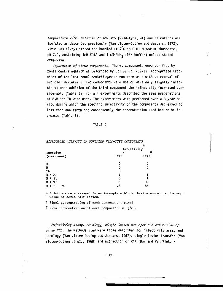
Separation of virus aomponents. The wt components were purified byzonal centrifugation as described by Bol et al. (1971). Appropriate frac-tions of the last zonal centrifugation run were used without removal ofsucrose. Mixtures of two components were not or were only slightly infec-tious; upon addition of the third component the infectivity increased con-siderably (Table I). For all experiments described the same preparationsof B,M and Tb were used. The experiments were performed over a 3 year pe-riod during which the specific infectivity of the components decreased toless than one-tenth and consequently the concentration used had to be in-creased (Table I).
TABLE I
BIOLOGICAL ACTIVITY OF PURIFIED WILD-TYPE COMPONENTS*
InfectivityInoculum t -t-(component) 1976 1979
BMTbB + MB + TbM + TbB + M + Tb
* Solutions were assayed in an incomplete block. Lesion number is the meanvalue of seven half leaves.
t Final concentration of each component 1 yg/ml.
T Final concentration of each component 12 yg/ml.
Infeativity assay* aerology, single lesion transfer and extraction ofvirus RNA. The methods used were those described for infectivity assay andserology (Van Vloten-Doting and Jaspars, 1967), single lesion transfer (VanVloten-Doting et al., 1968) and extraction of RNA (Bol and Van Vloten-
-39-
000100
78
000110
68
Doting, 1973).Preparation of virus coat protein. Protein was always prepared directly
before use by dissociation of nucleoprotein with MgCl« (final concentration5 mg/ml and 0.5 M, respectively; Kruseman et al., 1971). The RNA was removedby centrifugation and the supernatant containing the protein was diluted100-fold with distilled water.
Conservation of mutant stook. Systemically infected leaves of tobaccoplants infected with a mutant isolate, which had been passed through atleast three serial single lesion transfers, were dried over CaCl? at 4°C andstored at -20°C (Bos, 1969).
Determination of ts character. For all mutants the virus production ratiowas determined in the following way. Leaves from three to five tobaccoplants were inoculated with a homogenate of a systemically infected leaf.Directly after inoculation 10 discs (diam. 1.2 cm) were punched out. Thediscs were placed in an Erlenmeyer flask (50 ml) containing 15 ml 0.01M-Na2HP04, pH 7.0, pre-incubated in a waterbath at 30°C. From the sameleaves 10 more discs were punched out and placed in an Erlenmeyer flask withbuffer in a waterbath at 23°C. The discs were incubated at the indicatedtemperature for 4 or 5 days under continuous light (3000 lux, TL number 33).To enable a reliable estimate of the virus produced in the discs, thesewere homogenized with 2 ml PEN buffer and four serial dilutions of thehomogenate (according to the virus production expected) were assayed onbean {Phaseolus vulgavis L. var 'Berna'). Corresponding dilutions of homo-genates incubated at 30 and 23°C were always compared on half leaves of thesame plants. Only tests in which the number of lesions induced by the sampleincubated at 23°C was between 100 and 300 were taken into account. Wt AMVhad a virus production ratio of 60 to 80%; mutants suitable for furtherstudy had a virus production ratio of < 1%.
Supplementation test. Mutations were assigned to virus components usinga modified supplementation test (Bancroft and Lane, 1973; De Jager, 1976).A systemically infected leaf was homogenized in PEN buffer and divided intofour samples. To three samples an equal volume of the three purified wtcomponents (at a suitable concentration) was added separately and to thefourth sample an equal volume of PEN buffer was added. Each combination wasinoculated on to tobacco plants and the virus production ratio was deter-mined.
Complementation test. In the literature on ts mutants of animal or bac-terial viruses, complementation is usually expressed as the complementation
-40-
index (that is, yield of mixed infection divided by the sum of yields ofthe single infections at the restrictive temperature, e.g. Eckhart, 1977).Due to the variation in susceptibility of individual tobacco plants forvirus infection the complementation index would not be a reliable parameterfor the complementation of plant virus mutants. We have used the virus pro-duction ratio described above. Tobacco plants were infected with a mixtureof two nucleoprotein preparations or with a mixture of two homogenates ofleaves systemically infected with different mutants and the virus productionratio was determined. The ratio was also measured for the individual mu-tants. For these experiments, high concentrations of virus were used toenhance the possibility that cells were indeed inoculated with mixtures ofmutants.
RESULTS
Induction of mutantsPreliminary attempts to mutate AMV nucleoprotein with //-methyl-iV'-nitro-
soguanidine (Singer and Fraenkel-Conrat, 1967) failed, since the treatmentresulted in inactivation of the virus without the production of mutants.This is in contrast to the results of Hartmann et al. (1976) who were ableto obtain mutants of AMV strain S with this mutagen. Treatment with HN0? isunsuitable for AMV nucleoprotein, as at the pH required for this treatmentAMV nucleoprotein precipitates (Hull, 1969). Treatment of AMV RNA with HN02
(Gierer and Mundry, 1958) did induce some aberrant symptoms (8 out of 36)and one mutant proved to be thermosensitive. However, this method did notseem very attractive since it would require the purification of largeamounts of each of the AMV RNA species.
We had noticed that tobacco plants systemically infected with AMV andkept under continuous light from high pressure mercury lamps (Philips HLRG400 W), frequently gave yellow or green spots which were distinct from thenormal faint chlorotic symptoms; stable isolates were made from these dis-tinct symptoms and a high proportion (13 out of 25) of them were found tobe thermosensitive. Since this type of lamp is known to produce a slightleakage of u.v. light it seemed possible that the mutations may have beeninduced by u.v. light. Therefore we tried u.v. irradiation of purified nu-cleoprotein components. In Fig.l the loss of infectivity upon u.v. irradia-tion of Tb (assayed in the presence of untreated B + M) is shown. The Tb
-41-
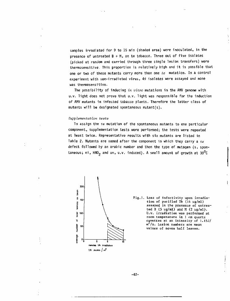
samples irradiated for 9 to 15 min (shaded area) were inoculated, in thepresence of untreated B + M, on to tobacco. Three out of five isolates(picked at random and carried through three single lesion transfers) werethermosensitive. This proportion is relatively high and it is possible thatone or two of these mutants carry more than one ts mutation. In a controlexperiment with non-irradiated virus, 49 isolates were assayed and nonewas thermosensitive.
The possibility of inducing in vitro mutations in the AMV genome withu.v. light does not prove that u.v. light was responsible for the inductionof AMV mutants in infected tobacco plants. Therefore the latter class ofmutants will be designated spontaneous mutant(s).
Supplementation testsTo assign the ts mutation of the spontaneous mutants to one particular
component, supplementation tests were performed; the tests were repeatedat least twice. Representative results with six mutants are listed inTable 2. Mutants are named after the component in which they carry a tsdefect followed by an arabic number and then the type of mutagen (s, spon-taneous; ni, HN02 and uv, u.v. induced). A small amount of growth at 30°C
Fig.l. Loss of infectivity upon irradia-tion of purified Tb (14 ug/ml)assayed in the presence of untrea-ted B (5 ug/ml) and M (2 ug/ml).U.v. irradiation was performed atroom temperature in 1 cm quartzcuvettes at an intensity of 1.I5J/m /s. Lesion numbers are meanvalues of seven half leaves.
minutes UV irradiation
1,15 Joules / n?
-42 -
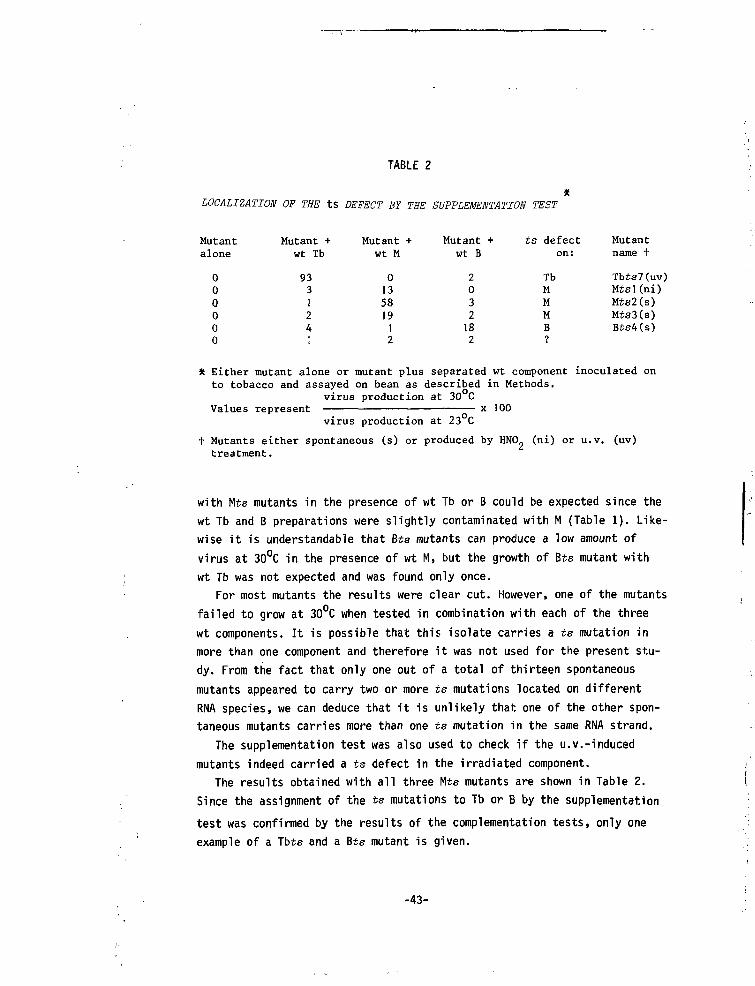
TABLE 2
LOCALIZATION OF THE tS DEFECT BY THE SUPPLEMENTATION TEST
Mutantalone
000000
Mutant +wt Tb
933124;
Mutant +wt M
013581912
Mutant +wt B
2032182
ts defecton:
TbMMMB
Mutantname +
Tbts7(uv)Mtsl(ni)Mts2(s)Mts3(s)Bis4(s)
* Either mutant alone or mutant plus separated wt component inoculated onto tobacco and assayed on bean as described in Methods,
virus production at 30 Cx 100Values represent
virus production at 23 C
t Mutants either spontaneous (s) or produced by HNO. (ni) or u.v. (uv)treatment.
with Mts mutants in the presence of wt Tb or B could be expected since thewt Tb and B preparations were slightly contaminated with M (Table 1). Like-wise it is understandable that Bts mutants can produce a low amount ofvirus at 30°C in the presence of wt M, but the growth of Bts mutant withwt Tb was not expected and was found only once.
For most mutants the results were clear cut. However, one of the mutantsfailed to grow at 30°C when tested in combination with each of the threewt components. It is possible that this isolate carries a ts mutation inmore than one component and therefore it was not used for the present stu-dy. From the fact that only one out of a total of thirteen spontaneousmutants appeared to carry two or more ts mutations located on differentRNA species, we can deduce that it is unlikely that one of the other spon-taneous mutants carries more than one ts mutation in the same RNA strand.
The supplementation test was also used to check if the u.v.-inducedmutants indeed carried a ts defect in the irradiated component.
The results obtained with all three Mts mutants are shown in Table 2.
Since the assignment of the ts mutations to Tb or B by the supplementation
test was confirmed by the results of the complementation tests, only one
example of a Tbte and a Bts mutant is given.
-43-
Stability of mutantsAll mutants except Bts l(uv) were stable in dried infected leaves stored
at -20°C. For Bts l(uv) we found that leaves stored at -20°C for more than4 years no longer contained thermosensitive virus, while material which hadbeen kept in culture was still thermosensitive. Apparently the originaldried material contained, besides the mutant, a very small amount of wt orrevertant virus, which 'survived' while the mutant was inactivated. A simi-lar observation has been made with mutant Ng from cowpea mosaic virus (C.P.De Jager, personal communication).
All mutants except Tbts 6(s) were stable during subculturing in tobacco.The virus production ratio of Jbts 6(s) was about 2% in initial experiments,but in later experiments this value increased to about 15%. Purified nucleo-protein from Tbts 6 (from one of the later experiments) showed a loss ofits thermosensitive character during storage at 4°C. These results suggestthat during subculturing this mutant became contaminated with a revertantor with wt; the contaminating virus multiplied faster and was more stableat 4°C than mutant Tbts 6(s).
Complementation testsTo gain information about the processes in which the product(s) of each
RNA species is (are) involved, we performed complementation tests withmutants carrying a ts mutation in the same RNA species.
The percentage of growth found at 30°C for the different combinationsof mutants varied from experiment to experiment. However, we found con-sistently that certain combinations of mutants were able to grow at 30°Cin contrast to others. The variability in the actual percentage of comple-mentation could be due to variations in the concentration of the mutantsin the leaf homogenatés used as inoculum. The use of purified nucleoproteinpreparations at concentrations inversely proportional to their specificinfectivity did not improve the reproducibility of the complementation test,probably because some mutant preparations lost their infectivity much fasterthan others.
Table 3(a) shows that all six Tbts mutants fall into one complementationgroup. The three Mts mutants fall into two complementation groups: group Iis formed by Mts l(ni) and group II by Mts 2(s) and Mts 3(s) (Table 3b).Bts l(uv) and Bts 4(s) belong to different complementation groups whileBts 2(s) and Bts 3(s) show no significant complementation with any of the
-44-
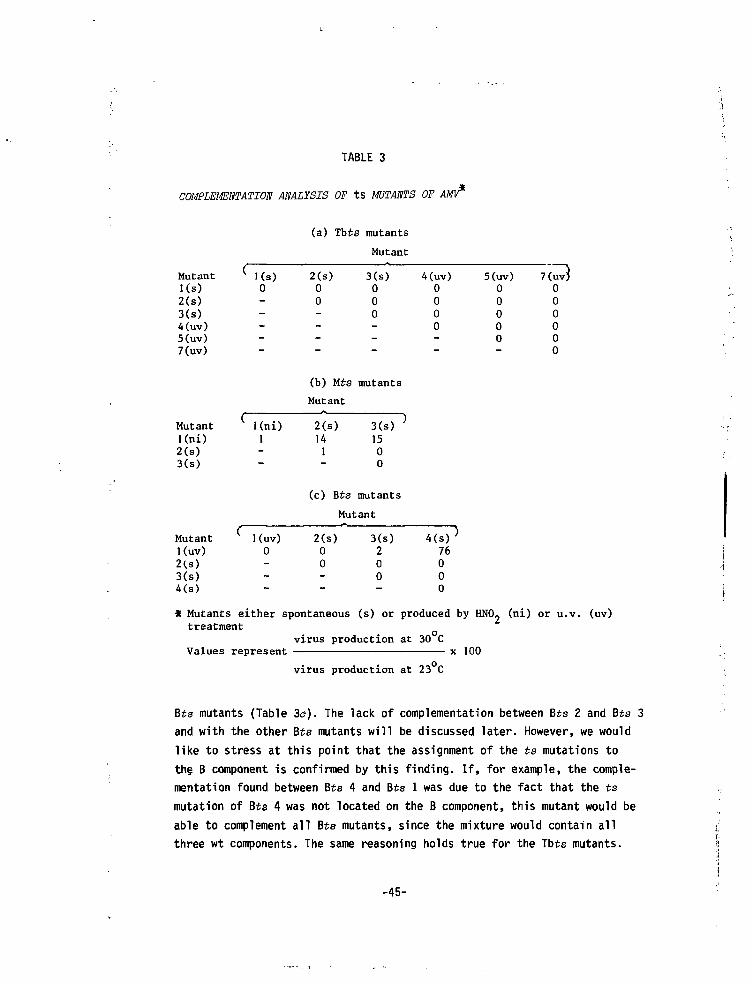
TABLE 3
COMPLEMENTATION ANALYSIS OF ts MUTANTS OF AMV*
(a) Tbts mutants
Mutant
Mutantl(s)2(s)3(8)4(uv)5(uv)7(uv)
Mutantl(ni)2(8)3(s)
Mutant
Kuv)2U)3(8)4(s)
2(s)00
3(s)000
4(uv)0000
(b) Mts mutants
Mutant
Kni) 2(s)141
3(8)1500
1
(c) Bts mutants
Mutant
Kuv)0
2(s)00
3(8)200
4(s)76000
5(uv)00000
* Mutants either spontaneous (s) or produced by HNO. (ni) or u.v. (uv)treatment
Values representvirus production at 30 C
x 100
virus production at 23 C
Bts mutants (Table 3a). The lack of complementation between Bts 2 and Bts 3and with the other Bts mutants will be discussed later. However, we wouldlike to stress at this point that the assignment of the ts mutations tothe B component is confirmed by this finding. If, for example, the comple-mentation found between Bts 4 and Bts 1 was due to the fact that the tsmutation of Bts 4 was not located on the B component, this mutant would beable to complement all Bts mutants, since the mixture would contain allthree wt components. The same reasoning holds true for the Tbts mutants.
-45-
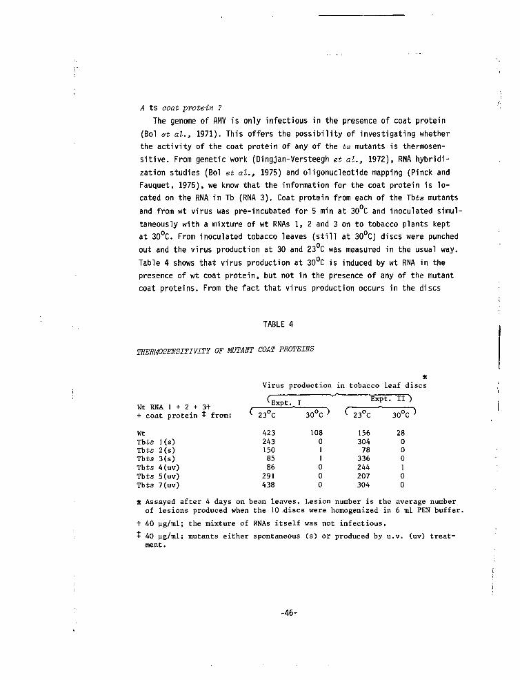
A ts aoat protein ?
The genome of AMV is only infectious in the presence of coat protein
(Bol et al., 1971). This offers the possibility of investigating whether
the activity of the coat protein of any of the ts mutants is thermosen-
sitive. From genetic work (Dingjan-Versteegh et at., 1972), RNA hybridi-
zation studies (Bol et al., 1975) and oligonucleotide mapping (Pinck and
Fauquet, 1975), we know that the information for the coat protein is lo-
cated on the RNA in Tb (RNA 3). Coat protein from each of the Tbts mutants
and from wt virus was pre-incubated for 5 min at 30 C and inoculated simul-
taneously with a mixture of wt RNAs 1, 2 and 3 on to tobacco plants kept
at 30°C. From inoculated tobacco leaves (still at 30°C) discs were punched
out and the virus production at 30 and 23°C was measured in the usual way.
Table 4 shows that virus production at 30°C is induced by wt RNA in the
presence of wt coat protein, but not in the presence of any of the mutant
coat proteins. From the fact that virus production occurs in the discs
TABLE 4
THERMOSENSITIVITÏ OF MUTANT COAT PROTEINS
xVirus production in tobacco leaf discs
I3t RNA 1 + 7 + 1+W L IViiri. 1 • & ~ J 1
+ coat protein -t- from:
WtTbts l(s)Tbts 2(s)Tbts 3(s)Tbts 4(uv)Tbts 5(uv)Tbts 7(uv)
(
( 23°C
4232431508586291438
30°C )
108011000
Expt_
( 23°C
15630478336244207304
• II )
30°C>
28000100
* Assayed after 4 days on bean leaves. Lesion number is the average numberof lesions produced when the 10 discs were homogenized in 6 ml PEN buffer.
t 40 ug/ml; the mixture of RNAs itself was not infectious.
t 40 ug/ml; mutants either spontaneous (s) or produced by u.v. (uv) treat-ment.
-46-
shifted to 23 C, we can conclude that the activity of the coat protein wasnot lost irreversibly. These results are consistent with the idea that thecoat protein from all six mutants is altered such that it cannot activatethe AMV genome at 30°C.
Tryptic peptide maps from Tbts 7(uv) revealed a small difference betweenwt AMV and the mutant (results not shown). Tbts l(s) has been shown to haveat least two amino acid substitutions (Kraal, 1975). None of the mutantscould be distinguished from wt AMV using anti serum raised against wt virus(results not shown).
Phenotype of mutantsThree of the four u.v.-induced mutants caused symptoms in both tobacco
and bean that were very similar or identical to those caused by wt AMV.Tbts 7(uv) caused chlorotic symptoms with a necrotic boundary in the in-fected leaves of tobacco; the symptoms in the systemically infected leaveswere also more necrotic than those of the wildtype (Fig.2). The symptomscaused by Tbts 7(uv) in bean were normal. It is possible that the altera-tion in the coat protein of Tbts 7 is responsible for the altered symptoms;however, it is also possible that the RNA 3 of Tbts 7 carries a second(non-)ts mutation.
As far as the spontaneous mutants and the HNO- mutant are concerned, adescription of the symptoms is probably irrelevant, because it is possiblethat they carry, besides a mutation on the indicated component, other mu-tations on one or both of the other components.
Effect of host on ts aharaeterAll thirteen mutants which displayed a ts character on tobacco were
assayed on cowpea. Unfortunately it was impossible to do the experimentsin the same way as with tobacco, because discs from cowpea leaves did notsurvive the incubation in phosphate buffer. We compared virus productionin cowpea plants at 30°C with that in plants at 23°C. Wt virus induces locallesions in cowpea leaves, both lesion size and lesion number decreasingwith increase in temperature. The amount of virus present in the locallesions is very small. Furthermore, virus production of wt at 30°C was lessthan 10$ of that at 23°C and therefore it did not seem worthwhile to com-pare the virus production of the mutants at 23°C and 30°C in this way.
Based on their ability to induce lesions on cowpea leaves none of themutants could be classified as ts on cowpea. This accords with the fact
-47-
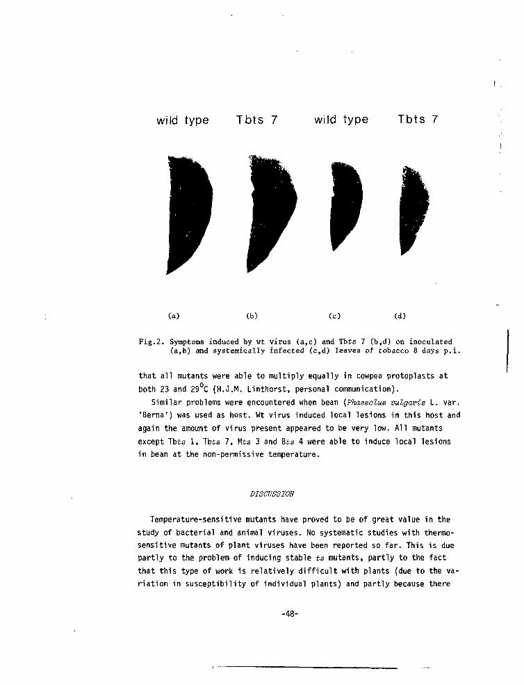
wild type Tbts 7 wild type Tbts 7
(a) (b) (c) (d)
Fig.2. Symptoms induced by wt virus (a,c) and Tbts 7 (b,d) on inoculated(a,b) and systemically infected (c,d) leaves of tobacco 8 days p.i.
that all mutants were able to multiply equally in cowpea protoplasts atboth 23 and 29°C (H.J.M. Linthorst, personal communication).
Similar problems were encountered when bean {Phaseolus vulgaris L. var.'Berna') was used as host. Wt virus induced local lesions in this host andagain the amount of virus present appeared to be very low. All mutantsexcept Tbts 1, Tbts 7, Mts 3 and Bts 4 were able to induce local lesionsin bean at the non-permissive temperature.
DISCUSSION
Temperature-sensitive mutants have proved to be of great value in thestudy of bacterial and animal viruses. No systematic studies with thermo-sensitive mutants of plant viruses have been reported so far. This is duepartly to the problem of inducing stable ts mutants, partly to the factthat this type of work is relatively difficult with plants (due to the va-riation in susceptibility of individual plants) and partly because there
-48-
is no quick selection method. For AMV the first problem seems to be solvedby the finding that u.v. irradiation yields a high percentage of stable tsmutants and the second problem is solved for tobacco plants by incubatingdiscs of the same leaves at different temperatures; the third problem isnot yet solved. In an attempt to find a suitable selection procedure for tsmutants of TMV Dawson and Jones (1976) produced only 25 mutants out of 225isolates. We found that from 25 isolates of AMV with deviating symptoms,13 were thermosensitive.
It was not expected that all six Tbts mutants would fall into one com-plementation group. Previous studies have shown that the RNA in Tb (Rna 3)contains the genetic information for the coat protein (Dingjan-Versteeghet al.t 1972) and for a 35000 protein (Mohier et al.3 1976; Van Vloten-Doting, 1976; Rutgers, 1977). As the activity of the coat protein from allsix mutants is lost at 30 C, it follows that each carries a ts mutationin the coat protein cistron. Possible explanations for the absence of mu-tants with a ts 35000 protein are that mutations in this protein are nearlyalways lethal and only very seldom ts, or that in this protein mutationscan be easily tolerated without rendering the protein ts.
The availability of mutants coding for proteins which can be switched'on and off' in their ability to activate the AMV genome may help to elu-cidate the precise role played by the coat protein during the infectioncycle. Ami no acid sequence data of the coat protein of Tbts mutants shouldyield information about the location and nature of the active site.
We found two complementation groups on M, while in vitro studies haveshown that RNA 2 (the RNA inside M) can be translated into one largeprotein corresponding to the total amount of genetic information presentin the RNA (Mohier et at., 1975; Van Tol and Van VlotGn-Doting, 1979).
The situation with B is more complicated than with M, since, besidestwo complementation groups, there are also two mutants which fail to com-plement any of the Bis mutants. This phenomenon is not due to interference(Cooper, 1977), since both mutants could be supplemented with wildtype B.As Bts 2(s) and Bis 3(s) are spontaneous mutants, it is unlikely that theyare double ts mutants. In vitro translation studies have shown that RNA 1(the RNA inside B) can be translated into one large protein (Mohier et at.,1975; Van Tol and Van Vloten-Doting, 1979).
Among the possible explanations which could reconcile the results fromthe in vitro translation studies with the genetic data are: (1) The largeprimary translation product is processed into smaller functional proteins
-49-
(Cooper, 1977). In Bis 2 and/or Bis 3 the processing is either impaired
or incorrect. Alternatively the primary product of RNA 1 is processed into
more functional units and Bis 2 and Bis 3 represent a third (or a third and
a fourth) complementation group. However, the protein(s) coded for by Bis 2
and /or Bis 3 is (are) cis-acting protein(s) (Spandidos and Graham, 1975a,
b). (2) In vivo subgenomic mRNA(s) is (are) generated from the genomic RNA
(these smaller RNA(s) may be read in the same or in a different reading
frame). (3) The protein functions as a multimer. Subunits with defects in
different regions of the sequence may be able to compensate each other,
restoring some of the activity of the multimer by intracistronic complemen-
tation (Crick and Orgel, 1964). The proteins encoded by Bis 2(s) and/or
Bis 3(s) are so distorted that no activity can be restored by association
with other subunits. This explanation is not very likely for the Bis mu-
tants since the complementation between Bis 1 and Bis 4 is efficient, while
the efficiency of this kind of intracistronic complementation is low
• (Jamieson and Subak-Sharpe, 1974; Williams et al.} 1974). (4) The protein
has two 'independent' functional domains which can be mutated separately
(Spoerel et al., 1979). The mutation of Bis 2(s) and/or Bis 3(s) is lo-
cated such that the two functional domains represented by Bis l(uv) and
Bis 4(ts) are both distored. Attempts to demonstrate that some of the
mutants induce in tobacco a replicase which is thermosensitive in vitro
have been unsuccessful (see Linthorst ei al., 1980).
It was unexpected and disappointing that none of the thirteen mutants
which were is on tobacco were ts on cowpea protoplasts assayed one degree
below the non-permissive temperature). It has been reported previously
that ts mutants of plant viruses behave differently in different hosts:
for example tobacco rattle virus (Robinson, 1973), tobacco mosaic virus
(Dawson and Jones, 1976) and cowpea chlorotic mottle virus (Bancroft et
at., 1972). The lack of ts activity on cowpea of the Tbis mutants is not
in conflict with the results obtained with tobacco (Table 4). In the latter
experiments the coat protein was pre-treated for 5 min at 30°C in the
absence of RNA. In the nucleoprotein particle the protein remains active,
probably because the RNA keeps the protein in the correct conformation.
This 'protected' protein is apparently sufficient to start the infection.
However, the virus replication will stop later since the newly synthesized
protein is inactive. The small amount of virus produced in the inoculated
cells will be below the detection level in the tobacco experiments, but it
-50-
may be sufficient to start lesion formation. The results with the Bis andMts mutants can be explained by assuming that the altered proteins are re-quired for systemic infection but not for local lesion assay. However, thisexplanation is not very attractive since the seven mutants described repre-sent at least four complementation groups and it is very unlikely that noneof these functions is required for local lesion induction. Neither is itvery probable that the mutants of these four complementation groups allcode for virus proteins involved in the transport of the virus (Nishiguchiet al.3 1978). It seems more likely that at least some of these functionsare performed by virus-coded proteins in close interaction with host-codedproteins, and that the difference between tobacco and cowpea suggests thatthis interaction (for at least some of these functions) is more sensitivein tobacco.
The present results represent the first unambiguous proof of complemen-tation between mutants of plant viruses which carry a mutation on the sameRNA. De Jager (1978, 1979) has reported that a mixture of the two mutantsN123 x N142 of cowpea mosaic virus middle component induces wild-typesymptoms, while each of the mutants alone induces another type of symptom.This has been interpreted as complementation between the two mutants. How-ever, since the basis of symptom induction is unknown it cannot be excludedthat the induction of wild-type symptoms by the mixture of the mutants isdue to some other phenomenon.
The number of ts mutants described in this study is rather small and itis quite possible that further work could reveal the presence of more com-plementation groups within the AMV genome.
We are grateful to A.G. Wesseling for cultivation of large numbers ofplants and to Ada Bax-Trimp, Dr. J.F. Bol, F.Th. Brederode, Dr. A. Castel,J.M. De Graaf, Annette Krouwels, Maaike van Hiele and D.K. Rauh for theircontribution to the experimental work. We thank B.Tank for helpful com-ments. It is a pleasure to acknowledge the stimulating discussions withDr. E.M.J. Jaspars. This work was financially supported by the NetherlandsOrganization for Advancement of Pure Research (Z.W.O.).
-51-
REFERENCES
Bancroft, J.B. and Lane, L.C. (1973). Journal of General Virology j ^ ,
381-389.
Bancroft, J.B., Rees, M.W., Dawson, J.R.O., McLean, G.D. and Short, M.N.
(1972). Journal of General Virology _1£, 69-81.
Bol, J.F. and Van Vloten-Doting, L. (1973). Virology 51,, 102-108.
Bol, J.F., Van Vloten-Doting, L. and Jaspars, E.M.J. (1971). Virology
46, 73-85.
Bol, J.F., Brederode, F.Th., Janze, G.C. and Rauh, D.K. (1975). Virology
65, 1-15.
Bos, L. (1969). Mededelingen Rijks faculteit Landbouwwetenschappen Gent
34, 875-887.
Cooper, P.D. (1977). In Comprehensive Virology £, 133-207. Edited by H.
Fraenke't-Conrat and ff.if. Wagner. New York: Plenum Press.
Crick, H.F.C. and Orgel, L.E. (1964). Journal of Moleaular Biology _8,
161-165.
Dawson, W.O. and Jones, G.E. (1976). Molecular and General Genetics 145,
307-309.
Dawson, W.O. and White, J.L. (1978). Virology 90, 209-213.
Dawson, W.O. and White, J.L. (1979). Virology 93, 104-110.
De Jager, C.P. (1976). Virology 0 , 151-163.
De Jager, C.P. (1978). Thesis, Landbouwhogeschool Wageningen.
De Jager, C.P. and Breekland, L. (1979). Virology 99, 312-318.
Dingjan-Versteegh, A., Van Vloten-Doting, L. and Jaspars, E.M.J. (1972).
Virology 49, 716-722.
Eckhart, W. (1977). In Comprehensive Virology 9_, 1-26. Edited by H. Fraen-
kel-Conrat and R.R. Wagner. New York: Plenum Press.
-52-
Gierer, A. and Mundry, K.W. (1958). Nature, London 1JS2, 1457-1458.
Habili, N. and Francki, R.I.B. (1974). Virology 61, 443-449.
Hartmann, D., Mohier, E., Leroy, C. and Hirth, L. (1976). virology 74,
470-480.
Heijtink, R.A., Houwing, C.J. and Jaspars, E.M.J. (1977). Biochemistry
.16, 4684-4693.
Hull, R. (1969). Advances in Virus Research _15, 365-433.
Jamieson, A.T. and Subak-Sharpe, Ü.H. (1974). Journal of General Virology
24, 481-492.
Kado, C.I. and Knight, C.A. (1966). Proceedings of the National Academy
of Sciences of the United States of America j>5_, 1276-1283.
Kraal, B. (1975). Virology 66, 336-340.
Kruseman, J., Kraal, B., Jaspars, E.M.J., Bol, J.F., Brederode, F.Th. and
Veldstra, H. (1971). Biochemistry iP_, 447-455.
Lane, L. (1974). Advances in Virus Research 2£, 151-220.
Linthorst, H.J.M., Bol, J.F. and Jaspars, E.M.J. (1980). Journal of Gene-
ral Virology 46, 511-515.
Marchoux, G., Marrou, J. and Quiot, J.-B. (1974). Comptes rendus hebdo-
madaires des séances de l'Académie des sciencess Série D 279, 1943-1946.
Mohier, E., Hirth, L., Le Meur, M.-A. and Gerlinger, P. (1975). Virology
68, 349-359.
Mohier, E., Hirth, L., Le Meur, M.-A. and Gerlinger, P. (1976). Virology
71, 615-618.
Nishiguchi, M., Motoyoshi, F. and Oshima, N. (1978). Jourmal of General
Virology 9 , 53-61.
Pi nek, L. and Fauquet, C. (1975). European Journal of Biochemistry 57,
441-451.
Robinson, D.J. (1973). Journal of General Virology 21, 499-506.
-53-
Robinson, D.J. (1977). Journal of General Virology 35^, 37-43.
Rutgers, A.S. (1977). Thesis, University of Leiden.
Singer, B. and Fraenkel-Conrat, H. (1967). Proceedings of the National
Academy of Sciences of the United States of America 58, 234-239.
Spandidos, D.A. and Graham, A.F. (1975a). Journal of Virology 15_, 954-963.
Spandidos, D.A. and Graham, A.F. (1975b). Journal of Virology J£, 1444-
1452.
Spoerel, N., Herrlich, P. and Bide, T. (1979). nature, London 278, 30-34.
Van Tol, R.G. L. and Van Vloten-Doting, L. (1979). European Journal of
Biochemistry 93, 461-468.
Van Vloten-Doting, L. (1976). Annales de I'Institute Pasteur 127A, 119-129.
Van Vloten-Doting, L. and Jaspars, E.M.J. (1967). Virology 3I3_, 684-693.
Van Vloten-Doting, L. and Jaspars, E.M.J. (1972). Virology 48, 699-708.
Van Vloten-Doting, L., Kruseman, J. and Jaspars, E.M.J. (1968). Virology
34, 728-737.
Williams, J.F., Young, C.S.H. and Austin, P.E. (1974). Cold Spring Harbor
Symposia on Quantitative Biology 39_, 427-437.
-54-
Accepted for publication
CHAPTER IV
COMPLEMENTATION AND INTERFERENCE OF ULTRAVIOLET-INDUCED Mts MUTANTS OF AL-FALFA MOSAIC VIRUS
Jan Roosien and Lous van Vloten-Doting.
Department of Biochemistry, State University of Leiden, P.O. Box 9505,2300 RA Leiden, The Netherlands.
SUMMARY
Ten new thermosensitive (ts) mutants of alfalfa mosaic virus (AMV), aplant virus with a tripartite genome, are described. Stable is mutantswere induced by u.v. irradiation of purified middle (M) component. Mutantswere isolated by subculturing in bean plants (instead of in the usualplant host, tobacco), in which unstable ts mutants apparently arise spon-taneously. As well as ts mutants, we found a mutant which in contrast tothe parent strain (AMV 425) was able to infect bean plants systemically.As expected, all stable mutations mapped on M component. Complementationanalysis confirmed the presence of two complementation groups on M compo-nent. This complementation is probably intracistronic. Furhermore, wefound that mutants belonging to the same complementation group often in-terfered with the multiplication of each other, even at the permissivetemperature. Taken together, these results could suggest that the productdirected by AMV RNA 2 (the RNA inside M component) has two functional do-mains and is active in a multimenc form.
-55-
INTRODUCTION
Although the size and composition of the genomes of a number of plantviruses (Matthews, 1979), and also the size of the proteins directed bythe(se) RNA(s) in a cell-free system (Davies, 1979; Atabekov and Morozov,1979; Van Vloten-Doting and Neeleman, 1982), are known our knowledge aboutthe function of the virus-coded proteins is very limited. To elucidate thedetails of the replication process of a virus, conditional lethal mutantsare of great potential value. Systematic studies with thermosensitive [ts)mutants of plant viruses are scarce (Bancroft et al., 1972; Dawson, 1978,1981; Dawson and White, 1978, 1979), mainly due to the fact that thereare no easy selection methods. Alfalfa mosaic virus (AMV) is a plant viruswith a tripartite genome; the three genomic RNAs (1, 2 and 3) are separate-ly encapsidated into bottom (B), middle (M) and top component b (Tb) res-pectively (Van Vloten-Doting et al., 1970; Bol et al., 1971). From 13 tsmutants only three mapped on M component (Van Vloten-Doting et al. , 1980)and were designated Hts. Complementation analysis indicated that thesemutants might represent two complementation groups. To obtain more proofabout the number of complementation groups on M component, we selected anew set of Mts mutants and confirmed the presence of two complementationgroups. Furthermore, interference was found between mutants belonging tothe same complementation group.
METHODS
Methods were as described previously (Van Vloten-Doting et al. , 1980)
except that some modifications were introduced as follows:
Single lesion transfer. Single lesion transfer was performed on beanplants (Phaseclus vulgaris L. var 'Berna') instead of on tobacco leaves.This has the advantage that (i) much larger numbers of isolates can besubcultured; (ii) local lesion development takes only 2 days; (iii) le-sions are more readily observed. Single lesion transfer on bean plants
-56-
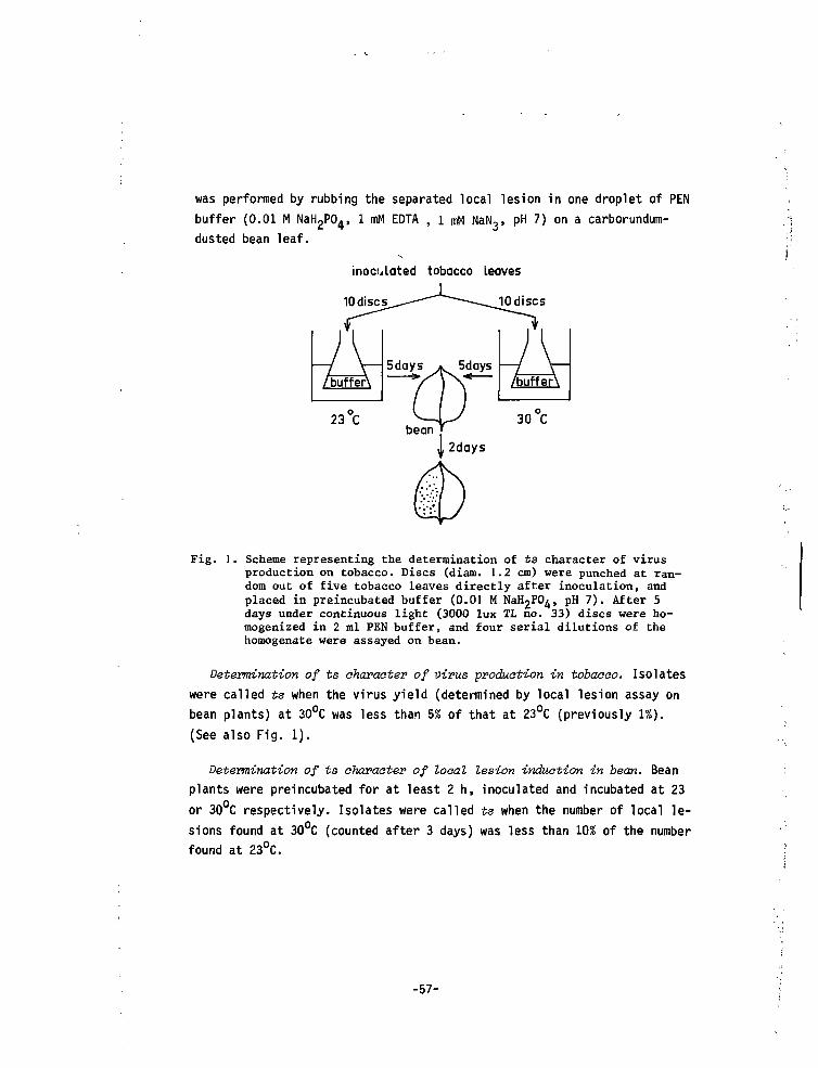
was performed by rubbing the separated local lesion in one droplet of PENbuffer (0.01 M NaH2P04> 1 mM EDTA , 1 mM NaN,, pH 7) on a carborundum-
dusted bean leaf.
inoculated tobacco Leaves
10discs
Fig. 1. Scheme representing the determination of ts character of virusproduction on tobacco. Discs (diam. 1.2 cm) were punched at ran-dom out of five tobacco leaves directly after inoculation, andplaced in preincubated buffer (0.01 M NaH2P0^, pH 7). After 5days under continuous light (3000 lux TL no. 33) discs were ho-mogenized in 2 ml PEN buffer, and four serial dilutions of thehomogenate were assayed on bean.
Determination of ts aharaater of virus production in tobaaoo. Isolateswere called ts when the virus yield (determined by local lesion assay onbean plants) at 30c
(See also Fig. 1).bean plants) at 30°C was less than 5% of that at 23°C (previously 1%).
Determination of ts character of local lesion induction in bean. Bean
plants were preincubated for at least 2 h, inoculated and incubated at 23or 30°C respectively. Isolates were called ts when the number of local le-sions found at 30°C (counted after 3 days) was less than 10% of the numberfound at 23°C.
-57-
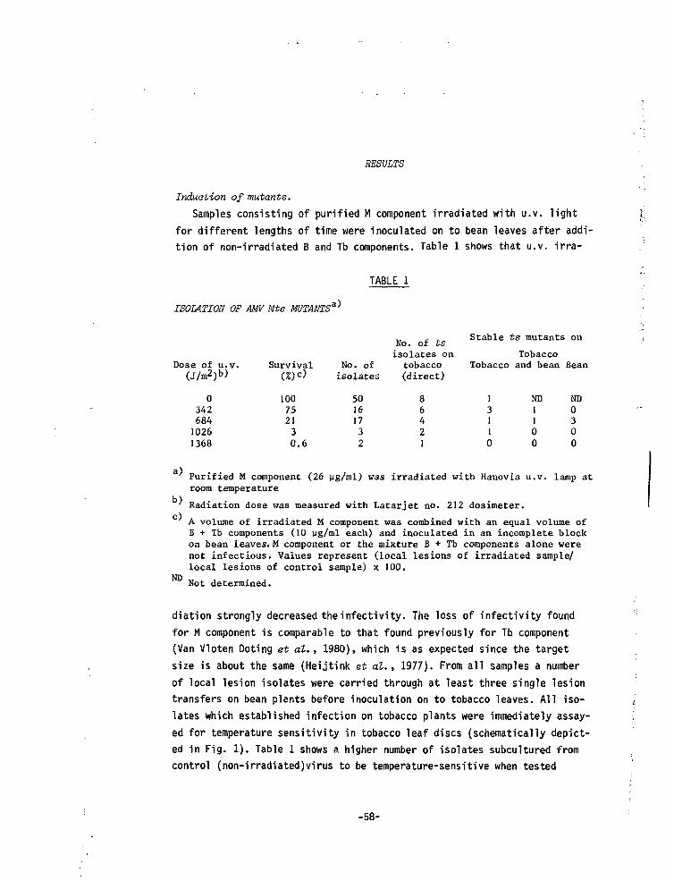
RESULTS
Induction of mutants.Samples consisting of purified M component irradiated with u.v. light
for different lengths of time were inoculated on to bean leaves after addi-tion of non-irradiated B and Tb components. Table 1 shows that u.v. irra-
ISOMTION OF MM Mts MUTANTS'a)
TABLE 1
Stable ts mutants on
Dose of u.v.(J/m2)b)
034268410261368
Survival(%)<=)
100752130.6
No. ofisolates
50161732
No. of tsisolates on
tobacco(direct)
86421
Tobacco
13110
Tobaccoand bean
ND1100
Bean
ND0300
a)
b)
c)
ND
Purified M component (26 ug/ml) was irradiated with Hanovia u.v. lamp atroom temperature
Radiation dose was measured with Latarjet no. 212 dosimeter.
A volume of irradiated M component was combined with an equal volume ofB + Tb components (10 ug/ml each) and inoculated in an incomplete blockon bean leaves. M component or the mixture B + Tb components alone werenot infectious. Values represent (local lesions of irradiated sample/local lesions of control sample) x 100.
Not determined.
diation strongly decreased theinfectivity. The loss of infectivity foundfor M component is comparable to that found previously for Tb component(Van Vloten Doting et al., 1980), which is as expected since the targetsize is about the same (Heijtink et at., 1977). From all samples a numberof local lesion isolates were carried through at least three single lesiontransfers on bean plants before inoculation on to tobacco leaves. All iso-lates which established infection on tobacco plants were immediately assay-ed for temperature sensitivity in tobacco leaf discs (schematically depict-ed in Fig. 1). Table 1 shows a higher number of isolates subcultured fromcontrol (non-irradiated)virus to be temperature-sensitive when tested
-58-
directly after transfer to tobacco. The ts character of only one isolatewas maintained upon prolonged cultivation (3 to 6 weeks with weekly trans-fers) in tobacco plants. The number of isolates with a ts character on to-bacco, measured directly after transfer from bean to tobacco, was signifi-cantly higher in the lines originating from irradiated virus than in thosefrom the control (Fisher test, P = 0.04). Upon cultivation in tobaccoleaves a number of isolates lost their ts character. Proportionally, thisnumber of unstable ts isolates is not different from that of the control,while the number of stable ts isolates is significantly higher (P = 0.01).All isolates originating from irradiated virus were also tested for ts cha-racter of local lesion induction in bean. Two out of the seven isolatesthat were ts on tobacco leaves were also ts on bean. Furthermore, we foundtwo mutants which only displayed a ts defect in local lesion induction inbean. Therefore, we now designate mutants with the first letter of the .-host on which they are ts. Differences in thermosensitivity of mutants indifferent hosts were also encountered in our previous study with AMV mu-tants (Van Vloten-Doting et at., 1980), as well as with mutants of a numberof other plant viruses (cowpea chlorotic mottle virus: Bancroft et al. ,1972; tobacco rattle virus: Robinson,1973; tobacco mosaic virus: Dawson andJones, 1976) and several animal viruses (Lee and Miller, 1979; Israël, 1980;Kowal and Stollar, 1981).
The appearance of unstable ts isolates in this experiment is probablydue to a host passage effect [virus normally maintained in tobacco -*- bean(4x) •+ tobacco], since in previous work we subcultured 49 AMV isolates ontobacco plants and none of these lines showed a ts character (Van Vloten-Doting et al., 1980).
Supplementation test.To check the localization of the ts defect a supplementation test was
performed with six of the mutants (Table 2). As expected, in all cases thets defect mapped on M component. The only stable isolate originating fromthe control virus also mapped on M conponent [Mts 13(s)t]. From this wededuced that one of the mutants originating from the irradiated virusmight be a spontaneous mutant. Supplementation tests with a mutant whichinduces deviating symptoms on bean plants (see below) showed that thismutation also mapped on M component (Fig. 2).
-59-
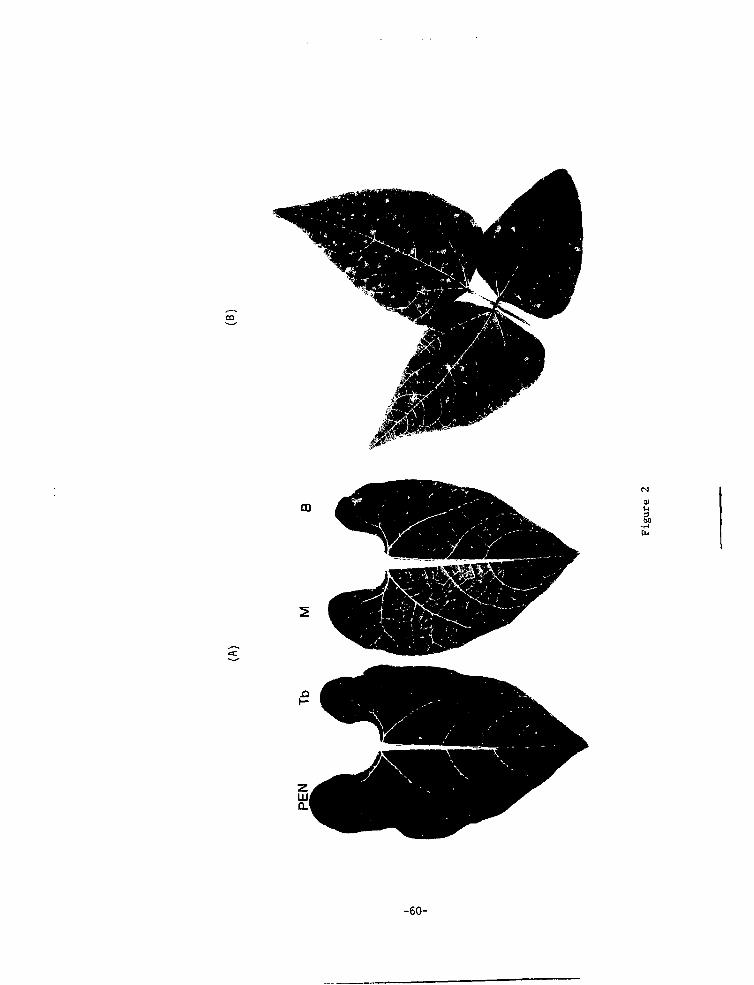
maiu3«O
-60-
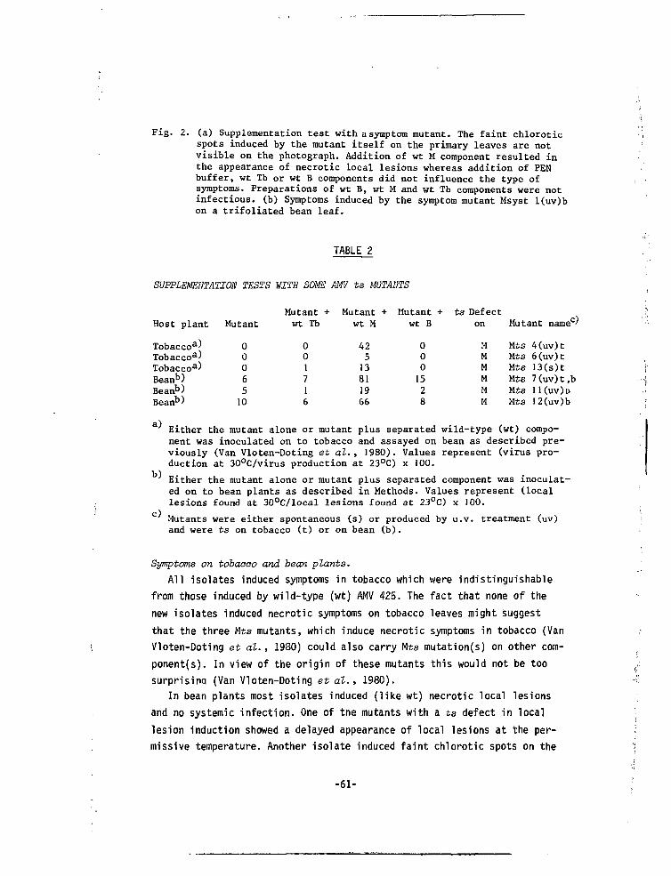
Fig. 2. (a) Supplementation test with a symptom mutant. The faint chloroticspots induced by the mutant itself on the primary leaves are notvisible on the photograph. Addition of wt M component resulted inthe appearance of necrotic local lesions whereas addition of PENbuffer, wt Tb or wt B components did not influence the type ofsymptoms. Preparations of wt B, wt M and wt Tb components were notinfectious, (b) Symptoms induced by the symptom mutant Msyst l(uv)bon a trifoliated bean leaf.
TABLE 2
SUPPLEMENTATION TESTS WITH SOME AMV ts MUTANTS
Host plant
Tobaccoa)Tobaccoa)Tobaccoa)Beanb)Beanb)Beanb)
Mutant
0006510
Mutant +wt Tb
001716
Mutant +wt M
42513811966
Mutant +wt B
0001528
ts Defecton
MMMMMM
Mutant name0'
Mts 4(uv)tMts 6(uv)tMts 13(s)tMts 7(uv)t,bMts 11(UV)D
Mts 12(uv)b
a)Either the mutant alone or mutant plus separated wild-type (wt) compo-nent was inoculated on to tobacco and assayed on bean as described pre-viously (Van Vloten-Doting et al., 1980). Values represent (virus pro-duction at 30°C/virus production at 23°C) x 100.
b)
c)
Either the mutant alone or mutant plus separated component was inoculat-ed on to bean plants as described in Methods. Values represent (locallesions found at 30°C/local lesions found at 23°C) x 100.
Mutants were either spontaneous (s) or produced by u.v. treatment (uv)and were ts on tobacco (t) or on bean (b).
Symptoms on tobacco and bean plants.All isolates induced symptoms in tobacco which were indistinguishable
from those induced by wild-type (wt) AMV 425. The fact that none of thenew isolates induced necrotic symptoms on tobacco leaves might suggestthat the three Mts mutants, which induce necrotic symptoms in tobacco (VanVloten-Doting et al. , 1980) could also carry Mts mutation(s) on other com-ponents). In view of the origin of these mutants this would not be toosurprising (Van Vloten-Doting et al. , 1980).
In bean plants most isolates induced (like wt) necrotic local lesionsand no systemic infection. One of the mutants with a ts defect in locallesion induction showed a delayed appearance of local lesions at the per-missive temperature. Another isolate induced faint chlorotic spots on the
-61-
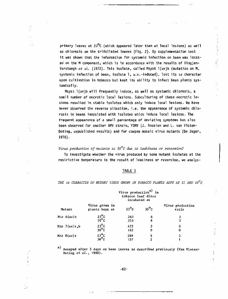
primary leaves at 23°C (which appeared later than wt local lesions) as well
as chlorosis on the trifoliated leaves (Fig. 2). By supplementation test
it was shown that the information for systemic infection on bean was locat-
ed on the M component, which is in accordance with the results of Dingjan-
Versteegh et at. (1972). This isolate, called Msyst l(uv)b (mutation on M,
systemic infection of bean, isolate 1, u.v.-induced), lost its ts character
upon cultivation in tobacco but kept its ability to infect bean plants sys-
temically.
Msyst l(uv)b will frequently induce, as well as systemic chlorosis, a
small number of necrotic local lesions. Subculturing of these necrotic le-
sions resulted in stable isolates which only induce local lesions. We have
never observed the reverse situation, i.e. the appearance of systemic chlo-
rosis in beans inoculated with isolates which induce local lesions. The
frequent appearance of a small percentage of deviating symptoms has also
been observed for another AMV strain, YSMV (J. Roosien and L. van Vloten-
Doting, unpublished results) and for cowpea mosaic virus mutants (De Jager,
1978).
Virus production of mutants at 30 C due to leakiness OP reversion?
To investigate whether the virus produced by some mutant isolates at the
restrictive temperature is the result of leakiness or reversion, we analys-
TABLE 3
THE ts CHARACTER OF MUTANT VIRUS GROWN IN TOBACCO PLANTS KEPT AT 23 AND 30°C
Virus production8 in
Mutant
Mts 6(uv)t
Mts 7(uv)t,b
Mts 8(uv)t
Virus grown inplants kept at
23°C30°C
23°C30°C
23°C3O°C
tobacco leafincubated
23°C
263255
472165
289157
discsat
30°C
68
20
52
Virus productionratio
23
00
21
Assayed after 5 days on bean leaves as described previously (Van Vloten-Doting et at. , 1980).
-62-
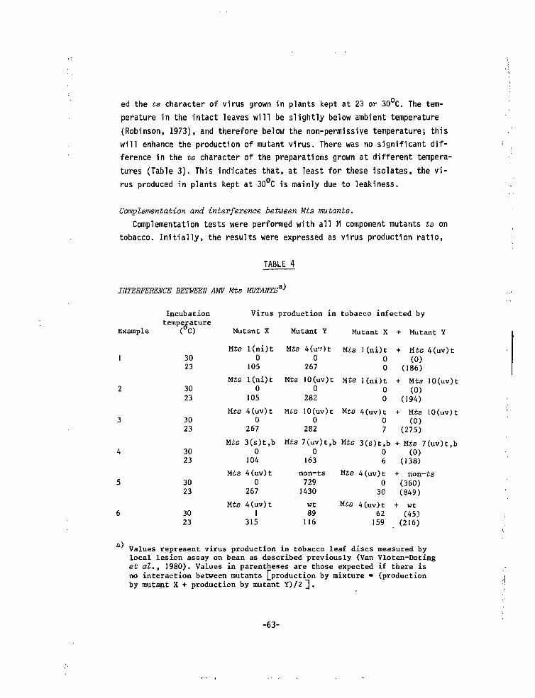
ed the ts character of virus grown in plants kept at 23 or 30 C. The tem-perature in the intact leaves will be slightly below ambient temperature(Robinson, 1973), and therefore below the non-permissive temperature; thiswill enhance the production of mutant virus. There was no significant dif-ference in the ts character of the preparations grown at different tempera-tures (Table 3). This indicates that, at least for these isolates, the vi-rus produced in plants kept at 30 C is mainly due to leakiness.
Complementation and interference between Mts mutants.Complementation tests were performed with all M component mutants ts on
tobacco. Initially, the results were expressed as virus production ratio,
TABLE 4
INTERFERENCE BETWEEN AMV Mts MUTANTS'a)
Example
Incubationtemperature
(°O
3023
3023
3023
3023
3023
3023
Virus
Mutant X
Mts l(ni)t0
105
Mts l(ni)t0
105
Mts 4(uv)t0
267
Mts 3(s)t,b0
104
Mts 4(uv)t0
267
Mts 4(uv)t1
315
production in
Mutant Y
Mts 4(ir.Ot0
267
Mts 10(uv)t0
282
Mts 10(uv)t0
282
Mts 7(uv)t,b0
163
non-ts7291430
wt89116
tobacco infected by
Mutant X + Mutant Y
Mts l(ni)t00
Mts l(ni)t00
Mts 4(uv)t07
Mts 3(s)t,b06
Mts 4(uv)t030
Mts 4(uv)t62159
+ Mts 4(uv)t(0)
(186)
+ Mts IO(uv)t(0)
(194) ;
+ Mts 10(uv)t(0)
(275)
+ Mts 7(uv)t,b(0)
(138)
+ non-ts(360)(849)
+ wt(45)
(216)
Values represent virus production in tobacco leaf discs measured bylocal lesion assay on bean as described previously (Van Vloten-Dotinget al., 1980). Values in parentheses are those expected if there isno interaction between mutants [production by mixture = (productionby mutant X + production by mutant Y)/2 ~\.
-63-
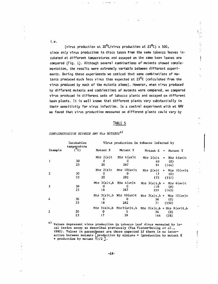
i.e.
(virus production at 30°C/virus production at 23°C) x 100,
since only virus production in discs taken from the same tobacco leaves in-cubated at different temperatures and assayed on the same bean leaves arecompared (Fig. 1). Although several combinations of mutants showed comple-mentation, the results were extremely variable between different experi-ments. During these experiments we noticed that some combinations of mu-tants produced much less virus than expected at 23°C (calculated from thevirus produced by each of the mutants alone). However, when virus producedby different mutants and combinations of mutants were compared, we comparedvirus produced in different sets of tobacco plants and assayed on differentbean plants. It is well known that different plants vary substantially intheir sensitivity for virus infection. In a control experiment with wt AMVwe found that virus production measured on different plants could vary by
TABLE 5
COMPLEMENTATION BETWEEN AMV Mts MUTANTS^
Example
Incubationtemperature
°
3023
3023
3023
3023
3023
Virus production in tobacco infected by
Mutant X Mutant Y Mutant X + Mutant Y
Mts 2(s)t020
Mts 2(s)t020
Mts 3(s)t,b018
Mts 3(s)t,b018
Mts 3(s)t,b017
Mts 4(uv)t0
267
Mts 10(uv)t0
232
Mts 4(uv)t0
267
Mts 10(uv)t0
282
Mts9(uv)t,b039
Mts 2(s)t4091
Mts 2(s)t17
173
+ Mts 4(uv)t(0)
(144)
+ Mts 10(uv)t(0)
(151)
Mts 3(s)t,b +110
Mts 4(uv)t(0)
231 (143)
Mts 3(s)t,b5671
Mts 10(uv)t(0)
(150)
Mts 3(s)t,b + Mts 9(uv)t,b36 (0)144 (28)
a) Values represent virus production in tobacco leaf discs measured by lo-cal lesion assay as described previously (Van Vloten-Doting et al.,1980). Values in parentheses are those expected if there is no inter-action between mutants [production by mixture = (production by mutant X+ production by mutant Y)/2 ] .
-64-
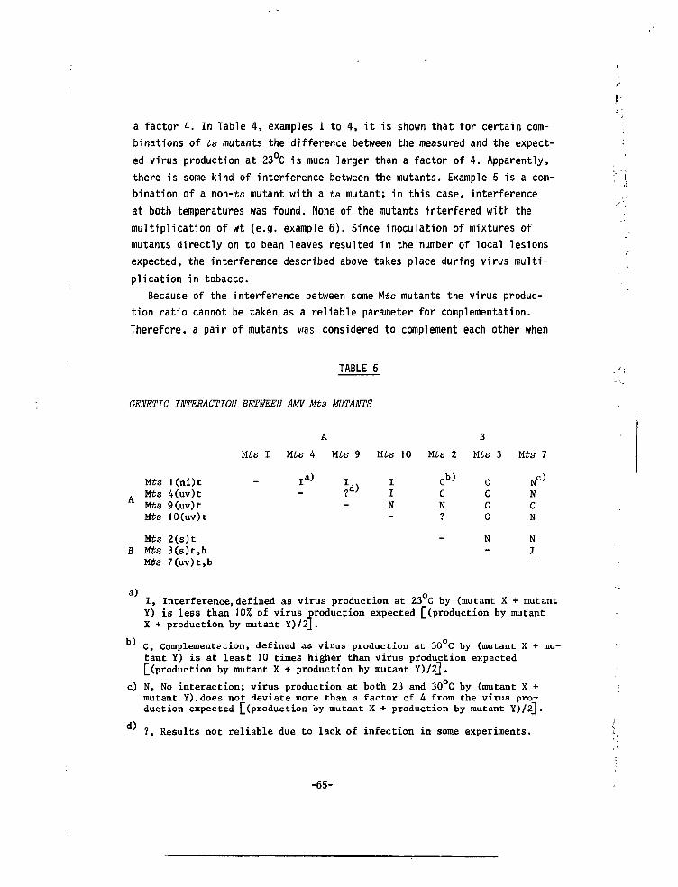
a factor 4. In Table 4, examples 1 to 4, it is shown that for certain com-
binations of ts mutants the difference between the measured and the expect-
ed virus production at 23°C is much larger than a factor of 4. Apparently,
there is some kind of interference between the mutants. Example 5 is a com-
bination of a non-ts mutant with a ts mutant; in this case, interference
at both temperatures was found. None of the mutants interfered with the
multiplication of wt (e.g. example 6). Since inoculation of mixtures of
mutants directly on to bean leaves resulted in the number of local lesions
expected, the interference described above takes place during virus multi-
plication in tobacco.
Because of the interference between some Mts mutants the virus produc-
tion ratio cannot be taken as a reliable parameter for complementation.
Therefore, a pair of mutants was considered to complement each other when
TABLE 6
GENETIC INTERACTION BETWEEN AM Mts MUTANTS
Mts
A M É SA Mts
Mts
MtsB Mts
Mts
l(ni)t4(uv)t9(uv)t10(uv)t
2(s)t3(s)t,b7(uv)t,b
Mts I
A
Mts 4 Mts 9 Mts 10
N
Mts 2
cb>cN
—
B
Mts 3
Cccc
N
Mts
NCN
NI
a) o
I, Interference,defined as virus production at 23 C by (mutant X + mutantY) is less than 10% of virus production expected L(Pr°duction by mutantX + production by mutant Y)/2J.
C, Complementation, defined as virus production at 30 C by (mutant X + mu-tant Y) is at least 10 times higher than virus production expected£ (production by mutant X + production by mutant Y)/2].
c) N, No interaction; virus production at both 23 and 30°C by (mutant X +mutant Y). does not deviate more than a factor of 4 from the virus pro-duction expected [^(production by mutant X + production by mutant Y)/2] .
?, Results not reliable due to lack of infection in some experiments.
-65-
the virus produced at 30°C by the mixture was at least 10 times higher thanexpected (the sum of the virus produced by the two mutants alone divided by2). Since all values exceeding four times (variation found in control expe-riments) the expected value could indicate complementation, to be on thesafe side we used a factor of 10. In Table 5 a number of examples of com-plementation between Mts mutants are listed. We have never observed a sig-nificantly higher virus production at 30°C by a pair of mutants which show-ed interference at 23°C.
Table 6 summarizes the results of several complementation experimentswith Mts mutants. The mutants Mts 5(uv)t, Mts 6(uv)t and Mts 8(uv)t are notincluded, since they were used in only a few complementation experiments.The mutants listed in Table 6 fall into two groups: (A) 1, 4, 9 and 10; (B)2, 3 and 7. Combination of mutants belonging to different groups often gives(not always: note the lack of interaction in combinations containing Mts 7)complementation, while combination of mutants belonging to the same groupoften (but not always) leads to interference.
DISCUSSION
AMV seems to be rather sensitive to host shifts since, for virus grown intobacco, four passages in bean resulted in the appearance of a high percen-tage of ts isolates. Upon cultivation of these isolates in the original host(tobacco) most of the isolates lost their ts character again. For a numberof plant viruses reversible changes in several properties have been observedupon passages in different hosts (see Yarwood, 1979). During virus multipli-cation there is always a low level of spontaneous mutation (Fulton, 1952; deJager, 1978; Donis-Keller et at. , 1981). In the original host most of themutants will have a disadvantage compared to wild-type; however, during pas-sages in another host, mutants better adapted to this host may be selected.The fact that the host passage effect is reversible might indicate that themutations involved in this phenomenon are located at 'hot spots'. The ap-pearance of unstable ts mutants upon passage of AMV through bean plantsnullifies the advantages of bean plants in single lesion transfers. In fu-ture experiments changes in host will be avoided.
In the present study, we found strong interference between several Mts
mutants. The fact that multiplication of both mutants is inhibited at the
-66-
permissive temperature suggests that interference takes place during the re-plication of virus in the primary infected cell and is not due to exclusionof one mutant by the other during infection of the neighbouring cells; Hulland Plaskitt (1970) showed that two AMV strains can multiply simultaneouslyin the same cell. There is a substantial literature about interference.Most reports describe the interference observed when two virus preparationsare inoculated at different times, which with plant viruses is often termedcross-protection (Matthews, 1970). There are very few reports about inter-ference between viruses or mutants of viruses when inoculated simultaneous-ly (Siegel, 1959; Wu and Rappaport, 1961; Schaffer et al., 1978; Sugita,1981). In contrast to the results obtained with AMV, interference betweenwt and ts mutants at the permissive or non-permissive temperature has beenreported for several animal viruses (Preble and Youngner, 1973; Stollar etal., 1974; Chakraborty et al., 1979). The level of interference possiblydepends upon the specific conditions of the test system. Another factor in-fluencing the level of interference is the m.o.i. (Wu and Rappaport, 1961;Ledinko, 1963; Sugita, 1981). In the case of a multicomponent virus probab-ly only the concentration of the component responsible for interference isof interest. Indeally, the concentration of biologically active M componentshould be constant in all experiments; unfortunately, this is technicallyimpossible.
Due to interference between mutants the virus production ratio used pre-viously is not always a reliable parameter. Fortunately, the three Mts mu-tants studied earlier (Van Vloten-Doting et aJ.,1980) showed no interferen-ce, so that the earlier conclusion that Mts l(ni)t belongs to another com-plementation group than Mts 2(uv)t and Mts 3(uv)t,b is still valid. More-over, this is confirmed in the present study.
Since a larger number of Mts mutants was investigated, a more detailedpicture of the product(s) directed by RNA 2 emerges. The simplest explana-tion for the results from Table 6 would be that RNA 2 codes for a proteinwhich is active in a multimeric form and which has two functional domains.This idea is based on the following, (i) It functions as a multimer becausecombinations of mutants within each group often interfere. In heteropoly-mers of subunits with different mutations in the same domain, the interac-tion between these subunits may lead to a decrease in the activity of thepolymer, even at the permissive temperature, A comparable situation was en-countered, for example, in the DNA polymerase cistron of herpes virus
-67-
(He less, 1981). (ii) It is one protein with two functional domains, sincenot all combinations of mutants belonging to different complementationgroups will complement each other, as would be expected if the primarytranslation product was processed into two functional units. Thus, the com-plementation with the Mts mutants is intracistronie. Intracistronic comple-mentation is, for example, found for influenza virus (Heller and Scholtis-sek, 1980; Thierry and Spring, 1981; Thierry et dl. , 1980). Two other ob-servations might support this proposal, (i) The mol. wt. of the in vitrotranslation product of AMV RNA 2 estimated by gel filtration under nonde-naturing conditions, is much higher than that of the monomer (Van Tol,1981). (ii) For brome mosaic virus, a plant virus belonging to the samefamily as AMV (Van Vloten-Doting et al., 1981), there is no evidence forpost-translational processing, since in BMV-infected protoplasts proteinswith the same mol. wt. as in the in vitro translation products were detect-ed (Okuna and Furusawa, 1979; Sakai et at. , 1979; Kiberstis et al. , 1981).The location of the mutation within a domain is very important for the in-teraction between subunits of a multimeric protein. It is conceivable thatthe frequent interference found in combinations containing Mts 1 (Table 6)might be due to the fact that the Mts 1 mutation (also at the permissivetemperature) severely disturbs the protein-protein interactions. The lackof such interactions in combinations containing Mts 7 might indicate thatthis mutation affects the structure of both domains at the non-permissivetemperature, whereby only the interaction with Mts 9 protein results in apartial restoration of function. Analysis of protoplasts infected with mu-tants of the two complementation groups might reveal more details about therole of the RNA 2 product in the virus replication cycle.
ACKNOWLEDGEMENTS
We gratefully acknowledge A.G. Wesseling for cultivating large numbersof plants, and the technical assistance of P. van Klaveren and D. Koot inpart of the experiments. We are grateful to Dr. E.M.J. Jaspars for his en-couraging discussions. This work was financially supported by The Nether-lands Organization for Advancement of Pure Research (Z.W.O.).
-68-
REFERENCES
Atabekov, J.G. and Morozov, S.Y. (1979). Advances in Virus Research j?5, 1-
91.
Bancroft, J.B., Rees, M.W., Dawson, J.R.O., McLean, G.D. and Short, M.N.
(1972). Journal of General Virology }£, 69-81.
Bol, J.F., Van Vloten-Doting, L. and Jaspars, E.M.J. (1971). Virology 46,
73-85.
Chakraborty, R.A., Ahmed, R. and Fields, B.N. (1979). Virology 94, 119-
127.
Davies, J.W. (1979). In Nuclei a Acids in Plants, vol. 2, pp. 111-148.
Edited by J. W. Davies and T.C. Hall, Florida: CRC Press.
Dawson, W.O. (1978). Virology 90, 112-118.
Dawson, W.O. (1981). Virology _115, 130-136.
Dawson, W.O. and Jones, G.E. (1976). Molecular and General Genetics 145,
307-309.
Dawson, W.O. and White, J.L. (1978). Virology 90, 209-213.
Dawson, W.O. and White, J.L. (1979). Virology 93, 104-110.
De Jager, C.P. (1978). Genetische analyse van cowpea-mozaiek virusmutanten.
Ph.D. thesis, Landbouwhogeschool Wageningen.
Dingjan-Versteegh, A., Van Vloten-Doting, L. and Jaspars, E.M.J. (1972).
Virology 49, 716-722.
Donis-Keller, H., Browning, K.S. and Clark, J.M., JR (1981). Virology 110,
43-54.
Fulton, R.W. (1952). Phytopathology 42, 156-158.
Heijtink, R.A., Houwing, C.J. and Jaspars, E.M.J. (1977). Biochemistry 16,
4684-4693.
Heller, E. and Scholtissek, C. (1980). Journal of General Virology 49, 133-
139.
Honess, R.W. (1981). Journal of General Virology 57, 297-306.
Hull, R. and Plaskitt, A. (1970). Virology £2, 773-776.
-69-
Israël, A. (1980). Virology _105, 1-12.
Kiberstis, P.A., Loesch-Fries, L.S. and Hall, T.C. (1981). Virology 112,
804-808.
Kowal, K.J. and Stollar, V. (1981). Virology UA, 140-148.
Ledinko, N. (1963). Virology 20, 29-44.
Lee, H.H. and Miller, L.K. (1979). Journal of Virology 31., 240-252.
Matthews, R.E.F. (1970). In Plant Virology, pp. 379-418. Edited by R.E.F.
Matthews, New York, Academic Press.
Matthews, R.E.F. (1979). Intervirology _12, 129-296.
Okuno, T. and Furusawa, I. (1979). Virology 99, 218-225.
Preble, O.T. and Youngner, J.S. (1973). Journal of Virology L2, 472-480.
Robinson, D.J. (1973). Journal of General Virology £1, 499-506.
Sakai, F., Dawson, J.R.O. and Watts, J.W. (1979). Journal of General Vi-
rology 42, 323-328.
Schaffner, P.A., Carter, V.C. and Timbury, M.C. (1978). Journal of Virology
2J_, 490-504.
Siegel, A. (1959). Virology 8, 470-477.
Stollar, V., Peleg, J. and Shenk, T.E. &1974). Intervirology £, 337-344.
Sugita, K. (1981). Journal of General Virology 55, 95-107.
Thierry, F. and Spring, S.B. (1981). Virology U5, 137-148.
Thierry, F., Spring, S.B. and Chanock, R.M. (1980). Virology JXU, 484-492.
Van Tol, R.G.L. (1981). Expression of the alfalfa mosaic virus genome.
Ph.D. thesis. Rijksuniversiteit Leiden.
Van Vloten-Doting, L. and Neeleman L. (1982). In Encyclopedia of Plant
Physiology, vol. 16. Edited by A Boulter and B. Parthier. New York and
Berlin : Springer-Verlag (in press).
Van Vloten-Doting, L., Dingjan-Versteegh, A. and Jaspars, E.M.J. (1970).
Virology 40, 419-430.
Van Vloten-Doting, L., Hasrat, J.A., Oosterwijk, E. ,Van 't Sant, P.,
Schoen, J.A. and Roosien, J. (1980). Journal of General Virology 46,
415-426.
-70-
Van Vioten-Doting, L., Francki, R.I.B.,Fulton, R.W., Kaper, J.M. and Lane,
L.C. (1981). Intervirology 15, 198-203.
Wu, J. and Rappaport, I. (1961). Virology 1±, 259-263.
Yarwood, C.E. (1979). Advances in Virus Research ^5_, 169-187.
-71-
Submitted fo r pub l ica t ion
CHAPTER V
FLOW CYTOMETRIC ANALYSIS OF TOBACCO AND COWPEA PROTOPLASTS INFECTED INVIVO AND IN VITRO WITH ALFALFA MOSAIC VIRUS
1 2 1Pieter van Klaveren , Jan Slats , Jan Roosien and Lous van Vloten-Doting.Department of Biochemistry, State University of Leiden, Wassenaarseweg64, 2333 AL Leiden, The Netherlands.
o
Department of Histochemistry and Cytochemistry, State University of Lei-den, Wassenaarseweg 72, 2333 AL Leiden, The Netherlands.
SUMMARY
Immunofluorescence flow cytometry was used to study the distribution ofviral antigen in protoplasts populations. Protoplasts were isolated fromhealthy and alfalfa mosaic virus (AMV) infected tobacco leaves (designatedin vivo infected). Furthermore isolated tobacco and cowpea protoplasts
were in vitro infected with AMV. Comparable immunofluorescence patterns4
(per sample 10 protoplasts were measured) were found for all infectedsamples. Infectious virus could be detected in in vivo infected tobaccoprotoplasts and in in vitro infected cowpea protoplasts, but not in invitro infected tobacco protoplasts.
INTRODUCTION
Immunofluorescence microscopy is often used to determine the percentageof cells containing viral antigen. This method does not provide data aboutthe amount of f1uorescence present in each protoplast, furthermore it israther time consuming. Flow cytometry offers the possibility to analysewithin a few minutes a large number of cells in a quantitative way (Melamedand Mullaney, 1979, and references therein). In this paper it is shownthat this method can also be applied to plant cell protoplasts.
We wanted to use this method for the analysis of tobacco protoplastsinfected in vitro with thermosensitive (ts) mutants of alfalfa mosaic vi-rus (AMV) (Van Vloten-Doting et al. , 1980; Roosien and Van Vloten-Doting,1982). These mutants produced in tobacco leaf discs kept at the non-per-missive temperature much less virus than in discs kept at the permissivetemperature. In the tobacco leaf disc system it is impossible to make adistinction between mutants which are conditionally defective in a proteininvolved in the virus replication cycle, or in a function associated withtransport through the host. The latter type of mutants will not show a tscharacter in isolated protoplasts (Nishiguchi et al., 1978; Leonard andZaitlin, 1982).
In our initial experiments concerned with the infection of tobacco pro-toplasts with wild-type AMV, we found a discrepancy between the detectionof viral antigen and infectivity. Such a discrepancy was not found in to-bacco protoplasts isolated from infected leaves nor in cowpea protoplastsinfected in vitro with AMV.
MATERIALS AND METHODS
Virus and plants: AMV strain 425 was grown on Nicotiana Tabaaim L. var."Samsun NN" and isolated according to Van Vloten-Doting and Jaspars (1972).Virus was frozen and kept at -20°C, aliquots were only thawed once.
Isolation of protoplasts. Tobacco protoplasts were isolated from 8 - 1 2cm long leaves of 6 - 8 weeks old Nicotiana Tabaeum L. var. "Samsun NN"plants. After removal of the upper (for practical conveniance) epidermisby rubbing carefully with a fingertip dipped in an abrasive (carborundum500 mesh). The leaves were floated upside down on a solution of 1% w/vcellulase (Onozuka R-10), 0.05% w/v Macerozym (R-10) in 0.7 M Mannitol pH5.5 till only the lower epidermis remained (2| - 3 hr). The released pro-toplasts were collected by centrifugation (5'; 50 x g) and washed two timeswith 0.7 M Mannitol to remove cell debris. Cowpea protoplasts were isolatedaccording to Alblas and Bol (1977).
Infeotion and incubation of protoplasts. Prior to infection protoplastswere incubated overnight in the dark (in analogy with plants, which are tobe infected) at 4°C (to ensure 100% survival) in 0.5% w/v cellulase, 0.01M CaCl2 in 0.7 M Mannitol pH 5.5 (to prevent renewed cell wall formation).The infection procedure consisted of a combination of several methods
-74-
(Motoyoshi et al., 1975; Cassels and Barlass 1978; Maule et al., 1980).Protoplasts received an osmotic shock (0.6 - 0.8 M Mannitol) followed by apreincubation for 10 min. in 0.7 M Mannitol containing 1 ug/ml Poly-L-orni-thin (PLO) and 0.05 M KH2P04 pH 5.2 at 5 x 10
5 protoplasts per ml. At thesame time virus (25 pg/ml) was preincubated at 0°C in 0.7 M Mannitol con-taining 1 yg/ml PLO. Both solutions were shaken with 80 excursions/min.after this period protoplasts were collected by centrifugation and resus-pended in the virus containing solution (concentration of protoplasts about107 per ml). After 10 min. at 0°C poly-ethylene-glycol (PEG) MW 6000 andCaCl- were added to a final concentration of 32% w/v and 2.4 mM respective-ly. After 10 - 15 sec. the solution was diluted 10 times with 0.7 M Manni-tol; 0.05 M KH2P04 pH 5.2 and incubated for 45 min. at 25°C in the dark.Finally protoplasts were washed 3 times with 0.7 M Mannitol, 0.001 M CaCl2
followed by incubation in the same medium with 200 ug/ml chloramphenicol(to suppress bacterial growth) and 0.1 M Tris-HCl pH 7.0 (to provide buf-fering capacity), at 23°C, 3000 lux (TL no 33). During the entire procedureviability was monitored using phenosaffranine staining (Widholm, 1972).Typically the percentage survival was greater than 65%. Infection and in-cubation of cowpea protoplasts was done according to Alblas and Bol (1977).
Immune precipitation and slab-gel analyses of labeled AMV aoat protein.Protoplasts were collected by centrifugation and homogenized by freezingand thawing under vigorously shaking with IB (50 mM NaH2P04; 50 mM NaCl;1% v/v Triton X-100; 0.5% w/v Sodium desoxycholate; 0.2% w/v Sodium dode-cylsulfaat pH 7.3). Immunoprecipitation was performed by adding 50 ug non-labeled AMV coat protein as carrier and 50 ul of a suitable antiserum,followed by an one hour incubation at 37°C. The precipitate was sedimentedby centrifugation (15'; 10,000 x g) and washed three times with IB. Theresulting pellet was subjected to slab-gel analyses on a 11% polyacrylamidegel, according to Laemmli (1970). After electrophoresis, the gel was dryedand fluorography was carried out according to Bonner and Laskey (1974).
Preparation of FITC conjugated antiserum. The antiserum used, was raisedagainst AMV in rabbits. Coupling with FITC (fluoresceinisothiocyanate) wasperformed as in Motoyoshi et al. (1973). The serum had a titer of 1/64 (asdetermined in a double diffusion assay; Ouchterlony, 1962). The conjugatedserum had a molar fluorescein/protein ratio of 2.3, calculated from opticaldensities at 425 and 280 nm. All experiments have been performed using thesame conjugated antiserum. The FITC conjugated serum was presaturated with
-75-
with an aceton powder of healthy tobacco leaves (Motoyoshi et al. , 1973).Presaturation with aceton powder of tobacco protoplasts did not improve spe-cificity.
Fixation of protoplasts. Protoplasts were fixed in 4% formaldehyde in0.7 M Mannitol (for tobacco) or 0.5 M Mannitol (for cowpea protoplasts).After 10 - 15 min. protoplasts were collected by centrifugation and washed3 times with PBS (0.01 M NaH2P04; 0.85% w/v NaCl pH 7.0) and kept at 4°C.
Immunofluoresaenae and floweytometrie analyses. Within 24 hr beforeanalysis, formaldehyde fixed protoplasts were collected by centrifugation(5'; 50 x g) and a drop of the undiluted FITC coupled antiserum was laid onthe pellet. After incubation for 3 hr at 37°C or overnight at 4°C in thedark, the protoplasts were washed three times with PBS. If necessary, pro-toplasts were pretreated in the same way with a preimmune serum. Finallyprotoplasts were suspended in PBS containing 0.1% w/v Tween 80. Tween 80does not interfere with the assay (results not shown). Prior to measurement,the suspension was filtered through a 50 p filter, to remove aggregatedmaterial.
Quantitation of immunofiuorescence was performed using a Fluorescence-Activated Cell Sorter (FACS IV) manufactured by Becton-Dickinson (for back-ground information see Herzenberg et at., 1976). Protoplasts were irradiat-ed with 488 nm blue laser light. Red autofluorescence was detected by onephotomultiplier using a long-pass 645 nm filter, green fluorescence was ana-lysed with a 525 nm band-pass filter. Based on red autofluorescence ten-thousand protoplasts were counted, and from each individual protoplast thegreen fluorescence was recorded. Values, except those presented in figure1A, are corrected for background fluorescence by substracting the valuefound for part of the sample on which no immunofluorescence staining wasperformed. Data are presented in histograms. Within figures, the relativefluorescence is comparable.
Infeotivity assay. Protoplast pellets were homogenized with PEN buffer(0.01 M NaH2P04> 1 mM EDTA, 1 mM NaNg pH 7.0). Homogenates were assayed onbean {Phaseolus vulgaris L. var. "Berna" (Van Vloten-Doting and Jaspars,1967).
-76-
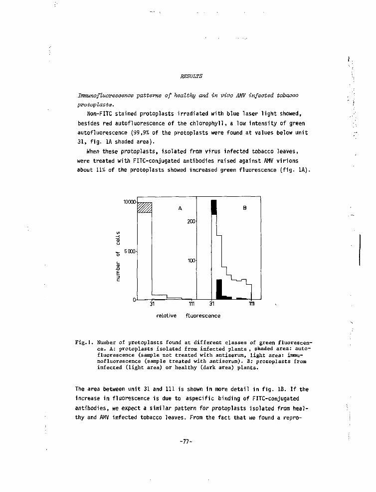
r.
RESULTS
Immunofluoresaenae -patterns of healthy and in vivo AMV infeoted tobaaooprotoplasts.
Non-FITC stained protoplasts irradiated with blue laser light showed,besides red autofluorescence of the chlorophyll, a low intensity of greenautofluorescence (99,9% of the protoplasts were found at values below unit31, fig. 1A shaded area).
When these protoplasts, isolated from virus infected tobacco leaves,were treated with FITC-conjugated antibodies raised against AMV virionsabout 11% of the protoplasts showed increased green fluorescence (fig. 1A).
10000-
5000-
n-
M A
200
100-
1 B
\
tl31 in 31
relative fluorescencem
Fig. I. Number of protoplasts found at different classes of green fluorescen-ce. A: protoplasts isolated from infected plants , shaded area: auto-fluorescence (sample not treated with antiserum, light area: immu-nofluorescence (sample treated with antiserum). B: protoplasts frominfected (light area) or healthy (dark area) plants.
The area between unit 31 and 111 is shown in more detail in fig. IB. If theincrease in fluorescence is due to aspecific binding of FITC-conjugatedantibodies, we expect a similar pattern for protoplasts isolated from heal-thy and AMV infected tobacco leaves. From the fact that we found a repro-
-77-
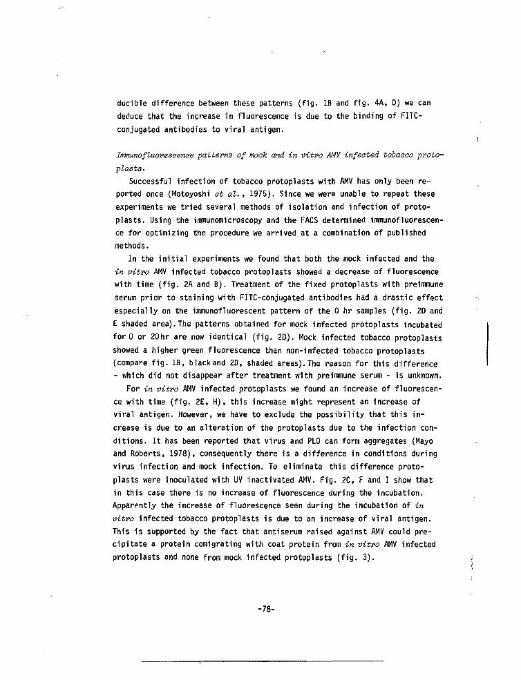
ducible difference between these patterns (fig. IB and fig. 4A, D) we candeduce that the increase in fluorescence is due to the binding of FITC-conjugated antibodies to viral antigen.
Immunofluoresoenoe patterns of moak and in vitro AMV infected tobaooo proto-
plasts.Successful infection of tobacco protoplasts with AMV has only been re-
ported once (Motoyoshi et at., 1975). Since we were unable to repeat theseexperiments we tried several methods of isolation and infection of proto-plasts. Using the immunomicroscopy and the FACS determined immunofluorescen-ce for optimizing the procedure we arrived at a combination of publishedmethods.
In the initial experiments we found that both the mock infected and thein vitro AMV infected tobacco protoplasts showed a decrease of fluorescencewith time (fig. 2A and B). Treatment of the fixed protoplasts with preimmuneserum prior to staining with FITC-conjugated antibodies had a drastic effectespecially on the immunofluorescent pattern of the 0 hr samples (fig. 2D andE shaded area). The patterns obtained for mock infected protoplasts incubatedfor 0 or 20hr are now identical (fig. 2D). Mock infected tobacco protoplastsshowed a higher green fluorescence than non-infected tobacco protoplasts(compare fig. IB, blackand 2D, shaded areas). The reason for this difference- which did not disappear after treatment with preimmune serum - is unknown.
For in vitro AMV infected protoplasts we found an increase of fluorescen-ce with time (fig. 2E, H), this increase might represent an increase ofviral antigen. However, we have to exclude the possibility that this in-crease is due to an alteration of the protoplasts due to the infection con-ditions. It has been reported that virus and PLO can form aggregates (Mayoand Roberts, 1978), consequently there is a difference in conditions duringvirus infection and mock infection. To eliminate this difference proto-plasts were inoculated with UV inactivated AMV. Fig. 2C, F and I show thatin this case there is no increase of fluorescence during the incubation.Apparently the increase of fluorescence seen during the incubation of invitro infected tobacco protoplasts is due to an increase of viral antigen.This is supported by the fact that antiserum raised against AMV could pre-cipitate a protein comigrating with coat protein from in vitro AMV infectedprotoplasts and none from mock infected protoplasts (fig. 3).
-78-
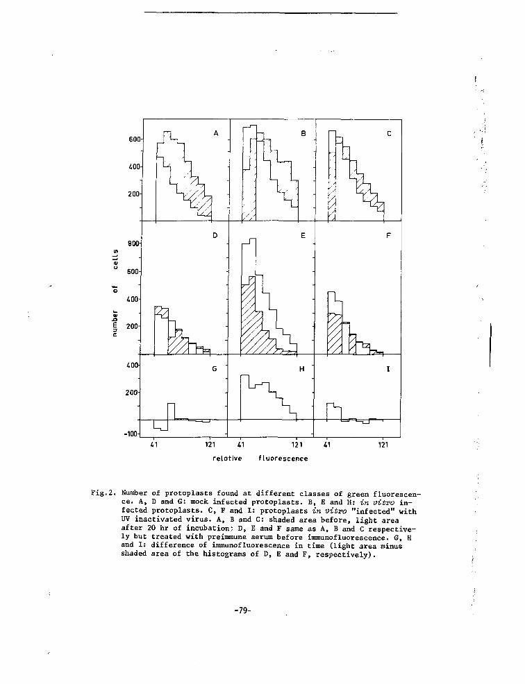
600
-100-
121
relative fluorescence
Fig.2. Number of protoplasts found at different classes of green fluorescen-ce. A, D and G: mock infected protoplasts. B, E and H: in vitro in-fected protoplasts. C, F and I: protoplasts in vitro "infected" withUV inactivated virus. A, B and C: shaded area before, light areaafter 20 hr of incubation; D, E and F same as A, B and C respective-ly but treated with preimmune serum before immunofluorescence. G, Hand I: difference of immunofluorescence in time (light area minusshaded area of the histograms of D, E and F, respectively).
-79-
Fig.3. Fluorogram of 11% slab-gel showing_ • - *fa5)"»
lT', immuno-precipitated 35s raethionine|i | ,. ?'fj< '*'f labeled proteins of mock infected
(M) and infected (I) protoplasts.Arrow indicates position of viralcoat protein.
iComparison of tobaeco protoplasts infeated in vivo and in vitro with AMV.
In fig. 4 the difference in immunofluorescence between in vivo AMV in-fected and healthy tobacco protoplasts is compared with the difference inimmunofluorescence between in vitro AMV and mock infected protoplasts. Boththe number of protoplasts showing immunofluorescence above the backgroundand the total amount of fluorescence found for the two samples is of thesame order.
To analyse the amount of infectious virus present in the samples, homo-genates (10 protoplasts in 0.5 ml PEN buffer) were inoculated onto bean.Homogenates from in vivo infected tobacco protoplasts induced more than 500local lesions per half leaf, while none of the 8 homogenates from in vitroinfected tobacco protoplasts was infectious. It is possible that in vitroinfected samples contain infectious virus together with a substance whichinhibits the local lesion assay. Since protoplast homogenates reduced theinfectivity of purified virus for more than 80% or even completely, itwould be difficult to establish whether or not mock infected protoplastsbecome even more inhibitory during incubation. Therefore we compared theinfectivity of in vivo infected protoplasts before and after 24 hr incuba-tion. The homogenates (25.10 protoplasts in 0.5 ml PEN buffer) induced 65(0 hr) and 55 (24 hr) local lesions per half leaf, respectively. If some
-80-
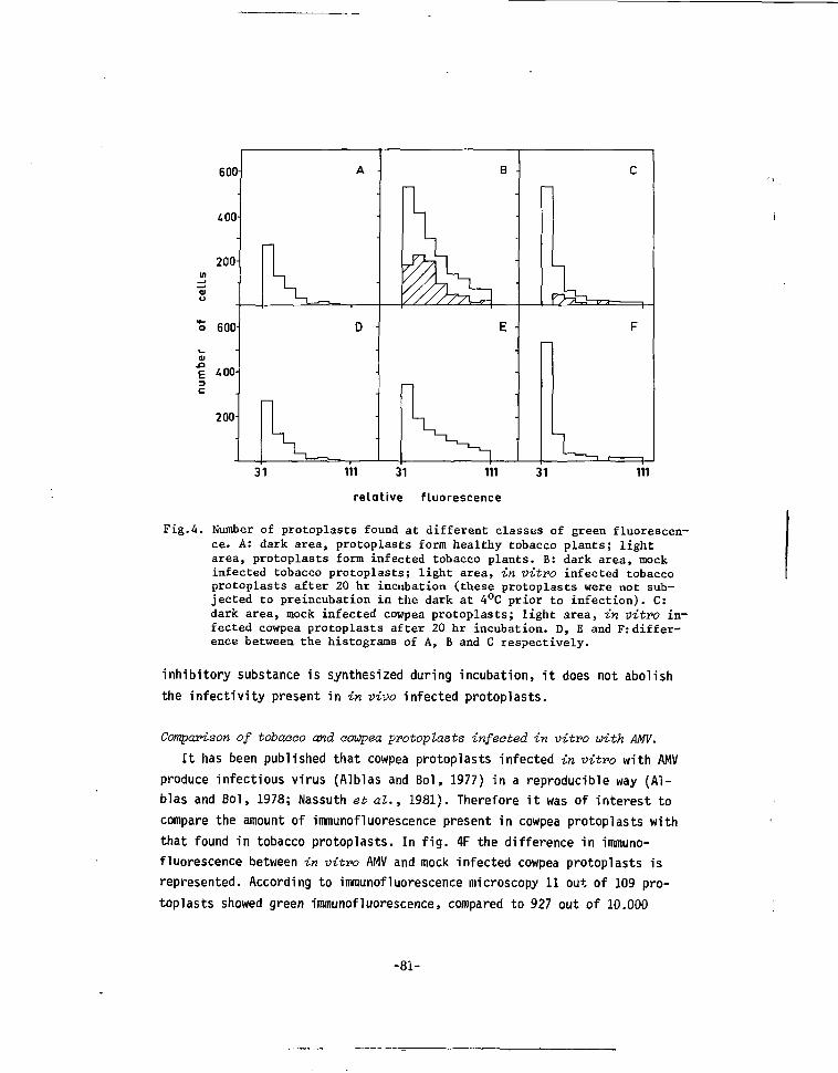
600-
400
200
o 600
400-
200-
B -
E
31 111 31 111 31
relative fluorescence
111
Fig.4. Number of protoplasts found at different classes of green fluorescen-ce. A: dark area, protoplasts form healthy tobacco plants; lightarea, protoplasts form infected tobacco plants. B: dark area, mockinfected tobacco protoplasts; light area, in vitro infected tobaccoprotoplasts after 20 hr incubation (these protoplasts were not sub-jected to preincubation in the dark at 4°C prior to infection). C:dark area, mock infected cowpea protoplasts; light area, in vitro in-fected cowpea protoplasts after 20 hr incubation. D, E and F: differ-ence between the histograms of A, B and C respectively.
inhibitory substance is synthesized during incubation, it does not abolishthe infectivity present in in vivo infected protoplasts.
Comparison of tobaaao and eowpea protoplasts infected in vitro with AMV.
It has been published that cowpea protoplasts infected in vitro with AMVproduce infectious virus (Alblas and Bol, 1977) in a reproducible way (Al-blas and Bol, 1978; Nassuth et al., 1981). Therefore it was of interest tocompare the amount of immunofluorescence present in cowpea protoplasts withthat found in tobacco protoplasts. In fig. 4F the difference in immuno-fluorescence between in vitro AMV and mock infected cowpea protoplasts isrepresented. According to immunofluorescence microscopy 11 out of 109 pro-toplasts showed green immunofluorescence, compared to 927 out of 10.000
-8Ï-
protoplasts as determined by the FACS. Comparison of fig. 4E with 4F showsthat the number of protoplasts with immunofluorescence above the backgroundas well as the total amount of fluorescence is again of the same order. Thehomogenate of the cowpea protoplasts (10 protoplasts in 0.5 ml PEN) was in-fectious as expected (207 local lesions per half leaf.
DISCUSSION
In the first part of the paper it is shown that immunofluorescence flowcytometry can be applied to plant cell protoplasts. By this method largenumbers of protoplasts can be analysed rapidly and quantitatively. The flowcytometric measurements are extremely reproducible within one batch of pro-toplasts (fig. 2). The variation seen between different figures is the varia-tion observed between different batches of protoplasts. In principle theFACS can also sort the protoplasts according to their relative fluorescence.Since it is doubtful whether antibodies can penetrate viable protoplastsand it is impossible to perform the measurements in an isotonic medium, pro-toplasts have to be treated with a fixative which may interfere with subse-quent analysis of the sorted protoplasts.
We found a reproducible difference in the immunofluorescence patterns(obtained for) protoplasts isolated from healthy tobacco with those obtainedof protoplasts isolated from AMV infected tobacco, indicating that the FITC-conjugatert antibodies can penetrate the formaldehyde fixed protoplasts. Asimilar difference was found between the patterns from mock infected and invitro AMV infected cowpea protoplasts, known to produce AMV (Alblas and Bol,1977; 1978; Nassuth et al. , 1981).
To study the replication of AMV ts mutants in protoplasts, tobacco proto-plasts had to be infected in vitro with AMV, since these ts mutants were se-lected on tobacco and several of them were not ts on cowpea plants (Van Vlo-ten-Doting et al. , 1980). Successful infection of tobacco (variety WhiteBurley) protoplasts with AMV has only been reported once (Motoyoshi et at. ,1975). In our laboratory it has been tried extensively to repeat these ex-periments (with several variaties of tobacco among which White Burley) with-out success (C.G. Bakhuisen, F. Alblas, J.F. Bol, A. Nassuth, personal com-munication and Van Klaveren, unpublished results). Therefore several methodsof isolation and infection of tobacco protoplasts were tried. Based on immu-nofluorescence data (both immunofluorescent microscopy and flow cytometry)
-82-
a modification of the published methods (involving a dark preincubation ofprotoplasts, an osmotic shock, PLO and PEG) was the most successful. The invitro infection procedure resulted in an increase of the aspecific bindingof antibodies (due to synthesis of cell wall component?), which could be re-duced by pretreatment of protoplasts with preimmune serum.
The immunofluorescence patterns obtained from in vitro AMV infected tobac-co protoplasts were very similar - both qualitatively and quantitatively -to those obtained from in vivo infected tobacco protoplasts and in vitroinfected cowpea protoplasts. However, infectious virus could not be detectedin in vitro infected tobacco protoplasts, while it was abundantly present inthe two other sampler. The absence of detectable infectious virus productionin in vitro infected tobacco protoplasts makes them unsuitable for the studyof the replication of ts mutants.
Although it is possible that further experiments may reveal more optimalconditions for the inoculation and incubation of tobacco protoplasts withAMVS the discrepancy between the detection of viral antigen and infectivityin these protoplasts deserves some comments.
(1) Maekawa et al. (1981) reported a discrepancy between the presence ofviral antigen and infectivity. Since in that case immunofluorescence butno infectivity was found in protoplasts from a non-host (Radish) of thevirus studied (brome mosaic virus), it was assumed that in the non-hostcells a substance was synthesized which inhibits virus multiplication.Neither of these explanations does apply to the present report on AMV intobacco protoplasts, since tobacco (Samsun NN) is an excellent systemichost for this particular virus.
(2) Loebenstein and Gera (1981) reported that in in vitro tobacco mosaicvirus infected tobacco protoplasts a substance is synthesized which in-hibits virus multiplication.
(3) There is a difference in aggregation of AMV particles in in vitro and invivo infected tobacco protoplasts. In -vivo AMV particles form a kind ofaggregates (Hull, Hills and Plaskitt, 1970) which may result in an under-estimation of the amount of viral antigen present. Furthermore particlesin these aggregates may be (partly) protected from inactivating substan-ces present in protoplasts homogenates. The latter is supported by thefact that non-aggregated purified AMV added to protoplast homogenates is(nearly) completely inactivated, while homogenates from in vivo infectedtobacco protoplats are infectious (even when diluted 200 times with anhomogenate from healthy protoplasts, results not shown).
-83-
(4) In in vitvu infected tobacco protoplasts there is a discrepancy betweenthe synthesis of coat protein and the synthesis of one or more of thethree genomic RNAs. In cowpea protoplasts the synthesis of RNA 1 and RNA2 is more sensitive to the presence of actinomycine D than the synthesisof RNA 3 (Nassuth et al. , to be published), indicating that the synthe-sis of the different genomic RNAs differs in sensitivity to disturbanceof the host cell metabolism.
The experiments presented are insufficient to differentiate between theseexplanations. However, the results clearly show that one should be carefulwith the interpretation of immunofluorescence data. The demonstration of in-crease of immunofluorescence in itself is insufficient to conclude thatvirus replication has taken place.
ACKNOWLEDGMENTS
We thank Annette Nassuth for infecting the cowpea protoplasts, FiekeAlblas for valuable advice on protoplast manipulation, and Dr. E.M.J. Jas-pars for critically reading the manuscript. We thank the Medical Facultyof the State University of Leiden for the use of their facilities. Thiswork was financially supported by the Netherlands Organization for Advance-ment of Pure Research (Z.W.O.).
-84-
REFERENCES
Alblas, F. and Bol, J.F. (1977). Journal of General Virology 36, 175-185.
Alblas, F. and Bol, J.F. (1978). Journal of General Virology 43., 653-656.
Bonner, W.M. and Laskey, R.A. (1974). European Journal of Biochemistry
46, 83-88.
Cassels, A.C. and Barlass, M. (1978). Virology 87, 459-462.
Herzenberg, L.A., Sweet, R.G. and Herzenberg, L.A. (1976). Scientific
American 234 (3), 108-117.
Hull, R., Hills, G.J. and Plaskitt, A. (1970). Virology 42, 753-772.
Laemnili, U.K. (1970). Nature 221, 680-685.
Leonard, D.A. and Zaitlin, M. (1982). Virology U 7 , 416-424.
Loebenstein, G. and Gera, A. (1981). Virology UA, 132-139.
Maekawa, K., Furusawa, I. and Okimo, T. (1981). Journal of General Virology
53, 353-356.
Maule, A.J., Boulton, M.I., Edmunds, C. and Wood, K.R. (1980). Journal of
General Virology 47, 199-203.
Mayo, M.A. and Roberts, I.M. (1978). Journal of General Virology 40, 19-
30.
Melamed, M.R. and Mullaney, P.F. (1979). Wiley Mediaal Publications, New
York. p. 3-9.
Motoyoshi, F., Bancroft, J.B., Watts, J.W. and Burgess, J. (1973). Journal
of General Virology 2Q. 177-193.
Motoyoshi, F., Hull, R. and Flack, I.H. (1075). Journal of General Virology
2J_, 263-266.
Nassuth, A., Alblas, F. and Bol, J.F. (1981). Journal of General Virology
J53, 207-214.
Nishiguchi, M., Motoyoshi, F. and Oshima, N. (1978). Journal of General
Virology 9_, 53-61.
Ouchterlony, 0. (1962). Progr. Allergy 6_, 30-154.
-85-
Roosien, J. and Van Vloten-Doting, L. (1982). Journal of General Virology,
£3, 139-198.
Van Vloten-Doting, L. and Jaspars, E.M.J. (1967). Virology 33, 684-693.
Van Vloten-üoting, L. and Jaspars, E.M.J. (1972). Virology 48, 699-708.
Van Vloten-Doting, L., Hasrat, J.A., Oosterwijk, E., Van 't Sant, P.,Schoen, M.A. and Roosien, J. (1980). Journal of General Virology 46,415-426.
Widholm, J.H. (1972). Stain Technology 47, 189-194.
-86-
Submitted for publication
CHAPTER VI
COMPETITION BETWEEN THE RNA 3 MOLECULES OF WILDltPE ALFALFA MOSAIC VIRUSAND THE TEMPERATURE-SENSITIVE MUTANT Tbts 7(uv)
Jan Roisien, Pieter van Klaveren and Lous van Vloten-Doting
Department of Biochemistry, State University of Leiden, P.O. Box 9505,
2300 RA Leiden, The Netherlands.
SUMMARY
In mixed infections of wildtype (wt) alfalfa mosaic virus (AMV) and atemperature-sensitive (is) mutant Tbts 7(uv), which carries a ts defect inthe early function of the coat protein, the mutant symptoms were not foundat 30°C. In the progeny from these mixed infections neither mutant coatprotein nor mutant RNA 3 could be detected. Even at 23°C there was someloss of mutant RNA 3 and coat protein from the progeny of the mixed infec-tions. Analysis and comparison of mutant and wt dsRNA preparations reveal-ed a lower dsRNA 3 content for the mutant preparation at 23°C. Also theamount of RNA 3 in virion preparations was lower for the mutant than forwt. These results point to a mutation in the RNA 3 of Tbts 7(uv) which di-minishes its affinity for the viral replicase.
-87-
INTRODUCTION
The coat protein of alfalfa mosaic virus (AMV) has at least two differ-ent functions in the multiplication of the tripartite genome (Bol et al. ,1971): activation of the viral genome (an early function, Van Vloten-Doting,1978) and the encapsidation of the newly formed viral RNAs (a late function,Nassuth et at., 1982). To obtain more insight in the functions of the coatprotein as well as the replication cycle of the plant virus AMV, we havemade a collection of temperature-sensitive {ts) mutants (Van Vloten-Dotinget at., 1980; Roosien and Van Vloten-Doting, 1982).
Several of our ts mutants showed a ts defect in the early function ofthe coat protein (Van Vloten-Doting et at., 1980; Smit et al., 1981). Thecoat protein of one of these mutants, Tbts 7(uv), can be distinguishedfrom wildtype (wt) coat protein by electrophoresis on SDS polyacrylamideslabgels (Smit, 1981). This mutant induces in tobacco plants symptoms dif-fering from those induced by wt (Van Vloten-Doting et al. , 1980), therebypermitting the detection of mutant RNA in the progeny from mixedly infect-ed plants. In this paper we show that the symptom mutation - as well asthe ccat protein mutation - maps on the smallest genomic RNA (RNA 3, pre-sent in the Top component b, Tb, particle. No mutant virus is produced intobacco leaf discs at the non-permissive temperature (30°C), while inwhole plants at 30°C some virus is produced and mutant symptoms do appear.
The properties of this mutant offer the possibility for an easy detec-tion of the presence of mutant RNA and mutant coat protein in virions pro-duced in plants infected with mixtures of mutant and wt virus at 23°C(permis-sive temperature) and 30°C.If the coat protein mutation only affects theearly function, it is to be expected that the progeny from mixed infectionsat 30°C will contain both types of RNA and both types of coat protein. Ifthe mutation affects both the early and the late function of the coat pro-tein, both types of RNA are expected to be encapsidated by wt coat proteinalone.
Surprisingly we found in the progeny of mixed infections at 30°C onlywt RNA and wt coat protein. Apparently the mutant RNA 3, instead of beingrescued, is outcompeted by wt RNA 3.
-88-
MATERIALS AND METHODS
Origin, propagation and infeativity assay of wt and Tbts 7(uv).
AIW strain 425 (Hagedorn and Hanson, 1963) was used. Mutant Tbts 7(uv)
was isolated from leaves inoculated with ultraviolet irradiated Tb together
with untreated Middle (M, containing RNA 2) and Bottom (B, containing RNA 1)
components of AMV 425 (Van Vloten-Doting et al., 1980).
Wt and mutant were propagated on tobacco (Niaotiana tabaoum L. var "Sam-
sun NN"). When virus was grown at 30°C, plants were incubated for 2-3 hours
at that temperature before inoculation.
Infectivity assays were carried out on bean plants (Phaseolus vulgaris L.
var Berna) as described by Van Vloten-Doting and Jaspars (1967).
Supplementation test.
Tbts 7(uv) was inoculated onto tobacco alone or in the presence of each
of the purified wt components as described before (Van Vloten-Doting et al.,
1980).
Isolation of virus and analysis of virion RNA.
Virus, both wt and Tbte 7(uv), was isolated as described by Van Vloton-
Doting and Jaspars (1972). Virion RNA was analysed by electrophoresis on 3 %
polyacrylamide gels (Loening, 1967). Virions (50-200 ug) were dissociated
with 1 % SDS and heated for 5 minutes at 60°C prior to loading onto the gel.
Gels (8 cm long) were run for 3.5 hours at roomtemperature at 5 mA per gel.
Isolation and Analysis of ds RNA.
Wt and mutant ds RNA was prepared as described by Morris and Dodds (1979).
Analysis was carried out by electrophoresis of these preparations on 2.6 %
polyacrylamide gels as described by Roosien and Van Vloten-Doting (1983).
Analysis of the virion ooat protein composition.
Viral protein was analysed by electrophoresis on polyacrylamide slabgels
of nucleoprotein dissociated with SDS according to Laemlli (1970), followed
by staining (Kedinger et al., 1974).
-89-
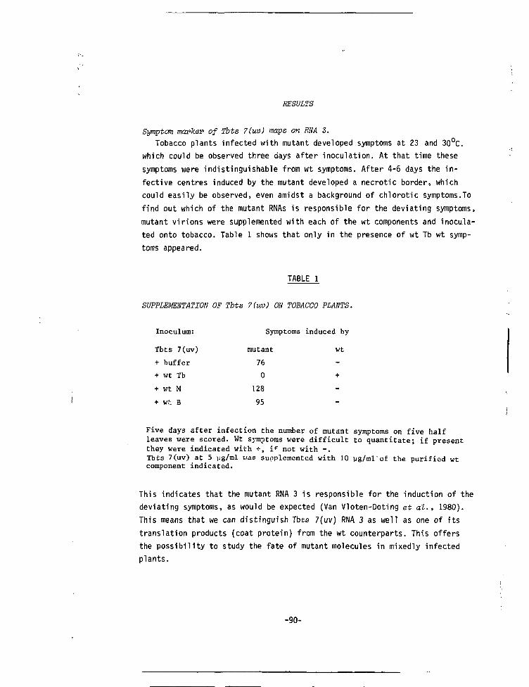
RESULTS
Symptom marker of Ibts 7(uv) maps on RNA 3.Tobacco plants infected with mutant developed symptoms at 23 and 30°C.
which could be observed three days after inoculation. At that time thesesymptoms were indistinguishable from wt symptoms. After 4-6 days the in-fective centres induced by the mutant developed a necrotic border, whichcould easily be observed, even amidst a background of chlorotic symptoms.Tofind out which of the mutant RNAs is responsible for the deviating symptoms,mutant virions were supplemented with each of the wt components and inocula-ted onto tobacco. Table 1 shows that only in the presence of wt Tb wt symp-toms appeared.
TABLE 1
SUPPLEMENTATION OF Tbts 7(uv) ON TOBACCO PLANTS.
Inoculum:
Tbts 7(uv)
+ buffer
+ wt Tb
+ wt M
+ wt B
Symptoms
mutant
76
0
128
95
induced by
wt
-
+
-
_
Five days after infection the number of mutant symptoms on five halfleaves were scored. Wt symptoms were difficult to quantitate; if presentthey were indicated with +, if not with -.Tbts 7(uv) at 5 pg/ml was supplemented with 10 ug/ml'of the purified wtcomponent indicated.
This indicates that the mutant RNA 3 is responsible for the induction of thedeviating symptoms, as would be expected (Van Vloten-Doting et al., 1980).This means that we can distinguish Tb*e 7(uv) RNA 3 as well as one of itstranslation products (coat protein) from the wt counterparts. This offersthe possibility to study the fate of mutant molecules in mixedly infectedplants.
-90-
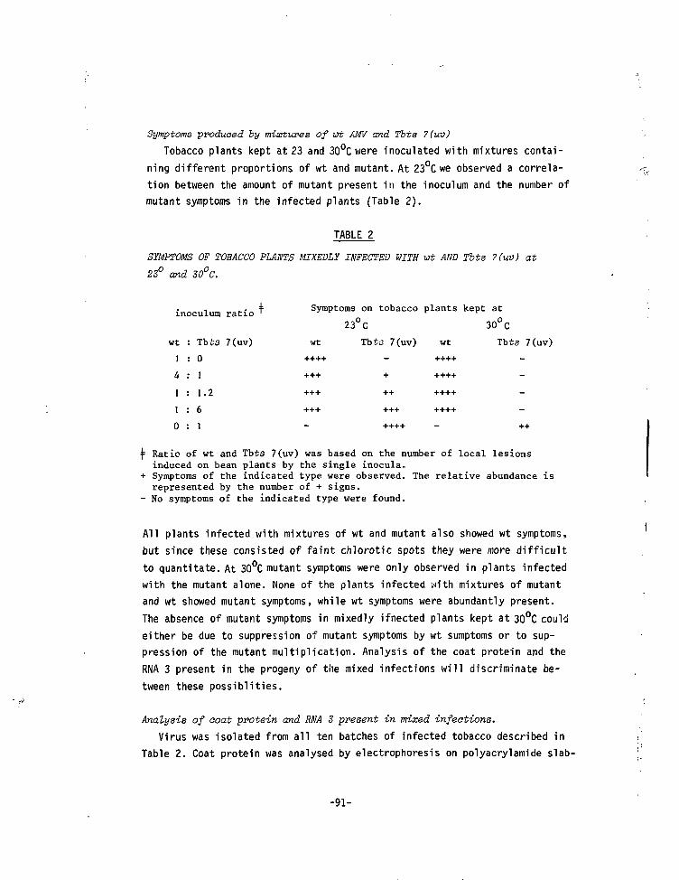
Symptoms produced by mixtures of wt AMV and Tbts 7(uv)
Tobacco plants kept at 23 and 30°C were inoculated with mixtures contai-ning different proportions of wt and mutant. At 23°C we observed a correla-tion between the amount of mutant present in the inoculum and the number ofmutant symptoms in the infected plants (Table 2).
TABLE 2
SYMPTOMS OF TOBACCO PLANTS MIXEDLX INFECTED WITH Wt AND Tbts 7(uv) at23° and 30°C.
inoculum
wt
1
1
1
0
: Tbts
: 0
1
1.2
6i
ratio
7(uv)
Symptoms on tobacco plants kept at
23°C 30°C
wt Tbts 7(uv) wt Tbte 7(uv)
^ Ratio of wt and Tbts 7(uv) was based on the number of local lesionsinduced on bean plants by the single inocula*
+ Symptoms of the indicated type were observed. The relative abundance isrepresented by the number of + signs.
- No symptoms of the indicated type were found.
All plants infected with mixtures of wt and mutant also showed wt symptoms,but since these consisted of faint chlorotic spots they were more difficultto quantitate. At 30°C mutant symptoms were only observed in plants infectedwith the mutant alone. None of the plants infected with mixtures of mutantand wt showed mutant symptoms, while wt symptoms were abundantly present.The absence of mutant symptoms in mixedly ifnected plants kept at 30°C couldeither be due to suppression of mutant symptoms by wt sumptoms or to sup-pression of the mutant multiplication. Analysis of the coat protein and theRNA 3 present in the progeny of the mixed infections will discriminate be-tween these possiblities.
Analysis of ooat protein and RNA 3 present in mixed infections.Virus was isolated from all ten batches of infected tobacco described in
Table 2. Coat protein was analysed by electrophoresis on polyacrylamide slab-
-91-
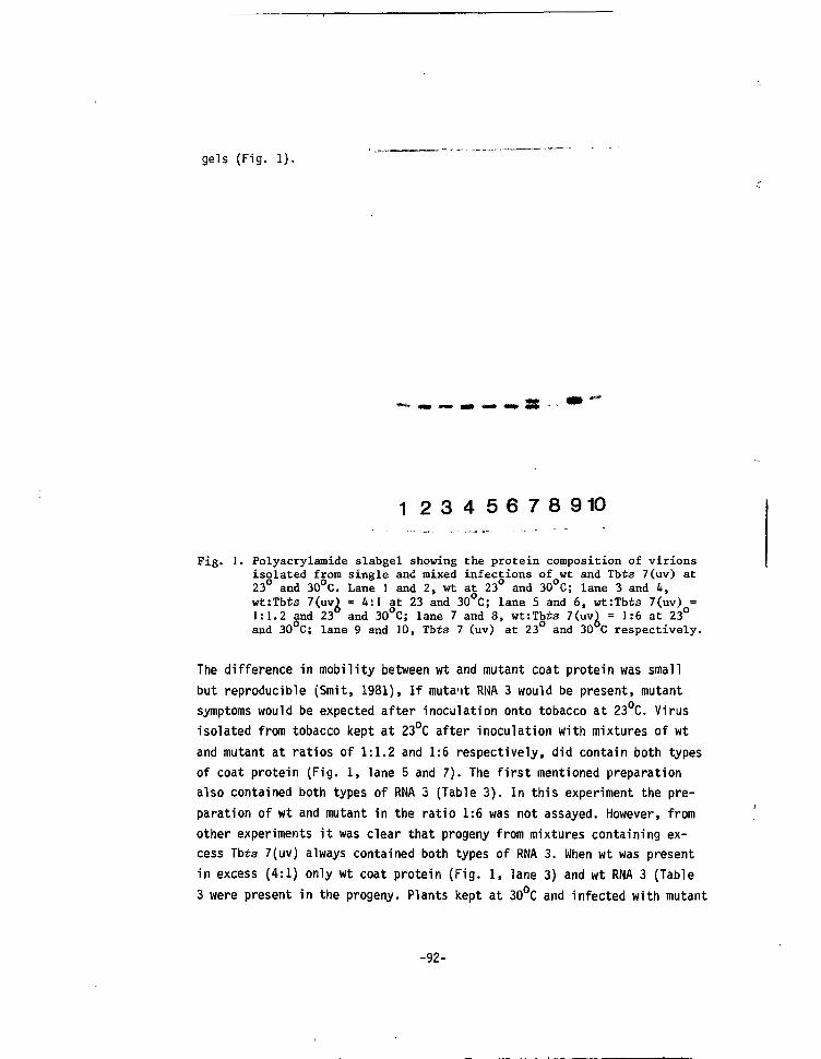
gels (Fig. 1).
M
1 2 3 4 5 6 7 8 9 10
Fig. 1. Polyacrylamide slabgel showing the protein composition of virionsisolated from single and mixed infections of wt and Tbts 7(uv) at23° and 30°C. Lane 1 and 2, wt at 23° and 30°C; lane 3 and 4,wt:Tbts 7(uv) = 4:1 at 23 and 30°C; lane 5 and 6, wt:Tbts 7(uv) =1:1.2 and 23 and 30 C; lane 7 and 8, wt:Tb£s 7(uv) = 1:6 at 23°and 30°C; lane 9 and 10, Tbts 7 (uv) at 23° and 30 C respectively.
The difference in mobility between wt and mutant coat protein was smallbut reproducible (Smit, 1981), If mutant RNA 3 would be present, mutantsymptoms would be expected after inoculation onto tobacco at 23°C. Virusisolated from tobacco kept at 23°C after inoculation with mixtures of wtand mutant at ratios of 1:1.2 and 1:6 respectively, did contain both typesof coat protein (Fig. 1, lane 5 and 7). The first mentioned preparationalso contained both types of RNA 3 (Table 3). In this experiment the pre-paration of wt and mutant in the ratio 1:6 was not assayed. However, fromother experiments it was clear that progeny from mixtures containing ex-cess Tbts 7(uv) always contained both types of RNA 3. When wt was presentin excess (4:1) only wt coat protein (Fig. 1, lane 3) and wt RNA 3 (Table3 were present in the progeny. Plants kept at 30°C and infected with mutant
-92-
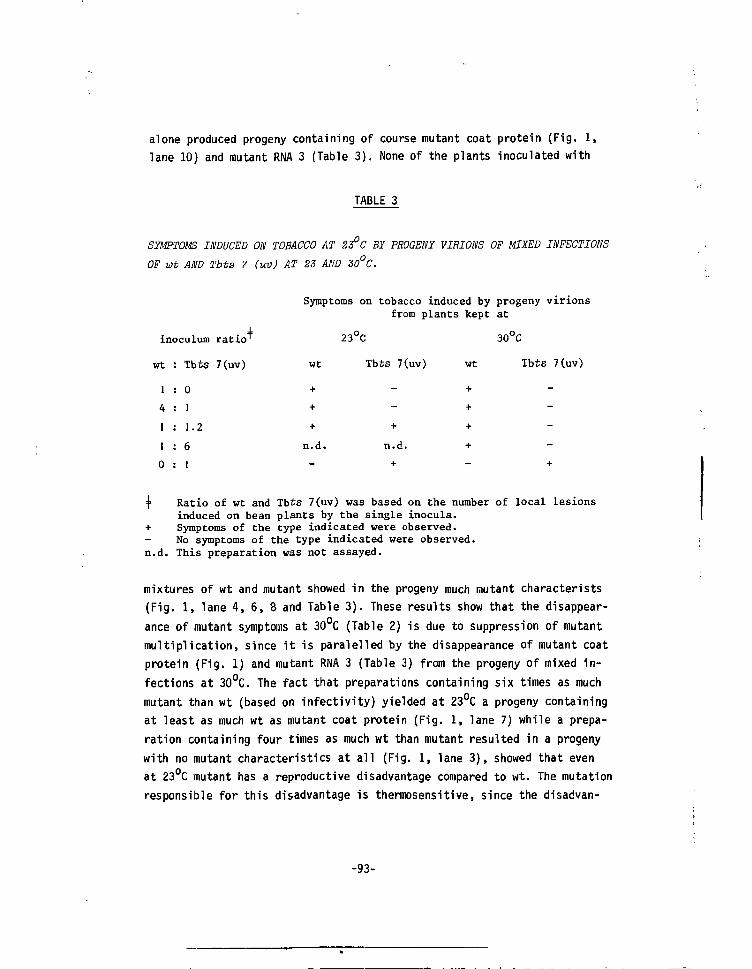
alone produced progeny containing of course mutant coat protein (Fig. 1,lane 10) and mutant RNA 3 (Table 3). None of the plants inoculated with
TABLE 3
SYMPTOMS INDUCED ON TOBACCO AT 2S°C BY PROGENY VIRIONS OF MIXED INFECTIONS
OF Wt AND Tbts 7 (uv) AT 23 AND 30°C.
Symptoms on tobacco induced by progeny virions
from plants kept at
23°C 30°C
wt Tbts 7(uv) wt Tbts 7(uv)
inoculum
rt
1
4
1
1
0
: Tbts
: 01
1.2
6
1
ratio
7(uv)
n.d. n.d.
+
=f= Ratio of wt and Tbts 7(uv) was based on the number of local lesionsinduced on bean plants by the single inocula.
+ Symptoms of the type indicated were observed.No symptoms of the type indicated were observed,
n.d. This preparation was not assayed.
mixtures of wt and mutant showed in the progeny much mutant characterists(Fig. 1, lane 4, 6, 8 and Table 3). These results show that the disappear-ance of mutant symptoms at 30°C (Table 2) is due to suppression of mutantmultiplication, since it is paralelled by the disappearance of mutant coatprotein (Fig. 1) and mutant RNA 3 (Table 3) from the progeny of mixed in-fections at 30°C. The fact that preparations containing six times as muchmutant than wt (based on infectivity) yielded at 23°C a progeny containingat least as much wt as mutant coat protein (Fig. 1, lane 7) while a prepa-ration containing four times as much wt than mutant resulted in a progenywith no mutant characteristics at all (Fig. 1, lane 3), showed that evenat 23°C mutant has a reproductive disadvantage compared to wt. The mutationresponsible for this disadvantage is thermosensitive, since the disadvan-
-93-
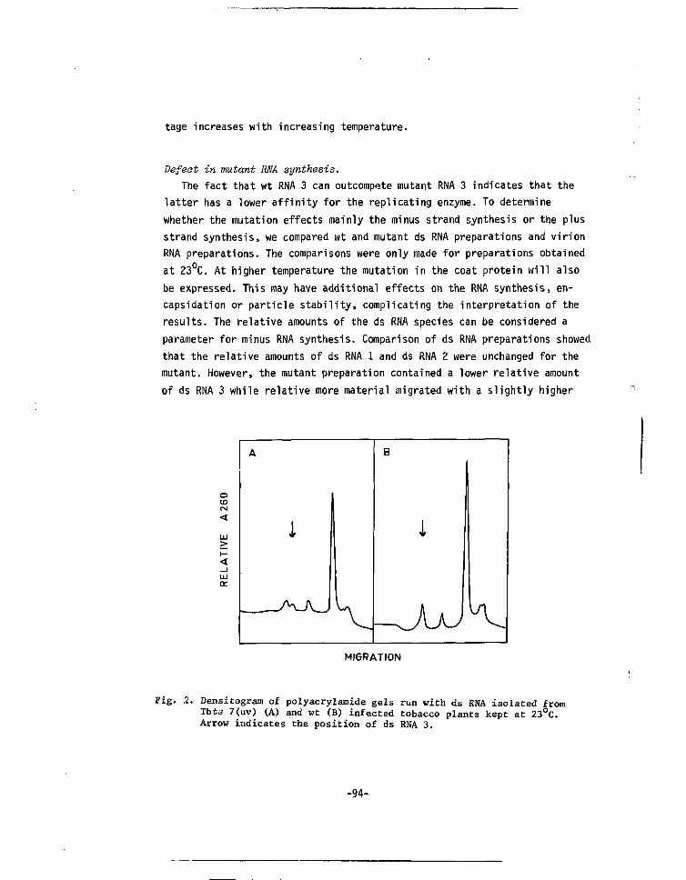
tage increases with increasing temperature.
Defeat in mutant RNA synthesis.The fact that wt RNA 3 can outcompete mutant RNA 3 indicates that the
latter has a lower affinity for the replicating enzyme. To determinewhether the mutation effects mainly the minus strand synthesis or the plusstrand synthesis, we compared wt and mutant ds RNA preparations and virionRNA preparations. The comparisons were only made for preparations obtainedat 23°C. At higher temperature the mutation in the coat protein will alsobe expressed. This may have additional effects on the RNA synthesis, en-capsidation or particle stability, complicating the interpretation of theresults. The relative amounts of the ds RNA species can be considered aparameter for minus RNA synthesis. Comparison of ds RNA preparations showedthat the relative amounts of ds RNA 1 and ds RNA 2 were unchanged for themutant. However, the mutant preparation contained a lower relative amountof ds RNA 3 while relative more material migrated with a slightly higher
o(0CM
UJ
ÜJ
cc
A B
Ï
MIGRATION
Fig. .2. Densitogram of polyacrylamide gels run with ds RNA isolated fromTbts 7(uv) (A) and wt (B) infected tobacco plants kept at 23°C.Arrow indicates the position of ds RNA 3.
-94-
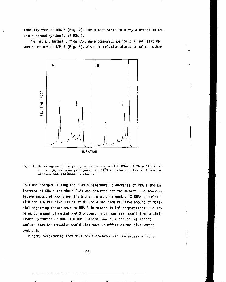
mobility than ds RNA 3 (Fig. 2). The mutant seems to carry a defect in theminus strand synthesis of RNA 3.
When wt and mutant virion RNAs were compared, we found a low relativeamount of mutant RNA 3 (Fig. 3). Also the relative abundance of the other
MIGRATION
Fig. 3. Densitogram of polyacrylamide gels run with RNAs of Vots 7(uv) (A)and wt (B) virions propagated at 23°C in tobacco plants. Arrow in-dicates the position of RNA 3.
RNAs was changed. Taking RNA 2 as a reference, a decrease of RNA 1 and anincrease of RNA 4 and the X RNAs was observed for the mutant. The lower re-lative amount of RNA 3 and the higher relative amount of X RNAs correlatewith the low relative amount of ds RNA 3 and high relative amount of mate-rial migrating faster than ds RNA 3 in mutant ds RNA preparations. The lowrelative amount of mutant RNA 3 present in virions may result from a dimi-nished synthesis of mutant minus strand RNA 3, although we cannotexclude that the mutation would also have an effect on the plus strandsynthesis.
Progeny originating from mixtures inoculated with an excess of Tbts
-95-
7(uv) and propagated at 23 C contained slightly less RNA 3 than wt virusand more X RNAs, a characteristic of the mutant (results not shown). TheRNA patterns from the progeny of all other mixed inocula from Table 2, pro-pagated at 23° or 30°C were comparable to that of wt at 23°C. This resultin combination with the results shown in Table 3 indicate that mutant RNA3 has no inhibitive effect on the relative amount o f wt RNA 3 in virions.Since wt RNAs are encapsidated for at least 80% (Nassuth et at. , 1983) wecan conclude that no inhibition of wt RNA 3 synthesis had occurred in themixedly infected plant.
DISCUSSION
The availability of a ts mutant, carrying two distinctive markers, onefor the coat protein and the other for the mutant RNA 3, enabled us tostudy the fate of this mutant when it was propagated in the presence of wtat 23° and 30°C. Instead of the expected rescue of mutant RNA 3 by encap-sidation in either wt or mutant coat protein, wt RNA 3 completely outcom-peted the mutant RNA 3 at 30°C. Even at 23°C wt RNA 3 had some advantageover mutant RNA 3. Since the effect was more pronounced at 30°C the mutationresponsible for the loss of mutant RNA 3 in the presume of wt can be con-sidered ts. Since wt RNA 3 synthesis is not inhibited, the mutation acts inais.
This mutation could be expressed at the protein level or at the RNAlevel. If the mutation is expressed at the protein level, one of the twoRNA 3 encoded products (Van Vloten-üoting and Jaspars, 1977) should act inais. Such a mutation might be responsible for the mutant characteristicsbut could not explain the results of the competition experiments.
Apparently the mutation is expressed at the RNA level, where it couldinterfere with the encapsidation or the replication. For tobacco mosaic vi-rus a ts mutant has been described which cannot be encapsidated correctlyat the non-permissive temperature. The mutation responsible for this beha-viour is expressed at the RNA level (Taliansky et al. , 1982a,b). A ts de-fect in the encapsidation of AMV mutant RNA 3 would diminish the presenceof this RNA in progeny virions but would not lower its expression in themixedly infected leaves. The fact that no mutant symptoms were observed(Table 2) points to the absence of mutant RNA 3 in the mixed infections andtherefore in the progeny virions. This is in line with the observation that
-96-
the progeny did not contain mutant coat protein. Therefore we conclude that
the defect affects the RNA synthesis. The virus coded functions involved in
the synthesis of AMV minus and probably also plus strand RNA are located on
RNA 1 and 2 (Nassuth and Bol, 1982). In the mutant these RNAs are assumed
to be unchanged. This is in accordance with the presence of the same rela-
tive amounts of RNA 1 and RNA 2 in mutant and wt ds RNA preparations. The
mutant contains a lower relative amount of ds RNA 3 which could be due to
a lower affinity of the mutant RNA 3 for the replicase. Wt RNA 3 provides
the replicase with a better substrate, resulting in its preferential replica-
tion. The difference in affinity of wt and mutant RNA 3 for the replicase
does not necessarily have to be big, since in whole plants multiple rounds
of infection take place and even a small difference in affinity could ex-
plain the loss of mutant RNA 3. Another example of competition between wt
and a ts derivative has been described for another member of the viruses
with a tripartite genome. For cowpea chlorotic mottle virus (CCMV) a mutant
with a ts mutation on RNA 3 was found to be dominant over wt at all tested
temperatures in mixedly infected protoplasts. However, this mutant required
some wt RNA 3 for its replication at the non-permissive temperature (Dawson
et at., 1975; Dawson and Watts, 1979). Whether in this case an increased
affinity of the mutant RNA 3 for the viral replicase played a role was un-
certain.
At present we do not know if (1) the changed symptoms, (2) the ts coat
protein, (3) the changed electrophoretic mobility of the coat protein and
(4) the reduced affinity of the mutant RNA 3 for the viral replicase all
have the same molecular basis, in other words, if they all result from the
same changein the RNA. If so, then the replicase recognition site would
overlap with the coding sequence. Comparison of the nucleotide sequence of
wt and mutant RNA 3 will provide more insight in signals involved in the
viral replication.
ACKNOWLEDGEMENTS
We thank Mr. A.G. Wesseling for cultivation of the plants and Dr. E.M.J.
Jaspars for his helpful discussions. This work was financially supported by
the Netherlands Organization for Advancement of Pure Research (Z.W.O.).
-97-
REFERENCES
Bol, J.F., Van Vloten-Doting, L. and Jaspars, E.M.J. (1971). Virology 46,
73-85.
Oawson, J.K.O., Motoyoshi, F., Watts, J.W. and Bancroft, J.B. (1975). J.
Gen. Virol. 29, 99-107.
Dawson, J.R.Ü. and Watts, J.W. (1979). J. Gen. Virol. 45, 133-137.
Hagedorn, D.J. and Hanson, E.W. (1963). Phytopathology 53, 188-192.
Kedinger, C , Gissinger, F. and Charnbon, P. (1974). Eur. J. Bioah. 44, 421-
436.
Laemmli, U.K. (1970). Nature (London) 227, 680-6a5.
Loening, U.E. (1967). Bioohem. J. 1M, 251-257.
Morris, T.J. and Dodds, J.A. (1979). Pythopathology 69, 854-858.
Nassuth, A. and Bol, J.F. (1982). Virology, in press.
rtassuth, A., Ten Bruggencate, G. and Bol, J.F. (1982). virology 123,in press.
Roosien, J. and Van Vloten-Doting, L. (1982). J. Gen. Virol. 63^, 189-198.
Roosien, J. and Van Vloten-Doting, L. (1983). virology, in press.
Smit, C.H. (1981). Ph.D. Thesis, University of Leiden.
Smit, C.H., Roosien, J., Van Vloten-Doting, L. and Jaspars, E.M.J. (1981).
Virology U2, 169-173.
Taliansky, M.E., Atabekova, T.I., Kaplan, I.B., Morozov, S.Yu., Malyshenko,
S.I. and Atabekov, J.G. (1982a). Virology 118, 301-308.
Taliansky, M.E., Kaplan, I.B., Yarvekulg, L.V., Atabekova, T.I., Agranovsky,
A.A. and Atabekov, J.G. (1982b). Virology 1^8, 309-316.
Van Vloten-Uoting, L. and Jaspars, E.M.J. (1967). Virology 33, 684-693.
Van Vloten-Doting, L. and Jaspars, E.M.J. (1972). Virology 48, 699-708.
Van Vloten-Doting, L. and Jaspars, E.M.J. (1977). In: Comprehensive Virology
(H. Fraenkel-Conrat and R.R. Wagner eds.) Plenum Press, New York. Vol. II
pp. 1-53.
-98-
Van V l o t e n - D o t i n g , L. ( 1978) . J. Gen. Virol. 4 1 , 649-652.
Van V l o t e n - D o t i n g , L . , H a s r a t , J . A . , O o s t e r w i j k , E . , Van ' t S a n t , P . ,Schoen, M.A. and Roos ien , J . ( 1980) . j . Gen. Virol. 4 6 , 415-426.
- 9 9 -
CHAPTER VII
Accepted for publication
A MUTANT OF ALFALFA MOSAIC VIRUS WITH AN UNUSUAL STRUCTURE
Jan Roosien and Lous van Vloten-DotingDepartment of Biochemistry, State University of Leiden, P.O. Box 9505,2300 RA Leiden, The Netherlands.
SUMMARY
A spontaneous mutant of alfalfa mosaic virus (AMV) with an alteredstructure is described. By analysis of pseudorecombinants the mutation(s)responsible for the altered structure could be assigned to RNA 3. By invitro translation and serology it is shown that both proteins (35 K proteinand coat protein) encoded by RNA 3 are changed. The mutation(s) present inRNA 3 have also an effect on the ratio of the three genomic RNAs, both invirion as well as in double-stranded RNA preparations. Compared to wildtype particles mutant particles (1) have a lower electrophoretic mobilityin polyacrylamide gels, (2) have an higher sedimentation velocity in sucro-se density gradients, (3) do contain the same percentage of RNA, (4) aremore prone to aggregation, (5) are somewhat less stable during storage and(6) are less sensitive to uncoating by AMV-RNA.
Electronmicrographs show that the mutant preparations contain some verylong bacilliform particles, however the majority of the particles is sphe-roidal and bears a strong resemblance to the particles of the ilarviruses.The structural properties of this mutant do support the classification ofAMV as an ilarvirus.
-101-
INTRODUCTION
Alfalfa mosaic virus (AMV) is a plant virus with a coat protein dependent
tripartite genome. The genomic KNAs (RNA 1, 2 and 3, in order of decrea-
sing molecular weight) are separately encapsidated into bacilliform par-
ticles of different lengths, called bottom component (B), middle component
(M) and top component b (Tb), respectively (Bol et al., 1971). In addition
virus preparations contain top component a (Ta) particles, which are main-
ly spheroidal (Heijtink and Jaspars, 1976 and references therein) and
contain two molecules of RNA 4 (Heijtink et at., 1977), a subgenomic mRNA
for the coat protein (Van Vloten-Doting and Jaspars, 1977). Two AMV strains
(Hull et al., 1969a; Hull, 1970a) have been discribed which form also lon-
ger bacilliform particles containing more than one RNA molecule (Heijtink
and Jaspars, 1974). Structural analysis has shown that the AMV co.jt pro-
tein in the tubular part of the particles is organised in a hexagonal
lattice, while it is assumed that in the rounded caps the 6-fold axes are
converted to 5-fold axes giving rise to a surface lattice similar to that
of an icosahedron (Mellema, 1975; Cremers et al. , 1981 and references the-
rein).
There is one other group of viruses with a coat protein dependent tri-
partite genome: the ilarviruses (Van Vloten-Doting, 1975; Gonsalves and
Garnsey, 1975; Huttinga and Mosch, 1976; Gonsalves and Fulton, 1977; Bos
et at. , 1980) with at least 13 members, type member is tobacco streak vi-
rus (TSV) (Matthews, 1979; \lan Vloten-Doting et al., 1981). A typical fea-
ture of the ilarviruses is that the centrifugal heterogeneity of the nu-
cleoprotein preparation is due to the presence of spheres of different si-
ze. Occasionally short bacilliform particles are found besides the sphe-
roidal particles (Basit and Francki, 1970; Halk and Fulton, 1978). The
structural differences between AMV and the ilarviruses have been an impor-
tant argument in favour of the classification of AMV in a separate group
(Matthews, 1979). Since AMV and the ilarviruses share several properties -
most notably the coat protein dependent tripartite genome - several authors
have proposed to include AMV in the ilarvirus group (Lister and Saksena,
1976; Gonsalves and Fulton, 1977; Halk and Fulton, 1978; Van Vloten-Doting
et at., 1981).
In this paper we discribe some properties of a mutant of AMV which pro-
-102-
duces, besides a small proportion of very long bacilliform particles, par-
ticles strongly resembling the particles described for the ilarviruses. The
structural properties of this mutant form an additional argument in favour
of the classification of AMV as an ilarvirus.
MATERIALS AND METHODS
Virus and virus culture. AMV strain 425 (Hagedorn and Hanson, 1963) and
mutants derived from this strain (Roosien and Van Vloten-Doting, 1982) were
propagated as described by Van Vloten-Doting and Jaspars(1972). T5V nucleo-
protein was a generous gift of dr. D.Zuidema of this laboratory.
Loeal lesion assay, single lesion transfer and determination of ts cha-
racter. The methods described by Van Vloten-Doting and Jaspars (1967),
Van Vloten-Doting et al. (1980) and Roosien and Van Vloten-Doting (1982)
were used.
Isolation of viral nuoleoprotein. Virus was isolated according to Van
Vloten-Doting and Jaspars (1972). Occasionally RNA and nucleoprotein com-
ponent composition was analysed directly after the first cycle of high and
low speed centrifugation. The second cycle of high and low centrifugation
does not change the component composition, only enchances the stability of
the nucleoprotein preparation during storage.
Isolation of RNA. Virion RNA was isolated according to Van Vloten-Doting
and Jaspars (1967). Double stranded RNA was isolated from tobacco leaves 5
days post infection by the method of Morris and Dodds (1979).
Polyaorylcmide gelelectrophoresis. Analytical gelelectrophoresis was car-
ried out according to Bol et al. (1971) with minor modifications. Gels were
8 cm long, loaded with 50 to 200 yg of nucleoprotein and run at room tem-
perature for 17 hr. To analyse the RNA content nucleoprotein was dissocia-
ted with 1% SDS. Before loading onto the gel, the preparation was heated
for 5 min. at 60°. Gels were run for 2.5 to 3.5 hr at room temperature.
Electrophoresis of double-stranded RNA was performed on 2.6% polyacry-
lamide gels run for 3.5 to 4.5 hr at 5mA per tube at room temperature.
Preparative gel electrophoresis was performed as described by Houwing and
Jaspars (1978), except that 0.5% agarose was incorporated in the gel and
the gel was 10 cm long.
-103-
Serology. Immudodiffusion tests were performed as described by Van Vlo-
ten-Doting et al. (1968).
Fractionation of viral nucleoprotein with Mg ions. Viral nucleoprotein
was separated into a Mg soluble and a Mg insoluble fraction according to
Van Voten-Doting and Jaspars (1972).
Sucrose density gradient oentrifugation. Nucleoprotein material was ana-
lysed on a density gradient of 60-300 mg sucrose/ml in PEN bufPer (10 mM
Na2HP04, lmM EDTA, lmM NaN3, pH 7.0). Gradients were centrifuged in an
SW 27 rotor equipped with large buckets for 2.5 hr at 25000 rpm and 4°C.
RNA-nucleoprotein mixtures were run in a SW 27 rotor as described by Van
Vloten-Uoting and Jaspars (1972).
Cesium chloride density gradient aentrifugation. To a nucleoprotein
preparation in PEN buffer + 1% formaldehyde a stock solution of CsCl (53%
in PEN + 1% formaldehyde) was added to a final density of 1,38 (refractive
index 1.3700). Tubes containing 8.5 ml of this solution were run in a 50
Ti rotor at 33 000 rpm for 63.5 hr at 10°C. After fractionation (Van Vlo-
ten-Doting and Jaspars, 1972) the refractive index was measured at room
temperature, corrected for the temperature difference and the density was
calculated according to Ifft et al. (1961).
Electron microscopy. Nucleoprotein preparations containing about 120 ug/
ml AMV and 10 yg/ml TMV as internal standard (width 18 nm, Matthews, 1979),
were negatively stained with 1% uranyl acetate on thin carbon film grids
and observed with a Philips EM 300 electron microscope.
In vitro translation. AMV-RNA (15 ug/ml) was translated in a mRNA depen-
dent rabbit reticulocyte lysate as described by Van Tol and Van Vloten-
Doting (1979) except that the amount of micrococcus nuclease was reduced
five-fold.In vitro translation products were analysed on l\% polyacrylamide
slab gels (Laemmli, 1970), followed by fluorography (Bonner and Laskey,
1974).
RESULTS
Origin of mutant.
During our studies on ts mutants of AMV (Van Vloten-Doting et at., 1980;
Smit, 1981; Smit et al.,1981; Roosien and Van Vloten-Doting, 1982) we rou-
-104-
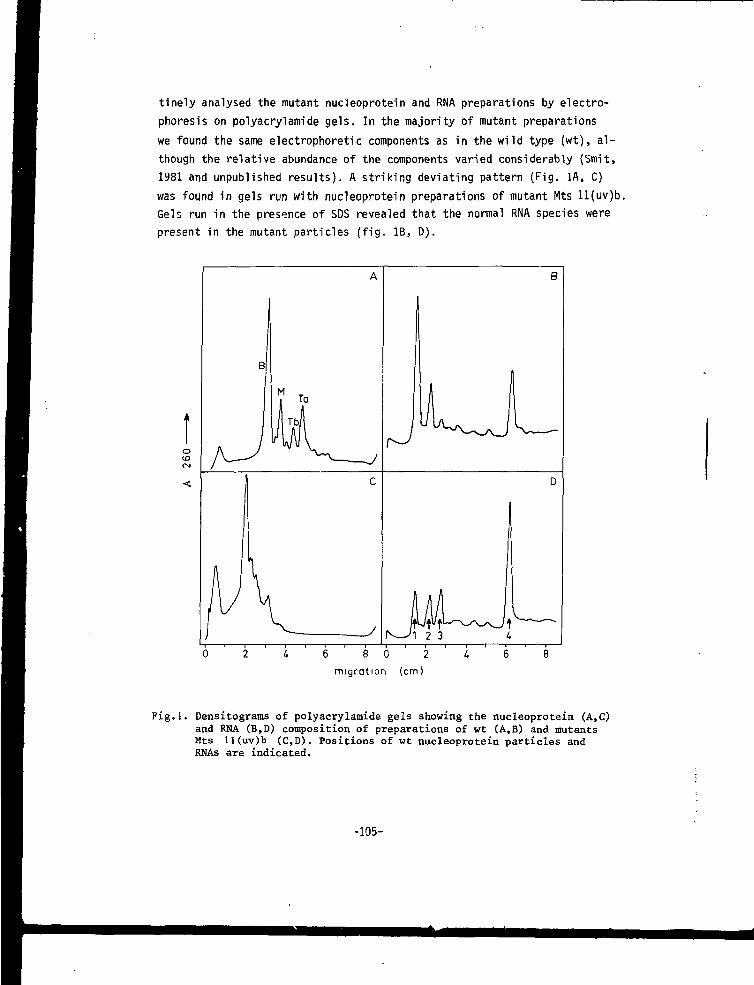
tinely analysed the mutant nucleoprotein and RNA preparations by electro-phoresis on polyacrylamide gels. In the majority of mutant preparationswe found the same electrophoretic components as in the wild type (wt), al-though the relative abundance of the components varied considerably (Smit,1981 and unpublished results). A striking deviating pattern (Fig. IA, C)was found in gels run with nucleoprotein preparations of mutant Mts ll(uv)b.Gels run in the presence of SDS revealed that the normal RNA species werepresent in the mutant particles (fig. IB, D).
oID
B
J1i
A
1
M
C
JLJ1
N-_Ji 2 3 U
B
(1
LD
L6 8 0 2
migration (cm)
Fig.l. Densitograms of polyacrylamide gels showing the nucleoprotein (A,C)and RMA (B,D) composition of preparations of wt (A,B) and mutantsMts 11(uv)b (C,D). Positions of wt nucleoprotein particles andRNAs are indicated.
-105-
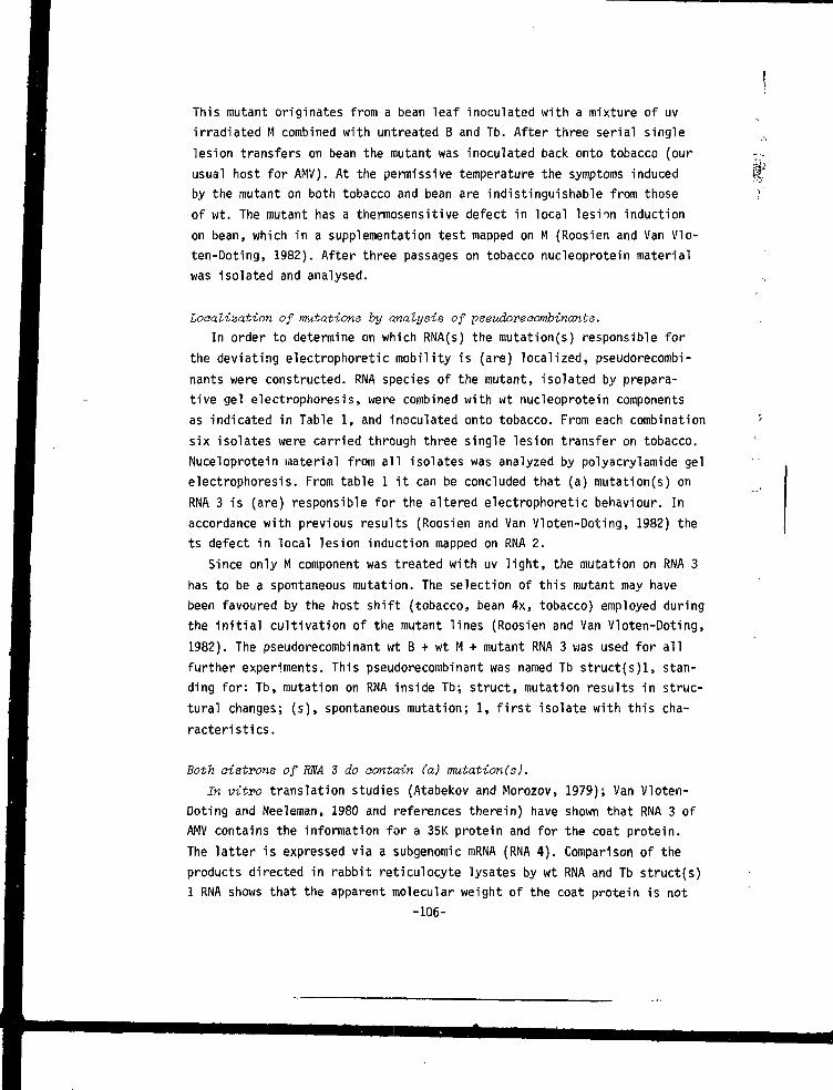
This mutant originates from a bean leaf inoculated with a mixture of uvirradiated M combined with untreated B and Tb. After three serial singlelesion transfers on bean the mutant was inoculated back onto tobacco (ourusual host for AMV). At the permissive temperature the symptoms inducedby the mutant on both tobacco and bean are indistinguishable from thoseof wt. The mutant has a thermosensitive defect in local lesion inductionon bean, which in a supplementation test mapped on M (Roosien and Van Vlo-ten-Doting, 1982). After three passages on tobacco nucleoprotein materialwas isolated and analysed.
Localization of mutations by analysis of pseudoreeombinants.In order to determine on which RNA(s) the mutation(s) responsible for
the deviating electrophoretic mobility is (are) localized, pseudorecombi-nants were constructed. RNA species of the mutant, isolated by prepara-tive gel electrophoresis, were combined with wt nucleoprotein componentsas indicated in Table 1, and inoculated onto tobacco. From each combinationsix isolates were carried through three single lesion transfer on tobacco.Nuceloprotein material from all isolates was analyzed by polyacrylamide gelelectrophoresis. From table 1 it can be concluded that (a) mutation(s) onRNA 3 is (are) responsible for the altered electrophoretic behaviour. Inaccordance with previous results (Roosien and Van Vloten-Doting, 1982) thets defect in local lesion induction mapped on RNA 2.
Since only M component was treated with uv light, the mutation on RNA 3has to be a spontaneous mutation. The selection of this mutant may havebeen favoured by the host shift (tobacco, bean 4x, tobacco) employed duringthe initial cultivation of the mutant lines (Roosien and Van Vloten-Doting,1982). The pseudorecombinant wt B + wt M + mutant RNA 3 was used for allfurther experiments. This pseudorecombinant was named Tb struct(s)l, stan-ding for: Tb, mutation on RNA inside Tb; struct, mutation results in struc-tural changes; (s), spontaneous mutation; 1, first isolate with this cha-racteristics.
Both oistrons of RNA 3 do contain (a) mutation(s).In vitro translation studies (Atabekov and Morozov, 1979); Van Vloten-
Doting and Neeleman, 1980 and references therein) have shown that RNA 3 ofAMV contains the information for a 35K protein and for the coat protein.The latter is expressed via a subgenomic mRNA (RNA 4). Comparison of theproducts directed in rabbit reticulocyte lysates by wt RNA and Tb struct(s)1 RNA shows that the apparent molecular weight of the coat protein is not
-106-
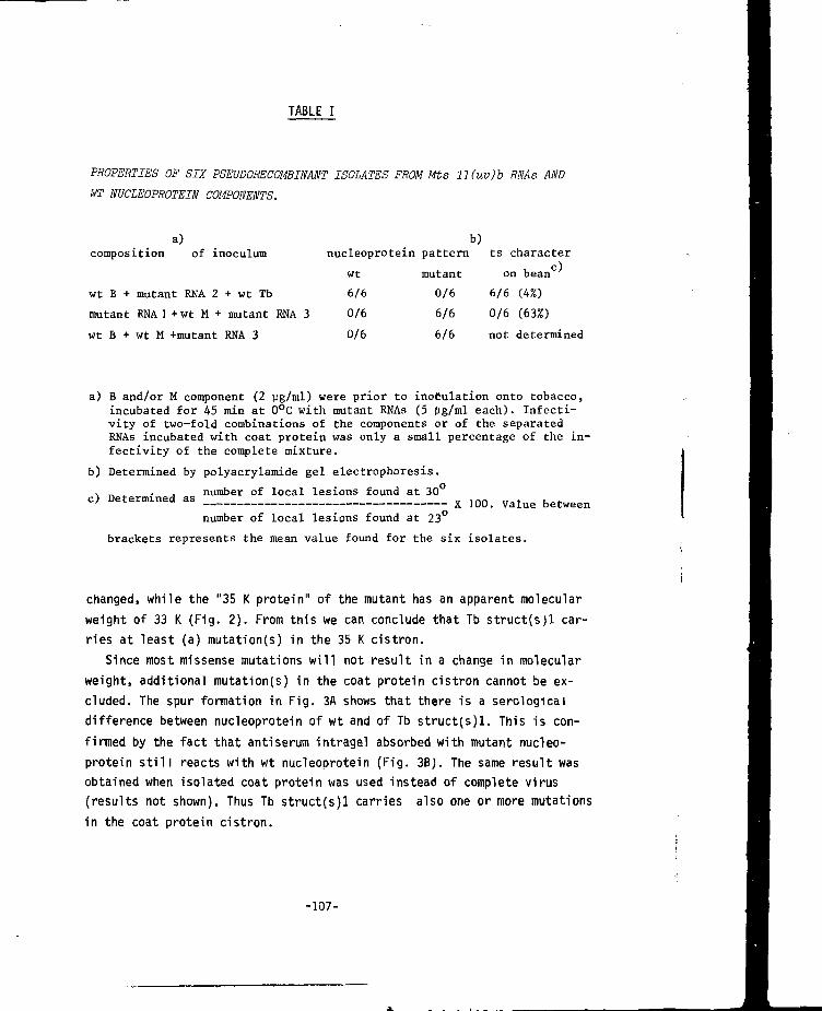
TABLE I
PROPERTIES OF SIX PSEUDORECOMBINANT ISOLATES FROM Mts ll(uv)b RNAs ANDWT NUCLEOPROTEIN COMPONENTS.
a)composition of inoculum
wt B + mutant RNA 2 + wt Tb
mutant RNA 1 + wt M + mutant RNA 3
wt B + wt M +mutant RNA 3
nucleoprotein pattern
wt mutant
6/6 0/6
0/6 6/6
0/6 6/6
b)ts character
on bean
6/6 (4%)
0/6 (63%)
not determined
a) B and/or M component (2 ug/ml) were prior to inoculation onto tobacco,incubated for 45 min at 0°C with mutant RNAs (5 (ig/ml each). Infecti-vity of two-fold combinations of the components or of the separatedRNAs incubated with coat protein was only a small percentage of the in-fectivity of the complete mixture.
b) Determined by polyacrylamide gel electrophoresis.
. number of local lesions found at 30
c) Determined as x ]00_ V a l u e b e t w e e n
number of local lesions found at 23°
brackets represents the mean value found for the six isolates.
changed, while the "35 K protein" of the mutant has an apparent molecularweight of 33 K (Fig. 2). From this we can conclude that Tb struct(s)l car-ries at least (a) mutation(s) in the 35 K cistron.
Since most missense mutations will not result in a change in molecularweight, additional mutation(s) in the coat protein cistron cannot be ex-cluded. The spur formation in Fig. 3A shows that there is a serologicaldifference between nucleoprotein of wt and of Tb struct(s)l. This is con-firmed by the fact that anti serum intragel absorbed with mutant nucleo-protein still reacts with wt nucleoprotein (Fig. 3BJ. The same result wasobtained when isolated coat protein was used instead of complete virus(results not shown). Thus Tb struct(s)l carries also one or more mutationsin the coat protein cistron.
-107-

35K-
CP-
Fig.2. Autoradiograph of polyacrylamide slabgel showing S methionine labeledproteins synthesized under the direc-tion of an RNA preparation enriched inRNA 3 of wt (lane 1) or Tb struct(s)I(lane 2). The positions of 35 K proteinand coat protein of wt are indicatedat the left.
Fig.3. Agar gel double diffusionprecipitation lines of60 (1,2), 30 (3,4) and15 (5,6) pg nucleoproteinof wt (1,3,5) and Tbstruct(s)l (2,4,6) uponreaction with 15 yl anti-serum raised against wt(A). (B) Same as (A)except that the middlewell was filled 24 hrsbefore with 60 vig Tbstruct(s)] nucleoprotein.
Comparison of the aomposition of double-stranded RNA isolated from tobaccoleaves infected with wt and mutant virus.
The composition of single stranded RNA present in wt and mutant virionsis very different (fig. 1B,D). If this difference is only due to a differentefficiency of encapsidation of the genomic RNAs in the mutant, we might ex-pect the same relative abundance of the double-stranded RNA species in wtand Tb struct(s)l infected tissue. Fig. 4 shows the double-stranded RNA pre-parations in mutant infected plants to be different from wt. In table 2 theamounts are quantified and it can be concluded that one or both of the RNA 3
-108-
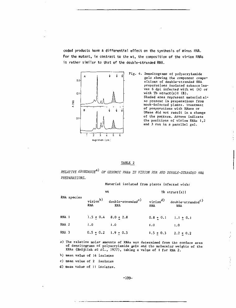
coded products have a differential effect on the synthesis of minus RNA.For the mutant, in contrast to the wt, the composition of the virion RNAsis rather similar to that of the double-stranded RNA.
o.v
Fig. 4. Densitograms of polyacrylamidegels showing the component compo-sitions of double-stranded RNApreparations isolated tobacco lea-ves 6 dpi infected with wt (A) orwith Tb struct(s)] (B).Shaded area represent material al-so present in preparations frommock-infected plants. Treatmentof preparations with RNase orDNase did not result in a changeof the pattern. Arrows indicatethe positions of virion RNAs 1,2and 3 run in a parallel gel.
1 2 3 i 5
migration (cm I
TABLE 2
RELATIVE ABUNDANCE*"* OF GENOMIC RNAs IN VIRION RNA AND DOUBLE-STRANDED RNA
PREPARATIONS.
Material isolated from plants infected with:
wt Tb struct(s)l
RNA species .virion double-strandedc
RNA RNA RNA RNAvirion double-stranded0
RNA 1
RNA 2
RNA 3
1.5 + 0.4 8.0 + 2.8
1.0 1.0
0.5 + 0.2 1.9 + 0.3
0.8+0.1 1.1+0.1
1.0 1.0
1.5 + 0.3 2.2 + 0.2
a) The relative molar amounts of RNAs was determined from the surface areaof densitograms of polyacrylamide gels and the molecular weights of theRNAs (Heijtink et al., 1977), taking a value of 1 for RNA 2.
b) mean value of 14 isolates
c) mean value of 2 isolates
d) mean value of II isolates.
-109-
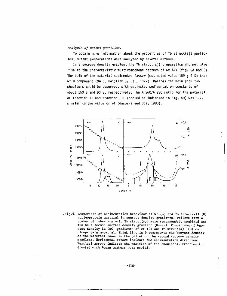
Analysis of mutant particles.To obtain more information about the properties of Tb struct(s)l partic-
les, mutant preparations were analysed by several methods.In a sucrose density gradient the Tb struct(s)l preparation did not give
rise to the characteristic multicomponent pattern of wt AMV (fig. 5A and B).The bulk of the material sedimented faster (estimated value 109 + 4 S) thanwt B component (94 S, Heijtink et al., 1977). Besides the main peak twoshoulders could be observed, with estimated sedimentation constants ofabout 150 S and 90 S, respectively. The A 260/A 280 ratio for the materialof fraction II and fraction III (pooled as indicated in Fig. 5D) was 1.7,similar to the value of wt (Jaspars and Bos, 1980).
-Fig.5. Comparison of sedimentation behaviour of wt (A) and Tb struct(s)] (B)nucleoprotein material in sucrose density gradients. Pellets from anumber of tubes run with Tb struct(s)] were resuspended, combined andrun on a second sucrose density gradient (B ). Comparison of buo-yant density in CsCl gradients of wt (C) and Tb struct(s)l (D) nu-cleoprotein material. Thick line in D represents the buoyant densityof the material found in the pellet of the second sucrose densitygradient. Horizontal arrov/s indicate the sedimantation direction.Vertical arrows indicate the position of the shoulders. Fraction in-dicated with Roman numbers were pooled.
-110-
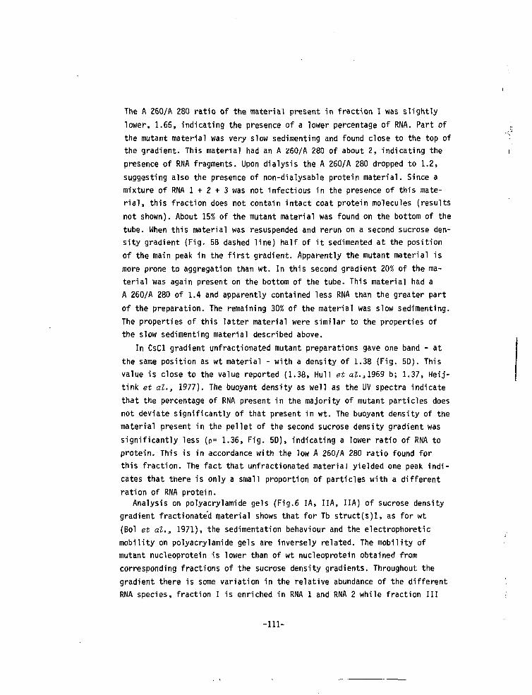
The A 260/A 280 ratio of the material present in fraction I was slightlylower, 1.65, indicating the presence of a lower percentage of RNA. Part ofthe mutant material was very slow segmenting and found close to the top ofthe gradient. This material had an A 260/A 280 of about 2, indicating thepresence of RNA fragments. Upon dialysis the A 260/A 280 dropped to 1.2,suggesting also the presence of non-dialysable protein material. Since amixture of RNA 1 + 2 + 3 was not infectious in the presence of this mate-rial, this fraction does not contain intact coat protein molecules (resultsnot shown). About 15% of the mutant material was found on the bottom of thetube. When this material was resuspended and rerun on a second sucrose den-sity gradient (Fig, 5B dashed line) half of it sedimented at the positionof the main peak in the first gradient. Apparently the mutant material ismore prone to aggregation than wt. In this second gradient 20% of the ma-terial was again present on the bottom of the tube- This material had aA 260/A 280 of 1.4 and apparently contained less RNA than the greater partof the preparation. The remaining 30% of the material was slow sedimenting.The properties of this latter material were similar to the properties ofthe slow sedimenting material described above.
In CsCl gradient unfractionated mutant preparations gave one band - atthe same position as wt material - with a density of 1.38 (Fig. 5D). Thisvalue is close to the value reported (1.38, Hull et al.,1969 b; 1.37, Heij-tink et al., 1977). The buoyant density as well as the UV spectra indicatethat the percentage of RNA present in the majority of mutant particles doesnot deviate significantly of that present in wt. The buoyant density of thematerial present in the pellet of the second sucrose density gradient wassignificantly less (p= 1.36, Fig. 5D), indicating a lower ratio of RNA toprotein. This is in accordance with the low A 260/A 280 ratio found forthis fraction. The fact that unfractionated material yielded one peak indi-cates that there is only a small proportion of particles with a differentration of RNA protein.
Analysis on polyacrylamide gels (Fig.6 IA, IIA, IIA) of sucrose densitygradient fractionated material shows that for Tb struct(s)l, as for wt(Bol et al., 1971), the sedimentation behaviour and the electrophoreticmobility on polyacrylamide gels are inversely related. The mobility ofmutant nucleoprotein is lower than of wt nucleoprotein obtained fromcorresponding fractions of the sucrose density gradients. Throughout, thegradient there is some variation in the relative abundance of the differentRNA species, fraction I is enriched in RNA 1 and RNA 2 while fraction III
-111-
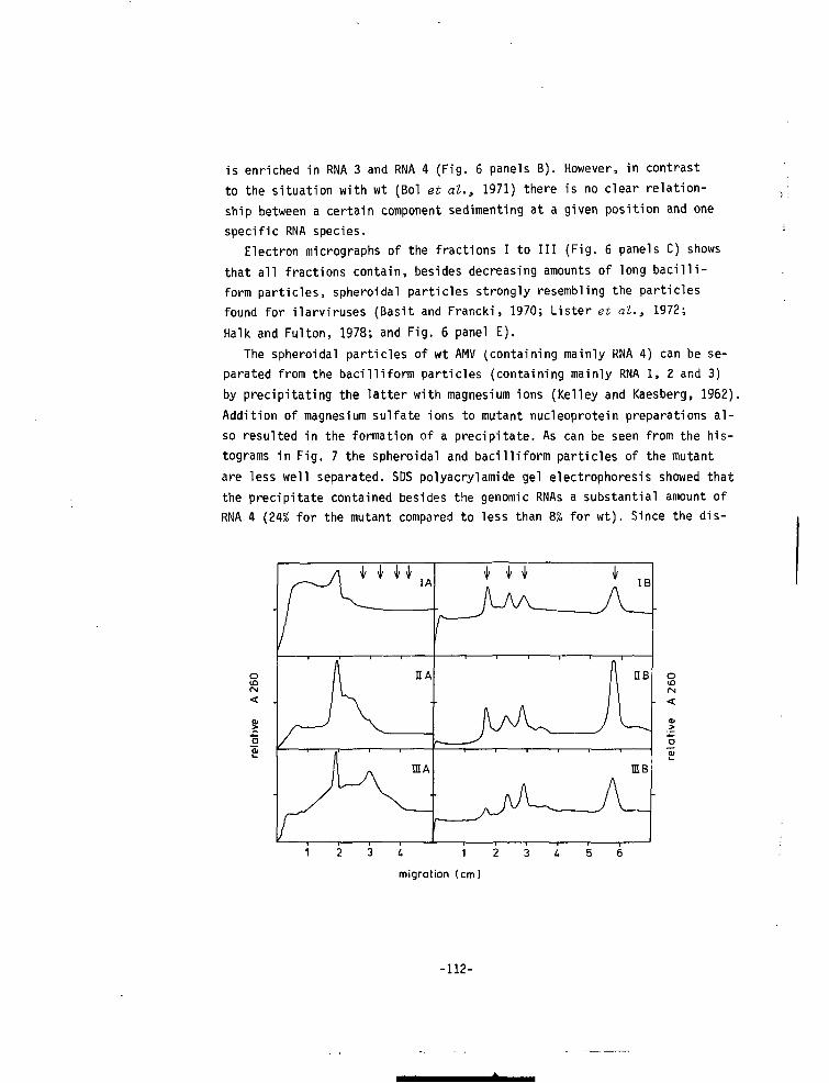
is enriched in RNA 3 and RNA 4 (Fig. 6 panels B). However, in contrastto the situation with wt (Bol et al., 1971) there is no clear relation-ship between a certain component sedimenting at a given position and onespecific RNA species.
Electron micrographs of the fractions I to III (Fig. 6 panels C) showsthat all fractions contain, besides decreasing amounts of long bacilli-form particles, spheroidal particles strongly resembling the particlesfound for ilarviruses (Basit and Francki, 1970; Lister et al., 1972;Halk and Fulton, 1978; and Fig. 6 panel E).
The spheroidal particles of wt AMV (containing mainly RNA 4) can be se-parated from the bacilliform particles (containing mainly RNA 1, Z and 3)by precipitating the latter with magnesium ions (Kelley and Kaesberg, 1962).Addition of magnesium sulfate ions to mutant nucleoprotein preparations al-so resulted in the formation of a precipitate. As can be seen from the his-tograms in Fig. 7 the spheroidal and bacilliform particles of the mutantare less well separated. SDS polyacrylamide gel electrophoresis showed thatthe precipitate contained besides the genomic RNAs a substantial amount ofRNA 4 (24% for the mutant compared to less than 8% for wt). Since the dis-
csID
U 1 2
migration (cm)
-112-
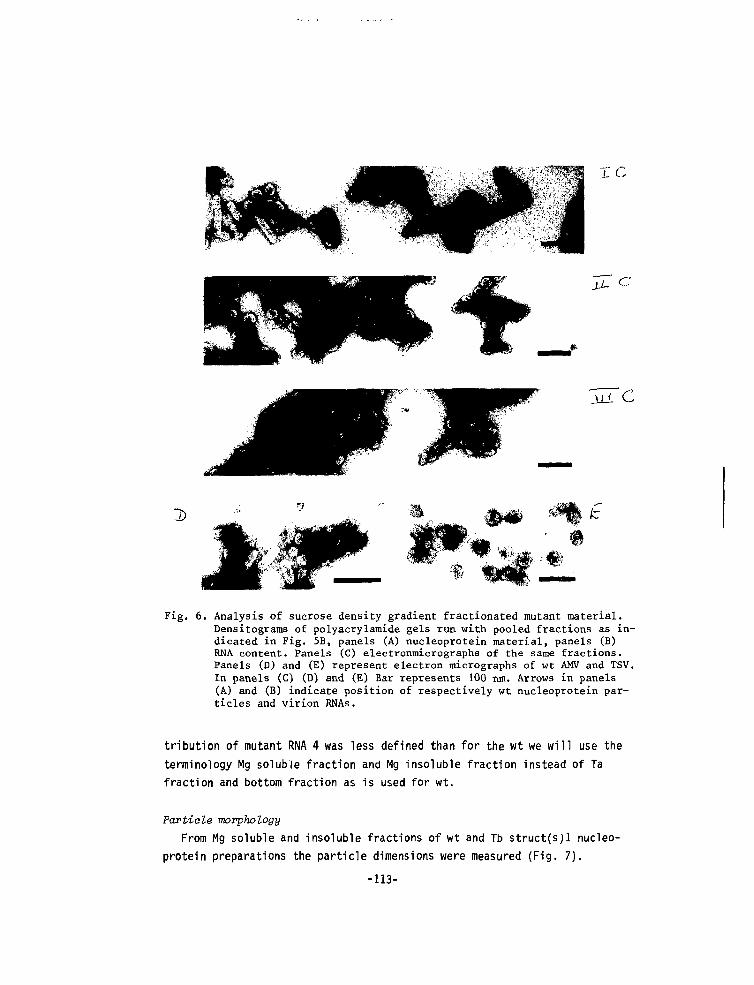
D
Fig. 6. Analysis of sucrose density gradient fractionated mutant material.Densitograms of polyaerylamide gels run with pooled fractions as in-dicated in Fig. 5B, panels (A) nucleoprotein material, panels (B)RNA content. Panels (C) electronmicrographs of the same fractions.Panels (D) and (E) represent electron micrographs of wt AMV and TSV.In panels (C) (D) and (E) Bar represents 100 nm. Arrows in panels(A) and (B) indicate position of respectively wt nucleoprotein par-ticles and virion RNAs.
tribution of mutant RNA 4 was less defined than for the wt we will use theterminology Mg soluble fraction and Mg insoluble fraction instead of Tafraction and bottom fraction as is used for wt.
Particle morphologyFrom Mg soluble and insoluble fractions of wt and Tb struct(s;i nucleo-
protein preparations the particle dimensions were measured (Fig. 7).
-113-
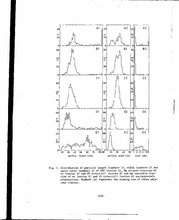
20 30 40 50 60 70particle length (nm)
80)80 20 30 40 50 1 2 3 4 5>5
particle width (nm) axial ratio
Fig. 7. Distribution of particle length (numbers 1), width (numbers 2) andaxial ratio (numbers 3) of TSV (series C), Mg soluble fraction ofwt (series D) and Tb struct(s)l (series B) and Mg insoluble frac-tion of wt (series E) and Tb struct(s)l (series A) nucleoproteinpreparations. Numbers (n) represent the running sum of three adja-cent classes.
-114-
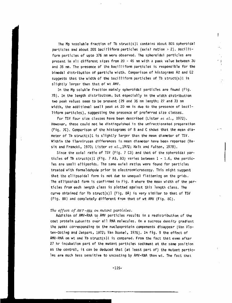
The Mg insoluble fraction of Tb struct(s)l contains about 80% spheroidalparticles and about 20% bacilliform particles (axial ration > 2). Baci l l i -form particles of upto 376 nm were observed. The spheroidal particles arepresent in al l different sizes from 20 - 45 nm with a peak value between 30and 35 nm. The presence of the bacilliform particles is responsible for thebimodal distribution of particle width. Comparison of histograms A2 and E2suggests that the width of the bacilliform particles of Tb struct(s)l isslightly larger than that of wt AMV.
In the Mg soluble fraction mainly spheroidal particles are found (Fig.7B). In the length distribution, but especially in the width distributiontwo peak values seem to be present (29 and 35 nm length; 27 and 33 nmwidth, the additional small peak at 20 nm is due to the presence of bacil-liform particles), suggesting the presence of preferred size classes.
For TSV four size classes have been described (Lister et al., 1972).However, these could not be distinguished in the unfractionated preparation(Fig. 7C). Comparison of the histograms of B and C shows that the mean dia-meter of Tb struct(s)l is slightly larger than the mean diameter of TSV.Within the ilarviruses differences in mean diameter have been reported (Ba-s i t and Francki, 1970; Lister et aZ.,,1972; Halk and Fulton, 1978).
Since the axial ratio of TSV (Fig. 7 C3) and that of the spheroidal par-ticles of Tb struct(s)l (Fig. 7 A3, B3) varies between 1 - 1.6, the partic-les are small ellipsoids. The same axial ratios were found for particlestreated with formaldehyde prior to electronmicroscopy. This might suggestthat the ellipsoidal form is not due to unequal flattening on the grids.The ellipsoidal form is confirmed in Fig. 8 where the mean width of the par-ticles from each length class is plotted against this length class. Thecurve obtained for Tb struct(s)l (Fig. 8A) is very similar to that of TSV(Fig. 8B) and completely different from that of wt AMV (Fig. 8CJ.
The effect of AMV -MA on mutant particles.Addition of AMV-RNA to AMV particles results in a redistribution of the
coat protein subunits over al l RNA molecules. On a sucrose density gradientthe peaks corresponding to the nucleoprotein components disappear (Van VI o-ten-Doting and Jaspars, 1972; Van Boxsel, 1976). In f i g . 9 the effect ofAMV-RNA on wt and Tb struct(s)l is compared. From the fact that even after27 hr incubation part of the mutant particles sediment at the same positionas the control, i t can be deduced that (at least part of) the mutant partic-les are much less sensitive to uncoating by AMV-RNA than wt. The fact that
' i
-115-
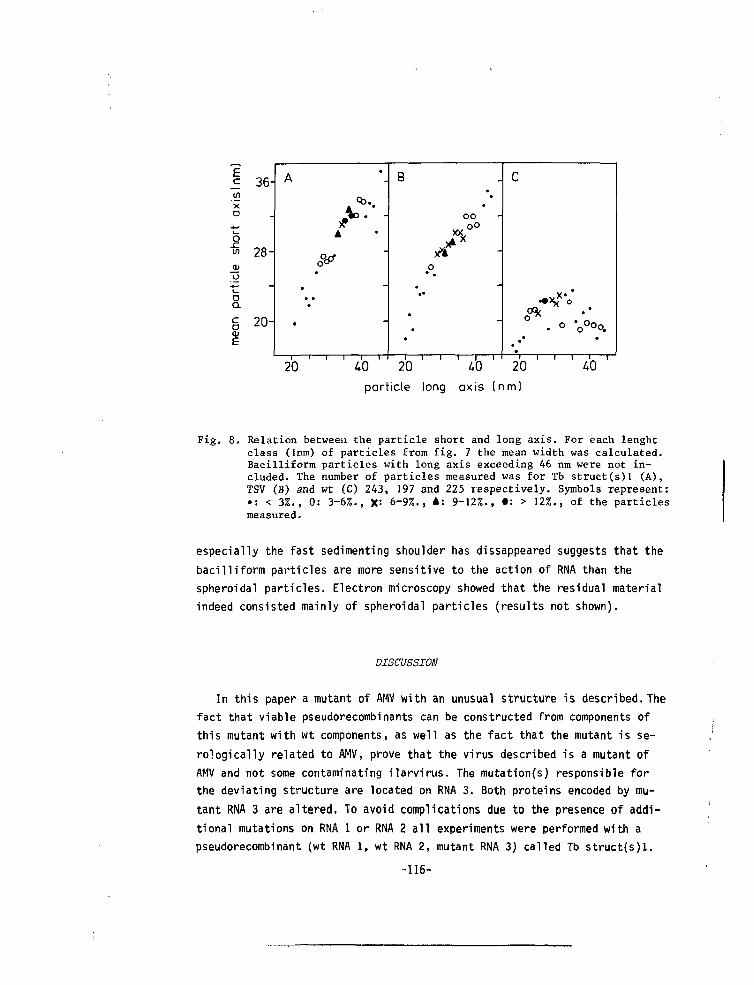
EcXo
o
oQ.co
36-
-
28-
-
20-
A
c••
• ••
•
,—,—
P:
A
ov
"l 1 r
•
• -
•
-
-
-
|—
B
•— r
-
•
oo
o• •
• —
*
•
•
—i—i—i—i—r
C
o *
••
' - . 1
•
•
•
~i—i—i—H
par t i c l e long ax i s ( n m )
Fig. 8. Relation between the particle short and long axis. For each lenghtclass (Inm) of particles from fig. 7 the mean width was calculated.Bacilliform particles with long axis exceeding 46 nm were not in-cluded. The number of particles measured was for Tb struct(s)l (A),TSV (B) and wt (C) 243, 197 and 225 respectively. Symbols represent:•: < 3%., 0: 3-6%., X= 6-9%., A: 9-12%., t: > 12%., of the particlesmeasured.
especially the fast sedimenting shoulder has dissappeared suggests that thebacilliform particles are more sensitive to the action of RNA than thespheroidal particles. Electron microscopy showed that the residual materialindeed consisted mainly of spheroidal particles (results not shown).
DISCUSSION
In this paper a mutant of AMV with an unusual structure is described. Thefact that viable pseudorecombinants can be constructed from components ofthis mutant with wt components, as well as the fact that the mutant is se-rologically related to AMV, prove that the virus described is a mutant ofAMV and not some contaminating ilarvirus. The mutation(s) responsible forthe deviating structure are located on RNA 3. Both proteins encoded by mu-tant RNA 3 are altered. To avoid complications due to the presence of addi-tional mutations on RNA 1 or RNA 2 all experiments were performed with apseudorecombinant (wt RNA l, wt RNA 2, mutant RNA 3) called Tb struct(s)l.
-116-
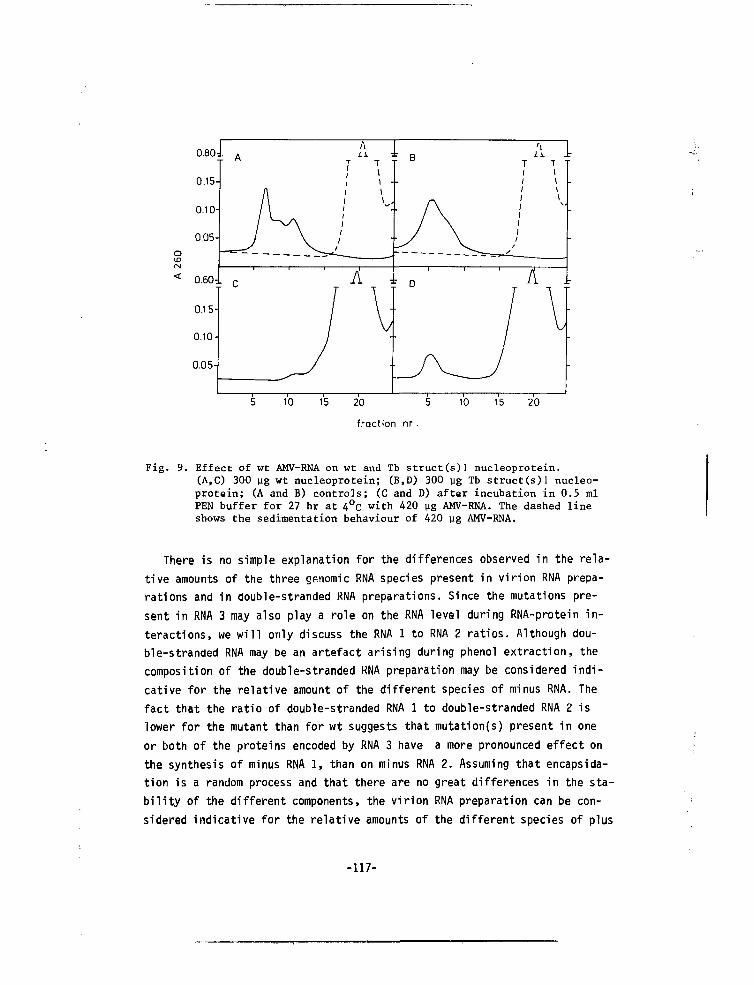
0.80-
0.15-
0.10-
0.05-
0.60:
0.15-
0.10-
0.05-
A
A\/ ^J
: C
\\ii
T
(
11
A7"
/
J
T\\ -\
VV
B
• A/ \
" D
/
Xi.T11
,'
11
1
/
AT1
JT11\\
1 Tv10 15 20 5
fraction nr .
10 15 20
Fig. 9. Effect of wt AMV-RHA on wt and Tb struct(s)] nucleoprotein.(A,C) 300 ug wt nucleoprotein; (B,D) 300 yg Tb struct(s)l nucleo-protein; (A and B) controls; (C and D) after incubation in 0.5 mlPEN buffer for 27 hr at 4°c with 420 ug AMV-RNA. The dashed lineshows the sedimentation behaviour of 420 ug AMV-RNA.
There is no simple explanation for the differences observed in the rela-tive amounts of the three genomic RNA species present in virion RNA prepa-rations and in double-stranded RNA preparations. Since the mutations pre-sent in RNA 3 may also play a role on the RNA level during RNA-protein in-teractions, we wi l l only discuss the RNA 1 to RNA 2 ratios. Although dou-ble-stranded RNA may be an artefact arising during phenol extraction, thecomposition of the double-stranded RNA preparation may be considered indi-cative for the relative amount of the different species of minus RNA. Thefact that the ratio of double-stranded RNA 1 to double-stranded RNA 2 islower for the mutant than for wt suggests that mutation(s) present in oneor both of the proteins encoded by RNA 3 have a more pronounced effect onthe synthesis of minus RNA 1, than on minus RNA 2. Assuming that encapsida-tion is a random process and that there are no great differences in the sta-b i l i t y of the different components, the virion RNA preparation can be con-sidered indicative for the relative amounts of the different species of plus
-117-
RNA. Based on these assumptions we can calculate that the amount of plus
strand RNA synthesized per minus strand RNA varies considerably. This might
again point to a difference in the role played by RNA 3 coded proteins
during the synthesis of the different RNA species.
Since both cistrons present in RNA 3 were found to contain mutations
there is only circumstantial evidence that changes in the coat protein are
responsable for changes in the structure. To our knowledgement there are no
indications that 35 K protein does function as a matrix protein. On the
contrary for several related plant viruses (Hiebert et al., 1968; Bancroft,
1970) it has been shown that purified RNA and coat protein can self assem-
ble into nucleoprotein particles indistinguishable from particles isolated
from infected leaves. The reconstitution of AMV particles from purified RNA
and protein is only partly successful (Driedonks et al., 1978, and refe-
rences therein). However the bacilliform particles can be reversely dis-
sociated and associated with high salt or high pH (Bol and Kruseman- 1969;
Verhagen et al., 1976) suggesting that the combination of RNA and coat
protein contains sufficient information for particle formation.
Mutant preparations do contain a large proportion of spheroidal particles
and a minor proportion (15-20%) of bacilliform particles. The majority of
these bacilliform particles is much longer than the particles in wt. Since
mutant nucleoprotein preparations do contain the same percentage of RNA
and the same RNA species as wt, these longer mutant particles have to con-
tain more than one RNA molecule. A similar situation has been described
previously for the long bacilliform particles observed in preparations from
two other AMV strains (VRU; Hull, 1970a and 15/64; Hull et al., 1969a). It
has been argued that these structures are due to an enchanced tendency of
the coat proteins from these strain to aggregate in hexamers (forming the
tubular part) (Cremers et al., 1981). The same reasoning would lead to the
suggestion that the spheroidal particles of Tb struct(s)l also do contain
a large proportion of hexamers. This however raises a serious structural
problem.
The spheroidal particles of Tb struct(s)l closely resemble the spheroi-
dal particles described for the ilarviruses (Fulton, 1967; Fulton, 1968;
Basit and Francki, 1970; Lister et al., 1972; Jones and Mayo, 1973; Garnsey,
1975; Van der Meer et al., 1976; Halk and Fulton, 1978; Bos et al., 1980).
Lister et al. (1972) described discrete size classes for the purified com-
ponents of TSV, however the dimensions of the particles did not fit into
a T = 7,4,3 and 1 series (Caspar and Klug, 1962). The structure of this type
-118-
of particles is unknown. The axial ratios of the spheroidal particles of
TSV and the AMV mutant do not differ significantly. A remarkable feature
for AMV (Heijtink et al.t 1977) and the ilarviruses (Lister et at., 1972;
Lister and Saksena, 1976; Halk and Fulton, 1978) is that all nucleoprotein
particles have a closely similar density, reflecting similar protein/
nucleic acid ratios. Apparently the protein/nucleic acid ratio is rather
strictly controled during assembly. According to the criteria defined by
Kaper and Geelen (1971), the nucleoprotein particles of these viruses are
primarily stabilized by RNA-protein interactions. For AMV it has been shown
that at alkaline pH the particles unfold without loss of coat protein (Ver-
hagen and Bol, 1972). AMV- and probably also the ilarvirus-particles can
be considered to be folded RNA-protein complexes. In the wt each of the
four RNA protein complexes has a preferred folding, leading to the four
major components. Alternative foldings are also realized, leading to part
of the 13 described minor components (part of the minor components is due
to the presence of intermediate sized RNAs) (Bol et al., 1971). In wt AMV
the folding is such that particles with symmetric features are formed. In
the mutant each of the RNA-protein complexes can probably fold in more than
one way so that a series of particles of all sizes is formed. This suggests
that the spheroidal particles consist of a supercoiled framework of RNA
complexed with protein subunits. These protein subunits may be organized in
a complex with local symmetry, but do not form a point group symmetry as
with classical spherical shells. Recently Cusack et al. f1981) proposed
that the structure of the spheroidal Ta-t particle of wt AMV was also
non-icosahedral and not built according to the hitherto accepted principles
of virus construction.
The folding of the RNA protein complexes is probably partly driven by
protein-protein interactions. The observation (Van Vloten-Doting, 1975;
Gonsalves and Garnsey, 1975) that ilarviruses as well as the spheroidal
mutant particles are less sensitive to uncoating by AMV-RNA suggests that
this protein-protein interaction is more important in the spheroidal par-
ticles than in the bacilliform particles.
In the near future we hope to compare the theoretical secondary structure
(Chou and Fassman, 1974) of mutant coat protein (the amino acid sequence
will be deduced from the nucleotide sequence of mutant RNA 4) with that of
wt coat protein and VRU coa\. protein (Castel et al., 1979). Such a com-
parison may reveal indications why different coat proteins yield different
assembly products.
-119-
The occurrence of an AMV mutant which can encapsidate the genomic RNAsin bacilliform particles as well as in particles closely resembling thespheroidal particles typical for ilarviruses, argues in favour of the pro-posed incorporation of AMV in the ilarvirus genus of the Tricornaviridaefamily (Van Vloten-Doting et al., 1981).
ACKNOWLEDGEMENTS.
We thank mr A.G. Wesseling for cultivation of plants and M. Huis- In 'tVeld, P.C.J. Krijgsman, G.T. Oostergetel and R. Ruigrok for excellent tech-nical assistence. We are grateful to dr. J.E. Mellema and dr E.M. Jasparsfor encouraging discussions. This work was financially supported by theNetherlands Organization for Advancement of Pure Research (Z.W.O.).
-120-
REFERENCES
Atabekov, J.G. and Morozov. S.Yu. (1979). Advan. Virus Res. j?5_, 1-91.
Bancroft, J.B. (1970). Advan. Virus Res. 1£, 99-134.
Basit, A.A. and Francki, R.I.B. (1970). Australian J. of Biol. Soi. Q,
1197-1206.
Bol, J.F. and Kruseman, J. (1969). virology 3]_, 485-488.
Bol, J.F., Van Vloten-Doting, L. and Jaspars, E.M.J. (1971). Virology 46,
73-85.
Bonner, W.M. and Laskey, R.A. (1974). Eur. J. Bioekem. 46, 83-88.
Bos, L., Huttinga, H. and Maat, D.Z. (1980). Neth. J. Pi. Path. 86, 79-98.
Caspar, D.L.D. and Klug, A. (1962). Cold Spring Harbor Symposia on Quan-
titative Biology T}_, 1-24.
Caste!, A., Kraal, B., de Graaf, J.M. and Bosch, L. (1979). Eur. J. Bioch.
102, 125-138.
Chou, P.Y. and Fasman, G.D. (1974). Biochemistry J^, 222-245.
Cremers, A.F.M., Oostergetel, G.T., Schilstra, M.J. and Mellema, J.E.(1981)
J. Mol. Biol. ^45, 545-561.
Cusack, S., Krijgsman, P.C.J., Mellema, J.E. and Oostergetel, G.T. (1980).
E.M.B.L. Research Reports 1980, c<*~ 246.
Dinjan-Versteegh, A., Van Vloten-Doting, L. and Jaspars, E.M.J. (1972).
Virology 49, 716-722.
Driedonks, R.A., Krijgsman, P.C.J. and Mellema, J.E. (1978). Eur. J. Bioch.
82, 405-417.
Fulton, R.W. (1967). Virology 32, 153.
Fulton, R.W. (1968). Wiss. Berlin Tagungsber. 97_, 123-138.
Garnsey, S.M. (1975). Phytopathology 6£, 50-57.
Gonsalves, u. and Garnsey, S.M. (1975). Virology (54, 23-31.
Gonsalves, b. and Fulton, R.W. (1977). Virology _81, 398-407.
Hagedorn, D.J. and Hanson, E.W. (1963). phytopathology 5_3, 188-192.
Halk, E.L. and Fulton, R.W. (1978). virology 91_, 434-443.
-121-
Hiebert, E., Bancroft, J.B. and Bracker, C E . (1968). virology 34, 492-508
Heijtink, R.A. and Jaspars, E.M.J. (1974). Virology 59, 371-382.
Heijtink, R.A. and Jaspars, E.M.J. (1976). Virology 69_, 75-80.
Heijtink, R.A., Houwing, C.J. and Jaspars, E.M.J. (1977). Biochemistry 16,
4684-4693.
Houwing, C.J. and Jaspars, E.M.J. (1978). Biochemistry 17, 2927-2933.
Hull, R., Rees, M.W. and Short, M.N. (1969a). virology 37, 404-415.
Hull, R., Hills, G.J. and Markham, R. (1969b). Virology 3_7, 416-428.
Huil, k. (1970a). Virology 42, 283.
Hull, R. (1970b). Virology 40, 34-47.
Huttinga, H. and Mosch, W.H.M. (1976). Acta Horticulturae 59, 113-118.
Ifft, J.B., Voet, D.H. and Vinograd, J. (1961). J. Phys. Chem. 65 , 1138-
1145.
Jaspars, E.M.J. and Bos, L. (1980). CMI/AAB. Description of plant viruses.
Nr. 229.
Jones, A.T. and Mayo, M.A. (1973). Ann.appl. Biol. 75_, 347-357.
Kaper, J.M. and Geelen, J.L.M.C. (1971). J. Mol. Biol. 56, 277-294.
Kelley, J.J. and Kaesberg, P. (1962). Bioahim. Biophys. Acta (51_, 865.
Lister, R.M. and Saksena, K.N. (1976). Virology 7£, 440-450.
Laemmli, U.K. (1970). Nature (London) 22&_, 680-685.
Lister, R.M., Gabrial, S.A. and Saksena, K.N. (1972). Virology 49, 290-299.
Matthews, R.E.F. (1979). Intervirology 12_, 3-5.
Meilema, J. (1975). J. Mol. Biol. 94, 643-648.
Morris, T.J. and Dodds, J.A. (1979). Phytopathology 69, 854-858.
Roosien, J. and Van Vloten-Doting, L. (1982). J. Gen. Virol. 63, 189-198.
Smit, C.H. (1981). Ph.D. Thesis, University of Leiden.
Smit, C.H., Roosien, J., Van Vloten-Doting, L. and Jaspars, E.M.J. (1981).
Virology JL12, 169-173.
Van Boxsel, J.A.M. (1976). Ph.D. Thesis, University of Leiden.
-122-
Van der Meer, F.A., Huttinga, H. and Maat, D.Z. (1976). Neth. j. PI. Puth.
82, 67-80.
Van Tol, R.G.L. and Van Vloten-Doting, L. (1979). Eur. J. Bioohem. 93_, 461-
468.
Van Vloten-Doting, L. (1975). Virology 65, 215-225.
Van Vloten-Doting, L. and Jaspars, E.M.J. (1967). Virology 3^, 684-693.
Van Vloten-Doting, L. and Jaspars, E.M.J. (1972). Virology 48, 699-708.
Van Vloten-Doting, L. and Jaspars, E.M.J. (1977). Comprehensive Virology
II, 1-53.
Van Vloten-Doting, L. and Neeleman, L. (1980). Genome Org. and Expr. in
Plants 511-527.
Van Vloten-Doting, L., Kruseman, J. and Jaspars, E.M.J. (1968). virology34, 728-737.
Van Vloten-Doting, L., Hasrat, J.A., Oosterwijk, E., Van 't Sar.;, P.,
Schoen, M.A. and Roosien, J. (1980). J.Gen. Virol 46_, 415-426.
Van Vloten-Doting, L., Francki, R.T.B., Fulton, R.W., Kaper, J.M. and Lane,
L.C. Intervirology ^£, 198-203.
Verhagen, W. and Bol, J.F. (1972). Virology 50, 431-439.
Verhagen, W., Van Boxsel, J.A.M., Bol, J.F., Van Vloten-Doting, L. and
Jaspars, E.M.J. (1976). Ann. miarobiol 127a, 165-172.
-123-
CHAPTER VIII
Submitted for publication
SYNTHESIS OF ALFALFA MOSAIC VIRUS RNA 2 IS LIMITED BY ITS OWN EXPRESSION
J. Roosien, A.N. Sarachu, F. Alblas and L. Van Vloten-DotingDepartment of Biochemistry, State University of Leiden, P.O. Box 9505,2300 RA Leiden, The Netherlands.
SUMMARY
An RNA 2 mutant of Alfalfa Mosaic Virus (AMV) is replicated more effi-ciently than the wildtype (wt) in cowpea protoplasts. Mutant preparationsisolated from infected cowpea protoplasts contained a higher amount ofMiddle component (M, containing RNA 2) than wt preparations. Supplemen-tation of the mutant with wt M resulted in a decrease of the total yieldand especially of the amount of M components produced. This indicates thatthe synthesis of AMV RNA 2 is limited by its own expression.
-125-
INTRODUCTION
AMV is a plantvirus with a tripartite genome consisting of RNA 1, 2 and3 encapsidated into B, M and Tb components respectively (Bol et al., 1971).In order to obtain more information about the replication process we haveproduced a collection of mutants (Van Vloten-Doting et al.3 1980; Roosienand Van Vloten-Doting, 1982) by UV irradiation of one purified component.One of these mutants (induced by UV irradiation of M) induced systemicchlorosis in bean plants, while the parent strain (AMV 425, Hagedorn andHanson, 1963) only induced necrotic local lesions (Roosien and Van Vloten-Doting» 1982). Recently we found a similar difference in symptoms inducedby this mutant and wt in cowpea plants. By comparing the properties ofmutant and wt virus we hope to obtain indications why the replication ofwt virus is confined to a small number of cells in the inoculated leaves,while the mutant can invade the whole plant. In the present paper we showthat in cowpea protoplasts the mutant is replicated more efficiently thanthe wt and an altered balance between viral nucleoproteins is observed.Supplementation experiments indicate that a change in a product encodedby RNA 2 is responsible for these differences.
-126-
RESULTS AND DISCUSSION
In Fig.IB the symptoms induced by the mutant on cowpea plants are pre-sented. From the supplementation emperiment (Fig.lA) we can deduce that themutation responsible for this change in symptoms is located - as expected -on RNA 2. The mutant had been called Msyst l(uv)b (Roosien and Van Vloten-Doting, 1982), standing for : mutation on M, systemic infection on bean,isolate j , m/ induced. In line with this nomenclature a letter c was addedto this name to indicate that the mutant also induced systemic symptomsin cowpea.
Cowpea protoplasts were isolated, infected (Nassuth et al., 1981) andincubated as before (Albas and Bol, 1977). In all experiments the incu-bation time was 40 hours, since it is known that at that time the virusproduction has reached a plateau (Nassuth et al.3 to be published). Sur-vival of protoplasts after infection with wt and Msyst l(uv)b,c was com-parable. The amount of nucleoprotein isolated (Van Vloten-Doting andJaspars, 1972, except that 0.1 M NaCl was added to the homogenization buf-fer) from cowpea protoplasts infected with mutant was reproducibly threetimes as much as that obtained from wt infected protoplasts (Table 1).Analysis of the nucleoprotein component composition showed, besides smallalterations in the relative amounts of all components, a striking increasein the amount of M in mutant preparations (Fig.2A,B). The increase in theamount of total nucleoprotein (Table I) is only partly due to the increasein the amount of M. Supplementation of mutant with wt M reduced the yieldto less than 40% (Table I) and resulted in the reappearance of wt componentcomposition (Fig.2C). The progeny from Msyst l(uv)b,c plus wt M infectedprotoplasts induced in bean both mutant- and wt-symptoms (Table I) indi-cating that both types of RNA 2 had been replicated. In a control experi-ment with wt virus plus additional wt M we observed a reduction to 65% inthe amount of nucleoprotein produced, reflecting the reduction of the othercomponents in the inoculum. No change in component composition was found(Fig.2D). The increase in M components observed in mutant infected proto-plasts can either reflect an increase in the relative amount of RNA 2 syn-thesized or an increase of its encapsidation. Since it is known that in wtinfected cowpea protoplasts at least 80% of the RNA 2 molecules present areencapsidated (Nassuth et al., to be published) an enhanced encapsidationcannot explain the increase on M component in the mutant.
-127-
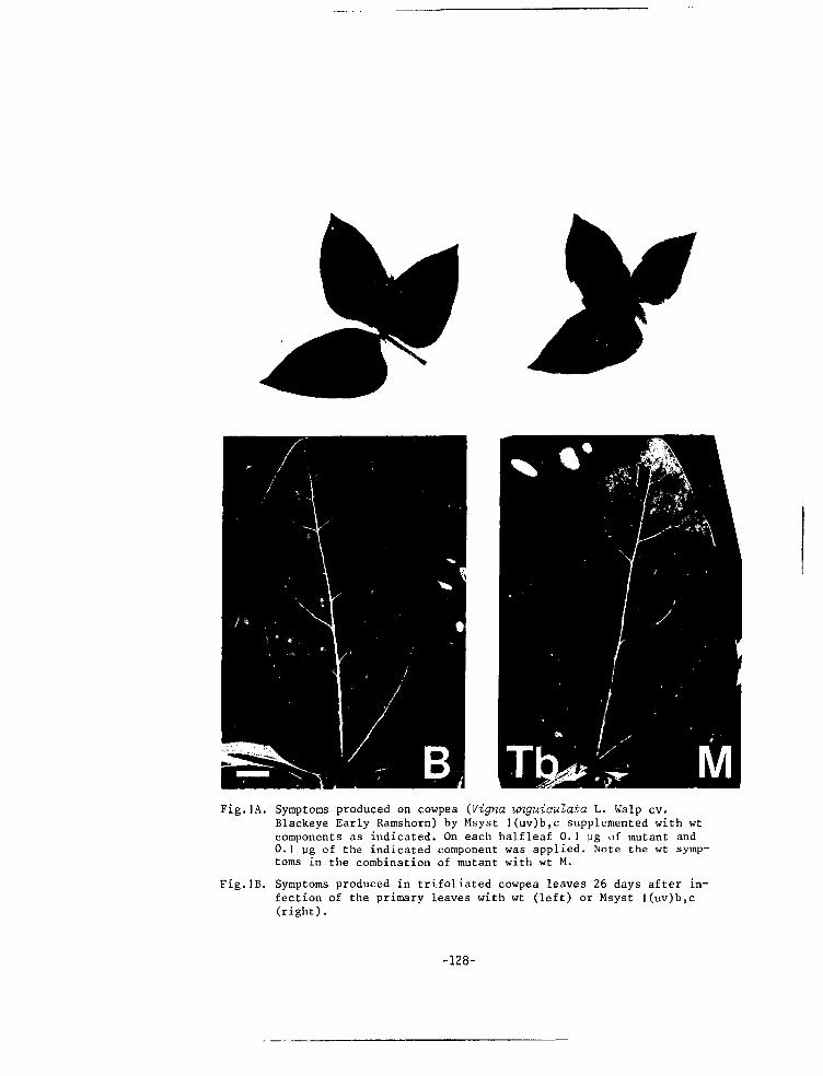
Fig.1A. Symptoms produced on cowpea (Vigna unguiaulata L. Walp cv.Blackeye Early Ramshorn) by Msyst l(uv)b,c supplemented with wtcomponents as indicated. On each halfleaf 0.1 yg of mutant and0. 1 (jg of the indicated component was applied. Note the wt symp-toms in the combination of mutant with wt M.
Fig.IB. Symptoms produced in trifoliated cowpea leaves 26 days after in-fection of the primary leaves with wt (left) or Msyst l(uv)b,c(right).
-128-
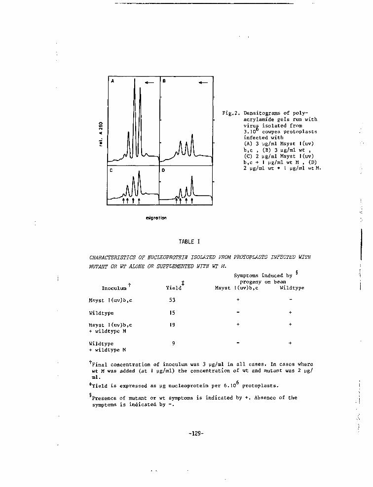
A
Jtt 1 1
B <
D
—ft 11
Fig.2. Densitograms of poly-acrylamide gels run withvirug isolated from3.10 cowpea protoplastsinfected with(A) 3 ug/ml Msyst 1(uv)b,c , (B) 3 ug/ml wt ,(C) 2 ug/ml Msyst I(uv)b,c + 1 ug/ml wt M , (D)2 ug/ml wt + 1 ug/ml wt M.
migration
TABLE I
CHARACTERISTICS OF NUCLEOPROTEIN ISOLATED FROM PROTOPLASTS INFECTED WITH
MUTANT OR WT ALONE OR SUPPLEMENTED WITH WT M.§
Symptoms induced by
x ± progeny on beanMsyst l(uv)b,c WildtypeInoculum
Msyst I(uv)b,c
Wildtype
Msyst l(uv)b,c+ wildtype M
Wildtype+ wildtype M
Yield
53
15
19
9
Final concentration of inoculum was 3 ug/ml in all cases. In cases wherewt M was added (at I ug/ml) the concentration of wt and mutant was 2 ug/ml.
*Yield is expressed as ug nucleoprotein per 6.10 protoplasts.
Presence of mutant or wt symptoms is indicated by +. Absence of thesymptoms is indicated by -.
-129-
Enhanced synthesis of RNA 2 can be due to an alteration of the RNA 2 mole-cule itself or by an alteration of its encoded protein. If the mutationis expressed at the RNA level we must assume that mutant RNA 2 has a higheraffinity for the replicating enzyme than wt RNA 2. However, it is difficultto envisage how such a mutation is repressed by the addition of wt RNA 2.Therefore we conclude that the mutation is expressed at the protein level.It is known that the RNA 2 encoded product does play a role in viral RNAsynthesis (Nassuth and Bol, 1982). From the observation that smalleramounts of RNA 2 are synthesized in the presence of both mutant and wtRNA 2 products than in the presence of mutant RNA 2 products alone, we candeduce that the wt RNA 2 product acts as a repressor for the synthesis ofRNA 2. The drastic reduction in yield upon addition of wt M to mutant viruscould indicate that the wt RNA 2 product also acts as a repressor for thesynthesis of the other AMV RNAs. However it is likewise possible that thisreduction is a secondary effect due to the decrease of RNA 2 product.
Previously we have shown that AMV Mts mutants do fall into two comple-mentation groups (Van Vloten-Doting et al.3 1980; Roosien and Van Vloten-Doting, 1982). We obtained indications that the complementation is intra-cistronic, meaning that the RNA 2 product has at least two functionaldomains (Roosien and Van Vloten-Doting, 1982). The repressor activity mayrepresent one of these domains or be a third function of the RNA 2 codedproduct. In view of the limited genetic information present in plant virusesit is not surprising that the virus coded proteins display several func-tions.
Both in cowpea plants and in cowpea protoplasts a wt phenotype is ob-tained upon addition of wt M to Msyst l(uv)b,c , leading to a possiblecorrelation between the regulation of viral RNA synthesis and the type ofplant reaction evoked by virus infection.
-130-
REFERENCES
Alblas, F. and Bol, J.F. (1977). Journal of General Urology 36, 175-185.
Bol, J.F., Van Vloten-Doting, L. and Jaspars, E.M.J. (1971). virology 46,
73-85.
Hagedorn, D.J. and Hanson, E.W. (1963). Phytopathology 53_, 188-192.
Nassuth, A., Albas, F. and Bol, J.F. (1981). Journal of General Virology
53_, 207-214.
Nassuth, A. and Bol, J.F. (1982). Virology in press.
Nassuth, A., Ten Bruggencate, G. and Bol, J.F. (1982). Virology in press.
Roosien, J. and Van Vloten-Doting, L. (1982). Journal of General Virology
63, 189-198.
Van Vloten-Doting, L. and Jaspars, E.M.J. (1972). Virology 48, 699-708.
Van Vloten-Doting, L., Hasrat, J.A., Oosterwijk, E., Van 't Sant, P.,
Schoen, M.A. and Roosien, J. (1980). Journal of General Virology 46,
415-126.
-131-
CHAPTER IX
CORRELATION BETWEEN RATE OF VIRUS MULTIPLICATION AND HYPERSENSITIVE REAC-
TION OF THE HOST
INTRODUCTION
Plant viruses are each year responsible for severe losses in variety of
crops. A hypersensitive reaction of the host to a virus, leading to micros-
copically or macroscopically visible necrotic local lesions with no sys-
temic spread, can give effective resistance in the field. The mechanism,
by which virus is localised within the necrotic local lesion, has been
studied extensively, but no coherent picture of the sequence of events has
evolved. It is known that the hypersensitive reaction is under the control
of both host and viral genes (Matthews, 1981). Most work has been concen-
trated on the host genes responsible for resistance by hypersensitivity.
For several viruses with a multipartite genome the study of pseudo-
recombinants has identified the genome part involved in the hypersensitive
reaction (Van Vloten-Doting and Jaspars, 1977). For some of the viruses
with a tripartite genome (Tricornaviridae, Van Vloten-Doting et at., 1981)
the middle component (M, containing RNA 2) has been found to carry the
information for a hypersensitive reaction in certain host plants. This type
of experiments was performed with natural strains, differing extensively,
which might complicate the interpretation of comparative studies. Recently
we isolated a mutant of alfalfa mosaic virus (AMV), a tricornavirus which,
in contrast to the parent strain, could invade bean (Roosien and Van Vlo-
ten-Doting, 1982) and cowpea (Roosien et al., 1983) plants systemically.
This mutant was induced by uv-irradiation of wildtype (wt) M (Roosien and
Van Vloten-Doting, 1982). Comparison of the properties of this mutant with
those of the wt may give indication why the multiplication of the mutant
is not restricted to a small zone around the initially infected cell.
RESULTS AND DISCUSSION
Cowpea leaves (Vigna unguiculata L. Walp cv. Black eye Early Ramshorn)
-133-
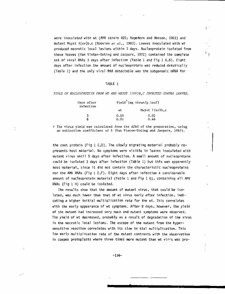
were inoculated with wt (AMV strain 425; Hagedorn and Hanson, 1963) and
mutant Msyst l(uv)b,c (Roosien et al., 1983). Leaves inoculated with wt
produced necrotic local lesions within 3 days. Nucleoprotein isolated from
these leaves (Van Vloten-Doting and Jaspars, 1972) contained the complete
set of viral RNAs 3 days after infection (Table 1 and Fig 1 A,B). Eight
days after infection the amount of nucleoprotein was reduced drastically
(Table 1) and the only viral RNA detectable was the subgenomic mRNA for
TABLE 1
YIELD OF NUCLEOPROTEIN FROM WT AND MSYST 1(UV)B,C INFECTED COWPEA LEAVES.
Days after Yield (mg virus/g leaf)infection
wt Msyst l(uv)b,c
3 0.05 0.028 0.01 0.46
t The virus yield was calculated from the A260 of the preparation, usingan extinction coefficient of 5 (Van Vloten-Doting and Jaspars, 1967).
the coat protein (Fig 1 C,D). The slowly migrating material probably re-
presents host material. No symptoms were visible in leaves inoculated with
mutant virus until 5 days after infection. A small amount of nucleoprotein
could be isolated 3 days after infection (Table 1) but this was apparently
host material, since it did not contain the characteristic nucleoproteins
nor the AMV RNAs (Fig 1 E,F). Eight days after infection a considerable
amount of nucleoprotein material (Table 1 and Fig 1 G), containing all AMV
RNAs (Fig 1 H) could be isolated.
The results show that the amount of mutant virus, that could be iso-
lated, was much lower than that of wt virus early after infection, indi-
cating a higher initial multiplication rate for the wt. This correlates
with the early appearance of wt symptoms. After 8 days, however, the yield
of the mutant had increased very much and mutant symptoms were observed.
The yield of wt decreased, probably as a result of degradation of the virus
in the necrotic local lesions. The escape of the mutant from the hyper-
sensitive reaction correlates with its slow in'tial multiplication. This
low early multiplication rate of the mutant contrasts with the observation
in cowpea protoplasts where three times more mutant than wt vip's was pro-
-134-
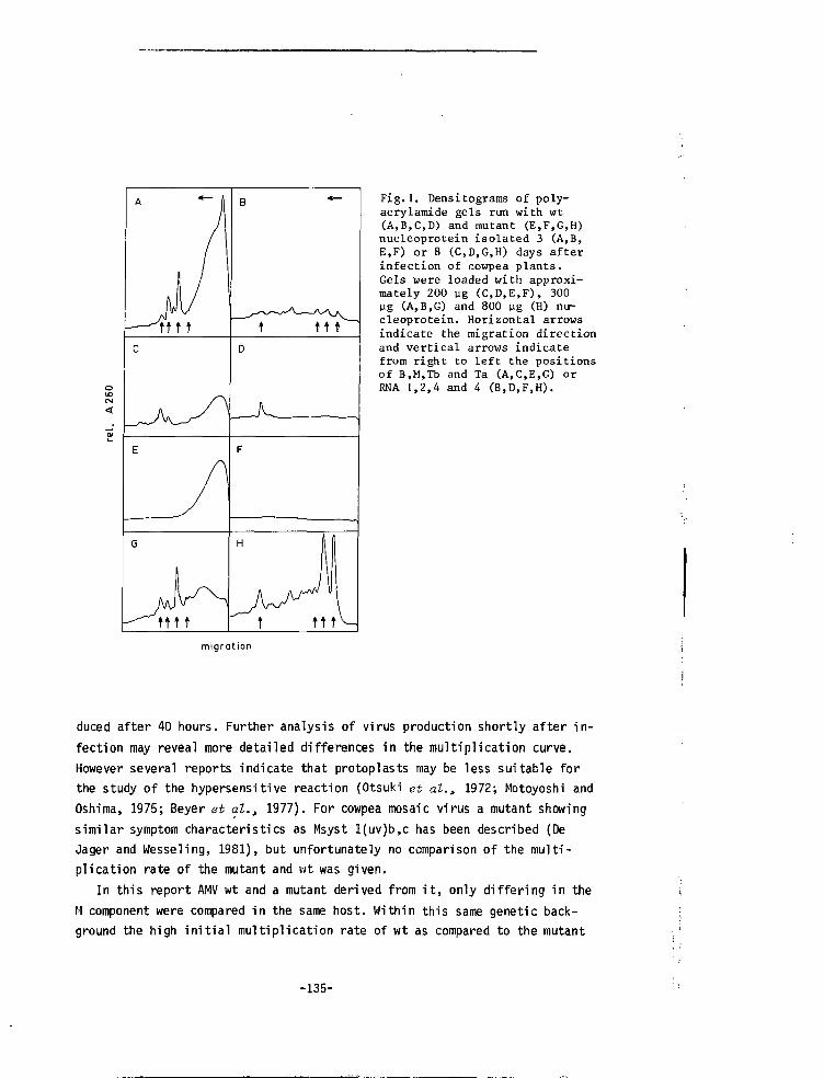
tttt
tttt
ttt
Fig.1. Densitograms of poly-acrylamide gels run with wt(A,B,C,D) and mutant (E,F,G,H)nucleoprotein isolated 3 (A,B,E,F) or 8 (C,D,G,H) days afterinfection of cowpea plants.Gels were loaded with approxi-mately 200 ug (C,D,E,F), 300yg (A,B,G) and 800 pg (H) nu-cleoprotein. Horizontal arrowsindicate the migration directionand vertical arrows indicatefrom right to left the positionsof B,M,Tb and Ta (A,C,E,G) orRNA 1,2,4 and 4 (B,D,F,H).
migration
duced after 40 hours. Further analysis of virus production shortly after in-fection may reveal more detailed differences in the multiplication curve.However several reports indicate that protoplasts may be less suitable forthe study of the hypersensitive reaction (Otsuki et al., 1972; Motoyoshi andOshima, 1975; Beyer et al., 1977). For cowpea mosaic virus a mutant showingsimilar symptom characteristics as Msyst l(uv)b,c has been described (DeJager and Wesseling, 1981), but unfortunately no comparison of the multi-plication rate of the mutant and wt was given.
In this report AMV wt and a mutant derived from it, only differing in theM component were compared in the same host. Within this same genetic back-ground the high initial multiplication rate of wt as compared to the mutant
-135-
could e l i c i t the hypersensitive host response. However further work with
this kind of mutants is needed to obtain more insight in the viral coded
functions, involved in evoking the host response.
REFERENCES
Beier, H., S i l e r , D.J., Russell, M.L. and Bruening, G. (1977). Phytopatho-
logy 67, 917-921.
De Jager, C.P and Wesseling, J.B.M. (1981). Physiological Plant Pathology
19, 347-358.
Hagedorn, D.J. and Hanson, E.W. (1963). Phytopathology 5j3, 188-192.
Matthews, R.E.F. (1981). Plant Virology, Second Edition, pp 393-447. Editor
R.E.F. Matthews. Academic Press. New York London Toronto Sydney San
Fransisco.
Hotoyoshi, F. and Oshima, N. (1975). Journal of General Virology 29_, 81-91.
Otsuki, Y., Shimomura, T. and Takebe, I . (1972). virology 50, 45-50.
Roosien, J . and Van Vloten-Doting, L. (198E). Journal of General Virology
63, 189-198.
Roosien, J . , Sarachu, A.N., Alblas, F. and Van Vloten-Doting, L. (1983).
Submitted for publ icat ion.
Van Vloten-Doting, L. and Jaspars, E.M.J. (1972). virology 48, 699-708.
Van Vloten-Doting, L. and Jaspars, E.M.J. (1977). Comprehensive Virology
11, 1-53. Edited by H.Fraenkel-Conrat and R.R. Wagner. New York: Plenum
Press.
Van V lo ten-Dot ing , L . , F ranck i , R . I . B . , F u l t o n , R.W., Kaper, J .M. and Lane,
L.C. (1981). Intervirology 15_, 198-203.
-136-
CHAPTER XY,
SUMMARY AND GENERAL DISCUSSION
Although plant viruses were the first viruses to be discovered, much
less is known about the molecular biology of these viruses than of animal
and bacterial viruses. This lagging is mainly due to the lack of infectable
cell cultures and the lack of conditional lethal mutants. Fifteen years ago
the technique of infecting isolated leaf cell protoplasts was developed and
in the last five years this technique was successfully applied for the
analysis of plant virus replication (review Nassuth, 1982). However the
study of conditional lethal mutants of plant viruses is still in its in-
fancy. In this thesis the isolation and characterization of a number of
mutants of alfalfa mosaic virus, a plant virus with a coat protein dependent
genome, is described (see for literature chapter 2). We selected thermo-
sensitive (ts) mutants since, at least theoretically, ts mutations can be
present in all virus coded functions. Most mutations will disturb only one
step in the virus multiplication process. When a cell inoculated with two
ts mutants does produce virus at the non-permissive temperature, we know
that the two mutants display defects in different functions. This type of
experiment (complementation test) will give insight in the number of essen-
tial virus coded functions involved in the replication process. For viruses
with a divided genome, like alfalfa mosaic virus (AMV), the mutations can be
assigned to the different genome parts by the supplementation test. Growth
of the mutant at the non-permissive temperature is measured in the presence
of each of the three purified wildtype (wt) components. Virus production
will only take place, when the wt component added, can compensate for the
non-functional product encoded on one of the mutant components.
It was found that a high percentage of spontaneous mutants, isolated be-
cause of their aberrant symptoms, were ts. The majority of these isolates
could grow at the non-permissive temperature in the presence of a single
wt component. To increase the mutation rate virus preparations were treated
with several mutagens. After nitrous acid treatment or irradiation with
ultraviolet light, an increase in the level of mutations was observed. Uv
irradiation was preferred since it did not require large amounts of puri-
fied viral components. During the preliminary characterization of potential
ts mutants we obtained also one structural and several symptom mutants which
-137-
were also analysed further (chapter 7, 8 and 9). The properties of the ts
mutants are described in chapter 3-7. Mutants were called after the genome
part (B, M, Tb) on which the mutation was located, followed by (1) an indi-
cation for the mutant character: ts for thermosensitive, "struct" f(K
structural deviation from wt, "syst" for systemic invasion of host plant,
normally reacting with hypersensitivity; (2) number of the isolate; (3)
origin of the mutation: "s" for spontaneous, "ni" for nitrous acid, "uv"
for ultraviolet irradiation; (4) first letter of the host plant(s) on which
the mutant character is displayed: "t" for tobacco, "b" for bean, "c" for
cowpea.
Among the isolated mutants one behaved quite different from wt when vi-
rion particles were analysed on polyacrylamide gels. After constructing
pseudohybrids of the purified mutant RNAs and wt components, RNA 3 was
found to be responsible for the mutant behaviour. It appeared that both
cistrons on RNA 3 were affected since in vitro translation or mutant RNA 3
yielded a 33 k product instead of the wt 35 k product and the coat protein
of the mutant was serologically different from wt. The RNA synthesis was
affected by one or both of these mutations on the single as well on the
double strand level. Analysis of mutant particles showed that in contrast
to the situation with wt several mutant RNAs were encapsidated together
into one particle. Electronraicroscopy showed some very long particles,
while the majority of the preparation consisted of spheroid particles of
different dimensions. These particle dimensions equal those measured for
the ilar virus tobacco streak virus. The properties of this mutant are an
argument in favor of the proposed AMV membership of the ilar virus genus.
Besides this structural mutant also a mutant of AMV was isolated which
induced different symptoms in cowpea and bean plants. Instead of the normal
hypersensitive reaction, resulting in necrotic local lesions, chlorotic
spots were observed on the primary infected leaves followed later by sys-
temic infection. Supplementation experiments, carried out on bean and cow-
pea plants showed for both hosts that the mutant carried (a) mutation(s)
on M (containing RNA 2). It was found that wt virus, causing the hypersen-
sitive reaction, had a much higher multiplication rate early after infec-
tion than the less severely reacting mutant. This could indicate that in
the same genetic background, there might be a causative relation between
the multiplication rate of the virus and the reaction of the host.
Comparison of mutant and wt virus production in cowpea protoplasts after
in vitro infection, showed that the mutant produced three times as much
-138-
virus as wt. The relative amount of mutant M component was much higher
than in wt. When the mutant was inoculated together with wt M component,
the virus yield dropped to the wt level and the component composition of
the preparation ressembled that of wt. From the decrease of the relative
abundance, especially of M, containing RNA 2, it was deduced that RNA 2
might encode a function which is repressive on the RNA synthesis.
The isolated ts mutants were divided into Tbts, Mts and Bts mutants by
means of supplementation experiments. The symptoms of these mutants on bean
and cowpea plants were like wt. On tobacco some mutants induced necrosis,
while others showed the wt chlorotic mottling. Some of these mutants, ts
in the tobacco leaf disc system, were also ts in local lesion induction in
bean plants, while on the other hand some mutants ts on bean were not ts
in the tobacco leaf disc system. Therefore the ts character is host-depen-
dent in many instances.
Since the ts mutants were qualified as such in tobacco leaf discs, a
distinction between a ts defect exhibited in the spreading of the virus
from cell to cell and a ts defect shown in the primary infected cell would
not be possible. Following the infection with the use of AMV antiserum
coupled to a fluorescing substance and a Fluorescence Activated Cell Sor-
ter, which monitors the number of fluorescing cells and the fluorescence
per cell, we tried to compare the infection process in protoplasts isolated
from infected leaves and in vitro infected protoplasts. For the latter pro-
toplasts no good correlation was found between infectivity and fluorescen-
ce.
Complementation experiments with the Tbts mutants showed them all to be
a member of the same group. Since it is known that wt RNA 3 carries two
cistrons we had to find out which of these would coincide with the comple-
mentation group observed. By checking the ability of the coat protein of
the mutants to render a mixture of the wt genomic RNAs infectious at the
non-permissive temperature, we found that all mutant coat proteins were ts
in this early functipn. This indicates that the complementation group re-
presents the coat protein cistron. Apparently none of the Tbts isolates
carried (only) a ts 35 k protein. The reason for this unequal distribution
of mutations over the two cistrons is unknown. To find out if mutants with
a defect in the early function of the coat protein would still be able to
encapsidate (a second function), we analysed the mixed infection of wt and
Tbts 7(uv), a mutant with an electrophoretically different coat protein and
a symptom marker mapping on RNA 3. At the non-permissive temperature no mu-
-139-
tant coat protein nor mutant RNA was produced in mixedly infected plants.
Analysis of Tbts 7(uv) virion and double stranded RNA obtained from plants
infected only with mutant at 23°C showed that the relative amounts of
single and double stranded RNA 3 were lower than in wt. Taken together,
these results might indicated the presence of a ts mutation in the mutant
RNA 3 itself, diminishing its affinity for the viral replicase.
The behaviour of the Bts mutants in complementation studies showed two
of them to complement each other, while two other Bts mutants did not com-
plement with any of the Bts mutants. We therefore conclude that B carries
at least two complementation groups. It is not clear whether the two non-
complementing mutants form a third or fourth group, representing some cis-
acting function. Biochemical studies showed both complementing mutants to
be disturbed in the minus strand RNA synthesis (Sarachu et al., 1983}.
In complementation experiments carried out originally with 3 Mts mutants
two complementation groups were observed. After isolating and testing more
Mts mutants, these results were confirmed, but in some combinations of mu-
tants a reduction in virus production at 23°C was found. This interference
was not found when these mutants were inoculated together with wt. Based
on complementation and interference behaviour the mutants could be divided
into two groups. Between these groups complementation was found, while
mutants inside these groups frequently gave rise to interference. To ex-
plain these results it is assumed that the RNA 2 encoded protein functions
as a multimer and has two functional domains which can be mutated indepen-
dently. In some combinations of mutants, carrying mutations in different
domains, the defective products form a heteropolymer which is functional
again. However combination of mutants carrying defects in the same domain
can result in heteropolymers still defective at 30°C and sometimes by
allosteric interaction even defective at 23°C. It is unknown which of these
domains, if any, represents the repressor function (chapter 8). As was
found for the Bts mutants the Mts mutants were also disturbed in the minus
strand RNA synthesis (Sarachu et al., 1983).
Recent supplementation experiments with some of the mutants described
in this thesis, have shown the presence of additional ts mutations on an-
other viral component (Sarachu et at., 1983). Probably as a result of the
high mutation rate of RNA viruses, some of the mutants might have acquired
these mutations during propagation. In future experiments batches of single
purified mutant components supplemented with the lacking purified wt compo-
nents will have to be used for biochemical analysis of the infection pro-
cess in protoplasts and plants.
-140-
Samenvatting
Hoewel de pi antevirussen als eerste onder de virussen werden ontdekt,is over hun moleculaire biologie veel minder bekend dan van dierlijke enbacteriële virussen. Dit achterblijven in kennis is voornamelijk te wijtenaan het ontbreken van infecteerbare celkweken en conditioneel lethale mu-tanten. Zo'n 15 jaar geleden werd de techniek ontwikkeld om bladcel proto-plasten te infecteren en gedurende met name de laatste vijf jaar is dezetechniek erg succesvol toegepast in de analyse van de replicatie van plan-tevirussen (zie voor overzicht: Nassuth, 1982). De bestudering van condi-tioneel lethale mutanten van plantevirussen staat echter nog in zijn kin-derschoenen. In dit proefschrift wordt isolatie en karakterizering beschre-ven van een aantal mutanten van alfalfa mozaiek virus, een plantevirus meteen manteleiwit afhankelijk driedelig genoom (zie voor literatuur hoofd-stuk 2). Temperatuur gevoelige (ts) mutanten werden gebruikt in deze stu-die, omdat tenminste theoretisch ts mutaties kunnen voorkomen in elke vi-raal gecodeerde functie. De meeste mutaties zullen slechts één stap in hetvirus vermeerderingsproces verstoren. Wanneer een cel, geinoculeerd mettwee ts mutanten, bij de restrictieve temperatuur virus produceert, wetenwe dat de twee mutanten gestoord zijn in verschillende functies. Dit soortproeven (complementatie test) kan inzicht verschaffen in een aantal essen-tieel viraal gecodeerde functies betrokken in het replicatieproces. Voorvirussen met een meerdelig genoom zoals alfalfa mozaiek virus (AMV) kunnende mutaties aan de verschillende genoomdelen worden toegekend door middelvan een supplemental e test. De groei van de mutant bij de restrictievetemperatuur wordt gemeten in aanwezigheid van elk van de gezuiverde wild-type (wt) componenten. Virus productie zal alleen dan optreden als de toe-gevoegde wt component kan compenseren voor het niet functionerende productgecodeerd door één van de componenten van de mutant.
Van de spontane mutanten, geisoleerd omdat ze afwijkende symptomen inplanten induceerden, bleek een groot percentage ts. De meerderheid van dezeisolaten kon bij de restrictieve temperatuur groeien in aanwezigheid vanéén enkele wt component. Om de mutatie graad te verhogen werden virus pre-paraten behandeld met verscheidene mutagentia. Na behandeling met salpete-rig zuur of bestraling met ultraviolet licht, werd een toename in het mu-tatie niveau geconstateerd. Aan uv bestraling werd de voorkeur gegeven om-
-141-

dat dit geen grote hoeveelheden gezuiverde virale componenten vereiste.
Tijdens de voorlopige karakterizering van potentiële ts mutanten vonden we
bovendien één structurele en meerdere symptoom mutanten. Deze werden ook
nader geanalyseerd (hoofdstuk 7,8 en 9). De eigenschappen van de ts mutan-
ten staan beschreven in hoofdstuk 3 tot 7. De mutanten werden naar het ge-
noomdeel (B,M,Tb) waarop de mutatie was gelegen genoemd, gevolgd door (1)
een aanduiding voor het mutante karakter: ts voor temperatuur gevoelig,
"struct" voor structureel afwijkend van wt, "syst" voor systemische infec-
tie van de waardplant die normaal reageert met een hypersensitieve reactie;
(2) het isolaat nummer; (3) oorsprong van de mutatie: "s" voor spontaan,
"ni" voor salpeterig zuur, "uv" voor ultraviolet bestraling; (4) de eerste
letter van de waardplant(en) waarop het mutante karakter tot uitdrukking
komt: "t" voor tabak, "b" voor boon, "c" voor cowpea.
Bij de analyse van virion deeltjes op polyacrylamide gels gedroeg één
van de de geisoleerde mutanten zich geheel verschillend van wt. Na met be-
hulp van de gezuiverde RNA's van de mutant en wt componenten een aantal
pseudo-hybriden te hebben gemaakt, bleek dat RNA 3 van de mutant verant-
woordelijk was voor het afwijkend gedrag. Het bleek dat beide cistrons op
RNA 3 een mutatie hadden want in vitro vertaling van mutant RNA 3 leverde
een product op van 33 k in plaats van het wt 35 k product en bovendien was
het manteleiwit van de mutant serologisch afwijkend van wt. De RNA synthese
bleek veranderd door één of beide mutaties zowel op enkel- als op dubbel-
strengs niveau. Analyse van deeltjes van de mutant liet zien dat in tegen-
stelling tot wt vescheidene mutante RNA's samen bemanteld werden tot een
deeltje. Met behulp van de electronen microscoop werden enige erg lange
deeltjes waargenomen, maar het grootste gedeelte van het preparaat bevatte
ellipsoïde deeltjes van verschillende afmetingen. De afmetingen van deze
deeltjes liggen in de buurt van die gemeten voor het ilar virus tabaks
strepen virus. De eigenschappen van deze mutanten dragen een argument aan
ten gunste van het rekenen van AMV tot het ilar virus genus.
Naast deze structurele mutant werd ook een mutant geisoleerd, die af-
wijkende symptomen induceerde in cowpea en boneplanten. In plaats van de
normaal optredende hypersensitieve reactie, die leidt tot necrotisch locale
lesies, waren chlorotische vlekken zichtbaar op de primair geinfecteerde
bladeren, later gevolgd door een systemische infectie. Supplementatie proe-
ven, uitgevoerd op bone- en cowpeaplanten toonden voor beide waardplanten
aan dat de mutant (een) mutatie(s) op M (dat RNA 2 bevat) had. Het bleek
-142-
dat vroeg na infectie wt virus, dat een hypersensitieve reactie oproept,
een veel hofere vermeerdenngssneiheid had dan de mutant, die niet zulke
sterke reacties in de plant teweeg bracht. Dit zou kunnen inhouden dat er,
binnen dezelfde genetische achtergrond een causaal verband bestaat tussen
de vermeerden'ngssnelheid van het virus en de reactie van de waardplant.
Vergelijking van de virus productie van de mutant en wt in cowpea pro-
toplasten na in vitro infectie bracht aan het licht dat de mutant drie keer
zoveel virus produceerde dan wt. De relatieve hoeveelheid M component was
veel hoger in de mutant dan in wt. Wanneer de mutant samen met wt M compo-
nent werd geinoculeerd zakte de virus opbrengst iiaar het niveau van wt en
de componenten samenstelling van het preparaat leek dan op dat van wt. Uit
de afname van vooral de relatieve hoeveelheid M, dat RNA 2 bevat, werd af-
geleid dat RNA 2 zou kunnen coderen voor een functie die onderdrukkend
werkt op de RNA synthese.
De geisoleerde ts mutanten werden door middel van supplemental e proeven
verdeeld in Tbts.Mts en Bts mutanten. De symptomen van deze mutanten op
bone- en cowpeaplanten waren als die van wt. Op tabak induceerden sommige
mutanten necrose, maar anderen vertoonden de wt chlorotische symptomen. Van
deze mutanten, die ts waren in het tabaksbladschijfjessysteem, bleken een
aantal ook ts in het vermogen om locale lesies in boneplanten te induceren,
terwijl anderzijds sommige mutanten die ts op bonen waren, dit niet in het
tabaksbladschijfjessysteem bleken. Het lijkt erop dat het ts karakter in
vele gevallen afhankelijk is van de waardplant.
Aangezien de ts mutanten als zodanig aangemerkt werden op grond van hun
gedrag in tabaksbladschijfjes, zou het onmogelijk zijn onderscheid te maken
tussen een ts defect, dat de verspreiding van het virus van cel tot cel
verstoort, en een ts defect dat de replicatie in de primair geinfecteerde
cel aantast. Door de infectie te volgen met behulp van AMV antiserum ge-
koppeld aan een fluorescerende verbinding en gebruik makend van een Flu-
orescence Activated Cell Sorter, die het aantal fluorescerende cellen en
de fluorescentie per cel kan registreren, hebben we getracht een verge-
lijking te maken tussen het infectie proces in protoplasten geisoleerd van
geinfecteerde bladeren en in vitvo geinfecteerde protoplasten. De laatst-
genoemde protoplasten vertoonden geen duidelijke correlatie tussen infec-
tiviteit en fluorescentie.
Complementatie proeven, uitgevoerd met de Tbts mutanten toonden aan dat
alle mutanten in dezelfde groep vielen. Aangezien wt RNA 3 twee cistrons
-143-
draagt moest uitgezocht worden welke van de twee de gevonden complementatiegroep zou vertegenwoordigen. Door het vermogen na te gaan van het mantel -eiwit van deze mutanten, om een mengsel van de wt genoom RNA's infectieuste maken bij de restrictieve temperatuur, werd gevonden dat alle mutanteneen manteleiwit hadden dat te was in die vroege functie. Dit betekent datde complementatiegroep het manteleiwit cistron vertegenwoordigt. Blijkbaarhad geen van de Tbts isolaten (alleen) een ts 35 k proteine. De oorzaakvoor deze ongelijke verdeling van de mutaties over de twee cistrons is on-bekend. Om uit te vinden of mutanten met een storing in de vroege functievan het manteleiwit nog in staat zouden zijn tot bemanteling (een tweedefunctie), hebben we gemengde infecties geanalyseerd van wt met Tbts 7(uv),een mutant met een electroforetisch van wt verschillend manteleiwit en eensymptoommutatie op RNA 3. Bij de restrictieve temperatuur werd geen mutantmanteleiwit en RNA 3 geproduceerd in gemengd geïnfecteerde planten. Analysevan Tbts 7(uv) virion en dubbelstrengs RNA, geisoleerd uit met alleen mu-tant geinfecteerde planten bij 23°C, liet zien dat de relatieve hoeveelheidenkel- en dubbelstrengs RNA 3 lager was dan in wt. Resumerend zouden dezeresultaten de aanwezigheid kunnen inhouden van een ts mutatie in het mu-tante RNA 3 molecuul resulterend in een verlaagde affiniteit voor het vi-rale replicase.
Het gedrag van de Bts mutanten in complementatie experimenten gaf aandat twee mutanten elkaar complementeerden, terwijl twee andere Bts mutantenmet geen enkele Bts mutant complementatie gaven. De conclusie werd getrok-ken dat de B component tenminste twee complementatie groepen draagt. Hetis onduidelijk of de twee niet complementerende mutanten een derde of zelfseen vierde groep vormen die de een of andere in ais werkende functie ver-tegenwoordigt. Biochemische analyse toonde aan dat beide elkaar complemen-terende mutanten gestoord waren in synthese van min streng RNA (Sarachuet al., 1983).
In complementatie experimenten, die in eerste instantie werden uitge-voerd met drie Mts mutanten, werden twee complementatie groepen waargeno-men. Na isolatie van meer Mts mutanten bleken deze in complementatie proe-ven ook in twee groepen uiteen te vallen. In sommige combinaties van dezemutanten trad echter een afname in virus productie bij 23°C op. Deze inter-ferentie werd niet gezien in combinaties van deze mutanten met wt. Geba-seerd op het complementatie en interferentie gedrag van deze mutanten wer-den ze in twee groepen verdeeld. Tussen de groepen werd complementatie ge-
-144-
vonden, maar binnen deze groepen werd veelvuldig interferentie waargenomentussen paren mutanten. Als verklaring voor deze resultaten werd aangenomendat het door RNA 2 gecodeerde eiwit als een multimeer functioneert en tweefunctionele domeinen heeft die afzonderlijk muteerbaar zijn. In combinatiesvan mutanten met mutaties in verschillende domeinen vormen de defecte gen-producten een heteropolymeer dat weer kan functioneren. Combinaties vanmutanten met een defect in hetzelfde domein kunnen echter resulteren inheteropolymere complexen die behalve bij 30°C soms ook door allosterischeinteracties zelfs bij 23 C niet meer goed functioneren. Het is niet bekendwelke van deze twee domeinen de repressor functie (hoofdstuk 8) vertegen-woordigt, zo dat al het geval is. Evenals de Bts mutanten bleken ook deMts mutanten gestoord in de synthese van min streng RNA (Sarachu et al.,1983).
Onlangs uitgevoerde supplementatie proeven, uitgevoerd met een paar vande in dit proefschrift beschreven mutanten, hebben de aanwezigheid vanextra ts mutaties op een andere virale component aangetoond (Sarachu et al., 1983). Ten gevolge van de hoge mutatie graad van RNA virussen zouden som-mige mutanten deze mutaties opgedaan kunnen hebben tijdens het opkweken.In toekomstige experimenten zullen afzonderlijke gezuiverde mutante com-ponenten, aangevuld tot een complete set met wt componenten, gebruikt moe-ten worden voor biochemische analyse van het infectie proces in protoplas-ten en planten.
-145-
CURRICULUM VITAE
Ik ben geboren op 9 juni 1948 te Gasselterboerveenschemond.In 1967 behaalde ik het diploma gymnasium s aan het Ubbo EmmiusLyceum te Stadskanaal. Datzelfde jaar volgde de eerste inschrij-ving aan de Rijksuniversiteit te Groningen, studierichting bio-logie.
In februari 1972 behaalde ik mijn kandidaatsexamen biologie(B4) en het doctoraalexamen volgde in juli 1975 met als hoofd-vak moleculaire genetica en de bijvakken experimentale phyto-morfologie en experimentele cytologie, het laatste verrichtaan het Anthonie van Leeuwenhoek Huis te Amsterdam.
Van augustus 1978 tot augustus 1982 was ik als wetenschap-pelijk assistent in dienst van de Rijksuniversiteit te Leidenen werkzaam op de afdeling PI antevirussen van het Biochemischlaboratorium.
-146-
NAWOORD
Aan de tot stand koming van dit proefschrift hebben veel mensen direct
of indirect een bijdrage geleverd. Hen allen wil ik daarvoor bedanken.
Niet onvermeld mag blijven dat de aangename werksfeer op het laboratorium
en met name de sfeer binnen de Plantevirus groep door mij altijd bijzonder
is gewaardeerd.
Een aantal mensen wil ik met name bedanken.
Aad Wesseling ben ik zeer erkentelijk voor het kweken van grote hoeveel-
heden planten zonder welke dit werk niet had kunnen worden
verricht.
Pieter van Klaveren wil ik dank zeggen voor de enthousiaste manier waarop
hij als doctoraal student een bijdrage heeft geleverd aan
dit proefschrift.
Fieke Alblas en Lyda Neeleman verdienen mijn dank voor de wijze waarop ze
een aantal experimenten voor mij hebben gedaan.
Leen Welrners ben ik zeer erkentelijk voor zijn teken en foto werk.
Het type werk werd op uitstekende wijze verzorgd door Sylvia van den Berg,
Nicoline van Hoek en Jeanne Pruyn.
I 'want to stress here that I enjoyed very much the cooperation with Alberto
Sarachu.
Tenslotte wil ik Annette Nassuth en Ben Cornelissen danken voor de morele
steun die ze mij hebben gegeven in de vier jaar die we samen in de Plante-
virus groep hebben doorgebracht.
-147-
STELLINGEN
1. De experimenten uitgevoerd door MSnnel en medewerkers geven geen enkele
indicatie dat het door hen geïsoleerde monoclonal e anti lichaam tegen
muizeinterferon cross reactiviteit vertoont met zowel de a als de e inter-
feron component.Hun suggestie dat muizeinterferon a en e een gemeenschap-
pelijk sterk geconserveerde sequentie bevatten is derhalve ongegrond.
MSnnel, D., De Maeyer, E., Kemler, R., Cachard, A. and De Maeyer-Guignard,J. (1982). Nature 296, 664-665.
2. Het ligt meer voor de hand te veronderstellen dat het manteleiwit van alf-
alfa mozaïek virus de virale min-streng RNA synthese afsluit door asso-
ciatie met het plus-streng matrijs-manteleiwit complex dan aan te nemen
dat dit gebeurt door associatie van het manteleiwit met het virale repli-
case, zoals door Nassuth en Bol is geponeerd.
Nassuth, A. and Bol, J.F. (1983). Virology 124, ter perse.Nassuth, A. (1982). Proefsahrift Rijksuniversiteit Leiden.
3. In tegenstelling tot wat Zurkowski in zijn artikel beweert is niet uit te
sluiten dat het sym plasmide pWZ2 van Rhizobium trifolii conjugatief is.
Zurkowski, U. (1981). Mol.Gen.Genet. 181,522-524.
4. Als de verklaring van Taliansky en medewerkers voor het thermosensitief
gedrag van de tabaksmozafek virus mutant Ni 2519 juist is, zal bij de
restrictieve temperatuur het messenger RNA voor het mantel eiwit in virus-
deeltjes aantoonbaar moeten zijn.
Taliansky, M.E., Kaplan, I.B., Yarvekulg, L.V., Atabekova, T.I., Agra-novsky, A.A. and Atabekov, J.G. (1982). Virology 118,309-316.
5. De conclusies van Wikman en medewerkers omtrent de interactieplaatsen van
twee Escherichia coli aminoacyl-tRNA~s met de elongatiefactor Tu worden
niet door hun resultaten gesteund.
Wikman, F.P., Siboska, G.E., Petersen, H.U. and Clark, B.F.C. (1982).The EMBO Journal 1,1095-1100.
6. De conclusies van Yamaoka en medewerkers betreffende de verschillende
regulatie van transcriptie van het viroplasma matrix eiwit en het mantel-
eiwit van het bloemkool mozaïek virus zijn onbewezen.
Yamaoka, N., Morita, T., Furusawa, I. and Yamamoto, N. (1982). J. Gen.Virology 61,283-287.
7. Beschouwingen over de biologische betekenis van de meerdeligheid van viralegenomen verliezen aan betekenis wanneer zij uitsluitend betrekking hebbenop hetzij pi antevirussen, hetzij diervirussen.Kilburn, E.D. (1981). Perspectives in Biology and Medicine.Autumn 1981.66-77.Fulton, R.W. (1980). Ann. Rev. of Phytopath. 18, 131-146.
8. De conclusie van Krisch en Allet dat het translatie initiatiegebied vanbacteriofaag T4 gen 32 betrokken is bij de translationele regulatie vandat gen door het eigen genproduct is niet bewezen.
Krisch, H.M. and Allet, B. (1982). PNAS 79, 4937-4941.
9. Het aan en uitschakelen vein de SOS functies in Escherichia coli is ver-klaarbaar vanuit de wisselwerking tussen de verschillende enkelstrengs-DNA-bindende eiwitten.
Litlle, J.W. and Mount, D.W. (1982). Cell 29,11-22.
10. Het verdient aanbeveling een RNA-afhankelijk-RNA polymerase pas RNA repli-case te noemen, wanneer is aangetoond dat het enzym inderdaad RNA kanrepliceren.
Baron, M.H. and Baltimore, D. (1982). J.Biol.Chem. 257,12359-12366.Gordon, K.H.J., Gill, D.S. and Symons, R.H. (1982). Virology 123,284-295.
11. üe door de regering voorgestelde tariefsverhogingen voor het openbaar ver-voer ter vermindering van het begrotingstekort kan leiden tot nog groteretekorten.
12. "Haastige spoed is zelden goed" geldt soms ook voor plantevirussen.Dit proefschrift, pp 133-136.
Leiden, 2 maart 1983 jan Roosien


