Embed Size (px)
Citation preview

This article appeared in a journal published by Elsevier. The attachedcopy is furnished to the author for internal non-commercial researchand education use, including for instruction at the authors institution
and sharing with colleagues.
Other uses, including reproduction and distribution, or selling orlicensing copies, or posting to personal, institutional or third party
websites are prohibited.
In most cases authors are permitted to post their version of thearticle (e.g. in Word or Tex form) to their personal website orinstitutional repository. Authors requiring further information
regarding Elsevier’s archiving and manuscript policies areencouraged to visit:
http://www.elsevier.com/authorsrights

Author's personal copy
N-terminal palmitoylation is required for Toxoplasma gondii HSP20inner membrane complex localization
M.G. De Napoli a, N. de Miguel a, M. Lebrun b, S.N.J. Moreno c, S.O. Angel a, M.M. Corvi a,⁎a Laboratorio de Parasitología Molecular, Instituto de Investigaciones Biotecnológicas-Instituto Tecnológico de Chascomus (IIB-INTECH), Universidad Nacional de San Martín (UNSAM),Consejo Nacional de Investigaciones Científicas y Técnicas (CONICET), Intendente Marino Km 8.2 (B7130) Chascomús, Provincia de Buenos Aires, Argentinab UMR 5235 CNRS, Université de Montpellier 2, 34095 Montpellier, Francec Department of Cellular Biology and Center for Tropical and Emerging Global Diseases, University of Georgia, Athens, 30602, USA
a b s t r a c ta r t i c l e i n f o
Article history:Received 23 December 2012Received in revised form 14 February 2013Accepted 19 February 2013Available online 26 February 2013
Keywords:Toxoplasma gondiiSmall heat shock protein 20PalmitoylationInner membrane complexEndodyogeny
Toxoplasma gondii is an obligate intracellular parasite and the causative agent of toxoplasmosis. Proteinpalmitoylation is known to play roles in signal transduction and in enhancing the hydrophobicity of proteinsthus contributing to their membrane association. Global inhibition of protein palmitoylation has been shownto affect T. gondii physiology and invasion of the host cell. However, the proteins affected by this modificationhave been understudied. This paper shows that the small heat shock protein 20 from T. gondii (TgHSP20) issynthesized as a mature protein in the cytosol and is palmitoylated in three cysteine residues. However, itslocalization at the inner membrane complex (IMC) is dependent only on N-terminal palmitoylation. Absenceor incomplete N-terminal palmitoylation causes TgHSP20 to partially accumulate in a membranous structure.Interestingly, TgHSP20 palmitoylation is not responsible for its interaction with the daughter cells IMCs.Together, our data describe the importance of palmitoylation in protein targeting to the IMC in T. gondii.
© 2013 Elsevier B.V. All rights reserved.
1. Introduction
Fatty acylation of proteins is a widespread feature of eukaryoticcells. This post-translational modification (PTM) plays a key role inmembrane localization of signal-transducing proteins [1,2] as wellas in regulation of mitochondrial metabolism [3,4]. Fatty acylationincludes myristoylation and palmitoylation. Whereas myristoylationrefers to the irreversible attachment of 14-carbon myristate in a co-[5,6] or post- [7–10] translational manner, palmitoylation is the post-translational attachment of the 16-carbon fatty acid palmitate onto cys-teine residues via a labile thioester bond. Palmitate is transferred ontoproteins enzymatically [11–13] or in a spontaneous manner [4,14]with both mechanisms being operational in cells.
Apicomplexan parasites are a large group of obligate intracellularparasites that include Eimeria spp., Cryptosporidium spp., Neosporaspp., Plasmodium spp., Theileria spp. and T. gondii. Protein palmitoylationhas been described to operate in Eimeria tenella [15], Plasmodiumfalciparum [16–19] and Toxoplasma gondii [15,20,21]. There are severalproteins known or predicted to be palmitoylated in T. gondii [22]. Ingeneral, protein palmitoylation is important for proper membranelocalization of themodified protein. In this regard, it has been describedthat mutation of the predicted palmitoylation sites in T. gondii inner
membrane complex sub-compartment protein 4 (ISP4) and calcium-dependent protein kinase 3 (CDPK3), alters their localization from thepellicle to the cytosol [23–25].We recently described the functional sig-nificance of this modification in T. gondii, where inhibition of proteinpalmitoylation was able to interfere with the parasite's physiologyand host-cell invasion [20]. Interestingly, the host-cell invasion processdepends on an actin- andmyosin-based complex located at the pellicle,between the plasma membrane (PM) and inner membrane complex(IMC) [26]. The same effect on host-cell invasion was observed inP. falciparum [27]. In general, these results underscore an importantrole for palmitoylation of IMC-associated proteins.
T. gondii small heat shock protein 20 (TgHSP20) displays interestingcharacteristics which could be used to study the role and dynamics ofprotein palmitoylation. TgHSP20 localizes to the parasite IMC in a setof discontinuous stripes [28]. Recombinant TgHSP20 forms multimers[29], binds phosphatidylinositol 4-phosphate and phosphatidylinositol4,5 biphosphate phospholipids [28] and has chaperone activity invitro [29]. It has been reported that antibodies against HSP20 reducehost-cell invasion by Babesia divergens, T. gondii and Neospora caninum[30,31]. In Plasmodium, deletion of the hsp20 gene results in altered spo-rozoite speed and substrate adhesion leading to an impaired naturalmalaria transmission in the host [32,33]. Although HSP20 is predictedto be synthesized in the cytosol, it localizes to the IMC in T. gondii [28],P. falciparum [32] and to the pellicle in Babesia [34]. Moreover, inT. gondii HSP20 is incorporated to the IMC at intermediate stages ofdaughter cell formation [28]. Here, we confirmed that all the TgHSP20cysteine residues are palmitoylated in vivo but only N-terminal
Biochimica et Biophysica Acta 1833 (2013) 1329–1337
⁎ Corresponding author at: Laboratorio de ParasitologíaMolecular, IIB-INTECH, IntendenteMarino Km 8.2, PO Box 164 (B7130IWA) Chascomús, Prov. Buenos Aires, Argentina.Tel.: +54 2241 430323; fax: +54 2241 424048.
E-mail address: [email protected] (M.M. Corvi).
0167-4889/$ – see front matter © 2013 Elsevier B.V. All rights reserved.http://dx.doi.org/10.1016/j.bbamcr.2013.02.022
Contents lists available at SciVerse ScienceDirect
Biochimica et Biophysica Acta
j ourna l homepage: www.e lsev ie r .com/ locate /bbamcr

Author's personal copy
palmitoylation is necessary for localization at the IMC. Interestingly,palmitoylation was not necessary for the interaction with the IMC ofdaughter cells during budding, as a non-palmitoylable version ofTgHSP20 localized to the IMC along the daughter cell development.Finally, we discuss a possible model of palmitoylation events and sub-cellular localization of T. gondii HSP20 to the IMC.
2. Materials and methods
2.1. Antibodies and reagents
Specialized and common reagents were from Sigma, unless speci-fied. [9,10-3H]-palmitic acid and [35S]-methionine/[35S]-cysteinewere from PerkinElmer Life and Analytical Sciences. ECL Plus wasfrom GE Biosciences. Alexa-conjugated secondary antibodies werefrom Molecular Probes. Tissue culture reagents were from Invitrogen.The serum anti-Ty was kindly provided by Dr Dubremetz (Universitéde Montpellier, France), anti-IMC1 antibody was generously providedby Dr Ward (University of Vermont, USA) and anti-SAG1 antibodywas kindly provided by Dr. Marina Clemente (Universidad de SanMartin, Argentina).
2.2. Toxoplasma and host-cell cultures
T. gondii tachyzoites of the RHΔhxgprt strain [35]were used through-out the study. Parasites were maintained by serial passage on confluentmonolayers of human foreskin fibroblasts (HFFs) in Dulbecco's ModifiedEagle Medium (DMEM; Gibco BRL) supplemented with 10% v/v bovineserum albumin (BSA), 100 i.u. (international units)/ml penicillin and100 μg/ml streptomycin (Gibco BRL).
2.3. Metabolic labeling with [3H]-palmitic acid and immunoprecipitation
Freshly lysed (109) tachyzoites were purified using a 3 μm polycar-bonate filter and incubated for 4 h with 100 μCi of [9,10-3H]-palmiticacid previously conjugated to BSA fatty acid free (1:1, mol:mol ratio).Then, the cells were washed twice with PBS and finally resuspendedin 2 ml of immunoprecipitation buffer (IP buffer; 20 mM Tris–HCl,150 mM NaCl, 1% TX-100 and complete protease inhibitor cocktail-Roche, pH 7.4). After incubation for 2 h with rotation at 4 °C, the lysatewas centrifuged at 10,000 ×g for 10 min at 4 °C, the pellet wasdiscarded and 5 μl of the appropriate antiserum was added to thesupernatant (rabbit anti-HSP20, rabbit preimmune or mouseanti-Ty, mouse preimmune). Protein A/G Plus sepharose (SantaCruz Biotechnology) was then added (50 μl of slurry previouslyequilibrated in IP buffer) and incubated 16–18 h with rotation at4 °C. The immunocomplexes were washed three times with IP bufferand finally resuspended in SDS-PAGE loading buffer without anyreducing agent. Each sample was divided in three: one gel was trans-ferred to a PVDF membrane and Western blotted for the proteins(loading control). The other two gels were subjected to hydroxyl-amine treatment and its control.
2.4. Western blot analysis
An SDS-PAGE gel was used as a loading control of the immunopre-cipitation. The gel was electrophoresed at 100 V for 1 h onto a PVDFmembrane and blocked in 5% w/v skim milk in PBS (137 mM NaCl,2.7 mMKCl, 10 mMNa2HPO4, 2 mMKH2PO4, pH 7.4) for 1 h. Then rab-bit anti-HSP20 (1/1000) or mouse anti-Ty (1/500) serum was addedand incubated for another hour followed by washes in PBS, PBS-T(PBS with 0.1% v/v Tween-20) and PBS. Secondary antibodies conjugat-ed toHRPwere added (1/10,000), incubated for 1 h followed bywashesin PBS, PBS-T and PBS. Finally proteins were immunodetected by ECLPlus (GE healthcare).
2.5. Hydroxylamine treatment
Gels were treated with either 1 M Tris–HCl pH 7.0 (control) orwith 1 M NH2OH-NaOH pH 7.0 for 48 h, changing for fresh solutionsevery 12 h. Then the gels were stained with Coomassie Brilliant Blue,destained and incubated for 30 min in ddH2O. Finally the gels wereincubated for 30 min in 1 M sodium salicylate pH 6.5, dried for 2 hat 80 °C using a gel dryer machine and exposed to autoradiographicfilm typically for a month.
2.6. In silico analyses
Analysis of HSP20 amino acid sequence in Apicomplexa wasperformed using T. gondii (TgHSP20; NCBI accession numberAAT66039), N. caninum (NcHSP20; NCBI accession number CD537778),E. tenella (EtHSP20; NCBI accession number AI758032), P. falciparum(PfHSP20; NCBI accession number CAD51208), Plasmodium berghei(PbHSP20; NCBI accession number AAL07529), Plasmodium vivax(PvHSP20; NCBI accession number XP_001615006), Plasmodium yoeliiyoelii (PyyHsp20; NCBI accession number EAA16935), Babesia bigemina(BbiHS20; NCBI accession number AAK11630), Babesia bovis (BbiHSP20;NCBI accession number AAK11624) and Theileria parva (TpHSP20; NCBIaccession number XP_763804). BioEdit software version 4.0 (Ibis Biosci-ences) was used for multiple alignment and sequence analysis.
2.7. Treatment with 2-bromopalmitate
A monolayer of HFF cells was infected with T. gondii in a 24-welldish. After 10 min on ice, the parasites were allowed to invade for 1 hat 37 °C. Then, the cells were washed twice with PBS and the mediawas changed with a mix of 1% v/v of 100 μM BSA fatty acid free and2-bromopalmitate respectively (final concentrations). The infectedcells were incubated for 16–18 h at 37 °C then were subjected to indi-rect immunofluorescence studies.
2.8. Indirect immunofluorescence studies
All the steps were carried out at room temperature. Media wasdiscarded and the cells were washed twice with PBS. Cells werefixed with formaldehyde 4% v/v in PBS for 30 min followed by a1 min wash in PBS. Then the cells were permeabilized with 0.3% v/vTriton X-100 in PBS for 20 min and blocked with 3% w/v BSA in PBSfor 30 min. After this, appropriate primary antibodies were addeddiluted in 3% w/v BSA in PBS for 60 min followed by extensive washes.Anti-species secondary antibodieswere added and incubated for another60 min followed by extensive washes. Finally the samples weremounted with Fluoromont (Abcam). The primary antibodies usedwere: mouse monoclonal anti-Ty (1/200), rabbit anti-IMC1 (1/1000),mouse anti-IMC1 (1/2000), and rabbit anti-HSP20 (1/1000). Parasiteswere imaged using a Delta Vision deconvolution microscope (AppliedPrecision).
2.9. Generation of TgHSP20 mutants
To generate the TgHSP20FL-Ty mutant version, the TgHSP20 ORFwas amplified as previously described [28]. To change cysteine in posi-tion 3 to serine in TgHSP20, we used the following forward primer:5′-AGATCTATGAGTTCCTGTGGCGGTAC-3′ (altered nucleotide under-lined), to change cysteine 4 to serine: 5′-AGATCTATGAGTTGCTCTGGCGGTAC-3′ and to change cysteines 3 and 4 to serines: 5′-AGATCTATGAGTTCCTCTGGCGGTAC-3′. In order to replace cysteine in posi-tion 160 to serinewe used the QuikChange® II XL site-directedmutagen-esis kit (Agilent Technologies), using as a forward primer 5′-GTTGGAGGTTAAAATCTCCTCCATTCAAACC-3′ and the reverse primer 5′-AGGAGATTTTAACCTCCAACAATCCATTTG-3′. The plasmid ptubHSP20FL-Tywas used as template to generate ptubHSP20C160S-Ty. The plasmid
1330 M.G. De Napoli et al. / Biochimica et Biophysica Acta 1833 (2013) 1329–1337

Author's personal copy
ptubHSP20C3,4S-Ty was used as template to generate TgHSP20C3,4,160S-Ty.Mutations were verified by sequencing and 50 μg of the wild type andeach mutagenized vector was transfected into RHΔhpt parasites. Thetransfected populations were selected under xanthine andmycophenolicacidpressure and clonedby limitingdilution, as previously described [36].
2.10. Transmission electron microscopy analysis
Infected monolayers were rinsed with 2% glutaraldehyde in 0.1 MNa-phosphate buffer pH 7.4. Intracellular parasites were then pelletedfor 2 min at full speed and floated in fresh fixative for 90 min. Aftertwo washes in phosphate buffer, cells were fixed using 1% osmiumtetroxide in phosphate buffer during 1 h at RT. After washing inddH2O twice, the sample was dehydrated in ethanol and embeddedin epon resin containing propylene oxide. Thin sections were obtainedwith a Leica ultramicrotome and collected in formvar-coated singlehole grids.
3. Results
3.1. HSP20 is palmitoylated in T. gondii and the predicted palmitoylationsites are conserved across the phylum
TgHSP20 localizes to the IMC (Fig. 1A; [28]). However, its aminoacid sequence neither contains a signal peptide (http://www.cbs.dtu.dk/services/SignalP), nor a predicted hydrophobic region or a
transmembrane domain (http://www.vivo.colostate.edu/molkit/hydropathy). In addition, there is no evidence for maturation (Fig. 1B)that could explain its IMC localization. Hence, we decided to studywhether post-translational modifications could account for its localiza-tion. In silico analysis shows that TgHSP20 could be subjected to severalpost-translational modifications (PTM) including phosphorylation,glycosylation with O-linked N-acetylglucosamine and palmitoylation(Fig. 1C). Of all these PTM, palmitoylation is the only modification thatcould facilitate TgHSP20 localization to the IMC. T. gondii HSP20contains three cysteine residues at positions 3, 4 and 160 (C3, C4 andC160) all of which are predicted to be palmitoylated (Fig. 1C).
In order to experimentally confirm whether TgHSP20 ispalmitoylated, we performed metabolic labeling of tachyzoites with[3H]-palmitic acid followed by immunoprecipitation. Fig. 1A shows thatthis protein is palmitoylated “in vivo”. Furthermore, TgHSP20 radiolabelwas removed by neutral hydroxylamine treatment, confirming that thepalmitic acid is bound to TgHSP20 through a labile thioester bond. It isimportant to highlight that the three cysteine residues found inTgHSP20 predicted to be palmitoylated, are conserved in Apicomplexa(Fig. 1B). These results could suggest that palmitoylation may be impor-tant for TgHSP20 localization and/or function in Apicomplexan parasites.
3.2. Palmitoylation of HSP20 is required for proper localization to the IMC
Since palmitoylation has been shown to localize soluble proteinsto membranes, we tested whether protein palmitoylation was
Fig. 1. TgHSP20 is palmitoylated in vivo and this modification is responsible for its IMC localization. A) TgHSP20 is palmitoylated in vivo. Metabolic labeling of wild type T. gondiiparasites using [3H]-palmitic acid followed by specific immunoprecipitation with anti-TgHSP20 antibody. After the immunoprecipitation, the sample was divided in three and runon SDS-PAGE: one sample was used for Western blotting as a loading control. The two remaining samples were used to detect the radiolabeled HSP20 as indicated in the Materialsand methods. B) Schematic representation of Apicomplexan HSP20 amino acid sequences. The analysis includes the amino acid sequences of HSP20 from Toxoplasma gondii(TgHSP20; NCBI accession number AAT66039), Neospora caninum (NcHSP20; NCBI accession number CD537778), Eimeria tenella (EtHSP20; NCBI accession number AI758032),Plasmodium falciparum (PfHSP20; NCBI accession number CAD51208), Plasmodium berghei (PbHSP20; NCBI accession number AAL07529), Plasmodium vivax (PvHSP20; NCBI acces-sion number XP_001615006), Plasmodium yoelii yoelii (PyyHSP20; NCBI accession number EAA16935), Babesia bigemina (BbiHSP20; NCBI accession number AAK11630),Babesia bovis (BbiHSP20; NCBI accession number AAK11624) and Theileria parva (TpHSP20; NCBI accession number XP_763804). Light-blue boxes indicate α-crystallin domain.Red boxes represent cysteine residues predicted to be palmitoylated. Black lines depict cysteine residues. C) TgHSP20 depends on palmitoylation for IMC localization. Intracellularparasites were treated for 16–18 h with either 100 μM 2-bromopalmitate (2-BP) or DMSO and then TgHSP20 localization was analyzed by indirect immunofluorescence. Thelocalization of the protein IMC1 was unaffected by the treatment. An unknown structure stained with TgHSP20 antibodies is shown in cells treated with 2-BP (arrowhead).Scale bars = 5 μm.
1331M.G. De Napoli et al. / Biochimica et Biophysica Acta 1833 (2013) 1329–1337

Author's personal copy
responsible for TgHSP20 localization to the IMC. In order to do this,intracellular tachyzoites were incubated with the palmitoylationinhibitor 2-bromopalmitate (2-BP; [37]). Fig. 1C shows that TgHSP20
changes localization from the IMC to the cytosol and formed anunidentified structure after 2-BP treatment (white arrowhead),suggesting that TgHSP20 is anchored to the IMC by palmitoylation.
Fig. 2. TgHSP20 is palmitoylated in all its cysteine residues but only N-terminal palmitoylation determines TgHSP20 localization to the IMC. A) Schematic representation of allTy-tagged constructs generated and used in this study. The red boxes indicate the putative palmitoylated cysteine residues. B) T. gondii HSP20 is palmitoylated on all of its cysteineresidues. Stable cell-lines over-expressing the different Ty-tagged constructs were metabolically labeled with [3H]-palmitic acid followed by anti-Ty immunoprecipitation. Then, thesamples were divided in three and run on SDS-PAGE: one sample was used for Western blot analysis as a loading control. The two remaining samples were used to detect if theradiolabel was incorporated to the protein. C) Palmitoylation of cysteines 3 and 4 is important to confer IMC localization. Intracellular tachyzoites overexpressing the differentTy-tagged constructs were fixed and the localization of the constructs was determined by indirect immunofluorescence assays. Differential interference contrast (DIC) microscopyis shown on the right. White arrowheads indicate a TgHSP20-postive unidentified structure. Scale bars = 5 μm.
1332 M.G. De Napoli et al. / Biochimica et Biophysica Acta 1833 (2013) 1329–1337

Author's personal copy
3.3. TgHSP20 is palmitoylated in all its cysteine residues
In order to determine which residue/s is/are palmitoylated, differ-ent additional copies of HSP20 in which each putative palmitoylatedcysteine residue (C3, C4 and C160) were changed to serines as indicat-ed in the scheme depicted in Fig. 2A. These mutated genes were clonedinto a Toxoplasma expression vector containing a Ty-tag and expressedin tachyzoites. These tachyzoites were metabolically labeled with[3H]-palmitic acid followed by immunoprecipitation with an anti-Tyantibody. Western blot analyses of the immunoprecipitated proteinsare shown in Fig. 2B. The endogenous TgHSP20 was visualized as a sin-gle band in Western blots using anti-TgHSP20 antibody (Figs. 1Aand S2). However, some of the Ty-constructs appeared as a doubletwhen using the anti-Ty antibody (Figs. 2B and S2) in which thelower band co-migrates with the endogenous TgHSP20 (Fig. 2). Massspectrometry analysis of these bands confirmed that both bandscorresponded to TgHSP20 (data not shown). These two bands may cor-respond to different post-translational modifications or structural dif-ferences of the protein. Nevertheless, more studies are required toelucidate the nature of the observed doublet of the TgHSP20-Ty con-structs. The expression of all constructs produced proteins with somelevel of palmitoylation, with the exception of TgHSP20C3,4,160S-Ty(Fig. 2B, lower panel). This result shows that TgHSP20 is palmitoylatedon all of its cysteine residues. Furthermore, neutral hydroxylaminetreatment removed the radiolabel from all the constructs, demonstrat-ing that the lipid attachmentwas due to a labile thioester bond (Fig. 2B).
3.4. N-terminal palmitoylation determines TgHSP20 localization
In order to establish the role of palmitoylation of each cysteineresidue on the localization of TgHSP20, we analyzed the sub-cellularlocalization of the different Ty-tagged TgHSP20s. Fig. 2C shows thatthe TgHSP20FL-Ty construct localized to the IMC, as previously reportedfor the endogenous TgHSP20 [28]. Interestingly, stable lines expressingTgHSP20C3S-Ty and TgHSP20C4S-Ty mutants also showed a faint IMClocalization and a strong signal in an unidentified structure (Fig. 2C,arrowheads). This structure was also observed when tachyzoites weretreated with 2-BP (Fig. 1C, arrowhead). Of note, this unidentifiedstructure did not react against IMC3 or SAG1 by indirect immunofluo-rescence (data not shown). TgHSP20C3,4S-Ty localized to the cytosoland displayed a very intense signal in the unidentified structure
(indicated with an arrowhead). The non-palmitoylable form ofTgHSP20 (TgHSP20C3,4,160S-Ty) displayed a cytosolic localizationwithout the presence of the unidentified structure. Localization ofTgHSP20C160S-Ty is similar to that of full length TgHSP20 (Fig. 2C).
In order to study the nature of the TgHSP20-positive unidentifiedstructure observed in some mutant lines, a transmission electronmicroscopy analysis was performed with tachyzoites stably expressingTgHSP20C3,4S-Ty. The image suggests the presence of some accumula-tion of membranes within the parasite (Fig. 3; [38]).
3.5. Palmitoylation is not required for localization of TgHSP20 in the IMCof daughter cells
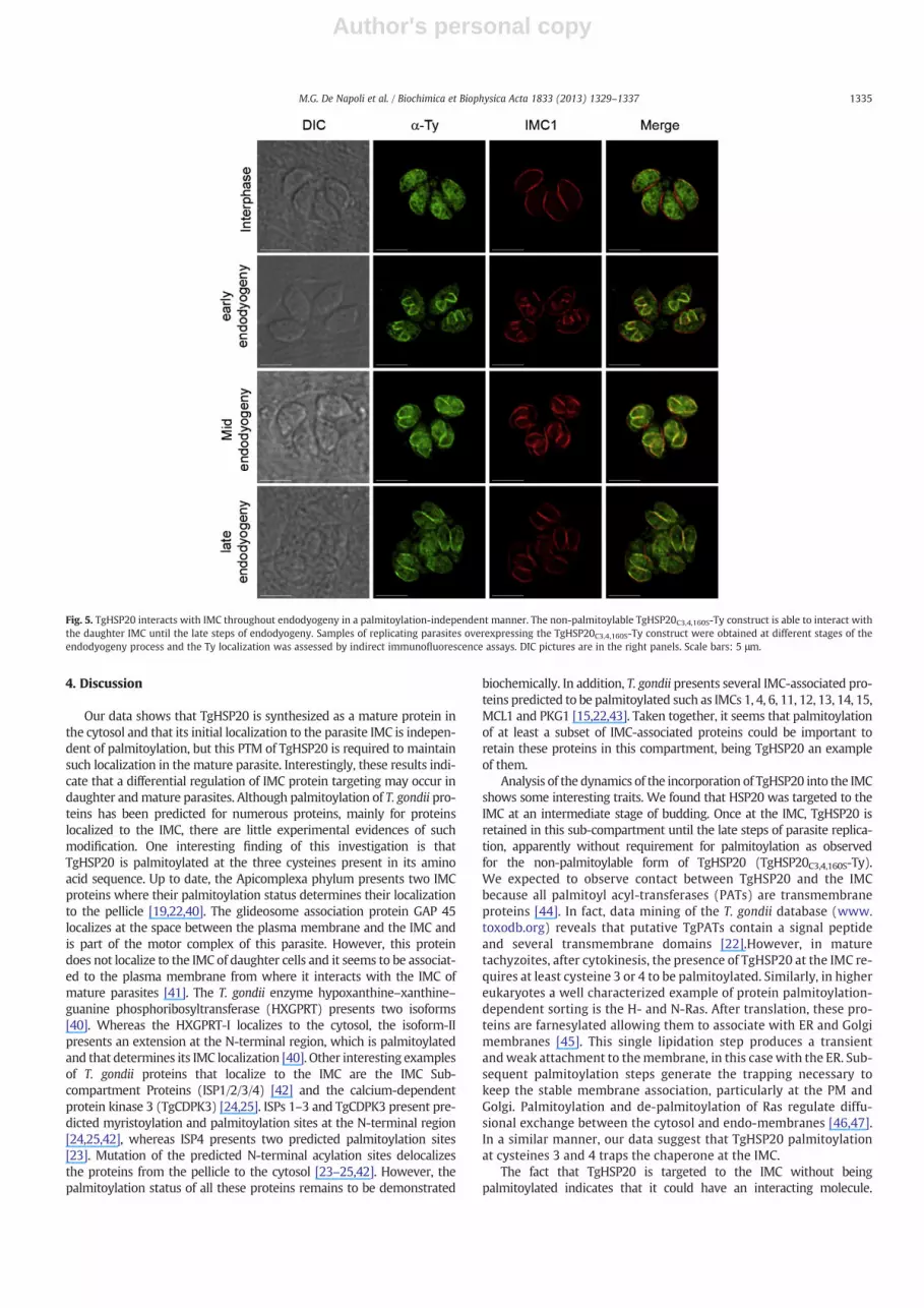
Apicomplexan parasites divide by internal budding into a variablenumber of daughter cells. T. gondii divides by the simplest mode inthe phylum, endodyogeny, and assembles two daughter cells everyround of replication. Prior to cytokinesis, daughter cells are dottedwith a fully formed pellicle, which includes the IMC [39]. In order todetermine how palmitoylation modulates TgHSP20 targeting to theIMC of daughter cells, intracellular replicating parasites were treatedwith 2-BP and analyzed by indirect immunofluorescence microscopywith anti-HSP20 antibodies. Interestingly, palmitoylation seems not toplay a role on TgHSP20 attachment to the IMC of daughter cells(Fig. 4A). To confirm this result, stable lines expressing differentpalmitoylable versions of TgHSP20 were analyzed for their sub-cellularlocalization in replicating parasites. All the mutant lines lysed theinfected monolayer at a similar time than the parental cell-line, notshowing any evident phenotype associated to parasite growth (datanot shown). All the Ty-tagged TgHSP20s localized to the IMC of daughtercells regardless of the cysteine that wasmodified (Fig. 4B). This indicatesthat the initial attachment of TgHSP20 to the IMC of daughter cells is aprocess independent of palmitoylation. Furthermore, the stable lineexpressing a non-palmitoylable form of TgHSP20 (TgHSP20C3,4,160S-Ty)localized to the IMC of daughter cells but to the cytosol of matureparasites. As such, we decided to investigate at which point of theendodyogeny process TgHSP20C3,4,160S-Ty changed its localization.Fig. 5 shows that TgHSP20C3,4,160S-Ty localizes to the IMC of daughtercells until late endodyogeny and moves to the cytosol only when para-sites are mature. As such, palmitoylation seems to be required to retainTgHSP20 at the mother IMC in intracellular parasites.
Fig. 3. Transmission electronmicroscopy of the TgHSP20C3,4S-Ty construct suggests thepresencemembrane accumulation. Lack or partial palmitoylation at the N-terminal of TgHSP20 formsan unidentified structure. A) Immunofluorescence assay showing the sub-cellular localization of full-length TgHSP20-Ty (TgHSP20FL-Ty; upper panel) or parasites over-expressing differentN-terminal palmitoylable TgHSP20 constructs (same result either with TgHSP20C3S-Ty, TgHSP20C4S-Ty or TgHSP20C3,4S-Ty; lower panel). Scale bars = 5 μm. B) Transmission electronmicroscopy of the parasites observed in panel A. The white arrowheads show the atypical localization of TgHSP20 with indirect immunofluorescence and black arrowheads in the electronmicroscopy analysis. PM: plasma membrane; Nu: nucleus; Cyt: cytoplasm. Scale bars = 1 μm and 200 nm for the zoomed images.
1333M.G. De Napoli et al. / Biochimica et Biophysica Acta 1833 (2013) 1329–1337

Author's personal copy
Fig. 4. Palmitoylation is not required for TgHSP20 attachment to the IMC of daughter cells. A) TgHSP20 localization at the daughter IMC is not affected by 2-BP treatment. Replicatingparasites were treated with 2-BP (100 μM) or DMSO for 16–18 h, then fixed and TgHSP20 localization was assessed by indirect immunofluorescence microscopy. Differentialinterference contrast (DIC) images are shown in the right panels. Scale bars = 5 μm. B) IMC localization at the daughter cell is maintained regardless of the TgHSP20 palmitoylationstatus. Intracellular replicating parasites overexpressing different Ty-tagged constructs were fixed and TgHSP20 localization was assessed by indirect immunofluorescence micros-copy. DIC pictures are in the right panels. The anti-IMC1 antibody labels the IMC from both the mother and daughter cells. Scale bars = 5 μm.
1334 M.G. De Napoli et al. / Biochimica et Biophysica Acta 1833 (2013) 1329–1337
Author's personal copy
4. Discussion
Our data shows that TgHSP20 is synthesized as a mature protein inthe cytosol and that its initial localization to the parasite IMC is indepen-dent of palmitoylation, but this PTM of TgHSP20 is required to maintainsuch localization in the mature parasite. Interestingly, these results indi-cate that a differential regulation of IMC protein targeting may occur indaughter andmature parasites. Although palmitoylation of T. gondii pro-teins has been predicted for numerous proteins, mainly for proteinslocalized to the IMC, there are little experimental evidences of suchmodification. One interesting finding of this investigation is thatTgHSP20 is palmitoylated at the three cysteines present in its aminoacid sequence. Up to date, the Apicomplexa phylum presents two IMCproteins where their palmitoylation status determines their localizationto the pellicle [19,22,40]. The glideosome association protein GAP 45localizes at the space between the plasma membrane and the IMC andis part of the motor complex of this parasite. However, this proteindoes not localize to the IMC of daughter cells and it seems to be associat-ed to the plasma membrane from where it interacts with the IMC ofmature parasites [41]. The T. gondii enzyme hypoxanthine–xanthine–guanine phosphoribosyltransferase (HXGPRT) presents two isoforms[40]. Whereas the HXGPRT-I localizes to the cytosol, the isoform-IIpresents an extension at the N-terminal region, which is palmitoylatedand that determines its IMC localization [40]. Other interesting examplesof T. gondii proteins that localize to the IMC are the IMC Sub-compartment Proteins (ISP1/2/3/4) [42] and the calcium-dependentprotein kinase 3 (TgCDPK3) [24,25]. ISPs 1–3 and TgCDPK3 present pre-dicted myristoylation and palmitoylation sites at the N-terminal region[24,25,42], whereas ISP4 presents two predicted palmitoylation sites[23]. Mutation of the predicted N-terminal acylation sites delocalizesthe proteins from the pellicle to the cytosol [23–25,42]. However, thepalmitoylation status of all these proteins remains to be demonstrated
biochemically. In addition, T. gondii presents several IMC-associated pro-teins predicted to be palmitoylated such as IMCs 1, 4, 6, 11, 12, 13, 14, 15,MCL1 and PKG1 [15,22,43]. Taken together, it seems that palmitoylationof at least a subset of IMC-associated proteins could be important toretain these proteins in this compartment, being TgHSP20 an exampleof them.
Analysis of the dynamics of the incorporation of TgHSP20 into the IMCshows some interesting traits. We found that HSP20 was targeted to theIMC at an intermediate stage of budding. Once at the IMC, TgHSP20 isretained in this sub-compartment until the late steps of parasite replica-tion, apparently without requirement for palmitoylation as observedfor the non-palmitoylable form of TgHSP20 (TgHSP20C3,4,160S-Ty).We expected to observe contact between TgHSP20 and the IMCbecause all palmitoyl acyl-transferases (PATs) are transmembraneproteins [44]. In fact, data mining of the T. gondii database (www.toxodb.org) reveals that putative TgPATs contain a signal peptideand several transmembrane domains [22].However, in maturetachyzoites, after cytokinesis, the presence of TgHSP20 at the IMC re-quires at least cysteine 3 or 4 to be palmitoylated. Similarly, in highereukaryotes a well characterized example of protein palmitoylation-dependent sorting is the H- and N-Ras. After translation, these pro-teins are farnesylated allowing them to associate with ER and Golgimembranes [45]. This single lipidation step produces a transientandweak attachment to themembrane, in this case with the ER. Sub-sequent palmitoylation steps generate the trapping necessary tokeep the stable membrane association, particularly at the PM andGolgi. Palmitoylation and de-palmitoylation of Ras regulate diffu-sional exchange between the cytosol and endo-membranes [46,47].In a similar manner, our data suggest that TgHSP20 palmitoylationat cysteines 3 and 4 traps the chaperone at the IMC.
The fact that TgHSP20 is targeted to the IMC without beingpalmitoylated indicates that it could have an interacting molecule.
Fig. 5. TgHSP20 interacts with IMC throughout endodyogeny in a palmitoylation-independent manner. The non-palmitoylable TgHSP20C3,4,160S-Ty construct is able to interact withthe daughter IMC until the late steps of endodyogeny. Samples of replicating parasites overexpressing the TgHSP20C3,4,160S-Ty construct were obtained at different stages of theendodyogeny process and the Ty localization was assessed by indirect immunofluorescence assays. DIC pictures are in the right panels. Scale bars: 5 μm.
1335M.G. De Napoli et al. / Biochimica et Biophysica Acta 1833 (2013) 1329–1337

Author's personal copy
Although these parasites express the endogenous TgHSP20 and the mu-tants versions could tag along with its localization, similar results wereobtained when TgHSP20C3,4,160S-Ty was expressed in a TgHSP20-knockout parasite (Fig. 3). Up to now, we were unable to detect any proteinpartner either as interactor and/or substrate [28] but one of them couldbe the specific PAT that palmitoylates TgHSP20, a speculation thatneeds to be further explored. Another possibility is that TgHSP20 bindsIMC lipids as it has been shown for the recombinant TgHSP20 [28]. Thisintrinsic property could account for the initial binding of TgHSP20 tothe IMC of nascent cells, whereas palmitoylation, as mentioned above,is required to maintain the IMC localization in mature intracellular para-sites. In this case, a change in membrane composition or fluidity at thefinal steps of parasite replication [48,49] could account for the generationof an unstable environment for non-palmitoylated TgHSP20. In fact, themature IMC present a different topology along the parasite withpalmitoylated proteins located at three different regions [42]. Thissuggests that once TgHSP20 is at the IMC, palmitoylation could conferproperties to stably interact or bind with specific regions along thissub-compartment. Actually, TgHSP20 presents a distribution in shortstripes from the basal to the polar region of the parasite. It is interestingto note that hsp20 expression at the mRNA level is clearly associated tocell cycle, peaking at the mitosis phase (Fig. 4), as described by otherIMC proteins and suggesting that it occurs because they accompany thebuilding of the new daughter cell [50]. This is in agreement with thepossibility that TgHSP20 matures together with the IMC during parasitereplication. Further studies would be necessary to address this point.
After treatment of tachyzoites with 2-BP, TgHSP20 showed a locali-zation similar to that observed for the TgHSP20C3,4S-Ty, demonstratingthat palmitoylation on cysteine 160 does not play a role in TgHSP20 trap-ping at the IMC. Considering that cysteine 160 is conserved, the role of itspalmitoylation remains to be elucidated. On the other hand, lack ofpalmitoylation at cysteine 160 (TgHSP20C160S-Ty) did not produce theTgHSP20-positive unidentified structure, suggesting that this structuredepends on palmitoylation at this residue. We speculate that this struc-ture may contain misfolded products of TgHSP20. Interestingly, thisstructure seems to contain membrane structures that are reminiscentof the membranous myelin fibers observed when the apicoplast biogen-esis was disturbed [38]. The significance of this finding needs to befurther studied. The fact that TgHSP20C3,4,160S-Ty did not produce anyunusual structure supports our hypothesis of the idea of a previousrequirement of TgHSP20 association to the IMC to generate such struc-ture. Even though the role of cysteine 160 is unclear, palmitoylation atthis site may stabilize the anomalous attachment of TgHSP20 to theIMC. Of note, these unidentified structures are not naturally observedin the parasite.
In conclusion, we observed that the T. gondii IMC protein HSP20 ispalmitoylated at three sites and that these palmitoylated sites havedifferent roles at least in the targeting of the chaperone to the IMC.We also observed that TgHSP20 is translated in the cytosol and doesnot require rapid palmitoylation to bind to the nascent IMC. However,a fine modulation of protein localization can occur at later steps ofparasite replication, including palmitoylation as one of such process.
Supplementary data to this article can be found online at http://dx.doi.org/10.1016/j.bbamcr.2013.02.022.
Acknowledgements
We would like to thank Maria E. Francia and Beth Richardson fortechnical assistancewith EM and the Plant Biology ElectronMicroscopyFacility at the University of Georgia. This work was supported by:ANPCyT grant BID 1728 OC-AR PICT 2010-1494 (MMC), a PIP grant2010-0190 (MMC) and a National Institutes of Health–National Insti-tute of Allergy and Infectious Diseases (NIH–NIAID) grant AI083162(to SOA and MMC). NDM, MMC and SOA are researchers from theNational Council of Research (CONICET) and UNSAM. MGDN is a PhDfellow from CONICET and was supported in part by a Fogarty
International Center Training Grant (NIH D43TW007888). Work inSNJM laboratory was supported by NIH grant AI096836.
References
[1] J.B. McCabe, L.G. Berthiaume, Functional roles for fatty acylated amino-terminaldomains in subcellular localization, Mol. Biol. Cell 10 (1999) 3771–3786.
[2] J.B. McCabe, L.G. Berthiaume, N-terminal protein acylation confers localization tocholesterol, sphingolipid-enriched membranes but not to lipid rafts/caveolae,Mol. Biol. Cell 12 (2001) 3601–3617.
[3] L. Berthiaume, I. Deichaite, S. Peseckis, M.D. Resh, Regulation of enzymatic activityby active site fatty acylation. A new role for long chain fatty acid acylation of proteins,J. Biol. Chem. 269 (1994) 6498–6505.
[4] M.M. Corvi, C.L. Soltys, L.G. Berthiaume, Regulation of mitochondrial carbamoyl-phosphate synthetase 1 activity by active site fatty acylation, J. Biol. Chem. 276(2001) 45704–45712.
[5] I. Deichaite, L.P. Casson, H.P. Ling, M.D. Resh, In vitro synthesis of pp 60v-src:myristoylation in a cell-free system, Mol. Cell. Biol. 8 (1988) 4295–4301.
[6] C. Wilcox, J.S. Hu, E.N. Olson, Acylation of proteins with myristic acid occurscotranslationally, Science 238 (1987) 1275–1278.
[7] T. Utsumi, Analysis of co- and post-translational modifications of proteins by invitro and in vivo metabolic labeling, Seikagaku 75 (2003) 373–378.
[8] S.M. Warden, C. Richardson, J. O'Donnell Jr., D. Stapleton, B.E. Kemp, L.A. Witters,Post-translational modifications of the beta-1 subunit of AMP-activated proteinkinase affect enzyme activity and cellular localization, Biochem. J. 354 (2001)275–283.
[9] D.D. Martin, G.L. Vilas, J.A. Prescher, G. Rajaiah, J.R. Falck, C.R. Bertozzi, L.G.Berthiaume, Rapid detection, discovery, and identification of post-translationallymyristoylated proteins during apoptosis using a bio-orthogonal azidomyristateanalog, FASEB J. 22 (2008) 797–806.
[10] G.L. Vilas, M.M. Corvi, G.J. Plummer, A.M. Seime, G.R. Lambkin, L.G. Berthiaume,Posttranslational myristoylation of caspase-activated p21-activated proteinkinase 2 (PAK2) potentiates late apoptotic events, Proc. Natl. Acad. Sci. U. S. A.103 (2006) 6542–6547.
[11] M. Fukata, Y. Fukata, H. Adesnik, R.A. Nicoll, D.S. Bredt, Identification of PSD-95palmitoylating enzymes, Neuron 44 (2004) 987–996.
[12] M.E. Linder, R.J. Deschenes, New insights into the mechanisms of proteinpalmitoylation, Biochemistry 42 (2003) 4311–4320.
[13] J.A. Duncan, A.G. Gilman, A cytoplasmic acyl-protein thioesterase that removespalmitate from G protein alpha subunits and p21(RAS), J. Biol. Chem. 273(1998) 15830–15837.
[14] M.C. Bano, C.S. Jackson, A.I. Magee, Pseudo-enzymatic S-acylation of a myristoylatedyes protein tyrosine kinase peptide in vitro may reflect non-enzymatic S-acylationin vivo, Biochem. J. 330 (Pt 2) (1998) 723–731.
[15] R.G. Donald, P.A. Liberator, Molecular characterization of a coccidian parasitecGMP dependent protein kinase, Mol. Biochem. Parasitol. 120 (2002) 165–175.
[16] N. Kato, T. Sakata, G. Breton, K.G. Le Roch, A. Nagle, C. Andersen, B. Bursulaya, K.Henson, J. Johnson, K.A. Kumar, F. Marr, D. Mason, C. McNamara, D. Plouffe, V.Ramachandran, M. Spooner, T. Tuntland, Y. Zhou, E.C. Peters, A. Chatterjee, P.G.Schultz, G.E. Ward, N. Gray, J. Harper, E.A. Winzeler, Gene expression signaturesand small-molecule compounds link a protein kinase to Plasmodium falciparummotility, Nat. Chem. Biol. 4 (2008) 347–356.
[17] C. Moskes, P.A. Burghaus, B.Wernli, U. Sauder, M. Durrenberger, B. Kappes, Export ofPlasmodium falciparum calcium-dependent protein kinase 1 to the parasitophorousvacuole is dependent on three N-terminal membrane anchormotifs, Mol. Microbiol.54 (2004) 676–691.
[18] I. Russo, A. Oksman, D.E. Goldberg, Fatty acid acylation regulates trafficking of theunusual Plasmodium falciparum calpain to the nucleolus, Mol. Microbiol. 72(2009) 229–245.
[19] R.R. Rees-Channer, S.R. Martin, J.L. Green, P.W. Bowyer, M. Grainger, J.E. Molloy,A.A. Holder, Dual acylation of the 45 kDa gliding-associated protein (GAP45) inPlasmodium falciparum merozoites, Mol. Biochem. Parasitol. 149 (2006) 113–116.
[20] A.M. Alonso, V.M. Coceres, M.G. De Napoli, A.F. Nieto Guil, S.O. Angel, M.M. Corvi,Protein palmitoylation inhibition by 2-bromopalmitate alters gliding, host cellinvasion and parasite morphology in Toxoplasma gondii, Mol. Biochem. Parasitol.184 (2012) 39–43.
[21] S.D. Gilk, E. Gaskins, G.E. Ward, C.J. Beckers, GAP45 phosphorylation controlsassembly of the Toxoplasmamyosin XIV complex, Eukaryot. Cell 8 (2009) 190–196.
[22] M.M. Corvi, L.G. Berthiaume, M.G. Napoli, Protein palmitoylation in protozoanparasites, Front. Biosci. (Schol. Ed.) 3 (2011) 1067–1079.
[23] C. Fung, J.R. Beck, S.D. Robertson, M.J. Gubbels, P.J. Bradley, Toxoplasma ISP4 is a centralIMC sub-compartment protein whose localization depends on palmitoylation but notmyristoylation, Mol. Biochem. Parasitol. 184 (2012) 99–108.
[24] J.M. McCoy, L. Whitehead, G.G. van Dooren, C.J. Tonkin, TgCDPK3 regulatescalcium-dependent egress of Toxoplasma gondii from host cells, PLoS Pathog. 8(2012) e1003066.
[25] E. Garrison, M. Treeck, E. Ehret, H. Butz, T. Garbuz, B.P. Oswald, M. Settles, J.Boothroyd, G. Arrizabalaga, A forward genetic screen reveals that calcium-dependent protein kinase 3 regulates egress in Toxoplasma, PLoS Pathog. 8 (2012)e1003049.
[26] C. Opitz, D. Soldati, ‘The glideosome’: a dynamic complex powering gliding motionand host cell invasion by Toxoplasma gondii, Mol. Microbiol. 45 (2002) 597–604.
[27] M.L. Jones, M.O. Collins, D. Goulding, J.S. Choudhary, J.C. Rayner, Analysis of proteinpalmitoylation reveals a pervasive role in Plasmodium development and pathogenesis,Cell Host Microbe 12 (2012) 246–258.
1336 M.G. De Napoli et al. / Biochimica et Biophysica Acta 1833 (2013) 1329–1337

Author's personal copy
[28] N. de Miguel, M. Lebrun, A. Heaslip, K. Hu, C.J. Beckers, M. Matrajt, J.F. Dubremetz,S.O. Angel, Toxoplasma gondii Hsp20 is a stripe-arranged chaperone-like proteinassociated with the outer leaflet of the inner membrane complex, Biol. Cell 100(2008) 479–489.
[29] N. de Miguel, N. Braun, A. Bepperling, T. Kriehuber, A. Kastenmuller, J. Buchner,S.O. Angel, M. Haslbeck, Structural and functional diversity in the family ofsmall heat shock proteins from the parasite Toxoplasma gondii, Biochim. Biophys.Acta 1793 (2009) 1738–1748.
[30] E. Precigout, A. Valentin, B. Carcy, A. Gorenflot, K. Nakamura, M. Aikawa, J. Schrevel,Babesia divergens: characterization of a 17-kDa merozoite membrane protein, Exp.Parasitol. 77 (1993) 425–434.
[31] V.M. Coceres, A.M. Alonso, M.L. Alomar, M.M. Corvi, Rabbit antibodies againstToxoplasma Hsp20 are able to reduce parasite invasion and gliding motility inToxoplasma gondii and parasite invasion in Neospora caninum, Exp. Parasitol.132 (2012) 274–281.
[32] G.N. Montagna, C.A. Buscaglia, S. Munter, C. Goosmann, F. Frischknecht, V.Brinkmann, K. Matuschewski, Critical role for heat shock protein 20 (HSP20) inmigration of malarial sporozoites, J. Biol. Chem. 287 (2012) 2410–2422.
[33] G.N. Montagna, K. Matuschewski, C.A. Buscaglia, Small heat shock proteins in cellularadhesion and migration: evidence from Plasmodium genetics, Cell Adh. Migr. 6(2012) 78–84.
[34] E. Montero, M. Rodriguez, L.M. Gonzalez, C.A. Lobo, Babesia divergens: identificationand characterization of BdHSP-20, a small heat shock protein, Exp. Parasitol. 119(2008) 238–245.
[35] R.G. Donald, D. Carter, B. Ullman, D.S. Roos, Insertional tagging, cloning, and expressionof the Toxoplasma gondii hypoxanthine–xanthine–guanine phosphoribosyltransferasegene. Use as a selectable marker for stable transformation, J. Biol. Chem. 271 (1996)14010–14019.
[36] O. Cerede, J.F. Dubremetz, M. Soete, D. Deslee, H. Vial, D. Bout, M. Lebrun, Synergisticrole of micronemal proteins in Toxoplasma gondii virulence, J. Exp. Med. 201 (2005)453–463.
[37] M.D. Resh, Use of analogs and inhibitors to study the functional significance ofprotein palmitoylation, Methods 40 (2006) 191–197.
[38] L. Tawk, J.F. Dubremetz, P. Montcourrier, G. Chicanne, F. Merezegue, V. Richard, B.Payrastre, M. Meissner, H.J. Vial, C. Roy, K.Wengelnik, M. Lebrun, Phosphatidylinositol3-monophosphate is involved in toxoplasma apicoplast biogenesis, PLoS Pathog. 7(2011) e1001286.
[39] B. Striepen, C.N. Jordan, S. Reiff, G.G. van Dooren, Building the perfect parasite:cell division in apicomplexa, PLoS Pathog. 3 (2007) e78.
[40] K. Chaudhary, R.G. Donald, M. Nishi, D. Carter, B. Ullman, D.S. Roos, Differen-tial localization of alternatively spliced hypoxanthine–xanthine–guaninephosphoribosyltransferase isoforms in Toxoplasma gondii, J. Biol. Chem. 280(2005) 22053–22059.
[41] K. Frenal, V. Polonais, J.B. Marq, R. Stratmann, J. Limenitakis, D. Soldati-Favre,Functional dissection of the apicomplexan glideosome molecular architecture,Cell Host Microbe 8 (2010) 343–357.
[42] J.R. Beck, I.A. Rodriguez-Fernandez, J. Cruz de Leon, M.H. Huynh, V.B. Carruthers,N.S. Morrissette, P.J. Bradley, A novel family of Toxoplasma IMC proteins displaysa hierarchical organization and functions in coordinating parasite division, PLoSPathog. 6 (2010).
[43] B.R. Anderson-White, F.D. Ivey, K. Cheng, T. Szatanek, A. Lorestani, C.J. Beckers, D.J.Ferguson, N. Sahoo, M.J. Gubbels, A family of intermediate filament-like proteins issequentially assembled into the cytoskeleton of Toxoplasma gondii, Cell. Microbiol.13 (2011) 18–31.
[44] D.A. Mitchell, A. Vasudevan, M.E. Linder, R.J. Deschenes, Protein palmitoylation bya family of DHHC protein S-acyltransferases, J. Lipid Res. 47 (2006) 1118–1127.
[45] E. Choy, V.K. Chiu, J. Silletti, M. Feoktistov, T. Morimoto, D. Michaelson, I.E. Ivanov,M.R. Philips, Endomembrane trafficking of ras: the CAAX motif targets proteins tothe ER and Golgi, Cell 98 (1999) 69–80.
[46] J.S. Goodwin, K.R. Drake, C. Rogers, L. Wright, J. Lippincott-Schwartz, M.R. Philips,A.K. Kenworthy, Depalmitoylated Ras traffics to and from the Golgi complex via anonvesicular pathway, J. Cell Biol. 170 (2005) 261–272.
[47] O. Rocks, A. Peyker, M. Kahms, P.J. Verveer, C. Koerner, M. Lumbierres, J. Kuhlmann, H.Waldmann, A. Wittinghofer, P.I. Bastiaens, An acylation cycle regulates localizationand activity of palmitoylated Ras isoforms, Science 307 (2005) 1746–1752.
[48] K. Hu, T. Mann, B. Striepen, C.J. Beckers, D.S. Roos, J.M. Murray, Daughter cell assemblyin the protozoan parasite Toxoplasma gondii, Mol. Biol. Cell 13 (2002) 593–606.
[49] T. Mann, E. Gaskins, C. Beckers, Proteolytic processing of TgIMC1 duringmaturation ofthemembrane skeleton of Toxoplasma gondii, J. Biol. Chem. 277 (2002) 41240–41246.
[50] M.S. Behnke, J.C. Wootton, M.M. Lehmann, J.B. Radke, O. Lucas, J. Nawas, L.D.Sibley, M.W. White, Coordinated progression through two subtranscriptomesunderlies the tachyzoite cycle of Toxoplasma gondii, PLoS One 5 (2010) e12354.
[51] M.J. Wichroski, J.A. Melton, C.G. Donahue, R.K. Tweten, G.E. Ward, Clostridiumsepticum alpha-toxin is active against the parasitic protozoan Toxoplasma gondiiand targets members of the SAG family of glycosylphosphatidylinositol-anchoredsurface proteins, Infect. Immun. 70 (2002) 4353–4361.
[52] S. Besteiro, A. Michelin, J. Poncet, J.F. Dubremetz, M. Lebrun, Export of a Toxoplasmagondii rhoptry neck protein complex at the host cell membrane to form the movingjunction during invasion, PLoS Pathog. 5 (2009) e1000309.
[53] B. Gajria, A. Bahl, J. Brestelli, J. Dommer, S. Fischer, X. Gao, M. Heiges, J. Iodice, J.C.Kissinger, A.J. Mackey, D.F. Pinney, D.S. Roos, C.J. Stoeckert Jr., H. Wang, B.P. Brunk,ToxoDB: an integrated Toxoplasma gondii database resource, Nucleic Acids Res. 36(2008) D553–D556.
[54] J.R. Radke, M.W. White, Expression of herpes simplex virus thymidine kinase inToxoplasma gondii attenuates tachyzoite virulence in mice, Infect. Immun. 67(1999) 5292–5297.
1337M.G. De Napoli et al. / Biochimica et Biophysica Acta 1833 (2013) 1329–1337


