Embed Size (px)
Citation preview
N6-Formylation of Lysine: A Pathological SecondaryModification of Proteins
by
Bahar Edrissi
B.S. in Biomedical EngineeringUniversity of Utah, 2003
ARCH&SMASSACHUSETTS INSTr'fTE
CHNOLOGY
A97MAY 29 2013
LLiBRARIES _SUBMITTED TO THE DEPARTMENT OF BIOLOGICAL ENGINEERING
IN PARTIAL FULFILLMENT OF THE REQUIREMENTS FOR THE DEGREE OF
DOCTOR OF PHILOSOPHY IN BIOLOGICAL ENGINEERING
AT THE
MASSACHUSETTS INSTITUTE OF TECHNOLOGY
FEBRUARY 2013
@ 2012 Massachusetts Institute of Technology. All rights reserved.
A u th or...........................<....... ............... . .....................................-.- -- -Department of Biological Engineering
December 5, 2012
Certified by........... ........................................... ...----Peter C. Dedon
Professor of Toxicology and Biological EngineeringThesis Supervisor
A ccepted by....................................Forest M. White
Associate Professor of Biological EngineeringChairman, Department Committee on Graduate Theses
Committee members who voted in favor of thesis:
Advisor: Peter C. DedonTitle: Professor of Toxicology and Biological Engineering
Chair: John M. EssigmannTitle: Professor of Chemistry and Biological Engineering
Other member: Steven R. TannenbaumTitle: Professor of Chemistry and Biological Engineering
Other member: John S. WishnokTitle: Senior Research Scientist
2
N6-Formylation of Lysine: A Pathological Secondary
Modification of Proteins
by
Bahar Edrissi
Submitted to the Department of Biological Engineeringon December 28, 2012, in partial fulfillment of the
requirements for the degree ofDoctor of Philosophy in Biological Engineering
Abstract
There is increasing recognition that aberrant protein modifications play animportant role in the pathophysiology of inflammation and oxidative stress in cells. Werecently discovered that N6-formylation of lysine is an abundant endogenousmodification of histone and chromatin proteins. The high abundance of N 6-formyllysinein histone proteins and its chemical similarity to the biologically important N6_acetyllysine has raised questions about its mechanisms of formation and biologicalconsequences. Using novel ultrasensitive and specific liquid chromatography-coupledtandem mass spectrometry methods (LC-MS/MS) to quantify N6-formyllysine lesions inproteins, we aimed to investigate the sources as well as the fate of this abundantendogenous protein modification. We present evidence that endogenous formaldehyde isa major source of N6-formyllysine and that this adduct is widespread among proteins inall cellular compartments. We observed in vitro as well as in vivo that formaldehydeexposure leads to a dose-dependent increase in N6 -formyllysine protein adducts, with theuse of isotopically-labeled formaldehyde to dissect endogenous from exogenousformaldehyde as sources of the adduct. Further, other isotope labeling studies revealedthat lysine demethylation in histone proteins is not a source of N -formyllysine. Withregard to N6-formyllysine persistence in cells, our investigation of histone deacetylasesrevealed that despite chemical similarity of N6-formyllysine to N6-acetyllysine, theformer is refractory to removal by histone deacetylases, which suggests that they willpersist throughout the life of individual histone proteins. If not repaired, lysineformylation could accumulate to significant levels. The resemblance of N -formyllysineto N 6-acetyllysine, together with recent studies that mapped its location on manyconserved lysine acetylation and methylation sites along histone proteins, support theidea that this abundant protein modification could interfere with normal regulation ofgene expression, potentially leading to an epigenetic mechanism of disruption of cellfunction.
Thesis Supervisor: Peter C. DedonTitle: Professor of Toxicology and Biological Engineering
3
Table of Contents
A bstract ............................................................................................................................... 3A bbreviations ..................................................................................................................... 6List of Figures.....................................................................................................................8List of Tables .................................................................................................................... 10A cknow ledgem ents .......................................................................................................... 11
Chapter 1. Introduction
G oals and sum m ary of this thesis ........................................................................ 15Post-translational modifications of proteins and their regulatory roles...............16Histone proteins and their post-translational modification.................................. 19Lysine acetylation is a key regulatory post-translational modification ............... 21Production of reactive chem ical species in cells.................................................. 23Protein damage due to reactive endogenous oxidants and electrophiles ............. 27N -Formyllysine as an adventitious protein modification ................................... 31The biological implications of N -formyllysine modification of histone............34References................................................................................................................36
Chapter 2. Development of a liquid chromatography-coupled tandem mass spectrometrymethod for quantification of N -formyllysine adducts in proteins
Abstract .................................................................................................................... 46Introduction..............................................................................................................47M aterials and M ethods......................................................................................... 48Results ...................................................................................................................... 57D iscussion................................................................................................................64References................................................................................................................68
Chapter 3. Quantitative analysis of histone modifications: Formaldehyde is a source ofpathological N 6-formyllysine that is refractory to histone deacetylases
Abstract .................................................................................................................... 70Introduction..............................................................................................................71M aterials and M ethods......................................................................................... 73Results......................................................................................................................78D iscussion................................................................................................................88References................................................................................................................92
Chapter 4. Formaldehyde-induced formation of N 6 -formyllysine protein adducts in rats
Abstract .................................................................................................................... 99Introduction............................................................................................................100M aterials and M ethods...........................................................................................102Results....................................................................................................................105
4
Discussion..............................................................................................................109References..............................................................................................................114
Chapter 5. Concluding remarks
Conclusions............................................................................................................118References..............................................................................................................122
5
Abbreviations
AGEs
C03'-
HATs
HDACs
HNE
HO'
H 2 0 2
HOCI
HPLC
LC
LC-MS/MS
LSDl
MDA
MRM
MS
NO
N0 2*
N027
N 20 3
102
02~
03
advanced glycation end products
carbonate radical anion
histone acetyl transferases
histone deacetylases
4-hydroxy-2-nonenal
hydroxyl radical
hydrogen peroxide
hypochlorous acid
high performance liquid chromatography
liquid chromatography
liquid chromatography-coupled tandem mass spectrometry
lysine-specific demethylase 1
malondialdehyde
multiple reaction monitoring
mass spectrometry
nitric oxide
nitrogen dioxide radical
nitrite
nitrous anhydride
singlet oxygen
superoxide anion radical
ozone
6
ONOO-
ONOOCO2
PITC
PTMs
QQQ
SAHA
SOD
SRM
TCE
TFA
peroxynitrite
nitrosoperoxycarbonate
phenylisothiocyanate
post-translational modifications of proteins
triple quadrupole mass spectrometer
suberoylanilidehydroxamic acid
superoxide dismutase
selected reaction monitoring
trichloroethylene
trifluoroacetic acid
7

List of Figures
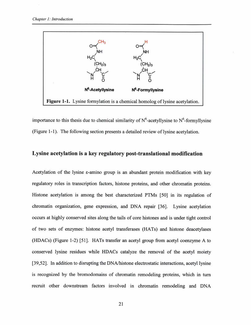
Figure 1-1. Lysine formylation is a chemical homolog of lysine acetylation..............21
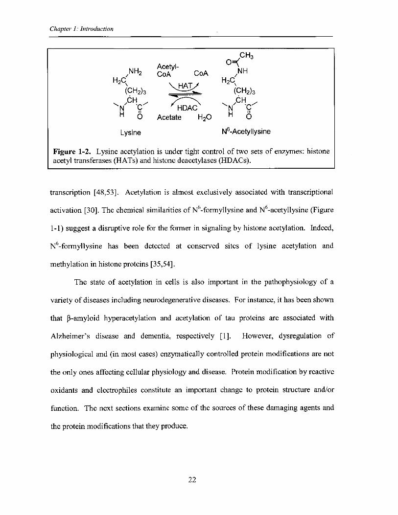
Figure 1-2. Lysine acetylation is under tight control of two sets of enzymes: histoneacetyltransferases (HATs) and histone deacetylases (HDACs)....................................22
Figure 1-3. Generation of reactive oxygen and nitrogen species.................................24
Figure 1-4. Examples of reactive aldehydes generated from damage tobiom olecules ...................................................................................................................... 26
Figure 1-5. Examples of protein adducts caused by reactive electrophiles ................. 29
Figure 1-6. Generation of N -formyllysine as a result of DNA oxidation..................32
Figure 2-1. The overall strategy for quantifying lysine adducts of interest in proteins...48
Figure 2-2. The structure and MW of lysine species monitored.................52
Figure 2-3. Proposed structures for product ions monitored ...................................... 54
Figure 2-4. Examples of product ions for each lysine species monitored, afteroptim ization ....................................................................................................................... 55
Figure 2-5. Examples of calibration curves for the isotope dilution LC-MS/MS analysisof lysine species ................................................................................................................. 56
Figure 2-6. Comparison of released lysine in protein digestion using Streptomycesgriseus protease versus proteinase K ............................................................................ 57
Figure 2-7. An example of different lysine species detected in purified histone H4 fromT K 6 cells............................................................................................................................59
Figure 2-8. Citrulline co-elutes with M+1 ion of N6-formyllysine.............................61
Figure 2-9. An example of co-elution of a contaminant peak with M+2 ion of N6-formyllysine in total histone sample from bovine liver............................................... 62
Figure 2-10. Dilution rate of N -formyllysine in TK6 cells ........................................ 63
Figure 3-1. Sources of N6-formyllysine...................................................................... 73
Figure 3-2. Formaldehyde is a source of N6-formyllysine...........................................81
8
Figure 3-3. Addition of [1 3C, 2H 2]-formaldehyde to TK6 cells distinguishes exogenousfrom endogenous sources of N6-formyllysine ............................................................... 83
Figure 3-4. Analysis of lysine demethylation as a source of N 6-formyllysine ........... 85
Figure 3-5. Effect of lysine deacetylases on N6-formyllysine .................................... 87
Figure 3-6. Summary of findings on N 6-formyllysine in histones...............................91
Figure 3-S1. Reversed-phase HPLC fractionation of histone proteins.......................96
Figure 3-S2. Lysine demethylation is not a source of N6-formyllysine in histones ........ 97
Figure 4-1. Inhalation of [13 C, 2H2]-formaldehyde distinguishes exogenous fromendogenous sources of N -formyllysine in rats .............................................................. 107
Figure 4-2. [13C,2 H2]-Formaldehyde causes a dose-response increase in exogenous N 6_form yllysine adducts........................................................................................................109
Figure 4-3. Comparison of exposure-response plots for ratios of exogenous toendo enous adducts in DNA and protein in the nasal epithelium of rats exposed to[' C , H 2]-form aldehyde ................................................................................................... 111
9
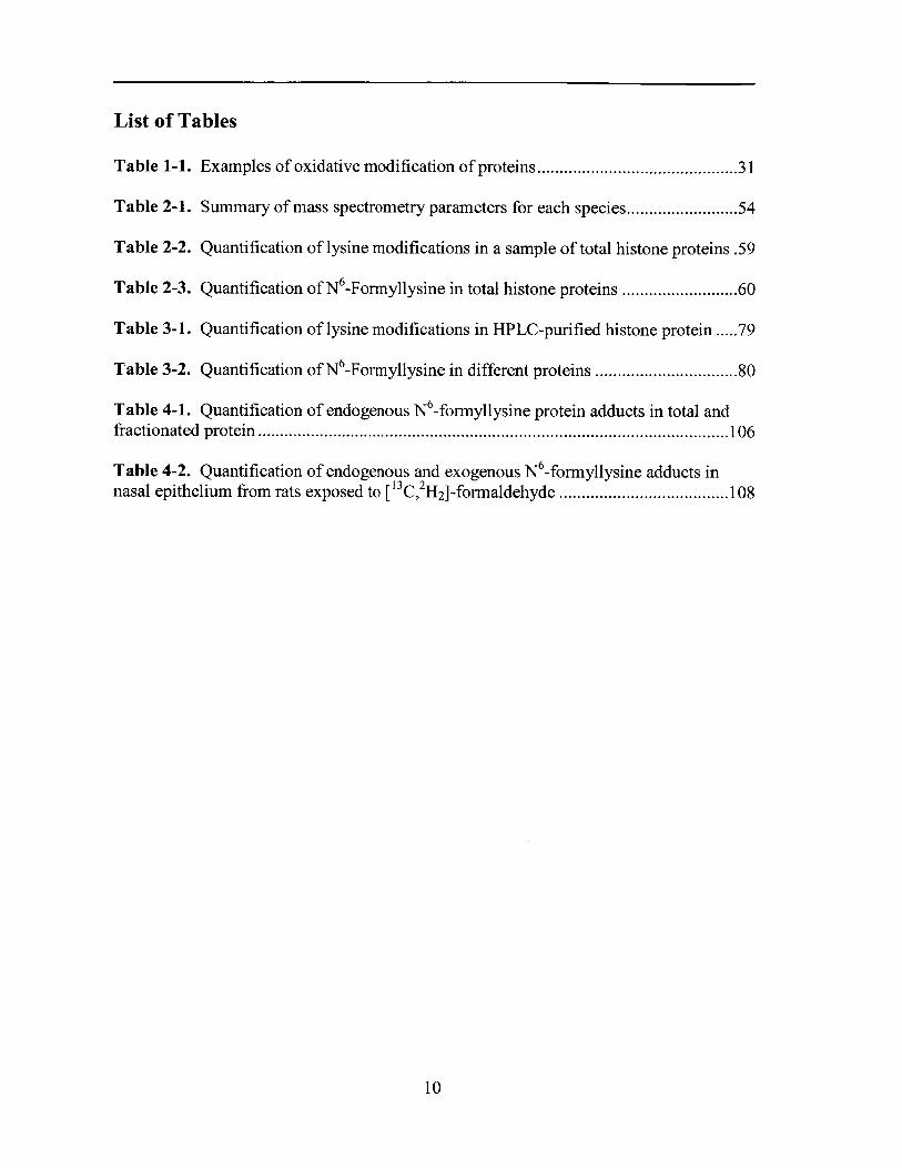
List of Tables
Table 1-1. Examples of oxidative modification of proteins........................................31
Table 2-1. Summary of mass spectrometry parameters for each species....................54
Table 2-2. Quantification of lysine modifications in a sample of total histone proteins .59
Table 2-3. Quantification of N6-Formyllysine in total histone proteins ...................... 60
Table 3-1. Quantification of lysine modifications in HPLC-purified histone protein ..... 79
Table 3-2. Quantification of N6-Formyllysine in different proteins ............................ 80
Table 4-1. Quantification of endogenous N -formyllysine protein adducts in total andfractionated protein .......................................................................................................... 106
Table 4-2. Quantification of endogenous and exogenous N -formyllysine adducts innasal epithelium from rats exposed to [13C,2H 2]-formaldehyde ...................................... 108
10
Acknowledgments
I consider myself very fortunate to have spent my graduate years at MIT, where Ibecame part of a wonderful and vibrant graduate community and met many greatindividuals who guided and supported me both academically and personally.
First and foremost, I would like to thank my thesis advisor Professor Peter C.Dedon. I am truly grateful for his guidance and mentorship, his patience andunderstanding through numerous technical challenges of my project, and his support andgenerosity over the years, particularly during my health challenges. Pete's hard work,enthusiasm about science, and dedication to research is inspiring. It amazes me how hethinks up so many interesting ideas in even brief meetings. I learned a great deal fromPete and I feel I was very lucky to work with him and be part of his lab.
Next, I would like to thank my committee members, Professor SteveTannenbaum, Professor John Essigmann, and Dr. John Wishnok for their very helpfuldiscussions and suggestions. I could not have asked for a better committee and I amforever indebted to them for their support, advice, and encouragement.
I am very thankful to all the past and present members of Dedon Lab for theirgreat friendship and help. They are all fantastic people and they made my everyday lifein the lab very enjoyable. I also have many wonderful memories of Dedon Labgatherings to cherish forever. I would like to extend a special thanks to Dr. MichaelDeMott and Dr. Ramesh Indrakanti for keeping the laboratory running smoothly, and toour wonderful administrative staff, Olga Parkin and Kristine Marzilli, for their help andkindness over the years. I am also very grateful to Dr. Koli Taghizadeh for hertremendous help with analytical method development and for being very kind and caring.Koli has always believed in me and I am very thankful for her support andencouragement over the years. I'd also like to thank Laura Trudel for her help withestablishing TK6 cell culture.
I am grateful to the BE Department, and particularly to Professor DouglasLauffenburger for all his efforts, guidance, and support of all BE students. I also wouldlike to thank the BE academic office, especially Dalia Fares and Aran Parillo for theirgreat help and friendship.
Our collaboration with Professor James Swenberg's lab has truly been valuablefor my dissertation. I really appreciate this opportunity, and I would like to thank them,especially Dr. Ben Moeller for his help and for promptly providing me with the animaltissues.
I am truly indebted to all my wonderful friends all over the world. I would like toespecially thank my girlfriends Andrea Bryan and Anusuya Das for their great friendshipthat started during the first year at MIT and continues today. We have had many funmemories that I will cherish forever. I am also very grateful for the love and support ofmy extended family, in particular my aunts Shahpar Ghodsi and Nasrin Jalili. I wouldalso like to thank my brother Afshin and his wife Melissa for their care and support. Ofcourse, I could not have achieved this if it was not for the love, support, and sacrifices of
11

my parents Nastaran Jalili and Mansour Edrissi. I am truly at a loss for words to expresshow thankful I am for the sacrifices you have made to provide my brother and me withgreat educational opportunities. Thank you for all you have done!
12
In loving memory of my grandmother,
Mahin Shahabpour
13
Chapter 1: Introduction
Chapter 1
Introduction
14
Chapter 1: Introduction
Goals and summary of this thesis
This thesis project is focused on defining the mechanism of formation and biological
significance of N6-formyllysine, a recently discovered abundant endogenous secondary
modification of proteins that resembles the physiologically important N6-acetyllysine in
histone proteins. This thesis is organized in 5 chapters. Chapter 1 presents a
comprehensive review of protein secondary modifications with an emphasis on lysine
acetylation, due to the resemblance of N6-formyllysine to N6-acetyllysine. The chapter
subsequently discusses the role of reactive oxidants and electrophiles in generating
pathological modifications of proteins and then focuses on N6-formyllysine and its
potential effects in terms of disrupting the epigenetic roles of histone modifications.
Chapter 2 provides detailed development of an ultrasensitive and specific liquid
chromatography-coupled tandem mass spectrometry method for quantifying this adduct
in proteins. Topics such as distribution of this adduct among different histone classes,
mechanisms for N6-formyllysine generation in addition to the DNA oxidation pathway
we originally reported, as well as its possible pathological consequences in cells are
explored in Chapter 3, with the discovery of formaldehyde as a major endogenous source
of N6-formyllysine. Chapter 4 extends our studies to measurement of N -formyllysine
protein adducts in tissues of rats exposed to formaldehyde by inhalation. In Chapter 5,
we conclude this thesis by summarizing our results regarding sources as well as fate of
N 6-formyllysine adducts and assess possible biological significance of this secondary
protein modification, particularly in terms of interfering with regulatory roles of histones.
N 6-Formylation of lysines in histones may represent an epigenetic mechanism of
disruption of cell function leading to cancer and other diseases.
15
Chapter 1: Introduction
Post-translational modifications of proteins and their regulatory roles
In eukaryotes, the level of complexity of the proteome is several orders of magnitude
greater than what is dictated by its genome sequence [1], with each gene effectively
encoding several proteins. In addition to alternative RNA splicing that generates
different mRNA transcripts from a single gene [2], the post-translational modification of
proteins (PTMs) significantly enhances the complexity of the proteome [1,3]. This type
of protein modification involves covalent alterations of the polypeptide backbone or side
chains, leading to changes in a protein's structure and/or function [1,3,4]. In most cases,
these modifications are reversible and under tight regulation of specific enzymes [5].
Eukaryotes dedicate a relatively high percentage of their genomes to enzymes modulating
PTMs. For instance, in humans, greater than 5% of the genes encode such enzymes [3].
Protein modifications modulate protein-protein and protein-nucleic acid
interactions and regulate the cellular environment by affecting a multitude of biological
functions and processes such as protein activity, localization, turnover, signaling
cascades, and cellular metabolism [1,5,6]. It is then of no surprise that dysregulation of
PTMs leads to a plethora of disorders and diseases.
From more than 300 PTMs discovered to date [5], the most common are
phosphorylation, acetylation, methylation, glycosylation, hydroxylation, palmitoylation,
sulfation, and ubiquitination [1,2,5], as briefly discussed:
- Phosphorylation. Phosphorylation and dephosphorylation of serine, threonine,
or tyrosine residues mediated by protein kinases (PKs) and phosphatases (PPs),
respectively, is a key modification with many roles in regulating cellular
16
Chapter 1: Introduction
physiology and metabolism, mainly through modulating enzymatic activities and
intracellular signaling pathways [2,7,8].
- Methylation. Another key PTM is methylation of lysine and arginine. Specific
residues in histones as well as non-histone proteins such as p53, NF-cB, ERa are
subject to methylation in cells [9]. In histones, lysine methylation is dynamically
regulated by histone methyltransferases and histone demethylases, with important
roles in gene expression and chromatin organization [10]. Protein methylation, in
addition to DNA methylation/demethylation, has implications in embryonic and
postnatal development [1,11,12].
- Glycosylation. Protein glycosylation, or the addition of sugar moiety to proteins,
is an enzymatic mediated and reversible PTM [13], as opposed to the random
non-enzymatic glycation that mostly renders proteins inactive [14]. Protein
glycans formed by glycosylation have important structural and modulatory roles
such as recognition elements for glycan binding proteins (GBPs), and therefore
regulate a variety of processes such as development and survival [15].
" Hydroxylation. Hydroxylation of amino acids such as proline, asparagine,
phenylalanine, tryptophan, and tyrosine is mediated by enzymes called
hydroxylases [16,17]. Hydroxylation is critical in cellular detoxification reactions
[1], as well as in the structural stability of proteins (e.g., hydroxyproline is an
essential element of collagen) [18].
- Palmitoylation. Covalent attachment of fatty acids to proteins is another type of
PTM. S-Palmitoylation of the cysteine thiol is a reversible modification with
important physiological roles in G-protein-coupled receptor (GPCR) trafficking
17
Chapter 1: Introduction
and protein activity, as well as localization (as attachment of the hydrophobic
moiety is important for membrane association) and T-cell signaling [19,20].
- Sulfation. The post-translational sulfation of tyrosine residues, a common
modification of secreted and transmembrane proteins, has essential roles in
chemokine signaling, leukocyte adhesion, immune response, extracellular
protein-protein interactions, intracellular protein transport, protein activity and
degradation [21-23].
- Ubiquitination. Ubiquitination is a covalent attachment of ubiquitin (Ub) to
lysine residues of proteins in order to tag them for degradation and recycling.
Attachment of Ub to proteins is a multi-step process, carried out in order with a
set of 3 enzymes: Ub activating enzymes (El), Ub conjugating enzymes (E2), and
Ub ligases (E3) [24]. The Ub tag (mediated by mono-Ub or poly-Ub), is a crucial
protein modification for proteasomal degradation, intracellular signaling cascades,
protein binding, localization, and activity [25,26].
- Acetylation. Lysine acetylation is a key protein modification with inherent
importance to this thesis due to the chemical similarity of lysine acetylation to
lysine formylation and the critical role of lysine acetylation of histone proteins in
regulating gene expression. To address this phenomenon, the next section of
thesis introduction addresses histone protein structure and function followed by
the biochemistry of histone acetylation.
18
Chapter 1: Introduction
Histone proteins and their post-translational modification
In eukaryotes, histone proteins are intimately associated with DNA and a plethora of
other protein complexes forming chromatin [27,28]. The fundamental repeating unit of
eukaryotic chromatin is nucleosome, an octamer of core histones (consisting of two H2A-
H2B dimers and a H3-H4 tetramer) around which 147 base pairs of DNA are tightly
wrapped in a left-handed super-helix 1.7 times around the core octamer [29-32]. The
linker histone HI binds the nucleosome and locks in place the DNA wrapped around the
core histones [33-35]. Additionally, it binds to linker DNA (10-60 base pairs of DNA
linking the nucleosomes) and allows for formation of the beads on a string structure and
higher order assemblies, such as 30 nm solenoids and 100 nm fibers [32,34,36].
Chromatin is a dynamic structure, capable of unfolding and refolding [37], with two
general states of tightly compact heterochromatin and the more open euchromatin that are
in general associated with transcriptionally silent and active genomic regions,
respectively [38,39].
In addition to serving as structural scaffolds for packaging DNA inside the
nucleus, histones have regulatory roles affecting a variety of cellular processes such as
chromatin organization and gene expression [33,40,41]. Besides the globular domains,
all core histones have long (-20-35 residues) and highly conserved amino-terminal tails
that protrude from the histone core domain [42] and compose -25-30% of the mass of
individual histones [43]. An exception is the histone H2A that has an additional long
carboxyl terminal segment protruding from the nucleosome [37]. These tail domains
may be structured or unstructured, may interact with nucleosomal or linker DNA, and
participate in protein-protein interactions [44].
19
Chapter 1: Introduction
Histone tail segments, rich in basic amino acids, are subject to a variety of post
translational modifications including acetylation, methylation, phosphorylation,
ubiquitylation, and ADP ribosylation with some locations modified in more than one way
[30,31,40,45,46]. For instance, acetylation can occur on lysines, methylation on lysines
and arginines, phosphorylation on serines and threonines, and ubiquitylation and
sumoylation on lysines. Just to add to the complexity, lysine can be mono-, di-, and tri-
methylated, and arginine can be mono- and di-methylated. Most of these enormous
secondary modifications identified to date occur on the long amino-terminal regions of
histones, although there are some that occur on the main globular region [37]. Singly or
in combination, these secondary modifications can act as "control switches" and play
diverse regulatory roles in chromatin remodeling, regulation of transcription, DNA
repair, and replication [40,47,48]. For instance, in higher eukaryotes, the combination of
H3 K14 acetylation, H4 K8 acetylation, and H3 S10 phosphorylation usually signals
transcriptional activation, and tri-methylation of H3 K9 and the lack of H3 and H4
acetylation are associated with transcriptional repression [37]. It is thought that through
these covalent modifications the tail structure or contacts change, thus modulating
histone:DNA interactions as well as creating new sites of dynamic interactions that can
attract binding of regulatory proteins and affect the chromatin stability and DNA
accessibility within the nucleosomes [40,44,49].
Most physiological histone modifications have been shown to be dynamic,
reversible, and under regulation of histone modifying enzymes [30]. An exception is
arginine demethylation during which arginine is changed into citrulline [30]. Lysine
acetylation in histone proteins is among the best characterized PTMs [50], with inherent
20
Chapter 1: Introduction
CH 3 H
NH NHH2CH2
(CH 2)3 (CH 2)3/CH /CH
N N 'CH " H C
N-Acetyllysine N-Formyllysine
Figure 1-1. Lysine formylation is a chemical homolog of lysine acetylation.
importance to this thesis due to chemical similarity of N6-acetyllysine to N6 -formyllysine
(Figure 1-1). The following section presents a detailed review of lysine acetylation.
Lysine acetylation is a key regulatory post-translational modification
Acetylation of the lysine E-amino group is an abundant protein modification with key
regulatory roles in transcription factors, histone proteins, and other chromatin proteins.
Histone acetylation is among the best characterized PTMs [50] in its regulation of
chromatin organization, gene expression, and DNA repair [36]. Lysine acetylation
occurs at highly conserved sites along the tails of core histones and is under tight control
of two sets of enzymes: histone acetyl transferases (HATs) and histone deacetylases
(HDACs) (Figure 1-2) [51]. HATs transfer an acetyl group from acetyl coenzyme A to
conserved lysine residues while HDACs catalyze the removal of the acetyl moiety
[39,52]. In addition to disrupting the DNA/histone electrostatic interactions, acetyl lysine
is recognized by the bromodomains of chromatin remodeling proteins, which in turn
recruit other downstream factors involved in chromatin remodeling and DNA
21
Chapter 1: Introduction
CH3
Acetyl- 0=NH/NH2 CoA CoA H NH
H2C H2C(CH2)3 (CH 2)3
/C H C /CHN C HDAC N ICH 0 Acetate H20 H 6
Lysine N6-Acetyllysine
Figure 1-2. Lysine acetylation is under tight control of two sets of enzymes: histoneacetyl transferases (HATs) and histone deacetylases (HDACs).
transcription [48,53]. Acetylation is almost exclusively associated with transcriptional
activation [30]. The chemical similarities of N6 -formyllysine and N6 -acetyllysine (Figure
1-1) suggest a disruptive role for the former in signaling by histone acetylation. Indeed,
N 6-formyllysine has been detected at conserved sites of lysine acetylation and
methylation in histone proteins [35,54].
The state of acetylation in cells is also important in the pathophysiology of a
variety of diseases including neurodegenerative diseases. For instance, it has been shown
that p-amyloid hyperacetylation and acetylation of tau proteins are associated with
Alzheimer's disease and dementia, respectively [1]. However, dysregulation of
physiological and (in most cases) enzymatically controlled protein modifications are not
the only ones affecting cellular physiology and disease. Protein modification by reactive
oxidants and electrophiles constitute an important change to protein structure and/or
function. The next sections examine some of the sources of these damaging agents and
the protein modifications that they produce.
22
Chapter 1: Introduction
Production of reactive chemical species in cells
A wide range of physiological or pathophysiological processes in cells generate oxidants
and electrophiles that directly damage biomolecules or lead to the formation of other
reactive intermediates [55,56], thus affecting crucial cellular processes.
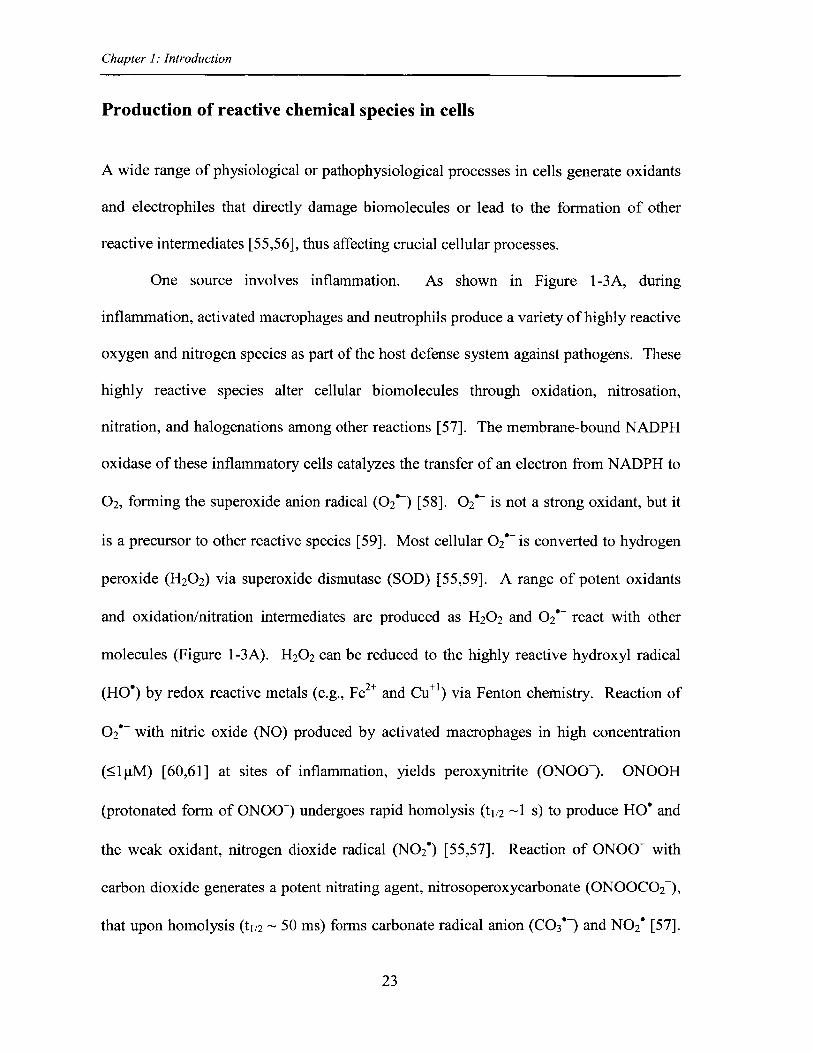
One source involves inflammation. As shown in Figure 1-3A, during
inflammation, activated macrophages and neutrophils produce a variety of highly reactive
oxygen and nitrogen species as part of the host defense system against pathogens. These
highly reactive species alter cellular biomolecules through oxidation, nitrosation,
nitration, and halogenations among other reactions [57]. The membrane-bound NADPH
oxidase of these inflammatory cells catalyzes the transfer of an electron from NADPH to
02, forming the superoxide anion radical (O2' ) [58]. 02' is not a strong oxidant, but it
is a precursor to other reactive species [59]. Most cellular 02' is converted to hydrogen
peroxide (H20 2) via superoxide dismutase (SOD) [55,59]. A range of potent oxidants
and oxidation/nitration intermediates are produced as H2 0 2 and 02' react with other
molecules (Figure 1-3A). H20 2 can be reduced to the highly reactive hydroxyl radical
(HO') by redox reactive metals (e.g., Fe2+ and Cue') via Fenton chemistry. Reaction of
O2' with nitric oxide (NO) produced by activated macrophages in high concentration
( 1p.M) [60,61] at sites of inflammation, yields peroxynitrite (ONOO-). ONOOH
(protonated form of ONOO~) undergoes rapid homolysis (t1/2 ~1 s) to produce HO' and
the weak oxidant, nitrogen dioxide radical (N0 2') [55,57]. Reaction of ONOO- with
carbon dioxide generates a potent nitrating agent, nitrosoperoxycarbonate (ONOOC0 2 ),
that upon homolysis (t1/2 ~ 50 ms) forms carbonate radical anion (CO3't) and N0 2' [57].
23
Chapter 1: Introduction
A
Illustration by Jeff Dixon, copyright Peter C. Dedon.
BATP synthase particles
Intermembrane space
Ribosomne rsa
Granule
Ou embran
Deoxyribonucleic acid (DNA)
1 e~ SOD02 -+ 02' -+ H20 2
Figure 1-3. Generation of reactive oxygen and nitrogen species.Inflammation (A) and mitochondrial respiration (B), are two majorsources for generation of reactive species. A and B are reproduced from[57] and [64], respectively.
Another nitrosating agent, nitrous anhydride (N20 3), is derived from oxidation of NO. In
activated neutrophils, myeloperoxidase-mediated reaction of H20 2 with Cl- yields
hypochlorous acid (HOCl), a strong oxidizing and halogenating agent [62].
24
Chapter 1: Introduction
Myeloperoxidase in these cells also mediates the conversion of nitrite (NO2) to N0 2'
[63].
The other sources of reactive oxygen species include mitochondrial respiration
(Figure 1-3B) and other 02 metabolism pathways. The use of molecular 02 for the
cellular production of energy is responsible for the generation of many reactive oxygen
species [65]. A variety of reactive molecules and free radicals are derived from 02 such
as 02' , HO', H20 2, peroxyl radical (ROO'), ozone (03), and singlet oxygen (102).
Reactive species such as 02' and H20 2 are produced as a result of electron leakage from
the mitochondrial electron transport chain during normal cellular metabolism [66,67].
Oxidoreductase enzymes such as NADPH oxidase, myeloperoxidase, and the cytochrome
P450 enzymes also produce reactive species [68].
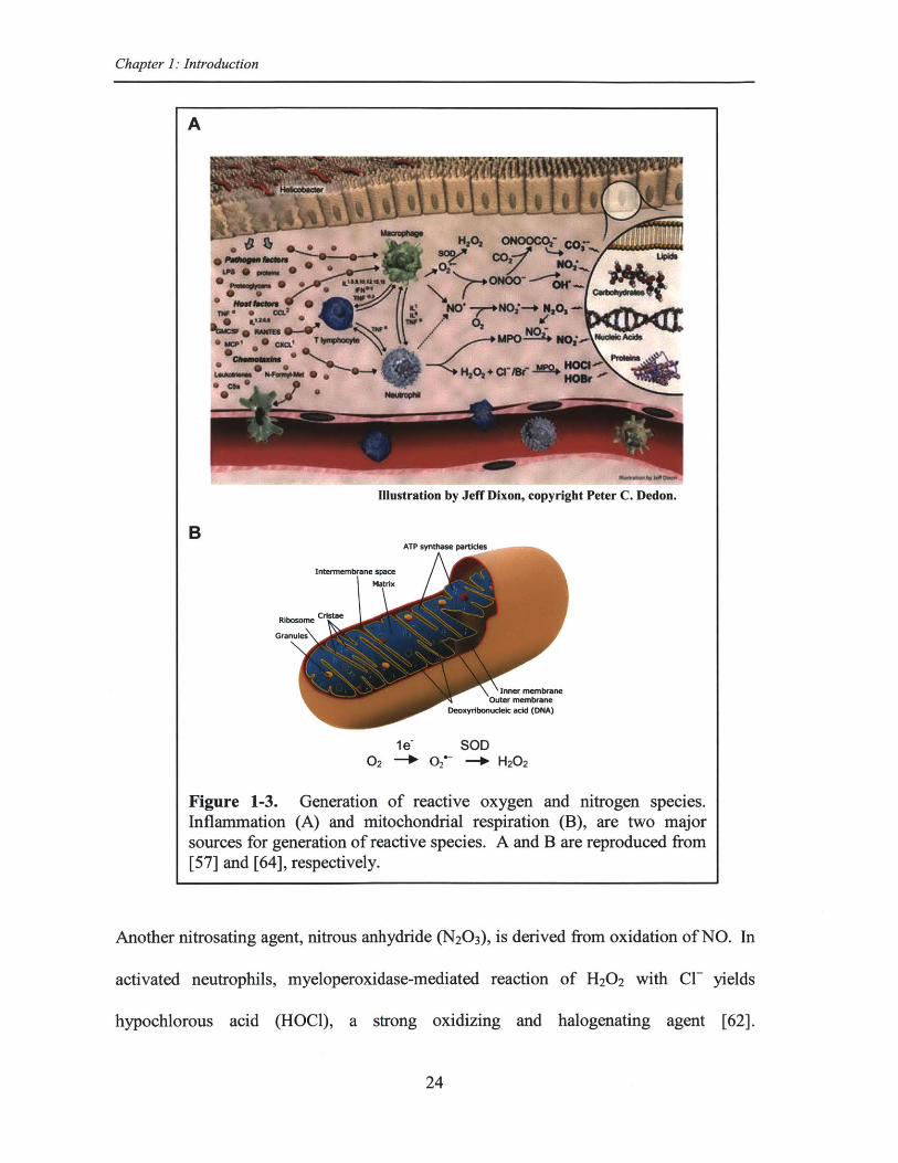
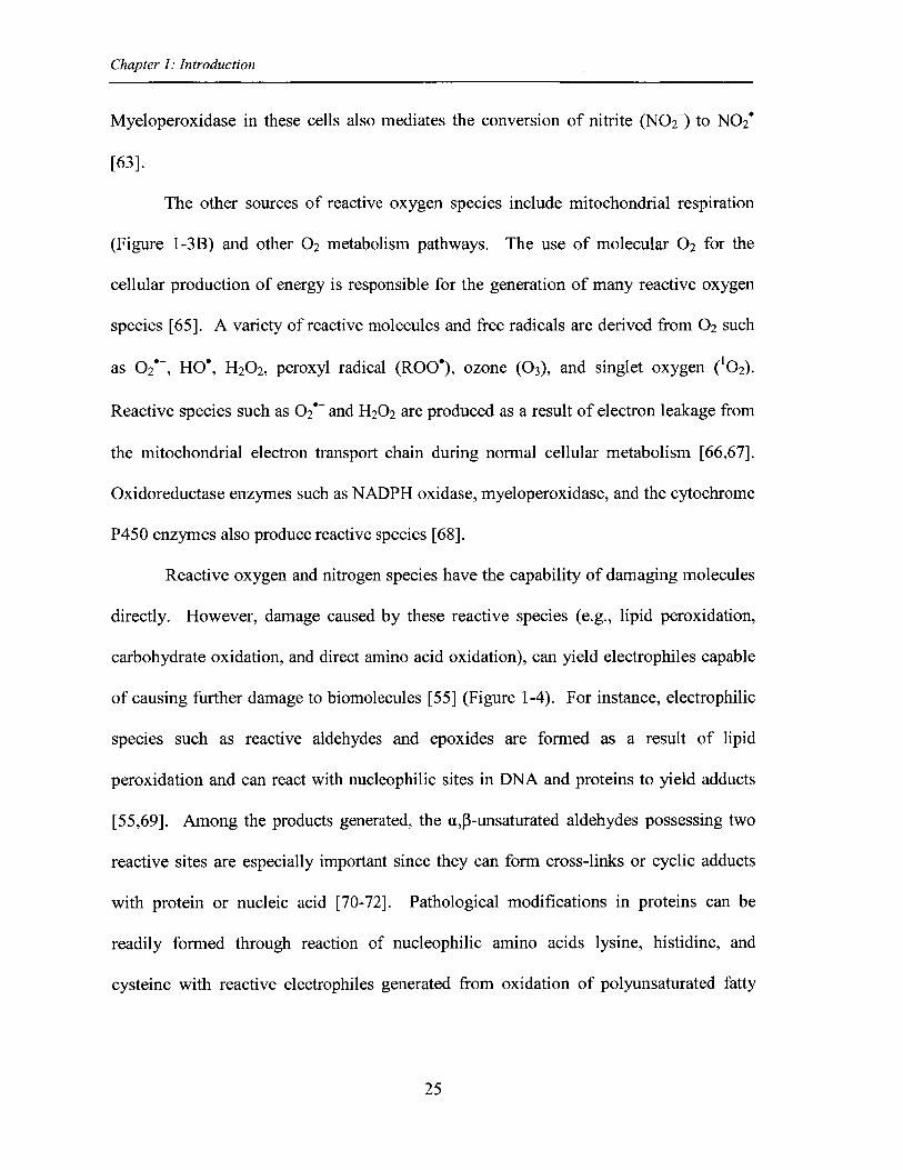
Reactive oxygen and nitrogen species have the capability of damaging molecules
directly. However, damage caused by these reactive species (e.g., lipid peroxidation,
carbohydrate oxidation, and direct amino acid oxidation), can yield electrophiles capable
of causing further damage to biomolecules [55] (Figure 1-4). For instance, electrophilic
species such as reactive aldehydes and epoxides are formed as a result of lipid
peroxidation and can react with nucleophilic sites in DNA and proteins to yield adducts
[55,69]. Among the products generated, the ap-unsaturated aldehydes possessing two
reactive sites are especially important since they can form cross-links or cyclic adducts
with protein or nucleic acid [70-72]. Pathological modifications in proteins can be
readily formed through reaction of nucleophilic amino acids lysine, histidine, and
cysteine with reactive electrophiles generated from oxidation of polyunsaturated fatty
25
Chapter 1: Introduction
acids and carbohydrates such as malondialdehyde, 4-hydroxy-2-nonenal (HNE), 4-
oxononenal, and glyoxal [73-77].
The next chapter covers in more detail these adventitious protein modifications
due to reaction with primary and secondary reactive species. It should be noted that
protein oxidative modification can result from other sources such as xenobiotics, cigarette
smoke, acetaminophen, y-irradiation, ultraviolet (UV) light, and ozone [68], among other
agents. However, for the purpose of this thesis, we only focus on protein damage due to
endogenously derived reactive oxidants and electrophiles.
From amino acid oxidation
HN
'
glutamate semialdehyde(from proline or arginine)
HN
0
Aminoadipate semialdehyde(from lysine)
acrolein(from threonine)
From lipid oxidation
O OH O O CO2H
OH OH
malondialdehyde (MDA) 4-hydroxy-2-nonenal (HNE) 9-hydroxy-1 2-oxo-1 0-dodecenoic acid(HODE)
From carbohydrate oxidation
o i 0
glyoxal
0 o
methylglyoxal
OH OHHO
OOH 0
3-deoxyglucosone
Figure 1-4. Examples of reactive aldehydes generated from damage to biomolecules.
26
Chapter 1: Introduction
Protein damage due to reactive endogenous oxidants and electrophiles
In addition to physiological secondary modifications that are mostly enzymatically
regulated, proteins are subjected to reactions with endogenous oxidants and electrophiles
generated by oxidative stress, inflammation, and normal cell metabolic processes [77-80].
Protein targets of reactive species are vast and include structural and membrane proteins,
metabolic and detoxification enzymes, or proteins involved in cell signaling and gene
expression [55]. Protein modification caused by a single oxidant or electrophile, such as
HNE can produce a diverse set of cellular responses with hundreds of genes upregulated
and downregulated [58]. Here we review representative examples of the types of damage
caused by these reactive species in cells.
Direct oxidation of amino acid side chains is one example of adventitious protein
modification. Oxidative modification of proteins is usually specific to type and location
of the residue and may occur on polypeptide backbone or the nucleophilic/redox-
sensitive side chains. As mentioned previously, modifications of proteins affect their
function in a positive or negative way, thus playing important roles in cellular physiology
as well as progress of disease. Cysteine, tyrosine, and methionine are among the amino
acids mostly modified by endogenous oxidants [55]. Cysteine sulfenic acid is formed via
oxidation of the sulfhydryl group of cysteine, helping to absorb the oxidative insult [81].
Cysteine sulfenic acid is unstable and forms disulfide bonds with glutathione or other
accessible thiols or undergoes further oxidation to cysteine sulfinic acid and cysteine
sulfonic acid [82]. S-nitrosylation of cysteine and 3-nitration of tyrosine residues in
proteins is another consequence of oxidative or nitrosative stress [83,84]. In fact, 3-
nitrotyrosine is used as a biomarker of protein damage associated with inflammation and
27
Chapter 1: Introduction
variety of diseases [84]. Methionine oxidation to form methionine sulfoxide, reversed by
methionine sulfoxide reductases, is speculated to serve as a defense mechanism against
oxidative stress by preventing other residues from oxidative damage [85]. Elevated
levels of oxidized aromatic amino acid residues (e.g., 3-nitrotyrosine, 3-chlorotyrosine,
and 3,4-dihydroxylphenylalanine) are seen in age-related diseases such as
neurodegenerative and cardiovascular diseases [55]. For instance, 3-nitrotyrosine
adducts were detected in tyrosine hydroxylase isolated from brain tissues of Parkinson's
disease model mice that exhibited reduced enzymatic activity [86]. Oxidation of proline,
arginine, and lysine residues result in protein carbonyls such as glutamate and
aminoadipate semialdehydes [73,78]. In general, protein carbonyls can be generated via
direct damage to polypeptide backbone, oxidation of amino acids proline, lysine,
arginine, threonine, glutamate, aspartic acid [1,55,73,78], or Michael addition of reactive
a4-unsaturated aldehydes to nucleophilic amino acids lysine, histidine, and cysteine [73-
77]. Protein carbonylation, an irreversible modification, often leads to loss of protein
function [87]. Indeed, total protein carbonyl content is regarded as a biomarker of
oxidative stress and inflammation and elevated levels are associated with a variety of
human diseases such as cardiovascular and neurodegenerative disorders, and processes
such as aging [73,75,78].
Another adventitious protein modification involves reaction of amino acid side
chains with physiological sugars and sugar oxidation products. Unlike the enzymatic
mediated and site specific glycosylation of proteins, these advanced glycation end
products (AGEs) are random events [1]. For instance, the reactive dicarbonyl sugar 3-
deoxyglucosone, synthesized via the Maillard reaction and the polyol pathway [88],
28
Chapter 1: Introduction
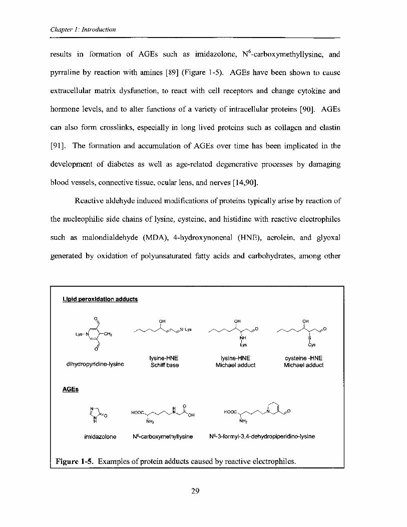
results in formation of AGEs such as imidazolone, N6-carboxymethyllysine, and
pyrraline by reaction with amines [89] (Figure 1-5). AGEs have been shown to cause
extracellular matrix dysfunction, to react with cell receptors and change cytokine and
hormone levels, and to alter functions of a variety of intracellular proteins [90]. AGEs
can also form crosslinks, especially in long lived proteins such as collagen and elastin
[91]. The formation and accumulation of AGEs over time has been implicated in the
development of diabetes as well as age-related degenerative processes by damaging
blood vessels, connective tissue, ocular lens, and nerves [14,90].
Reactive aldehyde induced modifications of proteins typically arise by reaction of
the nucleophilic side chains of lysine, cysteine, and histidine with reactive electrophiles
such as malondialdehyde (MDA), 4-hydroxynonenal (HNE), acrolein, and glyoxal
generated by oxidation of polyunsaturated fatty acids and carbohydrates, among other
Lipid peroxidation adducts
s OH OH OH
Lys-N CH3 N-Lys 0ONH SLys Cys
lysine-HNE lysine-HNE cysteine -HNEdihydropyridine-lysine Schiff base Michael adduct Michael adduct
AGEs
N H 0
HOOC N..) OH HOOC N O
H NH2 NH2
imidazolone N6-carboxymethyllysine N6-3-formyl-3,4-dehydropiperidino-lysine
Figure 1-5. Examples of protein adducts caused by reactive electrophiles.
29
Chapter 1: Introduction
biomolecules [74,75,77,79,80]. Michael adducts are among the major adducts formed,
for example, from the reaction of MDA with lysines residues (Figure 1-5) and 4-hydroxy-
2-alkenals with lysine, cysteine, and histidine amino acids [55]. Sequential addition of
two acrolein molecules to epsilon-amino group of lysine results in formation of cyclic
N6-3-formyl-3,4-dehydropiperidino-lysine adduct [92] (Figure 1-5). Schiff base adducts
are formed from reaction of 4-hydroxy-2-alkenals [93] as well as formaldehyde with
lysine residues [94]. Protein adducts generated by products of lipid peroxidation can alter
protein function and thus affect the vast range of biological functions they regulate,
leading to pathological processes associated with human diseases [77-80,95]. For
instance, HNE induces cell toxicity by covalently modifying IxB kinase, thus inhibiting
the subsequent transcription of NFKB-dependent genes needed for cell survival [58]. It
was also shown that HNE results in loss of microtubule network in neuroblastoma cells
by forming Michael adducts with tubulin isoforms [96].
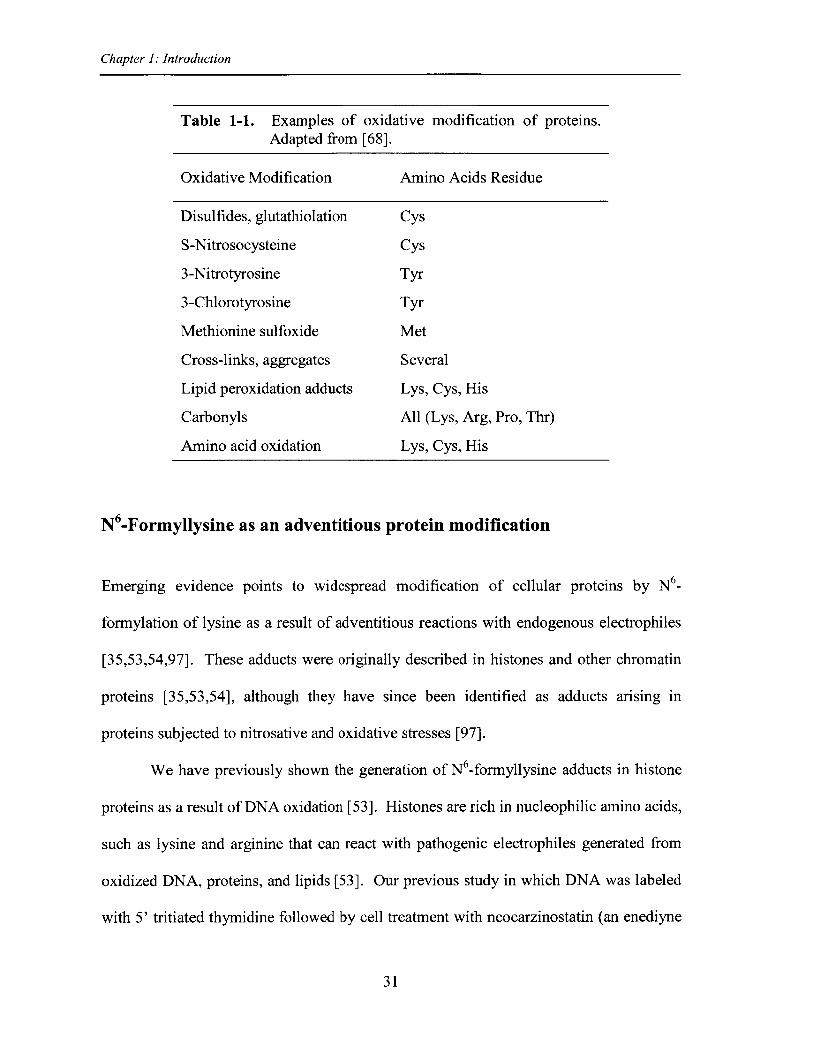
Among adventitious protein adducts (Table 1-1 and Figure 1-5), N6-formylation
of lysine has recently emerged as an abundant protein modification [35,53,54,97]. N6_
Formyllysine is a chemical homolog of the biologically important N6-acetyllysine and
thus may interfere with acetylation signaling in cells. The following section is a more
detailed introduction on this pathological protein modification that was originally
observed in histones and chromatin proteins.
30
Chapter 1: Introduction
Table 1-1. Examples of oxidative modification of proteins.Adapted from [68].
Oxidative Modification Amino Acids Residue
Disulfides, glutathiolation Cys
S-Nitrosocysteine Cys
3-Nitrotyrosine Tyr
3-Chlorotyrosine Tyr
Methionine sulfoxide Met
Cross-links, aggregates Several
Lipid peroxidation adducts Lys, Cys, His
Carbonyls All (Lys, Arg, Pro, Thr)
Amino acid oxidation Lys, Cys, His
N6-Formyllysine as an adventitious protein modification
Emerging evidence points to widespread modification of cellular proteins by N6_
formylation of lysine as a result of adventitious reactions with endogenous electrophiles
[35,53,54,97]. These adducts were originally described in histones and other chromatin
proteins [35,53,54], although they have since been identified as adducts arising in
proteins subjected to nitrosative and oxidative stresses [97].
We have previously shown the generation of N6-formyllysine adducts in histone
proteins as a result of DNA oxidation [53]. Histones are rich in nucleophilic amino acids,
such as lysine and arginine that can react with pathogenic electrophiles generated from
oxidized DNA, proteins, and lipids [53]. Our previous study in which DNA was labeled
with 5' tritiated thymidine followed by cell treatment with neocarzinostatin (an enediyne
31
Chapter 1: Introduction
[A
0 0O-P-0 H +I -0
3'-Formyl-phosphatE
0-I0-P=0
0O O
-P=O
+ O-P=0
0
0 0O-P-O Ha
3'-Formyl-phosphate
.NH2H2C
+ (CH 2)3IIsN'CH'C
H 01
Lysine
H
-NHH2C
(OH 2)3
N..CH,H O1
N6-Formyllysine
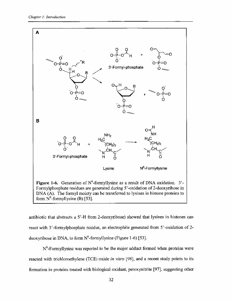
Figure 1-6. Generation of N6 -formyllysine as a result of DNA oxidation. 3'-Formylphosphate residues are generated during 5'-oxidation of 2-deoxyribose inDNA (A). The formyl moiety can be transferred to lysines in histone proteins toform N 6-formyllysine (B) [53].
antibiotic that abstracts a 5'-H from 2-deoxyribose) showed that lysines in histones can
react with 3'-formylphosphate residue, an electrophile generated from 5'-oxidation of 2-
deoxyribose in DNA, to form N6-formyllysine (Figure 1-6) [53].
N 6-Formyllysine was reported to be the major adduct formed when proteins were
reacted with trichloroethylene (TCE) oxide in vitro [98], and a recent study points to its
formation in proteins treated with biological oxidant, peroxynitrite [97], suggesting other
32
R
0-- 0
B
Chapter 1. Introduction
sources for its generation besides DNA oxidation. In 1985, N 6-formyllysine was reported
to be formed by reaction of 1-lysine with formaldehyde [99] and recently, it was shown it
could result from silver staining procedures that involve the use of formaldehyde [100].
Considering that formaldehyde reacts with amines to give a carbinolamine intermediate
that is only one oxidation state away from a formamide functional group, we
hypothesized that endogenous formaldehyde could serve as a major source of N6_
formyllysine residues in histone and other proteins.
Formaldehyde is a reactive aldehyde forming adducts with nucleophilic sites in
DNA and protein molecules resulting in products such as N 2-hydroxymethyl-dG DNA
adducts [101] and formaldehyde induced Schiff bases on lysine residues of histones [94],
in addition to protein-DNA crosslinks. It has been classified as a known human
carcinogen by the International Agency for Research on Cancer in 2005 [102] and by the
National Toxicology Program of the National Institute of Environmental Health Sciences
in 2011 [103], although it is still considered a probable human carcinogen by the U.S.
Environmental Protection Agency [104]. In addition to environmental and occupational
sources [101,105,106], formaldehyde arises endogenously from cellular processes such as
demethylation of histones and nucleic acids as well as biosynthesis of purines, thymidine,
and some amino acids [10,105,107]. Formaldehyde is a relatively abundant metabolite at
concentrations ranging from 13 to 97 pM in human plasma [105]. Thus, formaldehyde
induced N 6-formyllysine generation in proteins could be the major source of this adduct
compared to previous sources reported.
The relatively high abundance of N 6-formyllysine adducts in histones [35,53]
suggests it could interfere with the regulatory roles of histones. The following section
33
Chapter 1: Introduction
examines the potential disruptive role of N 6-formyllysine in cellular epigenetics.
The biological implications of N -formyllysine modification of histones
N -Formyl lysine has been reported to be a major protein adduct in histones occurring on
many conserved lysine residues involved in epigenetic regulations [35,53]. Even the
DNA oxidation pathway in itself can result in high abundance of this adduct in histones.
Indeed, there are thousands of oxidative events in a cell on a daily basis, resulting in
hundreds of deoxyribose oxidations in DNA with each site producing different
electrophilic products, leading to fonnation of 10's of N6-formyllysines per cell per day
[53,108]. As discussed later in this thesis, formaldehyde is found to be another major
source giving rise to this adduct in histones. Therefore, if not removed, this pathological
modification may accumulate to significant levels in cells.
The high rate of occurrence of N6-formyllysine in histones raises the possibility of
epigenetic interferences through affecting the regulatory roles of these proteins. In
chromatin proteins, N6-formyllysine has the potential to interfere with important
physiological modifications that perform signaling functions. Indeed, N6-formyllysine
has been detected at conserved sites of lysine acetylation and methylation in histones
[35,54]. In addition, the chemical similarities of N6-formyllysine and N6-acetyllysine
suggest a disruptive role for the former in signaling by histone acetylation. Thus, this
thesis project is focused on defining the mechanism of formation and biological
significance of N 6-formyllysine. To that end, we developed an ultrasensitive and specific
liquid chromatography-coupled tandem mass spectrometry method for quantifying this
adduct in proteins (Chapter 2), and used it to answer questions about its mechanisms of
34
Chapter 1: Introduction
formation and pathological consequences in cells (Chapter 3). Chapter 4 extends our
studies to measurement of N6-formyllysine protein adducts in tissues of rats exposed to
formaldehyde by inhalation, as we discovered formaldehyde to be a major endogenous
source of N 6-formyllysine (Chapter 3). We conclude this thesis in Chapter 5 by
summarizing our results regarding sources as well as fate of N6-formyllysine adducts and
assess possible biological significance of this secondary protein modification. The
presence of N 6-formylation of lysines in histones may have important biological
consequences leading to pathology and progress of variety of diseases.
35
Chapter 1: Introduction
References
1. Karve TM, Cheema AK (2011) Small changes huge impact: the role of proteinposttranslational modifications in cellular homeostasis and disease. J AminoAcids 2011: 207691.
2. Seet BT, Dikic I, Zhou MM, Pawson T (2006) Reading protein modificationswith interaction domains. Nat Rev Mol Cell Biol 7: 473-483.
3. Walsh CT (2006) Posttranslational Modification Of Proteins: Expanding Nature'sInventory: Roberts and Company Publishers.
4. Boeckmann B, Blatter MC, Famiglietti L, Hinz U, Lane L, et al. (2005) Proteinvariety and functional diversity: Swiss-Prot annotation in its biological context. CR Biol 328: 882-899.
5. Jensen ON (2000) Modification-specific proteomics: systematic strategies foranalysing post-translationally modified proteins. Trends Biotechnol 18: 36-42.
6. Doyle HA, Mamula MJ (2001) Post-translational protein modifications in antigenrecognition and autoimmunity. Trends Immunol 22: 443-449.
7. Hunter T (1995) Protein kinases and phosphatases: the yin and yang of proteinphosphorylation and signaling. Cell 80: 225-236.
8. Blom N, Gammeltoft S, Brunak S (1999) Sequence and structure-based predictionof eukaryotic protein phosphorylation sites. J Mol Biol 294: 1351-1362.
9. Yang XD, Lamb A, Chen LF (2009) Methylation, a new epigenetic mark forprotein stability. Epigenetics 4: 429-433.
10. Shi Y, Whetstine JR (2007) Dynamic regulation of histone lysine methylation bydemethylases. Mol Cell 25: 1-14.
11. Mann MR, Bartolomei MS (2002) Epigenetic reprogramming in the mammalianembryo: struggle of the clones. Genome Biol 3: REVIEWS 1003.
12. Litt MD, Simpson M, Gaszner M, Allis CD, Felsenfeld G (2001) Correlationbetween histone lysine methylation and developmental changes at the chickenbeta-globin locus. Science 293: 2453-2455.
13. Varki A, Esko JD, Colley KJ (2009) Cellular Organization of Glycosylation. In:Varki A, Cummings RD, Esko JD, Freeze HH, Stanley P et al., editors. Essentialsof Glycobiology. 2nd ed. Cold Spring Harbor (NY).
14. Ulrich P, Cerami A (2001) Protein glycation, diabetes, and aging. Recent ProgHorm Res 56: 1-21.
36
Chapter 1. Introduction
15. Varki A, Lowe JB (2009) Biological Roles of Glycans. In: Varki A, CummingsRD, Esko JD, Freeze HH, Stanley P et al., editors. Essentials of Glycobiology.2nd ed. Cold Spring Harbor (NY).
16. Lando D, Peet DJ, Whelan DA, Gorman JJ, Whitelaw ML (2002) Asparaginehydroxylation of the HIF transactivation domain a hypoxic switch. Science 295:858-861.
17. Fitzpatrick PF (2003) Mechanism of aromatic amino acid hydroxylation.Biochemistry 42: 14083-14091.
18. Kivirikko KI, Myllyla R, Pihlajaniemi T (1989) Protein hydroxylation: prolyl 4-hydroxylase, an enzyme with four cosubstrates and a multifunctional subunit.FASEB J 3: 1609-1617.
19. Martin BR, Wang C, Adibekian A, Tully SE, Cravatt BF (2012) Global profilingof dynamic protein palmitoylation. Nat Methods 9: 84-89.
20. Smotrys JE, Linder ME (2004) Palmitoylation of intracellular signaling proteins:regulation and function. Annu Rev Biochem 73: 559-587.
21. Seibert C, Sakmar TP (2008) Toward a framework for sulfoproteomics: Synthesisand characterization of sulfotyrosine-containing peptides. Biopolymers 90: 459-477.
22. Huang SY, Shi SP, Qiu JD, Sun XY, Suo SB, et al. (2012) PredSulSite: predictionof protein tyrosine sulfation sites with multiple features and analysis. AnalBiochem 428: 16-23.
23. Zhang Y, Jiang H, Go EP, Desaire H (2006) Distinguishing phosphorylation andsulfation in carbohydrates and glycoproteins using ion-pairing and massspectrometry. J Am Soc Mass Spectrom 17: 1282-1288.
24. Pickart CM (2001) Mechanisms underlying ubiquitination. Annu Rev Biochem70: 503-533.
25. Komander D (2009) The emerging complexity of protein ubiquitination. BiochemSoc Trans 37: 937-953.
26. Xu G, Jaffrey SR (2011) The new landscape of protein ubiquitination. NatBiotechnol 29: 1098-1100.
27. Verdone L, Agricola E, Caserta M, Di Mauro E (2006) Histone acetylation ingene regulation. Brief Funct Genomic Proteomic 5: 209-221.
28. Fierz B, Muir TW (2012) Chromatin as an expansive canvas for chemicalbiology. Nat Chem Biol 8: 417-427.
37
Chapter 1: Introduction
29. Luger K, Mader AW, Richmond RK, Sargent DF, Richmond TJ (1997) Crystalstructure of the nucleosome core particle at 2.8 A resolution. Nature 389: 251-260.
30. Kouzarides T (2007) Chromatin modifications and their function. Cell 128: 693-705.
31. Garcia BA, Barber CM, Hake SB, Ptak C, Turner FB, et al. (2005) Modificationsof human histone H3 variants during mitosis. Biochemistry 44: 13202-13213.
32. Horn PJ, Peterson CL (2002) Molecular biology. Chromatin higher order folding--wrapping up transcription. Science 297: 1824-1827.
33. Pesavento JJ, Kim YB, Taylor GK, Kelleher NL (2004) Shotgun annotation ofhistone modifications: a new approach for streamlined characterization of proteinsby top down mass spectrometry. J Am Chem Soc 126: 3386-3387.
34. Hayes JJ (2002) Changing chromatin from the inside. Nature Struct Biol 9: 161-163.
35. Wisniewski JR, Zougman A, Mann M (2008) Nepsilon-formylation of lysine is awidespread post-translational modification of nuclear proteins occurring atresidues involved in regulation of chromatin function. Nucleic Acids Res 36: 570-577.
36. Recht J, Tsubota T, Tanny JC, Diaz RL, Berger JM, et al. (2006) Histonechaperone Asfl is required for histone H3 lysine 56 acetylation, a modificationassociated with S phase in mitosis and meiosis. Proc Natl Acad Sci U S A 103:6988-6993.
37. Peterson CL, Laniel MA (2004) Histones and histone modifications. Curr Biol 14:R546-55 1.
38. Fischle W, Tseng BS, Dormann HL, Ueberheide BM, Garcia BA, et al. (2005)Regulation of HP 1 -chromatin binding by histone H3 methylation andphosphorylation. Nature 438: 1116-1122.
39. Hake SB, Xiao A, Allis CD (2004) Linking the epigenetic 'language' of covalenthistone modifications to cancer. Br J Cancer 90: 761-769.
40. Siuti N, Roth MJ, Mizzen CA, Kelleher NL, Pesavento JJ (2006) Gene-specificcharacterization of human histone H2B by electron capture dissociation. JProteome Res 5: 233-239.
41. Felsenfeld G, Groudine M (2003) Controlling the double helix. Nature 421: 448-453.
38
Chapter 1: Introduction
42. Munshi A, Shafi G, Aliya N, Jyothy A (2009) Histone modifications dictatespecific biological readouts. J Genet Genomics 36: 75-88.
43. Pesavento JJ, Bullock CR, LeDuc RD, Mizzen CA, Kelleher NL (2008)Combinatorial modification of human histone H4 quantitated by two-dimensionalliquid chromatography coupled with top down mass spectrometry. J Biol Chem283: 14927-14937.
44. Zheng C, Hayes JJ (2003) Structures and interactions of the core histone taildomains. Biopolymers 68: 539-546.
45. Kao CF, Osley MA (2003) In vivo assays to study histone ubiquitylation.Methods 31: 59-66.
46. Galasinski SC, Resing KA, Ahn NG (2003) Protein mass analysis of histones.Methods 31: 3-11.
47. Wiley EA, Ohba R, Yao MC, Allis CD (2000) Developmentally regulated rpd3phomolog specific to the transcriptionally active macronucleus of vegetativeTetrahymena thermophila. Mol Cell Biol 20: 8319-8328.
48. Boyne MT, 2nd, Pesavento JJ, Mizzen CA, Kelleher NL (2006) Precisecharacterization of human histones in the H2A gene family by top down massspectrometry. J Proteome Res 5: 248-253.
49. Angelov D, Vitolo JM, Mutskov V, Dimitrov S, Hayes JJ (2001) Preferentialinteraction of the core histone tail domains with linker DNA. Proc Natl Acad SciU S A 98: 6599-6604.
50. Grunstein M (1997) Histone acetylation in chromatin structure and transcription.Nature 389: 349-352.
51. Strahl BD, Allis CD (2000) The language of covalent histone modifications.Nature 403: 41-45.
52. Hildmann C, Riester D, Schwienhorst A (2007) Histone deacetylases--animportant class of cellular regulators with a variety of functions. Appl MicrobiolBiotechnol 75: 487-497.
53. Jiang T, Zhou X, Taghizadeh K, Dong M, Dedon PC (2007) N-formylation oflysine in histone proteins as a secondary modification arising from oxidativeDNA damage. Proc Natl Acad Sci U S A 104: 60-65.
54. LeRoy G, Weston JT, Zee BM, Young NL, Plazas-Mayorca MD, et al. (2009)Heterochromatin protein 1 is extensively decorated with histone code-like post-translational modifications. Mol Cell Proteomics 8: 2432-2442.
39
Chapter 1: Introduction
55. Marnett U, Riggins JN, West JD (2003) Endogenous generation of reactiveoxidants and electrophiles and their reactions with DNA and protein. J Clin Invest111: 583-593.
56. Winterboum CC (2008) Reconciling the chemistry and biology of reactiveoxygen species. Nat Chem Biol 4: 278-286.
57. Lonkar P, Dedon PC (2011) Reactive species and DNA damage in chronicinflammation: reconciling chemical mechanisms and biological fates. Int J Cancer128: 1999-2009.
58. Marnett LJ (2012) Inflammation and cancer: chemical approaches to mechanisms,imaging, and treatment. J Org Chem 77: 5224-5238.
59. Turrens JF (2003) Mitochondrial formation of reactive oxygen species. J Physiol552: 335-344.
60. Lewis RS, Tamir S, Tannenbaum SR, Deen WM (1995) Kinetic analysis of thefate of nitric oxide synthesized by macrophages in vitro. J Biol Chem 270: 29350-29355.
61. Mancardi D, Ridnour LA, Thomas DD, Katori T, Tocchetti CG, et al. (2004) Thechemical dynamics of NO and reactive nitrogen oxides: a practical guide. CurrMol Med 4: 723-740.
62. Hazen SL, d'Avignon A, Anderson MM, Hsu FF, Heinecke JW (1998) Humanneutrophils employ the myeloperoxidase-hydrogen peroxide-chloride system tooxidize alpha-amino acids to a family of reactive aldehydes. Mechanistic studiesidentifying labile intermediates along the reaction pathway. J Biol Chem 273:4997-5005.
63. Eiserich JP, Hristova M, Cross CE, Jones AD, Freeman BA, et al. (1998)Formation of nitric oxide-derived inflammatory oxidants by myeloperoxidase inneutrophils. Nature 391: 393-397.
64. Retrieved from http://en.wikipedia.org/wiki/Mitochondrion, November 18, 2012
65. Davies KJ (1995) Oxidative stress: the paradox of aerobic life. Biochem SocSymp 61: 1-31.
66. Imlay JA (2003) Pathways of oxidative damage. Annu Rev Microbiol 57: 395-418.
67. Trachootham D, Alexandre J, Huang P (2009) Targeting cancer cells by ROS-mediated mechanisms: a radical therapeutic approach? Nat Rev Drug Discov 8:579-591.
40
Chapter 1: Introduction
68. Shacter E (2000) Quantification and significance of protein oxidation inbiological samples. Drug Metab Rev 32: 307-326.
69. Esterbauer H, Schaur RJ, Zollner H (1991) Chemistry and biochemistry of 4-hydroxynonenal, malonaldehyde and related aldehydes. Free Radic Biol Med 11:81-128.
70. Stone MP, Cho YJ, Huang H, Kim HY, Kozekov ID, et al. (2008) InterstrandDNA cross-links induced by alpha,beta-unsaturated aldehydes derived from lipidperoxidation and environmental sources. Acc Chem Res 41: 793-804.
71. Minko IG, Yamanaka K, Kozekov ID, Kozekova A, Indiani C, et al. (2008)Replication bypass of the acrolein-mediated deoxyguanine DNA-peptide cross-links by DNA polymerases of the DinB family. Chem Res Toxicol 21: 1983-1990.
72. Minko IG, Kozekov ID, Harris TM, Rizzo CJ, Lloyd RS, et al. (2009) Chemistryand biology of DNA containing 1,N(2)-deoxyguanosine adducts of the alpha,beta-unsaturated aldehydes acrolein, crotonaldehyde, and 4-hydroxynonenal. ChemRes Toxicol 22: 759-778.
73. Slade PG, Williams MV, Chiang A, Iffrig E, Tannenbaum SR, et al. (2011) Afiltered database search algorithm for endogenous serum protein carbonylmodifications in a mouse model of inflammation. Mol Cell Proteomics 10: M 11007658.
74. Codreanu SG, Zhang B, Sobecki SM, Billheimer DD, Liebler DC (2009) Globalanalysis of protein damage by the lipid electrophile 4-hydroxy-2-nonenal. MolCell Proteomics 8: 670-680.
75. Tallman KA, Kim HY, Ji JX, Szapacs ME, Yin H, et al. (2007) Phospholipid-protein adducts of lipid peroxidation: synthesis and study of new biotinylatedphosphatidylcholines. Chem Res Toxicol 20: 227-234.
76. Connor RE, Marnett U, Liebler DC (2011) Protein-selective capture to analyzeelectrophile adduction of hsp90 by 4-hydroxynonenal. Chem Res Toxicol 24:1275-1282.
77. Jacobs AT, Marnett U (2010) Systems analysis of protein modification andcellular responses induced by electrophile stress. Acc Chem Res 43: 673-683.
78. Levine RL (2002) Carbonyl modified proteins in cellular regulation, aging, anddisease. Free Radic Biol Med 32: 790-796.
79. Thornalley PJ (2008) Protein and nucleotide damage by glyoxal andmethylglyoxal in physiological systems--role in ageing and disease. Drug MetabolDrug Interact 23: 125-150.
41
Chapter 1: Introduction
80. Dedon PC (2008) The chemical toxicology of 2-deoxyribose oxidation in DNA.Chem Res Toxicol 21: 206-219.
81. Rehder DS, Borges CR (2010) Cysteine sulfenic acid as an intermediate indisulfide bond formation and nonenzymatic protein folding. Biochemistry 49:7748-7755.
82. Wang Y, Vivekananda S, Men L, Zhang Q (2004) Fragmentation of protonatedions of peptides containing cysteine, cysteine sulfinic acid, and cysteine sulfonicacid. J Am Soc Mass Spectrom 15: 697-702.
83. Crabtree M, Hao G, Gross SS (2003) Detection of cysteine S-nitrosylation andtyrosine 3-nitration in kidney proteins. Methods Mol Med 86: 373-384.
84. Jones LH (2012) Chemistry and biology of biomolecule nitration. Chem Biol 19:1086-1092.
85. Levine RL, Mosoni L, Berlett BS, Stadtman ER (1996) Methionine residues asendogenous antioxidants in proteins. Proc Natl Acad Sci U S A 93: 15036-15040.
86. Ara J, Przedborski S, Naini AB, Jackson-Lewis V, Trifiletti RR, et al. (1998)Inactivation of tyrosine hydroxylase by nitration following exposure toperoxynitrite and 1-methyl-4-phenyl-1,2,3,6-tetrahydropyridine (MPTP). ProcNatl Acad Sci U S A 95: 7659-7663.
87. Madian AG, Regnier FE (2010) Proteomic identification of carbonylated proteinsand their oxidation sites. J Proteome Res 9: 3766-3780.
88. Niwa T (1999) 3-Deoxyglucosone: metabolism, analysis, biological activity, andclinical implication. J Chromatogr B Biomed Sci Appl 731: 23-36.
89. Jono T, Nagai R, Lin X, Ahmed N, Thornalley PJ, et al. (2004) Nepsilon-(Carboxymethyl)lysine and 3-DG-imidazolone are major AGE structures inprotein modification by 3-deoxyglucosone. J Biochem 136: 351-358.
90. Brownlee M (1995) Advanced protein glycosylation in diabetes and aging. AnnuRev Med 46: 223-234.
91. Bakris GL, Bank AJ, Kass DA, Neutel JM, Preston RA, et al. (2004) Advancedglycation end-product cross-link breakers. A novel approach to cardiovascularpathologies related to the aging process. Am J Hypertens 17: 23S-30S.
92. Kaminskas LM, Pyke SM, Burcham PC (2005) Differences in lysine adduction byacrolein and methyl vinyl ketone: implications for cytotoxicity in culturedhepatocytes. Chem Res Toxicol 18: 1627-1633.
42
Chapter 1: Introduction
93. Surh J, Kwon H (2005) Estimation of daily exposure to 4-hydroxy-2-alkenals inKorean foods containing n-3 and n-6 polyunsaturated fatty acids. Food AdditContam 22: 701-708.
94. Lu K, Boysen G, Gao L, Collins LB, Swenberg JA (2008) Formaldehyde-inducedhistone modifications in vitro. Chem Res Toxicol 21: 1586-1593.
95. Prasad A, Bekker P, Tsimikas S (2012) Advanced Glycation Endproducts andDiabetic Cardiovascular Disease. Cardiol Rev.
96. Neely MD, Sidell KR, Graham DG, Montine TJ (1999) The lipid peroxidationproduct 4-hydroxynonenal inhibits neurite outgrowth, disrupts neuronalmicrotubules, and modifies cellular tubulin. J Neurochem 72: 2323-2333.
97. Vana L, Kanaan NM, Hakala K, Weintraub ST, Binder LI (2011) Peroxynitrite-induced nitrative and oxidative modifications alter tau filament formation.Biochemistry 50: 1203-1212.
98. Cai H, Guengerich FP (2000) Acylation of protein lysines by trichloroethyleneoxide. Chem Res Toxicol 13: 327-335.
99. Tyihak E, Trezl L, Kolonits P (1985) The isolation of Nepsilon-formyl-L-lysinefrom the reaction between formaldehyde and L-lysine and its identification byOPLC and NMR spectroscopy. J Pharm Biomed Anal 3: 343-349.
100. Oses-Prieto JA, Zhang X, Burlingame AL (2007) Formation of epsilon-formyllysine on silver-stained proteins: implications for assignment of isobaricdimethylation sites by tandem mass spectrometry. Mol Cell Proteomics 6: 181-192.
101. Lu K, Moeller B, Doyle-Eisele M, McDonald J, Swenberg JA (2011) Moleculardosimetry of N2-hydroxymethyl-dG DNA adducts in rats exposed toformaldehyde. Chem Res Toxicol 24: 159-161.
102. Cogliano VJ, Grosse Y, Baan RA, Straif K, Secretan MB, et al. (2005) Meetingreport: summary of IARC monographs on formaldehyde, 2-butoxyethanol, and 1-tert-butoxy-2-propanol. Environ Health Perspect 113: 1205-1208.
103. (2011) NTP 12th Report on Carcinogens. Report on carcinogens : carcinogenprofiles / US Dept of Health and Human Services, Public Health Service, NationalToxicology Program: iii-499.
104. (2006) Integrated Risk Information System (IRIS): Formaldehyde. USEnvironmental Protection Agency.
105. Zhang L, Freeman LE, Nakamura J, Hecht SS, Vandenberg JJ, et al. (2010)Formaldehyde and leukemia: epidemiology, potential mechanisms, andimplications for risk assessment. Environ Mol Mutagen 51: 181-191.
43
Chapter 1: Introduction
106. Le Curieux F, Pluskota D, Munter T, Sjoholm R, Kronberg L (2000)Identification of fluorescent 2'-deoxyadenosine adducts formed in reactions ofconjugates of malonaldehyde and acetaldehyde, and of malonaldehyde andformaldehyde. Chem Res Toxicol 13: 1228-1234.
107. Begley TJ, Samson LD (2003) AlkB mystery solved: oxidative demethylation ofNi-methyladenine and N3-methylcytosine adducts by a direct reversalmechanism. Trends Biochem Sci 28: 2-5.
108. Dedon PC, Jiang ZW, Goldberg IH (1992) Neocarzinostatin-mediated DNAdamage in a model AGT.ACT site: mechanistic studies of thiol-sensitivepartitioning of C4' DNA damage products. Biochemistry 31: 1917-1927.
44
Chapter 2: Development of an LC-MS/MS method for quantification of 6-formyllysine adducts in protein
Chapter 2
Development of a liquid chromatography-coupled tandem mass spectrometry method for
quantification of N6-formyllysine adductsin proteins
45
Chapter 2: Development of an LC-MS/MS method for quantification ofht-formyllysine adducts in protein
Abstract
The initial step in investigating the mechanism of formation of N6-formyllysine andassessing its possible pathological consequences in cells involved developing anultrasensitive and specific liquid chromatography-coupled tandem mass spectrometry(LC-MS/MS) method for quantifying this adduct in proteins. The quantification methodwe developed previously (Jiang et al. PNAS 104: 60-5, 2007) proved to be relativelyinsensitive and biased, so a new method was developed, which involved proteolytichydrolysis of proteins to individual amino acids, normal phase chromatographicresolution of the amino acids followed by tandem electrospray mass spectrometry toquantify them. N6-Formyllysine is reported as a percentage of total lysines in order tonormalize it across samples with different amounts and types of proteins. The use ofStreptomyces griseus protease greatly diminished the background levels of lysine andformyllysine observed previously with proteinase K. Further, the direct analysis ofamino acids, as opposed to phenylisothiocyanate (PITC) derivatization, improved boththe sensitivity and specificity of the assay. In addition to lysine and N6-formyllysinespecies, N6-acetyl, mono-, di-, and tri-methyllysine modifications are monitored, withlimit of detection of 1-50 fmol for all species. Measurement of N6-formyllysine inhistones of TK6 cells indicates that this adduct is an abundant endogenous proteinmodification (~1 per 104 lysines). In addition, the dilution rate of the absolute quantity ofN -formyllysine in histone proteins of TK6 cells (grown in media containing isotopicallylabeled lysine) reveals that N6-formyllysine is a rather stable modification of histones,with a half-life closely matching TK6 cell doubling rate.
46
Chapter 2: Development of an LC-MS/MS method for quantification of -formyllysine adducts in protein
Introduction
The first step in defining the biological relevance of a new molecule is to quantify its
presence in cells and tissues. With this in mind, a new method was developed to quantify
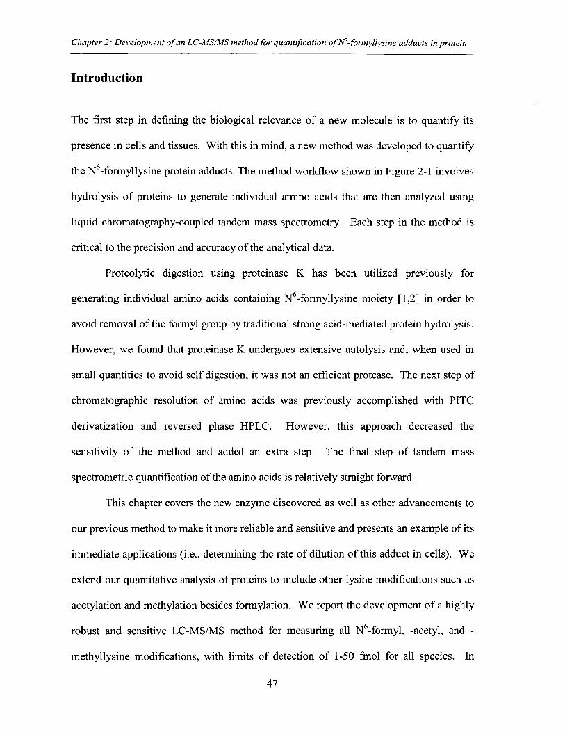
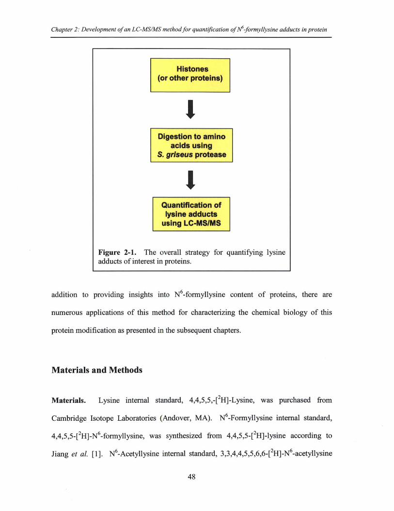
the N 6-formyllysine protein adducts. The method workflow shown in Figure 2-1 involves
hydrolysis of proteins to generate individual amino acids that are then analyzed using
liquid chromatography-coupled tandem mass spectrometry. Each step in the method is
critical to the precision and accuracy of the analytical data.
Proteolytic digestion using proteinase K has been utilized previously for
generating individual amino acids containing N6-formyllysine moiety [1,2] in order to
avoid removal of the formyl group by traditional strong acid-mediated protein hydrolysis.
However, we found that proteinase K undergoes extensive autolysis and, when used in
small quantities to avoid self digestion, it was not an efficient protease. The next step of
chromatographic resolution of amino acids was previously accomplished with PITC
derivatization and reversed phase HPLC. However, this approach decreased the
sensitivity of the method and added an extra step. The final step of tandem mass
spectrometric quantification of the amino acids is relatively straight forward.
This chapter covers the new enzyme discovered as well as other advancements to
our previous method to make it more reliable and sensitive and presents an example of its
immediate applications (i.e., determining the rate of dilution of this adduct in cells). We
extend our quantitative analysis of proteins to include other lysine modifications such as
acetylation and methylation besides formylation. We report the development of a highly
robust and sensitive LC-MS/MS method for measuring all N6-formyl, -acetyl, and -
methyllysine modifications, with limits of detection of 1-50 fmol for all species. In
47
Chapter 2: Development of an LC-MS/MS method for quantification of 1-formyllysine adducts in protein
Digestion to aminoacids using
S. griseus protease
'IQuantification of
lysine adductsusing LC-MSIMS
Figure 2-1. The overall strategy for quantifying lysineadducts of interest in proteins.
addition to providing insights into N6-formyllysine content of proteins, there are
numerous applications of this method for characterizing the chemical biology of this
protein modification as presented in the subsequent chapters.
Materials and Methods
Materials. Lysine internal standard, 4,4,5,5,-[2H]-Lysine, was purchased from
Cambridge Isotope Laboratories (Andover, MA). N6 -Formyllysine internal standard,
4,4,5,5-[ 2H]-N 6-formyllysine, was synthesized from 4,4,5,5-[2H]-lysine according to
Jiang et al. [1]. N -Acetyllysine internal standard, 3,3,4,4,5,5,6,6-[2 H]-N 6-acetyllysine
48
'I
Chapter 2: Development of an LC-MS/MS method for quantification of M-formyllysine adducts in protein
was obtained from CDN Isotopes (Pointe-Claire, Quebec, Canada). Lysine, N 6_
formyllysine, N 6-acetyllysine, Streptomyces griseus protease, and protease inhibitor
cocktail (for use with mammalian cell and tissue extracts) were obtained from Sigma-
Aldrich (St. Louis, MO). N -Mono-methyllysine, N6-di-methyllysine, and N6 -tri-
methyllysine were purchased from Bachem Bioscience Inc. (King of Prussia, PA).
Nonidet P-40 was from Roche Diagnostic Corporation (Indianapolis, IN). Human
lymphoblastoid TK6 cell line was a generous gift of Wogan Lab at Massachusetts
Institute of Technology.
TK6 cell culture and labeling. TK6 cells were cultured in RPMI 1640 medium
(Cellgro, Manassas, VA) supplemented with 10% heat-inactivated horse serum (Atlanta
Biologicals, Lawrenceville, GA), 10,000 U penicillin/ml and 10,000 ptg streptomycin/ml
(Lonza, Walkersville, MD), and 2mM L-glutamine (Lonza, Walkersville, MD) at 370 C
in a 5% CO 2 atmosphere. For labeling proteins with endogenous isotopically labeled N6_
formyllysine, TK6 cells were grown in a customized RPMI-1640 medium, with
everything identical to traditional RPMI 1640 medium (supplemented with horse serum,
antibiotics, and L-glutamine), except for the presence of deuterated lysine
(3,3,4,4,5,5,6,6-[ 2H]-Lysine) instead of non-labeled lysine. After one week of growth
(roughly 9 doubling times) in the customized medium, cells were washed and
resuspended in non-labeled RPMI medium. Histones were extracted from -10 million
cells at Oh, 18h, 36h, and 54h, and the quantity of isotopically labeled formyllysine, as a
percentage of total labeled and unlabeled lysines, was measured at each of those time
points.
49
Chapter 2: Development of an LC-MS/MS method for quantification of J-formyllysine adducts in protein
Histone extraction from TK6 cells and bovine liver tissue. Extraction of histones was
performed according to Boyne et al. [3], with modifications. Cells (~10 7 per sample)
were pelleted by centrifugation at 1000 x g for 5 min at 4 'C and the pellet was washed
once with PBS. Cell pellets were then lysed by resuspension in ice-cold lysis buffer (15
mM Tris-HCl, pH 7.5, 15 mM NaC1, 60 mM KCl, 1 mM CaCl2, 5 mM MgCl 2, 250 mM
sucrose, 1 mM dithiothreitol, 10 mM sodium butyrate) containing a 100:1 v:v dilution of
protease inhibitor cocktail in the presence of 0.03% Nonidet P-40 and incubation on ice
for 5 min with occasional gentle mixing. Nuclei were pelleted by centrifugation at 600 x
g for 5 min at 4 'C, and the pellet was washed twice with ice-cold lysis buffer without
Nonidet P-40. Histones were extracted by addition of ice-cold 0.4N H2 SO 4 and
incubation overnight on ice. The solution was centrifuged at 3000 x g for 5 min and
proteins in supernatant were precipitated by addition of 20% v/v trichloroacetic acid and
overnight incubation at 4 'C. Samples were then centrifuged at 14000 x g for 10 min at
40C, washed once with ice-cold acetone containing 0.1% HCl, and once with ice-cold
acetone. The extracts were air-dried and stored at -20 0 C until use. For extracting
histones from bovine liver tissue, 20 mg of tissue was cut into small pieces and washed
with PBS. Chromatin bound fraction was obtained using the Subcellular Protein
Fractionation Kit from Thermo Scientific (Waltham, MA) and a Kontes all-glass Dounce
homogenizer (10 strokes with a type B pestle). Proteins in chromatin bound fraction
were precipitated by addition of 20% v/v trichloroacetic acid and overnight incubation at
4 'C. Samples were then centrifuged at 14000 x g for 10 min at 4 'C, washed once with
ice-cold acetone containing 0.1% HCl, and once with ice-cold acetone. The extracts were
air-dried and stored at -20 0 C until use.
50
Chapter 2: Development of an LC-MS/MS method for quantification of N 6-formyllysine adducts in protein
Purification of individual histones. HPLC purification of total histones was performed
according to Boyne et al. [3], with modifications. Total histones ( 50 pg) were
dissolved in 0.1% trifluoroacetic acid (TFA) in water and fractionated by HPLC on an
Agilent 1100 series instrument (Agilent Technologies, Santa Clara, CA), using a Source
5RPC C18 reversed-phase column (4.6 x 150 mm, 5 jim particles; GE Healthcare Life
Sciences). The mobile phase flow rate was 1 mL/min and the solvent system was 0.1%
TFA in water (A) and 0.094% TFA in acetonitrile (B) with the elution starting at 0% B,
linearly increasing to 28% B over 28 min, reaching 37% B at 70 min, 38% B at 100 min,
60% B at 150 min, and finally 100% B at 151 min, before the column was re-equilibrated
to 0% B for 10 min. Protein elution was monitored by UV absorbance at 214 nm and
histones in each fraction were tentatively identified by resolution on a 13% SDS-PAGE
gel with Coomassie Blue staining (see Figure 3-S1).
Enzymatic hydrolysis of proteins. Histones extracted from TK6 cells and other protein
samples were dissolved in 50 piL of 100 mM ammonium bicarbonate buffer (pH 8.5),
4,4,5,5-_[2H]-lysine (2 nmol),4,4,5,5-[ 2H]-N 6-formyllysine (1 pmol), and 3,3,4,4,5,5,6,6-
[2H]-N 6-acetyl lysine (10 pmol) were added as internal standards, and the proteins
hydrolyzed by addition of Streptomyces griseus protease (freshly prepared solution each
time) with incubation at 37 'C for 16 h. S. griseus was used at a ratio of 1 pg of
enzyme per 10 pg of protein. Samples were then dried under vacuum and resuspended in
50 piL of water before mass spectrometry analysis.
Quantification of amino acids. N -Formyllysine and other amino acids were quantified
51
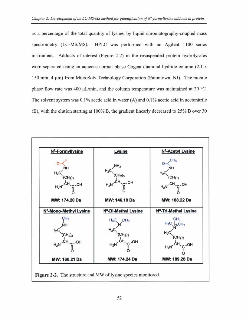
Chapter 2: Development of an LC-MS/MS method for quantification of M-formyllysine adducts in protein
as a percentage of the total quantity of lysine, by liquid chromatography-coupled mass
spectrometry (LC-MS/MS). HPLC was performed with an Agilent 1100 series
instrument. Adducts of interest (Figure 2-2) in the resuspended protein hydrolysates
were separated using an aqueous normal phase Cogent diamond hydride column (2.1 x
150 mm, 4 pm) from MicroSolv Technology Corporation (Eatontown, NJ). The mobile
phase flow rate was 400 IL/min, and the column temperature was maintained at 20 *C.
The solvent system was 0.1% acetic acid in water (A) and 0.1% acetic acid in acetonitrile
(B), with the elution starting at 100% B, the gradient linearly decreased to 25% B over 30
N-Formvllvsine,
H
H NHH
(CH2)3
IIH2N- CH'C'.OH
0
MW: 174.20 Da
Lysine
NH2
H(CH 2)3
H2NCH -OH
0
MW: 146.19 Da
N-Acetvl Lysine
O CH3
H2 NH
(CH2)3
IIH2N CHC'OH
MW: 188.22 Da
52
NB-Mono-Methyl Lysine NG-DI-Methyl Lysine N-Tri-Methyl LvsineCH3 H3C\ ,CH 3 CH3NH H3C\ CH 3
H2C / H2C C(CH2)3 (CH2)3 H2 \(CH1 1CH 2)3
H2NCH OH H2N 1CH -OH CH C'OH
0 0 01
MW: 160.21 Da MW: 174.24 Da MW: 189.28 Da
Figure 2-2. The structure and MW of lysine species monitored.
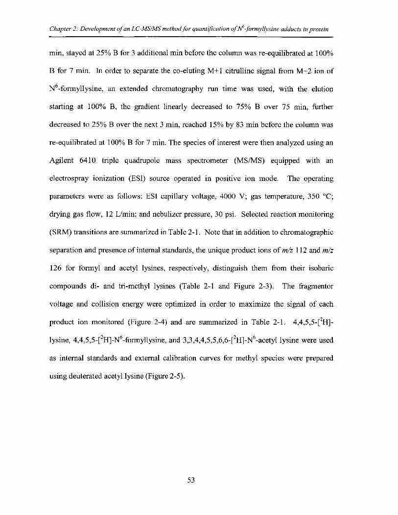
Chapter 2: Development of an LC-MS/MS method for quantification of M-formyllysine adducts in protein
min, stayed at 25% B for 3 additional min before the column was re-equilibrated at 100%
B for 7 min. In order to separate the co-eluting M+1 citrulline signal from M+2 ion of
N 6-formyllysine, an extended chromatography run time was used, with the elution
starting at 100% B, the gradient linearly decreased to 75% B over 75 min, further
decreased to 25% B over the next 3 min, reached 15% by 83 min before the column was
re-equilibrated at 100% B for 7 min. The species of interest were then analyzed using an
Agilent 6410 triple quadrupole mass spectrometer (MS/MS) equipped with an
electrospray ionization (ESI) source operated in positive ion mode. The operating
parameters were as follows: ESI capillary voltage, 4000 V; gas temperature, 350 'C;
drying gas flow, 12 L/min; and nebulizer pressure, 30 psi. Selected reaction monitoring
(SRM) transitions are summarized in Table 2-1. Note that in addition to chromatographic
separation and presence of internal standards, the unique product ions of m/z 112 and m/z
126 for formyl and acetyl lysines, respectively, distinguish them from their isobaric
compounds di- and tri-methyl lysines (Table 2-1 and Figure 2-3). The fragmentor
voltage and collision energy were optimized in order to maximize the signal of each
product ion monitored (Figure 2-4) and are summarized in Table 2-1. 4,4,5,5-[ 2H]-
lysine, 4,4,5,5-[ 2H]-N 6-formyllysine, and 3,3,4,4,5,5,6,6-[ 2H]-N 6 -acetyl lysine were used
as internal standards and external calibration curves for methyl species were prepared
using deuterated acetyl lysine (Figure 2-5).
53
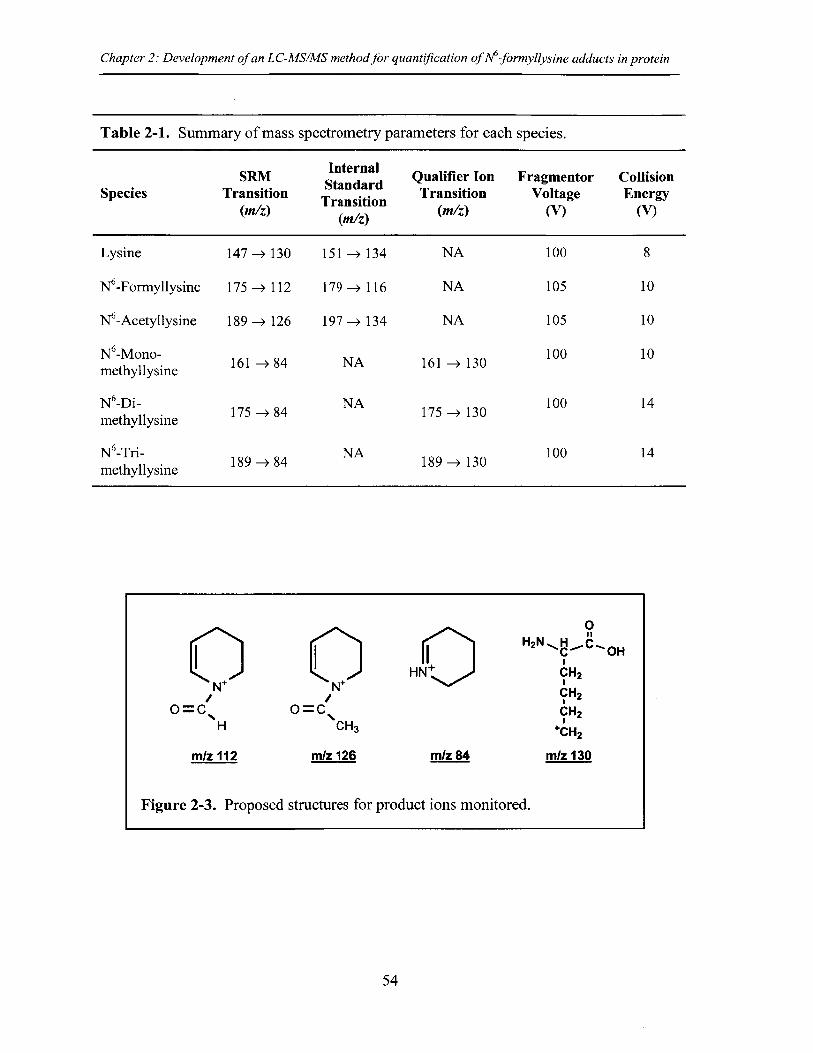
Chapter 2: Development of an LC-MS/MS method for quantification of M-formyllysine adducts in protein
Table 2-1. Summary of mass spectrometry parameters for each species.
SRMInternal Qualifier Ion Fragmentor CollisionSpecies Transition Standard Transition Voltage EnergyTranitio Transition (I/) (V) (V)(m/z) (m/)/V)(V
Lysine 147 -130 151 ->134 NA 100 8
N6-Formyllysine 175 ->112 179 -116 NA 105 10
N6 -Acetyllysine 189-> 126 197-> 134 NA 105 10
N6 -Mono- 161 84 NA 161 130 100 10methyllysine
N6-Di- NA 100 14methyllysine 175 -+ 84 175 -+ 130
N -Tri- NA 100 14methyllysine 189 -> 84 189 -> 130
N*+
O=C NCH3
m/z 126
HNQ
0H2N,, OH
CH2
CH2
CH 2
mCH 2
mlz 130
Figure 2-3. Proposed structures for product ions monitored.
54
m/z 84
N*
O=C,H
m/z 112
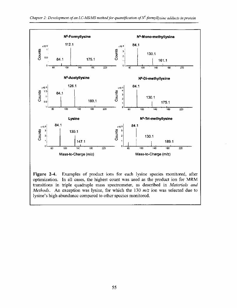
Chapter 2: Development of an LC-MS/MS method for quantification of M-formyllysine adducts in protein
N-Formyllysine N-Mono-methyllysine
112.1 xi0 5
o 2-
1175.1
140 180 220
N-Acetyllysine
126.1
84.1
00
0
0
X10
0
84.1
130.1
161.1
6 100 140 180 220
N-Di-methyllysine
84.1
130.1175.1
100 140 180 220
N-Tri-methyllysine
x10 5 84.1. 3
8 2-01
130.1
I 147.1
60 100 14C 180 220
Mass-to-Charge (m/z)
X10 5
IWO 3
S2-0
84.1
130.1189.1
60 100 140 180 220
Mass-to-Charge (m/z)
Figure 2-4. Examples of product ions for each lysine species monitored, afteroptimization. In all cases, the highest count was used as the product ion for MRMtransitions in triple quadruple mass spectrometer, as described in Materials andMethods. An exception was lysine, for which the 130 m/z ion was selected due tolysine's high abundance compared to other species monitored.
55
189.1
a0 100 140 180 220
x10 s1 5-
0.5-
Lysine
1
I
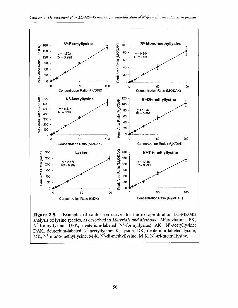
Chapter 2: Development of an LC-MS/MS method for quantification of A-formyllysine adducts in protein
180 - N6-Formyllysine 100 N6-Mono-methyllysineU-O 150 -0150 - y= 1.70x 80 ~ y =0.84xu- 120 - R2 =0.998 R2 =0.9990 . 601 90 -
M 4060 - 40
< 30 - 2044
0 a- 00 50 100 0 50 100
Concentration Ratio (FK/DFK) Concentration Ratio (MK/DAK)
700 - N 6-Acetyllysine 7 120 N-Di-methyllysine0<
600 - 0 100500 y =6.37xoR 2 0.998400 R2 0.999
60 6-c 300 w 40
.200
100 < 20
0 4
0 50 100 0 50 100
Concentration Ratio (AK/DAK) Concentration Ratio (M2K/DAK)
300 Lysine 180 - N-Tri-methyllysine2 250 - 150
0 y = 2.47x y = 1.44x200 R2 =0.999 0 R2 =0.996
a 150 90(D15
100 - 60
(D 50 30D 0
0 50 100 0 50 100
Concentration Ratio (K/DK) Concentration Ratio (M3K/DAK)
Figure 2-5. Examples of calibration curves for the isotope dilution LC-MS/MSanalysis of lysine species, as described in Materials and Methods. Abbreviations: FK,N6-formyllysine; DFK, deuterium-labeled N6-formyllysine; AK, N6 -acetyllysine;DAK, deuterium-labeled N -acetyllysine; K, lysine; DK, deuterium-labeled lysine;MK, N -mono-methyllysine; M 2K, N6-di-methyllysine; M 3K, N 6-tri-methyllysine.
56

Chapter 2: Development of an LC-MS/MS method for quantification of N 6-formyllysine adducts in protein
Results
An improved method to quantify N'-formyllysine in proteins. Our previous method
for quantifying N6-formyllysine in proteins involved proteinase K-mediated hydrolysis of
proteins, derivatization of the resulting amino acids with PITC, HPLC pre-purification of
amino acid derivatives, and final LC-MS/MS analysis of the derivatized amino acids [1].
This method proved to be relatively insensitive and biased as a result of using proteinase
K, which produced only partial hydrolysis of some proteins when used in small quantities
to minimize background autolysis (Figure 2-6). To overcome these problems, we used
Streptomyces griseus protease at a ratio of 1 pg enzyme per 10 pg proteins, which
resulted in efficient and complete digestion of all proteins as judged by the total amount
57
100 Theoretical /90807060 - S. griseus Protease
0 50Ee 40 /
3020 Proteinase K
100
0 20 40 60 80 100
Total histone (pg)
Figure 2-6. Comparison of released lysine in protein digestion using Streptomycesgriseus protease versus proteinase K. Proteinase K produced only partial hydrolysis ofTK6 histone proteins when used in small quantities to minimize background autolysis.
Chapter 2: Development of an LC-MS/MS method for quantification of M-formyllysine adducts in protein
of lysine released per pg of purified histone proteins (Figure 2-6). In addition, the
method was optimized to eliminate the HPLC pre-purification step and the need for PITC
derivatization to achieve chromatographic resolution of amino acids. This was achieved
using aqueous normal phase HPLC with a diamond-hydride column to resolve individual
amino acids. This chromatographic system resolved N6-acetyllysine, mono-, di-, and tri-
N6-methyllysines, as well as N6-formyllysine and lysine, as shown in Figure 2-7. With
isotopically labeled internal standards added prior to protease digestion, identification
and quantification of amino acids were accomplished by isotope-dilution LC-MS/MS in
positive ion mode, using multiple reaction monitoring (MRM) transitions. The limits of
detection were found to be 1 fmol for N6-formyl- and N6-acetyllysine, 10 fmol for lysine,
and 50 fmol for each of N6-mono-, di-, and tri-methyl lysine. Data for the various lysine
modifications are expressed here as proportions of the total number of lysines in the
sample.
Measurement of N6-formyllysine adducts in total histone proteins. The first
application of the new method involved quantification of the various modified lysines in
a mixture of total histone proteins extracted from TK6 lymphoblastoid cells (Table 2-2).
Data show that N6-formylation of lysine occurs at a low frequency (e.g., 1 per 104
lysines) in total histones compared to higher frequencies of acetylation and methylation
(40 - 230 per 104 lysines). Further, an analysis of total histone proteins, either extracted
from TK6 cells or from commercial sources, show a similar background level of N6_
formyllysine in all samples (Table 2-3). The next chapter covers the distribution of
various lysine adducts in different classes of histones separated by HPLC.
58

Chapter 2: Development of an LC-MS/MS method for quantification of M-formyllysine adducts in protein
Table 2-2. Quantification of lysine modifications in a sample of total histone proteins.
Formyll Acetyl Methyl Dimethyl Trimethyl
TK6 1.0 0.12 40 10 230 50 130 40 25 5Cells
'Column titles denote different N6-modifications of lysine.
2Data are expressed as modifications per 104 total lysines and represent mean ± SDfor 3 biological replicates.
59
x106 N-Acetyllysine1.6 N6-DI-
1.4 methyllysine
1.2 Lysine
00 0.8
NI-Tri-0.6 N-Formyllysine methyllysine
N6-Mono-0.4 methyllysine
0.2
10 12 14 16 18 20 22 24 26 28 30
Retention time, min
Figure 2-7. An example of different lysine species detected in purified histone H4
from TK6 cells. Lysine adducts were monitored by tandem mass spectrometry, as
described in Materials and Methods.
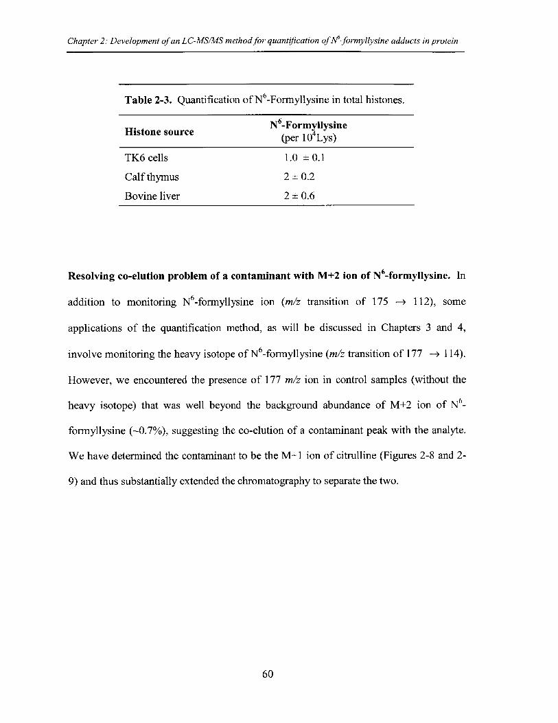
Chapter 2: Development of an LC-MS/MS method for quantification of M-formyllysine adducts in protein
Table 2-3. Quantification of N 6-Formyllysine in total histones.
N -FormyllysineHistone source (per 104 Lys)
TK6 cells 1.0 ± 0.1
Calf thymus 2± 0.2
Bovine liver 2 ± 0.6
Resolving co-elution problem of a contaminant with M+2 ion of N -formyllysine. In
addition to monitoring N6-formyllysine ion (m/z transition of 175 -> 112), some
applications of the quantification method, as will be discussed in Chapters 3 and 4,
involve monitoring the heavy isotope of N 6-formyllysine (m/z transition of 177 -+ 114).
However, we encountered the presence of 177 m/z ion in control samples (without the
heavy isotope) that was well beyond the background abundance of M+2 ion of N6_
formyllysine (-0.7%), suggesting the co-elution of a contaminant peak with the analyte.
We have determined the contaminant to be the M+1 ion of citrulline (Figures 2-8 and 2-
9) and thus substantially extended the chromatography to separate the two.
60
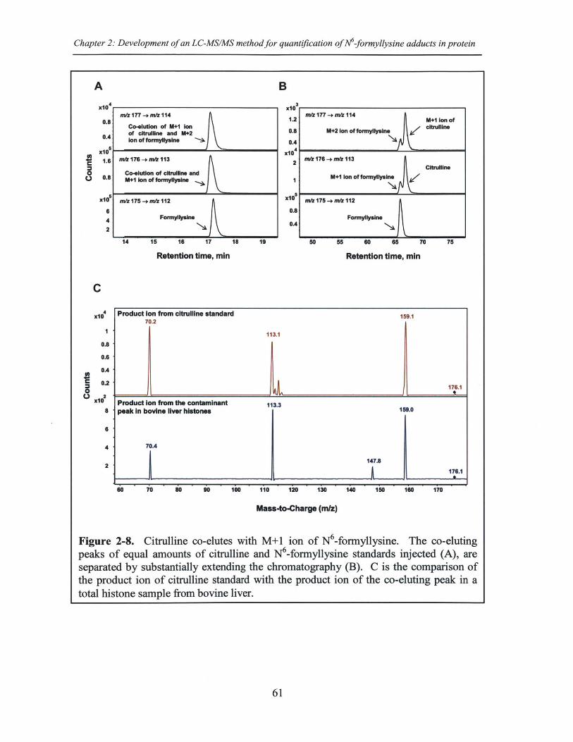
Chapter 2: Development of an LC-MS/MS method for quantification of N6-formyllysine adducts in protein
A B
x10n. /177 -* m/z 114
08 Co-elutlon of M*1 IonOf ciruln aM+12
0.4 Ion of foryllysAne .
A 101.6 mi 176 -+ m/ 113
Co lution of ciuns and0.6 M41 Ion of formyllyalne
x10e m/z 175 -+ miz 112
6
4 Formyllyulne
2
14 15 16 17 18 19
Retention time, min
6
4
2
x
x
X
1031.2 m/z177 -+ mtz 1141.2 M*I Ion of
0.8 M+2 Ion of formyllysine C~tfline
0.4
104
2 m/z 176 -+ m/z 113 A i
M+1 Ion of formylIysin Ctun
0 m/z 175 -+ m/z 112
0.8Formyllysine
OA
50 55 60 65 70 75
Retention time, min
Mass-to-Charge (mlz)
Figure 2-8. Citrulline co-elutes with M+1 ion of N6-formyllysine. The co-elutingpeaks of equal amounts of citrulline and N6-formyllysine standards injected (A), areseparated by substantially extending the chromatography (B). C is the comparison ofthe product ion of citrulline standard with the product ion of the co-eluting peak in atotal histone sample from bovine liver.
61
*1
C.,
C
x14xlO
10.8
0.6
0A
0.2
x12XlO
a
C.,
Product Ion from citrullne standard 159.170.2
113.1
176.1
Product Ion from the contaminant 113.3peak In bovine liver histones 159.0
70.4
147.8176.1
60 70 so 90 100 110 120 130 140 150 160 170
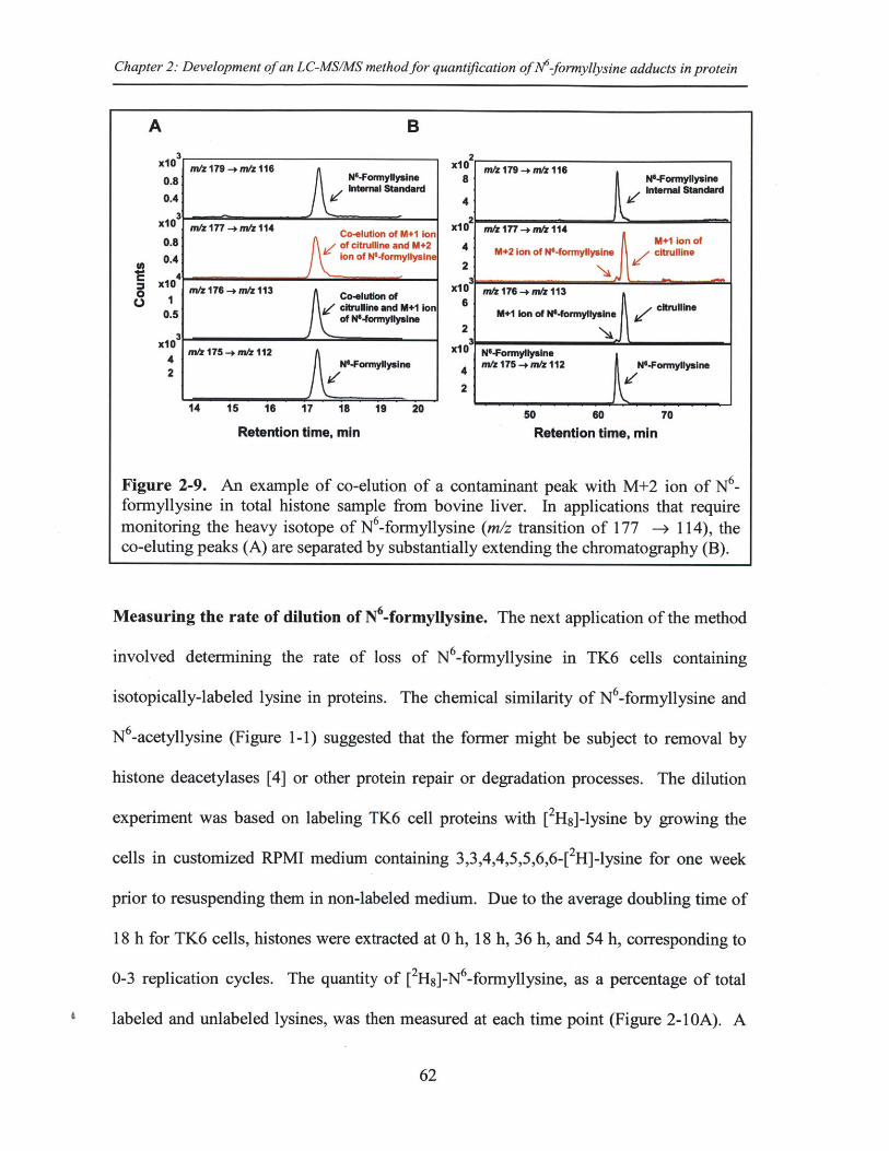
Chapter 2: Development of an LC-MS/MS method for quantification of M-formyllysine adducts in protein
A B
X n3 m/z179 -+ mI116 x1U nir179-+mz1160.8 N'-Fonrmy llyslne 8 N-Fonnyllyslne
Internal Standard Internal Standard0.41 l 4 l
x10 nu177 -+m 114 Co-eluton of M+1 Ion Me f/Z177.-.*flI0.8 of citrulline and M+2 4 M+2 Ion of W-fornly n M+1 Ion of
0.4 Ion of N-fonylysine 2 + n N r y citrullino
x10 n/z176 -+m/ 113 xCo0lu3on of n1176 -+ nm113
0.5citruflline and M+1 Ion 6 Mel Ion o0.5 W +n M+1Ion of N-frmyllysineJn
]jof N04ormyllyslne
x 1 175 - rnfl112 x10 N-Formyllysine
2 N.Formylyuine 4m& 175 -+m& 112 N-Formyllysine
2
14 15 16 17 18 19 20 50 60 70
Retention time, min Retention time, min
Figure 2-9. An example of co-elution of a contaminant peak with M+2 ion of N6_formyllysine in total histone sample from bovine liver. In applications that requiremonitoring the heavy isotope of N6-formyllysine (m/z transition of 177 --+ 114), theco-eluting peaks (A) are separated by substantially extending the chromatography (B).
Measuring the rate of dilution of N'-formyllysine. The next application of the method
involved determining the rate of loss of N6-formyllysine in TK6 cells containing
isotopically-labeled lysine in proteins. The chemical similarity of N6-formyllysine and
N 6-acetyllysine (Figure 1-1) suggested that the former might be subject to removal by
histone deacetylases [4] or other protein repair or degradation processes. The dilution
experiment was based on labeling TK6 cell proteins with [2H8]-lysine by growing the
cells in customized RPMI medium containing 3,3,4,4,5,5,6,6-[ 2H]-lysine for one week
prior to resuspending them in non-labeled medium. Due to the average doubling time of
18 h for TK6 cells, histones were extracted at 0 h, 18 h, 36 h, and 54 h, corresponding to
0-3 replication cycles. The quantity of [2Hs]-N6 -formyllysine, as a percentage of total
labeled and unlabeled lysines, was then measured at each time point (Figure 2-10A). A
62
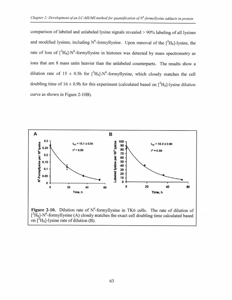
Chapter 2: Development of an LC-MS/MS method for quantification of N-formyllysine adducts in protein
comparison of labeled and unlabeled lysine signals revealed > 90% labeling of all lysines
and modified lysines, including N6-formyllysine. Upon removal of the [2H 8]-lysine, the
rate of loss of [2H8]-N 6-formyllysine in histones was detected by mass spectrometry as
ions that are 8 mass units heavier than the unlabeled counterparts. The results show a
dilution rate of 15 ± 0.5h for [2H8]-N 6 -formyllysine, which closely matches the cell
doubling time of 16 ± 0.9h for this experiment (calculated based on [2H8]-lysine dilution
curve as shown in Figure 2-1 OB).
63
A B
0.3 t 12 = 15.1 10.5h tw =16.2 0.9h0.25 r2 = 0.99
r 80 r 0.99S70-0.2 ' 60
30.15 . 5040
0.1 -30-20
0.05
0 20 40 so 0 20 40 60
Time, h wTIm, h
Figure 2-10. Dilution rate of N -formyllysine in TK6 cells. The rate of dilution of[2Hs)-N 6-formyllysine (A) closely matches the exact cell doubling time calculated basedon [ Hs]-lysine rate of dilution (B).
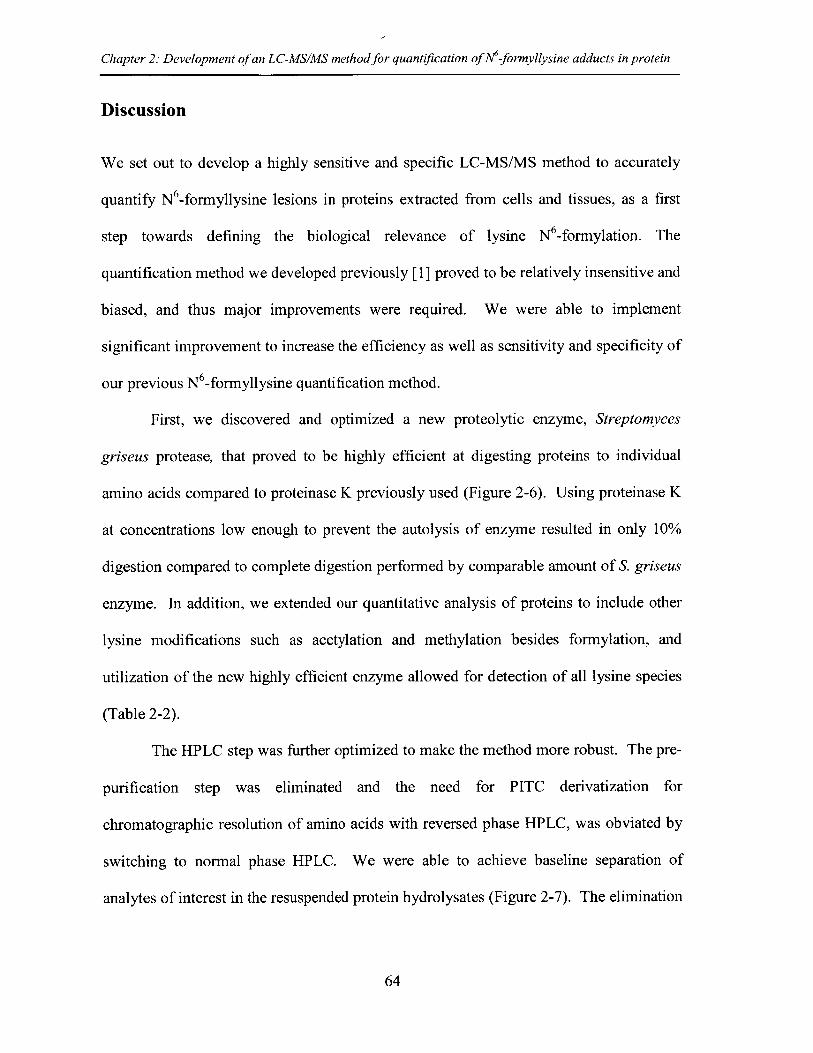
Chapter 2: Development of an LC-MS/MS method for quantification of Mf-formyllysine adducts in protein
Discussion
We set out to develop a highly sensitive and specific LC-MS/MS method to accurately
quantify N6-formyllysine lesions in proteins extracted from cells and tissues, as a first
step towards defining the biological relevance of lysine N -formylation. The
quantification method we developed previously [1] proved to be relatively insensitive and
biased, and thus major improvements were required. We were able to implement
significant improvement to increase the efficiency as well as sensitivity and specificity of
our previous N6-formyllysine quantification method.
First, we discovered and optimized a new proteolytic enzyme, Streptomyces
griseus protease, that proved to be highly efficient at digesting proteins to individual
amino acids compared to proteinase K previously used (Figure 2-6). Using proteinase K
at concentrations low enough to prevent the autolysis of enzyme resulted in only 10%
digestion compared to complete digestion performed by comparable amount of S. griseus
enzyme. In addition, we extended our quantitative analysis of proteins to include other
lysine modifications such as acetylation and methylation besides formylation, and
utilization of the new highly efficient enzyme allowed for detection of all lysine species
(Table 2-2).
The HPLC step was further optimized to make the method more robust. The pre-
purification step was eliminated and the need for PITC derivatization for
chromatographic resolution of amino acids with reversed phase HPLC, was obviated by
switching to normal phase HPLC. We were able to achieve baseline separation of
analytes of interest in the resuspended protein hydrolysates (Figure 2-7). The elimination
64
Chapter 2: Development of an LC-MS/MS method for quantification of M-formyllysine adducts in protein
of the derivatization step removed an extra step and further enhanced the sensitivity of
the method.
The identification and quantification of amino acids were accomplished by
isotope-dilution LC-MS/MS in positive ion mode, using MRM transitions. The
discovery of the unique product ions of m/z 112 and m/z 126 for formyl and acetyl
lysines, respectively, distinguished them from their isobaric compounds di- and tri-
methyl lysines (Table 2-1 and Figures 2-3 and 2-4) while lowering the limits of detection
by more than 10 fold (10 fmol to 1 fmol). The optimization of fragmentor voltage and
collision energies (Table 2-1 and Figure 2-4) further decreased the limits of detection for
all species. The new sensitive and specific method allows for robust quantification of all
N6-methyl-, -acetyl- and -formyl-lysine modifications, with limits of detection of 1 fmol
for N6 -formyl- and N6-acetyllysine, 10 finol for lysine, and 50 frnol for each of N6 -mono-
di-, and tri-methyl lysine.
In addition to providing insights into N 6-formyllysine content of proteins, there
are numerous applications of our quantification method for characterizing the chemical
biology of this protein modification, as presented in the subsequent chapters. Here, we
provided two examples of its immediate application. The first example was
quantification of the background levels of lysine adducts in a sample of total histones
extracted from TK6 cells. As shown in Table 2-2, N6-formyllysine adducts occur at a
low frequency (e.g., 1 per 10 4 lysines) in total histones compared to higher frequencies of
acetylation and methylation (40 - 230 per 104 lysines). The next chapter covers the
distribution of N6-formyllysine adducts in different classes of histones separated by
HPLC. Our quantification data is consistent with published studies of relative quantities
65
Chapter 2: Development of an LC-MS/MS method for quantification of AM-formyllysine adducts in protein
of histone modifications using immunologic and radiolabeling techniques [5-8].
Moreover, using our sensitive methods, we report ~1 lysine formylation per 104 lysines in
total histones extracted from TK6 cells compared to 4-10 modification per 104 previously
reported [1].
The other application of the quantification method presented here was to
determine the rate of dilution of [2H 8]-N6 -formyllysine in TK6 cells. Data showed that
the dilution rate of 15 h for [2H]-N6 -formyllysine closely matches the cell doubling time
of 16 h (Figure 2-10), suggesting that N6-formyllysine is either a stable modification of
histones or is formed and removed at the same frequency, resulting in no overall change
in its steady state levels. The observation that cell replication did not result in an
apparent repair or removal of N6-formyllysine adducts in histone proteins is consistent
with a previous study of conservation of histone carcinogen adducts during replication
[9].
Our robust and sensitive analytical method could readily be extended for
quantification of other amino acid modifications in proteins, with the advantage of using
a highly efficient yet mild proteolytic digestion as opposed to the traditionally used
strong hydrochloric acid for protein hydrolysis that could remove the adduct of interest.
The absolute quantification of a specific protein modification, in addition to mapping
studies, could provide insights on various biological pathways in cells. For instance,
mapping studies of N6-formyllysine sites reveal that this adduct occurs on conserved
lysines involved in epigenetic regulation [5], and through absolute quantification, we
showed that this modification is rather stable or is formed and removed at the same
frequency, resulting in no overall change in its steady state levels. Furthermore, this
66
Chapter 2: Development of an LC-MS/MS method for quantification of Nt-formyllysine adducts in protein
method is used in subsequent chapters to quantify various lysine modifications in
individual classes of histones. Histone modification-based signaling involves the
location and number of specific modification targets within a histone protein as well as
the frequency of modification of a target among all copies of a particular histone protein.
Our quantification data provide some insight into this issue. Further, N6-formyllysine
quantification is used to prove possible mechanisms for its formation and shed light on its
fate.
67
Chapter 2: Development of an LC-MS/MS method for quantification of M-formyllysine adducts in protein
References
1. Jiang T, Zhou X, Taghizadeh K, Dong M, Dedon PC (2007) N-formylation oflysine in histone proteins as a secondary modification arising from oxidativeDNA damage. Proc Nati Acad Sci U S A 104: 60-65.
2. Cai H, Guengerich FP (2000) Acylation of protein lysines by trichloroethyleneoxide. Chem Res Toxicol 13: 327-335.
3. Boyne MT, 2nd, Pesavento JJ, Mizzen CA, Kelleher NL (2006) Precisecharacterization of human histones in the H2A gene family by top down massspectrometry. J Proteome Res 5: 248-253.
4. Hake SB, Xiao A, Allis CD (2004) Linking the epigenetic 'language' of covalenthistone modifications to cancer. Br J Cancer 90: 761-769.
5. Wisniewski JR, Zougman A, Mann M (2008) Nepsilon-formylation of lysine is awidespread post-translational modification of nuclear proteins occurring atresidues involved in regulation of chromatin function. Nucleic Acids Res 36: 570-577.
6. Pasqualini JR, Mercat P, Giambiagi N (1989) Histone acetylation decreased byestradiol in the MCF-7 human mammary cancer cell line. Breast Cancer Res Treat14: 101-105.
7. Byvoet P, Barber M, Amidei K, Lowell N, Trudeau W (1986) Effect ofexogenous histone H5 on integration of histone HI in rat liver chromatin.Correlations with aberrant epsilon-N-methylation of histone HI. BiochimBiophys Acta 867: 163-175.
8. Wu M, Allis CD, Richman R, Cook RG, Gorovsky MA (1986) An interveningsequence in an unusual histone HI gene of Tetrahymena thermophila. Proc NatlAcad Sci U S A 83: 8674-8678.
9. Ozbal CC, Velic I, SooHoo CK, Skipper PL, Tannenbaum SR (1994)Conservation of histone carcinogen adducts during replication: implications forlong-term molecular dosimetry. Cancer Res 54: 5599-5601.
68
Chapter 3: Sources and fate ofNM-formyllysine adducts in proteins
Chapter 3
Quantitative analysis of histone modifications:Formaldehyde is a source of pathological
N6-formyllysine that is refractory to histonedeacetylases
69
Chapter 3: Sources and fate of M-formyllysine adducts in proteins
Abstract
Aberrant protein modifications play an important role in the pathophysiology of manyhuman diseases, in terms of both dysfunction of physiological modifications and theformation of pathological modifications by reaction of proteins with endogenouselectrophiles. Recent studies have identified a chemical homolog of lysine acetylation,N 6-formyllysine, as an abundant modification of histone and chromatin proteins, onepossible source of which is the reaction of lysine with 3'-formylphosphate residues fromDNA oxidation. Using a new liquid chromatography-coupled to tandem massspectrometry method to quantify all N -methyl-, -acetyl- and -formyl-lysinemodifications, we now report that endogenous formaldehyde is a major source of N6_formyllysine and that this adduct is widespread among cellular proteins in allcompartments. N -Formyllysine was evenly distributed among different classes ofhistone proteins from human TK6 cells at 1-4 modifications per 10 4 lysines, whichcontrasted strongly with lysine acetylation and mono-, di- and tri-methylation levels of1.5-380, 5-870, 0-1400 and 0-390 per 104 lysines, respectively. While isotope labelingstudies revealed that lysine demethylation is not a source of N6-formyllysine in histones,formaldehyde exposure was observed to cause a dose-dependent increase in N6-formyllysine, with use of [13C, 2 H2 ]-formaldehyde revealing unchanged levels of adductsderived from endogenous sources. Inhibitors of class I and class II histone deacetylasesdid not affect the levels of N6-formyllysine in TK6 cells and the class III histonedeacetylase, SIRTI, had minimal activity (<10%) with a peptide substrate containing theformyl adduct. These data suggest that N6-formyllysine is refractory to removal byhistone deacetylases, which supports the idea that this abundant protein modificationcould interfere with normal regulation of gene expression if it arises at conserved sites ofphysiological protein secondary modification.
70
Chapter 3: Sources and fate of M-formyllysine adducts in proteins
Introduction
In addition to physiological secondary modifications, proteins are subjected to reactions
with endogenous electrophiles generated by oxidative stress, inflammation, and normal
cell metabolic processes [1-5]. These adventitious or pathological modifications
typically arise by reaction of the nucleophilic side chains of lysine, histidine, and cysteine
with reactive electrophiles such as malondialdehyde, 4-hydroxynonenal (HNE), and
glyoxal generated by oxidation of polyunsaturated fatty acids and carbohydrates, among
other biomolecules [2-4,6,7]. The resulting adducts, which can alter protein function and
lead to protein degradation, are associated with a variety of pathological processes and
human diseases [1-5,8]. Among these pathological adducts, N6-formylation of lysine has
recently emerged as an abundant protein modification [5,9-11]. While originally
described in chromatin proteins [9-11], it has since been identified as an adduct arising in
proteins subjected to nitrosative and oxidative stresses [5]. In chromatin proteins, N 6_
formyllysine has the potential to interfere with the functions of other post-translational
modifications that perform signaling functions [12-15], such as acetylation, methylation,
phosphorylation, ubiquitylation, and ADP ribosylation, with some locations modified in
more than one way (e.g., refs. [16-18]). The chemical similarities of N6-formyllysine and
N 6-acetyllysine suggest a disruptive role for the former in signaling by histone
acetylation. Indeed, N6-formyllysine has been detected at conserved sites of lysine
acetylation and methylation in histones [10,11].
While N 6-formyllysine adducts are now well recognized as abundant protein
modifications in cells, the source of these pathological adducts remains unclear. We
recently showed that some portion of N -formyllysine arises in chromatin proteins by
71
Chapter 3: Sources and fate of M-formyllysine adducts in proteins
reaction of lysine side chains with the 3'-formylphosphate residue derived from 5'-
oxidation of 2-deoxyribose in DNA in cells (Figure 3-1) [9]. However, the observation
of this adduct in proteins treated with the biological oxidant, peroxynitrite, suggests other
sources for the formyl species [5]. Considering that formaldehyde reacts with amines to
give a carbinolamine (N6-hydroxymethyllysine) intermediate that is only one oxidation
state away from a formamide functional group (Figure 3-1), we hypothesized that
endogenous formaldehyde could serve as a source of N6-formyllysine residues in histone
and other proteins. In addition to environmental and occupational sources [19-21],
formaldehyde arises from cellular processes such as oxidative demethylation of nucleic
acid and histone proteins, as well as biosynthesis of purines, thymidine, and some amino
acids [20,22,23], making it a relatively abundant metabolite at concentrations ranging
from 13 to 97 pM in human plasma [20]. To test this hypothesis and to clarify the
cellular locations and quantities of N 6-formyllysine relative to other major histone
modifications, we developed a novel liquid chromatography-coupled to electrospray
tandem mass spectrometry (LC-MS/MS) method to quantify all N6-methyl-, -acetyl-, and
-formyl-lysine modifications. Application of this method reveals that endogenous
formaldehyde is a major source of N 6-formyllysine, that this adduct is widespread among
proteins in all cellular compartments, and that, in chromatin proteins, it is refractory to
removal by histone deacetylases.
72
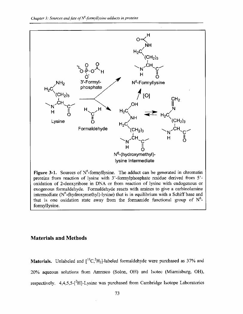
Chapter 3: Sources and fate of M-formyllysine adducts in proteins
I H
0-P-0 H
3'-Formyl-phosphate
H,
110
Formaldehyde
NHH2C
(CH2)3H
H0
N6-Formyllysine
1]
/OHH2 C
NH/
H2C\(CH2)3
NCH'C-N ~H 0
N6-(hydroxymethyl)-lysine Intermediate
Figure 3-1. Sources of N6-formyllysine. The adduct can be generated in chromatinproteins from reaction of lysine with 3'-formylphosphate residue derived from 5'-oxidation of 2-deoxyribose in DNA or from reaction of lysine with endogenous orexogenous formaldehyde. Formaldehyde reacts with amines to give a carbinolamineintermediate (N6-(hydroxymethyl)-lysine) that is in equilibrium with a Schiff base andthat is one oxidation state away from the formamide functional group of N6_formyllysine.
Materials and Methods
Materials. Unlabeled and [13C, 2H2]-labeled formaldehyde were purchased as 37% and
20% aqueous solutions from Amresco (Solon, OH) and Isotec (Miamisburg, OH),
respectively. 4,4,5,5-[ 2H]-Lysine was purchased from Cambridge Isotope Laboratories
73
/NH 2
H2C
NH C0
Lysine
CH211N
H2C\(CH2)3
N-CHH 0
(CHt2)3
Chapter 3: Sources and fate of N6-formyllysine adducts in proteins
(Andover, MA). 4,4,5,5-[2H]-N6-Formyllysine was synthesized from 4,4,5,5-[2H]-lysine
according to Jiang et al. [9]. 3,3,4,4,5,5,6,6-[ 2H]-N 6-acetyllysine were obtained from
CDN Isotopes (Pointe-Claire, Quebec, Canada). L-Methionine-([13 C 2H3 ]-methyl) was
obtained from Isotec (Miamisburg, OH). Lysine, N6-formyllysine, N6-acetyllysine,
bovine serum albumin, human recombinant HMG- 1, human IgG, Streptomyces griseus
protease, and protease inhibitor cocktail (for use with mammalian cell and tissue extracts)
were obtained from Sigma-Aldrich (St. Louis, MO). N6-Mono-methyllysine, N6-di-
methyllysine, and N6-tri-methyllysine were purchased from Bachem Bioscience Inc.
(King of Prussia, PA). Nonidet P-40 was from Roche Diagnostic Corporation
(Indianapolis, IN). Suberoylanilidehydroxamic acid (SAHA) and SIRTI (human
recombinant) enzyme were purchased from Cayman chemical (Ann Arbor, MI). Peptide
substrates for SIRTI (GGAKRHR and its lysine-acetylated and -formylated forms) were
synthesized at Massachusetts Institute of Technology Biopolymers Laboratory. The
human lymphoblastoid TK6 cell line was a generous gift of Prof. Gerald Wogan
(Massachusetts Institute of Technology).
TK6 cell culture, exposure, and labeling. TK6 cells were cultured in RPMI 1640
medium (Cellgro, Manassas, VA) supplemented with 10% heat-inactivated horse serum
(Atlanta Biologicals, Lawrenceville, GA), 10,000 U penicillin/ml and 10,000 ptg
streptomycin/ml (Lonza, Walkersville, MD), and 2 mM L-glutamine (Lonza,
Walkersville, MD) at 37 *C in a 5% CO2 atmosphere. For formaldehyde exposure
studies, TK6 cells were pelleted, washed, and resuspended in RPMI 1640 medium
without any supplements, prior to addition of formaldehyde to the medium. Following
74
Chapter 3: Sources and fate of M-formyllysine adducts in proteins
addition of formaldehyde, cells were incubated at 37 'C for 2 h with occasional mixing
prior to extracting chromatin proteins. Histones were extracted from ~107 cells after
exposure and the quantity of formyllysine, as a percentage of total lysine, was measured
as described below. For lysine demethylation studies, TK6 cells were grown in a
customized RPMI-1640 medium identical to the traditional medium (e.g., supplemented
with horse serum, antibiotics, and L-glutamine), except for the presence of labeled
methionine (L-methionine-([ 13C,2 H3]-methyl)) instead of non-labeled methionine.
Histones (from ~107 cells) were extracted every 2 d for 20 d in order to investigate the
formation of N 6[13C,2 H]-formyllysine. For histone deacetylase studies, TK6 cells were
incubated with the histone deacetylase inhibitor, SAHA, for 20 h at 37 'C in a 5% CO2
atmosphere prior to histone extraction. SAHA was dissolved in a 50:50 solution of
DMSO:PBS prior to addition to cell media. Control cells ( 107) were treated with the
DMSO:PBS vehicle.
Histone extraction from TK6 cells and subcellular protein fractionation from tissue.
For detailed procedure regarding histone extraction, refer to Materials and Methods
section of Chapter 2. For collecting membrane, cytosolic, and nuclear fractions from
tissues, 20 mg of bovine liver tissue was cut into small pieces and washed with PBS, and
proteins were fractionated using the Subcellular Protein Fractionation Kit from Thermo
Scientific (Waltham, MA) and a Kontes all-glass Dounce homogenizer (10 strokes with a
type B pestle). Proteins in subcellular extracts were precipitated by addition of 20% v/v
trichloroacetic acid and overnight incubation at 4 'C. Samples were then centrifuged at
14000 x g for 10 min at 4 'C, washed once with ice-cold acetone containing 0.1% HCl,
75
Chapter 3: Sources and fate of M-formyllysine adducts in proteins
and once with ice-cold acetone. The extracts were air-dried and stored at -20 0 C until
use.
Purification of individual histones. HPLC purification of total histones was performed
according to Boyne et al. [24] with modifications. For detailed procedure of histone
purification into individual amino acids, refer to Materials and Methods section of
Chapter 2.
Enzymatic hydrolysis of proteins. Proteins were hydrolyzed to individual amino acids
using S. griseus protease (freshly prepared solution each time) with incubation at 37 'C
for 16 h. For detailed procedure regarding protein digestion into individual amino
acids, refer to Materials and Methods section of Chapter 2.
Quantification of amino acids. N6-Formyllysine and other amino acids of interest were
quantified as a percentage of the total quantity of lysine, by liquid chromatography-
coupled mass spectrometry (LC-MS/MS). For detailed procedure regarding LC-MS/MS
based quantification of individual amino acids, refer to Materials and Methods section of
Chapter 2.
SIRTI peptide experiment. SIRTI peptide substrates (GGAKRHR, GGAKacetyiRHR,
and GGAKrmy1RHR) were HPLC purified on an Agilent 1100 series instrument using
Vydac 218TP52 C18 reverse-phase column (2.1 x 250 mm, 5 [im) from Grace Vydac
(Hesperia, CA). The mobile phase flow rate was 200 .L/min, and the column
76
Chapter 3: Sources and fate of M-formyllysine adducts in proteins
temperature was maintained at 30 'C. The solvent system was 0.05% trifluoroacetic acid
in water (A) and 0.05% trifluoroacetic acid in 80% acetonitrile (B), with the isocratic
elution of 5% B for the first 5 min, then a linear increase to 42% B over 25 min, reaching
100% B at 31 min followed by column re-equilibration at 5% B for 10 min. Each
purified SIRTi peptide substrate (100 pmol) was incubated overnight (12 h) at 25 *C
with 1 pg SIRTi, in 50 mM Tris-HCl (pH 8) buffer containing 137 mM NaCl, 2.7 mM
KCl, 1 mM MgCl 2, and 6 mM NAD*. The removal of acetyl and formyl groups from
SIRTi peptide substrates was determined using liquid chromatography-coupled mass
spectrometry. HPLC was performed on an Agilent 1100 series instrument using Agilent
Exclipse XDB C18 reverse-phase column (4.6 x 150 mm, 5 jim). The mobile phase flow
rate was 200 p.L/min, and the column temperature was maintained at 40 *C. The solvent
system was 0.1% acetic acid in water (A) and 0.1% acetic acid in acetonitrile (B), with
the elution starting at 20% B, the gradient linearly increased to 50% B over 5 min,
reached 100% B at 6 min, and kept at 100% B for 9 minutes before the column was re-
equilibrated at 20% B for 10 min. The species of interest were then analyzed using the
Agilent 6410 MS/MS system equipped with an electrospray ionization (ESI) source
operated in positive ion mode. The operating parameters were as follows: ESI capillary
voltage, 3500 V; gas temperature, 345 'C; drying gas flow, 8 L/min; and nebulizer
pressure, 30 psi. Multiple reaction monitoring (MRM) transitions were as follow:
GGAKRHR peptide, m/z 781.1 - 625.2; GGAKfrmyRHR peptide, m/z 809.4 -> 516.3;
and GGAKacetyiRHR peptide, m/z 823.4 -> 530.4. The fragmentor voltage and collision
energy were 200 V and 40 V for GGAKRHR peptide, respectively; 100 V and 46 V for
GGAKormy1RHR peptide; and 100 V and 52 V for GGAKacetyiRHR peptide.
77
Chapter 3: Sources and fate of At-formyllysine adducts in proteins
Results
Quantification of N -lysine modifications in proteins. In order to quantify N6-
formyllysine adducts, proteins are first subjected to proteolytic digestion to generate
individual amino acids that are then analyzed using our improved LC-MS/MS based
method, as outlined in detail in Chapter 2. To validate the new analytical method for
lysine modifications, we compared the frequency of N6-formyllysine among different
classes of histone proteins extracted from TK6 cells and resolved by reversed-phase
HPLC. As shown in Figure 3-S1A, all of the major histone classes were separated, with
further resolution of variant forms of histones HI and H3 (SDS-PAGE verification in
Figure 3-SiB), which is consistent with previous observations in cultured human cells
[10]. N6 -Formyllysine was detected in all histone classes at a frequency of 1-4
modifications per 104 lysines. This 3- to 4-fold variation among histone classes stands in
contrast to the 10- to 100-fold variation in the frequency of other modifications (Table 3-
1). The data in Table 3-1 represent the first absolute quantification of the various lysine
acetyl and methyl modifications in histone proteins, and are consistent with published
studies of relative quantities of histone modifications using immunologic and
radiolabeling techniques [10,25-27]. Histone modification-based signaling involves the
location and number of specific modification targets within a histone protein, as well as
the frequency of modification of a target among all copies of a particular histone protein.
Our data provide some insight into this issue. For example, we observed low-level
acetylation and methylation in histone HI, which is consistent with studies using
radiolabeled acetate [25], while this low level of modification maps to specific sites in the
globular domain and N-terminal tail of histone HI [10]. This low-level of acetylation
78
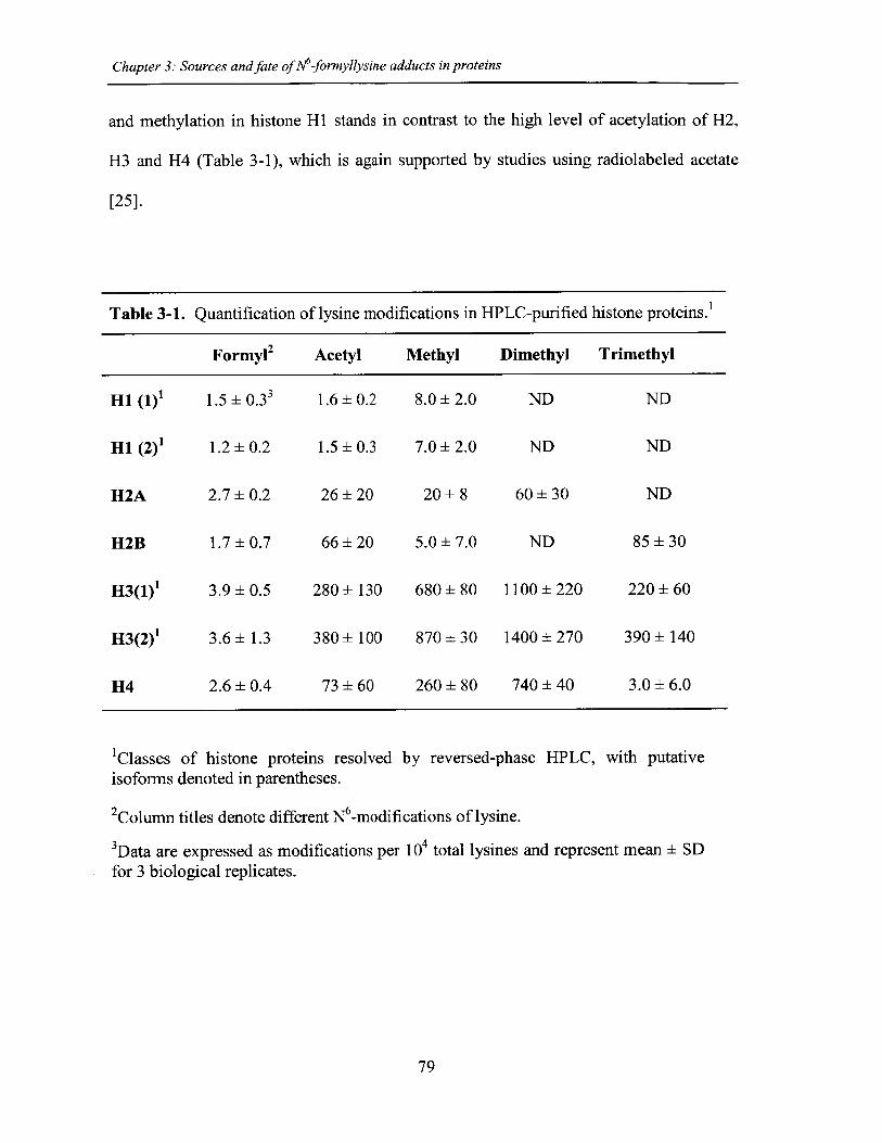
Chapter 3: Sources and fate of A16-formyllysine adducts in proteins
and methylation in histone Hi stands in contrast to the high level of acetylation of H2,
H3 and H4 (Table 3-1), which is again supported by studies using radiolabeled acetate
[25].
Table 3-1. Quantification of lysine modifications in HPLC-purified histone proteins.1
Formy12 Acetyl Methyl Dimethyl Trimethyl
H1 (1)' 1.5 0.33 1.6 ±0.2 8.0 ±2.0 ND ND
11 (2)1 1.2 0.2 1.5 ±0.3 7.0 ±2.0 ND ND
112A 2.7 0.2 26 ±20 20 8 60 ±30 ND
H2B 1.7 0.7 66 ±20 5.0 7.0 ND 85 30
H3(1)1 3.9 0.5 280 ±130 680 80 1100 ±220 220 60
H3(2)1 3.6 1.3 380 ±100 870 30 1400 ±270 390 140
H4 2.6 0.4 73 ±60 260 80 740 ±40 3.0 6.0
'Classes of histone proteins resolved by reversed-phase HPLC, with putativeisoforms denoted in parentheses.
2Column titles denote different N6-modifications of lysine.
3Data are expressed as modifications per 104 total lysines and represent mean ± SDfor 3 biological replicates.
79
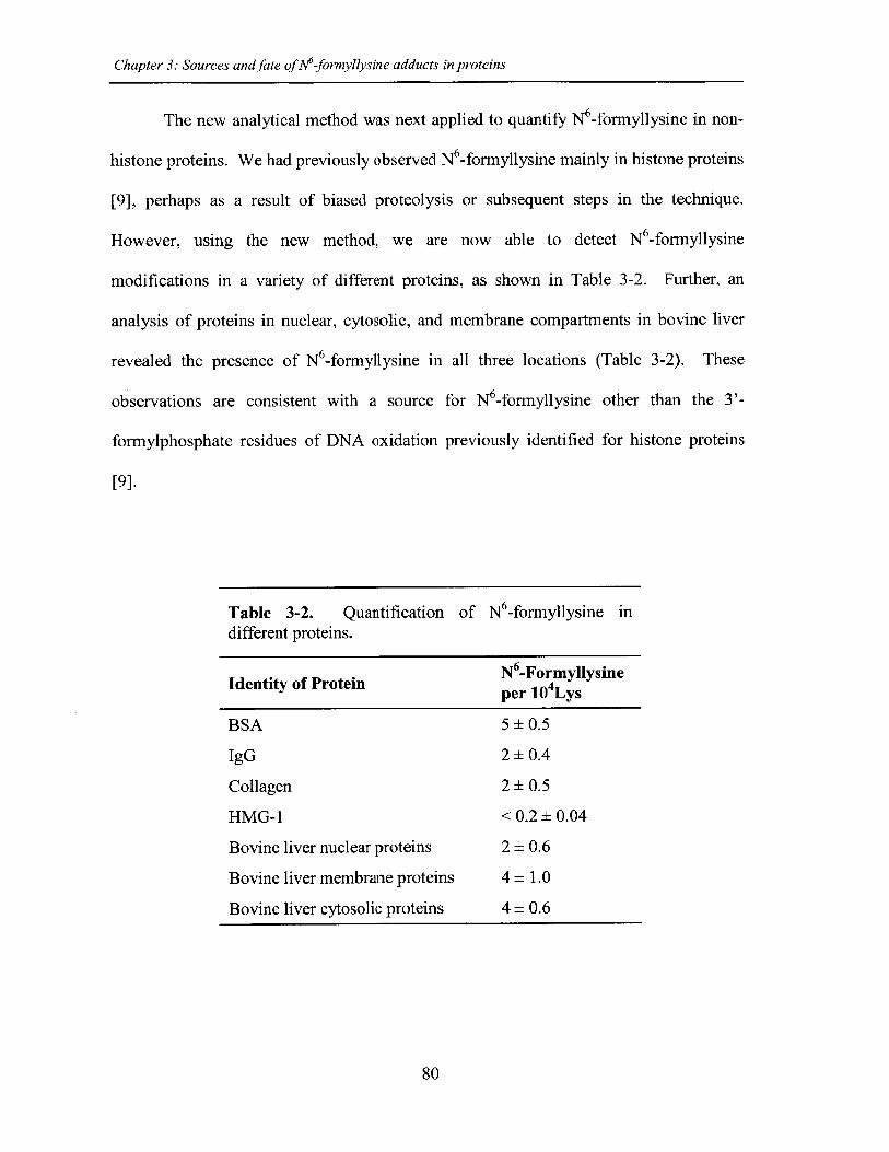
Chapter 3: Sources and fate ofN6-formyllysine adducts in proteins
The new analytical method was next applied to quantify N6-formyllysine in non-
histone proteins. We had previously observed N6 -formyllysine mainly in histone proteins
[9], perhaps as a result of biased proteolysis or subsequent steps in the technique.
However, using the new method, we are now able to detect N -formyllysine
modifications in a variety of different proteins, as shown in Table 3-2. Further, an
analysis of proteins in nuclear, cytosolic, and membrane compartments in bovine liver
revealed the presence of N6-formyllysine in all three locations (Table 3-2). These
observations are consistent with a source for N6-formyllysine other than the 3'-
formylphosphate residues of DNA oxidation previously identified for histone proteins
[9].
Table 3-2. Quantification of N6 -formyllysine indifferent proteins.
N 6-FormyllysineIdentity of Protein ery 4NLysper 10 Lys
BSA 5±0.5
IgG 2 ±0.4
Collagen 2 ±0.5
HMG-l <0.2± 0.04
Bovine liver nuclear proteins 2 ± 0.6
Bovine liver membrane proteins 4± 1.0
Bovine liver cytosolic proteins 4± 0.6
80
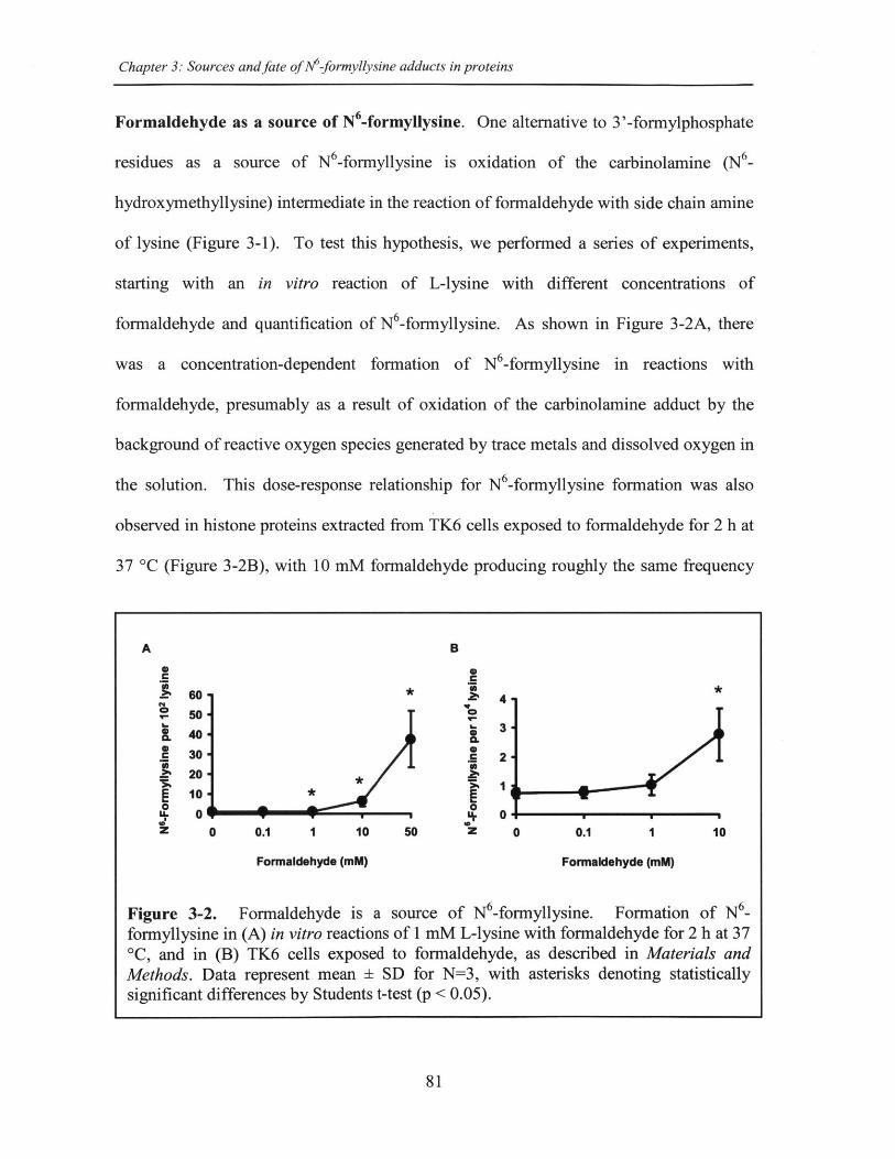
Chapter 3: Sources and fate of N6-formyllysine adducts in proteins
Formaldehyde as a source of N6-formyllysine. One alternative to 3'-formylphosphate
residues as a source of N6-formyllysine is oxidation of the carbinolamine (N6-
hydroxymethyllysine) intermediate in the reaction of formaldehyde with side chain amine
of lysine (Figure 3-1). To test this hypothesis, we performed a series of experiments,
starting with an in vitro reaction of L-lysine with different concentrations of
formaldehyde and quantification of N6-formyllysine. As shown in Figure 3-2A, there
was a concentration-dependent formation of N 6-formyllysine in reactions with
formaldehyde, presumably as a result of oxidation of the carbinolamine adduct by the
background of reactive oxygen species generated by trace metals and dissolved oxygen in
the solution. This dose-response relationship for N6-formyllysine formation was also
observed in histone proteins extracted from TK6 cells exposed to formaldehyde for 2 h at
37 'C (Figure 3-2B), with 10 mM formaldehyde producing roughly the same frequency
A B
C C
60 * 4.~4
50*340
c 30- 220-
10 *
. 0 0 W 02 0 0.1 1 10 50 z 0 0.1 1 10
Formaldehyde (mM) Formaldehyde (mM)
Figure 3-2. Formaldehyde is a source of N6-formyllysine. Formation of N -formyllysine in (A) in vitro reactions of 1 mM L-lysine with formaldehyde for 2 h at 370C, and in (B) TK6 cells exposed to formaldehyde, as described in Materials andMethods. Data represent mean ± SD for N=3, with asterisks denoting statisticallysignificant differences by Students t-test (p < 0.05).
81
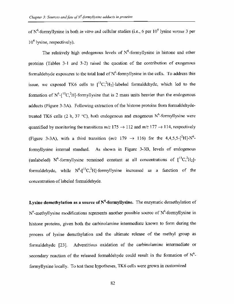
Chapter 3: Sources and fate of1 6-formyllysine adducts in proteins
of N6 -formyllysine in both in vitro and cellular studies (i.e., 6 per 104 lysine versus 3 per
104 lysine, respectively).
The relatively high endogenous levels of N6-formyllysine in histone and other
proteins (Tables 3-1 and 3-2) raised the question of the contribution of exogenous
formaldehyde exposures to the total load of N6-formyllysine in the cells. To address this
issue, we exposed TK6 cells to ["C,2H2 ]-labeled formaldehyde, which led to the
formation of N6_ 13C, 2H]-formyllysine that is 2 mass units heavier than the endogenous
adducts (Figure 3-3A). Following extraction of the histone proteins from formaldehyde-
treated TK6 cells (2 h, 37 C), both endogenous and exogenous N6 -formyllysine were
quantified by monitoring the transitions m/z 175 -> 112 and m/z 177 -> 114, respectively
(Figure 3-3A), with a third transition (m/z 179 -> 116) for the 4,4,5,5-[ 2H]-N 6_
formyllysine internal standard. As shown in Figure 3-3B, levels of endogenous
(unlabeled) N6-formyllysine remained constant at all concentrations of [ 3C,2H2]-
formaldehyde, while N6_[13C, 2H]-formyllysine increased as a function of the
concentration of labeled formaldehyde.
Lysine demethylation as a source of N -formyllysine. The enzymatic demethylation of
N 6-methyllysine modifications represents another possible source of N6-formyllysine in
histone proteins, given both the carbinolamine intermediate known to form during the
process of lysine demethylation and the ultimate release of the methyl group as
formaldehyde [23]. Adventitious oxidation of the carbinolamine intermediate or
secondary reaction of the released formaldehyde could result in the formation of N6_
formyllysine locally. To test these hypotheses, TK6 cells were grown in customized
82
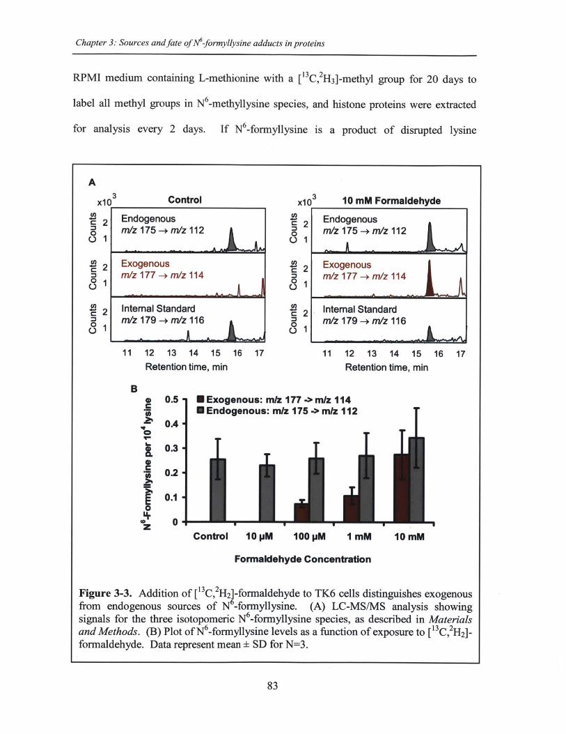
Chapter 3: Sources and fate of M-formyllysine adducts in proteins
RPMI medium containing L-methionine with a ["C,2H3]-methyl group for 20 days to
label all methyl groups in N6-methyllysine species, and histone proteins were extracted
for analysis every 2 days. If N6-formyllysine is a product of disrupted lysine
A3
Xl0
0
0
0
0
0
004.
0
2
1
2
I
2
1
Control 3x10
S2001
0C
0
2
1
2
1
10 mM Formaldehyde
11 12 13 14 15 16
Retention time, min
B
C
z
0.5-
OA -
0.3-
0.2-
0.1 -
0-Control
17 11 12 13 14 15 16Retention time, min
r
10 PM 100 PM 1 mM 10 mM
Formaldehyde Concentration
Figure 3-3. Addition of [13C,2H2 -formaldehyde to TK6 cells distinguishes exogenousfrom endogenous sources of N -formyllysine. (A) LC-MS/MS analysis showingsignals for the three isotopomeric N6-formyllysine species, as described in Materialsand Methods. (B) Plot of N6-formyllysine levels as a function of exposure to [1c,2H2]-formaldehyde. Data represent mean ± SD for N=3.
83
Endogenousm/z 175 -+ mz 112
Exogenousm/z 177 -+ m/z 114 - -
Intemal Standardmz 179 -+ m/z 116
Endogenousm/z 175 -+ m/z 112
Exogenousm/z 177 -+ m/z 114
Intemal Standardm/z 179 -+ m/z 116
17
U Exogenous: m/z 177 - m/z 114N Endogenous: m/z 175 -> mz 112
T
Chapter 3: Sources and fate of M-formyllysine adducts in proteins
demethylation in histones and is formed via oxidation of the carbinolamine intermediate
known to form during the process of lysine demethylation [23], or by reaction of lysine
with the formaldehyde released at the last step of successful lysine demethylation [23],
then one would expect to see an increase of 2 mass units corresponding to formation of
N6 [ 13C, 2H]-formyllysine (m/z 177 -+ 114 transition). In order to increase the signal-to-
noise ratio for N6 13C,2H]-formyllysine, N -formyllysine was HPLC-pre-purified in all
samples before LC-MS/MS analysis. Figure 3-4 depicts an example of the analysis using
the day 6 sample. As shown in Figure 3-4, N6-mono-methyllysine and N6-di-
methyllysine are predominately labeled (>90%) with heavy isotope methyl groups (i.e.,
[ 1C,2 H3]-methyl). In contrast to methyllysines, the level of N6-13C,2H]-formyllysine did
not increase beyond the natural isotope abundance level of -0.7% for the [M+2] ion of
N6-formyllysine (Figure 3-4C and Figure 3-S2). Note that the HPLC gradient was
changed here to fully resolve a contaminant signal from the TK6 cells (identified as the
[M+1] ion of citrulline) that otherwise co-eluted with N6-formyllysine and produced an
m/z value similar to the [M+2] isotopomer of N6-formyllysine.
84
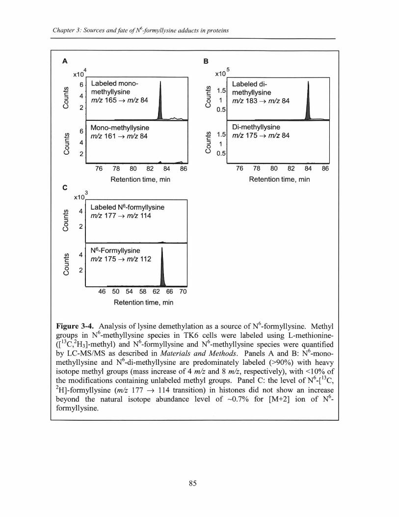
Chapter 3: Sources and fate ofNM-fornyllysine adducts in proteins
A B
Labeled mono-methyllysinem/z 165 -+ m/z 84
Mono-methyllysinem/z 161 -+ m/z 84
76 78 80 82 84 86
Retention time, min
3
46 50 54 58 62 66 70
X10 5
Cn00
U,
00
1.5
10.5
1.5.
0.5
76 78 80 82
Retention time,
Retention time, min
Figure 3-4. Analysis of lysine demethylation as a source of N6-formyllysine. Methylgroups in N6-methyllysine species in TK6 cells were labeled using L-methionine-([ 13C, 2H 3]-methyl) and N6-formyllysine and N6-methyllysine species were quantifiedby LC-MS/MS as described in Materials and Methods. Panels A and B: N -mono-methyllysine and N6-di-methyllysine are predominately labeled (>90%) with heavyisotope methyl groups (mass increase of 4 m/z and 8 m/z, respectively), with <10% ofthe modifications containing unlabeled methyl groups. Panel C: the level of N6[13C,2H]-formyllysine (m/z 177 -* 114 transition) in histones did not show an increasebeyond the natural isotope abundance level of -0.7% for [M+2] ion of N6_formyllysine.
85
4x10
6
4
2
6
4
2
U,
0
0
0
Labeled di-methyllysinem/z 183 -+ m/z 84
Di-methyllysinem/z 175 -> m/z 84
Cx10
84 86
min
U,W
00
0
0
4
2
4
2
Labeled N6-formyllysinem/z 177 - m/z 114
N6-Formyllysinem/z 175 - m/z 112
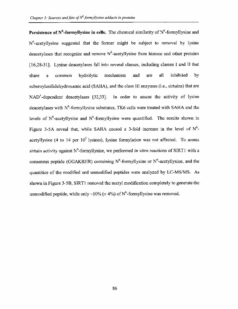
Chapter 3: Sources and fate of N6-formyllysine adducts in proteins
Persistence of N6 -formyllysine in cells. The chemical similarity of N -formyllysine and
N 6-acetyllysine suggested that the former might be subject to removal by lysine
deacetylases that recognize and remove N6-acetyllysine from histone and other proteins
[16,28-31]. Lysine deacetylases fall into several classes, including classes I and II that
share a common hydrolytic mechanism and are all inhibited by
suberoylanilidehydroxamic acid (SAHA), and the class III enzymes (i.e., sirtuins) that are
NAD*-dependent deacetylases [32,33]. In order to assess the activity of lysine
deacetylases with N -formyllysine substrates, TK6 cells were treated with SAHA and the
levels of N6-acetyllysine and N6-formyllysine were quantified. The results shown in
Figure 3-5A reveal that, while SAHA caused a 3-fold increase in the level of N6_
acetyllysine (4 to 14 per 103 lysines), lysine formylation was not affected. To assess
sirtuin activity against N6-formyllysine, we performed in vitro reactions of SIRTi with a
consensus peptide (GGAKRHR) containing N6-formyllysine or N -acetyllysine, and the
quantities of the modified and unmodified peptides were analyzed by LC-MS/MS. As
shown in Figure 3-5B, SIRT1 removed the acetyl modification completely to generate the
unmodified peptide, while only -10% (± 4%) of N6-formyllysine was removed.
86
Chapter 3: Sources and fate of N6-formyllysine adducts in proteins
A a
C
0.8-
C 0.6-aC
0.4-
0.2-
z 0-
O FK *
OAK.
-H
C C SAHA SAHA
- SIRTI
Acetylated Peptidem/z 823.4 -+ m/z 530.4
Unmodified Peptidem/z 781.1 -+ m/z 625.2
0 2 3 4
- SIRTI
0 1 2 3Retention time, min
4
x105-
3-
5-
3-
1-
x102
1.2 -0.8 -
0.4 -
1.2 -
0.8 -
0.4 -
+ SIRTI
Acetylated Peptidem/z 823.4 -+ m/z 530.4
Unmodified Peptidem/z 781.1 -+ m/z 625.2
0 2 3 4
+ SIRTI
0 1 2 3Retention time, min
4
Figure 3-5. Effect of lysine deacetylases on N -formyllysine. (A) TK6 cells weretreated with the class I and class II histone deacetylase inhibitor, SAHA, as describedin Materials and Methods. Data represent mean ± SD for N=3, with asterisks denotingstatistically significant differences by Students t-test (p < 0.05). (B) Treatment of apeptide substrate containing N6-formyllysine with the class III histone deacetylase,SIRT1.
87
18 a16 0141210 5"8 *6 *42 -
0
B
x105.
3.
5.
3.
1-
2x10
1.2 .0.8 -
0.4 .
1.2 .
0.8 .0.4
0
0
0
0
m/z 809.4 -+ m/z 516.3
Unmodified Peptidem/z 781.1 -+ m/z 625.2
Formylated Peptidem/z 809.4 -+ m/z 516.3
Unmodified Peptidem/z 781.1 -+ m/z 625.2
I
Chapter 3: Sources and fate of M-formyllysine adducts in proteins
Discussion
N -Formyllysine was first reported in 1985 in reactions of lysine with formaldehyde in
vitro [34] and, more recently, it was shown to form during in vitro silver-staining
procedures that involve the use of formaldehyde [35]. The recent discovery of N6_
formyllysine as a relatively abundant endogenous posttranslational modification of
histones and other nuclear proteins in cells [9-11] has raised questions about its
mechanism of formation and its potential for interfering with the regulatory function of
lysine N -acetylation. With respect to formation, we previously presented evidence that
N 6-formyllysine in histones could arise from reactions with 3'-formylphosphate residues
derived from DNA oxidation [9]. However, formaldehyde emerged as an alternative
source given the high reactivity of formaldehyde toward primary amines, such as the side
chain of lysine, and the potential for endogenous oxidation to convert a formaldehyde-
derived carbinolamine to a stable formamide (Figure 3-1). We have now demonstrated in
vitro and in cells that formaldehyde exposure leads to formation of N6 -formyllysine
residues in proteins. The fact that this modification arises in proteins other than
chromatin proteins and in cellular compartments other than the nucleus (Table 3-2)
suggests that 3'-formylphosphate residues in oxidized DNA do not account for all N6_
formyllysine adducts. This is consistent with recent studies in which N6-formyllysine
was detected in a protein oxidized with peroxynitrite in vitro [5]. The absence of
detectable N6-formyllysine arising from demethylation of N6-methyllysine species
(Figure 3-4C and Figure 3-S2) suggests that interruption of histone demethylation
reactions to form the carbinolamine precursor of N6-formyllysine occurs at low
frequency, or that the formaldehyde produced by complete lysine demethylation [23]
88
Chapter 3: Sources and fate of N6-formyllysine adducts in proteins
does not occur at concentrations high enough to drive formylation of lysine or cause
substantial changes in N6-formyllysine levels detected by our current analytical method.
Furthermore, there is the possibility that the formaldehyde released during lysine
demethylation may be scavenged before it could react with lysines in histones. A recent
study reports that lysine-specific demethylase 1 (LSD1) is a folate binding protein [36],
which led the authors to hypothesize that the biological function of folate is to trap the
formaldehyde that is generated during lysine demethylation [36]. In addition, the
observation that the formaldehyde equivalent derived from histone demethylation might
not account for a significant portion of formyllysine residues is not surprising in light of
the abundance of formaldehyde from other cellular processes [20,22,23]. This is clear
from the high steady-state concentrations of formaldehyde in human plasma (13-97 pM)
[20].
The relative abundance of N -formyllysine in histone and other proteins (Tables
3-1 and 3-2) [9-11] and the persistence of these adducts in histone proteins provides
insights into both their source and their potential effects on cell function. The N6_
formyllysine residues are relatively evenly distributed among different classes of histone
proteins (Table 3-1), while the other functional modifications show very biased
distributions over a large frequency range, which is consistent with the known function
and conserved locations for lysine methylation and acetylation [16-18]. This random
distribution of formyllysine adducts in histone proteins suggests that they are adventitious
and not physiological. The fact that N -formyllysine levels are similar in histone and
non-nuclear proteins and in all cell compartments also suggests that the sources of this
protein modification are equally balanced in the various compartments and proteins, or
89
Chapter 3: Sources and fate of A 6-formyllysine adducts in proteins
that there is a single dominant source that distributes uniformly throughout the cell. With
regard to their persistence in cells, the N 6-formyllysine adducts appear to be refractory to
removal by histone deacetylase enzymes, which suggests that they will persist throughout
the life of individual histone proteins. Figure 3-6 summarizes our findings presented here
on N6-formyllysine adducts.
The isotope labeling studies revealed that lysine demethylation is not a source for
N 6-formyllysine in histones. Furthermore, our data suggest that N6-formyllysine is
refractory to removal by histone deacetylases, which is consistent with the persistence of
this pathological adduct throughout the life of individual histone proteins. That the N6_
formyllysine adducts have been observed at many of the known conserved functional
locations for lysine acetylation and methylation in histones [10,11] suggests that N6_
formyllysine could interfere with signaling processes associated with physiological
histone modifications [16,28]. The association of N6-formyllysine with a variety of
different cell and organismal processes, including metabolism, and the oxidative and
nitrosative stresses of inflammation [5,9], suggest that this adduct may play a role in
many pathophysiological processes in humans.
90
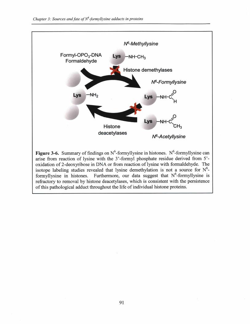
Chapter 3: Sources and fate of N6-formyllysine adducts in proteins
Ne-Methyllysine
Formyl-OP0 3-DNA NH-CH3Formaldehyde
Histone demethylases
N-Formyllysine
0
H
NH-CHistone H3
deacetylases NAcetllysine
Figure 3-6. Summary of findings on N6-formyllysine in histones. N6-formyllysine canarise from reaction of lysine with the 3'-formyl phosphate residue derived from 5'-oxidation of 2-deoxyribose in DNA or from reaction of lysine with formaldehyde. Theisotope labeling studies revealed that lysine demethylation is not a source for N6_formyllysine in histones. Furthermore, our data suggest that N6-formyllysine isrefractory to removal by histone deacetylases, which is consistent with the persistenceof this pathological adduct throughout the life of individual histone proteins.
91
Chapter 3: Sources and fate of N6-formyllysine adducts in proteins
References
1. Levine RL (2002) Carbonyl modified proteins in cellular regulation, aging, anddisease. Free Radic Biol Med 32: 790-796.
2. Jacobs AT, Marnett LJ (2010) Systems analysis of protein modification andcellular responses induced by electrophile stress. Acc Chem Res 43: 673-683.
3. Thomalley PJ (2008) Protein and nucleotide damage by glyoxal andmethylglyoxal in physiological systems--role in ageing and disease. Drug MetabolDrug Interact 23: 125-150.
4. Dedon PC (2008) The chemical toxicology of 2-deoxyribose oxidation in DNA.Chem Res Toxicol 21: 206-219.
5. Vana L, Kanaan NM, Hakala K, Weintraub ST, Binder LI (2011) Peroxynitrite-induced nitrative and oxidative modifications alter tau filament formation.Biochemistry 50: 1203-1212.
6. Codreanu SG, Zhang B, Sobecki SM, Billheimer DD, Liebler DC (2009) Globalanalysis of protein damage by the lipid electrophile 4-hydroxy-2-nonenal. MolCell Proteomics 8: 670-680.
7. Tallman KA, Kim HY, Ji JX, Szapacs ME, Yin H, et al. (2007) Phospholipid-protein adducts of lipid peroxidation: synthesis and study of new biotinylatedphosphatidylcholines. Chem Res Toxicol 20: 227-234.
8. Prasad A, Bekker P, Tsimikas S (2012) Advanced Glycation Endproducts andDiabetic Cardiovascular Disease. Cardiol Rev.
9. Jiang T, Zhou X, Taghizadeh K, Dong M, Dedon PC (2007) N-formylation oflysine in histone proteins as a secondary modification arising from oxidativeDNA damage. Proc Natl Acad Sci U S A 104: 60-65.
10. Wisniewski JR, Zougman A, Mann M (2008) Nepsilon-formylation of lysine is awidespread post-translational modification of nuclear proteins occurring atresidues involved in regulation of chromatin function. Nucleic Acids Res 36: 570-577.
11. LeRoy G, Weston JT, Zee BM, Young NL, Plazas-Mayorca MD, et al. (2009)Heterochromatin protein 1 is extensively decorated with histone code-like post-translational modifications. Mol Cell Proteomics 8: 2432-2442.
12. Pesavento JJ, Kim YB, Taylor GK, Kelleher NL (2004) Shotgun annotation ofhistone modifications: a new approach for streamlined characterization of proteinsby top down mass spectrometry. J Am Chem Soc 126: 3386-3387.
92
Chapter 3: Sources and fate of M-formyllysine adducts in proteins
13. Felsenfeld G, Groudine M (2003) Controlling the double helix. Nature 421: 448-453.
14. Chi P, Allis CD, Wang GG (2010) Covalent histone modifications--miswritten,misinterpreted and mis-erased in human cancers. Nat Rev Cancer 10: 457-469.
15. Rando OJ (2012) Combinatorial complexity in chromatin structure and function:revisiting the histone code. Curr Opin Genet Dev.
16. Kouzarides T (2007) Chromatin modifications and their function. Cell 128: 693-705.
17. Siuti N, Roth MJ, Mizzen CA, Kelleher NL, Pesavento JJ (2006) Gene-specificcharacterization of human histone H2B by electron capture dissociation. JProteome Res 5: 233-239.
18. Garcia BA, Barber CM, Hake SB, Ptak C, Turner FB, et al. (2005) Modificationsof human histone H3 variants during mitosis. Biochemistry 44: 13202-13213.
19. Lu K, Moeller B, Doyle-Eisele M, McDonald J, Swenberg JA (2011) Moleculardosimetry of N2-hydroxymethyl-dG DNA adducts in rats exposed toformaldehyde. Chem Res Toxicol 24: 159-161.
20. Zhang L, Freeman LE, Nakamura J, Hecht SS, Vandenberg JJ, et al. (2010)Formaldehyde and leukemia: epidemiology, potential mechanisms, andimplications for risk assessment. Environ Mol Mutagen 51: 181-191.
21. Le Curieux F, Pluskota D, Munter T, Sjoholm R, Kronberg L (2000)Identification of fluorescent 2'-deoxyadenosine adducts formed in reactions ofconjugates of malonaldehyde and acetaldehyde, and of malonaldehyde andformaldehyde. Chem Res Toxicol 13: 1228-1234.
22. Begley TJ, Samson LD (2003) AlkB mystery solved: oxidative demethylation ofNi-methyladenine and N3-methylcytosine adducts by a direct reversalmechanism. Trends Biochem Sci 28: 2-5.
23. Shi Y, Whetstine JR (2007) Dynamic regulation of histone lysine methylation bydemethylases. Mol Cell 25: 1-14.
24. Boyne MT, 2nd, Pesavento JJ, Mizzen CA, Kelleher NL (2006) Precisecharacterization of human histones in the H2A gene family by top down massspectrometry. J Proteome Res 5: 248-253.
25. Pasqualini JR, Mercat P, Giambiagi N (1989) Histone acetylation decreased byestradiol in the MCF-7 human mammary cancer cell line. Breast Cancer Res Treat14: 101-105.
93
Chapter 3: Sources and fate of M-formyllysine adducts in proteins
26. Byvoet P, Barber M, Amidei K, Lowell N, Trudeau W (1986) Effect ofexogenous histone H5 on integration of histone HI in rat liver chromatin.Correlations with aberrant epsilon-N-methylation of histone HI. BiochimBiophys Acta 867: 163-175.
27. Wu M, Allis CD, Richman R, Cook RG, Gorovsky MA (1986) An interveningsequence in an unusual histone HI gene of Tetrahymena thermophila. Proc NatlAcad Sci U S A 83: 8674-8678.
28. Strahl BD, Allis CD (2000) The language of covalent histone modifications.Nature 403: 41-45.
29. de Ruijter AJ, van Gennip AH, Caron HN, Kemp S, van Kuilenburg AB (2003)Histone deacetylases (HDACs): characterization of the classical HDAC family.Biochem J 370: 737-749.
30. Hildmann C, Riester D, Schwienhorst A (2007) Histone deacetylases--animportant class of cellular regulators with a variety of functions. Appl MicrobiolBiotechnol 75: 487-497.
31. Hake SB, Xiao A, Allis CD (2004) Linking the epigenetic 'language' of covalenthistone modifications to cancer. Br J Cancer 90: 761-769.
32. Chen L (2011) Medicinal chemistry of sirtuin inhibitors. Curr Med Chem 18:1936-1946.
33. Dokmanovic M, Clarke C, Marks PA (2007) Histone deacetylase inhibitors:overview and perspectives. Mol Cancer Res 5: 981-989.
34. Tyihak E, Trezl L, Kolonits P (1985) The isolation of Nepsilon-formyl-L-lysinefrom the reaction between formaldehyde and L-lysine and its identification byOPLC and NMR spectroscopy. J Pharm Biomed Anal 3: 343-349.
35. Oses-Prieto JA, Zhang X, Burlingame AL (2007) Formation of epsilon-formyllysine on silver-stained proteins: implications for assignment of isobaricdimethylation sites by tandem mass spectrometry. Mol Cell Proteomics 6: 181-192.
36. Luka Z, Moss F, Loukachevitch LV, Bornhop DJ, Wagner C (2011) Histonedemethylase LSD1 is a folate-binding protein. Biochemistry 50: 4750-4756.
94
Chapter 3: Sources and fate ofN6-formyllysine adducts in proteins
Supplementary Figures
95
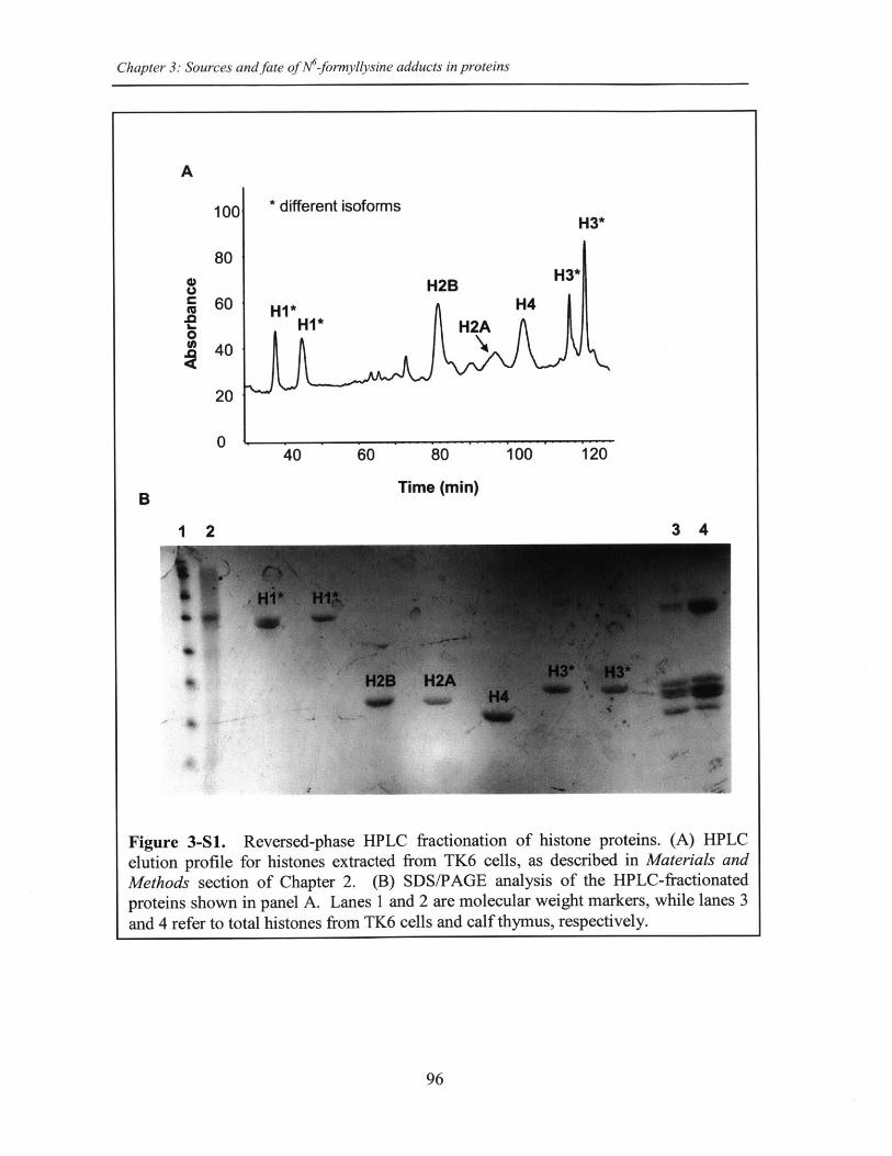
Chapter 3: Sources and fate of M-formyllysine adducts in proteins
A
0||
(U.0
10C
80
60
40
20
0
* different isoforms
H1*HI*
H3*
H2BH4
40 60 80 100 120
Time (min)B
1 2 3 4
Figure 3-S1. Reversed-phase HPLC fractionation of histone proteins. (A) HPLCelution profile for histones extracted from TK6 cells, as described in Materials andMethods section of Chapter 2. (B) SDS/PAGE analysis of the HPLC-fractionatedproteins shown in panel A. Lanes 1 and 2 are molecular weight markers, while lanes 3and 4 refer to total histones from TK6 cells and calf thymus, respectively.
96
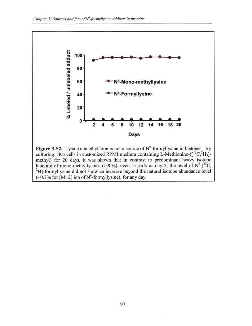
Chapter 3: Sources and fate of M-formyllysine adducts in proteins
97
t5
Z 100
-o 80.2 80
60 - N-Mono-methyllysineC
" 40 *N-Formyllysine
S20-1
2 4 6 8 10 12 14 16 18 20
Days
Figure 3-S2. Lysine demethylation is not a source of N -formyllysine in histones. Byculturing TK6 cells in customized RPMI medium containing L-Methionine-([ C,2H3]-methyl) for 20 days, it was shown that in contrast to predominant heavy isotopelabeling of mono-methyllysines (>90%), even as early as day 2, the level of N6-[13C,2H]-formyllysine did not show an increase beyond the natural isotope abundance level(-0.7% for [M+2] ion of N6-formyllysine), for any day.
Chapter 4: Formaldehyde-induced formation of N6-formyllysine protein adducts in rats
Chapter 4
Formaldehyde-induced formation ofN -formyllysine protein adducts in rats
98
Chapter 4: Formaldehyde-induced formation ofN6-formyllysine protein adducts in rats
Abstract
The emergence of lysine N6-formylation as an abundant endogenous modification ofproteins has raised questions about its mechanism of formation. In addition to DNAoxidation pathway previously reported (Jiang et al,. PNAS 104: 60-5, 2007), wedemonstrated in vitro and in cells that formaldehyde exposure leads to formation of N -formyllysine residues in histone proteins (Chapter 3). Here, we extend our studies toquantify N6-formyllysine protein adducts in rats exposed to isotope-labeled formaldehydeby inhalation in order to calculate the contribution of exogenous versus endogenoussources of formaldehyde to the total load of N6-formyllysine in cells. Moreover, ourstudies complement previous studies of formaldehyde-induced N2-hydroxymethyl-dGDNA adducts in exposed rats (Lu et al., Chem. Res. Toxicol. 24: 159-61, 2011), where weaim to correlate the protein and DNA adducts given the carcinogenic potency offormaldehyde. Using our ultrasensitive and specific liquid chromatography-coupledtandem mass spectrometry method, we quantified endogenous and exogenous N 6_
fromyllysine protein adducts in total and fractionated proteins (cytoplasmic, membrane,and nuclear) extracted from nasal epithelium, lung, and liver tissues of rats exposed tosingle-dose, nose-only inhalation of 0.7, 2, 6, and 10 ppm [13C 2 H2 ]-formaldehyde for 6 h.Similar to the DNA study, exogenous N -formyllysine adducts were only detectable innasal epithelium and not in distant tissues, with a dose-dependent formation in total aswell as in fractionated proteins. At 10 ppm exposure, nearly one-third of the total load ofN 6-formyllysine was derived from exogenous formaldehyde (0.9 ± 0.1 exogenous versus2 ± 0.1 endogenous N 6-formyllysine adducts per 104 lysines in total proteins). Moreover,there was a general decrease in exogenous adducts observed in nuclear proteins comparedto proteins obtained from other compartments, consistent with expected decrease ofinhaled formaldehyde concentration as it reacts with other biomolecules on its way tonucleus. In histone proteins, the exogenous to endogenous adduct ratios for N6_formyllysine are observed to occur about one third of ratios observed for DNA adduct,although they follow a similar pattern of increase in response to formaldehyde dosage(e.g., an increase in formaldehyde dosage from 6 ppm to 10 ppm, resulted in -3-foldincrease in exogenous versus endogenous ratios for both protein and DNA adducts). Ourformaldehyde studies shed light on yet another potential pathway associated withformaldehyde toxicity and carcinogenicity, that is through epigenetic disruption ofhistone proteins in cells by permanently formylating conserved sites of importantphysiological modifications.
99
Chapter 4: Formaldehyde-induced formation of N-formyllysine protein adducts in rats
Introduction
Aberrant protein modifications due to oxidants and electrophiles generated during
physiological or pathophysiological processes can lead to altered cellular processes and
progression of disease[1-5]. N6-Formylation of lysines has recently emerged as an
abundant adventitious protein modification [6-9], raising questions about its mechanism
of formation and biological consequences in cells. In histone proteins, the chemical
similarities of N 6-formyllysine to N6-acetyllysine suggest a disruptive role for the former
in signaling by histone acetylation, and the occurrence at many conserved lysine
acetylation and methylation sites along histone proteins [7] suggests possible interference
with the regulatory roles of post-translational modifications of these proteins. Studies to
date point to formaldehyde exposure as a major source of this adduct (Chapter 3) [10,11]
in addition to oxidative and nitrosative stresses of inflammation [6,9].
Formaldehyde exposure from environmental and occupational sources[12-14] as
well as endogenous cellular processes [13,15,16] make it an abundant electrophile in the
human body, as indicated by its concentration range of 13 to 97 pM in plasma [13,17].
Formaldehyde, classified as a known human carcinogen according to IARC [18,19], is
reported to cause squamous cell carcinoma in rats [20,21] with sufficient epidemiological
evidence for causing nasopharyngeal cancer but limited evidence for leukemia in humans
[13,18,19,22].
As a reactive aldehyde, formaldehyde readily forms adducts with nucleophilic
sites in DNA and proteins such as N2-hydroxymethyl-dG DNA adducts [12] and Schiff
bases on the side chain amine of lysines in proteins [23], in addition to extensive DNA-
protein and DNA-DNA crosslinks [24]. Our recent studies revealed N6-formyllysine
100
Chapter 4: Formaldehyde-induced formation of N6-formyllysine protein adducts in rats
formation in histone proteins of TK6 cells from the reaction of formaldehyde with the c-
amino group of lysine (Chapter 3). Our data showed a dose-dependent formation of N -
formyllysine residues as a function of formaldehyde concentration, with use of [13C, 2H2]-
formaldehyde revealing unchanged levels of N6-formyllysinespecies due to endogenous
sources.
Here, we extend our cell studies to an in vivo assessment of formaldehyde-
induced lysine N6-formylation in rats exposed by inhalation, with the use of [13C,2H2]-
formaldehyde to differentiate endogenous from exogenous adducts. Using our
ultrasensitive and specific liquid chromatography-coupled tandem mass spectrometry
(LC-MS/MS) method, we quantified the extent to which exposure to [1C,2H2]-
formaldehyde by inhalation induces N [13C, 2 H]-formyllysine. Endogenous N 6[ 2C, IH]-
formyllysine and exposure-induced N6_[ 13C, 2H]-formyllysine adducts were quantified in
total and fractionated (cytoplasmic, membrane, and nuclear) proteins extracted from nasal
epithelium, lung, and liver tissues of rats exposed to single-dose, nose-only inhalation of
0.7, 2, 6, and 10 ppm [ 3C2H2 ]-formaldehyde for 6 h. These studies complement previous
studies of [13C,2 H2 ]-formaldehyde-induced N 2-hydroxymethyl-dG DNA adducts in rats
from the same formaldehyde exposure studies [12], which allows us to correlate the
protein and DNA adducts arising with the same formaldehyde dose.
Application of our sensitive analytical methods revealed that exposure to
[ 13C,2H2 ]-formaldehyde vapor leads to the formation of exogenous N 6[3C, 2H]-
formyllysine in proteins of nasal epithelium, and that the exogenous adducts are formed
in proteins extracted from all cellular compartments in a dose-dependent manner, with
endogenous adducts dominating at all exposure conditions. Similar to the DNA study,
101
Chapter 4: Formaldehyde-induced formation of M-formyllysine protein adducts in rats
the exogenous N6-formyllysine adducts were only detected in nasal epithelium and not in
distant tissues, with exogenous to endogenous adduct ratios in total proteins closely
matching increases seen in DNA adducts.
Materials and Methods
Materials. Rat tissues were provided by Prof. James Swenberg's Lab at University of
North Carolina at Chapel Hill [12]. Subcellular Protein Fractionation Kit was purchased
from Thermo Scientific (Waltham, MA). Lysine internal standard, 4,4,5,5,-[2H]-Lysine,
was purchased from Cambridge Isotope Laboratories (Andover, MA). N -Formyllysine
internal standard, 4,4,5,5-[2 H]-N 6-fonnyllysine, was synthesized from 4,4,5,5-[ 2H]-lysine
according to Jiang et al. [6]. Streptomyces griseus protease was purchased from Sigma-
Aldrich (St. Louis, MO).
Rat exposure to formaldehyde vapor. Rat exposure to [13C2H2 ]-formaldehyde was
performed at the Lovelace Respiratory Research Institute, Albuquerque, NM, by our
collaborator, Prof. James Swenberg, according to appropriate and approved protocols for
the use of vertebrate animals in experiments [12]. Fischer rats (6 w old, male) were
exposed to formaldehyde vapor by single-dose, nose-only inhalation of [ 3C2H2]-
formaldehyde for 6 h to produce final target exposure concentrations of 0.7, 2, 6, and 10
ppm. Rats were euthanized using an intraperitoneal barbiturate injection. For nasal
mucosa collection, the skull was split with a slight bias to one side (to preserve septal
mucosa) and maxilloturbinates collected, wrapped in aluminum foil, immediately snap
102
Chapter 4: Formaldehyde-induced formation of A-formyllysine protein adducts in rats
frozen in liquid nitrogen and held at -80 0C pending analyses. Other tissue samples were
processed in a similar fashion.
Subcellular protein fractionation from tissue. For extracting proteins from tissues, ~10
mg of nasal epithelium, lung, or liver tissue was cut into small pieces and washed with
PBS. Tissues were homogenized using a Kontes all-glass Dounce homogenizer, with 10
strokes of a type B pestle, and proteins were separated into cytoplasmic, membrane,
soluble nuclear, and chromatin bound fractions using the Subcellular Protein
Fractionation Kit from Thermo Scientific. Total protein was obtained by centrifuging
tissue lysate, prior to fractionation, for 14000 x g for 15 min at 4 'C and collecting the
supernatant. Protein precipitation from each fraction was accomplished by adding 20%
v/v trichloroacetic acid and incubating overnight at 4 'C. Samples were centrifuged at
14000 x g for 10 min at 4 'C, washed once with ice-cold acetone containing 0.1% HCl,
and once with ice-cold acetone before being air-dried and stored at -20 0 C until use. N 6_
Formyllysine adducts were found to be stable under these conditions.
Enzymatic hydrolysis of proteins. Extracted proteins from tissues were dissolved in 50
p L of 100 mM ammonium bicarbonate buffer (pH 8.5), 4,4,5,5-[ 2H]-lysine (2 nmol), and
4,4,5,5-[2 H]-N 6-formyllysine (1 pmol) were added as internal standards, and the proteins
were hydrolyzed by addition of 2 p1 of freshly prepared S. griseus protease solution
(1Ig/pl) with incubation at 37'C for 16 h. Samples were dried under vacuum and
resuspended in 50 pL of water prior to mass spectrometry analysis.
103
Chapter 4: Formaldehyde-induced formation of M-formyllysine protein adducts in rats
Quantification of amino acids. N6-Formyllysine amino acids in the resuspended protein
hydrolysates were quantified as a percentage of lysine content by liquid chromatography-
coupled mass spectrometry (LC-MS/MS). Analytes in total, cytoplasmic, and membrane
protein hydrolysates were analyzed without a pre-purification step. Nuclear protein
hydrolysates were first HPLC pre-purified to enhance the signal for the M+2 ion of N6_
formyllysine. HPLC pre-purification was done using an Agilent 1100 series system, with
an aqueous normal-phase Cogent diamond hydride column (2.1 x 150mm, 4pm) from
MicroSolv Technology Corporation (Eatontown, NJ), with mobile phase flow rate of 400
pL/min and column temperature of 20 'C. The solvent system was 0.1% acetic acid in
water (A) and 0.1% acetic acid in acetonitrile (B), with the elution starting at 100% B, the
gradient linearly decreased to 25% B over 30 min and held at 25% B for 3 additional min
before the column was re-equilibrated with 100% B for 7 min. For N6-formyllysine
quantification, an extended chromatography was used, in order to separate the M+l ion
of citrulline from M+2 ion of N6-formyllysine (Figure 2-8). The same solvent system of
0.1% acetic acid in water (A) and 0.1% acetic acid in acetonitrile (B) was used, with the
elution starting at 100% B, the gradient linearly decreased to 75% B over 75 min,
reaching 25% B over the next 3 min and 15% B by 83 min. The column was then re-
equilibrated with 100% B for 7 min. N6-Formyllysine and lysine were then analyzed
using an Agilent 6410 triple quadrupole mass spectrometer (MS/MS) equipped with an
electrospray ionization (ESI) source operated in positive ion mode. The operating
parameters were as follows: ESI capillary voltage, 4000 V; gas temperature, 350 'C;
drying gas flow, 12 L/min; and nebulizer pressure, 30 psi, with selected reaction
monitoring (SRM) transitions of m/z 175-+112 and for N6-formyllysine (with internal
104
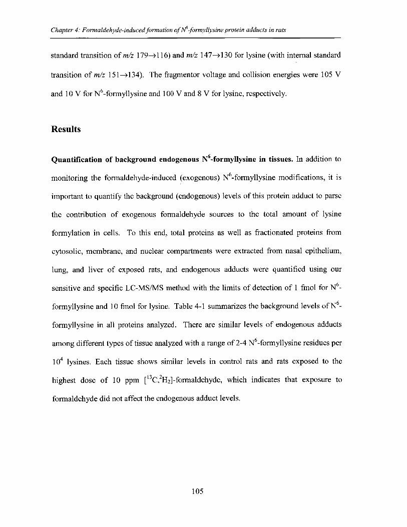
Chapter 4: Formaldehyde-induced formation of M-formyllysine protein adducts in rats
standard transition of m/z 179-> 116) and m/z 147-+130 for lysine (with internal standard
transition of m/z 151 ->134). The fragmentor voltage and collision energies were 105 V
and 10 V for N6-formyllysine and 100 V and 8 V for lysine, respectively.
Results
Quantification of background endogenous N6-formyllysine in tissues. In addition to
monitoring the formaldehyde-induced (exogenous) N -formyllysine modifications, it is
important to quantify the background (endogenous) levels of this protein adduct to parse
the contribution of exogenous formaldehyde sources to the total amount of lysine
formylation in cells. To this end, total proteins as well as fractionated proteins from
cytosolic, membrane, and nuclear compartments were extracted from nasal epithelium,
lung, and liver of exposed rats, and endogenous adducts were quantified using our
sensitive and specific LC-MS/MS method with the limits of detection of 1 fmol for N6_
formyllysine and 10 fmol for lysine. Table 4-1 summarizes the background levels of N6_
formyllysine in all proteins analyzed. There are similar levels of endogenous adducts
among different types of tissue analyzed with a range of 2-4 N 6-formyllysine residues per
104 lysines. Each tissue shows similar levels in control rats and rats exposed to the
highest dose of 10 ppm [13C, 2H2 ]-formaldehyde, which indicates that exposure to
formaldehyde did not affect the endogenous adduct levels.
105
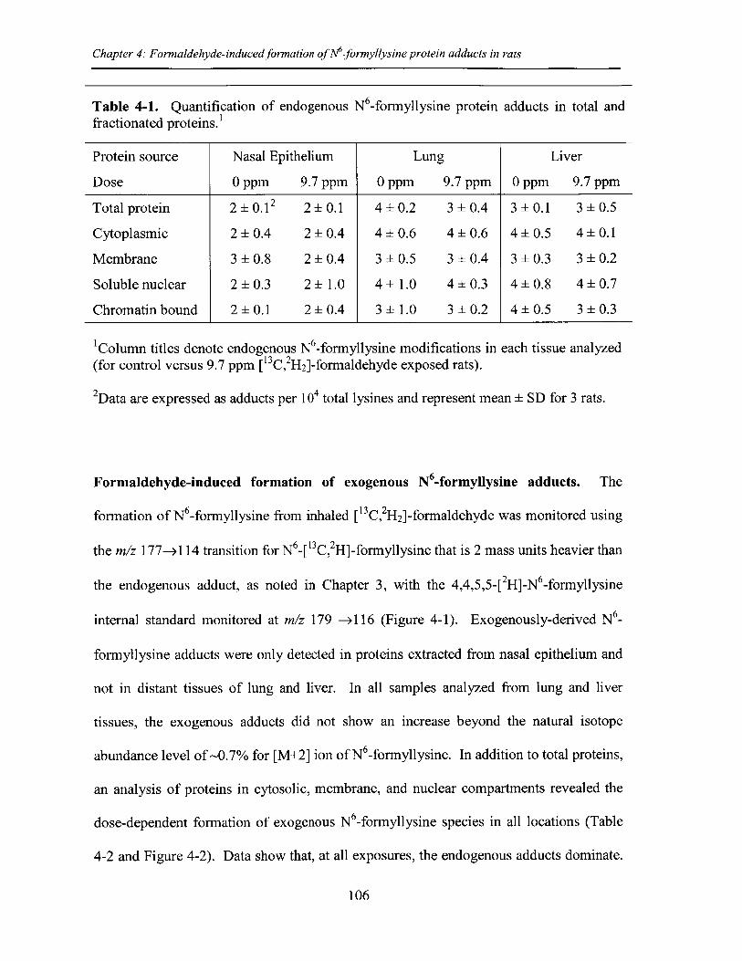
Chapter 4: Formaldehyde-induced formation ofNM-formyllysine protein adducts in rats
Table 4-1. Quantification of endogenous N6-formyllysine protein adducts in total andfractionated proteins.I
Protein source Nasal Epithelium Lung Liver
Dose 0 ppm 9.7 ppm 0 ppm 9.7 ppm 0 ppm 9.7 ppm
Total protein 2 ±0.12 2 ±0.1 4 ±0.2 3 ±0.4 3 ±0.1 3 ±0.5
Cytoplasmic 2 ±0.4 2 ±0.4 4 ±0.6 4 ±0.6 4 ±0.5 4 ±0.1
Membrane 3 ±0.8 2 ±0.4 3 ±0.5 3 ±0.4 3 ±0.3 3 ±0.2
Soluble nuclear 2 ±0.3 2 ±1.0 4 ±1.0 4 ±0.3 4 ±0.8 4 ±0.7
Chromatin bound 2 ±0.1 2 ±0.4 3 ±1.0 3 ±0.2 4 ±0.5 3 ±0.3
Column titles denote endogenous N6-formyllysine modifications in each tissue analyzed(for control versus 9.7 ppm [13C, 2H2]-formaldehyde exposed rats).
2Data are expressed as adducts per 104 total lysines and represent mean ± SD for 3 rats.
Formaldehyde-induced formation of exogenous N6-formyllysine adducts. The
formation of N6-formyllysine from inhaled ["C,2H2].-.formaldehyde was monitored using
the m/z 177-> 114 transition for N6- 13c 2H]-formyllysine that is 2 mass units heavier than
the endogenous adduct, as noted in Chapter 3, with the 4,4,5,5-[ 2H]-N 6-formyllysine
internal standard monitored at m/z 179 ->116 (Figure 4-1). Exogenously-derived N6_
formyllysine adducts were only detected in proteins extracted from nasal epithelium and
not in distant tissues of lung and liver. In all samples analyzed from lung and liver
tissues, the exogenous adducts did not show an increase beyond the natural isotope
abundance level of ~0.7% for [M+2] ion of N6-formyllysine. In addition to total proteins,
an analysis of proteins in cytosolic, membrane, and nuclear compartments revealed the
dose-dependent formation of exogenous N -formyllysine species in all locations (Table
4-2 and Figure 4-2). Data show that, at all exposures, the endogenous adducts dominate.
106
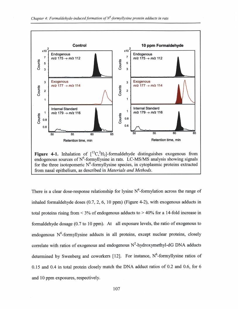
Chapter 4: Formaldehyde-induced formation of M-formyllysine protein adducts in rats
Control 10 ppm Formaldehydex102 x10 2
Endogenous Endogenous7 m/z 175 -rmz 112 7 m/z 175 -+ m/z 112
c5 5
03
50 55 60 65 ' 0 O o
Retention time, min Retention time, min
Figure 4-1. Inhalation of [IC,2H2]-formaldehyde distinguishes exogenous fromendogenous sources of N6-formyllysine in rats. LC-MS/MS analysis showing signalsfor the three isotopomeric N6-formyllysine species, in cytoplasmic proteins extractedfrom nasal epithelium, as described in Materials and Methods.
There is a clear dose-response relationship for lysine N6-formylation across the range of
inhaled formaldehyde doses (0.7, 2, 6, 10 ppm) (Figure 4-2), with exogenous adducts in
total proteins rising from < 3% of endogenous adducts to > 40% for a 14-fold increase in
formaldehyde dosage (0.7 to 10 ppm). At all exposure levels, the ratio of exogenous to
endogenous N6-formyllysine adducts in all proteins, except nuclear proteins, closely
correlate with ratios of exogenous and endogenous N2-hydroxymethyl-dG DNA adducts
determined by Swenberg and coworkers [12]. For instance, N6-formyllysine ratios of
0.15 and 0.4 in total protein closely match the DNA adduct ratios of 0.2 and 0.6, for 6
and 10 ppm exposures, respectively.
107
Exogenousm/z 177 -+ mlz 114
Internal Standardm/z 179 -+ m/z 116
4 2
0
0.3S0.8
3
0.
0.6
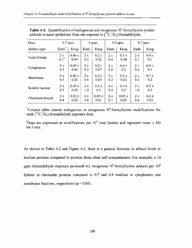
Chapter 4: Formaldehyde-induced formation ofN6-formyllysine protein adducts in rats
Table 4-2. Quantification of endogenous and exo enous N6-formyllysine proteinadducts in nasal epithelium from rats exposed to [I C, 2H2]-formaldehyde.
Dose 0.7 ppm 2 ppm 5.8 ppm 9.7 ppm
Adduct type Endo' Exog Endo Exog Endo Exog Endo Exog
Total Protein 2 0.06± 2± 0.2± 2± 0.3± 2± 0.9±0.12 0.04 0.2 0.02 0.4 0.04 0.1 0.1
. 2 0.05± 3± 0.2± 2± 0.4± 2± 0.8±Cytoplasmic 0.3 0.04 0.2 0.07 0.8 0.2 0.4 0.1
2 0.06± 2± 0.2± 3± 0.3± 2± 0.7±Membrane 0.3 0.02 0.8 0.03 0.2 0.02 0.4 0.2
Soluble nuclear 2 0.05± 2± 0.2± 4± 0.4± 2± 0.5±0.3 0.05 1.0 0.1 0.4 0.2 1.0 0.2
2 0.02± 2± 0.03 2± 0.05 2± 0.2±Chromatinbound 0.4 0.02 0.8 0.01 0.1 0.05 0.4 0.01
Column titles denote endogenous or exoeach [13C,2H 2]-formaldehyde exposure dose.
genous N6-formyllysine modifications for
2Data are expressed as modifications per 104 total lysines and represent mean ± SDfor 3 rats.
As shown in Table 4-2 and Figure 4-2, there is a general decrease in adduct levels in
nuclear proteins compared to proteins from other cell compartments. For example, a 10
ppm formaldehyde exposure produced 0.2 exogenous N6-formyllysine adducts per 104
lysines in chromatin proteins compared to 0.7 and 0.8 residues in cytoplasmic and
membrane fractions, respectively (p < 0.05).
108
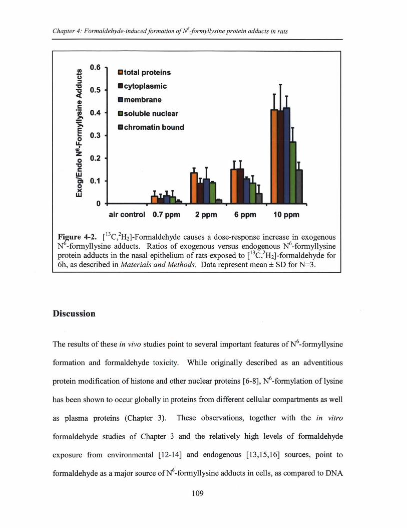
Chapter 4: Formaldehyde-inducedformation of N6-formyllysine protein adducts in rats
0.6 Etotal proteins
Z 0.5 Ecytoplasmic* U membraneC
0.4 Usoluble nuclear
*chromatin bound0.3
o0-U-
z.80.2
" 0.10
air control 0.7 ppm 2 ppm 6 ppm 10 ppm
Fi ure 4-2. [ 3C, 2H2]-Formaldehyde causes a dose-response increase in exogenousN -formyllysine adducts. Ratios of exogenous versus endogenous N -formyllysineprotein adducts in the nasal epithelium of rats exposed to [13 C,2H2 ]-formaldehyde for6h, as described in Materials and Methods. Data represent mean ± SD for N=3.
Discussion
The results of these in vivo studies point to several important features of N6-formyllysine
formation and formaldehyde toxicity. While originally described as an adventitious
protein modification of histone and other nuclear proteins [6-8], N6-formylation of lysine
has been shown to occur globally in proteins from different cellular compartments as well
as plasma proteins (Chapter 3). These observations, together with the in vitro
formaldehyde studies of Chapter 3 and the relatively high levels of formaldehyde
exposure from environmental [12-14] and endogenous [13,15,16] sources, point to
formaldehyde as a major source of N6-formyllysine adducts in cells, as compared to DNA
109
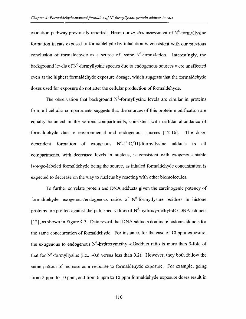
Chapter 4: Formaldehyde-induced formation of M-formyllysine protein adducts in rats
oxidation pathway previously reported. Here, our in vivo assessment of N6-formyllysine
formation in rats exposed to formaldehyde by inhalation is consistent with our previous
conclusion of formaldehyde as a source of lysine N6-formylation. Interestingly, the
background levels of N6-formyllysine species due to endogenous sources were unaffected
even at the highest formaldehyde exposure dosage, which suggests that the formaldehyde
doses used for exposure do not alter the cellular production of formaldehyde.
The observation that background N 6-formyllysine levels are similar in proteins
from all cellular compartments suggests that the sources of this protein modification are
equally balanced in the various compartments, consistent with cellular abundance of
formaldehyde due to environmental and endogenous sources [12-16]. The dose-
dependent formation of exogenous N6_[ 13C,2H]-formyllysine adducts in all
compartments, with decreased levels in nucleus, is consistent with exogenous stable
isotope-labeled formaldehyde being the source, as inhaled formaldehyde concentration is
expected to decrease on the way to nucleus by reacting with other biomolecules.
To further correlate protein and DNA adducts given the carcinogenic potency of
formaldehyde, exogenous/endogenous ratios of N 6-formyllysine residues in histone
proteins are plotted against the published values of N2-hydroxymethyl-dG DNA adducts
[12], as shown in Figure 4-3. Data reveal that DNA adducts dominate histone adducts for
the same concentration of formaldehyde. For instance, for the case of 10 ppm exposure,
the exogenous to endogenous N2-hydroxymethyl-dGadduct ratio is more than 3-fold of
that for N6-formyllysine (i.e., ~0.6 versus less than 0.2). However, they both follow the
same pattern of increase as a response to formaldehyde exposure. For example, going
from 2 ppm to 10 ppm, and from 6 ppm to 10 ppm formaldehyde exposure doses result in
110
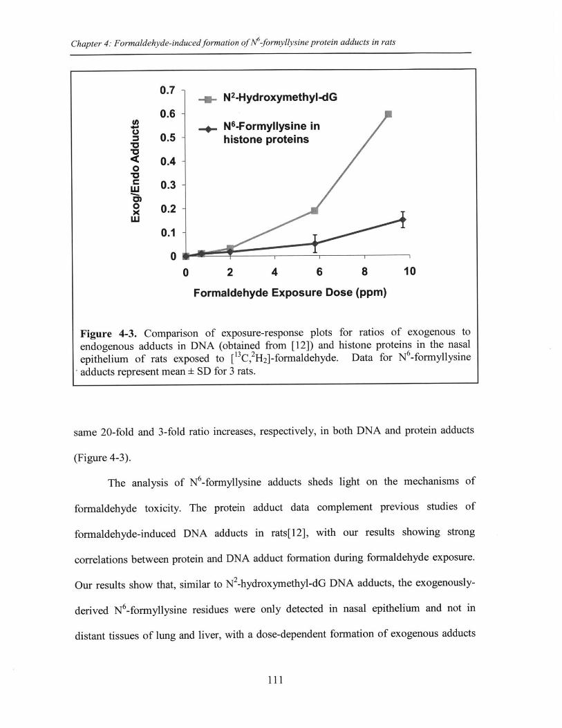
Chapter 4: Formaldehyde-induced formation of N-formyllysine protein adducts in rats
0.7-0 - N2-Hydroxymethyl-dG
0.6 -.. N6-Formyllysine in
0.5 - histone proteins
0.4 -0M0 0.3 -
uJ0 0.2
0.1 -
0 M -
0 2 4 6 8 10
Formaldehyde Exposure Dose (ppm)
Figure 4-3. Comparison of exposure-response plots for ratios of exogenous toendogenous adducts in DNA (obtained from [12]) and histone proteins in the nasalepithelium of rats exposed to [13C,2H2 ]-formaldehyde. Data for N6-formyllysineadducts represent mean ± SD for 3 rats.
same 20-fold and 3-fold ratio increases, respectively, in both DNA and protein adducts
(Figure 4-3).
The analysis of N6-formyllysine adducts sheds light on the mechanisms of
formaldehyde toxicity. The protein adduct data complement previous studies of
formaldehyde-induced DNA adducts in rats[12], with our results showing strong
correlations between protein and DNA adduct formation during formaldehyde exposure.
Our results show that, similar to N2-hydroxymethyl-dG DNA adducts, the exogenously-
derived N6-formyllysine residues were only detected in nasal epithelium and not in
distant tissues of lung and liver, with a dose-dependent formation of exogenous adducts
111
Chapter 4: Formaldehyde-induced formation of N6-formyllysine protein adducts in rats
in total proteins as well as proteins from specific cellular compartments (Figure 4-2 and
Table 4-2). With the exception of nuclear proteins, the levels of N6-formyllysine
formation in proteins closely correlates with N2-hydroxymethyl-dG adduct formation in
DNA. However, as shown in Figure 4-3,the proportion of DNA adducts derived from
exogenous formaldehyde was significantly higher than that for the histone adducts for the
same concentration of formaldehyde, even though theoretically the same number of
exogenous formaldehyde molecules should reach DNA and histones. This difference in
ratios could be due to several factors such as DNA guanine content compared to histone
lysine content, faster kinetics of N2-hydroxymethyl-dGformation versus N -formyllysine
formation, as well as possible greater solvent exposure of guanine N2-positions compared
to lysine amino side chains in histone proteins. Nonetheless, both N2-hydroxymethyl-dG
and N 6-formyllysine adducts follow the same pattern of increase as a response to
formaldehyde dosage (Figure 4-3).
There have been extensive studies to date on possible mechanisms for
formaldehyde toxicity and carcinogenicity. For instance, some studies found a non-linear
dose-dependent formation of DNA damage products in rats and non-human primates
exposed to formaldehyde inhalation [12,17,25,26] with other studies showing long term
formaldehyde exposure of higher than 6 ppm dosage resulting in a substantial increases
in squamous cell carcinomas in rats [20,21,27]. On the path to understanding
formaldehyde's biological consequences, our results shed light on yet another pathway,
that is through formation of N6-formyllysine adducts in histones. N6-Formyllysine
adducts have been mapped on many conserved lysine acetylation and methylation sites
along histones [7,8]. This observation along with chemical similarity of lysine N6_
112
Chapter 4: Formaldehyde-induced form ation of M-formyllysine protein adducts in rats
formylation to lysine N -acetylation, as well as our results showing N6-formyllysine is
refractory to removal by histone deacetylases (Chapter 3) suggests that N6 -formyllysine
could interfere with the epigenetic roles associated with physiological histone
modifications [28,29]. In other words, our studies suggest that prolonged exposure to
formaldehyde from environmental and occupational sources can drive substantial and
permanent formylation of conserved sites of important physiological modifications in
histone proteins, thus interfering with their regulatory roles and further contributing to
toxicity and carcinogenicity associated with formaldehyde exposure.
113
Chapter 4: Formaldehyde-induced formation of N6-formyllysine protein adducts in rats
References
1. Marnett LJ, Riggins JN, West JD (2003) Endogenous generation of reactiveoxidants and electrophiles and their reactions with DNA and protein. J Clin Invest111: 583-593.
2. Levine RL (2002) Carbonyl modified proteins in cellular regulation, aging, anddisease. Free Radic Biol Med 32: 790-796.
3. Stadtman ER, Berlett BS (1997) Reactive oxygen-mediated protein oxidation inaging and disease. Chem Res Toxicol 10: 485-494.
4. Jacobs AT, Marnett LJ (2010) Systems analysis of protein modification andcellular responses induced by electrophile stress. Acc Chem Res 43: 673-683.
5. Dedon PC (2008) The chemical toxicology of 2-deoxyribose oxidation in DNA.Chem Res Toxicol 21: 206-219.
6. Jiang T, Zhou X, Taghizadeh K, Dong M, Dedon PC (2007) N-formylation oflysine in histone proteins as a secondary modification arising from oxidativeDNA damage. Proc Natl Acad Sci U S A 104: 60-65.
7. Wisniewski JR, Zougman A, Mann M (2008) Nepsilon-formylation of lysine is awidespread post-translational modification of nuclear proteins occurring atresidues involved in regulation of chromatin function. Nucleic Acids Res 36: 570-577.
8. LeRoy G, Weston JT, Zee BM, Young NL, Plazas-Mayorca MD, et al. (2009)Heterochromatin protein 1 is extensively decorated with histone code-like post-translational modifications. Mol Cell Proteomics 8: 2432-2442.
9. Vana L, Kanaan NM, Hakala K, Weintraub ST, Binder LI (2011) Peroxynitrite-induced nitrative and oxidative modifications alter tau filament formation.Biochemistry 50: 1203-1212.
10. Tyihak E, Trezl L, Kolonits P (1985) The isolation of Nepsilon-formyl-L-lysinefrom the reaction between formaldehyde and L-lysine and its identification byOPLC and NMR spectroscopy. J Pharm Biomed Anal 3: 343-349.
11. Oses-Prieto JA, Zhang X, Burlingame AL (2007) Formation of epsilon-formyllysine on silver-stained proteins: implications for assignment of isobaricdimethylation sites by tandem mass spectrometry. Mol Cell Proteomics 6: 181-192.
114
Chapter 4: Formaldehyde-induced formation of N-formyllysine protein adducts in rats
12. Lu K, Moeller B, Doyle-Eisele M, McDonald J, Swenberg JA (2011) Moleculardosimetry of N2-hydroxymethyl-dG DNA adducts in rats exposed toformaldehyde. Chem Res Toxicol 24: 159-161.
13. Zhang L, Freeman LE, Nakamura J, Hecht SS, Vandenberg JJ, et al. (2010)Formaldehyde and leukemia: epidemiology, potential mechanisms, andimplications for risk assessment. Environ Mol Mutagen 51: 181-191.
14. Le Curieux F, Pluskota D, Munter T, Sjoholm R, Kronberg L (2000)Identification of fluorescent 2'-deoxyadenosine adducts formed in reactions ofconjugates of malonaldehyde and acetaldehyde, and of malonaldehyde andformaldehyde. Chem Res Toxicol 13: 1228-1234.
15. Begley TJ, Samson LD (2003) AlkB mystery solved: oxidative demethylation ofNi-methyladenine and N3-methylcytosine adducts by a direct reversalmechanism. Trends Biochem Sci 28: 2-5.
16. Shi Y, Whetstine JR (2007) Dynamic regulation of histone lysine methylation bydemethylases. Mol Cell 25: 1-14.
17. Moeller BC, Lu K, Doyle-Eisele M, McDonald J, Gigliotti A, et al. (2011)Determination of N2-hydroxymethyl-dG adducts in the nasal epithelium and bonemarrow of nonhuman primates following 13CD2-formaldehyde inhalationexposure. Chem Res Toxicol 24: 162-164.
18. Cogliano VJ, Grosse Y, Baan RA, Straif K, Secretan MB, et al. (2005) Meetingreport: summary of IARC monographs on formaldehyde, 2-butoxyethanol, and 1-tert-butoxy-2-propanol. Environ Health Perspect 113: 1205-1208.
19. Humans IWGotEoCRt (2006) Formaldehyde, 2-butoxyethanol and 1-tert-butoxypropan-2-ol. IARC Monogr Eval Carcinog Risks Hum 88: 1-478.
20. Monticello TM, Swenberg JA, Gross EA, Leininger JR, Kimbell JS, et al. (1996)Correlation of regional and nonlinear formaldehyde-induced nasal cancer withproliferating populations of cells. Cancer Res 56: 1012-1022.
21. Swenberg JA, Kerns WD, Mitchell RI, Gralla EJ, Pavkov KL (1980) Induction ofsquamous cell carcinomas of the rat nasal cavity by inhalation exposure toformaldehyde vapor. Cancer Res 40: 3398-3402.
22. Zhang L, Tang X, Rothman N, Vermeulen R, Ji Z, et al. (2010) Occupationalexposure to formaldehyde, hematotoxicity, and leukemia-specific chromosomechanges in cultured myeloid progenitor cells. Cancer Epidemiol Biomarkers Prev19: 80-88.
23. Lu K, Boysen G, Gao L, Collins LB, Swenberg JA (2008) Formaldehyde-inducedhistone modifications in vitro. Chem Res Toxicol 21: 1586-1593.
115
Chapter 4: Formaldehyde-induced formation ofN6-formyllysine protein adducts in rats
24. Lu K, Collins LB, Ru H, Bermudez E, Swenberg JA (2010) Distribution of DNAadducts caused by inhaled formaldehyde is consistent with induction of nasalcarcinoma but not leukemia. Toxicol Sci 116: 441-451.
25. Casanova M, Morgan KT, Steinhagen WH, Everitt JI, Popp JA, et al. (1991)Covalent binding of inhaled formaldehyde to DNA in the respiratory tract ofrhesus monkeys: pharmacokinetics, rat-to-monkey interspecies scaling, andextrapolation to man. Fundam Appl Toxicol 17: 409-428.
26. (2006) Integrated Risk Information System (IRIS): Formaldehyde. USEnvironmental Protection Agency.
27. Kerns WD, Pavkov KL, Donofrio DJ, Gralla EJ, Swenberg JA (1983)Carcinogenicity of formaldehyde in rats and mice after long-term inhalationexposure. Cancer Res 43: 4382-4392.
28. Kouzarides T (2007) Chromatin modifications and their function. Cell 128: 693-705.
29. Strahl BD, Allis CD (2000) The language of covalent histone modifications.Nature 403: 41-45.
116
Chapter 5: Concluding remarks
Chapter 5
Concluding remarks
117
Chapter 5: Concluding remarks
The recent discovery of N6-formyllysine as a relatively abundant endogenous
secondary modification of histones and other nuclear proteins [1-3] has raised questions
about its mechanism of formation and its potential for interfering with the regulatory
roles of these proteins, particularly in terms of disrupting histone acetylation signaling
[4,5] due to chemical similarity of N6-formyllysine to N6-acetyllysine (Figure 1-1). With
respect to formation, we considered formaldehyde as a source because of its high
reactivity toward primary amines, such as the side chain of lysine, and the potential for
endogenous oxidation converting the formaldehyde-derived carbinolamine to a
formamide bond (Figure 3-1). Using our ultrasensitive and specific liquid
chromatography-coupled tandem mass spectrometry method to quantify N6-formyllysine
lesions in proteins, we presented evidence for formaldehyde being a major source of this
pathological protein modification in addition to products of oxidative DNA damage
previously reported [1]. More specifically, the work presented in this thesis demonstrated
in vitro as well as in vivo (Chapters 3 and 4) that formaldehyde exposure leads to a dose-
dependent lysine N6-formylation in all cellular proteins, with the use of [ 3C,2H2]-
formaldehyde to differentiate endogenous from exogenous adducts. Indeed, our study of
formaldehyde exposure in TK6 cells showed that the exogenous formaldehyde
contributed to half of the total load of formyllysine in histone proteins at 10 mM
concentration (Chapter 3), while in rats exposed to 10 ppm formaldehyde by inhalation,
nearly one-third of the total load of N6-formyllysine was derived from exogenous sources
in total proteins extracted from nasal epithelium (Chapter 4).
The formaldehyde studies of Chapters 3 and 4 and the high incident of
formaldehyde exposure due to environmental [6-8] and endogenous [7,9,10] sources
118
Chapter 5: Concluding remarks
together with our discovery of global lysine formylation occurring in plasma proteins and
proteins from all cellular compartments (Tables 3-2 and 4-1), point to formaldehyde as
the major source of N6-formyllysine adducts as compared to DNA oxidation pathway [1].
The fact that N6-formyllysine levels are similar in histone and non-nuclear proteins and in
all cell compartments also suggests that the sources of this protein modification are
equally balanced in the various compartments and proteins, or that there is a single
dominant source that distributes uniformly throughout the cell. Though it is not yet
possible to distinguish N6-formyllysine adducts arising from endogenous formaldehyde
versus other sources, the fact that human serum concentration of formaldehyde occur in
the range of 10 to 100 ptM [7,11,12] is consistent with formylation of lysine in vivo.
In terms of other sources of this adduct, we considered disrupted histone
demethylation that would leave the carbinolamine precursor of N 6-formyllysine for
subsequent oxidation and the formaldehyde produced by complete lysine demethylation
[10]. However, we presented evidence in Chapter 3 that lysine demethylation process
does not serve as a source of lysine N6-formylation. The absence of detectable N6_
formyllysine arising from N6-methyllysine species (Figure 3-4C) suggests that disrupted
histone demethylation occurs at low frequency, or that the formaldehyde produced at the
last step of lysine demethylation does not occur at concentrations high enough to drive
formylation of lysine or cause substantial changes in N6-formyllysine levels detected by
our current analytical methods. In addition, there is the possibility that the formaldehyde
released during lysine demethylation may be scavenged before it could react with lysines
in histone proteins. Indeed, a recent study reports lysine-specific demethylase 1 (LSD1)
as a folate binding protein and suggests folate serves as a trap for the formaldehyde
119
Chapter 5: Concluding remarks
generated during lysine demethylation [13].
With regard to N6-formyllysine persistence in cells, there is still no evidence
supporting the enzymatic removal of N6-formyllysine. Our investigation of histone
deacetylases (Chapter 3) revealed that despite chemical similarity of N -formyllysine to
N 6-acetyllysine, the former is not removed by histone deacetylases, suggesting that they
will persist throughout the life of individual histone proteins. Further, the [2 H]-N -
formyllysine dilution studies (Chapter 2) indicate that at one extreme N6-formyllysine
persists in cells for the life of histone proteins and at the other extreme, its steady state
levels doesn't change as it is formed and removed at the same frequency. Although the
studies presented in this thesis point to the stability of lysine N6-formylation in proteins,
we cannot rule out the possibility of an enzyme that would remove this modification from
selected conserved lysine sites in histone proteins, resulting in small change in the
quantity of formyllysine that is simply not detectable by our current analytical method.
With respect to pathological consequences of N6-formyllysine in cells, the
abundance of lysine N6-formylation in histone proteins points to its potential for
epigenetic alterations in gene expression. The observation that N6-formyllysine adducts
are randomly and relatively evenly distributed among different classes of histone
proteins, as opposed to the very biased distributions of other functional modifications
(Table 3-1), suggests that they are adventitious and not physiological. The high
abundance of lysine N6-formylation in histone proteins (Table 2-3) as well as its
occurrence on many conserved functional locations for lysine acetylation and methylation
[2,3] suggest a disruptive role for N6 -formyllysine with regards to signaling processes
associated with physiological histone modifications [4,5]. In other words, if N6_
120
Chapter 5: Concluding remarks
formyllysine is not removed, it could accumulate to significant levels in histone proteins
and interfere with their epigenetic regulatory processes through sabotaging the lysine
acetylation signaling or blocking the conserved lysines sites and preventing them from
undergoing important physiological modifications needed for proper functions of cells.
This potential pathological consequence of N6-formyllysine in cells is consistent with
toxicity and carcinogenicity associated with formaldehyde. Indeed, on the path to
understanding formaldehyde's biological consequences, our results shed light on yet
another pathway, that is through formation of N6-formyllysine adducts in histone
proteins. This potential epigenetic mechanism of disruption of cell function together with
the association of N6-formyllysine with oxidative and nitrosative stresses of inflammation
[1,14], suggest that this adduct may play a role in many pathophysiological processes in
humans.
121
Chapter 5: Concluding remarks
References
1. Jiang T, Zhou X, Taghizadeh K, Dong M, Dedon PC (2007) N-formylation oflysine in histone proteins as a secondary modification arising from oxidativeDNA damage. Proc Natl Acad Sci U S A 104: 60-65.
2. Wisniewski JR, Zougman A, Mann M (2008) Nepsilon-formylation of lysine is awidespread post-translational modification of nuclear proteins occurring atresidues involved in regulation of chromatin function. Nucleic Acids Res 36: 570-577.
3. LeRoy G, Weston JT, Zee BM, Young NL, Plazas-Mayorca MD, et al. (2009)Heterochromatin protein 1 is extensively decorated with histone code-like post-translational modifications. Mol Cell Proteomics 8: 2432-2442.
4. Kouzarides T (2007) Chromatin modifications and their function. Cell 128: 693-705.
5. Strahl BD, Allis CD (2000) The language of covalent histone modifications.Nature 403: 41-45.
6. Lu K, Moeller B, Doyle-Eisele M, McDonald J, Swenberg JA (2011) Moleculardosimetry of N2-hydroxymethyl-dG DNA adducts in rats exposed toformaldehyde. Chem Res Toxicol 24: 159-161.
7. Zhang L, Freeman LE, Nakamura J, Hecht SS, Vandenberg JJ, et al. (2010)Formaldehyde and leukemia: epidemiology, potential mechanisms, andimplications for risk assessment. Environ Mol Mutagen 51: 181-191.
8. Le Curieux F, Pluskota D, Munter T, Sjoholm R, Kronberg L (2000)Identification of fluorescent 2'-deoxyadenosine adducts formed in reactions ofconjugates of malonaldehyde and acetaldehyde, and of malonaldehyde andformaldehyde. Chem Res Toxicol 13: 1228-1234.
9. Begley TJ, Samson LD (2003) AlkB mystery solved: oxidative demethylation ofNi-methyladenine and N3-methylcytosine adducts by a direct reversalmechanism. Trends Biochem Sci 28: 2-5.
10. Shi Y, Whetstine JR (2007) Dynamic regulation of histone lysine methylation bydemethylases. Mol Cell 25: 1-14.
11. Heck HD, Casanova-Schmitz M, Dodd PB, Schachter EN, Witek TJ, et al. (1985)Formaldehyde (CH2O) concentrations in the blood of humans and Fischer-344rats exposed to CH20 under controlled conditions. Am Ind Hyg Assoc J 46: 1-3.
122
Chapter 5: Concluding remarks
12. Heck HD, White EL, Casanova-Schmitz M (1982) Determination offormaldehyde in biological tissues by gas chromatography/mass spectrometry.Biomed Mass Spectrom 9: 347-353.
13. Luka Z, Moss F, Loukachevitch LV, Bornhop DJ, Wagner C (2011) Histonedemethylase LSD1 is a folate-binding protein. Biochemistry 50: 4750-4756.
14. Vana L, Kanaan NM, Hakala K, Weintraub ST, Binder LI (2011) Peroxynitrite-induced nitrative and oxidative modifications alter tau filament formation.Biochemistry 50: 1203-1212.
123


