Embed Size (px)
Citation preview
Kankala et al. Journal of Nanobiotechnology (2022) 20:126 https://doi.org/10.1186/s12951-022-01315-x
REVIEW
Nanoarchitectured prototypes of mesoporous silica nanoparticles for innovative biomedical applicationsRanjith Kumar Kankala1,2,3* , Ya‑Hui Han1,2, Hong‑Ying Xia1,2, Shi‑Bin Wang1,2,3 and Ai‑Zheng Chen1,2,3
Abstract
Despite exceptional morphological and physicochemical attributes, mesoporous silica nanoparticles (MSNs) are often employed as carriers or vectors. Moreover, these conventional MSNs often suffer from various limitations in biomedi‑cine, such as reduced drug encapsulation efficacy, deprived compatibility, and poor degradability, resulting in poor therapeutic outcomes. To address these limitations, several modifications have been corroborated to fabricating hierarchically‑engineered MSNs in terms of tuning the pore sizes, modifying the surfaces, and engineering of siliceous networks. Interestingly, the further advancements of engineered MSNs lead to the generation of highly complex and nature‑mimicking structures, such as Janus‑type, multi‑podal, and flower‑like architectures, as well as streamlined tadpole‑like nanomotors. In this review, we present explicit discussions relevant to these advanced hierarchical archi‑tectures in different fields of biomedicine, including drug delivery, bioimaging, tissue engineering, and miscellaneous applications, such as photoluminescence, artificial enzymes, peptide enrichment, DNA detection, and biosensing, among others. Initially, we give a brief overview of diverse, innovative stimuli‑responsive (pH, light, ultrasound, and thermos)‑ and targeted drug delivery strategies, along with discussions on recent advancements in cancer immune therapy and applicability of advanced MSNs in other ailments related to cardiac, vascular, and nervous systems, as well as diabetes. Then, we provide initiatives taken so far in clinical translation of various silica‑based materials and their scope towards clinical translation. Finally, we summarize the review with interesting perspectives on lessons learned in exploring the biomedical applications of advanced MSNs and further requirements to be explored.
Keywords: Surface immobilization, Metal‑impregnation, Degradability, Biocompatibility, Drug delivery, Tissue engineering, Immune therapy
© The Author(s) 2022. Open Access This article is licensed under a Creative Commons Attribution 4.0 International License, which permits use, sharing, adaptation, distribution and reproduction in any medium or format, as long as you give appropriate credit to the original author(s) and the source, provide a link to the Creative Commons licence, and indicate if changes were made. The images or other third party material in this article are included in the article’s Creative Commons licence, unless indicated otherwise in a credit line to the material. If material is not included in the article’s Creative Commons licence and your intended use is not permitted by statutory regulation or exceeds the permitted use, you will need to obtain permission directly from the copyright holder. To view a copy of this licence, visit http:// creat iveco mmons. org/ licen ses/ by/4. 0/. The Creative Commons Public Domain Dedication waiver (http:// creat iveco mmons. org/ publi cdoma in/ zero/1. 0/) applies to the data made available in this article, unless otherwise stated in a credit line to the data.
Open Access
Journal of Nanobiotechnology
*Correspondence: [email protected] Institute of Biomaterials and Tissue Engineering, Huaqiao University, Xiamen 361021, Fujian, People’s Republic of ChinaFull list of author information is available at the end of the article

Page 2 of 67Kankala et al. Journal of Nanobiotechnology (2022) 20:126
IntroductionSince the mid-1950s, immense progress has evidenced the development of various nanoplatforms (size of 1 to 100 nm) with intrinsic functionalities for diverse appli-cations, such as agriculture, adsorption, engineering, medicine, energy, and environment-related fields [1]. These nano-sized particulate forms offer unique advan-tages such as scalability, ease of synthesis, tunable phys-icochemical features (electronic, mechanical, magnetic, and optical), and unique morphological attributes, dis-tinctively varied from their bulk counterparts [2, 3]. In addition, the predominant importance is gained from researchers due to prominent features of great surface chemistry and large surface-to-volume ratio, facilitat-ing the encapsulation of various guest species [4, 5]. Owing to different precursors, several kinds of inor-ganic-based materials have been fabricated, including but not limited to silver (Ag), gold (Au), platinum (Pt), calcium phosphates (hydroxyapatite), titanium, black phosphorus (BP) based-quantum dots (QDs), cadmium-conjugated selenium, and tellurium-based QDs, layered double hydroxides (LDHs), palladium, silica, rhodium, and zinc nanoconstructs [1, 3, 6–8]. In medicine, these nanocontainers encapsulated with certain therapeutic guest species alter their biobehavior by overcoming vari-ous biological barriers and delivering them to the target site to improve the therapeutic efficacy both in-vitro and in-vivo.[9] In addition to highly advantageous phys-icochemical and morphological attributes, the tailoring convenience and functionalization of surfaces substan-tially improve their intrinsic properties of compatibility,
versatility, and stability (thermal/colloidal) towards potential biomedical applications, such as biosensing, drug delivery, peptide enrichment, artificial enzymes, photoluminescence, bioimaging, and tissue engineer-ing [8, 10–14]. Recently, several advancements towards fabricating innovative therapeutic platforms have been evidenced using these diverse constructs and their com-posites with other species, resulting in the magnetic- [15, 16], ultrasound- [17, 18], and photo- [18–20] responsive materials for synergistic therapeutic effects [9].
Among diverse inorganic-based nanomaterials avail-able, silica has gathered enormous attentiveness in medi-cine owing to its considerable compatibility. Moreover, several facts of endogenous availability in bone and as an excipient in oral formulations have rendered the silica-based materials safe by the United States Food and Drug Administration (US-FDA) for medicine. The versatile mesoporous silica nanoparticles (MSNs) pre-sent highly advantageous physicochemical features and attractive morphological attributes, such as parti-cle sizes of 30–200 nm, the surface area of ~ 1500 m2/g, tunable mesopores of 2–10 nm, and arbitrary sizes, as well as shapes (spheres, fibers, gyroids, and tubules). Such impressive attributes are of particular interest in adsorption [21], catalysis [22], optical devices [23], poly-meric fillers [24], and biomedicine-related applications [25, 26]. Specifically, several other attractive features of exceptional hydrophilic surface topology and porous interior, facilitate ease of surface functionalization, col-loidal stability, and high dispersity. These advantageous characteristics have enabled their applicability in various
Graphical Abstract

Page 3 of 67Kankala et al. Journal of Nanobiotechnology (2022) 20:126
biomedical applications, such as bio-imaging [27], bio-sensing [18], biocatalysts [28], tissue engineering [29], and therapeutic cargo (drug/protein/gene) delivery [26, 30–32]. Compared to various organic-based nanofor-mulations, these inorganic silica matrices offer higher efficacy in conveying the therapeutic cargo due to the porous architectures and exceptional colloidal and ther-mal stabilities. Accordingly, the conventional MSNs have been extensively investigated both in-vitro and in-vivo to explore the performance efficacy and safety attributes. Nonetheless, some of the performance attributes, such as long-term circulation and distribution efficacy, fail to result in satisfactory results due to a complex biological environment, hindering their applicability [33]. While understanding the complexity and exploring to solve these inherent problems of traditional MSNs, several advancements have been evidenced in the fabrication of various advanced prototypes of MSNs [34–36]. Con-sidering the characteristics of advantageous tunable sili-ceous frameworks and highly reactive surface hydroxyl groups, it is highly convenient to fabricate advanced MSNs by harnessing their physicochemical and morpho-logical properties, in terms of modifying the surface with the supramolecular networks, impregnating molecular species (metals), and improvising the porosity towards augmenting their applicability for innovative biomedi-cal applications [9, 37, 38]. Notably, these advancements have explored the versatility of MSNs, guiding their chances in the progression to their translation to address the therapeutic needs.
Although several reviews based on MSNs have been highlighted over the past two decades [9, 23, 25, 26, 29–31, 33, 37, 39–49], this article substantially differs with them in emphasizing the critical notified advancements of the MSNs, highlighting their biomedical applicability in diverse biomedical applications reported in the past 5 years and exploration to clinics (Fig. 1). In most of the instances, the published reviews from us and others are focused on either of the aspects of advancements, for instance, polymer coating/surface modification [50], or capping [33, 51], or framework modification [52], or dis-cussions restricted to stimuli-responsive delivery [53], and one of the specific biomedical applications of can-cer therapy [26, 31, 54], tissue engineering [29], as well as bioimaging [55]. To be precise, in our previous review on advanced MSNs, we were intended to explore the advanced prototypes of MSNs, in which the discussions were predominantly focused only on the physicochemi-cal features and morphological attributes after modify-ing the MSNs with different ways of surface modification, pore alteration, and molecular impregnation in MSN frameworks [43]. In another review, we have discussed the precise encapsulation of various metallic constituents
in MSNs at different positions for diverse applications of catalysis, adsorption, and medicine [42]. In another instance, we have demonstrated the modification of the framework alone with diverse molecular species towards improving the applicability [52]. Although the context of these published review articles is based on medicine, the biomedical applicability of these advanced MSNs was not well explored, in which the discussions were constricted to the modifications and their effects on the physico-chemical and morphological attributes. In this article, we are intended to provide the detailed insights of vari-ous advanced prototypes of MSNs in different biomedi-cal applications in the past few years, highlighting the effects of altered modifications in drug delivery, bioimag-ing, tissue engineering, and miscellaneous applications (DNA detection, artificial enzymes, peptide enrichment, and photoluminescence). In addition, recent advances in the past 2 years reported after the previous article[43] are also emphasized, for instance, cancer immune thera-peutic advances and streamlined MSNs (tadpole-like) with tunable curvature. Moreover, we have provided the fundamentals and critical properties of these designed advanced prototypes of MSNs in medicine for their exploration to clinical translation.
In the further sections, we briefly introduce various types of MSNs and detailed fabrication processes to pro-vide insights, emphasizing the factors influencing the MSNs formation and mechanisms involving reaction kinetics. Further, the advanced prototypes of MSNs, such as modified surfaces, engineered frameworks, and altered
Fig. 1 Schematic illustrating the different advanced prototypes of MSNs for varied biomedical applications

Page 4 of 67Kankala et al. Journal of Nanobiotechnology (2022) 20:126
porous architectures, as well as the highlight complex Janus-type and nature-mimicking architectures, are dis-cussed for better insight. Then, we comprehensively illus-trate various biomedical applications of these advanced MSNs, with insights on drug delivery, exploring the stimuli-responsiveness (pH/light/thermos/ultrasound), targeted delivery, as well as cancer immune therapy, and various other ailments, such as diabetes, cardiac-, vascu-lar-, and central nervous systems. Further, explicit dis-cussions on other important biomedical applications of these advanced MSNs are provided, such as biosensing, tissue engineering/wound healing, peptide enrichment, bioimaging, photoluminescence, artificial enzymes, and deoxyribose nucleic acid (DNA) extraction, among oth-ers, opting a set of examples with critical advancements in recent times. Finally, we provide the summary with exciting perspectives, emphasizing the lessons learned so far in applying these advanced MSNs and future oppor-tunities and challenges in their translation to clinics.
Types and fabrication strategiesIn the early 1990s, mesoporous silica-based hierarchical crystalline molecular sieves were first testified by Kresge et al. [56], naming them as Mobil Composition of Mat-ter (MCM)-41. These molecular sieves were fabricated by combining the amphiphilic surfactant with silica precursor, resulting in ordered hexagonal mesophases (15–10 nm) in high yields [40]. Further, several proto-types with altered physicochemical properties have been fabricated by changing the reaction conditions and pre-cursors, the silica source, and the surfactant templates, such as MCM-based materials (MCM-48 and MCM-50), Santa Barbara Amorphous (SBA)-type materials-1, 15, and 16, using the amphiphilic triblock copolymers as structure-directing agents [40], as well as Institute of Bio-engineering and Nanotechnology (IBN-X, 2–5) using the fluorocarbons as structure-directing agents and trimethyl benzene (TMB) as a swelling agent [32, 33, 57, 58]. Com-paratively, these advanced types with improved porosity are often preferred in catalysis due to significant thermal and mechanical stabilities [9, 23, 40, 59]. The synthesis of
the ordered mesoporous silica species is generally based on templating method, utilizing the tetraethoxysilane (TEOS) as the silica source and amphiphilic surfactants as structure-directing templates (for instance, cetyltri-methylammonium bromide, CTAB) (Fig. 2). Although no convincing principles have been established, it is con-venient to draw the MSN formation mechanism using the typical particle generation principles. In a surfactant-templating strategy, the dissolved surfactant molecules are initially arranged as micelles at a critical micelle con-centration (CMC) in an alkaline medium. Further, the added silica gets deposited over the micelles through pre-cise electrostatic interactions between the inorganic and organic constituents, resulting in their nucleation and subsequent condensation to uniform-sized MSN-based structures. Notably, the critical assessment of these two steps of nucleation and growth is often required to con-trol the size and overall morphology of MSNs, focusing on the thermodynamics guiding the assembly of silica and surfactant, as well as the controlled reaction kinetics.
Notably, the precise control over the interfacial ten-sions during the self-assembly of surfactant micelles and subsequent silica condensation rates lead to controllable particle size and shapes, as well as tunable pore sizes [60]. Owing to these facts, it is appropriate to construct vari-ous mesostructures ranging from disordered structures to ordered lamellar architectures. However, optimizing reaction conditions is often necessary for the precise syn-thesis of uniform-sized mesoporous nanoarchitectures [23, 60]. Although the excellent topological and morpho-logical attributes of the MSNs offer desirable properties, these features could be well-regulated by altering various factors of the synthesis conditions, i.e., formulation (sur-factant, silica, and solvent) and processing/reaction (pH, temperature, and stirring speed) [23, 40, 61]. Notably, the choice of suitable surfactant and its concentration play critical roles in resulting in the ordered mesoporous arrays upon removal. The changes in the surfactant spe-cies would influence the particle size and pore dimen-sions. In addition to CTAB to establish the supposed interactions with the silica during its condensation, other
Fig. 2 Schematic illustrating the sequential steps towards the fabrication of conventional MSNs

Page 5 of 67Kankala et al. Journal of Nanobiotechnology (2022) 20:126
cationic surfactants have been applied, such as cetyltri-methylammonium chloride (CTAC) or cetyltrimethox-ysilane (CTMS). Despite the success in forming ordered mesoporous arrays, the CTAB surfactant would result in the MSNs with a diameter over 50 nm. To further reduce the particle size, CTAC species have been applied, which, however, resulted in disordered pore arrays. In addi-tion to these facile surfactants, complex species, such as Pluronic copolymer and fluorocarbon surfactants with altered hydrophobicity, have been applied, resulting in the 3D cubic large porous architectures (5–30 nm) [57]. Further, several soft- and hard-templating strategies of polymers and metals as the surfactant templates have resulted in different innovative architectures, such as hol-low architectures and nanorods. In addition, binary sur-factants with different Mol. Wt., have been utilized, such as CTAB with (polystyrene-b-poly(acrylic acid), PS-b-PAA) to generate dual-mesoporous core–shell structures [62]. In addition to surfactants, the utilization of several external additives, swelling agents, or pore-expanders (for instance, TMB and octane) results in MSNs with enlarged pores towards improving the encapsulation of large-sized biomolecules, such as proteins [63–65]. The concentration of surfactant species plays a significant role, as in some instances, the changes in the concen-tration lead to irreversible aggregation and subsequent large-particle sizes with irregular pores. In addition to the most-commonly applied precursor, TEOS, TMOS can be replaced to synthesize MSNs. According to the modi-fied Stober process, ammonia is often preferred as the reaction medium, i.e., solvent, to provide the favorable alkalinity for the fabrication of MSNs. Further improve-ments have been made in replacing ammonia, referred to as pseudomorphic synthesis, widely appropriate for the transformation processes. This approach utilizes sodium hydroxide to establish the alkaline conditions for rapid condensation of silica over the surfactant micelles. In addition, triethanolamine was applied to provide alka-line conditions in the reaction medium as an alterna-tive to sodium hydroxide [66]. The major advantage of triethanolamine is the generation of small-sized particles of ~ 20 nm, avoiding aggregation due to rapid hydrolysis.
To this end, the reaction-based factors include pH value, temperature, and stirring speed. Among these fac-tors, the pH value plays a crucial role in silica’s charge affecting the hydrolysis and subsequent co-condensation rates [32]. The charge of silica species differs depending on the pH value in terms of the isoelectric point, where the reaction medium attains a negative charge at a pH over the IEP value and vice versa [23]. Accordingly, the alkaline pH medium facilitates the highly negatively-charged silica, improving the assembly and condensation over the positively-charged species (cationic surfactants)
through interactions between the contrary-charged spe-cies. Similar to the charge density, the silica condensa-tion rate differs in displaying the mixed behaviors with changes in the pH value, i.e., increased rate of conden-sation to pH-7.5 and then decreases due to instabil-ity of silicates. In addition to the formation, the overall changes in the final shape of the particles are evident, possibly happened by using the different mixtures of the cationic surfactant species. Accordingly, it is highly con-venient to fabricate MSNs with arbitrary sizes (several tens to hundreds) and shapes (spherical to irregular) by regulating the formulation and reaction conditions. In some instances, surface functionalization of MSNs utiliz-ing diverse organosilanes may substantially control their morphology and help anchor for gatekeeping and immo-bilization of various guest species [67]. Accordingly, mesoporous architectures with uniform morphological attributes and altered physicochemical characteristics have been fabricated by systematically adjusting the con-ditions, such as surfactant templates pH and silica source [25, 40]. In this vein, several efforts have been dedicated by numerous research groups, including but not limited to Grun, Lin, Cai, Shi, as well as Mou et al. to fabricating MSNs with uniform particle sizes and ordered pore sizes, along with satisfied biocompatibility attributes [68–73]. Therefore, it should be noted that the appropriate parti-cle size and surface functionalization are often taken into account for establishing the interactions with the biologi-cal membranes and improving the delivery of encapsu-lated guest species [43, 74, 75].
Despite the success in utilizing cationic surfactants as structure-directing agents, the fabrication process of conventional MSNs requires complex removal and sub-sequent immobilization procedures. To overcome these aspects, drugs can be co-loaded with the surfactants as chemosensitizers. The performance efficacy of the retained structure-directing micelles and their complexes is explored by monitoring the compatibility issues sur-factants towards cancer therapy [76]. Similarly, the newly designed synthetic templates, i.e., drug-structure-direct-ing complexes, have been employed to fabricate MSNs with exceptional functional and structural advantages [77–79]. Compared to traditional surfactants, these drug-complex templates exhibit higher stability and robust-ness, resulting in the MSNs with high surface area and pore volume. In this vein, Morales et al. designed several drug-based structure-directing templates to fabricate MSNs using various long-chain organic templates, such as fatty acids (decanoyl, palmitoyl, lauroyl, and oleoyl, stearoyl chloride) [78, 80]. The drug-complex templates-based MSN composites provided excellent pharmaco-logical and nutraceutical efficacies, avoiding additional surfactant removal steps and functionalization and drug

Page 6 of 67Kankala et al. Journal of Nanobiotechnology (2022) 20:126
immobilization process [78]. Interestingly, the encapsu-lated drugs offered the sustained release of the encap-sulated drugs both intracellularly and extracellularly for over months. In another case, the oil-in-water (O/W) emulsion-based synthesis of hollow-shell MSNs using the L-tryptophan with palmitoyl chloride as the drug-structure directing agent resulted in the peapod-like morphology [81]. Interestingly, the lamellar pillars were grown inside the hollow shells of MSNs. Further, the authors demonstrated the fabrication of MSNs to deliver the lipidic derivatives of cilastatin, a kidney protector [79]. Similarly, Stewart et al. explored the encapsulation of surfactant-like drugs (for instance, octenidine dihy-drochloride) to fabricate the drugs structure-directing agents’ concept for antimicrobial therapy [77]. Despite the success, these complex templates resulted in large-sized MSNs of 100–700 nm with multi-dispersion and non-uniform distribution. Moreover, specific chemical linkages are often employed to fabricate the drug com-plex and the long-chain template molecule. However, the selection of template and drug, as well as suitable link-age reaction play crucial roles, determining their release and performance efficacies. Since reported recently, strict optimization of processing parameters is still required to explore their morphological and physicochemical param-eters and suitability to various drugs.
Engineered MSN prototypesIndeed, MSNs are known for highly advantageous phys-icochemical characteristics and controllable morphologi-cal features. However, these stable siliceous constructs are merely reinforced as carriers for encapsulating and transporting diverse therapeutic guests (drugs/con-trast agents) [29, 42]. Despite the success, several short-comings include poor drug encapsulation and delivery capacities, degradability and compatibility issues, as well as reduced cellular internalization efficiency, limit-ing their applicability in medicine. Depending on the affinities between MSNs and guest molecules, these sili-ceous frameworks often result in poor encapsulation due to weak interactions between them, leading to the quick release of therapeutic cargo while loading with an exchange of surrounding ions [26, 82]. In general, the conventional hydrothermal approach often results in the robust siloxane (–Si–O–Si–) framework, which is highly challenging to be degraded in the physiological environ-ment [83]. Although more stable than other similar gen-eration materials, such as polymers and liposomes, to a considerable extent, the surface siloxane species could be degraded by slow hydrolysis, depending on the condensa-tion rate during synthesis, specific surface area, porosity, and particle size, as well as the presence of immobilized groups [43, 83, 84]. In some instances, the presence of
metal ions in the dissolution medium and polyethylene glycol coating (PEGylation) may facilitate partial degra-dation of MSNs [9, 85, 86]. Nonetheless, monitoring the uncontrolled degradation behavior of siliceous frame-works is highly challenging [76]. To this end, MSNs are considered biologically compatible due to two major reasons of the presence of surface hydroxyl groups and resultant silicic acid species from degradation. However, several contrary reports have enunciated that the MSNs would result in toxic signs in various cell lines in-vitro due to delayed degradation, exhibiting the accumulation-induced toxicity risks [87]. Moreover, it must present the improved cellular uptake efficacy to exhibit desired effi-cacy, irrespective of the cell surface. To this end, the neg-atively-charged MSNs are often limited with the cellular internalization efficacy as cell membranes are similarly charged, which, however, could be internalized through receptor-mediated endocytosis due to small size.
MSNs have been modified to fabricate advanced pro-totypes due to these critical limitations over the past two decades. In general, these advanced prototypes have been so far confined to different crucial aspects: (1) modifying the hydrophilic MSN surface by coating with biocompat-ible polymers/peptides/biological membranes [88–90]; (2) engineering the siliceous frameworks to improve their degradation with enriched properties [41, 91, 92]; (3) tuning the mesopore ordering toward fabricating hol-low and cage-like structures [62, 93]; and (4) modifying the overall structure forming the complex architectural forms, such as Janus-type and flower-like architectures (Fig. 3) [43]. Notably, these precise modifications result in considerable changes not only in the resultant mor-phology (altered particle sizes and shapes) but also spe-cific physicochemical characteristics, such as colloidal stability and surface characteristics, facilitating their improved applicability in various fields of biomedicine [6, 26, 33, 40, 42, 94–96]. Although the discussed series of modifications were described explicitly in our previous article, [43] herein, we briefly emphasize these advance-ments to provide insights, highlighting their pros and cons towards substantially enriching their applicability in biomedical applications.
Modified MSN surfaceDue to the extensive surface hydroxyl moieties, the hydrophilic surface of MSNs can be employed to con-veniently modify through chemical functionalization and immobilize various functionalities on both the interior and exterior surfaces. The convenient immobilization of multiple functionalities on the versatile MSNs hydro-philic surface is often facilitated by either electrostatic interactions or covalent conjugation through post-graft-ing of organosilanes [6, 97–99]. Comparatively, the post

Page 7 of 67Kankala et al. Journal of Nanobiotechnology (2022) 20:126
grafting approach is safer and more efficient, requiring an additional step to immobilize the organosilane over the facile electrostatic interactions [37, 38, 100]. To this end, the electrostatic interactions depend on the attrac-tions between the surface silanol groups of MSNs and cationic polymers (for instance, polyethylenimine, PEI) [101]. The surface engineering of MSNs through apply-ing innovative chemistries, in terms of coating them with the supramolecular systems, offer numerous advantages: gate-keeping of the enclosed guest species improves their biological half-life (for instance, genes and proteins); enriching the biodegradability of the siliceous frame-works with susceptible coating materials; and ability to immobilize ligands (TAT peptides and nuclear localiza-tion signal, NLS) [102–104] for choking specific physi-ological barriers towards enriched, safe, and targeted delivery [73, 89, 105–109]. In this vein, several compo-nents can be employed to modify the surface of MSNs for exploring safe delivery. To explore these aspects, the modification of MSN surface with different materials is
broadly divided into two different types of nanocompos-ites, i.e., organic (polymers, liposomes, biomembranes, and protein)-inorganic (mSiO2) hybrids and inorganic (metal/metal oxide shield)-inorganic (mSiO2) hybrids.
Organic coatingAmong various organic modifiers to fabricating organic–inorganic hybrids, polymer coating plays a crucial role in modifying the MSN surface, which acts as one of the efficient controlled delivery vehicles in improving the performance and fate of various guest molecules through prolonging the therapeutic effects due to the structural diversities and different chemical functionalities [24]. Various classic polymers used in coating over MSNs include chitosan, alginate, polyethylene glycol (PEG), poly(2-vinyl pyridine), pyridine disulfide hydrochloride (PDS) Pluronic P123, and poly(2-(methacryloyloxy)ethyl ferrocenecarboxylate) (PFcMA) [85, 109, 110]. Some of these polymers provide additional benefits of offer-ing sensitivity to various biological (pH and glutathione,
Fig. 3 Illustration representing various categories of advanced prototypes of MSNs, including modified surfaces with various compositions, altered porosities, engineered siliceous frameworks, and specialized cutting‑edge architectural designs (Janus & multipodal, bullet type, flower‑shaped, and deformable architectures)

Page 8 of 67Kankala et al. Journal of Nanobiotechnology (2022) 20:126
GSH) and external (light, ultrasound, and temperature) stimuli, triggering the precise release of therapeutic guests from MSNs in the desired microenvironments and avoiding adverse events [17, 88, 111, 112]. Several examples of stimuli-responsive polymers coated over MSNs include poly(acrylic acid) (PAA), polyvinylfer-rocene (PVFc), chitosan, poly(N-isopropyl acrylamide) (pNIPAAm), poly(2-(diethylamino)-ethyl methacrylate) (PDEAEMA), and poly(2-phenyl-1,3-dioxan-5-yl meth-acrylate) [106, 109, 113–116]. Nonetheless, the chemi-cal functionalities play crucial roles in the post-grafting of polymers over MSNs. Despite the success in coating with various polymers, in some instances, it may limit the targeted delivery of therapeutic guests [64], thus requir-ing to immobilize a targeting ligand to enrich the tar-geted delivery. For instance, Arg-Gly-Asp (RGD) ligand was immobilized over PDS and PEG-coated MSNs for receptor-mediated internalization [112]. In addition, the targeting ligands immobilized over MSNs would substan-tially improve the delivery efficiency intracellularly by overcoming the macrophage uptake and combating the multi-drug resistance in cancer and bacteria [109, 117]. In addition, the bilayered liposome is coated over the structure to offer additional compatibility features and improved delivery efficacy [19, 117]. However, it is often preferred to add positively charged constituents to form a protective coat due to a similar charge. Similarly, bio-logical membrane-based supramolecular architectures are cloaked over the MSNs surface, such as extracted cell membranes of cancer, RBC, and macrophages, among others [90, 118–121]. Comparatively, these biomem-branes directly camouflaged as coats over MSNs are more advantageous over polymers and liposomes, in terms of improving hemocompatibility, offering long-term cir-culation, providing macrophage escape, and delaying renal clearance. These properties of camouflaged-MSNs enable targeted tumor precision therapy and improved internalization efficacy through substantial entanglement with the cell membranes due to similar composition [120–122]. Nonetheless, the progress remained at the infant stage, requiring mechanistic exploration in terms of interactions between the bio-nano interfaces within the composite, as well as cloaked composites and cell membranes.
In addition to complete biological membranes, a unique strategy of coating biological constituents (for instance, proteins) has also been demonstrated to estab-lish specific bionanomaterials for different biomedical applications [63, 123]. In general, the successful encapsu-lation of proteins in the interiors of frameworks can safe-guard from denaturation and enable to deliver towards improving their efficacy [63, 123–125]. Owing to the abundant hydrophilic surface, MSNs can be coated with
the proteins towards improving the interactions with the cellular membranes, facilitating the improved internali-zation and gating of encapsulated guest species towards subsequent enriched delivery in-vivo [126]. The classic examples of various proteins include albumin, hemo-globin, streptavidin, Concanavalin A (Con A), biotin-avidin complex, and biotinylated transferrin [100, 127]. In addition, the stimuli-responsiveness (glucose, pH, thermos, and light) of the gated proteins over MSNs has subsequently improved the delivery efficacy of thera-peutic guests precisely [100, 127]. In addition to precise delivery, some proteins covered over MSNs through covalent interactions may result in stable corona, opti-mum for biosensing [128]. For instance, covalently-linked Hb and glucose oxidase (GOD) were coated over MSNs as multiple layers, which would be applied as biocompat-ible biomarkers due to the autofluorescence property and catalytic devices [128]. Despite the convenience of immo-bilizing such macromolecules, these surface-coated pro-teins might be prone to denaturation when administered in-vivo.
Inorganic shieldingAnother type of surface-modified nanohybrids includes inorganic-inorganic composites. Several metal nanopar-ticles (MNPs) can be arranged as shields over the MSN surface, for instance, iron (Fe), cobalt (Co), gold (Au), copper (Cu), silver (Ag). Noticeably, these arranged MNP-based shielding offers excellent physicochemi-cal and optoelectronic attributes [129]. In addition, spe-cies like iron-based constructs allow the composites to explore the paramagnetic behavior in applying them not only for magnetic field-assisted targeted drug delivery, leaving the surrounding tissues safe but also for magnetic resonance imaging (MRI), enabling ease of diagnosis [130–132]. Moreover, such paramagnetic behavior facili-tates the ease of separation towards the reusability of the constructs [129, 133–135]. In this vein, diverse MNPs (iron oxide, cadmium sulfide, CdS, AuNPs, and AgNPs) have been utilized towards modifying the surface of MSNs, via tethering molecular or supramolecular gating groups for drug delivery (drugs, biocides, genes, proteins, and dyes) [33, 68, 88, 97, 136–145]. Despite the success in exploring the stimuli responsiveness-assisted delivery of encapsulated guests [140], the compatibility issues of the surface-modified MNPs, along with the silica density, would result in adverse events, for instance, inflamma-tion, which yet remained to be addressed [146].
Altered porosityIndeed, MSNs were named due to their porosity range of 2 to 50 nm based on the International Union of Pure and Applied Chemistry (IUPAC) nomenclature [9, 30]. These

Page 9 of 67Kankala et al. Journal of Nanobiotechnology (2022) 20:126
porous architectures facilitate the harboring of various guest molecules and their delivery without any signs of destabilization in their highly inert siliceous frameworks [2]. Although the MSNs possess highly ordered porous architectures, it is highly challenging to encapsulate a high amount of guests and their release, as well as large-sized and dense therapeutic guests, towards improved therapeutic efficacy [147, 148]. Owing to these facts, the porosity of ordered MSNs can be reorganized towards fabricating MSNs with enlarged pores and hollow-type MSNs (HMSNs) for accommodating large-sized or highly dense biomacromolecules.
Enlarged pore sizeTo encapsulate bulk molecules, the mesopore size is often tuned by using various auxiliary agents, i.e., swell-ing agents or enhancers (for instance, TMB, n-octane, n-decane, N, N′ dimethylalkylamines) [36, 63, 69, 149–151]. Despite the increased mesopore sizes, the major limitation lies in the fact that it is highly challenging to preserve these ordered mesostructures [152]. Further, several polymers/surfactants have been applied in gener-ating different mesoporous features, such as disordered or ordered, unimodal or bimodal, and diameters of 5 to 30 nm [23, 153, 154]. These classic additives include PEO-PPO-PEO, polystyrene-poly(ethylene oxide) [155], and polystyrene-polybutadiene-polystyrene [156–158] as well as Brij surfactants (Brij 56 and 76) [159, 160]. Although the generation of large-pore sizes is highly conducive by adding auxiliary pore expanders, it is highly challenging to control the collapse of crystalline mesostructures’ geo-metrical phase change after addition [149]. Notably, the type of composition of surfactants plays a crucial role in eventual mesoscopic characteristics. Interestingly, the large-pore-sized MSNs were fabricated using PS-b-PAA copolymer at an altered CTAB concentration, result-ing in the cubic (Fd3m), hexagonal, and lamellar porous architectures [36]. In addition to pore size, the overall pore volume can be regulated by altering the other reac-tion conditions, such as additional treatment with N, N′ dimethyldecylamine, and increased temperature, among others [153, 161, 162].
HMSNsIn this vein, pore engineering has been employed to fur-ther address the shortcomings of conventional MSNs in accordance with the storage and transportation of bulk proteins [148, 163, 164]. These HMSNs can be prepared using soft- and hard-templating strategies, in which the former utilize amphiphilic surfactants as single vesicles [165, 166], micelles [167], and microemulsion drop-lets [168], as well as the latter, employ dissolvable or
combustible interiors (polymer beads based on PS, poly-methylmethacrylate, PMMA, polyvinyl pyrrolidone, PVP, and pNIPAAm, as well as metal/metal oxides/semicon-ductor nanoparticles of CdS, Ag, Au, zinc sulfide, silica, hydroxyapatite, and calcium phosphate) as the hard templates [11, 148, 164, 169]. Among various facile soft-templating approaches, the micelle-based strategy results in tiny HMSNs, which are not appropriate for deliver-ing large amounts of therapeutic cargo. To a consider-able extent, hydrophobic expansion agents, for instance, poly(styrene-b-2-vinylpyridine-b-ethylene oxide) (PS-PVP-PEO), can be added to improve the inner hollow void spaces [170]. Notably, the final size of HMSNs can be improved by packing the micelles at a high packing parameter, leading to large-sized globular hollow archi-tectures of 100 nm and core–shell composites [23, 171]. To this end, a microemulsion system can be applied as the complex soft templating approach employing hybrid phases (O/W) to fabricate HMSNs [168]. Applying the expansion agents in this complex templating approach has significantly improved mesopore sizes, such as kippah-shaped HMSNs [172, 173].
To this end, the hard-templating method utilizing various polymeric beads and MNPs is the most effective approach to generating discrete, uniform, and mono-dispersed HMSNs from several tens of nanometers to over a micron in diameter. Utilizing polymer beads offers more advantages as they are cheaper than the MNPs and can be extracted through facile acid-dissolution, solvent extraction, and calcination (400 °C) strategies [11, 148]. On the other hand, the MNPs are more expensive and require hazardous or corrosive solvents to extract from the core [148]. Notably, this hard templating approach can be applied to accommodate metal/metal oxide cores, resulting in customized core–shell hybrid nanoarchitec-tures in the optimal reaction conditions. The function-alities and advantages of the individual components can be applied for synergistic theranostics [6, 174–180]. The removal of the core MNPs through various extraction approaches may result in the formation of HMSNs, which is notably expensive and time-consuming [180, 181]. In some instances, the generation of core–shell architec-tures has been improved by coating surfactants or poly-mers over the MNPs before silica casting, resulting in the uniform distribution of silica shell coating over the MNPs [15, 174–176]. Moreover, the major limitation of the hard-templating approach is the controlled synthesis of HMSNs in small sizes with MNPs in interiors [182]. In addition, several other approaches have been applied to fabricate yolk-shell architectures, such as selective etch-ing, Ostwald ripening, bottom-up, template-free, ship-in-bottle, Kirkendall effect-based strategies [174–177, 180,

Page 10 of 67Kankala et al. Journal of Nanobiotechnology (2022) 20:126
183–187]. Nonetheless, the generation of uniform-sized constructs is conducive by applying a binary surfactant mixture of anionic and zwitterionic surfactants, leading to vesicular structures encapsulated with highly stable MNPs [183, 188, 189].
Re‑engineered siliceous frameworksDue to the intrinsic stability of highly dense siliceous frameworks, MSNs often suffer from major disadvan-tages of deprived biological compatibility and poor deg-radability, resulting in the reduced elimination-induced toxicity risks invivo [9, 41, 42, 83]. Notably, the degrada-bility of MSN frameworks in the highly complex biologi-cal environment is one of the critical attributes related to their compatibility. Moreover, the encapsulation of diverse therapeutic guests in the interiors of MSNs is often limited due to the poor interactions between the silica host and the guest species [110, 190]. Despite vari-ous modifications of modifying the surface with biocom-patible polymers and altered porosity, several concerns of compatibility and degradability in medicine remain unaddressed, leading to their inadequate clinical transla-tion. To this end, the third kind of modification, i.e., the precise engineering of siliceous networks, can address these intrinsic shortcomings through impregnating various species with contrary charge and modifying the patterns of siloxane species arrangement, for instance, organic (periodic mesoporous organosilicas, PMOs) [83, 147, 190] or inorganic (transition metals, divalent and trivalent) moieties [82, 92, 96, 110]. The supramolecu-lar arrangement of the condensed silica species with the other species has generated massive scope in fabricat-ing advanced prototypes of MSNs, which subsequently improve the degradability of MSNs and facilitate encap-sulation efficacy as well as the stimuli-responsive release of guest species [179, 190, 191]. These successive modi-fications have unlocked new-fangled prospects for these emerging materials in diverse biomedical applications [179, 192].
PMOsPMOs are often referred to as an innovative class of hybrid-ized mesoporous covalently-bonded siliceous frameworks containing organic and inorganic components distrib-uted homogeneously to offer new expanding possibilities and reconnoitering pioneering applications [45, 190, 193]. Similar to traditional MSNs, these PMO matrices are gen-erally fabricated by sol–gel process, involving the cocon-densation of organic groups-bridged silanes instead of sole silica precursor (TEOS) [147, 194]. Parallel efforts from Stein, Inagaki, and Ozin generated diverse PMOs initially using the low Mol. Wt. organo-silanes (methane, benzene, ethylene, and ethane-bridged groups), which substantially
improved the siliceous frameworks’ physicochemical attributes and mesopore ordering [45, 194, 195]. Although incorporating organic moieties improves the degradabil-ity features, the arrangement of pore walls is of significant concern. Initially, Mizoshita et al. presented that the pore walls were arranged as regularly packed columnar assem-blies due to the hydrogen bonds facilitating molecular-scale ordering of pore walls [196]. Further research has explored that the change in the bridging organic moiety would certainly facilitate improved scope in their appli-cation in medicine due to improved degradability and compatibility attributes and reduced sizes for effective drug delivery [83]. Further efforts have been dedicated to altering the organo-siloxane, for instance, ethylene [197], biphenyl [198], divinylbenzene [199], thiophene [200], bis-imidazolium [86], and 2,20-bipyridine [201], resulting in the assorted varieties of PMOs (20–500 nm) and diverse shapes (wormlike to spherical) [190, 202]. Interestingly, several studies have reported that the utilization of mixed organosilane precursors would improve the surface area significantly [83, 203]. These diverse varieties of PMOs have been applied in various fields of adsorption, catalysis, applied as synthetic templates, enzyme immobilization, protein separation, and drug delivery [83, 179, 192, 204].
Due to the lower silica density than pristine MSNs, PMOs show improved degradation and compatibility attributes, facilitating their augmented applicability in medicine, for instance, disulfide-bridged composites [66, 83, 190, 205, 206]. Further, the applicability of PMOs could be improved through impregnating larger-sized organic functionalities with stimuli-responsive linkers in the sili-ceous frameworks towards fabricating hybrid composites [83]. Nonetheless, the colloidal stability of the fabricated hybrid systems must be of prior importance while fabricat-ing such systems. Moreover, strict optimization is required to regulate the morphological attributes and encapsula-tion parameters for effective loading and improved appli-cability. Further, the mechanistic elucidations related to encapsulation and favorable release of therapeutic guests precisely for improved therapeutics. In addition to improving the fabrication and performance efficacies, the viewpoint and detailed mechanistic insights of safety and in-vivo fate attributes must be considered, as these are the necessary factors for the clinical translation owing to the highly complex biological microenvironment [207].
Metal impregnationTo a considerable extent, the surface engineering of MSNs with contrary-charged polymers and camouflag-ing biomembranes could modify their in-vivo fate through overcoming the extracellular repulsions. However, the intracellular fate of MSNs remains a significant challenge during the drug delivery application in terms of reduced

Page 11 of 67Kankala et al. Journal of Nanobiotechnology (2022) 20:126
degradability and performance efficacy. Although the MSNs offer exceptional mechanical and thermal stabili-ties, the amorphous character of silica hinders their appli-cability [208]. To address these intrinsic limitations, the re-engineering of MSNs would considerably facilitate the altered charge density of the MSN framework. Similar to PMOs, the generation of altered MSN frameworks by incorporating positively charged metal species, such as transition metals (Co, Fe, Cu, and Ni) with excellent elec-tronic architecture, is appropriate for varying the overall charge density [82, 95, 96, 129, 209, 210]. Notably, several attributes must be considered while metal incorporation, such as the reactant concentrations of silica-to-metal ratio and arrangement of pores. The eventual mesoporous frameworks get distorted or disordered at higher concen-trations, resulting in irregular shapes and separation as metal oxides [208]. The impregnated meal species in the siliceous pool would considerably affect their performance at a lower concentration due to deprived loading efficiency. After critical optimization, our group had observed that the concentration of silica-to-metal ratio of 30:1 in Cu spe-cies could be optimum for biomedical applications con-sidering the morphological attributes and subsequently augmented encapsulation and delivery efficacies [82, 96, 110, 129]. However, it should be noted that the concentra-tion may vary with the change in the metal species [92]. Further, the arrangement of mesopores must be consid-ered while metal impregnation, such that it should result in deep and large volume pores to improve the loading effi-ciency of the therapeutic guest molecules significantly [82, 129, 209].
Concerning the biomedical applications, these pris-tine MSNs often suffer from the deprived encapsulation efficacy of therapeutic guests due to poor interactions between the inert silica host and therapeutic guests. The incorporation of therapeutic guests in conventional MSNs is often favorable through physical adsorption, resulting in their rapid withdrawal from the mesopores in exchange with the surrounding ions during encapsulation [26, 116]. In this vein, our group has presented several studies to impregnate metals in the MSN frameworks, i.e., divalent, Cu(II), and trivalent, Fe(III). These impreg-nated metal species substantially improved the loading efficacy of therapeutics through establishing stable coor-dination interactions that are pH-responsive, specifi-cally in the acidic environment of cancerous tissue and infectious bacterial site [82, 96]. In addition to building the high-loaded metal-impregnated carriers, these com-posites present the improved cellular internalization and favored delivery due to the altered surface charge density that facilitates the enhancement of the interactions of the carriers with the negatively charged biomembranes over the inert silica carrier [96]. Nonetheless, it should be
noted that the coordination linkage is often established with the amine functional groups, limiting their universal applicability with all kinds of therapeutic drugs.
In addition to encapsulation and delivery, the impreg-nated metal species in the siliceous frameworks of MSNs would participate in the performance of these innovative carriers. The versatile metal species can act as nanoma-chinery elements and transform the naturally available molecules (hydrogen peroxide, H2O2) to deadly cyto-toxic radicals (i.e., reactive oxygen species, ROS) at the diseased site. This specific chemical transformation is referred to as Fenton-like reactions, which happen to be favorable due to the specific electronic architectures of transition elements (Cu and Fe species), resulting in tumor ablation [96]. Notably, this process is often higher in the diseased sites due to higher H2O2 levels as a part of antioxidant defenses than normal cells [129]. Further, our group had explored the incorporation of such metal species to explore the synergistic effects of chemody-namic therapy [92]. Interestingly, the similarly charged transition metal species changed the shape of MSNs due to repulsions between the impregnated transition metals, leading to sphero-ellipsoid. Such altered shapes with improved intrinsic functionalities of MSNs would undoubtedly open new burgeoning opportunities in uti-lizing them towards innovative applications.
Specialized architectural designsApart from the conventional modifications of MSNs in terms of modifying surfaces, altering porosity, and engi-neering intrinsic siliceous frameworks, several other advancements in the past decade have evidenced the generation of redesigned architectures through reshuf-fling their molecular arrangement patterns [124, 211, 212]. Despite the rigid siliceous mesoporous frameworks of MSNs, re-engineering siliceous frameworks into het-ero-nanostructures by changing their shape and geom-etry leads to such specialized architectural designs of Janus-type and multi-podal structures as well as dynam-ically-regulated deformable solids and advanced nature-simulating (flower-shaped) and biologically-inspiring (tadpole-like) architectures.[25, 26, 213–215] With the application necessities as principal concerns, these spe-cialized designs offer exceptional performance in terms of stimuli-responsive release, as well as degradation, and compatibility in-vivo [210].
Janus‑type architecturesThe innovative Janus-type composites, often referred to as irregular-shaped anisotropic structures, offer aug-mented physicochemical and morphological attributes owing to their distinguished surface properties and structural attributes [16, 121, 216–218]. Notably, these
Page 12 of 67Kankala et al. Journal of Nanobiotechnology (2022) 20:126
architectures not only overcome the shortcomings of conventional Janus structures made of silica and poly-mers but also address the limitations of pristine MSNs, such as poor encapsulation efficacy and augmented physicochemical and morphological attributes [211, 219, 220]. In most instances, additional constituents, such as diverse varieties of MNPs, are often applied to prepare the versatile Janus-type architectures, which would instead provide improved stability due to altered charge density and superfluous corresponding opti-cal, electrical, and magnetic characteristics [16, 212, 221–223]. In one case, UCNPs-enclosed MSN shells with dual-independent mesophases were fabricated to encapsulate two different guests for therapeutic co-delivery [211]. In another case, electron beam evap-oration-assisted metal coating resulted in irregularly deposited islands on MSN surface, towards power-ing them as self-thermophoresis-based cargo delivery [224]. Similarly, vacuum sputtering led to the fabrica-tion of metal coating over MSNs as hemispherical cap-like Janus structures [212]. These irregularly distributed metal coats over the MSN surface could be applicable as therapeutic carriers or nanocatalysts [218, 224–226]. Despite the success in exploring the versatile function-alities, further studies in terms of performance optimi-zation are needed to be explored, as safe transportation through self-thermophoresis is always a challenging issue, requiring additional protection in terms of gate-keepers or capping. Moreover, the multi-step fabri-cation of Janus-type architectures is indeed another challenge to be addressed as the multiple synthetic steps might result in mechanical abrasion, leading to poor morphological features and deprived loading efficacy, as well as therapeutic performances. To over-come this shortcoming, our group has demonstrated the utilization of multiple transition elements (Cu and Fe) to alter MSN networks using the modified Stober process, resulting in the Janus-type architectures. The metal species with similar charges were rearranged due to repulsions during the condensation, resulting in the sphero-ellipsoid nanoarchitectures [92]. Inter-estingly, the reduced inert silica content and increased metal-based coordination linkages between the silica and metal species had improved the drug loading and releasing efficiencies precisely and resulted in the degra-dation of the nanocontainers.
Multipodal and nature‑mimicking designsIn addition to Janus-type architectures that are gener-ally referred to as a single pod-containing structure, MSNs with multiple pods are another type of highly complex morphological architectures [227, 228]. Similar
to Janus-type architectures, the multi-pod-like struc-tures offer selective encapsulation of multiple thera-peutic guests in different pods towards their precise delivery. Initially, Wiesner et al. generated silica-based two-dimensional (2D P6mm)-hexagonal pods on their cubic cores (Pm3n) porosity using the epitaxial growth [229]. Further, Croissant et al. fabricated hybrid, crystal-like multipodal architectures of PMOs, in which the pods of ethylene-bridge PMOs were condensed over the core of benzene-bridged PMO [230]. Although these inter-esting designs presented exceptional performances, sev-eral aspects of reproducibility at the optimal synthetic conditions and exploring the performance attributes at various in-vitro and in-vivo levels yet remained to be addressed comprehensively.
Apart from the diverse varieties of synthetic archi-tectures, several nature-inspired designs have been fabricated, specifically flower-shaped and streamlined tadpole-like MSNs. In a case, MSNs with the core-cone structure were prepared by packing silica cones over the MSN core in water-chlorobenzene, resulting in the MSN nanoflowers [124]. The cones arranged over the core with enlarged openings provided high pore volume, which could be convenient for delivering large-sized/high Mol. Wt. therapeutic guests (for instance, proteins). Despite the success in fabricating flower-like arrangements, the stability of arranged cones is challenging, resulting in detached cones. In addition, several radially porous, flower-mimicking designs, referred to as dendritic MSNs, have been fabricated to encapsulate a large amount of cargo along with complex molecules, such as proteins, nucleic acids, and vaccines [231, 232]. Interestingly, Lee et al. generated the iron oxide-based clusters as core materials decorated with the large porous siliceous shell and PEI for vaccine delivery [233]. The addition of ethyl acetate in the ammonium hydroxide solution prior to TEOS addition resulted in the extra-large-porous shell by reducing the polarity of the sol–gel process for enhanced cancer vaccine delivery (further details of vaccine deliv-ery are provided in the Cancer Immune Therapy) [234].
In addition to several non-flexible designs, recent research has evidenced the fabrication of some innova-tive nature-mimicking, highly flexible, streamlined archi-tectures, which look like tadpoles, fishes, and sperm-like structures. Ma et al. designed the asymmetric stream-lined MSN architectures with tunable surface curvatures and larger inner cavities [235]. These structures with ellipsoidal heads and a contracted tail offered a low fluid resistance coefficient for enhanced locomotivity. Employ-ing the dynamic migration strategy resulted in adjusting the tail length to over 500 nm by controlling the reac-tion time and manipulating the inner cavity from open

Page 13 of 67Kankala et al. Journal of Nanobiotechnology (2022) 20:126
to closed (Fig. 4). Initially, the W/O emulsion containing CTAB as a stabilizer with oil phase as pentanol, in which the streamlined tadpole-like structures were obtained by dynamic migration of the interfaces between pen-tanol and water and the stretching effects of the phases resulted in the curvature radius. Moreover, regulat-ing alkyl chain lengths at the dynamic surface enabled the tuning of the head curvatures. The authors demon-strated several physicochemical attributes and functional coefficients that explore the synthesis optimization and functional characteristics. Owing to the streamlined morphology and diffusional performances of these large porous containers, iron oxide was enclosed to explore the H2O2 driven motor-like movements, which would undoubtedly open the new burgeoning area towards bio-medical applications.
Fig. 4 A The illustration of interfacial dynamic migration mechanism. a The formation process of the streamlined mesoporous silica nanoparticles. b Schematic illustration for the curvature regulation of the streamlined mesoporous nanoparticles by using surfactants with different chain lengths. c–e TEM images of the streamlined silica nanoparticles with different curvatures. All scale bars are 50 nm. B Streamlined hollow mesoporous silica nanotadpoles. a, c, d TEM images with different magnifications, b SEM image, and e structural model of the mesoporous silica nanotadpoles prepared by the interfacial dynamic migration strategy. f Element mapping of the mesoporous silica nanotadpoles. Scale bars represent 500 nm in panels a and b, 100 nm in panels c, d, and h. Insets in panel a: digital photo of the tadpoles (upper right corner) and the corresponding distribution histograms of the body length of the mesoporous silica nanotadpoles (lower right corner). Reproduced with permission from Ref [235]. Copyright 2021, American Chemical Society
Deformable solidsIndeed, MSNs are highly stable due to their rigid sili-ceous frameworks. The irreversible stiffness of intricate siliceous frameworks, in some instances, may limit their penetration efficacy owing to complex biological barri-ers [55]. Nonetheless, it is possible to engineer and con-veniently make these siliceous frameworks deformable by impregnating relatively flexible cross-linked organic groups in MSNs [210]. These flexible groups allow changes in the shape of MSNs in the physiological envi-ronments, referred to as deformability due to biological stress, while crossing the biological barriers, enabling the augmented internalization more conveniently than traditional MSNs [144, 236]. Moreover, this deform-able attribute offers improved bioavailability of various drugs in tumors along with long-term circulation. In a

Page 14 of 67Kankala et al. Journal of Nanobiotechnology (2022) 20:126
case, PMOs-based nanocapsules with highly deformable frameworks were fabricated using the preferential-etch-ing process [210]. These capsules substantially improved the cellular uptake by altering their shape while crossing the biological barrier from sphere to oval, resulting in the augmented bioavailability by 26-fold over traditional MSNs. Although these materials look soft like liposomes and polymers, these deformable materials are mesostruc-tured with certain exceptional physicochemical features, which could be reliable in treating and diagnosing vari-ous diseases [210].
Biomedical applicationsDrug deliveryIndeed, the conventional therapeutic molecules alone or in combination with specific polymeric hosts often result in poor therapeutic performance due to limited bioavailability of drugs, enormous intrinsic toxicity, poor colloidal stability, and non-specific interactions with the biomolecules in-vivo. Since the magic bullet concept by Paul Ehrlich was proposed, the nanotechnology-based devices and their advancements have evidenced the fabri-cation of diverse varieties of controlled and targeted drug delivery nanoplatforms, as well as diagnostic and sens-ing systems towards versatile theranostic nanomedicine. These intelligent carriers could offer exceptional phys-icochemical and morphological attributes to improving the bioavailability and pharmacokinetic biobehaviour of drugs [220]. In addition, these versatile nanomedicines offer high encapsulation, avoiding premature leakage, stimuli (pH/thermos/light)-controlled delivery in a spa-tial-controlled manner towards achieving desired thera-peutic action at the pathophysiological region of interest. In this regard, MSNs have been employed as carriers for delivering various therapeutic guest molecules due to their ordered pore fabrication and high surface area, pro-viding the mesostructured architectures for hosting the drug cargo. In addition, this attribute of encapsulation ability facilitates the enhancement of solubility of hydro-phobic drugs through adjusting the porous morphology [74, 82]. To further enhance the performance of these robust and highly stable carriers, several advancements have been made to significantly control the release of drugs from the mesoporous by surface functionalization due to abundant surface chemistry. In this vein, installing various molecular and metallic species in MSNs not only enriches their encapsulation but also offers controlled as well as targeted delivery efficacies for augmented thera-peutic effects. Moreover, the beneficial properties of MSNs as innovative carriers can be harnessed by deco-rating with uniquely designed gate-keeper molecules over the mesopores, facilitating the controlled release of drugs. This section provides an emphasis on these
advanced MSNs-based carriers with a set of notable examples of molecular linkages and metal species immo-bilized in the siliceous frameworks, responding to one or more stimuli for diverse theranostic applications.
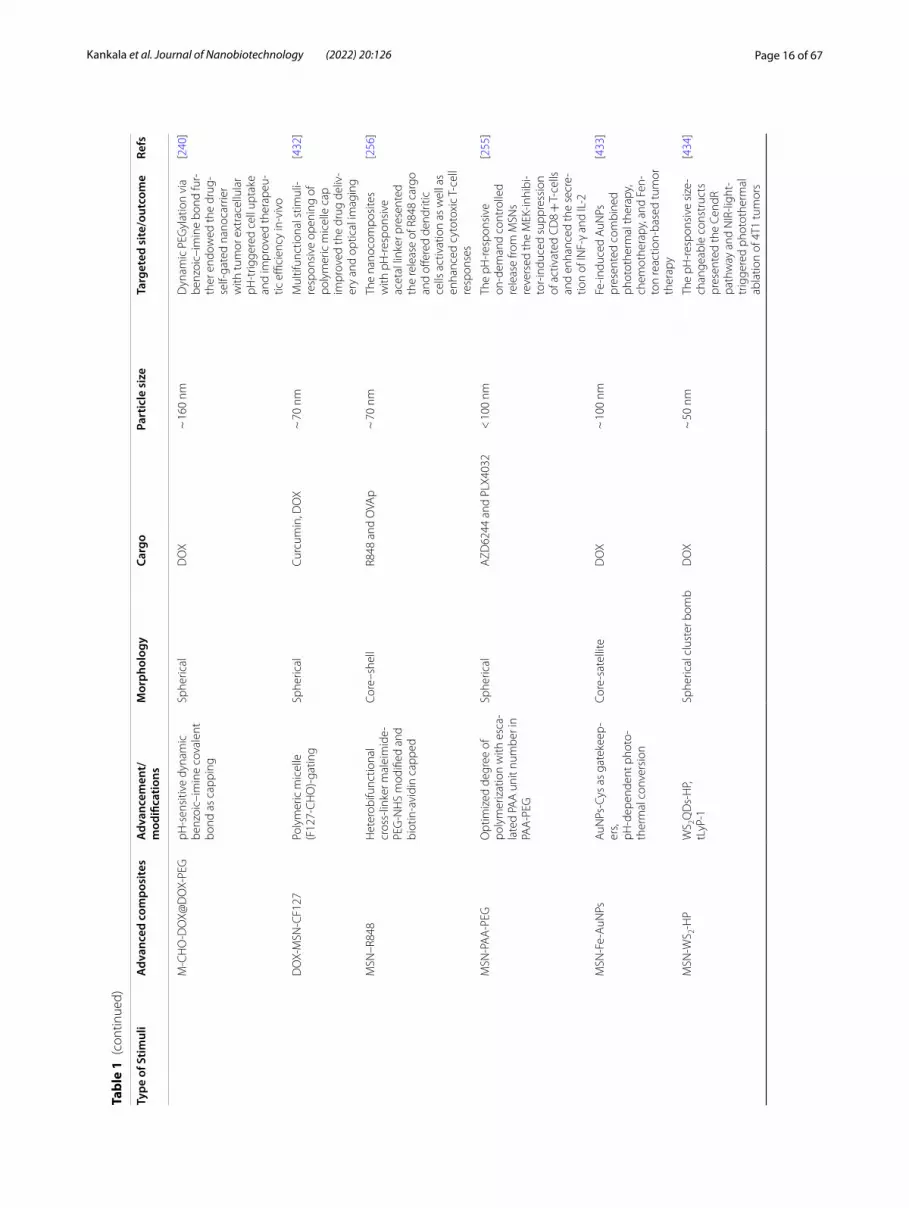
Controlled delivery of chemotherapeuticsMolecular and supramolecular linkages Over the dec-ades, enormous progress has been evidenced by incorpo-rating various selected promising molecular or supramo-lecular linkers in MSNs, which could be well controlled through different complicated mechanisms. These inno-vative switches include biological stimuli (pH/molecu-lar) and externally-applied triggers (light/ultrasound/temperature/magnetic field), which effectively stimulate the delivery of various drugs/genes/therapeutic peptides (Table 1) [8, 237–242]. In cancer, these theranostic nano-medicines carrying the chemotherapeutics are targeted passively to tumors based on the enhanced permeation and retention (EPR) effect due to leaky vasculature, result-ing in their accumulation specifically in tumors. Notably, these stimuli-responsive strategies are expected to afford time-, dose- and spatiotemporal-controlled release in the tumors both in-vitro and in-vivo systems [11, 243–245]. Collectively, these effective stimuli-responsive systems show significant impact in not only drug delivery with improved drug loading and release but also in protecting encapsulated drug cargo from premature release under physiological conditions reducing adverse effects and extravasation by RES uptake [246].
pH-responsiveness Typically, the infectious and cancer-ous sites, including intracellular and extracellular regions, possess lower pH levels (4.5–6.0) than the surrounding physiological fluids (the blood, pH of 7.4). More often, the designed nanoplatforms are passively targeted and accu-mulated due to the EPR effect based on the leaky vascula-ture, specifically, solid tumors. In this context, the delivery systems upon internalization through endocytosis face an extreme shift in the pH value to 6.0 in the surrounding medium of early endosomes, which gradually reduces to pH-4.5 in late endosomes and lysosomes due to the excess availability of proteases and hydrolases, as well as supply of protons into their lumen as a part of defense mecha-nisms against foreign bodies. A simultaneous shift in pH of the environment could be used as an effective biologi-cal trigger, favoring the release of therapeutic guest mole-cules, specifically anticancer and antimicrobial drugs. This potent triggering effect of responsiveness by specific func-tionalities immobilized on the advanced nanocontainers could address numerous issues associated with the con-ventional approaches, such as premature leakage of drugs, combating MDR toward effective therapy [247]. Several examples of specific functional groups include ionizable

Page 15 of 67Kankala et al. Journal of Nanobiotechnology (2022) 20:126
Tabl
e 1
Vario
us a
dvan
ced
prot
otyp
es o
f MSN
s co
nvey
ing
ther
apeu
tic c
argo
for v
ario
us s
timul
i‑res
pons
ive
deliv
ery
Type
of S
timul
iA
dvan
ced
com
posi
tes
Adv
ance
men
t/m
odifi
catio
nsM
orph
olog
yCa
rgo
Part
icle
siz
eTa
rget
ed s
ite/o
utco
me
Refs
pH‑r
espo
nsiv
eV7
‑RU
BYW
orm
hole
por
e, C
hito
san‑
coat
ed, a
nd V
7 pe
ptid
e‑m
odifi
ed
Sphe
rical
IR78
0 dy
e, p
aclit
axel
, or
carb
opla
tin<
40
nmTh
e tu
mor
‑spe
cific
‑tar
‑ge
ted
rele
ase
pres
ente
d im
prov
ed th
erap
eutic
eff
ects
aga
inst
the
orth
o‑to
pic
ovar
ian
tum
ors
[258
]
LB‑M
SN‑O
VALi
pid
bila
yer‑
coat
ed o
ver
the
surf
ace
Rect
angu
lar
Ova
lbum
in ~
200
nm
MSN
s‑en
caps
ulat
ed
in m
icro
need
le a
rray
s sh
owed
exc
eptio
nal i
ntra
‑de
rmal
ant
igen
del
iver
y
[260
]
CS‑
PtN
Ps@
Zn‑M
SNs
CS‑
coat
ed o
ver t
he M
SNs
Sphe
rical
DO
X ~
100
nm
pH‑r
espo
nsiv
e C
S de
grad
atio
n fa
cilit
ated
th
e co
nven
ient
del
iver
y of
MSN
s in
trac
ellu
larly
, ov
erca
me
the
MD
R, a
nd
offer
ed P
tNPs
‑ass
iste
d de
ep tu
mor
pen
etra
tion
[110
]
Trip
le‑la
bele
d M
SNs
YQRL
GC
‑pep
tide
conj
u‑ga
ted,
PEI
/PEG
/TH
PMP
Sphe
rical
FITC
, OG
, and
RIT
C ~
200
nm
Thes
e ly
soso
me‑
targ
eted
na
nopr
obes
enr
iche
d th
e un
ders
tand
ing
of th
e fa
te
of M
SNs
intr
acel
lula
rly
[257
]
FCA
@m
SiO
2Fe
3O4 c
oate
d ca
rbon
/si
lver
(FC
A) a
s co
re a
nd
mes
opor
ous
silic
a as
she
ll
Core
–she
llFe
2+, a
rtem
isin
in ~
200
nm
Art
emis
inin
‑load
ed
FCA
@m
SiO
2 pre
sent
ed
the
acid
‑spe
cific
rele
ase
of F
e2+ io
ns to
non
‑en
zym
atic
ally
con
vert
ar
tem
isin
in to
toxi
c sp
e‑ci
es fo
r can
cer a
blat
ion
[430
]
Cu‑F
e‑M
SNs
Dua
l met
al‑im
preg
nate
d co
nstr
ucts
Janu
s‑ty
peD
OX
~ 1
00 n
mIm
preg
natin
g tw
o si
mila
rly‑c
harg
ed m
etal
s fa
cilit
ated
sha
pe c
hang
es
and
prom
oted
the
ROS‑
assi
sted
CD
T
[92]
HM
SNs‑
β‑C
D‑A
D‑P
EGM
ultip
le s
urfa
ce‑m
odifi
ed
pH‑r
espo
nsiv
e lin
kers
,be
nzoi
c im
ine
and
boro
nic
acid
est
er
Sphe
rical
DO
X ~
100
nm
Thes
e PE
G‑c
oate
d, C
D‑
gate
d, h
ollo
w M
SNs
with
ca
scad
e pH
stim
uli c
leav
‑in
g th
e be
nzoi
cim
ine
bond
s an
d bo
roni
c ac
id
este
r pre
sent
ed e
xcel
lent
in
trac
ellu
lar d
eliv
ery
[431
]
Page 16 of 67Kankala et al. Journal of Nanobiotechnology (2022) 20:126
Tabl
e 1
(con
tinue
d)
Type
of S
timul
iA
dvan
ced
com
posi
tes
Adv
ance
men
t/m
odifi
catio
nsM
orph
olog
yCa
rgo
Part
icle
siz
eTa
rget
ed s
ite/o
utco
me
Refs
M‑C
HO
‑DO
X@D
OX‑
PEG
pH‑s
ensi
tive
dyna
mic
be
nzoi
c–im
ine
cova
lent
bo
nd a
s ca
ppin
g
Sphe
rical
DO
X ~
160
nm
Dyn
amic
PEG
ylat
ion
via
benz
oic–
imin
e bo
nd fu
r‑th
er e
ndow
ed th
e dr
ug‑
self‑
gate
d na
noca
rrie
r w
ith tu
mor
ext
race
llula
r pH
‑trig
gere
d ce
ll up
take
an
d im
prov
ed th
erap
eu‑
tic e
ffici
ency
in‑v
ivo
[240
]
DO
X‑M
SN‑C
F127
Poly
mer
ic m
icel
le
(F12
7‑C
HO
)‑ga
ting
Sphe
rical
Curc
umin
, DO
X ~
70
nmM
ultif
unct
iona
l stim
uli‑
resp
onsi
ve o
peni
ng o
f po
lym
eric
mic
elle
cap
im
prov
ed th
e dr
ug d
eliv
‑er
y an
d op
tical
imag
ing
[432
]
MSN
–R84
8H
eter
obifu
nctio
nal
cros
s‑lin
ker m
alei
mid
e‑PE
G‑N
HS
mod
ified
and
bi
otin
‑avi
din
capp
ed
Core
–she
llR8
48 a
nd O
VAp
~ 7
0 nm
The
nano
com
posi
tes
with
pH
‑res
pons
ive
acet
al li
nker
pre
sent
ed
the
rele
ase
of R
848
carg
o an
d off
ered
den
driti
c ce
lls a
ctiv
atio
n as
wel
l as
enha
nced
cyt
otox
ic T
‑cel
l re
spon
ses
[256
]
MSN
‑PA
A‑P
EGO
ptim
ized
deg
ree
of
poly
mer
izat
ion
with
esc
a‑la
ted
PAA
uni
t num
ber i
n PA
A‑P
EG
Sphe
rical
AZD
6244
and
PLX
4032
< 1
00 n
mTh
e pH
‑res
pons
ive
on‑d
eman
d co
ntro
lled
rele
ase
from
MSN
s re
vers
ed th
e M
EK‑in
hibi
‑to
r‑in
duce
d su
ppre
ssio
n of
act
ivat
ed C
D8 +
T‑ce
lls
and
enha
nced
the
secr
e‑tio
n of
INF‑
γ an
d IL
‑2
[255
]
MSN
‑Fe‑
AuN
PsA
uNPs
‑Cys
as
gate
keep
‑er
s,pH
‑dep
ende
nt p
hoto
‑th
erm
al c
onve
rsio
n
Core
‑sat
ellit
eD
OX
~ 1
00 n
mFe
‑indu
ced
AuN
Ps
pres
ente
d co
mbi
ned
phot
othe
rmal
ther
apy,
ch
emot
hera
py, a
nd F
en‑
ton
reac
tion‑
base
d tu
mor
th
erap
y
[433
]
MSN
‑WS 2‑
HP
WS 2Q
Ds‑
HP,
tLyP
‑1Sp
heric
al c
lust
er b
omb
DO
X ~
50
nmTh
e pH
‑res
pons
ive
size
‑ch
ange
able
con
stru
cts
pres
ente
d th
e Ce
ndR
path
way
and
NIR
‑ligh
t‑tr
igge
red
phot
othe
rmal
ab
latio
n of
4T1
tum
ors
[434
]

Page 17 of 67Kankala et al. Journal of Nanobiotechnology (2022) 20:126
Tabl
e 1
(con
tinue
d)
Type
of S
timul
iA
dvan
ced
com
posi
tes
Adv
ance
men
t/m
odifi
catio
nsM
orph
olog
yCa
rgo
Part
icle
siz
eTa
rget
ed s
ite/o
utco
me
Refs
Lipi
d‑PE
G c
oate
d si
licas
‑om
esLi
pid
bila
yer a
nd P
EG‑
coat
ed c
onst
ruct
s fo
r co
‑adm
inis
trat
ion
of a
nti‑
PD‑1
ant
ibod
y
Silic
asom
eD
AC
HPt
~ 1
40 n
mD
AC
HPt
sili
caso
me
by a
nti‑P
D‑1
ant
ibod
y pr
esen
ted
exce
llent
ch
emot
hera
py a
nd IC
D
resp
onse
in o
rtho
topi
c Kr
as‑d
eriv
ed p
ancr
eatic
ca
ncer
[262
]
PEG
and
lipi
d bi
laye
r coa
tA
link
ed d
owns
trea
m
casc
ade
Core
–she
ll si
licas
ome
IRIN
< 1
00 n
mTh
ese
com
posi
tes
with
sca
le‑u
p fe
atur
es
pres
ente
d im
prov
ed
ther
apeu
tic e
ffica
cy
agai
nst r
obus
t tre
atm
ent‑
resi
stan
t Kra
s‑in
duce
d pa
ncre
atic
can
cer
[263
]
USM
O@
MSN
sU
ltras
mal
l man
gane
se
oxid
e‑ca
ppin
g ov
er M
SNs
Core
–she
ll st
ruct
ures
DO
X ~
50
nmTh
e de
sign
ed n
ano‑
com
posi
tes
pres
ente
d M
RI‑g
uide
d pH
‑sw
itchi
ng
ther
anos
tic p
erfo
rman
ce
for s
ynch
rono
us M
RI
diag
nosi
s an
d ch
emo‑
ther
apy
[435
]
GSH
‑res
pons
ive
MSN
‑S–S
‑NA
C‑T
rpD
isul
fide
bond
and
sho
rt
pept
ide
as c
appi
ng
agen
ts
Sphe
rical
DO
X ~
90
nmA
bol
t‑lik
e bl
ocki
ng
nano
valv
e pr
esen
ted
GSH
‑res
pons
ive
rele
ase
for H
eLa
cell
apop
tosi
s
[291
]
DM
SN‑D
P@C
MM
CF‑
7 m
embr
ane
coat
edCo
re/s
hell
DN
A fu
el s
tran
ds24
3 nm
GSH
‑res
pons
ive
DN
A
stra
nds
in D
MSN
s po
sed
to Im
mun
e es
cape
and
ho
mot
ypic
‑tar
getin
g
[364
]
MSN
s‑S–
S‑si
RNA
Dis
ulfid
e ca
ppin
gSp
heric
alD
OX
and
Bcl‑2
siR
NA
80 n
mSy
nerg
istic
tum
or g
row
th
inhi
bitio
n in
‑viv
o sh
owed
po
tent
ial c
hem
othe
rapy
an
d ge
ne th
erap
y
[292
]
CD
s@M
SN‑T
PP@
AuN
PsTP
P an
d A
uNPs
coa
ted
over
the
MSN
sur
face
Sphe
rical
DO
X~
40
nmG
SH‑r
espo
nsiv
e et
chin
g of
AuN
Ps p
rovi
ded
effec
‑tiv
e ca
ncer
ther
apy
and
mito
chon
dria
l‑tar
gete
d im
agin
g
[436
]

Page 18 of 67Kankala et al. Journal of Nanobiotechnology (2022) 20:126
Tabl
e 1
(con
tinue
d)
Type
of S
timul
iA
dvan
ced
com
posi
tes
Adv
ance
men
t/m
odifi
catio
nsM
orph
olog
yCa
rgo
Part
icle
siz
eTa
rget
ed s
ite/o
utco
me
Refs
MSN
‑ss‑
AD
DA
‑TC
PPD
isul
fide‑
base
d Ta
t48‑
60,
RGD
S, A
DD
A, p
eptid
e‑ba
sed
amph
iphi
le
capp
ing
Sphe
rical
DO
X~
120
nm
Targ
etin
g an
d G
SH‑
resp
onsi
ve d
eliv
ery
of
DO
X to
αvβ
3 in
tegr
in
over
expr
esse
d tu
mor
ce
lls
[317
]
HM
SNs
TEO
S an
d BT
ESPD
with
di
sulfi
de li
nkag
esH
ollo
w m
esop
orou
s sh
ell
DO
X ~
100
nm
Thes
e co
nstr
ucts
resu
lted
in h
igh
load
ing
capa
city
, an
d G
SH‑r
espo
nsiv
e co
ntro
lled
degr
adat
ion
[293
]
Fa‑P
EG‑M
MSN
sFa
‑PEG
coa
ted
Mn2+
‑dop
ed M
SNs
Sphe
rical
DH
A~
100
nm
Acc
umul
atin
g PL
‑PU
FA‑
OO
H o
xidi
zed
by ·O
H a
nd
dest
roye
d th
e st
ruct
ure
of p
olyu
nsat
urat
ed fa
tty
acid
s
[437
]
Au@
MSN
@H
P N
PsH
A, H
S, a
nd H
P gl
ycos
a‑m
inog
lyca
n m
odifi
catio
nCo
re–s
hell
DO
X ~
100
nm
GSH
‑ass
iste
d de
grad
a‑tio
n of
the
disu
lfide
bon
d be
twee
n G
AG
and
MSN
s fa
vore
d pr
ecis
e sy
nerg
is‑
tic c
hem
opho
toth
erm
al
trea
tmen
t
[438
]
PDA
/MnO
2 coa
ted
MSN
sPD
A/M
nO2 c
oatin
g ov
er
MSN
sSp
heric
alD
OX
150–
300
nmG
SH‑a
ssis
ted
tran
sfor
ma‑
tion
of M
nO2 t
o M
n2+
led
to th
e re
leas
e of
dru
g ca
rgo
[439
]
FMSN
‑MnO
2‑BC
QBS
A‑m
odifi
ed,
NIR
‑II s
mal
l mol
ecul
e an
d M
RI re
port
er
Fusi
form
/rod
‑like
MnO
2 and
CQ
4Tw
idth
‑ ~ 1
5 nm
, le
ngth
‑ ~ 9
0 nm
TME‑
activ
ated
tum
or‑
deep
del
iver
y sy
stem
for
dual
‑mod
e im
agin
g an
d se
lf‑re
info
rcin
g ch
emod
y‑na
mic
ther
apy
[440
]
Ultr
asou
nd‑r
espo
nsiv
ePV
‑MSN
sPl
atel
et v
esic
les‑
coat
ed
over
the
surf
ace
Sphe
roid
CA
and
IR78
010
0 nm
IR78
0‑ba
sed
SDT
and
the
CA
‑bas
ed G
SH d
eple
tion
impr
oved
can
cer a
blat
ion
[441
]
MSN
‑FA
‑TA
N‑M
BFA
‑imm
obili
zed
over
the
surf
ace
MB
TAN
~ 1
10 n
mTh
is m
ultif
unct
iona
l veh
i‑cl
e sh
owed
exc
eptio
nal
ultr
asou
nd re
spon
sive
pr
oper
ties
tow
ards
tum
or
targ
etin
g an
d im
agin
g
[442
]
FITC
‑labe
lled
MSN
sSu
bmic
ron
cavi
tatio
n nu
clei
Sphe
rical
Rhod
amin
e B
–U
ltras
ound
‑indu
ced
iner
‑tia
l cav
itatio
n en
hanc
ed
the
extr
avas
atio
n of
the
nano
carr
iers
[290
]

Page 19 of 67Kankala et al. Journal of Nanobiotechnology (2022) 20:126
Tabl
e 1
(con
tinue
d)
Type
of S
timul
iA
dvan
ced
com
posi
tes
Adv
ance
men
t/m
odifi
catio
nsM
orph
olog
yCa
rgo
Part
icle
siz
eTa
rget
ed s
ite/o
utco
me
Refs
HYB
RID
L‑PE
G‑R
GD
Biot
in o
r RG
D p
eptid
e co
ated
Sphe
rical
DO
X~
220
nm
Ultr
asou
nd‑r
espo
nsiv
e ra
ndom
cop
olym
er
enha
nced
cel
lula
r upt
ake
and
canc
er‑k
illin
g effi
cacy
[443
]
MSN
s‑PE
GPE
G‑c
oate
d ov
er s
urfa
ceSp
heric
alG
d(D
TPA
)2– ~
92
nmM
RgH
IFU
stim
ulat
ed
carg
o re
leas
e fa
cilit
ated
by
ultr
asou
nd‑r
espo
nsiv
e PE
G fo
r MRI
‑gui
ded
ther
apy
[444
]
MN
P@M
SNs‑
AM
A‑C
DBu
lky
hydr
ophi
lic β
‑CD
ca
ppin
gCo
re–s
hell
DO
X ~
55
nmH
IFU
‑stim
ulat
ed c
leav
age
of A
CVA
C −
N b
onds
fa
cilit
ated
the
ultr
asou
nd‑
resp
onsi
ve re
leas
e
[445
]
Mag
netic
‑res
pons
ive
EuSP
ION
@M
SNs
Pola
rizat
ion
anis
otro
py
(r) o
f tw
o lu
min
esce
nce
emis
sion
ban
ds
Core
–she
ll–
–N
éel r
elax
atio
n as
the
dom
inan
t hea
ting
mec
hani
sm re
sulte
d in
un
ders
tand
ing
hype
rthe
r‑m
ia‑b
ased
dru
g re
leas
e
[296
]
SPN
C@
MSN
MnF
e 2O4@
CoFe
2O4
core
and
cap
ped
with
Ph
e −
Phe
− G
ly −
Gly
(N
− C
)
Core
–she
llFl
uore
scei
n or
dau
no‑
rubi
cin
120
nmLo
caliz
ed m
agne
tic
heat
ing
pres
ente
d hi
gh
cyto
toxi
city
on
panc
reat
ic
carc
inom
a ce
lls
[295
]
MM
SNs‑
PEG
PEG
and
ther
mor
espo
n‑si
ve p
olym
er‑c
oate
d ov
er
the
surf
ace
Core
–she
llD
OX
160
nmH
eate
d m
agne
tic s
peci
es
in th
e co
re fa
cilit
ated
the
poly
mer
tran
sitio
n an
d op
enin
g to
war
ds d
rug
rele
ase
from
MSN
s
[446
]
Fe3O
4‑m
SiO
2Ja
nus
Janu
s‑ty
peBe
rber
ine
~ 3
00 n
mTh
e su
perp
aram
agne
tic
cons
truc
ts w
ith h
igh
drug
‑load
ing
amou
nts,
supe
rior e
ndoc
ytic
abi
lity,
an
d lo
w c
ytot
oxic
ity
acte
d ag
ains
t hep
atoc
el‑
lula
r car
cino
ma
[447
]
Mag
@M
SNs
Ther
mo‑
resp
onsi
ve
poly
mer
‑coa
ted
core
–sh
ell M
SNs
Core
–she
llFl
uore
scei
n55
nm
Thes
e co
re–s
hell
cons
truc
ts a
void
ed th
e ris
k of
indu
cing
tum
or
met
asta
sis
gene
rate
d by
hy
pert
herm
ia
[294
]

Page 20 of 67Kankala et al. Journal of Nanobiotechnology (2022) 20:126
Tabl
e 1
(con
tinue
d)
Type
of S
timul
iA
dvan
ced
com
posi
tes
Adv
ance
men
t/m
odifi
catio
nsM
orph
olog
yCa
rgo
Part
icle
siz
eTa
rget
ed s
ite/o
utco
me
Refs
SPIO
N@
MSN
Retr
o‑D
iels
Ald
er re
actio
nD
A, M
al, o
r CD
Sphe
reFl
uore
scei
n70
–80
nmN
on‑in
vasi
ve e
xter
nal
actu
atio
n th
roug
h al
tern
atin
g m
agne
tic
field
s im
prov
ed th
e dr
ug
rele
ase
[448
]
MA
RSZn
NC
s Cu
curb
it[6]
uril
Core
–she
llD
OX
< 2
00 n
mTh
e no
n‑in
vasi
ve
cont
rolle
d de
liver
y w
as
achi
eved
aft
er b
eing
ex
pose
d to
the
AC
fiel
d fo
r tre
atin
g br
east
can
cer
cells
[449
]
Tem
pera
ture
‑res
pons
ive
THI@
HM
S@P(
NIP
AA
m‑
MA
A)
P(N
IPA
Am
‑co‑
MA
A)‑
coat
ed H
MSN
sH
ollo
w M
SNs
THI
~ 1
70 n
mTh
e st
rong
ly te
mpe
r‑at
ure‑
depe
nden
t and
di
stan
ce‑li
miti
ng m
echa
‑ni
sm w
as d
emon
stra
ted
usin
g po
sitiv
e te
mpe
ra‑
ture
coe
ffici
ent p
estic
ide
[450
]
MSN
s‑M
NFs
P(N
IPA
Am
‑co‑
HM
AA
m)‑
enca
psul
ated
with
M
ET‑M
SNs
Sphe
res
in th
e el
ectr
o‑sp
un n
anofi
bers
MET
MSN
s‑ <
100
nm
MN
Fs‑d
iam
eter
of
420
nm
ON
–OFF
’ sw
itchi
ng o
f A
MF
show
ed e
xcel
‑le
nt h
eat g
ener
atio
n effi
cacy
and
sub
sequ
ent
cyto
toxi
city
on
B16F
10
mel
anom
a ce
lls
[451
]
MSN
‑PEG
RAFT
pol
ymer
izat
ion
of
PEG
Sphe
rical
Sulfo
rhod
amin
e B,
PD
I14
0 nm
A te
mpe
ratu
re‑c
ontr
olle
d “p
umpi
ng” m
echa
nism
w
as d
emon
stra
ted
for d
rug
rele
ase
from
m
esop
ores
[452
]
MSN
‑the
rmor
espo
nsiv
e po
lym
erD
isul
fide‑
cont
aini
ng c
ys‑
tam
ine
linke
d th
erm
ore‑
spon
sive
pol
ymer
Sphe
rical
DO
X50
–100
nm
UC
ST p
olym
ers
coat
ed
over
the
surf
ace
pre‑
sent
ed re
spon
sive
rele
ase
agai
nst b
reas
t can
cer
cells
(SK‑
BR‑3
)
[283
]
Ligh
t‑re
spon
sive
Porp
hyrin
cap
ped‑
MSN
sPo
rphy
rin c
appi
ngSp
heric
alRB
P, TO
P, or
CA
L13
0 nm
Visi
ble
radi
atio
n‑as
sist
ed
gene
ratio
n of
RO
S‑cl
eav‑
able
link
ages
allo
wed
the
rele
ase
of T
OP
[277
]
AuN
Ps‑M
SNs
AuN
Ps‑c
appi
ng w
ith
phot
olia
ble
linke
rSp
heric
alPT
X10
0 nm
Low
pow
er p
hoto
irrad
ia‑
tion‑
assi
sted
cle
avag
e of
lin
kers
faci
litat
ed th
e ze
ro
prem
atur
e re
leas
e fo
r ch
emot
hera
py
[136
]
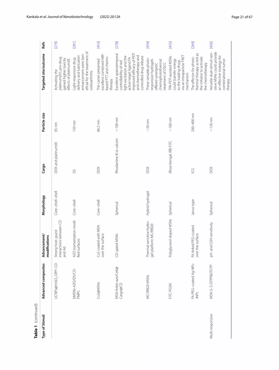
Page 21 of 67Kankala et al. Journal of Nanobiotechnology (2022) 20:126
Tabl
e 1
(con
tinue
d)
Type
of S
timul
iA
dvan
ced
com
posi
tes
Adv
ance
men
t/m
odifi
catio
nsM
orph
olog
yCa
rgo
Part
icle
siz
eTa
rget
ed s
ite/o
utco
me
Refs
UC
NPs
@m
SiO
2‑D
PP–C
DSt
rong
hos
t–gu
est
inte
ract
ions
bet
wee
n C
D
and
Ad
Core
–she
ll–sh
ell
DO
X an
d pl
atin
um(II
)65
nm
Act
ivat
ing
the
plat
inum
(IV),
pro‑
drug
ga
ined
hig
her t
oxic
ity
effec
ts o
f pla
tinum
(II)
[279
]
bMSN
s‑A
ZO/D
S/C
D‑
PMPC
AZO
isom
eriz
atio
n‑m
odi‑
fied
surf
aces
Core
–she
llD
S15
0 nm
Ligh
t‑re
spon
sive
dru
g de
liver
y an
d lu
bric
atio
n en
hanc
emen
t wer
e be
n‑efi
cial
for t
he tr
eatm
ent o
f os
teoa
rthr
itis
[281
]
CuS@
MSN
sCu
S co
ated
with
MSN
ov
er th
e su
rfac
eCo
re–s
hell
DO
X86
.2 n
mTh
e ca
rrie
r pre
sent
ed
exce
llent
com
bine
d N
IR‑
base
d PT
T an
d ch
emo‑
ther
apy
[453
]
MSN
‑link
er‑a
zo/C
e6@
Carg
o@C
DC
D‑g
ated
MSN
sSp
heric
alRh
odam
ine
B or
cal
cein
~ 1
00 n
mEx
celle
nt s
patio
tem
pora
l co
ntro
llabi
lity
of re
d lig
ht e
xcita
tion
and
the
activ
e ta
rget
liga
nd F
A
impr
oved
effi
cacy
of P
DT
and
chem
othe
rapy
and
co
ntro
lled
drug
rele
ase
[278
]
MC
/IR82
0‑M
SNs
Ther
mal
‑sen
sitiv
e hy
dro‑
gel p
latfo
rm M
C/IR
820
Hyb
rid h
ydro
gel
DO
X ~
50
nmTh
ese
vers
atile
pho
to‑
resp
onsi
ve h
ydro
gels
off
ered
syn
ergi
stic
ch
emop
hoto
ther
mal
tr
eatm
ent o
f OSC
C
[454
]
FITC
‑PG
SNPo
lygl
ycer
ol‑d
oped
MSN
sSp
heric
al(R
ose
beng
al, R
B) F
ITC
~ 1
00 n
mTP
A‑P
DT‑
assi
sted
MSN
s co
uld
tran
sfer
ene
rgy
to th
e lo
adin
g dr
ugs
via
an in
trap
artic
le F
RET
mec
hani
sm
[455
]
FA‑P
EG–c
oate
d A
g‑N
Ps‑
JNPs
FA‑li
nked
PEG
‑coa
ted
over
the
surf
ace
Janu
s‑ty
peIC
G20
0–40
0 nm
The
effec
tor f
or p
hoto
‑th
erm
al th
erap
y ac
ted
as
the
initi
ator
to a
ctiv
ate
the
chem
othe
rapy
[269
]
Mul
ti‑re
spon
sive
MSN
‑S–S
‑DTP
P&D
TCPP
pH‑ a
nd G
SH‑s
ensi
tivity
Sphe
rical
DO
X ~
120
nm
Vers
atile
dua
l‑stim
uli‑s
en‑
sitiv
e M
SNs
coul
d pr
ovid
e an
effe
ctiv
e st
rate
gy fo
r co
mbi
natio
nal t
umor
th
erap
y
[456
]

Page 22 of 67Kankala et al. Journal of Nanobiotechnology (2022) 20:126
Tabl
e 1
(con
tinue
d)
Type
of S
timul
iA
dvan
ced
com
posi
tes
Adv
ance
men
t/m
odifi
catio
nsM
orph
olog
yCa
rgo
Part
icle
siz
eTa
rget
ed s
ite/o
utco
me
Refs
Seru
m a
lbum
in a
nd
myo
glob
in‑g
ated
UC
NP@
mSi
O2
pH, G
SH, o
r H
2O2‑
resp
onsi
veCo
re–s
hell
sphe
rical
na
nost
ruct
ures
with
w
orm
‑like
por
es in
she
lls
DO
X64
nm
Thes
e na
noco
mpo
site
s sh
owed
spa
tiote
mpo
rally
ta
rget
ed d
rug
deliv
ery
for
canc
er c
hem
othe
rapy
[298
]
Dm
@TM
SN‑P
EIRe
dox‑
enha
nced
pH
‑re
spon
sive
Sphe
rical
mor
phol
ogy
with
wor
mlik
e m
eso‑
stru
ctur
e
DO
X an
d m
iRN
A‑1
45~
183
nm
The
nano
com
posi
tes
with
affi
nity
to g
luco
se‑
regu
late
d pr
otei
n 78
(G
RP78
), a
cell
surf
ace
prot
ein
over
expr
esse
d in
co
lore
ctal
car
cino
ma
is
deve
lope
d
[297
]
MSN
‑AN
A‑H
FnRe
dox‑
and
pH
‑trig
gere
dSp
heric
alD
OX
100
nmH
Fn c
appe
d M
SNs
prov
ided
TfR
1 ta
rget
ing
on s
uppr
essi
on o
f tum
or
grow
th
[299
]
TTTM
SNs
pH‑ a
nd re
dox‑
dual
‑re
spon
sive
MSN
‑S–S
‑Pep
tide‑
MPE
G
Rect
angu
lar
DO
X ~
125
nm
RGD
FFFF
C‑a
ssis
ted
targ
etin
g, b
enzo
ic‑im
ine
bond
‑bas
ed p
H‑
resp
onsi
ve, a
nd d
i‑sul
fide
clea
vage
‑bas
ed re
dox‑
resp
onsi
ve e
nhan
ced
the
tum
or‑t
arge
ting
effica
cy
[457
]
MSN
@p(
NIP
AA
m‑c
o‑M
A)
Ther
mal
‑ and
pH
‑res
pon‑
sive
p(N
IPA
Am
‑co‑
MA
)
Sphe
rical
EVO
and
BBR
~ 1
60 n
mTh
ese
com
posi
tes
with
du
al d
rugs
pro
vide
d ex
celle
nt th
erap
eutic
eff
ects
aga
inst
EM
T‑6
mou
se m
amm
ary
carc
i‑no
ma
tum
or a
llogr
aft
[261
]
MSN
s@PD
A@
kera
tinpH
and
GSH
dua
l res
pon‑
sive
Ker
atin
as
capp
ing
Sphe
rical
/elli
psoi
dal
DO
X ~
100
nm
Thes
e co
mpo
site
s se
lect
ivel
y sh
owed
hig
her
toxi
city
aga
inst
A54
9 ce
lls
than
nor
mal
cel
ls
[301
]
MSN
‑SS‑
PDA
Redo
x/pH
/NIR
‑mul
ti‑de
pend
ent,
Dis
ulfid
e lin
ked
PDA
‑coa
ting
Sphe
rical
DO
X ~
130
nm
Thes
e co
mpo
site
s ex
hib‑
ited
exce
llent
pho
to‑t
her‑
mal
con
vers
ion
abili
ty,
mul
ti‑st
imul
i res
pons
ive
drug
rele
ase,
che
mo/
phot
othe
rmal
syn
ergi
stic
th
erap
y eff
ect
[458
]

Page 23 of 67Kankala et al. Journal of Nanobiotechnology (2022) 20:126
Tabl
e 1
(con
tinue
d)
Type
of S
timul
iA
dvan
ced
com
posi
tes
Adv
ance
men
t/m
odifi
catio
nsM
orph
olog
yCa
rgo
Part
icle
siz
eTa
rget
ed s
ite/o
utco
me
Refs
MSN
‑S–S
‑N =
C‑H
ApH
‑ and
redo
x‑re
spon
sive
HA
‑g‑C
DSp
here
with
hig
hly
orde
red
hone
ycom
b ch
anne
ls
DO
X~
100
nm
The
com
posi
tes
with
du
al‑r
espo
nsiv
enes
s pr
ovid
ed C
D44
ove
r‑ex
pres
sed
canc
er c
ell
targ
etin
g eff
ects
[300
]
MSN
‑Au
GSH
‑ and
NIR
‑trig
gere
dA
uNPs
Ellip
soid
DO
X~
250
nm
A c
ombi
natio
n of
che
mo‑
ther
apy
and
phot
othe
r‑m
al th
erap
y to
war
d A
549
cells
[302
]
β-CD
: β-C
yclo
dext
rin; A
CVA
: 4,4′-A
zobi
s(4-
cyan
oval
eric
aci
d); A
D- 1
-Ada
man
tane
met
hyla
min
e; A
DD
A-T
CPP:
C12
-CG
RKKR
RQRR
RPPQ
RGD
S; A
MA
: 1-A
dam
anty
lam
ine;
AuN
Ps-C
ys: L‐
Cyst
eine
‐der
ivat
ized
gol
d na
nopa
rtic
les;
A
ZO: A
zobe
nzen
e; B
BR: B
erbe
rine;
BFO
: Bis
mut
h fe
rrite
; BTE
SPD
: Bis
[3-(t
rieth
oxys
ilyl)p
ropy
l] di
sulfi
de; C
A: C
inna
mal
dehy
de; C
AL:
Cal
cein
; CD
s: C
arbo
n na
nodo
ts; C
e6: C
hlor
in e
6; C
endR
: Neu
ropi
lin-1
(NRP
-1)-
depe
nden
t en
docy
tic/e
xocy
tic tr
ansp
ort;
CM: C
oum
arin
; CS-
PtN
Ps@
Zn-M
SNs:
Chi
tosa
n-Pl
atin
um n
anop
artic
les
coat
ed Z
inc-
dope
d M
SNs;
CuS
: Cop
per s
ulfid
e; C
u-Fe
-MSN
s Co
pper
and
iron
-dop
ed M
SNs;
DAC
HPt
: Act
ivat
ed
oxal
ipla
tin (1
,2-d
iam
min
ocyc
lohe
xane
pla
tinum
(II);
DH
A: D
ihyd
roar
tem
isin
in; D
MSN
s-de
ndrit
ic M
SNs;
DO
X: D
oxor
ubic
in; D
S: D
iclo
fena
c so
dium
; EVO
: Evo
diam
ine;
EuS
PIO
N: E
urop
ium
-dop
ed s
uper
para
mag
netic
iron
ox
ide
nano
part
icle
; FA
: Fol
ate;
FaP
EG: F
olat
e-gr
afte
d PE
G; F
CA-F
e 3O4 c
oate
d ca
rbon
/silv
er; F
ITC:
Flu
ores
cein
isot
hioc
yana
te; F
RET:
Flu
ores
cenc
e re
sona
nce
ener
gy tr
ansf
er; G
d(D
TPA
)2–: G
adop
ente
tate
dim
eglu
min
e; G
SH
-glu
tath
ione
; HA
: Hya
luro
nic
acid
; HA
p: H
ydro
xyap
atite
; Hfn
: Hum
an H
cha
in fe
rriti
n; H
MA
Am
: N-h
ydro
xym
ethy
lacr
ylam
ide;
HM
SiO
2 /H
MSN
s/H
MS:
Hol
low
mes
opor
ous
silic
a na
nopa
rtic
les;
HN
Ps: h
arm
onic
nan
opar
ticle
s;
HP:
Hep
arin
; HS:
Hep
arin
sul
fate
; HYB
RID
: Hyb
rid m
esop
orou
s si
lica
nano
carr
ier;
IBU
: Ibu
prof
en; I
CG: I
ndoc
yani
ne g
reen
; IN
F-In
terf
eron
; IL-
Inte
rleuk
in; J
NPs
: Jan
us-t
ype
MSN
s; L
B-M
SN-O
VA: L
ipid
bila
yer-
MSN
-ova
lbum
in;
MA
: Met
hacr
ylic
aci
d; M
al: M
alei
mid
opro
pyl t
rieth
oxys
ilane
1; M
ARS
: Mag
netic
ally
act
ivat
ed re
leas
e sy
stem
; MB:
Mic
robu
bble
; MC:
Met
hylc
ellu
lose
; MET
: Met
form
in; m
iRN
A: M
icro
RNA
; MM
SNs:
Man
gane
se-d
oped
M
SNs;
MN
Fs: M
agne
tic n
anofi
bers
; MN
Ps: M
nFe 2O
4@Co
Fe2O
4 nan
opar
ticle
s; M
RI: M
agne
tic re
sona
nce
imag
ing;
MRg
HIF
U: M
RI-g
uide
d hi
gh-in
tens
ity fo
cuse
d ul
tras
ound
; mSi
O2-
mes
opor
ous
silic
a; M
SNs-
mes
opor
ous
silic
a na
nopa
rtic
les;
MSN
-PO
LY: R
AFT
pol
ymer
izat
ion
on th
e su
rfac
e of
MSN
s; N
IPA
Am
: N-is
opro
pyla
cryl
amid
e; O
G: O
rego
n gr
een;
OSC
C: O
ral s
quam
ous
cell
carc
inom
a; O
VAp:
Ova
lbum
in; P
AA
: Pol
yacr
ylic
aci
d; P
DA
: Po
lydo
pam
ine;
PEG
: Pol
yeth
ylen
e gl
ycol
; PEI
: Pol
yeth
ylen
imin
e; P
GSN
: Pol
ygly
cero
l-dop
ed M
SNs;
PL-
PUFA
-OO
H: L
ipid
per
oxid
es; P
MPC
: Pol
y(2-
met
hacr
yloy
loxy
ethy
l pho
spho
rylc
holin
e); p
(NIP
AA
m-c
o-M
A) :
Pol
y(N
-is
opro
pyla
cryl
amid
e-co
-met
hacr
ylic
aci
d); P
V: P
late
let m
embr
ane
vesi
cle;
NAC
: N-a
cety
l- l-c
yste
ine;
SD
T: S
onod
ynam
ic th
erap
y; s
iRN
A: S
mal
l int
erfe
ring
RNA
; SPN
C: S
uper
para
mag
netic
nan
opar
ticle
cor
es; T
AN
: Ta
nshi
none
IIA
; TEO
S: Te
trae
thyl
ort
hosi
licat
e; T
fR1:
Tra
nsfe
rrin
rece
ptor
1; T
HI:
Thia
met
hoxa
m; T
HPM
P: 3
-trih
ydro
xysi
lyl p
ropy
lmet
hylp
hosp
hona
te; T
OP:
Topo
teca
n; T
PA-P
DT:
Tw
o-ph
oton
act
ivat
ed-p
hoto
dyna
mic
th
erap
y; T
PP: T
riphe
nylp
hosp
hine
; Trp
: Try
ptop
han;
pD
NA
: Pla
smid
DN
A; P
V-co
ated
MSN
s: P
late
let v
esic
les-
coat
ed M
SNs;
RB:
Ros
e be
ngal
; RBP
: [Ru
(bip
y)3]
Cl2;
RITC
: Rho
dam
ine
B is
othi
ocya
nate
; RO
S: re
activ
e ox
ygen
sp
ecie
s; U
CNPs
: Upc
onve
rsio
n na
nopa
rtic
les;
USM
O: U
ltras
mal
l man
gane
se o
xide
; V7-
RUBY
: Wor
mho
le m
esop
orou
s si
lica
nano
part
icle
s; Y
QRL
GC:
lyso
som
al s
ortin
g pe
ptid
es; W
S 2-H
P: T
ungs
ten
disu
lfide
qua
ntum
dot
s;
ZnN
Cs: Z
inc-
dope
d iro
n ox
ide
nano
crys
tals

Page 24 of 67Kankala et al. Journal of Nanobiotechnology (2022) 20:126
weakly acidic/basic functional groups, such as hydra-zone, disulfide linkage, and dinitroimidazole, among oth-ers [14, 248]. In addition, some of the currently available polymers contain pH-responsive linkers, such as chitosan and dextran from the natural origin, as well as carboxylic and amine functional groups containing polymers from synthetic origins. More often, these pH-responsive moie-ties are applied by conjugating them in the mesopores or coated over the surface for immobilizing the therapeutic
molecules for triggered release either alone or through a capping agent [74, 114, 127, 143, 226, 249–252].
The advanced MSNs with tunable morphology and appropriately immobilized functional groups exhibit precise pH-responsiveness in attaining the advanced delivery and exceptional cancer therapy performance. In this vein, several studies have been reported by utiliz-ing various pH-responsive linkers in the efficient deliv-ery of therapeutic guest molecules, specifically at the
Fig. 5 Various advanced MSNs for pH‑responsive delivery of drugs. A Methodological comparison between the traditional MSNs‑based nano‑drug delivery systems by the drug post‑loading route and the proposed MSNs by the drug in situ co‑loading route. B In‑vitro pH‑responsive drug release behaviors of DOX@CTAB@MSNs in the release media of different pH values, which were used to simulate the alkalescent conditions in normal tissues and blood (pH ~ 7.4) and the acidic conditions in the tumor (pH = 4–6.8). Reproduced with permission from Ref. [76] Copyright 2011, Elsevier. C TEM images of nanomicelles with (i) and without (ii) PEG segments at different pH values. D Confocal microscopic images of HeLa cells treated by rhodamine B‑loaded and FITC‑labeled micelles for 4 h at different conditions: (i) pH = 7.4, 37 °C; (ii) pH = 7.4, 39 °C; (iii) pH = 6.4, 37 °C; (iv) pH = 6.4, 39 °C (scale bar: 80 μm). Reproduced with permission from Ref. [253] Copyright 2010, American Chemical Society. E Schematic showing the fabrication of MSNs for tumor‑triggered targeting drug delivery. Functionalization protocol of the MSN; drugs loaded under physiological condition; removal of PASP protection layer in response to MMP at a tumor site; cell uptake through RGD‑mediated interaction; GSH‑triggered drug release inside the cell; and apoptosis of tumor cells. Reproduced with permission from Ref. [254] Copyright 2013, American Chemical Society

Page 25 of 67Kankala et al. Journal of Nanobiotechnology (2022) 20:126
targeted tumor and infection sites [116]. In one case, He et al. demonstrated the release of CTAB and doxorubicin (DOX) from positively-charged micelles/MSNs compos-ites through interactions with the proton, specifically in a high proton environment compared to traditional post-loaded drug systems for overcoming MDR in can-cer (Fig. 5A, B) [76]. The interesting feature of this design included a facile single-step synthesis, avoiding the sur-factant removal and drug encapsulation procedures sepa-rately. In another case, the surface-coated PEG protective layer over the MSNs could be detached at a pH lower than 6.8, for instance, the cancer microenvironment. The encapsulated MSNs were subsequently internalized by the cancer cells with the identification of the RGD motif (Fig. 5C, D) [253]. Similarly, Zhang et al. fabricated versatile envelope-type MSNs to achieve pH-triggered, tumor-targeted delivery of DOX [254]. Further, upon the hydrolysis of matrix metalloproteinase substrate pep-tide, the polyanion outer layer was eliminated, result-ing in the exposure of the RGD motif. Moreover, the GSH-assisted disintegration of disulfide, along with the improved uptake of tumor cells, significantly contributed to the rapid drug release through cyclodextrin gatekeeper (Fig. 5E). In this regard, various polymers, for instance, chitosan, PEI, and PEG, PAA [111, 251, 255–259], and lipids [109, 260, 261], as well as the mixture of polymers and lipids [262, 263], have been coated over the MSNs for the pH-responsive delivery of chemotherapeutics from MSNs. These complex designs significantly facilitated versatile functionalities eventually, resulting in the apop-tosis of the cancer cells. Although the design presented multiple successful functionalities, the multiple immobi-lized groups require multiple-step synthetic procedures, which could substantially limit the design in bulk manu-facturing and compatibility explorations. Moreover, the eventual tracking of performance efficacy and exploring the pharmacokinetic behavior of the MSNs-based com-plex designs are further challenges to be addressed.
In the vein of addressing the establishment of innova-tive therapeutic modalities against cancer therapy, most studies have focused on the establishment of pH-respon-sive linkers for drug delivery and coating of pH-respon-sive polymers for capping of MSNs towards successful intracellular delivery [264]. These innovative modifica-tions have successfully addressed cancer therapy to a con-siderable extent, including delivering chemotherapeutics and overcoming the drug resistance towards improved biological performance [243, 265]. Despite the delivery success, the complete eradication of cancer remained a significant challenge as most solid tumors possess tight junctions with impermeable barriers, limiting the entry of nanomedicines into deep tumors and failing their clin-ical translation [50]. Over the years, these highly complex
anatomical features of tumors resulted in unsuitable therapeutic strategies, resulting in the ablation of surface tumor cells alone, and the deep tumor led to recurrence. To a considerable extent, several surface modification strategies have been employed to address the deep pene-tration via piercing the tight junctions of tumors through surface charge reversal and size reduction, among others [50, 266]. In this regard, MSNs and their advanced pro-totypes have been employed to address the deep tumor penetration using the surface-modified MSNs. In a case, MSNs were coated with the up/downconverting lumi-nescent nanoparticles (U/DCNPs) via acid-labile (ben-zoic-imine) linkages to form core–shell assemblies and subsequently coated with the charge reversible polymers, poly(allylamine)-dimethyl maleic anhydride–polyethyl-ene glycol (PAH-DMMA-PEG) [266]. The charge rever-sal of coated polymer from negative to positive in the acidic microenvironment induced the disintegration into isolated MSNs and UCNPs, facilitating the successful penetration into solid tumors. In another case, Mannose-decorated MSNs were coated with the PDA-Gd-PEG composite for MRI and deep penetration through charge reversal for synergistic photothermal and visualized therapy [267]. Although the success in the fabrication of these innovative constructs, in several instances, the degradation of the resultant MSNs after degradation of the surface-modified organic components are the fur-ther challenges remained to be addressed. In an attempt to address this aspect to a considerable extent, our group has demonstrated the fabrication of the nanoplatforms based on the pH-responsive chitosan-encapsulated with Pt nanoparticles coated over the metal-impregnated MSNs [110]. The decoration of ultrasmall Pt-based nano-constructs facilitated the advanced therapeutic capabili-ties of deep tumor penetration through influencing the adherens junctions between the tumor cells. Interest-ingly, the metal-impregnated MSNs, certainly showed the degradation of MSNs due to low silica density and coordination arrangement of metal species in the MSN framework. However, further studies of addressing the controlled degradation ability and monitoring the result-ant byproducts remained to be explored.
Light-responsiveness The light-based therapies, i.e., photodynamic therapy (PDT) and photothermal therapy (PTT), offer several beneficial qualities, such as selectivity, preferential localization of photosensitizer at the tissue of interest, and substantial activation execute the therapeu-tic function, among others [268, 269]. However, severe aggregation of photosensitizers due to hydrophobicity is one of the critical issues of such light-induced therapies, limiting their applicability [269, 270]. To a considerable extent, the utilization of various carriers with adequate

Page 26 of 67Kankala et al. Journal of Nanobiotechnology (2022) 20:126
carrying capacity enables the successful transportation of photosensitizers to the desired sites and substantially facilitates their clearance from the body [271]. Despite the limited efficacy and vulnerable damage by ultraviolet (UV) radiation, it is highly desirable to establish NIR light to avoid that quick attenuation, which further prevents the biological samples and living organs tenuously from being destructed due to its more profound tissue penetra-tion efficacy [252, 272, 273]. Investigations on light (vis-ible/UV/NIR)-induced therapeutic efficacy using MSNs-based nanocarriers have been carried out, attributing to the light-triggered characteristics in a spatiotemporal manner [129, 136, 140, 268, 274–276]. In a case, a novel drug carrier was fabricated based on MSNs coated with porphyrin caps grafted with ROS cleavable bonds [277]. The visible light, i.e., common light,-assisted cleavage of decorated porphyrin caps could release the encapsulated drug cargo, topotecan, from MSNs. The highly compatible light and dual effects of ROS production, in terms of trig-gered release and therapeutic effects, substantially dem-onstrated the antitumor effects in HOS cells. Although the design is advantageous, several performance- and
compatibility-related attributes in-vivo yet remain to be explored as the light penetration and depth of tumor may hinder the efficacy of light exposure. Moreover, the com-prehensive evaluations of type of light utilization, expo-sure time, and distance, must be selectively optimized. In this regard, several studies with some smart linkers, such as azo-isomerization, and light-assisted opening of gatekeepers, enabled the fabrication of various intelligent nanocarriers using MSNs [278–281]. In another case, Liu et al. fabricated MSNs-based upconversion core–shell nano-constructs, which were highly appealing toward the exertion of NIR light [272]. Considering the fascinating feature of photo-triggered luminescence ability, the can-cer-therapeutic drug (DOX) was loaded in the mesopores of silica constructs, in which azobenzene (azo) group is selected as “stirrer” (Fig. 6), on absorbing the NIR light (980 nm), the nanoparticles significantly transmitted photons in UV band that instantly absorbed by the light-responsive azo group. Further, the isomerization gener-ated the cyclic movement of rotation-inversion, promot-ing the release of DOX as an impeller via simultaneous UV–Vis light emission. The interesting switching of light
Fig. 6 Light‑responsive advanced MSNs. NIR light‑triggered DOX release by using the upconversion property of UCNPs and trans–cis photo‑isomerization of azo molecules grafted in the mesopore network of MSNs. Reproduced with permission from Ref. [272] Copyright 2013, John Wiley and Sons

Page 27 of 67Kankala et al. Journal of Nanobiotechnology (2022) 20:126
frequencies by cyclic movement of rotation-inversion would be convenient in exploring through the in-vitro investigations, which, however, is undoubtedly challeng-ing in executing in in-vivo as the tissue microenvironment is highly complex.
Thermos-responsiveness Indeed, the environmental temperature is another fascinating and promising stimu-lus, in which the controlled increase of the temperature at the diseased site may substantially trigger the con-trolled release of therapeutic guests. In this context, various spatiotemporal thermos-responsive units have been immobilized in these MSN-based architectures, exhibiting an exciting feature of reversibly responding to temperature, such as pNIPAAm, and sulfobetaine meth-acrylate (SBMA), or depositing specific metal species like graphene oxide or Pt nanoparticles [176, 252, 282–285]. Instinctively, the system is operated based on a revers-ible switching-on/off mechanism at the mesopore entry via diverse internal or external decoration or modification over the surface. These composites at elevated environ-mental temperatures exhibit phase transition, enabling the release of drugs through changes in their hydropho-bic interactions [284, 285]. More often, the thermogelling polymers, such as pNIPAAm, present an exciting feature of gelation at elevated temperatures, such that they facili-tate the localized delivery of drugs in a controlled fash-ion [285]. Several reports have focused on manipulating temperature-induced drug release, showing a tremendous challenge to distinguish between fatal tumor sites and healthy peripheral tissues [286]. For instance, Sun et al. demonstrated the novel temperature-responsive polyam-pholyte-coated MSNs by reversible addition-fragmenta-tion chain transfer polymerization on their surface for the first time, displaying the controlled release behaviors and high biosafety [282]. In another case, Tian et al. applied a thermo-sensitive polymer (pNIPAAm) and coated it over SBA-15-type silica to synthesize an ordered mesostruc-ture with the low critical solution temperature collated [287]. Notably, different from the capping agents acted by super valves, the flexible polymeric shells over MSNs offer significant advantages over agglomerating and providing other partitions for cargo loading apart from the pores of MSNs [252, 283, 284]. Apart from the delivery through thermos-responsiveness, specific light-responsive mol-ecules, such as indocyanine green (ICG), could facilitate the synergistic therapeutic efficacy through augmented temperatures, referred to as hyperthermia, at the tumor sites, resulting in the substantial ablation through singlet oxygen generation. Notably, the MSNs-based advanced nanosystems with synergistic light- and temperature-induced ablation offer more advantages over the single-responsive system.
Ultrasound-responsiveness The ultrasound-based respon-siveness acts by applying a higher frequency wave that exceeds human auditory limitations. The sonochemical pro-cess is classified based on frequency and penetration depth. In general, the low frequency (< 1 MHz) shows a superior penetration depth irrespective of the site. In comparison, high (5–10 MHz) and medium (1–5 MHz) frequencies dis-play inferior penetration capacity along with the destruction of living tissues [288]. Regarding the biomedical applica-tions, the approach has recently garnered enormous atten-tion towards tumor ablation, either by mechanical or thermal methods conducted on subcellular disruption or coagulative necrosis, respectively [289]. Inertial acoustic cavitation con-cept by the oscillation raised from fluidic gas bubbles, either linear or non-linear related, has been acknowledged as the most significant mechanical ultrasound effect for biomedical applications [276]. Nevertheless, an unstable phenomenon exists of the drastic collapse of the growing bubbles under the fluidic inertia. In one case, interest on drug carriers and nuclei for inertia cavitation stimulation, Paris et al. enhanced the extravasation of MSNs with the combination of acous-tic-sensitive polymeric particles, resulting in the initiation of inertia cavitation process by reducing the required pres-sure under the focused ultrasound conditions (Fig. 7A, B) [290]. In another study, they fabricated ultrasound-sensitive MSNs, presenting significant effects by controlled release of encapsulated therapeutic guests through gate-opening strat-egy [17]. The coated polymer over MSNs was collapsed ini-tially and then opened its conformation like a coil after the hydrophobicity change, triggering the cargo release with the preservation of ultrasound-sensitive capacity (Fig. 7C–E). In-depth analyses are required to establish the mechanistic stud-ies that are yet to be explored.
In addition to the notified most common stimuli trig-gers (pH, light, and thermos), various advancements have been evidenced in employing advanced MSNs that are responsive to other stimuli, such as GSH-, and magnetic-responsive. More often, the internal trigger, GSH-respon-sive, linkages are decorated either on the surface of MSNs or in the siliceous frameworks, for instance, di-sulfide-linkages [291, 292]. The decoration of GSH-responsive linkers containing polymers on the surface act as gate keepers, facilitating the release of the encapsulated thera-peutic cargo from the mesopores [291]. In addition, such responsiveness can be employed to encapsulate the drugs or genes for controlled release, specifically in the GSH-rich environment, i.e., cancers [292]. In most instances, the GSH-responsive disulfide linkages were bridged in the organosilica frameworks towards fabricating the bio-degradable MSNs [41, 83, 293]. These smart disulfide bridged MSNs not only present the successful delivery of the therapeutic guests in the intracellular environ-ment precisely but also offer the impressive and required

Page 28 of 67Kankala et al. Journal of Nanobiotechnology (2022) 20:126
biodegradability attribute. To this end, the MSNs encap-sulated with the magnetic species, for instance, iron oxide, as core–shell architectures are often employed for the localized delivery using the externally applied mag-netic field [294–296]. Apart from a single trigger, several studies have explored the utilization of multiple stimuli (two or more) either to control the release of therapeutic guests by unlocking the capping or the conveyance of the MSNs [297]. In this regard, various multiple stimuli com-binations employed in the fabrication of advanced pro-totypes of MSNs include pH, GSH, or H2O2-responsive [298], redox-enhanced pH-responsive [297, 299], ther-mos- and pH-responsive [261], pH- and GSH-responsive [300, 301], GSH- and NIR-triggered [302] nanocompos-ites. These composites with multiple stimuli-triggered delivery or degradation offer more advantages than the single stimuli-based composites. However, they require various mechanistic elucidations and comprehensive evaluations in orchestrating their synergistic effects for their successful translation to clinics.
Metallic linkersDespite the advantageous morphological attributes and well-defined porous architectures, it is evident that the traditional siliceous frameworks can be supported only as carriers concerning the drug delivery application [42,
43]. However, specific modifications according to the requirements often lead to enhanced performance and substantial applicability of the carrier. In addition to numerous aforementioned molecular and supramolecu-lar stimuli-responsive constituents [13, 33, 243, 249, 265, 303], various transition metal species with exceptional potential can be employed to establish the responsive linkages within the siliceous frameworks due to their unique electronic properties and structural attributes [220, 304, 305]. Establishing the transition metals (Cu, Fe, Co, and Zn)-based functional coordination linkages offer enormous advantages over traditional MSNs in drug delivery, in terms of improved drug loading efficiency, precise control over their on-demand release, and con-trolled degradability. In recent times, lanthanides (La3+ ions) have also been doped into the siliceous frameworks for drug delivery applications, which, however, require further optimization and safety considerations compre-hensively [306]. In addition to safety issues, the loading efficiency of therapeutic guests within the porous con-tainers must be addressed as it explores the therapeutic levels at the target site to perform its therapeutic action and eliminate the irregularities in the dosage frequency.
Previous reports indicated that the loading efficiency of the therapeutic guest molecules in the MSN pores is significantly poor as the affinities between the guest and
Fig. 7 MSNs‑based ultrasound responsive drug delivery. A Schematic of combining MSNs with SSPs. B Representative microscopy images of nanoparticle delivery in the agarose phantom model (MSN in combination with SSPs), showing bright field (left), green fluorescence (center), and green fluorescence in a cross‑section of the flow channel (right) (n = 3). Scale bar = 500 µm. Reproduced with permission from Ref. [290] Copyright 2017, Elsevier. C Schematic illustration of the behavior in an aqueous medium of the dual responsive release system. D Release profiles of fluorescein from hybrid‑MSNs in PBS solution versus time with ultrasound exposure (10 min and 1.3 MHz, 100 W) (i) and without ultrasound (ii). E Fluorescence microscopy images of LNCaP cells incubated with Rhodamine B‑labeled hybrid‑MSNs with fluorescein loaded before (top) and after (bottom) ultrasound irradiation. Reproduced with permission from Ref. [17] Copyright 2015, American Chemical Society
Page 29 of 67Kankala et al. Journal of Nanobiotechnology (2022) 20:126
host predominantly depend on the physical adsorption by weak hydrogen bonding or other electrostatic interac-tions, leading to a simultaneous exchange with the ions in the surrounding medium [26, 116, 307]. In addition, these weak interactions often result in premature loss of therapeutic molecules not only during the encapsulation but also while conveyance through physiological fluids, resulting in deprived therapeutic efficiency due to their insufficient levels at the desired site. To a considerable extent, these issues have been addressed by installing var-ious stimuli-responsive capping molecules, such as the utilization of quantum dots [68, 250], AuNPs [139, 145], and protein or polymer assembly [88, 89, 97, 127, 259]. However, it should be noted that these complex strategies require multi-step synthesis, leading to mechanical abra-sion-induced changes in MSN shapes [92]. In an attempt to address this issue, we have done enormous work by fabricating innovative metal-encapsulated MSNs through a single-step, facile approach by impregnating positively-charged first-transition-row metals species viz. Cu metal species in the mesoporous frameworks [82, 96]. These metal species in the frameworks offered coordination linkages with the therapeutics (herein, DOX molecules), resulting in their improved loading efficiency compared to the pristine MSNs (Fig. 8A) [82, 96]. In addition to improved loading efficacy, these specific interactions were highly susceptible in the acidic environment (pH-5.0) and facilitated disassembling through the protona-tion of nitrogen atoms in the guest molecules (Fig. 8B, C) [96, 308]. Despite the efficient loading and precise release of the encapsulated guest species, the doped metal spe-cies could merely facilitate the coordination interactions to certain guest species with terminal amine-functional groups [82]. Further efforts in terms of the incorporation of various metals, such as Fe, Zn, have been reported [91, 110]. Hitherto, only very few metals have been doped, requiring enormous investigations to further explore the multiple combinations and optimized conditions for incorporating diverse metal species. Although reduced silica density improves the compatibility attributes, the safety and toxicity considerations must be considered while doping various metals in siliceous frameworks.
Compared to molecular and supramolecular link-ers, these transition metal-based MSNs offer a specific advantage of participating in the metabolic activation cascade reactions due to the unique electronic architec-ture. These metal species, such as Cu and Fe, participate in catalysis and significantly facilitate the conversion of the intracellularly available H2O2 molecules to toxic ROS. These deadly free radical species promote the substantial activation of apoptotic cascades and cell membrane dete-rioration, leading to cell death. In an attempt to explore these events, we fabricated the Cu metal-impregnated
and multi-metal (Cu/Fe)-doped MSNs, which resulted in the generation of highly toxic free radicals by partici-pating in the redox chemistry mediated by chemody-namic therapy (Fig. 8D). These deadly species damaged the intracellular organelles, such as mitochondria, by disturbing the electron transport chain for energy pro-duction and substantially enhancing the stimulation of various apoptotic cascades. Notably, the rationale behind choosing these metals was that they could participate actively in the redox reactions and lead to the generation of higher amounts of free radicals from H2O2 molecules, which were in higher amounts in cancer cells over nor-mal cells. Moreover, these metals would cause no adverse effects and are regarded as safe as these metals are essen-tial trace elements in the body. Moreover, incorporating various metals, being one of them as Fe metal, facilitated the change of the overall shape of MSNs to sphero-ellip-soid due to the dependent arrangement of positively-charged metals by strong repulsion forces and played a significant role in the tumor ablation through Ferroptosis compared to the free DOX molecules. Despite the suc-cess, further in-depth analysis in biosafety and delivery efficiency is required. Moreover, impregnating different combinations of the metals with substantial altered phys-icochemical features would inevitably provide the syner-gistic effects in multiple aspects of improved optical and electrical properties as well as therapeutic events. How-ever, the critical advancements in the utilization of vari-ous metal species would result in severe effects on the elimination rates imposing severe health risks, requiring in-depth analysis for addressing these issues.
Targeted delivery of chemotherapeuticsMore often, the advancements based on pristine MSNs and their subsequent composites have been predomi-nantly focused on various stimuli-controlled nanoformu-lations towards improving the encapsulation and delivery efficiencies. Although successful in terms of controlled delivery avoiding premature leakage, most of these formulations lack targeting ability as they are simply dependent on the EPR effect (loose endothelial/vascular connections facilitate their entry specifically in cancer), hampering their translation to clinics [309]. Despite the advantageous attributes to a considerable extent, the effi-cacy of the EPR effect-based passive targeting was ques-tioned due to inappropriate accumulation, off-target toxicity, and development of MDR, among others [310]. To this end, several active targeting approaches of MSN-based nanocomposites have been designed using various targeting ligands, such as folic acid (FA), prostate-specific membrane antigen (PSA), HA, lactoferrin, RGD, trans-ferrin, and lactobionic acid, among others [309, 311–316]. These ligands immobilized either on the surfaces

Page 30 of 67Kankala et al. Journal of Nanobiotechnology (2022) 20:126
directly or through the polymer conjugation specifically act by targeting the overexpressed corresponding recep-tors on cancer precisely to increase the internalized delivery and reduce adverse events in normal cells. In
addition to these small targeting molecules, several long-chain multifunctional peptides have been decorated to improve tumor-targeting efficacy [145, 317]. The exter-nal stimuli-based targeting efficacy can also be employed
Fig. 8 Metallic linkers in MSNs for diverse applications. A Schematic illustration showing the synthesis and cell internalization of designed hierarchical metal‑impregnated MSN nanoformulation elucidating the delivery of DOX and plausible mechanism of surpassing MDR. Reproduced with permission from Ref. [82] Copyright 2017, American Chemical Society. B pH‑responsive release of DOX from Janus‑type (Cu‑Fe‑MSNs) architectures at various time intervals in simulated fluids, PBS at pH 5.0 and 7.4. compared to naked MSNs. C Cellular internalization illustrating the lysosomal escape of free DOX and Janus‑type nanocontainers (Cu‑Fe‑MSN‑DOX) in HeLa cell line and their respective RGB fluorescence intensity profiles of DAPI, LysoTracker, and DOX at the selected region (yellow line) from the samples (i) Free DOX, and (ii) Cu‑Fe‑MSN‑DOX for 2 h, respectively. D CLSM images showing the ROS levels correlating to DCF‑DA fluorescence in HeLa cells after treatment with bare MSNs, Cu‑MSNs, Fe‑MSNs, Cu‑Fe‑MSNs, and Cu‑Fe‑MSN‑DOX at 80 μg/mL for 3 h. Reproduced with permission from Ref. [92] Copyright 2019, Elsevier

Page 31 of 67Kankala et al. Journal of Nanobiotechnology (2022) 20:126
to precisely target the designed nanoformulations, for instance, external applied magnetic field [7]. Further efforts have been continued to explore diverse targeting strategies for precise cancer therapy. In a case, Cheng et al. fabricated a multifunctional platform based on the advanced prototypes of MSNs encapsulated with siRNA and DOX species, gated with the PDA and SH-PEG-FW coatings subsequently for targeted delivery (Fig. 9A-C) [318]. These nanoconstructs encapsulated with DOX and siRNA further enhanced the cytotoxic effects in MCF-7/ADR cells through overcoming the resistance, resulting in the siRNA-medicated downregulation of P-gp inhibi-tion and retention of DOX. In another case, Gisbert-Gar-zarán et al. fabricated biotin-conjugated MSNs, which were first immobilized with the silylated redox-respon-sive linker and further functionlized with the thiol-PEG-NH2 [319]. The targeted and redox-responsive delivery platform presented the controlled premature drug release and subsequent targeting to the tumor, inducing specific and enhanced internalization by HAVAB moieties into the tumor spheroids. Further, the designed nanocompos-ites showed excellent tumor reducing efficacy in chicken embryos, which, however, improved the internalization through a passive accumulation in tumors. Similarly, the transferrin ligand-conjugated dendritic large-pore MSNs through the poly(methacrylic acid) shell, for the delivery of water-insoluble paclitaxel for cancer therapy (Fig. 9D) [232]. These designed targeted nanocomposites
with excellent drug loading efficacy and colloidal stability presented substantial targeting efficiency to A549 tumor-bearing mice compared to pure drugs.
In most instances, the targeting ligand-immobilized polymeric shells coated over the MSNs have been pre-dominantly developed to address the targeting of cancer and improved cellular internalization for intracellular delivery of therapeutic cargo [320]. However, the intra-cellular fate of these delivered MSNs with the aid of tar-geting ligands has become another issue to be addressed [102, 103, 320]. In this vein, to further explore the mech-anistic views of therapeutics and their fate along with the interactions with the subcellular organelles, several ligands that recognize the intracellular organelles have been designed or discovered to target various organelles such as TAT peptide, triphenylphosphonium, and cou-marin, among others [171, 320–322]. Predominantly, the nucleus and mitochondria are the specific organelles that would substantially facilitate the room for cancer therapy due to their participation in many cellular growth events. However, it should be noted that several features must be considered while fabricating the delivery systems, in which the eventual particle size of the MSNs must not exceed over 100 nm, as the passage of the large-sized constructs is challenging intracellularly. More often, the cellular and organelle targeting are two different aspects with altered surface features, which resulted in multiple
Fig. 9 Schematic illustration of A the dynamic interaction between DOX and benzaldehyde via pH‑sensitive benzoic–imine bond. B The synthesis route of M‑R@D‑PDA‑PEG‑FA‑D and C the combined photothermal chemo gene‑targeted therapy of tumors. Reproduced with permission from Ref [318]. Copyright 2017, John Wiley and Sons. D Schematic illustration of the synthesis and cancer cell internalization of the PTX@DMSN@PMAsh‑Tf nanocarriers with both tumor‑targeting and GSH‑responsive deconstruction of the protective PMAsh shell for the efficient delivery of water‑insoluble PTX in cancer therapy. Reproduced with permission from Ref. [232] Copyright 2021, Elsevier

Page 32 of 67Kankala et al. Journal of Nanobiotechnology (2022) 20:126
camouflaged layers over the MSNs with multi-step syn-thesis procedures [323]. Considering these aspects, it is required to develop the common targeting ligands for cell membrane and specific organelles to deliver therapeutic guests more effectively.
Cancer immune therapyDespite the success in utilizing numerous non-targeted and targeted delivery systems, the conventional cancer therapy based on various delivery systems for convey-ing small molecular drugs and genes offer limitations, such as adverse effects due to overdose, complex surgical
strategies, and cancer recurrence [233]. To address these challenges, cancer immune therapy has emerged as the potential therapeutic approach, which is increasingly recognized for preventing cancer recurrence due to can-cer specificity and immune memory [234]. Notably, the immunotherapy acts by provoking the adaptive immune antigen-specific responses through several subsequent steps of draining to lymph nodes, internalization by den-dritic cells and their activation and maturation, as well as presenting the MHC-1 complexes to CD8 + cells [324]. In a case, Lee et al. employed the extra-large porous MSNs (pore size of 20–30 nm), similar to flower-like
Fig. 10 A, a Schematic of the immunological sequence of H‑XL‑MSN cancer vaccine to suppress tumor growth. b Level of SIINFEKL‑specific CTLs in the lymph node. Seven days after vaccination (n = 4) (error bars, mean ± SD; **P < 0.0005). Reproduced with permission from Ref. [233] Copyright 2020, American Chemical Society. B Schematic illustration of the overall vaccination process using extra‑large pore MSNs (XL‑MSNs) loaded with antigen and TLR9 agonist to invoke antigen‑specific cytotoxic T lymphocytes (CTLs) to suppress tumor growth. Reproduced with permission from Ref. [234] Copyright 2018, American Chemical Society. Further permissions related to this material excerpted should be directed to the ACS

Page 33 of 67Kankala et al. Journal of Nanobiotechnology (2022) 20:126
constructs for loading the cancer vaccine to activate the dendritic cells (Fig. 10A) [233]. The extra-large porous constructs enabled the high loading of proteins, and the surface modification of these MSNs with PEI facili-tated the slow release of the encapsulated antigen. Fur-ther, in-vitro and in-vivo experiments demonstrated that the cancer vaccine enhanced the antigen-specific cytotoxic cells and subsequent suppression of tumor growth. In another instance, the similar research group aimed at invoking the adaptive immune responses for tumor elimination using the co-delivering of antigen protein and toll-like receptors 9 agonist (Fig. 10B) [234]. Together, these innovative composites of advanced MSN prototypes presented excellent antigen-carrying capacity towards cancer immunotherapy. Although the encapsu-lation of antigens is favorable in MSNs, the influence of various physicochemical properties remained unclear, which would certainly influence their encapsulation and delivery efficacies, leading to the altered activation and maturation in the in-vivo cascade. To explore these aspects, Hong et al. synthesized the MSNs with an aver-age size of 80 nm and different pore sizes (7.8, 10.3, and 12.9 nm) that were encapsulated with ovalbumin antigen [324]. The experimental results showed that the influence of pore size played a major role in the cross-presentation efficacy in the last step of the in-vivo cascade, which in turn presented that the lymph-node targeted constructs with B16F10 tumor antigens yielded excellent anti-tumor effects. Although this study explored the effect of pore size on the release of encapsulated antigens and subse-quent activation of immune responses in-vivo, several other physicochemical properties, for instance, surface functionalities, yet remained to be explored. Further, the targeted delivery of the antigens are yet required to be demonstrated towards efficacious anticancer therapy.
Other ailmentsIn addition to enormous research on cancer therapy using the conventional and advanced stimuli-responsive delivery patterns, MSNs and their advanced prototypes have been utilized to deliver various therapeutics to other organs for better health, for instance, heart, brain, and others [325, 326]. In this section, we discuss various aspects of delivery systems using the MSN-based nano-systems for innovative therapeutics, highlighting their ability to convey the therapeutics overcoming the com-plex physiological barriers. In a case, a combination of therapies based on advanced MSN prototypes was employed to treat myocardial infection by RNA deliv-ery for anti-inflammation and anti-angiogenesis [326]. A minimally-invasive, pH-responsive hydrogel matrix was encapsulated with the MSNs for the on-demand controlled release of microRNA-21-5p in the acidic
microenvironment, which substantially acted by reducing the inflammatory responses through inhibiting the polar-ization of M1 macrophages and conquering the TLR/NFκB signaling pathway. Moreover, the additional intrin-sic effects of localized delivery of microRNA by MSNs on the further delivery to endothelial cells improved the local neovascularization by targeting SPRY1 and trig-gering VEGF-induced ERK-MAPK signaling. The subse-quent stages of stimuli-responsive release of RNA from the MSNs-based hydrogel matrix addressed inflamma-tion in a couple of stages and neovascularization, leading to the protection of affected cardiomyocytes. Despite the successful exploration of in-vitro and in-vivo (porcine model) demonstrations and their targeted effects, as well as mechanistic views, the injectable administration of the hydrogel matrix lacks the precise targeting efficacy, which could be the reason for hampering their transla-tion to clinics.
In addition, these advanced MSNs have been applied to deliver various therapeutics towards treating neuro-degenerative disorders, such as Alzheimer’s and Parkin-son’s diseases [327, 328]. Despite the ability to deliver the drugs (for instance, Dopamine, L-Dopa) by MSNs, the complex blood–brain barrier would undoubtedly limit the overpass of MSNs. In several instances, the uptake and transportation through the complex blood–brain barrier of advanced nanocomposites, such as polymer-coated MSNs, were demonstrated in various in-vitro models (rat brain endothelial cells, RBE4). The surface-functionalized MSNs presented an enhanced uptake efficacy compared to conventional MSNs [327]. Despite the success, it is required to demonstrate in-vivo elucida-tions of these nanocomposites as the different and com-plex anatomical features in-vivo always result in altered pharmacokinetic behaviors compared to in-vitro models. Similarly, AuNPs-encapsulated MSNs were employed to demonstrate the metal ions induced reduction in the amyloid-β aggregation towards the H2O2-responsive con-trolled release for Alzheimer’s treatment [139]. Moreo-ver, these nanocomposites were supported to overcome the adverse effects of the metal chelators when adminis-tered alone, which were evident from the clinical studies [139]. Recently, Morales et al. utilized the drug-structure directing template to fabricate the L-Dopa-decanoyl chloride and synthesized anionic complex structures through amidation and subsequent MSNs for Parkinson’s disease [80]. These strategies enclosing the therapeutic agents and the templates presented the reduced release in the gastric pH compared to conventional MSNs. In contrast, the release was improved in the intestinal pH in a sustained fashion to relieve the symptoms of Parkin-son’s disease. Although the release was controlled well when administered through the oral route in treating the
Page 34 of 67Kankala et al. Journal of Nanobiotechnology (2022) 20:126
neurodegenerative disorder, considering the complexity of the blood–brain barrier, it is too early to confirm the delivery accomplishments, requiring several other inves-tigations in-vitro and in-vivo monitoring various parame-ters exploring PK-PD attributes. In an attempt to address the targeting ability of the delivery system to the brain, Song et al. fabricated lactoferrin molecules-conjugated silica nanoparticles, in which these iron-binding cationic glycoprotein molecules could effectively target the recep-tors overexpressed in the vascular endothelial cells of the blood–brain barrier [329]. The biocompatible carri-ers were modified with PEG to avoid protein adsorption and demonstrated the transportation efficiency in-vitro in three different cell lines of endocytes, pericytes, and astrocytes. The surface-immobilized targeting ligands substantially facilitated the crossing of the complex bar-rier through the receptor-mediated transcytosis pathway. Notably, these ligands were more efficient than the trans-ferring molecules, which would be considered an excel-lent choice for delivering the theranostics to the brain through the blood–brain barrier. Recently, polysorbate 80-immobilized, calcium-doped MSNs were employed to deliver rivastigmine to brain [330]. These nanosys-tems with excellent compatibility in blood and brain cells presented improved pharmacokinetics, exhibiting better brain uptake clearance of the designed nanocomposites compared to free drug. In addition, the performance attributes concerning the therapeutic efficacy and mech-anistic views of the delivered drugs must be explored comprehensively. Further, the elimination kinetics of the traversed advanced prototypes should also be considered while developing these advanced prototypes.
Diabetes is one of the major health problems globally affecting millions, in which one in five adults is diagnosed with this serious metabolic disorder. In general, several treatment strategies have been employed based on the type of diabetes, for instance, direct administration of insulin for type-1 diabetes and delivery of various thera-peutics promoting the insulin release from the β-cells of the pancreas for type-2 diabetes. Since ever the pancre-atic insulin was purified in 1922 by Leonard Thompson, the insulin-based formulations have been well established for type-1 diabetes. However, the direct administration of insulin often poses various limitations of multiple self-administration doses, invasiveness, and irregularities, causing serious complications, such as dizziness, uncon-sciousness, and even death [331]. In this vein, insulin was primarily delivered by applying conventional MSNs, pre-senting their release in sustained and controlled fashions, owing to their extensive carrying ability and compatibility [331]. Although the encapsulation and carrying abilities are exceptional for delivering the insulin by MSNs, the appropriate time and position to release insulin remained
as query for employing these delivery systems. Further advancements have resulted in the fabrication of glucose-responsive insulin delivery. In a case, Xu et al. demon-strated the fabrication of a glucose-responsive insulin delivery system using the hollow MSNs, which were further coated with the enzyme-polymer layer-by-layer composite system [332]. Interestingly, polymer-coated MSNs presented the insulin release threshold at a varied range for the first time in response to glycemia. The bio-efficacy measurements were demonstrated in type-1 dia-betic mice, in which the glycemic levels were regulated up to 84 h. In another case, boronic acid-functionalized MSNs were designed for glucose-responsive controlled delivery of insulin and cyclic adenosine monophosphate (cAMP), in which the insulin-FITC conjugates acted as gatekeepers, facilitating the balanced release of encapsu-lated cAMP in different pH conditions [333]. In addition, the mSiO2 particles were coated with glutaraldehyde-linked enzymes, enabling the release of insulin from their mesopores in response to external glucose levels [334]. Although these investigations proposed extensive insulin delivery, the appropriate release of insulin in the blood-stream and the insulin release, as well as long-acting effi-ciencies yet remained explicitly explored. Moreover, the extended-release efficacy and safety of degraded silica remained comprehensively investigated.
In addition to insulin delivery, several sensitizers have been delivered using MSNs to promote the secretion of insulin release from the pancreas. In a case, our group has demonstrated the encapsulation of a natural dipeptidyl peptidase-4 inhibitor in MSNs, i.e., 16-Hydroxycleroda-3,13-Dine-16,15-Olide, for potential hypoglycemia in diabetic mice [325]. Although the encapsulated natural molecule explored the GLP-1 degradation and reduced the blood glucose levels, the optimal parameters and application of large porous molecules for the encapsu-lation of such large-sized natural molecule must be fur-ther explored. Recently, poly(3-acrylamidophenylboronic acid) (PAPBA)-coated HMSNs-encapsulated were inte-grated into the transcutaneous microneedle patches to deliver metformin [335]. The polymer graft over HMSNs prevented the drug release in the normoglycemic lev-els, while the rapid glucose-responsive drug release was evidenced in the hyperglycemic levels due to a change in the hydrophilicity of the PAPBA at a high concentra-tion of glucose. The transdermal administration in rats presented excellent hypoglycemic effects over the sub-cutaneous injection. However, challenges such as com-prehensive safety attributes, stability of the needles after administering, and extended-release for prescribed time intervals remain to be explored.
Although the bloodstream in the body is highly accessi-ble, several ailments often lack accessibility for treatment,

Page 35 of 67Kankala et al. Journal of Nanobiotechnology (2022) 20:126
similar to various major organs, such as the brain. Among such ailments, venous thrombosis has been facing several treatment challenges, such as poor penetration ability and short half-life of the therapeutics [336]. To address these challenges, Wan et al. explored the fabrication of innovative nanomotors (flower-like) based on platelet-modified mesoporous/macroporous silica/platinum nanomotors to delivery of thrombolytics (urokinase) and anticoagulants (heparin) for thrombus treatment (Fig. 11A) [337]. The proteins in the decorated platelet modification regulated the targeting ability, and its subse-quent NIR-assisted degradation enhanced the release of the encapsulated urokinase rapids and heparin sustain-ably in 3 h and 20 days, respectively. Moreover, the inter-esting motion ability of the nanomotors could effectively
enhance the thrombolysis effect in the rat models with static and dynamic thrombus (Fig. 11B). Although the targeting ability and their stimuli-responsive controlled release abilities for effective thrombolysis, the optimiza-tion of the asymmetric Pt encapsulation is still required to be demonstrated for exploring the effective motion abilities.
BioimagingIndeed, one of the exciting attributes of nanomedicine is the use of nanoparticles as contrast agents, which sup-port the visualization of the functional and anatomical features in delineating diseased sites from the healthy tis-sues. This imaging-based approach substantially allows the clinicians to recommend appropriate treatment
Fig. 11 A Schematic illustration of the fabrication of MMNM/Hep/UK/PM nanomotors and their application for thrombus targeting and thrombolytic treatment. B Photographs of blood vessels and thrombus at 0, 3, and 7 days of different samples and corresponding relative volume of thrombus (a, blank; b, UK; c, MMNM/Hep/UK/PM with NIR irradiation; scale bar, 0.5 cm). Reproduced with permission from Ref. [337] Copyright 2020, American Association for the Advancement of Science

Page 36 of 67Kankala et al. Journal of Nanobiotechnology (2022) 20:126
options [338]. In this context, most of the nanoparticles are often employed for imaging as some of them lack tracking capacity but carrying ability of various fluores-cent species [4, 5, 338]. To this end, various inorganic species with intrinsic optical and magnetic properties have been employed for bioimaging applications. Nota-bly, compared to the organic-based fluorescent molecules that suffer from aggregation-caused quenching, the nan-odevices, such as organics-encapsulated in nanoparti-cles and inorganic nanoparticles-based contrast agents, potentially offer higher contrast efficacy. To overcome the limitations of various organic- and inorganic-based materials, various nanoengineering strategies have been employed to encapsulate various contrast agents for mul-tiple diagnostic modalities, such as fluorescence imag-ing, MRI, computed tomography (CT), positron emission tomography (PET), single-photon emission computed tomography (SPECT), and photoacoustic imaging (PAI) [339–343]. However, critical advancements in material development to enable the localization and potentially high efficiency of contrast agents play crucial roles in facilitating the significant visualization of biological tis-sues for all the mentioned approaches.
MSNs have been utilized as versatile nanoplatforms by encapsulating various contrast agents for subsequent diagnostic and imaging modalities due to their unique mesostructured attributes, such as high specific surface area, tunable pore volumes, flexible surface decoration features, and intrinsic biocompatibility. Moreover, these attributes of MSNs can offer substantial advantages towards addressing the aforementioned limitations of contrast agents. The contrast agents-integrated MSNs improve the diagnostic performance of the imaging probes, leading to early detection, substantial real-time therapeutic monitoring through drug release profil-ing, and subsequent validation in providing pathophysi-ological changes for effective therapeutics. This section provides insights into notified bioimaging techniques, specifically, MRI, optical/fluorescence, PET, and other multi-modal imaging modalities, considering the impor-tance of the imaging modality and amount of published literature based on advanced prototypes MSNs (as listed in Table 2).
MRIVarious advanced metals/contrast agents-encapsulated MSNs have been employed for more precise and effec-tive MRI-based diagnostic purposes, such as gadolin-ium complexes and iron oxide composites [344–347]. In general, these contrast agents act by depending on the proton relaxation stimulated by an externally-applied magnetic field, resulting in the production of endog-enous contrast ability in the time course [338, 343].
Moreover, this imaging modality is one of the most used approaches clinically due to the superior 3D tissue con-trast efficiency, high stability, and high spatial resolution [338]. Despite the various advantages over other imag-ing techniques, MRI suffers from a significant drawback of limited sensitivities of its contrast agents. Although gadolinium complexes as the exogenous contrast agents for clinical MRI have shown excellent resolution and sensitivity, in-depth investigations on the improvement of relaxivity and diagnostic performances are required to detect pathophysiological conditions of diseases. Moreover, the FDA has cautioned that the Gd com-plexes could cause severe complications of nephrogenic systemic fibrosis (NSF) in patients, leading to the search for effective alternatives to these composites. Recently, manganese-based MRI agents have been found for uti-lizing them as an alternative to Gd complexes as these species contain five unpaired electrons, variable valence states with strong oxidizing ability, and long electronic relaxation time [348]. In a case, MnO2 was doped into the porous networks of MCM-48, enabling the labe-ling of cells and tracking these nanocomposites [349]. These nanocomposites with excellent colloidal stabil-ity offered high longitudinal relaxivity values and highly positive contrast properties at a low concentration, facilitating them as effective cell tracking composites. In another case, manganese oxide-MSNs were fabricated for targeting prostate-specific membrane antigen, which subsequently offered excellent colloidal stability, target-ing efficacy, and good T1 relaxivity [314]. Although the designed composites offered excellent contrast efficiency and excellent relaxivities, in some instances, the compat-ibility issues might have significantly hampered the utili-zation of these MSNs due to the accumulation-induced risk of silica and MnO2. To overcome these aspects, Yu et al. fabricated Mn-doped MSNs in which the EPR-assisted accumulation of these nanocomposites offered excellent T1-weighted MRI [350]. Interestingly, the doped Mn species were released specifically in tumor environ-ment precisely, referred to as Mn-extraction, resulting in the selective degradation of the Mn-doped composites in the tumor. However, further optimization of Mn doping amount and investigations related to improved metabo-lism, distribution, specificity, and targeting efficacy are required.
On the other hand, highly crystalline superparamag-netic iron oxide nanoconstructs are used as highly effi-cient T2-weighted MRI platforms [351]. The iron oxide composites based on the thermal decomposition synthe-sis approach often possess highly crystalline structures with hydrophobic surfaces, which limit their applicabil-ity in-vivo. To a considerable extent, these limitations can be addressed by coating the superparamagnetic

Page 37 of 67Kankala et al. Journal of Nanobiotechnology (2022) 20:126
Table 2 Various examples of advanced prototypes of MSNs for bioimaging
Mode Imaging modalities Advanced composites Particle size (nm) Pore size (nm) Contrast agent Animal model Refs
Single‑mode T1‑MRI MnOx‑HMSNs 200 4.4 MnOx Rats [459]
MRI fmSiO4@SPIONs 50 2.8 SPIONs Mice [345]
T1 MRI mSiO2@GGO NPLNPs 50 – Cr3+, Nd3+, Gd3Ga5O12 Kunming mice, BALB/c mice
[347]
T1/T2 MRI MnFe2O4@ mSiO2 50 2.4 MnFe2O4 Kunming mice [344]
T1/T2 MRI Fe3O4@MnO/mSiO2 45 2.5 Fe3O4@MnO – [460]
Dual‑mode T1/T2 MRI, FL Fe3O4@mSiO2 45–105 2–3 FITC, RITC Balb/c mice [15]
T2 MRI, FL Fe3O4‑MSN 70 ± 6 2.3 FITC, RITC Balb/c mice [461]
PET, CT 89Zr‑labelled MSNs 180 8 89Zr SCID mice [366]
FL, T1 MRI MSN@QDs 101.2 3.9 Mn‑dopedZnSe QDs
Balb/c mice [182]
UCL, CT UCNPs@mSiO2‑POM @FC
120 – Yb Kunming mice [462]
FL, PAI 17AAG@HMONs‑Gem‑PEG
60 – ICG Balb/c mice [463]
TPF,T2 MRI
Fe3O4@CDs@mSiO2@PTX@mSiO2
91 2.9 Fe3O4, CDs Balb/c mice [464]
T2 MRI, PL CoFe2O4@mSiO2 52 8 ‑ – [465]
T1 MRI, FL MS‐Gd2O3:Eu@PEG 190 in length, 70 in width
~ 4 Gd2O3 – [211]
T2 MRI, FL Fe3O4@mSiO2‑CD‑FA
100–150 4.72 CD, Fe3O4 – [466]
T1 MRI, PL GdVO4:Eu3+@mSiO2 75 3.3 GdVO4, Eu3+ – [361]19F MRI, FL Au‑FMSNs ~ 100 < 1.2 Au, C6F6, FITC – [467]
T2 MRI, FL MSN‑Gd > 75 1.4–2.9 Gd3+ – [468]
UCL, PAI UCNP@mSiO2‑ICG ~ 60 – Nd, Er, ICG ND4 Swiss Webster mice
[270]
T2 MRI, CT M‑MSN(Dox/Ce6)/PEM/P‑gp shRNA
280 ± 17 2.482 Fe3O4‑Au Balb/c mice [264]
T1 MRI, UCL NaYF4: Yb/Er@NaGdF4@SiO2@mSiO2
240–265 2.23, 4.01 Gd Balb/c mice [469]
T1 MRI, FL Gd‑Al@MSNs 57 2.5 Gd‑Al, Cy5 Balb/c mice, Kunming mice
[470]
UCL, CT Y2O3:Yb, Er@mSiO2 340 4.6 Y2O3:Yb,Er
Balb/c mice [471]
FL, CT A‑AuNC@PAA/mSiO2 120 2 Au Balb/c mice [472]
MRI, UCL β‐NaYF4:Yb3+, Er3+ @β‐NaGdF4:Yb3+@mSiO2
78 2.9 β‐NaYF4:Yb3+, Er3+ @β‐NaGdF4:Yb3+
Kunming mice [473]
T1 MRI, UCL MUCNCs@SNTs 95 in wall thickness 5 NaYF4:Yb/Er/Gd
– [474]
T2 MRI, FL Hydrazine‑MSN‑FITC‑Fe3O4‑PEG
– – Fe3O4, FITC – [353]
Multi‑mode FL, MRI, CT WS2‑IO@MS‑PEG – 2.48 WS2, Iron oxide Balb/c mice [141]
PAI, PET, FL CuSNDs@DOX‑MSNs 100 6 CuSNDs Nude mice [475]
US, CT, PAI, Thermal HMSs@Au‑PFH‑mPEG NSs
200 3.8 Au Balb/c mice [374]
T2 MRI, FL Thermal Au‑NRs‑MMSNEs 300 × 180 2–3.5 Au, Fe3O4 SD mice [142]
PT, FL, T1 MRI RGD‑CCmMC 116.5 2.9 ± 0.3 Mn‑Cdots, gold cube‑in‑cube
Balb/c mice [476]
T2 MRI, US, FL mSiO2‑MNPs 9.4 ± 1.2 2, 4 Fe3O4, FITC – [370]

Page 38 of 67Kankala et al. Journal of Nanobiotechnology (2022) 20:126
constructs with the hydrophilic mesoporous silica shell [71]. Due to the abundant surface chemistry and the eventual constructs can be further immobilized with the high Mol. Wt. polymers and targeting moieties aug-ment their circulation time in the physiological fluids and targeted diagnosis [351, 352]. In a case, Kim et al. fabri-cated mesoporous silica-coated iron oxide composites, in which the silica coating significantly improved the relax-ivities of the iron oxide composites in the physiological fluids [15]. Further, PEGylation of these silica-coated iron oxide composites enhanced their accumulation in the tumor tissues of mice after intravenous administra-tion via the EPR effect, which was confirmed by the sig-nal enhancement in the T2-weighted MRI. In addition, these accumulated particles were retained for more than 24 h, indicating their suitability for long-term bioimag-ing application [353]. Moreover, chelation of Gd3+ into the mesopores of the iron oxide-coated mSiO2 core–shell composites could further enhance the transverse relax-ivity (r2), demonstrating that the combinatorial efficacy of both these contrast agents could yield in the effective
Table 2 (continued)
Mode Imaging modalities Advanced composites Particle size (nm) Pore size (nm) Contrast agent Animal model Refs
TPL, TPEF, FL, PAI, PT GNR@mSiO2‑5‑FU 54.14 ± 4.39 in length and 15.87 ± 1.28 in width, a shell of 19.85 ± 1.67
GNRs, ICG Balb/c mice [477]
SR‐STXM, CT, T1 MRI, PA Au@SiO2(Gd)@HA 232.7 – Gd Balb/c mice [346]
T2 MRI, PAI, BL GRMNBs 130 2.1 Au, Fe3O4 C57BL/6 mice [371]
PAI, MRI, FL Au@SiO2 200 4.6 AuQDs CD1 mice [478]
UCL, CT, PT,T1/T2 MRI
GdOF: Ln@SiO2–ZnPc‑CDs‑FA
293 3.53 GdOF, ZnPc, CDs Balb/c mice [479]
T1 MR, CT, UCL UCMSNs ‑ 3.1 Yb3+, Gd3+ Balb/c mice [273]
T1 MRI, PET, FL MSN@Gd@64Cu ~ 60 ‑ Gd3+, 64Cu, FITC Balb/c mice [358]
T1 MRI, FL, US MSN‑Gd 384 ± 134 nm 4.1 ± 1.1 nm GdCl3, Fluorescein nude mice [480]
US, CEUS,T2 MRI
Fe‑HSNs 200.3 ± 15.2 6.03 ‑ C57BL mice, ApoE‑/‑ mice
[368]
T2 MRI, CT, FL i‑fmSiO4@SPIONs 50 2.7 Fe3O4 – [373]
17-AAG: 17-N-allylamino-17-demethoxygeldanamycin or Tanespimycin; 5-FU: 5-Fluorouracil; A-AuNC: Aggregated gold nanocluster; Al: Aluminum; Au: Gold; BL: Bioluminescent; CDs: Carbon dots; CEUS: Contrast-enhanced ultrasound; Ce6: Chlorin e6; CPT: Camptothecin; CT: Computed tomography; Cu: Copper; Cy 5: Cyanine 5; DOX: Doxorubicin; FA: Folic acid; FITC: Fluorescein isothiocynate; FC: Folate-chitosan; FL: Fluorescence; FMSNs: Fluorescein-functionalized MSNs; Gd: Gadolinium; Gem: Gemcitabine; GNR@mSiO2: Mesoporous silica‐coated gold nanorods; GRMNBs: Multi‐gold nanorods crystal‐seeded magnetic mesoporous silica nanobeads; HA: Hyaluronic acid; HMONs: Hollow mesoporous organosilica nanocapsule; HMS -Hollow mesoporous silica; HSN: Hollow silica nanoparticles; i-fmSiO4@SPIONs: Iodinated oil-loaded mesoporous silica-coated superparamagnetic iron oxide nanoparticles; ICG: Indocyanine green; mC: Magnetic carbon; MMSNEs- Magnetic mesoporous silica nanoellipsoids; Mn: Manganese; MNPs: Mesoporous silica-coated magnetic nanoparticles; mPEG: Methoxypoly(ethylene glycol); MRI: Magnetic resonance imaging; mSiO2 or MS: Mesoporous silica; MSNs: mesoporous silica nanoparticles; MUCNCs@SNTs: Silica nanotubes functionalized with NaYF4:Yb/Er/Gd nanocrystals; NPLNPs: Near-infrared persistent luminescence nanoparticles; NRs: Nanorods; NSs: Nanostars; P-gp: P-glycoprotein; PAA: Polyacrylic acid; PAI: Photo acoustic imaging; PEG: Poly(ethylene glycol); PEM: Alginate/chitosan-based polyelectrolyte multilayers; PET: Positron emission tomography; PFH: Perfluorohexane; PL: Photoluminescence; POM: Polyoxometalate; PT: Photothermal; PTX: Paclitaxel; QDs: Quantum dots; RITC: Rhodamine isothiocyanate; ShRNA: small hairpin ribose nucleic acid; SR-STXM: Synchrotron radiation scanning transmission X‐ray microscopy; TA: Trimethylammonium groups; TPEF: Two‐photon excitation fluorescence; TPF: Two‐photon fluorescence; TPL: Two‐photon luminescence; TRF: Transferrin; UCNPs: Upconversion nanoparticles; UCL: Upconverting luminescence; UMSCs: Umbilical cord‐derived mesenchymal stem cells; UCMSNs: NaGdF4:Yb,Er@NaGdF4:Yb@NaNdF4@mSiO2-CuS-PEG; US: Ultrasound; Yb3+: ytterbium trivalent ion
MRI in-vivo.[47, 351, 354] Interestingly, the mesoporous silica shell coating over these contrast agents offers enor-mous advantages toward highly efficient MRI. In particu-lar, the porous shell with unique morphological attributes of well-ordered structural features provides accessibility of water molecules to the paramagnetic centers, allowing their anisotropic diffusion to improve the imaging effi-ciency [9, 348]. In addition to improved imaging efficacy, these iron-oxide-based constructs require no auxiliary targeting ligands as these could be managed by exter-nally applied magnetic field-guided targeting to various cancers in-vivo. Notably, this physical targeting approach offers more advantages over the active and passive target-ing approaches in terms of controlled “ON–OFF” strat-egy and no requirement of multi-functionalization steps. Moreover, the silica thickness could be optimized for controlled and improved MRI contrast efficiency. Despite the compatibility to a certain extent, it is a long way to go for the FDA approval to further clinical translation of these iron-oxide-based MSN nanocomposites.

Page 39 of 67Kankala et al. Journal of Nanobiotechnology (2022) 20:126
Optical imagingOptical imaging is another innovative modality used for bioimaging with comparatively offering advantageous features over MRI, such as cost-effective, high sensitiv-ity, a relatively wide selection of fluorescent probes, and biosafety, among others [355]. Various types of fluores-cent moieties can be used to execute optical imaging, including fluorescent organic molecules and inorganic nanoparticles, such as quantum dots and rare-earth up-conversion constructs for an imaging application. To this end, the classic NIR fluorescence agent, ICG, has been approved for clinical application of surgical resection, owing to low toxicity [270, 355]. As mentioned earlier, these organic-based fluorescent molecules suffer from specific drawbacks of aggregation-caused quenching and photo-bleaching effect. Even the classic ICG is no excep-tion, as it suffers from deprived solubility in the physi-ological environment and poor fluorescence quantum yields in the aqueous environment [356]. To a consider-able extent, these fluorescence molecules integrated with the carriers like MSNs can overcome such limitations due to their unique structural attributes. Moreover, these siliceous matrices offer optical transparency, simultane-ous optical imaging, and therapeutic functionalities by transporting therapeutic guests, such as fluorescein and ZW800 [17, 253, 357]. Moreover, these innovative strat-egies result in not only the dynamic optical imaging of tumors but also help in demonstrating the biodistribu-tion behavior of the nanocarriers [75, 358]. In a case, ICG and sorafenib were encapsulated in MSNs to improve real-time fluorescence imaging with enhanced cellular internalization and longer red fluorescence signal reten-tion [359]. To further improve the fluorescence imaging, the polymer-grafted MSNs enclosed with the fluorescent molecules have been fabricated to cap the mesopores with the polyelectrolytes [360]. Moreover, core–shell nanocomposites encapsulated with fluorescent nanocrys-tals have been explored for molecular imaging, which resulted in simultaneous enhancement of stability and performance efficiency of the fluorescent probes [117]. Other advantages of high quantum yield and resistance to photobleaching effects made them very promising for optical bioimaging. Moreover, other fluorogenic mol-ecules have been used as alternatives to conventional fluorescent organic molecules with tunable fluorescent attributes, such as aggregation-induced emission (AIE) fluorogen (PhENH2) and up-conversion luminescent quantum dots [361, 362]. Compared to the convention-ally immobilized fluorescent molecules, the conjugated AIE fluorogenic species present more stability. Although these are highly compatible and efficient bio-imagers, extensive investigations regarding the synthesis are
required to explore the physicochemical attributes and their effects in-vivo for their use in the future.
MiscellaneousApart from these imaging modes based on MRI and fluo-rescent organic agents-based optical imaging, various imaging techniques have been utilized by encapsulating diverse contrast agents in the encapsulated MSNs such as PET, ultrasound imaging, PAI, and computed tomogra-phy [363–365]. In most instances, these imaging modali-ties are often favorable in imaging in-vivo toward the early diagnosis of various ailments. In this framework, several advancements have been evidenced in fabricat-ing various contrast agents-encapsulated formulations towards improving their intrinsic imaging amplitude [270, 366]. PET imaging has garnered enormous inter-est toward the early diagnosis of tumors in-vivo due to its high sensitivity, unlimited penetration depth, non-invasive, and the broad range of probe selectivity [363]. Moreover, it should be noted that this imaging modality is the only currently employed approach for metabolic and functional imaging of organs for any ailments. This imaging technique is often employed in nuclear medi-cine, in which the carriers, for instance, MSNs, are uti-lized to encapsulate radionuclides (64Cu) for improved imaging efficiency [358, 366, 367].
In addition, ultrasound-based imaging has been employed to screen various diseases due to several benefits of cost-effective, non-invasiveness, flexibility, real-time monitoring, and non-ionizing [276, 290]. The ultrasonography approach requires high efficient contrast agents due to its poor resolution efficiency and sensitivity compared to other imaging modes, such as MRI and PET [365]. In general, this imaging modality comprises micro-bubbles stabilized in polymeric or lipid shells to improve the imaging resolution [368, 369]. Despite using organic bubbles for contrast enhancement, the micron-sized bubbles with instability seem highly challenging to fulfill the required tasks of early detection of tumors and other diagnostic applications. In a way, Jin et al. fabricated superhydrophobic silica spheres with high surface area for improving their performance as ultrasound agents with strong contrast intensity for over 30 min [369]. In another instance, hollow silica spheres have been devel-oped to overcome these limitations by exhibiting excep-tional echogenic behavior with excellent stability [7]. In terms of cancer imaging for early diagnosis, this imag-ing modality is often dependent on the EPR effect after administering contrast agents. To overcome this aspect, Pilapong et al. fabricated a targeted ultrasound imaging agent based on EpCAM aptamer-conjugated MSN-iron oxide nanoparticles to visualize EpCAM-positive cells (HepG2) [370]. Moreover, these silica-based bubbles

Page 40 of 67Kankala et al. Journal of Nanobiotechnology (2022) 20:126
significantly reduced the toxicity attributes compared to the organic bubbles as these innovative architectures with surface functionalities are convenient to immobi-lize targeting ligands. Despite the success, most of the research is focused on improving the contrast efficacy, requiring further investigations to address the targeting efficacy, and in-vivo fate of these nanoformulations in terms of distribution, metabolism, and sensitivity.
PAI, an advanced non-ionizing imaging technique inte-grated with ultrasound and optical imaging. Notably, the PET approach offers deeper penetration efficacy than the optical method and higher contrast efficiency than ultra-sound imaging. Similar to ultrasound imaging, the PAI efficacy has been improved further in exploring the diag-nostic characteristics by utilization of the MSNs [270, 364, 371]. In addition to encapsulating typical optical agents (ICG, DOX), nanocomposite forms of MSNs with the 2D materials (MXene) significantly provided excel-lent optical and ultrasound imaging effects due to SPR effects along with the photothermal effects in hepatocel-lular carcinoma [372]. It should be noted that the forma-tion of core–shell improved the theranostic effects of the nanocomposite forms, which, however, require stringent control over the optimum synthetic conditions to enrich silica shell thickness and pore engineering. In addition, the careful considerations relevant to the compatibility attributes are yet remained to be explored.
To this end, CT has gained enormous interest in can-cer diagnosis due to various advantages of rapid and detailed image generation and cost-effectiveness. This modality is based on utilizing small iodinated molecules as contrast agents, which suffer from short-term imag-ing due to rapid excretion, hampering their applicability [373]. The applicability can be improved by employing materials with X-ray attenuation properties [343, 374]. In a case, gold nanorods-encapsulated MSNs were uti-lized for the early diagnosis of cancer and improved pho-tothermal therapy [375]. In another case, to address the reduced radiation-absorption efficacy of conventional MSNs, Janus-type mesoporous silica-coated gold nano-materials were fabricated as a versatile platform for can-cer theranostics. These nanocomposites conjugated with folic acid improved the targeting of hepatocellular car-cinoma, resulting in reduced systemic toxicity. Overall, the versatile Janus platform presented significant tumor inhibition and CT imaging-assisted HCC diagnosis [376]. Further advancements on fabricating core-mSiO2 shell materials are required to be explored, which, however, needs to address several optimization parameters and the effect of silica shell on the CT properties. These advanced nanocomposites would certainly offer great potential to address the existing limitations.
Multi‑modal imagingIn addition, there has been growing interest in estab-lishing the advanced type of multimode imaging as a theranostic platform using MSNs to provide high accu-racy and high imaging efficiency. Moreover, these imag-ing modes possess pros and cons, critically requiring multimodal imaging to overcome these demerits and combine their merits to allow better therapeutic out-comes through accurate diagnosis. Such active imag-ing probes encapsulated within the highly porous MSNs would facilitate multimode imaging purposes (Table 2) [358]. Moreover, these mesostructured architectures offer the augmented stability and intracellular retention time of nanoprobes, enabling long-term imaging. In one case, Li et al. designed distinct, versatile nanostructures based on Au nanostars-coated and perfluorohexane-encapsulated hollow MSNs modified with mPEG-SH for multimodal ultrasound/CT/PA/thermal imaging and PTT (Fig. 12A–D) [374]. Initially, the fabricated HMSNs were modified with thiol groups for coating Au through Au–S linkages. Further, several steps were performed to fabricate an eventual versatile theranostic nanoplatform, such as the growth of Au nanostars on MSN surface, encapsulation of perfluorohexane in the mesopores, and the surfaces modified with the thiolated PEG. Owing to their accumulation specifically in the tumor site due to the EPR effect of cancer, the intravenously adminis-tered nanocomposites exhibited exceptional stability in the physiological environment and augmented the con-trast efficiency, reaching the highest signal intensity at 2 h post-injection in the tumor region. In addition, these composites resulted in NIR-assisted photothermal con-version efficiency of around 67.1%. These experimen-tal results suggested that the fabricated biocompatible composites can be a potential theranostic platform for multimodal imaging and PTT for cancer ablation. In another case, Mn-doped ZnSe QDs were incorporated in the mesopores for improved synergistic dual (FL and MRI) mode imaging modalities, which was demonstrated with superior and complementary performance over the single-mode imaging [182]. Oftentimes, these MSNs enriched the encapsulated composites with large stokes shift towards highly luminescent/paramagnetism Mn-doped ZnSe QDs assembly. Similarly, Ma et al. fabricated uniform AuNRs-gated magnetic core/mesoporous silica shell nanoellipsoids (AuNRs-MMSNEs) and integrated MR/thermal/optical imaging in one platform (Fig. 12E–I), and synergistic chemo- and photothermal therapies were demonstrated [142]. Interestingly, the resultant versatile nanocomposites exhibited high intracellular localization, apparent darkening of the tumor site in 0.5 h post intratumoral injection, and enhanced T2 relaxivity coefficient with increased concentration of Fe species,

Page 41 of 67Kankala et al. Journal of Nanobiotechnology (2022) 20:126
Fig. 12 Advanced MSNs for bioimaging application. A High‑resolution TEM images of HMSNs@Au nanostars. B Ultrasonographic images of water, HMSNs@Au nanostars@ perfluorohexane (HAP), and HAP@mPEG (HAPP) with different concentrations under contrast mode. C CT images of (1) HAPP and (2) Omnipaque with different concentrations of the radiodense element (Au or I). D PA images of HAPP with different Au concentrations. Reproduced with permission from Ref. [374] Copyright 2017, American Chemical Society. E T2 phantom images of AuNRs‑MMSNEs at different Fe concentrations. F Relaxation rate 1/T2 of AuNRs‑MMSNEs as a function of Fe concentration. G In‑vivo MRI of a mouse before and after intratumor injection of Au NRs‑MMSNEs. H Photographs of AuNRs‑MMSNEs dispersed in water before (left) and after (right) an external magnetic attraction. I Infrared thermal imaging under the photothermal heating by 808 nm laser irradiation for different periods in AuNRs‑MMSNEs‑injected tumor under 2 W cm−2 irradiation. Reproduced with permission from Ref. [142] Copyright 2012, Elsevier

Page 42 of 67Kankala et al. Journal of Nanobiotechnology (2022) 20:126
demonstrating that these nanocomposites could be used for T2-weighted MRI. In addition, the high loading effi-ciency of the chemotherapeutic agent and substantial release in the tumor environment due to the electrostatic interactions between the guest species and MSN host and augmented photothermal conversion efficiency of AuNRs indicated that the designed versatile composites could be used for synergistic therapy.
Tissue engineeringTissue engineering aims to repair malfunctioned tissues by fabricating 3D biomimetic constructs at arbitrary geometries while managing the physiological microenvi-ronment and biochemical cues by emulating their native counterparts due to the shortage of donors for organ replacement therapy [377, 378]. In this context, numer-ous biodegradable constructs have been fabricated, such as porous 3D scaffolds, photo-crosslinkable hydrogels, and nano/microfibrous biocompatible materials. These innovative scaffolds mimic the natural extracellular matrix (ECM), facilitating the proliferation and differen-tiation of encapsulated cells, as well as providing efficient control over the physiological microenvironments for tis-sue growth [10, 378]. Despite the advances in tissue engi-neering, it still faces specific challenges concerning the macro and micro-sized tissue constructs, such as estab-lishing coordination with the adjacent cells, the inability to mimic the complex biochemical environment, and insufficient migration capability. These critical shortcom-ings led the researchers to integrate the nanostructured components with the fabricated biodegradable con-structs, providing enormous potential in improving func-tionalities and structural restoration of the biomimetic scaffolds [12]. The intrinsic molecular characteristics of the nanocomposites adjust the biobehavior of the macro-sized constructs by altering their spatiotemporal arrange-ment. These nanocomposites with the ability to convey certain guest species can also deliver therapeutic guest species (for instance, growth factors and small therapeu-tic molecules), which can synergistically augment tissue growth [29].
Indeed, MSNs are one of such innovative carriers recently applied for tissue engineering due to their intrin-sic physicochemical features of colloidal stability, high dispersity, abundant surface chemistry, morphological attributes with high surface area, and tunable pores for encapsulating diverse guest species [74, 96, 379–381]. In addition, the surface hydroxyl groups of MSNs offer-ing enormous hydrophilicity to the frameworks facilitate the enhanced interactions with the biological membranes and subsequent tissue growth [379]. Notably, silica has been recognized as one of the most biocompatible mate-rials among various available inorganic constructs due
to the several facts of existence as endogenous material in bone and commercial utility as an excipient in oral formulations [40]. More often, these innovative MSN-based nanoconstructs can be used for delivering protec-tive drugs concerning the tissue engineering application, owing to their high porosity facilitating encapsulation efficiency of therapeutic guests for cellular engineering and stem cell therapy [382]. Numerous stem cells have been preferred in engineering biomimetic tissue con-structs due to their intrinsic differentiation efficacy into various cell types. However, deprived efficacy based on hard-to-transfect property, in a few instances, such as embryonic stem cells (ESCs), significantly affect the cells in achieving differentiation efficiency and substantial pro-liferation of target tissues, which could limit their appli-cability in tissue engineering [379, 380]. In this context, MCM-41-type MSNs were used to design a delivery plat-form for efficiently monitoring the transfection of ESCs in situ [379]. The surface coating of PAH over the MSNs acted as shielding on pores, facilitating the delivery of 5-azacytidine (5-Aza) from MSNs, specifically at pH 5.0. The delivered therapeutic guests successfully induced/regulated their differentiation of ESCs (P19 cells) to car-diomyocytes [379]. Ren et al. fabricated ascorbic acid (AA)-loaded MSNs and subsequently demonstrated their potential for myocardial repair (Fig. 13A) [380]. Notably, MSN nanoconjugates significantly augmented the differ-entiation efficiency of ESCs in comparison to AA alone by substantially upregulating the expressions of various myocardial genes and subsequently downregulating the stemness genes (Fig. 13B). Although the in-vitro inves-tigations are successful to a considerable extent, these studies warrant further feasibility toward investigations in-vivo and their substantial translation [380].
To fabricate minimally invasive surgery practice toward tissue engineering, Buchtova et al. developed an inject-able cellulose derivative modified with pendant silanol or alkoxysilane groups, allowing the sol–gel pathway to reversible polycondensation, which consequently showed access to fulfill and reserve the scope of vulnerable car-tilage areas [377]. The microarchitecture of the scaffold provided strong support for cell adhesion and prolifera-tion. Interestingly, the well-interconnected porous struc-ture of MSNs facilitated the adhesion to cells. In addition to repairing the vital organs by delivering therapeutic molecules, mesoporous silica-based materials can gen-erate biological structures, such as bone, owing to their intrinsic bioefficacy of silanol groups, which can react with the physiological fluids, resulting in the carbonated nano-sized apatites. Subsequent delivery of antibiotics toward protection against inflammation and infection during tissue growth is another appealing, functional attribute of MSNs [383]. In a case, Trejo et al. fabricated

Page 43 of 67Kankala et al. Journal of Nanobiotechnology (2022) 20:126
parathyroid hormone-related protein (PTHrP)-encapsu-lated SBA-15-type MSNs [384]. The delivered hormone significantly promoted localized bone formation than the control groups, i.e., SBA-15 alone and media. Con-trarily, it was reported that the SBA-15-type MSNs and metal-encapsulated mesoporous materials might facili-tate the bone growth assisted with calcium phosphate and bone-matrix interforce enhancement. In another case, Zhu et al. utilized MSNs to co-deliver antituber-cular drugs (isoniazid and rifampicin) toward long-term osteoarticular tuberculosis therapy in-vivo [385]. The
MSNs-β-tricalcium phosphate (TCP) composites loaded with bioactive glass exhibited higher drug loading effi-ciencies for two kinds of drugs and excellent sustained releasing abilities for about effective concentrations over 42 days to the pure β-TCP. The long-term sustained release ability of MSNs was due to the bioactive glass coating over them, which substantially resulted in the eradication of Mycobacterium tuberculosis culture in the seeded plates in 28 days. Contrarily, the β-TCP groups exhibited the tuberculosis strain in the 14-day treatment period. Various MSN-based advanced nanocomposites with various polymeric biocompatible constructs are listed in Table 3.
In addition to metal-based materials as hard tissue replacement materials for mounting, there has been a significant interest in developing traditional ceramic replacement materials for bone tissue engineering due to their similar composition and structure to human bone. However, these ceramic substances suffer from certain drawbacks of inflexibility and fragility. These limita-tions can be addressed by various innovative composites based on the biodegradable and compatible polymers encapsulated with MSNs to mimic the natural bone ECM. To address these aspects, Zhou et al. generated PLGA-SBA-15 membranes based on the electrospinning approach to strengthen the adhesion and subsequent proliferation of human bone marrow-derived mesen-chymal stem cells (Fig. 13C–F) [386] for promoting the activity of osteogenic differentiation. Although a few of the notified examples have exhibited the growth of tissue considering the intrinsic properties of both scaffolds and encapsulated silica-based constructs, there is still a long way for MSNs toward tissue engineering application. Moreover, in-depth analyses in understanding the behav-ior of tissue growth and the influence of degraded prod-ucts remained to be explored. Further, these advanced prototypes of MSNs have been employed to deliver vari-ous antibacterial agents, such as AgNPs, and antibiotics, to improve the wound healing and subsequent regenera-tion ability of damaged skin (Table 3) [387, 388].
MiscellaneousRecently, in addition to drug delivery and bioimag-ing, these complex types of advanced MSNs have been applied in diverse advanced biomedical applications, such as peptide enrichment, photoluminescence, artifi-cial enzymes or nanozymes, and DNA detection, among others. Owing to the exceptional physicochemical and morphological attributes, integrating MNPs offers excit-ing and intriguing features to this versatile MSN carrier. Herein, we give a brief synopsis and insights on the spe-cific functional attributes of these advanced prototypes of
Fig. 13 MSNs for tissue engineering and regenerative medicine. A Fluorescent images of human ESCs treated with tetramethylrhodamine (TRITC)‑loaded MSNs (TMSNs) (i) TRITC and (ii) overlaid with a bright‑field image of ESCs. B TMSN‑AA nanoconjugates induced the differentiation of human ESCs toward cardiomyocytes (subjected to western blot analysis using antibodies against myocardial marker genes cTnI and FLK‑1. Anti‑actin was used as a loading control). Reproduced with permission from Ref. [380] Copyright 2015, Elsevier. C–F SEM images showing the attachment and spreading of hMSCs to the membranes after 3 and 24 h: (C1‑C2) pure poly(lactide‑co‑glycolide) (PLGA); (D1‑D2) PLGA‑10% SBA‑15. Cellular morphology and alignment of the hMSCs on different membranes: Cellular actin filaments stained with fluorescein isothiocyanate‑phalloidin (green) and nuclei counterstained with 4′,6′‑diamidino‑2‑phenylindole (blue) E pure PLGA; F PLGA‑10% SBA‑15. Reproduce with permission from Ref. [386] Copyright 2014, American Chemical Society

Page 44 of 67Kankala et al. Journal of Nanobiotechnology (2022) 20:126
Tabl
e 3
Seve
ral e
xam
ples
sho
win
g th
e ad
vanc
ed p
roto
type
s of
MSN
s fo
r tis
sue
engi
neer
ing
and
othe
r mis
cella
neou
s bi
omed
ical
app
licat
ions
App
licat
ion
Adv
ance
d co
mpo
site
sO
rien
tatio
nPa
rtic
le s
ize
Out
com
eRe
fs
Tiss
ue e
ngin
eerin
gSi
O2‑
NaH
A/p
NIP
AA
mCo
re–s
hell
450–
1000
nm
Inje
ctab
le p
olym
er‑c
oate
d na
noge
ls
impr
oved
the
stim
ulus
–res
pons
e an
d m
echa
nica
l str
engt
h to
war
ds b
iopr
int‑
ing
[481
]
PAH
‑MSN
sPA
H m
odifi
ed o
ver t
he s
urfa
ce17
5 nm
The
deliv
ery
of 5
‑AZA
regu
late
d th
e di
ffere
ntia
tion
of P
19 c
ells
to c
ardi
omyo
‑cy
tes
[379
]
AA
‑MSN
sA
A‑e
ncap
sula
ted
in M
SNs
94.9
± 8
.3 n
mA
A d
eliv
ery
from
MSN
s in
duce
d th
e di
ffere
ntia
tion
of h
uman
ES
cells
into
ca
rdio
myo
cyte
s effi
cien
tly
[380
]
MSN
sPr
istin
e M
SNs
~ 2
00 n
mTh
ese
MSN
s pr
ovid
ed e
nhan
ced
oste
o‑bl
ast d
iffer
entia
tion,
bon
e m
iner
aliz
atio
n an
d pr
omot
ed a
ngio
gene
sis
[482
]
Ant
ibio
tics/
antim
ycot
ics‑
spid
er s
ilk–
MSN
sM
SNs‑
disp
erse
d in
spi
der s
ilk h
ydro
gels
MSN
s‑ 7
2 nm
Com
posi
te‑ >
2 μ
mTh
ese
antim
icro
bial
s‑lo
aded
MSN
s pr
o‑vi
ded
exce
llent
ant
imic
robi
al p
rope
rtie
s, pr
omot
ing
fibro
blas
t cel
l adh
esio
n an
d pr
olife
ratio
n
[483
]
MSN
‑Dex
@C
S/PL
LAC
S‑co
ated
MSN
s in
PLL
A78
3 ±
342
nm
Alte
red
phys
icoc
hem
ical
and
mec
hani
‑ca
l pro
pert
ies
of P
LLA
offe
red
favo
rabl
e in
terf
aces
for M
SCs
prol
ifera
tion
and
oste
ogen
ic d
iffer
entia
tion
[484
]
MSN
s‑IG
FIG
F‑lo
aded
MSN
s38
4 ±
134
nm
IGF
deliv
ery
from
bio
degr
adab
le M
SNs
incr
ease
d th
e ce
ll su
rviv
al ra
te o
f mes
en‑
chym
al s
tem
cel
ls to
war
ds c
ardi
ac s
tem
ce
ll th
erap
y
[480
]
D‑M
SNs
DM
OG
‑load
ed in
mes
opor
es90
nm
Sust
aine
d re
leas
e of
DM
OG
from
MSN
s fa
cilit
ated
ang
ioge
nesi
s an
d os
teog
en‑
esis
tow
ards
bon
e tis
sue
rege
nera
tion
[485
]
BMP‑
2 +
Dex
@C
S‑M
SNs
Chi
tosa
n‑co
ated
ove
r the
sur
face
100–
200
nmpH
‑res
pons
ive
rele
ase
of D
ex a
nd B
MP‑
2 fro
m M
SNs
coat
ed w
ith c
hito
san
stim
u‑la
ted
oste
obla
st d
iffer
entia
tion
[486
]
Eu‑M
SNs
Eu‑d
oped
in M
SNs
280–
300
nmEu
3+ d
oped
MSN
s st
imul
ated
the
pro‑
infla
mm
ator
y re
spon
se o
f mac
roph
ages
an
d fu
rthe
r act
ivat
ed o
steo
geni
c di
f‑fe
rent
iatio
n of
BM
SCs
and
angi
ogen
ic
activ
ity o
f HU
VEC
s
[487
]
PA‑A
CP@
AF‑
eMSN
Expa
nded
mes
opor
es a
nd P
A‑A
CP‑
load
ed M
SNs
200
nmPo
lym
er‑s
tabi
lized
inte
rmed
iate
pr
ecur
sors
of c
alci
um p
hosp
hate
wer
e de
liver
ed fo
r int
rafib
rilla
r min
eral
izat
ion
of c
olla
gen
for i
n‑si
tu m
iner
aliz
atio
n of
bo
ne a
nd te
eth
[488
]
DM
OG
‑NPS
NPs
DM
OG
‑load
ed in
am
ine‑
mod
ified
sili
cas
45 n
mTh
ese
nano
poro
us s
ilica
s in
duce
d hy
poxi
c co
nditi
ons
and
prom
oted
blo
od
vess
el fo
rmat
ion
[489
]
Page 45 of 67Kankala et al. Journal of Nanobiotechnology (2022) 20:126
Tabl
e 3
(con
tinue
d)
App
licat
ion
Adv
ance
d co
mpo
site
sO
rien
tatio
nPa
rtic
le s
ize
Out
com
eRe
fs
HA
‑DM
SNCa
lciu
m‑d
oped
den
driti
c M
SNs
> 2
μm
Incr
ease
d su
rfac
e ar
ea, c
alci
um d
epos
i‑tio
n, a
nd s
urfa
ce ro
ughn
ess
impr
oved
bo
ne re
pair
in a
rat c
rani
al b
one
defe
ct
mod
el
[490
]
AD
A‑G
EL/Ic
a‑M
SNM
SNs
in h
ydro
gels
> 2
μm
Thes
e hy
drog
el c
ompo
site
s w
ith M
SNs
enha
nced
ost
eobl
ast p
rolif
erat
ion,
ad
hesi
on, a
nd d
iffer
entia
tion
abili
ty fo
r bo
ne re
pair
[491
]
DBM
‑MSN
/152
RMD
BM s
caffo
lds
mod
ified
with
MSN
s >
100
μm
Thes
e sc
affol
ds p
rom
oted
cel
l mig
ratio
n,
diffe
rent
iatio
n, p
rolif
erat
ion,
rege
nera
‑tio
n, a
nd a
ngio
gene
sis
by re
gula
tion
of
PTP1
B
[492
]
CS‑
SF/M
SNs/
GN
Hyb
rid g
el w
ith s
ilk fi
broi
n an
d ch
itosa
n >
2 μ
mW
ell‑d
efine
d in
ject
abili
ty a
nd th
erm
o‑re
spon
sive
ness
enh
ance
d m
echa
nica
lly
stro
ng a
nd e
last
ic c
hara
cter
istic
s of
the
com
posi
tes
impr
oved
gro
wth
, mat
rix
depo
sitio
n, a
nd o
steo
geni
c de
velo
p‑m
ent o
f cel
ls
[493
]
Ca‑m
SiO
2Ca
‑dop
ed M
SNs
230
nmTh
e M
SNs
as fi
llers
pre
sent
ed th
e ab
il‑ity
to in
duce
min
eral
izat
ion,
enh
ance
m
echa
nica
l pro
pert
ies,
and
prev
ent
seco
ndar
y ca
ries
[494
]
Wou
nd h
ealin
gM
SN‑C
eria
Ceria
nan
ocry
stal
s de
cora
ted
over
the
surf
ace
< 1
00 n
mTh
ese
com
posi
tes
offer
ed s
tron
g tis
sue
adhe
sion
str
engt
h, re
stric
ted
ROS
in th
e w
ound
, acc
eler
ated
the
wou
nd h
ealin
g pr
oces
s
[388
]
Ag‑
MSN
sA
g‑do
ped
MSN
s ~
200
nm
DN
ase
I del
iver
ed fr
om A
g‑M
SNs
show
ed e
nhan
ced
antib
acte
rial e
ffect
s th
roug
h pr
omot
ing
biofi
lm b
iom
ass
disp
ersi
ng a
bilit
y
[495
]
Ag‑
MSN
s@C
TAB
Janu
s‑ty
pe28
0–38
0 nm
pH‑r
espo
nsiv
e re
leas
e of
ant
ibio
tics
offer
ed e
ffici
ent a
ntib
acte
rial a
bilit
y[4
96]
Fe3O
4@Si
O2@
RF&P
MO
sJa
nus‑
type
260
(cen
ter) +
150
nm
(pod
s)Th
ese
tetr
a‑po
ds o
ffere
d hi
gh b
acte
ria
adhe
sion
effi
cien
cy a
nd h
igh
antib
i‑ot
ic lo
adin
g to
war
ds tr
eatin
g ba
cter
ial
infe
ctio
ns
[387
]
MSN
LPβ‑
CD
‑cap
ped,
PEI
‑mod
ified
MSN
s90
–110
nm
Thes
e na
noco
mpo
site
s w
ere
able
to
load
larg
e‑m
olec
ular
med
icin
e (A
MPs
) to
erad
icat
e pa
thog
enic
bio
film
s
[497
]
Pept
ide
enric
hmen
tl‑C
ys‑F
e 3O4@
mSi
O2
Core
–she
ll,Cy
s‑m
odifi
ed o
ver t
he s
urfa
ce25
0 nm
The
com
posi
tes
with
mag
netic
fiel
d as
sist
ance
, and
str
ong
hydr
ophi
lic‑
ity, p
rese
nted
hig
hly
effec
tive
agai
nst
enric
hing
end
ogen
ous
glyc
opep
tides
[498
]

Page 46 of 67Kankala et al. Journal of Nanobiotechnology (2022) 20:126
Tabl
e 3
(con
tinue
d)
App
licat
ion
Adv
ance
d co
mpo
site
sO
rien
tatio
nPa
rtic
le s
ize
Out
com
eRe
fs
Fe3O
4@m
SiO
2‑Cu
2+Co
re–s
hell,
Cu2+
imm
obili
zed
in th
e m
esop
ores
350
nmH
igh
dens
e Cu
2+ io
ns im
mob
ilize
d in
th
e m
esop
ores
impr
oved
the
enric
h‑m
ent o
f hyd
roph
obic
and
hyd
roph
ilic
pept
ides
from
sta
ndar
d pe
ptid
es
solu
tion
[187
]
Fe3O
4@Ti
O2‑
ZrO
2@m
SiO
2Co
re–s
hell
886.
5 nm
The
mag
netic
MSN
s w
ith b
inar
y m
etal
ox
ides
sho
wed
enh
ance
d en
richm
ent
perf
orm
ance
tow
ards
mon
o‑ a
nd m
ulti‑
phos
pho‑
pept
ides
with
bet
ter s
ensi
tivity
[393
]
Ti4+
‑Mag
gra
phen
e@Si
O2
Core
–she
ll, T
i4+ im
mob
ilize
d in
the
mes
opor
es25
0 nm
Thes
e co
mpo
site
s pr
esen
ted
grea
t cap
a‑bi
lity
of fa
st e
nric
hmen
t for
end
ogen
ous
phos
phor
ylat
ed p
eptid
es
[394
]
Fe3O
4@m
SiO
2‑Cu
2+Co
re–s
hell
400
nmIm
prov
ed h
ydro
phili
c an
d bi
ocom
pat‑
ibili
ty o
f des
igne
d co
mpo
site
s en
riche
d th
e en
doge
nous
pep
tides
from
hum
an
saliv
a an
d op
timiz
ed th
e de
tect
ion
of
endo
geno
us p
eptid
es
[499
]
Fe3O
4@m
SiO
2@Ti
4+‑Z
r4+Co
re–s
hell
300
nmTh
e co
mpo
site
s w
ith m
ore
robu
st
spec
ifici
ty, h
ighe
r sen
sitiv
ity, a
nd b
ette
r effi
cien
cy p
rese
nted
enr
ichm
ent a
bilit
y to
war
ds b
oth
mon
o‑ a
nd m
ulti‑
phos
‑ph
oryl
ated
pep
tides
[500
]
Art
ifici
al e
nzym
esH
is‑B
MSs
, Pro
‑BM
Ss, T
rp‑B
MSs
Am
ino
acid
‑tem
plat
ed M
SNs
150–
270
nm, 1
30–2
70 n
m, 1
50–2
90 n
mCo
mpa
rativ
e st
udie
s in
dica
ted
that
the
com
posi
tes
with
the
high
est w
etta
bilit
y an
d th
e fa
stes
t deg
rada
tion
rate
sho
wed
th
e lo
wes
t bra
in d
istr
ibut
ion
abili
ty a
nd
good
bio
com
patib
ility
[501
]
EMSN
‑PtN
Cs
Pt lo
aded
in th
e m
esop
ore
200
nmTh
e en
clos
ed P
t nan
oclu
ster
s sh
owed
hi
gher
cat
alyt
ic a
ctiv
ity fo
r H2O
2 rel
ease
d fro
m li
ving
cel
ls
[502
]
Au‑
MSN
Au
NPs
inco
rpor
ated
into
the
pore
wal
l ~
1 μ
mTh
ese
com
posi
tes
pres
ente
d ex
celle
nt
enzy
mat
ic p
erox
idas
e‑lik
e ac
tivity
for
dete
rmin
ing
the
dopa
min
e co
ncen
tra‑
tion
[503
]
T‑D
MSN
s@A
uA
u lo
ad in
por
es o
f thi
olat
ed D
MSN
s16
2 nm
Thes
e th
iola
ted
nano
com
posi
tes
with
di
ffere
nt d
ensi
ties
of th
iol g
roup
s an
d al
tere
d A
u si
zes
show
ed s
igni
fican
t im
pact
with
the
high
est s
ensi
tive
pero
xida
se‑li
ke a
ctiv
ity a
t an
Au
size
is
1.9
nm
[504
]

Page 47 of 67Kankala et al. Journal of Nanobiotechnology (2022) 20:126
Tabl
e 3
(con
tinue
d)
App
licat
ion
Adv
ance
d co
mpo
site
sO
rien
tatio
nPa
rtic
le s
ize
Out
com
eRe
fs
PEG
/Ce‑
Bi@
DM
SNCo
re–s
hell
nano
rods
~ 1
20 n
mTh
e na
noco
mpo
site
s ex
hibi
ted
pero
xida
se‑m
imic
and
cat
alas
e‑m
imic
ca
taly
tic a
ctiv
ities
, GSH
dep
letio
n, a
nd
high
er h
ydro
phili
city
[505
]
Au‑
MS
Janu
s‑ty
pe ~
100
nm
Thes
e na
node
vice
s pr
esen
ted
impr
oved
en
zym
atic
pro
cess
es o
f inv
erta
se a
nd
gluc
ose
oxid
ase
[506
]
PdCo
@M
SNs
PdCo
NPs
‑coa
ted
over
the
surf
ace
200–
250
nmTh
ese
nano
com
posi
tes
exhi
bite
d pe
roxi
dase
‑mim
ic a
nd c
atal
ase‑
mim
ic
cata
lytic
act
iviti
es
[507
]
Au@
Pt@
SiO
2Co
re–s
hell
~ 1
10 n
mTh
e an
tigen
‑load
ed c
ompo
site
s pr
e‑se
nted
sus
cept
ible
per
oxid
ase‑
like
activ
‑ity
with
cat
alyt
ic s
tabi
lity
and
robu
stne
ss
[508
]
Nuc
leic
aci
d de
tect
ion
CaF 2:
Yb/H
o@M
SNs
load
on
TPU
@G
OD
NA
pro
bes‑
linke
d G
O >
2 μ
mCo
‑hyb
ridiz
atio
n be
twee
n ta
rget
miR
NA
se
quen
ces
and
the
DN
A p
robe
enr
iche
d th
e ac
cura
cy o
f miR
NA
det
ectio
n
[404
]
MB@
MSN
s–D
NA
DN
A‑g
ated
MSN
s ~
100
nm
DN
A H
1 ha
s m
iRN
A re
spon
se, M
B re
leas
e, a
nd in
terc
alat
e in
dsD
NA
en
hanc
ed s
igni
fican
t ele
ctro
chem
ical
re
spon
se
[509
]
Ca:R
E3+@
MSN
sCa
:RE3+
load
ed in
por
es ~
100
nm
Diff
eren
t rar
e ea
rth
elem
ents
pro
vide
d di
ffere
nt lu
min
esce
nce
to d
etec
t diff
er‑
ent m
iRN
As
[510
]
AuN
P@g‑
C3N
4QD
s@m
SiO
2Co
re–s
hell
~ 2
20 n
mEn
hanc
es E
CL
sign
al a
nd h
igh
stab
ility
to
war
ds d
eter
min
ing
Shig
a to
xin–
gene
r‑at
ing
E. C
oli S
TEC
gen
e
[403
]
DN
A‑M
‑PS4
0Co
re–s
hell
1350
± 5
0 nm
The
high
er s
urfa
ce a
rea
of th
e co
mpo
s‑ite
s al
low
ed to
une
quiv
ocal
ly d
etec
tion
the
diffe
rent
hig
h‑ a
nd lo
w‑r
isk
HPV
DN
A
type
s
[511
]
DN
A@
MSN
sM
odifi
ed o
ver t
he s
urfa
ce60
± 4
nm
dsD
NA
ope
n an
d Rh
B re
stor
ed fl
uore
s‑ce
nce
for d
evel
opin
g in
nova
tive
early
di
seas
e di
agno
sis
and
cell
scre
enin
g as
say
[512
]
MSN
@H
RP‑D
NA
HRP
‑mod
ified
on
the
surf
ace
26.3
nm
CRE
T, h
ighe
r loa
ding
, and
low
CL
back
‑gr
ound
impr
oved
the
sens
itivi
ty a
nd
sele
ctiv
ity o
f miR
NA
det
ectio
n
[513
]
NaY
F 4:Yb,
Er@
NaY
F 4@m
SiO
2Co
re–s
hell
40 n
mTh
e de
sign
ed L
RET
nano
prob
es a
ccu‑
rate
ly d
etec
ted
targ
et m
iRN
A[5
14]
Larg
e po
re‑M
SNs
Thio
l fun
ctio
naliz
ed M
SNs
5 μm
Larg
e m
esop
ores
and
bin
ding
site
s de
tect
ed s
hort
DN
A s
eque
nces
~ 2
0 bp
[402
]

Page 48 of 67Kankala et al. Journal of Nanobiotechnology (2022) 20:126
Tabl
e 3
(con
tinue
d)
App
licat
ion
Adv
ance
d co
mpo
site
sO
rien
tatio
nPa
rtic
le s
ize
Out
com
eRe
fs
ssD
NA
‑Au@
SiO
2H
emis
pher
ical
coa
t of A
u on
sili
ca45
0 nm
Exce
llent
SER
S eff
ect w
ith h
igh
sens
itiv‑
ity p
rovi
ded
ultr
asen
sitiv
e de
tect
ion
of
CpG
met
hyltr
ansf
eras
e
[515
]
Rub‑
Pt@
mSi
O2
Mod
ifica
tion
380
nmSt
rong
EC
L si
gnal
of e
ncap
sula
ted
nano
‑co
mpo
site
s pr
esen
ted
high
sen
sitiv
ity o
f m
iRN
A le
t‑7b
[516
]
Au‑
GN
ST/S
iNP‑
IL/S
PELo
aded
on
elec
trod
e ~
600
nm
Spec
ific
reco
gniti
on o
f CTC
s, w
ith D
NA
pr
obe
and
RCA
pro
cess
, gen
erat
ed a
st
rong
ele
ctro
chem
ical
sig
nal
[517
]
Phot
olum
ines
cenc
eM
CM
‑41‑
SH‑T
b(D
PA) 3
Gra
fted
usi
ng th
e ch
elat
ing
ligan
d,
4‑Vi
nylp
yrid
ine2
,6‑d
icar
boxy
lic a
cid
> 2
00 n
mTh
ese
lant
hani
des
imm
obili
zed
con‑
stru
cts
pres
ente
d br
ight
gre
en a
nd re
d em
issi
on, l
ong
lum
ines
cenc
e lif
etim
e,
high
qua
ntum
effi
cien
cy
[518
]
CsP
b 2Br 5N
Cs‑
MS
Core
–she
ll23
0 nm
Thes
e co
nstr
ucts
offe
red
brig
ht e
mis
sion
an
d en
hanc
ed w
ater
sta
bilit
y, th
erm
al
stab
ility
, and
pho
tost
abili
ty
[519
]
SiO
2@A
NA
‑Si‑E
u‑ph
en@
SiO
2Co
re–s
hell‑
shel
l20
0 nm
Thes
e m
ulti‑
shel
l con
stru
cts
exhi
bite
d st
rong
lum
ines
cenc
e pr
oper
ties
and
exce
llent
lum
ines
cenc
e st
abili
ty
[520
]
QD
s/M
SF/A
uM
ultil
ayer
ed m
etal
s on
the
Mic
a m
em‑
bran
e >
2 μ
mLu
min
esce
nce
resu
lted
in th
e ne
ar‑
infra
red
regi
on w
ith s
uffici
ent s
tabi
lity
and
low
radi
atio
n lo
ss
[521
]
R‑C
Ds@
MPS
Mod
ifica
tion
on th
e su
rfac
e82
nm
Redu
cing
the
aggr
egat
ion‑
indu
ced
self‑
quen
chin
g of
R‑C
Ds
enha
nced
lum
i‑ne
scen
t effi
cien
cy a
nd C
RI o
f WLE
D
[522
]
CsP
bX3@
MSN
Dep
osite
d in
por
es12
0 nm
CsP
bX3 p
erov
skite
nan
ocry
stal
s‑en
cap‑
sula
tion
in M
SNs
offer
ed b
ette
r sta
bilit
y,
achi
evin
g hi
gh P
L qu
antu
m y
ield
[523
]
Er2O
3@M
SNs
Dep
osite
d in
por
es–
The
diffe
rent
par
ticle
siz
es o
f the
hos
t m
ater
ials
hav
e sh
own
a bi
ggis
h in
flu‑
ence
on
the
lum
ines
cenc
e ch
arac
ters
[524
]
152R
M:
PTP1
B ty
rosi
ne-1
52-r
egio
n m
imic
king
pep
tide;
5-A
ZA:
5-A
zacy
tidin
e; β
-CD
: β-
Cycl
odex
trin
; A
A:
Asc
orbi
c ac
id;
ACP:
am
orph
ous
calc
ium
pho
spha
te;
AD
A-G
EL:
Alg
inat
e di
alde
hyde
-gel
atin
; Ag
-MSN
s:
Silv
er-d
oped
MSN
s; A
gNPs
: Si
lver
nan
opar
ticle
s; A
MPs
: an
timic
robi
al p
eptid
es;
AN
A-S
i: 5-
N-b
is(a
mid
opro
pyltr
ieth
oxys
ilyl)
nico
tinic
aci
d; A
u-M
SNs:
Gol
d-in
corp
orat
ed M
SNs;
AuN
Ps:
Gol
d na
nopa
rtic
les;
BM
P-2:
Bo
ne m
orph
ogen
etic
pro
tein
-2;
BM
Ss: B
iom
imet
ical
ly-s
ynth
esiz
ed M
SNs;
BM
SCs:
Bon
e m
arro
w s
trom
al c
ells
; Ca-
mSi
O2:
Calc
ium
-dop
ed s
ilica
; CL:
Che
milu
min
esce
nce;
CRE
T: C
hem
ilum
ines
cenc
e re
sona
nce
ener
gy
tran
sfer
; CS:
Chi
tosa
n; C
sPbX
3: Ce
sium
lead
hal
ide;
CsP
b 2Br 5 N
Cs: C
esiu
m le
ad b
rom
ide
pero
vski
te n
anoc
ryst
als;
CTA
B: C
etyl
trim
ethy
l am
mon
ium
bro
mid
e; C
TCs:
Circ
ulat
ing
tum
or c
ells
; Cys
: Cys
tein
e; D
BM: D
emin
eral
ized
bo
ne m
atrix
; Dex
: dex
amet
haso
ne; D
MO
G: D
imet
hylo
xally
l gly
cine
; DPA
: pyr
idin
e-2,
6-di
carb
oxyl
ic a
cid;
dsD
NA
: Dou
ble
stra
nded
-deo
xyrib
ose
nucl
eic
acid
; ECL
: Ele
ctro
chem
ilum
ines
cenc
e; E
MSN
s: E
xpan
ded
MSN
s; E
r 2O3:
Erbi
um(II
I) ox
ide;
Eu-
MSN
s: E
urop
ium
-dop
ed M
SNs;
g-C
3N4
QD
s: g
raph
ite p
hase
car
bon
nitr
ide
quan
tum
dot
s; G
N: G
enip
in; G
NST
s: G
old
nano
star
str
uctu
res;
GO
: Gra
phen
e ox
ide;
HA
-DM
SN: H
ydro
xyap
atite
-den
driti
c M
SNs;
HA
: Hya
luro
nic
acid
; His
-BM
S: C
16- L-
His
tidin
e-te
mpl
ated
BM
S; H
PV: H
uman
pap
illom
aviru
s; H
RP: H
orse
radi
sh p
erox
idas
e; H
UVE
Cs: H
uman
um
bilic
al v
ein
endo
thel
ial
cells
; ICA
: Ica
riin;
IG
F: I
nsul
in-li
ke g
row
th
fact
or; L
RET:
Lum
ines
cent
reso
nanc
e en
ergy
tran
sfer
; MB:
Met
hyle
ne b
lue;
miR
NA
: mic
ro ri
bose
nuc
leic
aci
d; M
SCs:
mes
ench
ymal
ste
m c
ells
; MSN
LP: L
arge
por
ous
MSN
s; M
SNs:
Mes
opor
ous
silic
a na
nopa
rtic
les;
NPS
NPs
: N
anop
orou
s si
lica
nano
part
icle
s; P
A-A
CP: P
olya
cryl
ic a
cid-
stab
ilize
d am
orph
ous
calc
ium
pho
spha
te; P
AH
: Pol
y(al
lyla
min
e hy
droc
hlor
ide)
; PEI
: Pol
yeth
ylen
imin
e; P
EG: P
oly(
ethy
lene
gly
col);
PLL
A: P
oly-
L-la
ctic
aci
d; P
MO
s:
perio
dic
mes
opor
ous
orga
nosi
licas
; pN
IPA
Am
: Pol
y(N
-isop
ropy
lacr
ylam
ide)
; Pro
-BM
S: C
16- L-
polin
e-te
mpl
ated
BM
S; P
S: P
olys
tyre
ne; P
tNCs
: Pla
tinum
nan
oclu
ster
s; Q
Ds:
Qua
ntum
dot
s; R
-CD
s: R
ed c
arbo
n do
ts; R
CA: R
ollin
g ci
rcle
am
plifi
catio
n; R
hB: R
hoda
min
e B;
RF:
Res
orci
nol–
form
alde
hyde
res
in; R
OS:
Rea
ctiv
e ox
ygen
spe
cies
; Rub
: Rub
rene
; SER
S: S
urfa
ce-e
nhan
ced
Ram
an s
pect
rosc
opy;
SiN
Ps: S
ilica
nan
opar
ticle
s; S
F: S
ilk fi
broi
n; S
PE:
Wor
king
ele
ctro
de s
urfa
ce; S
TEC:
Shi
ga to
xin-
gene
ratin
g E.
coli;
TPU
: The
rmop
last
ic p
olyu
reth
ane;
Trp
-BM
S: C
16- L-
tryp
toph
an-t
empl
ated
BM
S; W
LED
: whi
te li
ght-
emitt
ing
diod
e
Page 49 of 67Kankala et al. Journal of Nanobiotechnology (2022) 20:126
MSNs for various innovative miscellaneous biomedical applications.
Peptide enrichmentCurrently, enormous interest has been gained by researchers in exploring the enrichment of peptides from biological samples towards safe diagnosis and other bio-medical applications. Several conventional approaches have been utilized based on biomarkers for physical sepa-ration towards the quantitative and qualitative assess-ments of various endogenous peptides in the biological specimens (urine and blood). In this context, centrifugal ultrafiltration is the most acceptable strategy to obtain pure peptides by removing the high Mol. Wt. molecules based on the mechanism of size-exclusion [389]. To this end, MSNs have garnered enormous interest in pep-tidome research due to the several benefits of exten-sive porous channels and high surface area, facilitating the improved adsorption efficacy. These advantageous attributes of MSNs allow the efficient retaining of low molecular weight peptides from the biological samples through weak hydrophobic interactions with the inte-rior pore walls [390–392]. Despite the success in pep-tide retaining ability of mesopores over other particles, such as multi-walled carbon nanotubes (MWCNTs), the stringent efficiency of peptide enrichment is still limited due to the weak hydrophobic interactions with MSN host, which may result in the escape of the adsorbed species. Combining the advancements of immobilized metal ions affinity toward chromatography technique, the typical ordered silica-based mesoporous materi-als exhibit sophisticated performance in proteomics via size-exclusion and magnetic field-induced separation. The advanced prototypes, metals-encapsulated MSNs (M-MSNs), could serve as potential peptide enrich-ment biomarkers, ensuring high safety and sensitivity in proteome research. In this framework, many endog-enous peptides have been used to record the pathophysi-ological conditions in the body toward disease-specific diagnostic information [187]. In a case, Chen et al. fab-ricated iron oxide species-encapsulated MSNs with the hydrophobic surface to enhance their specificity toward hydrophobic endogenous peptides in a fast, rapid, and selective enrichment [391]. It was demonstrated that the high throughput screening of their peptide enrichment applicability was feasible through combinatorial efficacy of superparamagnetic properties of enclosed iron-oxide species and intrinsic porous architectures of MSNs [393]. Nonetheless, these composites are often preferred to enrich only hydrophobic moieties. On the other hand, they extended the research by encapsulating Cu metal ions in the nanoporous channels of the core–shell archi-tectures to selectively improve the encapsulation of
hydrophilic endogenous peptides from serum specimens efficiently (Fig. 14A) [187]. Sun et al. prepared size-selec-tive magnetic mesoporous silica complexes with titanium (IV) immobilization steps (Fig. 14B, C), significantly sim-plifying the whole procedure of phosphorylated peptide enrichment [394]. Although the protein enrichment was successful in human serum and urine samples, several mechanistic elucidations are still required to be accom-plished to evaluate their performance toward peptide enrichment thoroughly.
Artificial enzymesThe artificial enzymes often referred to as nanozymes, have emerged as promising alternatives to biological enzymes due to the specific limitations of natural bio-catalysts or enzymes, such as difficulties in recovery and recycling, higher efficiency to particular substrates, high sensitivity to external conditions, and low operational stability, which limited their applicability in practical applications [395–399]. Despite the success in fabricat-ing wide varieties of inorganic nanomaterials-based arti-ficial enzyme-mimetic catalytic cascades, there still exist certain limitations, such as lack of potential enzyme-mimicking behavior and deprived stability attributes in terms of distribution and aggregation, which limited their performance and substantial applicability [399]. Several investigations demonstrated that the metal-impregnated MSNs could be appropriate and more convincing as nanozymes due to their intrinsic inert nature, enzyme-mimicking complexity and functional attributes, advan-tageous physicochemical properties, attractive catalytic, optical, and electronic properties. An exceptionally stable and self-activated, enzyme-simulated catalytic cascade system was fabricated by Lin et al. using AuNPs-encapsu-lated expanded MSNs (AuNPs-EMSN) (Fig. 14D) [395]. This innovative cascade system mimicked both the glu-cose oxidase and peroxidase-like artificial enzymes sub-stantially. Subsequently, the AuNPs-EMSN catalytically oxidized glucose yielding gluconic acid in PBS, activating the peroxidase-like catalytic activity at reduced pH. Sev-eral advanced prototypes of MSNs utilized as artificial nanozymes are listed in Table 3. Despite the noteworthy paybacks of M-MSNs as artificial nanozymes, the appli-cability of advanced MSNs is still in their infant stage as the optimization experiments, and mechanistic studies in evaluating the performance as well as their subsequent advancements relevant to scalability are yet remained to be explored.
Nucleic acid detectionIn addition to peptide enrichment, the detection of nucleic acids is one of the essential tools relevant to gene therapy and clinical diagnosis [400]. Various

Page 50 of 67Kankala et al. Journal of Nanobiotechnology (2022) 20:126
Fig. 14 Innovative advanced metal‑encapsulated mesoporous silica frameworks for miscellaneous applications. A Schematic representation illustrating the fast and efficient approach of selective peptide enrichment process using Fe3O4@mSiO2‑Cu2+ core–shell containers and subsequent matrix‑assisted laser desorption ionization‑time of flight mass spectrometry (MALDI‑TOF MS) analysis. Reproduced with permission from Ref. [187] Copyright 2010, John Wiley and Sons. B, C Schematic showing the synthesis of Ti4+‑MGMSs. Reproduced with permission from Ref. [394] Copyright 2014, American Chemical Society. D Rational design of AuNPs‑EMSN as an intelligent enzyme mimic for realizing higher functions (self‑organized artificial catalytic cascade). Reproduced with permission from Ref. [395] Copyright 2013, Elsevier. E Schematic showing the formation of ZnO nanoparticles in the mesopores after subsequent calcination for photoluminescence (PL) applications and subsequent PL spectra at different loadings of ZnO. Reproduced with permission from Ref. [406] Copyright 2011, American Chemical Society
conventional approaches to detect DNA include DNA sequencing and polymerase chain reaction (PCR). These approaches suffer from several disadvantages, such as time-consuming and complex labeling [215, 400, 401]. To address these shortcomings, several efforts have been dedicated to the utilization of inorganic-based
materials, including silica-based mesoporous materials with safe and cost-effective attributes for DNA detection. Despite the success, the applicability of conventional MSNs loaded with dyes (for instance, Rhodamine B) is limited due to poor encapsulation efficacy and unclear fundamental mechanistic studies on adsorption. In a

Page 51 of 67Kankala et al. Journal of Nanobiotechnology (2022) 20:126
case, Wang et al. fabricated specially designed advanced prototypes of MSNs based on Pt nanoparticles-gated MSNs (Pt@mSiO2) for label-free nucleic acid detection [401]. Extensive electrostatic interactions facilitated by Pt nanoparticles significantly augmented the adsorp-tion of single-stranded DNA probes than the Pt@mSiO2 containers. The efficacy was confirmed by substantially preventing the intrinsic peroxidase-like catalysis of its specific substrate, TMB. Further, upon DNA hybridiza-tion in the presence of complementary DNA for detec-tion, the Pt nanoparticles were typically available for catalysis function. In addition, the authors demonstrated that this label-free approach presented efficient, and self-signal amplifying detection, requiring no auxiliary labeling with ssDNA. Further, several efforts have been dedicated recently to exploring the applicability of DNA detection using these advanced prototypes of MSNs (Table 3) [402–404]. Despite the efficacy, in-depth analy-sis, and further improvements are significantly required as the fluorescence resonance energy transfer (FRET) procedure possessed similar sensitivity to that of the innovative metal-encapsulated MSNs.
PhotoluminescenceWith beneficial quantum confinement properties of metal oxides, the advanced MSNs with enclosed MNPs possess some exceptional characteristics and make them one of the most impressive materials for optical appli-cations, such as photovoltaic sensing, photocatalysis, and diagnostics [405]. Compared to their bulk coun-terparts, the confined metal oxide nanoarchitectures would exhibit exceptional luminescence properties due to exceptional magnetic, electronic, and optical proper-ties. Nonetheless, it is highly challenging to achieve con-trol over the determination of functional attributes and altering the configuration of these remarkable materials. Moreover, they may lose their inherent luminescence property due to undesirable severe aggregation over time, which could be addressed by dispersing these photoac-tive nanoarchitectures into porous matrices like silica, hampering their particle–particle interactions [405]. Although MSNs exhibit specific advantages, it is conveni-ent for these semi-conductive particles to be encapsu-lated in narrow-sized mesoporous channels, resulting in the enhancement of the bandgap of these semiconductor-based advanced architectures. In addition, some of these photoluminescent particles, for instance, ZnO nanoparti-cles, can be accumulated over the external surface of sil-ica. To overcome this limitation, Niu et al. demonstrated the fabrication of ZnO nanoparticles-encapsulated MSNs by employing the chelating-template strategy. The zinc ions were initially captured by N-hexadecylethylenedi-amine triacetate (HED3A) and then guided the formation
of mesophase, enabling the deposition of ZnO species inside the mesopores during calcination (Fig. 14E) [406]. The simplified and versatile approach could be extended to various other MNPs with excellent dispersibility, size-dependent light emission, and quantum confinement performance. In addition, these composites, includ-ing luminescent semiconductor nanoparticles and dye-doped nanocontainers, could be applicable in bioanalysis, drug delivery, and diagnosis to explore their targeting ability to specific tissues and escape from the RES uptake [407]. In this framework, luminescent bio-labeling was proposed for the first time. The concept of encapsulat-ing transition metal-based complexes of Eu3+ on the surface of silica walls via a bifunctional ligand elucidated the cellular labeling under light excitation in the range of 355–365 nm [408, 409]. On the other hand, a similar type of transition complexes were impregnated in MSNs, and subsequent silica shell coating was achieved, which led to the deprived premature leakage of the encapsulated dye [408, 409].
Steps towards scale‑up and clinical translationDespite the success in exploring the performance effi-cacy of these innovative advanced MSNs both in-vitro and in-vivo, there still exists a significant gap between the synthesis of advanced MSNs in lab-scale and indus-trial production, as well as their clinical applicability [37, 146]. Several researchers have explored the scale-up pro-cessing of these MSNs and their advanced prototypes. In a case, using mild conditions, Zhang et al. fabricated MSNs at a kilogram scale based on the templating sol–gel conditions, employing the mineralizing agent to control the morphologies through a three-step forma-tion mechanism [410]. In another instance, Šoltys et al. generated batch synthesis of hollow mesoporous micro-spheres of silica using micro-emulsion-based process-ing with successful scale-up to 40 times [411]. In recent times, Liu et al. generated the advanced lipid-coated MSNs for irinotecan delivery at a large scale of around 100 gm, involving a multi-round optimization of vari-ous processing and formulation parameters of around 70 in number [412]. Interestingly, these lipid-coated MSNs presented an enhanced therapeutic efficacy and reduced gastrointestinal and bone marrow toxicities in a pancre-atic cancer model due to excellent stability and irinote-can loading capacity. Moreover, the MSNs-based delivery platform significantly outperformed the lipid formula-tion (Onivyde) in clinical trials and the encapsulated pure drugs. These studies apparently provided immense con-fidence in researchers using silica and their conjugates towards applying them in human trials.
Despite the extensive research in the lab scale along with excellent safety and efficacy considerations, the

Page 52 of 67Kankala et al. Journal of Nanobiotechnology (2022) 20:126
translation rate of silicas is lower compared to the other organic-based formulations, for instance, liposomes [413, 414]. Notably, MSNs are yet to be introduced to clinical trials due to insufficient preclinical investigations, which is the deciding factor for the clinical settings. To this end, several instances of human trials have been performed to explore their proof-of-concept of mesoporous silicas and silica-based hybrids. Owing to exemplary experimental results in the application of silica-based materials, both in-vitro and in-vivo evaluations, several instances of well-off exploration in human trials have been evidenced. For instance, dye-doped silica-based C-dots, referred to as core–shell nanosilica, have been approved by US-FDA for bioimaging applications [363, 413–415]. The interest-ing attributes of these dots included biological stability, non-toxicity, and high quantum effectiveness, ensuring their applicability in clinics. In another instance, nano-materials-first-in-man (NANOM-FIM) trials of the sil-ica-AuNPs-dispersed on-artery patch were performed [275, 379, 416, 417]. In an attempt to explore the bio-availability and reliability of silica-based formulations, a Phase-I study was conducted to assess the oral bioavaila-bility of silica-lipid hybrids for delivering ibuprofen, con-firming the efficacy of these nanostructured formulations and demonstrating their safe utilization for the delivery of poor-water soluble compounds (Ibuprofen and simv-astatin) [418, 419]. In another case, a proof-of-concept in man study was performed to explore the bioavaila-bility-enhancing potential of the ordered mesoporous silicas while delivering the fenofibrate. Interestingly, the absorption efficacy of the silica-assisted formulation was higher compared to micronized marketed formulation of fenofibrate [420]. In addition to lipid-based formula-tions, gold–silica hybrids were designed to execute the ultrafocal photothermal ablation of prostate cancer based on localized hyperthermia. The multiparametric MRI and postprocedure mpMRI/ultrasound targeted fusion biopsies-based treatment options presented that these hybrids were safe for men with low and intermediate risk of prostate cancers [421].
Although some efforts on clinical trials by employ-ing silica-based formulations have been performed, it is required to elucidate various necessary parameters in terms of scale-up and comprehensive biosafety, as well as silica matrix degradability, attributes to further boost their commercialization and clinical application [4, 37, 146, 244]. Initially, essential assessments of the scale-up are required in terms of applying cheap sources and critical optimization of synthesis (processing and formu-lation) parameters. In this vein, several synthesis parame-ters of controlled reaction time, utilization of eco-friendly solvents, reduced silica density, and facile synthesis steps rather than multiple functionalization procedures
must be employed. Moreover, regulating the norms and designing procedures for the systematic assessments in morphological and physicochemical attributes such as particle sizes, surface attributes would undoubtedly lead to improved industrial scale-up of these compos-ites. Although the amorphous silica is predominantly employed in various clinical applications (for example, bone regeneration and dental applications) along with necessary evaluations, the resultant advanced MSNs are always different from their silica precursors in terms of physicochemical attributes, requiring the necessary biosafety evaluations. Moreover, several studies have reported biosafety both in-vitro in various cells and in-vivo in various animal models, requiring extensive biosafety evaluations, including but not limited to geno-toxicity, immunotoxicity, and organ toxicity, due to the multiple components involved in the advanced proto-types. In addition to safety, siliceous matrix degradation is another necessary attribute for translating these inno-vative architectures to clinics. Notably, the degradation of the siliceous matrix is one way or the other related to the biosafety considerations of MSNs [42]. In most instances, the degradation of the rigid siliceous frameworks has been highly challenging for their clinical translation. To a considerable extent, the modified siliceous frameworks in terms of organosilicas, metal-impregnation, stimuli-responsive coordination linkages in the frameworks, and polymer-modified MSNs have presented controlled degradation [52]. For instance, the metal-impregnated MSNs, stimuli-responsive disulfide, and iron-coordi-nated linkages in the frameworks significantly boosted the degradation of MSNs [41, 91]. These studies dem-onstrated that the controlled degradation, in turn, sub-stantially improved the biosafety of the advanced MSNs. However, the challenging aspects of the degradability of advanced MSNs include the tracking of the controllable degradation and monitoring their subsequent excretion rates, which could be supportive in exploring the toxic-ity attributes of these advanced MSNs. In contrast, some other advanced types resulted in no considerable degra-dation, resulting in undesired agglomeration and leading to thrombosis and death.
Prior to evaluating their performance and safety con-siderations, it should be noted that critical optimiza-tion of dosage and subsequent enumeration of PK-PD parameters are crucial to be explored with careful con-sideration in human subjects, as patient-specific efficacy is another challenge. In a case, Ferreira et al. fabricated ultrasmall porous silica nanoparticles to improve their pharmacokinetics towards cancer theranostics [422]. The isotopic pair 86Y/90Y encapsulated ultrasmall constructs improved the in-vivo behavior with superlong circulation by avoiding the RES uptake and excellent PET imaging

Page 53 of 67Kankala et al. Journal of Nanobiotechnology (2022) 20:126
with rapid and selective distribution in the targeted tis-sues for internal radiotherapy. Considering these aspects, particle size played a crucial role in the pharmacokinetics of the silica constructs in terms of enhanced absorption, excellent distribution, and elimination kinetics. Contra-rily, in several instances, the internalization of various nanocomposites happened to be favorable in terms of surface modification with polymeric chains and target-ing ligands, for instance, crossing the blood–brain bar-rier. To further improve the biomedical applicability of these advanced prototypes in the clinical trials, improved synthesis with optimal morphological features with sim-ple modifications, as well as detailed toxicity along with PK-PD profiles, and comprehensive biological assess-ment tests of the notified parameters are necessary to be evaluated in human patients, requiring various in-vivo tests for biosafety evaluation initially and subsequent performance efficacy measurements of these MSNs and their advanced prototypes.
Conclusions and outlookThis review has provided a critical emphasis on the fab-rication and biomedical applications of multifunctional advanced MSNs, in terms of different modified surfaces, engineered frameworks, altered porosity, and specially designed architectures with improved functionalities. Initially, we have provided a brief explanation of synthe-sizing the MSNs and the critical emphasis of the possible factors, both formulation and processing points-of-view. Further, engineered MSNs with different modifications concerning modified surfaces, altered porosity, engi-neered frameworks, and various innovative and complex architectures are emphasized, highlighting their pros and cons towards improving the physicochemical and mor-phological attributes. By integrating the realms of chem-istry, physics, and medicine, these innovative prototypes of MSNs have opened up new burgeoning opportunities towards diverse biomedical applications. Furthermore, we provided several exciting discussions in the applica-tion of these advanced MSNs towards diverse, innovative stimuli-responsive (pH/light/thermos/ultrasound)-based drug delivery strategies and imaging, as well as artificial enzymes, peptide enrichment, photoluminescence, and biosensing, among others, highlighting their structural and physicochemical attributes in biomedical applica-tions. Finally, discussions on the critical necessities and progress in translating these innovative architectures to clinics are provided.
Although several efforts have been dedicated to exploring various progressions in the generation and applicability of these innovative MSNs, there still exist some challenges in terms of fabrication and perfor-mance attributes towards their adequate translation and
commercialization. The astonishing gap between the current progress in lab-scale and future advancement towards their applicability and commercialization lies in various factors, such as biosafety, colloidal stability, and degradability regarding performance attributes and scale-up in terms of processing [412]. In terms of perfor-mance, the safety of these advanced MSNs at a compre-hensive scale has yet remained to be addressed. Although some investigations in-vitro in cell models and in-vivo in animal models have been performed, comprehensive tox-icity evaluations with detailed insights on nephrotoxic-ity, immunogenicity, and others have yet remained to be addressed. These studies, in several instances, have been studied for various silica-based platforms, including con-ventional MSNs [423–427]. However, the changes in the fabrication procedures, altered morphologies of nano-platforms, in this case, modified surfaces and siliceous frameworks through impregnating various conjugates, may alter their safety profile, requiring extensive biosafety investigations at all the levels of preclinical stages. Sec-ond, depending on the size, shape, and surface chemis-try, the fate of the designed formulation is correlated to the success of the delivery system. Among such various features, colloidal stability of the nanoformulation plays a crucial role in the dosage distribution and their fate in the systemic circulation, delivery patterns, and eventual clearance [294, 425, 428, 429]. MSNs are generally nega-tive in charge and stable in physiological fluids, which has limited their successful delivery as the biological membranes possess negative charge, resulting in repul-sion and poor delivery. However, this limitation has been overcome by modifying the surfaces with polymers and siliceous frameworks with transition metals towards improving colloidal stability. In addition to such changes, it is required to address the aggregation phenomena through stringently controlling the eventual particle size of these advanced prototypes and optimizing the surface charge for appropriate application as a paramount inter-est with a necessity in avoiding multiple functionaliza-tions’. Third, the controlled degradability of the advanced prototypes of MSNs must be considered, besides safety. Notably, these two features are one way or around related in biomedical applications, which are often referred to as essential prerequisites as non-degradable materials would often lead to accumulation-induced toxicity risk. To a considerable extent, the modified frameworks of MSNs (PMOs and metal-impregnated MSNs) and degra-dable polymers-coated MSNs led to change in their deg-radability profiles. Moreover, strict regulation of safety procedures and fundamental concepts for establishing rapid degradability profiles with standard operations and guidelines must be established. Similarly, we are anticipating various modifications in the MSNs that will

Page 54 of 67Kankala et al. Journal of Nanobiotechnology (2022) 20:126
undoubtedly improve the degradability and safety attrib-utes, alleviating the chances for clinical translation.
Since their inception, several reports have demon-strated the scalability and reproducibility of conventional MSNs. As the lab scale is different from the industrial scale in processing, several predominant reasons include the batch-to-batch challenges relevant to reproducibil-ity maintaining uniform-size distribution, encapsulation efficacy in high yields, and non-availability of cheap sil-ica sources, resulting in increased cost. These challenges always hindered the adoption of these materials. Con-cerning the advanced prototypes, it is still a long way to go towards the scale-up of these innovative prototypes, requiring the facile synthesis procedures, cheap silica sources, reduced fabrication steps, saving time of pro-duction. However, the rapid pace of research and steps towards translation would certainly address these chal-lenges and see the advanced prototypes in the future.
Authors’ contributionsRKK, YHH, and HYX wrote the review along with the schematic illustrations, figure arrangement, and compilation of tables. RKK, SBW, and AZC revised the manuscript with some meticulous discussions. All authors read and approved the final manuscript.
FundingThis study was supported by the National Natural Science Foundation of China (NSFC, 31800794, 81971734, and 32071323), Natural Science Founda‑tion of Fujian Province (2019J01076), the support by the Fundamental Research Funds for the Central Universities (ZQN‑713), and Program for Inno‑vative Research Team in Science and Technology in Fujian Province University.
Availability of data and materialsAll relevant figures and tables are included in this manuscript.
Declarations
Ethics approval and consent to participateNot applicable.
Consent for publicationNot applicable.
Competing interestsThe authors declare that they have no competing interests.
Author details1 Institute of Biomaterials and Tissue Engineering, Huaqiao University, Xiamen 361021, Fujian, People’s Republic of China. 2 College of Chemical Engineering, Huaqiao University, Xiamen 361021, Fujian, People’s Republic of China. 3 Fujian Provincial Key Laboratory of Biochemical Technology (Hua‑qiao University), Xiamen 361021, Fujian, People’s Republic of China.
Received: 24 January 2022 Accepted: 17 February 2022
References 1. Lohse SE, Murphy CJ. Applications of colloidal inorganic nanoparticles:
from medicine to energy. J Am Chem Soc. 2012;134:15607–20. 2. Huczko A. Template‑based synthesis of nanomaterials. Appl Phys A.
2000;70:365–76.
3. Kuthati Y, Kankala RK, Lee C‑H. Layered double hydroxide nanoparticles for biomedical applications: current status and recent prospects. Appl Clay Sci. 2015;112:100–16.
4. Farokhzad OC, Langer R. Nanomedicine: developing smarter therapeu‑tic and diagnostic modalities. Adv Drug Deliv Rev. 2006;58:1456–9.
5. Riehemann K, Schneider SW, Luger TA, Godin B, Ferrari M, Fuchs H. Nanomedicine–challenge and perspectives. Angew Chem Int Ed. 2009;48:872–97.
6. Liong M, Lu J, Kovochich M, Xia T, Ruehm SG, Nel AE, Tamanoi F, Zink JI. Multifunctional inorganic nanoparticles for imaging, targeting, and drug delivery. ACS Nano. 2008;2:889–96.
7. Chen Y, Chen H, Zhang S, Chen F, Zhang L, Zhang J, Zhu M, Wu H, Guo L, Feng J, Shi J. Multifunctional mesoporous nanoellipsoids for biologi‑cal bimodal imaging and magnetically targeted delivery of anticancer drugs. Adv Func Mater. 2011;21:270–8.
8. Ma Y, Huang J, Song S, Chen H, Zhang Z. Cancer‑targeted nanothera‑nostics: recent advances and perspectives. Small. 2016;12:4936–54.
9. Chen Y, Chen H, Shi J. In vivo bio‑safety evaluations and diagnostic/therapeutic applications of chemically designed mesoporous silica nanoparticles. Adv Mater. 2013;25:3144–76.
10. Wan ACA, Ying JY. Nanomaterials for in situ cell delivery and tissue regeneration. Adv Drug Deliv Rev. 2010;62:731–40.
11. Lou XW, Archer LA, Yang Z. Hollow micro‑/nanostructures: synthesis and applications. Adv Mater. 2008;20:3987–4019.
12. Kankala RK, Zhu K, Sun X‑N, Liu C‑G, Wang S‑B, Chen A‑Z. Cardiac tissue engineering on the nanoscale. ACS Biomater Sci Eng. 2018;4:800–18.
13. Zhu J, Niu Y, Li Y, Gong Y, Shi H, Huo Q, Liu Y, Xu Q. Stimuli‑responsive delivery vehicles based on mesoporous silica nanoparticles: recent advances and challenges. J Mater Chem B. 2017;5:1339.
14. Sun B, Luo C, Yu H, Zhang X, Chen Q, Yang W, Wang M, Kan Q, Zhang H, Wang Y, et al. Disulfide bond‑driven oxidation‑ and reduction‑responsive prodrug nanoassemblies for cancer therapy. Nano Lett. 2018;18:3643–50.
15. Kim J, Kim HS, Lee N, Kim T, Kim H, Yu T, Song IC, Moon WK, Hyeon T. Multifunctional uniform nanoparticles composed of a magnetite nanocrystal core and a mesoporous silica shell for magnetic resonance and fluorescence imaging and for drug delivery. Angew Chem Int Ed. 2008;47:8438–41.
16. Shao D, Li J, Zheng X, Pan Y, Wang Z, Zhang M, Chen Q‑X, Dong W‑F, Chen L. Janus “nano‑bullets” for magnetic targeting liver cancer chemo‑therapy. Biomaterials. 2016;100:118–33.
17. Paris JL, Cabanas MV, Manzano M, Vallet‑Regi M. Polymer‑grafted mesoporous silica nanoparticles as ultrasound‑responsive drug carriers. ACS Nano. 2015;9:11023–33.
18. Ge S, Lan F, Liang L, Ren N, Li L, Liu H, Yan M, Yu J. Ultrasensitive photoelectrochemical biosensing of cell surface N‑Glycan expression based on the enhancement of nanogold‑assembled mesoporous silica amplified by graphene quantum dots and hybridization chain reaction. ACS Appl Mater Interfaces. 2017;9:6670–8.
19. Feng L, Cheng L, Dong Z, Tao D, Barnhart TE, Cai W, Chen M, Liu Z. Theranostic liposomes with hypoxia‑activated prodrug to effectively destruct hypoxic tumors post‑photodynamic therapy. ACS Nano. 2016;11:927–37.
20. Zhang L, Chen Y, Li Z, Li L, Saint‑Cricq P, Li C, Lin J, Wang C, Su Z, Zink JI. Tailored synthesis of octopus‑type janus nanoparticles for synergistic actively‑targeted and chemo‑photothermal therapy. Angew Chem Int Ed. 2016;55:2118–21.
21. Etale A, Tutu H, Drake DC. Mesoporous silica nanoparticles for the adsorptive removal of Cu(II), Mn(II), and U(VI) from acid mine drainage. Mine Water Environ. 2015;34:231–40.
22. Lee C‑H, Wong S‑T, Lin T‑S, Mou C‑Y. Characterization and biomimetic study of a hydroxo‑bridged dinuclear phenanthroline cupric complex encapsulated in mesoporous silica: models for catechol oxidase. J Phys Chem B. 2005;109:775–84.
23. Wu S‑H, Mou C‑Y, Lin H‑P. Synthesis of mesoporous silica nanoparticles. Chem Soc Rev. 2013;42:3862–75.
24. Suzuki N, Zakaria MB, Chiang Y‑D, Wu KCW, Yamauchi Y. Thermally stable polymer composites with improved transparency by using colloidal mesoporous silica nanoparticles as inorganic fillers. Phys Chem Chem Phys. 2012;14:7427–32.

Page 55 of 67Kankala et al. Journal of Nanobiotechnology (2022) 20:126
25. Argyo C, Weiss V, Bräuchle C, Bein T. Multifunctional mesoporous silica nanoparticles as a universal platform for drug delivery. Chem Mater. 2014;26:435–51.
26. Slowing II, Vivero‑Escoto JL, Wu C‑W, Lin VSY. Mesoporous silica nanoparticles as controlled release drug delivery and gene transfection carriers. Adv Drug Deliv Rev. 2008;60:1278–88.
27. Lin Y‑S, Tsai C‑P, Huang H‑Y, Kuo C‑T, Hung Y, Huang D‑M, Chen Y‑C, Mou C‑Y. Well‑ordered mesoporous silica nanoparticles as cell markers. Chem Mater. 2005;17:4570–3.
28. Hartmann M. Ordered mesoporous materials for bioadsorption and biocatalysis. Chem Mater. 2005;17:4577–93.
29. Rosenholm JM, Zhang J, Linden M, Sahlgren C. Mesoporous silica nanoparticles in tissue engineering—a perspective. Nanomedicine. 2016;11:391–402.
30. Trewyn BG, Slowing II, Giri S, Chen H‑T, Lin VSY. Synthesis and functionalization of a mesoporous silica nanoparticle based on the sol‑gel process and applications in controlled release. Acc Chem Res. 2007;40:846–53.
31. Mekaru H, Lu J, Tamanoi F. Development of mesoporous silica‑based nanoparticles with controlled release capability for cancer therapy. Adv Drug Deliv Rev. 2015;95:40–9.
32. Han L, Gao C, Wu X, Chen Q, Shu P, Ding Z, Che S. Anionic surfactants templating route for synthesizing silica hollow spheres with different shell porosity. Solid State Sci. 2011;13:721–8.
33. Wen J, Yang K, Liu F, Li H, Xu Y, Sun S. Diverse gatekeepers for mesoporous silica nanoparticle based drug delivery systems. Chem Soc Rev. 2017;46:6024–45.
34. Díez P, Sánchez A, Gamella M, Martínez‑Ruíz P, Aznar E, de la Torre C, Murguía JR, Martínez‑Máñez R, Villalonga R, Pingarrón JM. Toward the design of smart delivery systems controlled by integrated enzyme‑based biocomputing ensembles. J Am Chem Soc. 2014;136:9116–23.
35. Zhu C‑L, Lu C‑H, Song X‑Y, Yang H‑H, Wang X‑R. Bioresponsive con‑trolled release using mesoporous silica nanoparticles capped with aptamer‑based molecular gate. J Am Chem Soc. 2011;133:1278–81.
36. Niu D, Liu Z, Li Y, Luo X, Zhang J, Gong J, Shi J. Monodispersed and ordered large‑pore mesoporous silica nanospheres with tunable pore structure for magnetic functionalization and gene delivery. Adv Mater. 2014;26:4947–53.
37. Tarn D, Ashley CE, Xue M, Carnes EC, Zink JI, Brinker CJ. Mesoporous silica nanoparticle nanocarriers: biofunctionality and biocompatibility. Acc Chem Res. 2013;46:792–801.
38. Lim MH, Stein A. Comparative studies of grafting and direct syntheses of inorganic−organic hybrid mesoporous materials. Chem Mater. 1999;11:3285–95.
39. Yanes RE, Tamanoi F. Development of mesoporous silica nanomaterials as a vehicle for anticancer drug delivery. Ther Deliv. 2012;3:389–404.
40. Lee C‑H, Lin T‑S, Mou C‑Y. Mesoporous materials for encapsulating enzymes. Nano Today. 2009;4:165–79.
41. Du X, Kleitz F, Li X, Huang H, Zhang X, Qiao S‑Z. Disulfide‑bridged organosilica frameworks: designed, synthesis, redox‑triggered biodegradation, and nanobiomedical applications. Adv Func Mater. 2018;28:1707325.
42. Kankala RK, Zhang H, Liu C‑G, Kanubaddi KR, Lee C‑H, Wang S‑B, Cui W, Santos HA, Lin K, Chen A‑Z. Metal species‑encapsulated mesoporous silica nanoparticles: current advancements and latest breakthroughs. Adv Func Mater. 2019;29:1902652.
43. Kankala RK, Han Y‑H, Na J, Lee C‑H, Sun Z, Wang S‑B, Kimura T, Ok YS, Yamauchi Y, Chen A‑Z. Wu KC‑W: nanoarchitectured structure and surface biofunctionality of mesoporous silica nanoparticles. Adv Mater. 2020;32:1907035.
44. Yang B, Chen Y, Shi J. Mesoporous silica/organosilica nanoparticles: synthesis, biological effect and biomedical application. Mater Sci Eng R Rep. 2019;137:66–105.
45. Croissant JG, Cattoen X, Wong MC, Durand JO, Khashab NM. Syntheses and applications of periodic mesoporous organosilica nanoparticles. Nanoscale. 2015;7:20318–34.
46. Rosenholm JM, Sahlgren C, Lindén M. Towards multifunctional, tar‑geted drug delivery systems using mesoporous silica nanoparticles—opportunities & challenges. Nanoscale. 2010;2:1870–83.
47. Lee JE, Lee N, Kim T, Kim J, Hyeon T. Multifunctional mesoporous silica nanocomposite nanoparticles for theranostic applications. Acc Chem Res. 2011;44:893–902.
48. Croissant JG, Fatieiev Y, Almalik A, Khashab NM. Mesoporous silica and organosilica nanoparticles: physical chemistry, biosafety, delivery strate‑gies, and biomedical applications. Adv Healthc Mater. 2018;7:1700831.
49. Teng Z, Li W, Tang Y, Elzatahry A, Lu G, Zhao D. Mesoporous organo‑silica hollow nanoparticles: synthesis and applications. Adv Mater. 2019;31:1707612.
50. Li Z, Shan X, Chen Z, Gao N, Zeng W, Zeng X, Mei L. Applications of surface modification technologies in nanomedicine for deep tumor penetration. Adv Sci. 2021;8:2002589.
51. Huang P, Lian D, Ma H, Gao N, Zhao L, Luan P, Zeng X. New advances in gated materials of mesoporous silica for drug controlled release. Chin Chem Lett. 2021.
52. Kankala RK, Wang S‑B, Chen A‑Z. Nanoarchitecting hierarchical mesoporous siliceous frameworks: a new way forward. iScience. 2020;23:101687.
53. Lin F‑C, Xie Y, Deng T, Zink JI. Magnetism, ultrasound, and light‑stimu‑lated mesoporous silica nanocarriers for theranostics and beyond. J Am Chem Soc. 2021;143:6025–36.
54. Abbasi M, Ghoran SH, Niakan MH, Jamali K, Moeini Z, Jangjou A, Izadpanah P, Amani AM. Mesoporous silica nanoparticle: heralding a brighter future in cancer nanomedicine. Microporous Mesoporous Mater. 2021;319:110967.
55. Li Z, Barnes JC, Bosoy A, Stoddart JF, Zink JI. Mesoporous silica nanopar‑ticles in biomedical applications. Chem Soc Rev. 2012;41:2590–605.
56. Kresge CT, Leonowicz ME, Roth WJ, Vartuli JC, Beck JS. Ordered mesoporous molecular sieves synthesized by a liquid‑crystal template mechanism. Nature. 1992;359:710–2.
57. Han Y, Ying JY. Generalized fluorocarbon‑surfactant‑mediated synthesis of nanoparticles with various mesoporous structures. Angew Chem Int Ed. 2005;44:288–92.
58. Tao Z. Mesoporous silica‑based nanodevices for biological applications. RSC Adv. 2014;4:18961–80.
59. Kohane DS, Langer R. Biocompatibility and drug delivery systems. Chem Sci. 2010;1:441–6.
60. Duan L, Wang C, Zhang W, Ma B, Deng Y, Li W, Zhao D. Interfacial assem‑bly and applications of functional mesoporous materials. Chem Rev. 2021;121:14349–429.
61. Lang N, Tuel A. A fast and efficient ion‑exchange procedure to remove surfactant molecules from MCM‑41 materials. Chem Mater. 2004;16:1961–6.
62. Niu D, Ma Z, Li Y, Shi J. Synthesis of core‑shell structured dual‑mesoporous silica spheres with tunable pore size and controllable shell thickness. J Am Chem Soc. 2010;132:15144–7.
63. Tu J, Boyle AL, Friedrich H, Bomans PH, Bussmann J, Sommerdijk NA, Jiskoot W, Kros A. Mesoporous silica nanoparticles with large pores for the encapsulation and release of proteins. ACS Appl Mater Interfaces. 2016;8:32211–9.
64. Li X, Chen Y, Wang M, Ma Y, Xia W, Gu H. A mesoporous silica nanoparti‑cle–PEI–Fusogenic peptide system for siRNA delivery in cancer therapy. Biomaterials. 2013;34:1391–401.
65. Chang JH, Tsai PH, Chen W, Chiou SH, Mou CY. Dual delivery of siRNA and plasmid DNA using mesoporous silica nanoparticles to differenti‑ate induced pluripotent stem cells into dopaminergic neurons. J Mater Chem B. 2017;5:3012–23.
66. Yamada H, Urata C, Aoyama Y, Osada S, Yamauchi Y, Kuroda K. Prepara‑tion of colloidal mesoporous silica nanoparticles with different diam‑eters and their unique degradation behavior in static aqueous systems. Chem Mater. 2012;24:1462–71.
67. Huh S, Wiench JW, Trewyn BG, Song S, Pruski M, Lin VS. Tuning of particle morphology and pore properties in mesoporous silicas with multiple organic functional groups. Chem Commun. 2003. https:// doi. org/ 10. 1039/ b3062 55d.
68. Lai C‑Y, Trewyn BG, Jeftinija DM, Jeftinija K, Xu S, Jeftinija S, Lin VSY. A mesoporous silica nanosphere‑based carrier system with chemically removable CdS nanoparticle caps for stimuli‑responsive controlled release of neurotransmitters and drug molecules. J Am Chem Soc. 2003;125:4451–9.

Page 56 of 67Kankala et al. Journal of Nanobiotechnology (2022) 20:126
69. Slowing II, Trewyn BG, Lin VSY. Mesoporous silica nanoparticles for intracellular delivery of membrane‑impermeable proteins. J Am Chem Soc. 2007;129:8845–9.
70. Zhou H, Cai W, Zhang L. Synthesis and structure of indium oxide nano‑particles dispersed within pores of mesoporous silica. Mater Res Bull. 1999;34:845–9.
71. Zhao W, Gu J, Zhang L, Chen H, Shi J. Fabrication of uniform magnetic nanocomposite spheres with a magnetic core/mesoporous silica shell structure. J Am Chem Soc. 2005;127:8916–7.
72. Grün M, Lauer I, Unger KK. The synthesis of micrometer‑ and submi‑crometer‑size spheres of ordered mesoporous oxide MCM‑41. Adv Mater. 1997;9:254–7.
73. Lee C‑H, Lin T‑S, Mou C‑Y. (VO)2+ ions immobilized on functionalized surface of mesoporous silica and their activity toward the hydroxylation of benzene. J Phys Chem B. 2003;107:2543–51.
74. Lee C‑H, Cheng S‑H, Huang IP, Souris JS, Yang C‑S, Mou C‑Y, Lo L‑W. Intracellular pH‑responsive mesoporous silica nanoparticles for the controlled release of anticancer chemotherapeutics. Angew Chem Int Ed. 2010;49:8214–9.
75. Lee CH, Cheng SH, Wang YJ, Chen YC, Chen NT, Souris J, Chen CT, Mou CY, Yang CS, Lo LW. Near‑infrared mesoporous silica nanoparticles for optical imaging: characterization and in vivo biodistribution. Adv Func Mater. 2009;19:215–22.
76. He Q, Gao Y, Zhang L, Zhang Z, Gao F, Ji X, Li Y, Shi J. A pH‑responsive mesoporous silica nanoparticles‑based multi‑drug delivery system for overcoming multi‑drug resistance. Biomaterials. 2011;32:7711–20.
77. Stewart CA, Finer Y, Hatton BD. Drug self‑assembly for synthesis of highly‑loaded antimicrobial drug‑silica particles. Sci Rep. 2018;8:895.
78. Morales V, Gutiérrez‑Salmerón M, Balabasquer M, Ortiz‑Bustos J, Chocarro‑Calvo A, García‑Jiménez C, García‑Muñoz RA. New drug‑structure‑directing agent concept: inherent pharmacological activity combined with templating solid and hollow‑shell mesostructured silica nanoparticles. Adv Func Mater. 2016;26:7291–303.
79. Martinez‑Erro S, Navas F, Romaní‑Cubells E, Fernández‑García P, Morales V, Sanz R, García‑Muñoz RA. Kidney‑protector lipidic cilastatin deriva‑tives as structure‑directing agents for the synthesis of mesoporous silica nanoparticles for drug delivery. Int J Mol Sci. 2021;22:7968.
80. Morales V, McConnell J, Pérez‑Garnes M, Almendro N, Sanz R, García‑Muñoz RA. l‑Dopa release from mesoporous silica nanoparticles engineered through the concept of drug‑structure‑directing agents for Parkinson’s disease. J Mater Chem B. 2021;9:4178–89.
81. Morales V, Pérez‑Garnes M, Balabasquer M, González‑Casablanca J, García‑Muñoz RA. Oil‑in‑water synthesis of hollow‑shell mesoporous peapod‑like silicates: electron microscopy insights. Microporous Mesoporous Mater. 2018;264:43–54.
82. Kankala RK, Liu C‑G, Chen A‑Z, Wang S‑B, Xu P‑Y, Mende LK, Liu C‑L, Lee C‑H, Hu Y‑F. Overcoming multidrug resistance through the synergistic effects of hierarchical pH‑sensitive, ROS‑generating nanoreactors. ACS Biomater Sci Eng. 2017;3:2431.
83. Croissant J, Cattoën X, Man MWC, Gallud A, Raehm L, Trens P, Maynadier M, Durand J‑O. Biodegradable ethylene‑bis(Propyl)disulfide‑based peri‑odic mesoporous organosilica nanorods and nanospheres for efficient in‑vitro drug delivery. Adv Mater. 2014;26:6174–80.
84. Shen D, Yang J, Li X, Zhou L, Zhang R, Li W, Chen L, Wang R, Zhang F, Zhao D. Biphase stratification approach to three‑dimensional dendritic biodegradable mesoporous silica nanospheres. Nano Lett. 2014;14:923–32.
85. He Q, Zhang J, Shi J, Zhu Z, Zhang L, Bu W, Guo L, Chen Y. The effect of PEGylation of mesoporous silica nanoparticles on nonspecific binding of serum proteins and cellular responses. Biomaterials. 2010;31:1085–92.
86. Dinker MK, Kulkarni PS. Insight into the PEG‑linked bis‑imidazolium bridged framework of mesoporous organosilicas as ion exchangers. Microporous Mesoporous Mater. 2016;230:145–53.
87. Hudson SP, Padera RF, Langer R, Kohane DS. The biocompatibility of mesoporous silicates. Biomaterials. 2008;29:4045–55.
88. Gao W, Cao W, Sun Y, Wei X, Xu K, Zhang H, Tang B. AuNP flares‑capped mesoporous silica nanoplatform for MTH1 detection and inhibition. Biomaterials. 2015;69:212–21.
89. Niedermayer S, Weiss V, Herrmann A, Schmidt A, Datz S, Muller K, Wag‑ner E, Bein T, Brauchle C. Multifunctional polymer‑capped mesoporous
silica nanoparticles for pH‑responsive targeted drug delivery. Nanoscale. 2015;7:7953–64.
90. Shao D, Li M, Wang Z, Zheng X, Lao Y‑H, Chang Z, Zhang F, Lu M, Yue J, Hu H, et al. Bioinspired diselenide‑bridged mesoporous silica nanopar‑ticles for dual‑responsive protein delivery. Adv Mater. 2018;30:1801198.
91. Wang L, Huo M, Chen Y, Shi J. Coordination‑accelerated “Iron Extraction” enables fast biodegradation of mesoporous silica‑based hollow nano‑particles. Adv Healthc Mater. 2017;6:1700720.
92. Liu C‑G, Han Y‑H, Zhang J‑T, Kankala RK, Wang S‑B, Chen A‑Z. Rerouting engineered metal‑dependent shapes of mesoporous silica nanocon‑tainers to biodegradable Janus‑type (sphero‑ellipsoid) nanoreactors for chemodynamic therapy. Chem Eng J. 2019;370:1188–99.
93. Wang Y, Nor YA, Song H, Yang Y, Xu C, Yu M, Yu C. Small‑sized and large‑pore dendritic mesoporous silica nanoparticles enhance antimicrobial enzyme delivery. J Mater Chem B. 2016;4:2646–53.
94. Shao D, Lu MM, Zhao YW, Zhang F, Tan YF, Zheng X, Pan Y, Xiao XA, Wang Z, Dong WF, et al. The shape effect of magnetic mesoporous silica nanoparticles on endocytosis, biocompatibility and biodistribu‑tion. Acta Biomater. 2017;49:531–40.
95. Landau MV, Dafa E, Kaliya ML, Sen T, Herskowitz M. Mesoporous alu‑mina catalytic material prepared by grafting wide‑pore MCM‑41 with an alumina multilayer. Microporous Mesoporous Mater. 2001;49:65–81.
96. Kankala RK, Kuthati Y, Liu C‑L, Mou C‑Y, Lee C‑H. Killing cancer cells by delivering a nanoreactor for inhibition of catalase and catalytically enhancing intracellular levels of ROS. RSC Adv. 2015;5:86072–81.
97. Croissant JG, Zhang D, Alsaiari S, Lu J, Deng L, Tamanoi F, AlMalik AM, Zink JI, Khashab NM. Protein‑gold clusters‑capped mesoporous silica nanoparticles for high drug loading, autonomous gemcitabine/doxorubicin co‑delivery, and in‑vivo tumor imaging. J Control Release. 2016;229:183–91.
98. Yildirim A, Demirel GB, Erdem R, Senturk B, Tekinay T, Bayindir M. Plu‑ronic polymer capped biocompatible mesoporous silica nanocarriers. Chem Commun. 2013;49:9782–4.
99. Asefa T, Tao Z. Biocompatibility of mesoporous silica nanoparticles. Chem Res Toxicol. 2012;25:2265–84.
100. Martínez‑Carmona M, Baeza A, Rodriguez‑Milla MA, García‑Castro J, Vallet‑Regí M. Mesoporous silica nanoparticles grafted with a light‑responsive protein shell for highly cytotoxic antitumoral therapy. J Mater Chem B. 2015;3:5746–52.
101. Yiu H, McBain S, El Haj A, Dobson J. A triple‑layer design for polyethyl‑enimine‑coated, nanostructured magnetic particles and their use in DNA binding and transfection. Nanotechnology. 2007;18:435601.
102. Jhaveri A, Torchilin V. Intracellular delivery of nanocarriers and targeting to subcellular organelles. Expert Opin Drug Deliv. 2016;13:49–70.
103. Liu C‑G, Han Y‑H, Kankala RK, Wang S‑B, Chen A‑Z. Subcellular performance of nanoparticles in cancer therapy. Int J Nanomed. 2020;15:675–704.
104. Yu C, Qian L, Ge J, Fu J, Yuan P, Yao SCL, Yao SQ. Cell‑Penetrating poly(disulfide) assisted intracellular delivery of mesoporous silica nano‑particles for inhibition of miR‑21 function and detection of subsequent therapeutic effects. Angew Chem Int Ed. 2016;55:9272–6.
105. Ngamcherdtrakul W, Morry J, Gu S, Castro DJ, Goodyear SM, Sangvanich T, Reda MM, Lee R, Mihelic SA, Beckman BL, et al. Cationic polymer modified mesoporous silica nanoparticles for targeted SiRNA delivery to HER2+ breast cancer. Adv Func Mater. 2015;25:2646–59.
106. Singh N, Karambelkar A, Gu L, Lin K, Miller JS, Chen CS, Sailor MJ, Bhatia SN. Bioresponsive mesoporous silica nanoparticles for triggered drug release. J Am Chem Soc. 2011;133:19582–5.
107. Kim SM, Jeon M, Kim KW, Park J, Lee IS. Postsynthetic function‑alization of a hollow silica nanoreactor with manganese oxide‑immobilized metal nanocrystals inside the cavity. J Am Chem Soc. 2013;135:15714–7.
108. Kim CW, Pal U, Park S, Kim J, Kang YS. Synthesis of multifunctional metal‑ and metal oxide Core@Mesoporous silica shell structures by using a wet chemical approach. Chemistry. 2012;18:12314–21.
109. Zhang X, Li F, Guo S, Chen X, Wang X, Li J, Gan Y. Biofunctionalized polymer‑lipid supported mesoporous silica nanoparticles for release of chemotherapeutics in multidrug resistant cancer cells. Biomaterials. 2014;35:3650–65.
110. Kankala RK, Liu C‑G, Yang D‑Y, Wang S‑B, Chen A‑Z. Ultrasmall plati‑num nanoparticles enable deep tumor penetration and synergistic

Page 57 of 67Kankala et al. Journal of Nanobiotechnology (2022) 20:126
therapeutic abilities through free radical species‑assisted catalysis to combat cancer multidrug resistance. Chem Eng J. 2020;383:123138.
111. Chen M, He X, Wang K, He D, Yang S, Qiu P, Chen S. A pH‑responsive polymer/mesoporous silica nano‑container linked through an acid cleavable linker for intracellular controlled release and tumor therapy in vivo. J Mater Chem B. 2014;2:428–36.
112. Palanikumar L, Choi ES, Cheon JY, Joo SH, Ryu J‑H. Noncovalent polymer‑gatekeeper in mesoporous silica nanoparticles as a targeted drug delivery platform. Adv Func Mater. 2015;25:957–65.
113. Bhattacharyya S, Wang HS, Ducheyne P. Polymer‑coated mesoporous silica nanoparticles for the controlled release of macromolecules. Acta Biomater. 2012;8:3429–35.
114. Park C, Oh K, Lee SC, Kim C. Controlled release of guest molecules from mesoporous silica particles based on a pH‑responsive polypseudoro‑taxane motif. Angew Chem Int Ed. 2007;46:1455–7.
115. Elbert J, Krohm F, Rüttiger C, Kienle S, Didzoleit H, Balzer BN, Hugel T, Stühn B, Gallei M, Brunsen A. Polymer‑modified mesoporous silica thin films for redox‑mediated selective membrane gating. Adv Func Mater. 2014;24:1591–601.
116. Xing L, Zheng H, Cao Y, Che S. Coordination polymer coated mesoporous silica nanoparticles for pH‑responsive drug release. Adv Mater. 2012;24:6433–7.
117. Pan J, Wan D, Gong J. PEGylated liposome coated QDs/mesoporous silica core‑shell nanoparticles for molecular imaging. Chem Commun. 2011;47:3442–4.
118. Xie W, Deng WW, Zan MH, Rao L, Yu GT, Zhu DM, Wu WT, Chen B, Ji LW, Chen LB, et al. Cancer cell membrane camouflaged nanoparticles to realize starvation therapy together with checkpoint blockades for enhancing cancer therapy. ACS Nano. 2019;13:2849–57.
119. Xuan M, Shao J, Gao C, Wang W, Dai L, He Q. Self‑propelled nanomotors for thermomechanically percolating cell membranes. Angew Chem Int Ed. 2018;57:12463–7.
120. Xuan M, Shao J, Zhao J, Li Q, Dai L, Li J. Magnetic mesoporous silica nanoparticles cloaked by red blood cell membranes: applications in cancer therapy. Angew Chem Int Ed. 2018;57:6049–53.
121. Zhong C, Su S, Xu L, Liu Q, Zhang H, Yang P, Zhang M, Bai X, Wang J. Preparation of NiAl‑LDH/Polypyrrole composites for uranium(VI) extrac‑tion from simulated seawater. Colloids Surf A. 2019;562:329–35.
122. Cheng H, Jiang X‑Y, Zheng R‑R, Zuo S‑J, Zhao L‑P, Fan G‑L, Xie B‑R, Yu X‑Y, Li S‑Y, Zhang X‑Z. A biomimetic cascade nanoreactor for tumor targeted starvation therapy‑amplified chemotherapy. Biomaterials. 2019;195:75–85.
123. Zeno WF, Hilt S, Risbud SH, Voss JC, Longo ML. Spectroscopic characteri‑zation of structural changes in membrane scaffold proteins entrapped within mesoporous silica gel monoliths. ACS Appl Mater Interfaces. 2015;7:8640–9.
124. Xu C, Yu M, Noonan O, Zhang J, Song H, Zhang H, Lei C, Niu Y, Huang X, Yang Y, Yu C. Core‑cone structured monodispersed mesoporous silica nanoparticles with ultra‑large cavity for protein delivery. Small. 2015;11:5949–55.
125. Li W‑Q, Sun L‑P, Xia Y, Hao S, Cheng G, Wang Z, Wan Y, Zhu C, He H, Zheng S‑Y. Preoccupation of empty carriers decreases endo‑/lysosome escape and reduces the protein delivery efficiency of mesoporous silica nanoparticles. ACS Appl Mater Interfaces. 2018;10:5340–7.
126. Clemments AM, Botella P, Landry CC. Spatial mapping of protein adsorption on mesoporous silica nanoparticles by stochastic optical reconstruction microscopy. J Am Chem Soc. 2017;139:3978–81.
127. Wu S, Huang X, Du X. Glucose‑ and pH‑responsive controlled release of cargo from protein‑gated carbohydrate‑functionalized mesoporous silica nanocontainers. Angew Chem Int Ed. 2013;52:5580–4.
128. Yang Y, Jia Y, Gao L, Fei J, Dai L, Zhao J, Li J. Fabrication of autofluores‑cent protein coated mesoporous silica nanoparticles for biological application. Chem Commun. 2011;47:12167–9.
129. Kuthati Y, Kankala RK, Busa P, Lin S‑X, Deng J‑P, Mou C‑Y, Lee C‑H. Phototherapeutic spectrum expansion through synergistic effect of mesoporous silica trio‑nanohybrids against antibiotic‑resistant gram‑negative bacterium. J Photochem Photobiol, B. 2017;169:124–33.
130. Lin Y‑S, Wu S‑H, Hung Y, Chou Y‑H, Chang C, Lin M‑L, Tsai C‑P, Mou C‑Y. Multifunctional composite nanoparticles: magnetic, luminescent, and mesoporous. Chem Mater. 2006;18:5170–2.
131. Leslie‑Pelecky DL, Rieke RD. Magnetic properties of nanostructured materials. Chem Mater. 1996;8:1770–83.
132. Gandhi S, Sethuraman S, Krishnan UM. Synthesis, characterization and biocompatibility evaluation of iron oxide incorporated magnetic mesoporous silica. Dalton Trans. 2012;41:12530–7.
133. Kowalczyk A, Borcuch A, Michalik M, Rutkowska M, Gil B, Sojka Z, Indyka P, Chmielarz L. MCM‑41 modified with transition metals by template ion‑exchange method as catalysts for selective catalytic oxidation of ammonia to dinitrogen. Microporous Mesoporous Mater. 2017;240:9–21.
134. Rao CNR, Kulkarni GU, Thomas PJ, Edwards PP. Metal nanoparticles and their assemblies. Chem Soc Rev. 2000;29:27–35.
135. Zhu Q‑L, Xu Q. Metal‑organic framework composites. Chem Soc Rev. 2014;43:5468–512.
136. Vivero‑Escoto JL, Slowing II, Wu CW, Lin VS. Photoinduced intracel‑lular controlled release drug delivery in human cells by gold‑capped mesoporous silica nanosphere. J Am Chem Soc. 2009;131:3462–3.
137. Giri S, Trewyn BG, Stellmaker MP, Lin VSY. Stimuli‑responsive controlled‑release delivery system based on mesoporous silica nanorods capped with magnetic nanoparticles. Angew Chem Int Ed. 2005;44:5038–44.
138. Xu C, Lin Y, Wang J, Wu L, Wei W, Ren J, Qu X. Nanoceria‑triggered synergetic drug release based on CeO2‑capped mesoporous silica host‑guest interactions and switchable enzymatic activity and cellular effects of CeO2. Adv Healthcare Mater. 2013;2:1591–9.
139. Yang L, Yin T, Liu Y, Sun J, Zhou Y, Liu J. Gold nanoparticle‑capped mesoporous silica‑based H2O2‑responsive controlled release system for Alzheimer’s disease treatment. Acta Biomater. 2016;46:177–90.
140. Aznar E, Marcos MD, Martinez‑Manez R, Sancenon F, Soto J, Amoros P, Guillem C. pH‑ and photo‑switched release of guest molecules from mesoporous silica supports. J Am Chem Soc. 2009;131:6833–43.
141. Zhang Z, Liu C, Bai J, Wu C, Xiao Y, Li Y, Zheng J, Yang R, Tan W. Silver nanoparticle gated, mesoporous silica coated gold nanorods (AuNR@MS@AgNPs): low premature release and multifunctional cancer thera‑nostic platform. ACS Appl Mater Interfaces. 2015;7:6211–9.
142. Ma M, Chen H, Chen Y, Wang X, Chen F, Cui X, Shi J. Au capped magnetic core/mesoporous silica shell nanoparticles for combined photothermo‑/chemo‑therapy and multimodal imaging. Biomaterials. 2012;33:989–98.
143. Gan Q, Lu X, Yuan Y, Qian J, Zhou H, Lu X, Shi J, Liu C. A magnetic, revers‑ible pH‑responsive nanogated ensemble based on Fe3O4 nanoparti‑cles‑capped mesoporous silica. Biomaterials. 2011;32:1932–42.
144. Chen P‑J, Hu S‑H, Hsiao C‑S, Chen Y‑Y, Liu D‑M, Chen S‑Y. Multifunctional magnetically removable nanogated lids of Fe3O4‑capped mesoporous silica nanoparticles for intracellular controlled release and MR imaging. J Mater Chem. 2011;21:2535–43.
145. Chen G, Xie Y, Peltier R, Lei H, Wang P, Chen J, Hu Y, Wang F, Yao X, Sun H. Peptide‑decorated gold nanoparticles as functional nano‑capping agent of mesoporous silica container for targeting drug delivery. ACS Appl Mater Interfaces. 2016;8:11204–9.
146. Naahidi S, Jafari M, Edalat F, Raymond K, Khademhosseini A, Chen P. Bio‑compatibility of engineered nanoparticles for drug delivery. J Control Release. 2013;166:182–94.
147. Asefa T, MacLachlan MJ, Coombs N, Ozin GA. Periodic mesoporous organosilicas with organic groups inside the channel walls. Nature. 1999;402:867–71.
148. Hu J, Chen M, Fang X, Wu L. Fabrication and application of inorganic hollow spheres. Chem Soc Rev. 2011;40:5472–91.
149. El‑Safty SA, Hanaoka T. microemulsion liquid crystal templates for highly ordered three‑dimensional mesoporous silica monoliths with controllable mesopore structures. Chem Mater. 2004;16:384–400.
150. Huo Q, Margolese DI, Stucky GD. Surfactant control of phases in the synthesis of mesoporous silica‑based materials. Chem Mater. 1996;8:1147–60.
151. Corma A, Kan Q, Navarro MT, Pérez‑Pariente J, Rey F. Synthesis of MCM‑41 with different pore diameters without addition of auxiliary organics. Chem Mater. 1997;9:2123–6.
152. Corma A. From microporous to mesoporous molecular sieve materials and their use in catalysis. Chem Rev. 1997;97:2373–420.
153. Souris JS, Chen NT, Cheng SH, Chen CT, Lo LW. Silica nanoparticle platform. Cambridge: Academic Press; 2014.

Page 58 of 67Kankala et al. Journal of Nanobiotechnology (2022) 20:126
154. Zhang F, Yan Y, Meng Y, Xia Y, Tu B, Zhao D. Ordered bimodal mesoporous silica with tunable pore structure and morphology. Microporous Mesoporous Mater. 2007;98:6–15.
155. Deng Y, Yu T, Wan Y, Shi Y, Meng Y, Gu D, Zhang L, Huang Y, Liu C, Wu X, Zhao D. Ordered mesoporous silicas and carbons with large accessible pores templated from amphiphilic diblock copolymer poly(ethylene oxide)‑b‑polystyrene. J Am Chem Soc. 2007;129:1690–7.
156. Zhao D, Feng J, Huo Q, Melosh N, Fredrickson GH, Chmelka BF, Stucky GD. Triblock copolymer syntheses of mesoporous silica with periodic 50 to 300 angstrom pores. Science. 1998;279:548–52.
157. Torad NL, Lian H‑Y, Wu KCW, Zakaria MB, Suzuki N, Ishihara S, Ji Q, Mat‑suura M, Maekawa K, Ariga K, et al. Novel block copolymer templates for tuning mesopore connectivity in cage‑type mesoporous silica films. J Mater Chem. 2012;22:20008–16.
158. Yu C, Yu Y, Miao L, Zhao D. Highly ordered mesoporous silica structures templated by poly(butylene oxide) segment di‑ and tri‑block copoly‑mers. Microporous Mesoporous Mater. 2001;44–45:65–72.
159. Gao Y, Chen Y, Ji X, He X, Yin Q, Zhang Z, Shi J, Li Y. Controlled intracel‑lular release of doxorubicin in multidrug‑resistant cancer cells by tuning the shell‑pore sizes of mesoporous silica nanoparticles. ACS Nano. 2011;5:9788–98.
160. McGrath KM, Dabbs DM, Yao N, Aksay IA, Gruner SM. Formation of a silicate L3 phase with continuously adjustable pore sizes. Science. 1997;277:552–6.
161. Sayari A, Kruk M, Jaroniec M, Moudrakovski IL. New approaches to pore size engineering of mesoporous silicates. Adv Mater. 1998;10:1376–9.
162. Sayari A. Unprecedented expansion of the pore size and volume of periodic mesoporous silica. Angew Chem Int Ed. 2000;39:2920–2.
163. Sen Karaman D, Gulin‑Sarfraz T, Zhang JX, Rosenholm JM. One‑pot synthesis of pore‑expanded hollow mesoporous silica particles. Mater Lett. 2015;143:140–3.
164. Zhu Y, Shi J, Chen H, Shen W, Dong X. A facile method to synthesize novel hollow mesoporous silica spheres and advanced storage prop‑erty. Microporous Mesoporous Mater. 2005;84:218–22.
165. Yeh Y‑Q, Chen B‑C, Lin H‑P, Tang C‑Y. Synthesis of hollow silica spheres with mesostructured shell using cationic−anionic‑neutral block copolymer ternary surfactants. Langmuir. 2006;22:6–9.
166. Liu J, Hartono SB, Jin YG, Li Z, Lu GQ, Qiao SZ. A facile vesicle template route to multi‑shelled mesoporous silica hollow nanospheres. J Mater Chem. 2010;20:4595–601.
167. Liu J, Yang Q, Zhang L, Yang H, Gao J, Li C. Organic−Inorganic hybrid hollow nanospheres with microwindows on the shell. Chem Mater. 2008;20:4268–75.
168. Schmidt‑Winkel P, Glinka CJ, Stucky GD. Microemulsion templates for mesoporous silica. Langmuir. 2000;16:356–61.
169. Yi DK, Lee SS, Papaefthymiou GC, Ying JY. Nanoparticle architec‑tures templated by SiO2/Fe2O3 nanocomposites. Chem Mater. 2006;18:614–9.
170. Liu D, Sasidharan M, Nakashima K. Micelles of poly(styrene‑b‑2‑vinylpyr‑idine‑b‑ethylene oxide) with blended polystyrene core and their appli‑cation to the synthesis of hollow silica nanospheres. J Colloid Interface Sci. 2011;358:354–9.
171. Pan L, He Q, Liu J, Chen Y, Ma M, Zhang L, Shi J. Nuclear‑targeted drug delivery of TAT peptide‑conjugated monodisperse mesoporous silica nanoparticles. J Am Chem Soc. 2012;134:5722–5.
172. Kao K‑C, Tsou C‑J, Mou C‑Y. Collapsed (kippah) hollow silica nanoparti‑cles. Chem Commun. 2012;48:3454–6.
173. Hao N, Wang H, Webley PA, Zhao D. Synthesis of uniform periodic mesoporous organosilica hollow spheres with large‑pore size and efficient encapsulation capacity for toluene and the large biomol‑ecule bovine serum albumin. Microporous Mesoporous Mater. 2010;132:543–51.
174. Gorelikov I, Matsuura N. Single‑step coating of mesoporous silica on cetyltrimethyl ammonium bromide‑capped nanoparticles. Nano Lett. 2008;8:369–73.
175. Lin KJ, Chen LJ, Prasad MR, Cheng CY. Core‑shell synthesis of a novel, spherical, mesoporous silica/platinum nanocomposite: Pt/PVP@MCM‑41. Adv Mater. 2004;16:1845–9.
176. Joo SH, Park JY, Tsung C‑K, Yamada Y, Yang P, Somorjai GA. Thermally sta‑ble Pt/mesoporous silica core‑shell nanocatalysts for high‑temperature reactions. Nat Mater. 2009;8:126–31.
177. Yang Y, Zhang W, Yang F, Zhou B, Zeng D, Zhang N, Zhao G, Hao S, Zhang X. Ru nanoparticles dispersed on magnetic yolk–shell nanoarchitectures with Fe3O4 core and sulfoacid‑containing periodic mesoporous organosilica shell as bifunctional catalysts for direct con‑version of cellulose to isosorbide. Nanoscale. 2018;10:2199–206.
178. Yang G, Gong H, Liu T, Sun X, Cheng L, Liu Z. Two‑dimensional mag‑netic WS2@Fe3O4 nanocomposite with mesoporous silica coating for drug delivery and imaging‑guided therapy of cancer. Biomaterials. 2015;60:62–71.
179. Zhang L, Qiao SZ, Jin YG, Chen ZG, Gu HC, Lu GQ. Magnetic hollow spheres of periodic mesoporous organosilica and Fe3O4 nanocrystals: fabrication and structure control. Adv Mater. 2008;20:805–9.
180. Chen Z, Cui Z‑M, Niu F, Jiang L, Song W‑G. Pd nanoparticles in silica hol‑low spheres with mesoporous walls: a nanoreactor with extremely high activity. Chem Commun. 2010;46:6524–6.
181. Feng Z, Li Y, Niu D, Li L, Zhao W, Chen H, Li L, Gao J, Ruan M, Shi J. A facile route to hollow nanospheres of mesoporous silica with tunable size. Chem Commun. 2008. https:// doi. org/ 10. 1039/ b8045 94a.
182. Zhou R, Sun S, Li C, Wu L, Hou X, Wu P. Enriching Mn‑Doped ZnSe Quantum dots onto mesoporous silica nanoparticles for enhanced fluorescence/magnetic resonance imaging dual‑modal bio‑imaging. ACS Appl Mater Interfaces. 2018;10:34060–7.
183. Wu X‑J, Xu D. Formation of Yolk/SiO2 shell structures using surfactant mixtures as template. J Am Chem Soc. 2009;131:2774–5.
184. Wang Z, Li L, Han D, Gu F. CO oxidation on Au@CeO2 yolk–shell nano‑particles with high catalytic stability. Mater Lett. 2014;137:188–91.
185. Egodawatte S, Datt A, Burns EA, Larsen SC. Chemical insight into the adsorption of chromium(III) on iron oxide/mesoporous silica nanocom‑posites. Langmuir. 2015;31:7553–62.
186. Liu H, Tao K, Xiong C, Zhou S. Controlled synthesis of Pd‑NiO@SiO2 mesoporous core‑shell nanoparticles and their enhanced catalytic performance for p‑chloronitrobenzene hydrogenation with H2. Catal Sci Technol. 2015;5:405–14.
187. Liu S, Chen H, Lu X, Deng C, Zhang X, Yang P. Facile synthesis of copper(II)immobilized on magnetic mesoporous silica microspheres for selective enrichment of peptides for mass spectrometry analysis. Angew Chem Int Ed. 2010;49:7557–61.
188. Lin Y‑S, Wu S‑H, Tseng C‑T, Hung Y, Chang C, Mou C‑Y. Synthesis of hollow silica nanospheres with a microemulsion as the template. Chem Commun. 2009. https:// doi. org/ 10. 1039/ b9026 81a.
189. Fang X, Chen C, Liu Z, Liu P, Zheng N. A cationic surfactant assisted selective etching strategy to hollow mesoporous silica spheres. Nanoscale. 2011;3:1632–9.
190. Du X, Li X, Xiong L, Zhang X, Kleitz F, Qiao SZ. Mesoporous silica nano‑particles with organo‑bridged silsesquioxane framework as innovative platforms for bioimaging and therapeutic agent delivery. Biomaterials. 2016;91:90–127.
191. Djojoputro H, Zhou XF, Qiao SZ, Wang LZ, Yu CZ, Lu GQ. Periodic mesoporous organosilica hollow spheres with tunable wall thickness. J Am Chem Soc. 2006;128:6320–1.
192. Matos JR, Kruk M, Mercuri LP, Jaroniec M, Asefa T, Coombs N, Ozin GA, Kamiyama T, Terasaki O. Periodic mesoporous organosilica with large cagelike pores. Chem Mater. 2002;14:1903–5.
193. Dang M, Li W, Zheng YY, Su XD, Ma XB, Zhang YL, Ni QQ, Tao J, Zhang JJ, Lu GM, et al. Mesoporous organosilica nanoparticles with large radial pores via an assembly‑reconstruction process in bi‑phase. J Mater Chem B. 2017;5:2625–34.
194. Inagaki S, Guan S, Fukushima Y, Ohsuna T, Terasaki O. Novel mesoporous materials with a uniform distribution of organic groups and inorganic oxide in their frameworks. J Am Chem Soc. 1999;121:9611–4.
195. Park SS, Park DH, Ha C‑S. Free‑standing periodic mesoporous organosil‑ica film with a crystal‑like wall structure. Chem Mater. 2007;19:2709–11.
196. Mizoshita N, Inagaki S. Periodic mesoporous organosilica with molecu‑lar‑scale ordering self‑assembled by hydrogen bonds. Angew Chem Int Ed. 2015;54:11999–2003.
197. Urata C, Yamada H, Wakabayashi R, Aoyama Y, Hirosawa S, Arai S, Takeoka S, Yamauchi Y, Kuroda K. Aqueous colloidal mesoporous nano‑particles with ethenylene‑bridged silsesquioxane frameworks. J Am Chem Soc. 2011;133:8102–5.

Page 59 of 67Kankala et al. Journal of Nanobiotechnology (2022) 20:126
198. Grosch L, Lee YJ, Hoffmann F, Froba M. Light‑harvesting three‑chromo‑phore systems based on biphenyl‑bridged periodic mesoporous organosilica. Chemistry. 2015;21:331–46.
199. Sayari A, Wang W. Molecularly ordered nanoporous organosilicates pre‑pared with and without surfactants. J Am Chem Soc. 2005;127:12194–5.
200. Cho E‑B, Kim D, Gorka J, Jaroniec M. Three‑dimensional cubic (Im3m) periodic mesoporous organosilicas with benzene‑ and thiophene‑bridging groups. J Mater Chem. 2009;19:2076–81.
201. Maegawa Y, Inagaki S. Iridium‑bipyridine periodic mesoporous orga‑nosilica catalyzed direct C‑H borylation using a pinacolborane. Dalton Trans. 2015;44:13007–16.
202. Waki M, Fujita S, Inagaki S. Ionic conductivity of mesoporous electro‑lytes with a high density of pyridinium groups within their framework. J Mater Chem A. 2014;2:9960–3.
203. Croissant J, Salles D, Maynadier M, Mongin O, Hugues V, Blanchard‑Desce M, Cattoën X, Wong Chi Man M, Gallud A, Garcia M, et al. Mixed periodic mesoporous organosilica nanoparticles and core‑shell systems, application to in vitro two‑photon imaging, therapy, and drug delivery. Chem Mater. 2014;26:7214–20.
204. Liu J, Yang HQ, Kleitz F, Chen ZG, Yang T, Strounina E, Lu GQ, Qiao SZ. Yolk‑shell hybrid materials with a periodic mesoporous organosilica shell: ideal nanoreactors for selective alcohol oxidation. Adv Func Mater. 2012;22:591–9.
205. Yang Y, Wan J, Niu Y, Gu Z, Zhang J, Yu M, Yu C. Structure‑dependent and glutathione‑responsive biodegradable dendritic mesoporous organosilica nanoparticles for safe protein delivery. Chem Mater. 2016;28:9008–16.
206. Zhou M, Du X, Li W, Li X, Huang H, Liao Q, Shi B, Zhang X, Zhang M. One‑pot synthesis of redox‑triggered biodegradable hybrid nanocap‑sules with a disulfide‑bridged silsesquioxane framework for promising drug delivery. J Mater Chem B. 2017;5:4455–69.
207. Williams DF. On the mechanisms of biocompatibility. Biomaterials. 2008;29:2941–53.
208. Aspromonte SG, Sastre Á, Boix AV, Cocero MJ, Alonso E. Cobalt oxide nanoparticles on mesoporous MCM‑41 and Al‑MCM‑41 by supercritical CO2 deposition. Microporous Mesoporous Mater. 2012;148:53–61.
209. Parvulescu V, Su BL. Iron, cobalt or nickel substituted MCM‑41 molecu‑lar sieves for oxidation of hydrocarbons. Catal Today. 2001;69:315–22.
210. Teng Z, Wang C, Tang Y, Li W, Bao L, Zhang X, Su X, Zhang F, Zhang J, Wang S, et al. Deformable hollow periodic mesoporous organosilica nanocapsules for significantly improved cellular uptake. J Am Chem Soc. 2018;140:1385–93.
211. Li X, Zhou L, Wei Y, El‑Toni AM, Zhang F, Zhao D. Anisotropic growth‑induced synthesis of dual‑compartment janus mesoporous silica nanoparticles for bimodal triggered drugs delivery. J Am Chem Soc. 2014;136:15086–92.
212. Xuan M, Wu Z, Shao J, Dai L, Si T, He Q. Near infrared light‑powered janus mesoporous silica nanoparticle motors. J Am Chem Soc. 2016;138:6492–7.
213. Cheng YJ, Zhang YD, Deng WJ, Hu J. Antibacterial and anticancer activi‑ties of asymmetric lollipop‑like mesoporous silica nanoparticles loaded with curcumin and gentamicin sulfate. Colloids Surf B: Biointerfaces. 2020;186:110744.
214. Rosenholm JM, Mamaeva V, Sahlgren C, Linden M. Nanoparticles in targeted cancer therapy: mesoporous silica nanoparticles entering preclinical development stage. Nanomedicine. 2012;7:111–20.
215. Niu Y, Yu M, Meka A, Liu Y, Zhang J, Yang Y, Yu C. Understanding the contribution of surface roughness and hydrophobic modification of silica nanoparticles to enhanced therapeutic protein delivery. J Mater Chem B. 2015;4:212–9.
216. Li XM, Zhao TC, Lu Y, Wang PY, El‑Toni AM, Zhang F, Zhao DY. Degrada‑tion‑restructuring induced anisotropic epitaxial growth for fabrication of asymmetric diblock and triblock mesoporous nanocomposites. Adv Mater. 2017. https:// doi. org/ 10. 1002/ adma. 20170 1652.
217. Li XM, Zhou L, Wei Y, El‑Toni AM, Zhang F, Zhao DY. Anisotropic encap‑sulation‑induced synthesis of asymmetric single‑hole mesoporous nanocages. J Am Chem Soc. 2015;137:5903–6.
218. Boujakhrout A, Sánchez E, Díez P, Sánchez A, Martínez‑Ruiz P, Parrado C, Pingarrón JM, Villalonga R. Single‑walled carbon nanotubes/Au–mesoporous silica janus nanoparticles as building blocks for the prepa‑ration of a bienzyme biosensor. ChemElectroChem. 2015;2:1735–41.
219. Wang YS, Shao D, Zhang L, Zhang XL, Li J, Feng J, Xia H, Huo QS, Dong WF, Sun HB. Gold nanorods‑silica Janus nanoparticles for theranostics. Appl Phys Lett. 2015;106:173705.
220. Karimi M, Sahandi Zangabad P, Baghaee‑Ravari S, Ghazadeh M, Mirshekari H, Hamblin MR. Smart nanostructures for cargo delivery: uncaging and activating by light. J Am Chem Soc. 2017;139:4584–610.
221. Vega MS, Martinez AG, Cucinotta F. Facile strategy for the synthesis of Gold@Silica hybrid nanoparticles with controlled porosity and janus morphology. Nanomaterials. 2019;9:348.
222. Wang X, He YP, Liu C, Liu YL, Qiao ZA, Huo QS. A controllable asym‑metrical/symmetrical coating strategy for architectural mesoporous organosilica nanostructures. Nanoscale. 2016;8:13581–8.
223. Ma X, Sanchez S. Bio‑catalytic mesoporous Janus nano‑motors pow‑ered by catalase enzyme. Tetrahedron. 2017;73:4883–6.
224. Ma X, Hahn K, Sanchez S. Catalytic mesoporous Janus nanomotors for active cargo delivery. J Am Chem Soc. 2015;137:4976–9.
225. Villalonga R, Diez P, Sanchez A, Aznar E, Martinez‑Manez R, Pingarron JM. Enzyme‑controlled sensing‑actuating nanomachine based on Janus Au‑mesoporous silica nanoparticles. Chemistry. 2013;19:7889–94.
226. Shao D, Zhang X, Liu W, Zhang F, Zheng X, Qiao P, Li J, Dong WF, Chen L. Janus silver‑mesoporous silica nanocarriers for SERS traceable and pH‑sensitive drug delivery in cancer therapy. ACS Appl Mater Interfaces. 2016;8:4303–8.
227. Sun Y, Xia Y. Shape‑controlled synthesis of gold and silver nanoparticles. Science. 2002;298:2176–9.
228. Zhang J‑T, Kankala RK, Zhou Y‑H, Dong J‑C, Chen A‑Z, Wang Q. Dual functional modification of alkaline amino acids induces the self‑assem‑bly of cylinder‑like tobacco mosaic virus coat proteins into gear‑like architectures. Small. 2019;15:1805543.
229. Suteewong T, Sai H, Hovden R, Muller D, Bradbury MS, Gruner SM, Wiesner U. Multicompartment mesoporous silica nanoparticles with branched shapes: an epitaxial growth mechanism. Science. 2013;340:337–41.
230. Croissant J, Cattoën X, Wong Chi Man M, Dieudonné P, Charnay C, Raehm L, Durand J‑O. One‑pot construction of multipodal hybrid periodic mesoporous organosilica nanoparticles with crystal‑like archi‑tectures. Adv Mater. 2015;27:145–9.
231. Wang Y, Song H, Yu MH, Xu C, Liu Y, Tang J, Yang YN, Yu CZ. Room tem‑perature synthesis of dendritic mesoporous silica nanoparticles with small sizes and enhanced mRNA delivery performance. J Mater Chem B. 2018;6:4089–95.
232. Deng C, Liu Y, Zhou F, Wu M, Zhang Q, Yi D, Yuan W, Wang Y. Engineer‑ing of dendritic mesoporous silica nanoparticles for efficient delivery of water‑insoluble paclitaxel in cancer therapy. J Colloid Interface Sci. 2021;593:424–33.
233. Lee JY, Kim MK, Nguyen TL, Kim J. Hollow mesoporous silica nanoparti‑cles with extra‑large mesopores for enhanced cancer vaccine. ACS Appl Mater Interfaces. 2020;12:34658–66.
234. Cha BG, Jeong JH, Kim J. Extra‑large pore mesoporous silica nanopar‑ticles enabling co‑delivery of high amounts of protein antigen and toll‑like receptor 9 agonist for enhanced cancer vaccine efficacy. ACS Cent Sci. 2018;4:484–92.
235. Ma Y, Lan K, Xu B, Xu L, Duan L, Liu M, Chen L, Zhao T, Zhang J‑Y, Lv Z, et al. Streamlined mesoporous silica nanoparticles with tunable curva‑ture from interfacial dynamic‑migration strategy for nanomotors. Nano Lett. 2021;21:6071–9.
236. Chung TH, Wu SH, Yao M, Lu CW, Lin YS, Hung Y, Mou CY, Chen YC, Huang DM. The effect of surface charge on the uptake and biological function of mesoporous silica nanoparticles in 3T3‑L1 cells and human mesenchymal stem cells. Biomaterials. 2007;28:2959–66.
237. Basuki JS, Qie F, Mulet X, Suryadinata R, Vashi AV, Peng YY, Li L, Hao X, Tan T, Hughes TC. Photo‑modulated therapeutic protein release from a hydrogel depot using visible light. Angew Chem Int Ed. 2016;129:986–91.
238. Shen T, Zhang Y, Kirillov AM, Cai H, Huang K, Liu W, Tang Y. Two‑photon sensitized hollow Gd2O3:Eu(3+) nanocomposites for real‑time dual‑mode imaging and monitoring of anticancer drug release. Chem Commun. 2015;52:1447–50.
239. Sung SE, Hwang M, Kim AY, Lee EM, Lee EJ, Hwang SK, Kim SY, Kim HK, Jeong KS. MYOD overexpressed equine adipose‑derived stem

Page 60 of 67Kankala et al. Journal of Nanobiotechnology (2022) 20:126
cells enhanced myogenic differentiation potential. Cell Transpl. 2016;25:2017–26.
240. Zeng X, Liu G, Tao W, Ma Y, Zhang X, He F, Pan J, Mei L, Pan G. A drug‑self‑gated mesoporous antitumor nanoplatform based on pH‑sensitive dynamic covalent bond. Adv Funct Mater. 2017;27:1605985.
241. Zou Z, Li S, He D, He X, Wang K, Li L, Yang X, Li H. A versatile stimulus‑responsive metal–organic framework for size/morphology tunable hol‑low mesoporous silica and pH‑triggered drug delivery. J Mater Chem B. 2017;5:2126–32.
242. Zhang K, Xu H, Jia X, Chen Y, Ma M, Sun L, Chen H. Ultrasound‑triggered nitric oxide release platform based on energy transformation for targeted inhibition of pancreatic tumor. ACS Nano. 2016;10:10816.
243. Sobot D, Mura S, Couvreur P. How can nanomedicines overcome cellu‑lar‑based anticancer drug resistance? J Mater Chem B. 2016;4:5078–100.
244. Farokhzad OC, Langer R. Impact of nanotechnology on drug delivery. ACS Nano. 2009;3:16–20.
245. Lin Y‑S, Haynes CL. Impacts of mesoporous silica nanoparticle size, pore ordering, and pore integrity on hemolytic activity. J Am Chem Soc. 2010;132:4834–42.
246. Tan MC, Chow GM, Ren L, Zhang Q. Inorganic nanoparticles for biomedical applications. In: Shi D, editor. NanoScience in biomedicine. Berlin: Springer; 2009. p. 272–89.
247. Lage H. An overview of cancer multidrug resistance: a still unsolved problem. Cell Mol Life Sci. 2008;65:3145–67.
248. Popat A, Hartono SB, Stahr F, Liu J, Qiao SZ, Lu GQ. Mesoporous silica nanoparticles for bioadsorption, enzyme immobilisation, and delivery carriers. Nanoscale. 2011;3:2801–18.
249. Wu L, Zhang Z, Li H, Cai J, Wei W, Liu S. Fe3O4‑capped mesoporous silica foam for pH‑responsive drug delivery. J Nanosci Nanotechnol. 2016;16:6781–7.
250. Huang S, Song L, Xiao Z, Hu Y, Peng M, Li J, Zheng X, Wu B, Yuan C. Graphene quantum dot‑decorated mesoporous silica nanoparticles for high aspirin loading capacity and its pH‑triggered release. Anal Methods. 2016;8:2561–7.
251. Zhao C‑X, Yu L, Middelberg APJ. Magnetic mesoporous silica nanoparti‑cles end‑capped with hydroxyapatite for pH‑responsive drug release. J Mater Chem B. 2013;1:4828–33.
252. Wang P, Chen S, Cao Z, Wang G. NIR light‑, temperature‑, pH‑, and redox‑responsive polymer‑modified reduced graphene oxide/mesoporous silica sandwich‑like nanocomposites for controlled release. ACS Appl Mater Interfaces. 2017;9:29055–62.
253. Quan CY, Chen JX, Wang HY, Li C, Chang C, Zhang XZ, Zhuo RX. Core‑shell nanosized assemblies mediated by the alpha‑beta cyclo‑dextrin dimer with a tumor‑triggered targeting property. ACS Nano. 2010;4:4211–9.
254. Zhang J, Yuan ZF, Wang Y, Chen WH, Luo GF, Cheng SX, Zhuo RX, Zhang XZ. Multifunctional envelope‑type mesoporous silica nanoparti‑cles for tumor‑triggered targeting drug delivery. J Am Chem Soc. 2013;135:5068–73.
255. Liu X, Feng Y, Xu G, Chen Y, Luo Y, Song J, Bao Y, Yang J, Yu C, Li Y, et al. MAPK‑targeted drug delivered by a pH‑sensitive MSNP nanocarrier synergizes with PD‑1 blockade in melanoma without T‑Cell suppres‑sion. Adv Func Mater. 2019;29:1806916.
256. Wagner J, Gossl D, Ustyanovska N, Xiong M, Hauser D, Zhuzhgova O, Hocevar S, Taskoparan B, Poller L, Datz S, et al. Mesoporous silica nanoparticles as pH‑responsive carrier for the immune‑activating drug resiquimod enhance the local immune response in mice. ACS Nano. 2021;15:4450–66.
257. Zhang RL, Pratiwi FW, Chen BC, Chen P, Wu SH, Mou CY. Simultaneous single‑particle tracking and dynamic pH sensing reveal lysosome‑targetable mesoporous silica nanoparticle pathways. ACS Appl Mater Interfaces. 2020;12:42472–84.
258. Samykutty A, Grizzle WE, Fouts BL, McNally MW, Chuong P, Thomas A, Chiba A, Otali D, Woloszynska A, Said N, et al. Optoacoustic imag‑ing identifies ovarian cancer using a microenvironment targeted theranostic wormhole mesoporous silica nanoparticle. Biomaterials. 2018;182:114–26.
259. Choi SR, Jang D‑J, Kim S, An S, Lee J, Oh E, Kim J. Polymer‑coated spheri‑cal mesoporous silica for pH‑controlled delivery of insulin. J Mater Chem B. 2014;2:616–9.
260. Tu J, Du G, Reza Nejadnik M, Monkare J, van der Maaden K, Bomans PHH, Sommerdijk N, Slutter B, Jiskoot W, Bouwstra JA, Kros A. Mesoporous silica nanoparticle‑coated microneedle arrays for intrader‑mal antigen delivery. Pharm Res. 2017;34:1693–706.
261. Feng Y, Li NX, Yin HL, Chen TY, Yang Q, Wu M. Thermo‑ and pH‑respon‑sive, lipid‑coated, mesoporous silica nanoparticle‑based dual drug delivery system to improve the antitumor effect of hydrophobic drugs. Mol Pharm. 2019;16:422–36.
262. Liu X, Jiang J, Chang CH, Liao YP, Lodico JJ, Tang I, Zheng E, Qiu W, Lin M, Wang X, et al. Development of facile and versatile platinum drug deliv‑ering silicasome nanocarriers for efficient pancreatic cancer chemo‑immunotherapy. Small. 2021;17:e2005993.
263. Liu X, Jiang J, Liao YP, Tang I, Zheng E, Qiu W, Lin M, Wang X, Ji Y, Mei KC, et al. Combination chemo‑immunotherapy for pancreatic cancer using the immunogenic effects of an irinotecan silicasome nanocarrier plus anti‑PD‑1. Adv Sci. 2021;8:2002147.
264. Yang H, Chen Y, Chen ZY, Geng Y, Xie XX, Shen X, Li TT, Li S, Wu CH, Liu YY. Chemo‑photodynamic combined gene therapy and dual‑modal cancer imaging achieved by pH‑responsive alginate/chitosan multilayer‑modified magnetic mesoporous silica nanocomposites. Biomater Sci. 2017;5:1001–13.
265. Yang D, Wang T, Su Z, Xue L, Mo R, Zhang C. Reversing cancer multidrug resistance in xenograft models via orchestrating multiple actions of functional mesoporous silica nanoparticles. ACS Appl Mater Interfaces. 2016;8(34):22431–41.
266. Chen L, Zhao T, Zhao M, Wang W, Sun C, Liu L, Li Q, Zhang F, Zhao D, Li X. Size and charge dual‑transformable mesoporous nanoassem‑blies for enhanced drug delivery and tumor penetration. Chem Sci. 2020;11:2819–27.
267. Li Z, Yang Y, Wei H, Shan X, Wang X, Ou M, Liu Q, Gao N, Chen H, Mei L, Zeng X. Charge‑reversal biodegradable MSNs for tumor synergetic chemo/photothermal and visualized therapy. J Control Release. 2021;338:719–30.
268. Sun Q, You Q, Pang X, Tan X, Wang J, Liu L, Guo F, Tan F, Li N. A photoresponsive and rod‑shape nanocarrier: single wavelength of light triggered photothermal and photodynamic therapy based on AuNRs‑capped & Ce6‑doped mesoporous silica nanorods. Biomaterials. 2017;122:188.
269. Wang Z, Chang Z, Lu M, Shao D, Yue J, Yang D, Li M, Dong W. Janus silver/silica nanoplatforms for light‑activated liver cancer chemo/pho‑tothermal therapy. ACS Appl Mater Interfaces. 2017;9:30306–17.
270. Lv RC, Wang DP, Xiao LY, Chen GY, Xia J, Prasad PN. Stable ICG‑loaded upconversion nanoparticles: silica core/shell theranostic nanoplatform for dual‑modal upconversion and photoacoustic imaging together with photothermal therapy. Sci Rep. 2017;7:15753.
271. Pan L, Liu J, Shi J. Cancer cell nucleus‑targeting nanocomposites for advanced tumor therapeutics. Chem Soc Rev. 2018;47:6930–46.
272. Liu J, Bu W, Pan L, Shi J. NIR‑triggered anticancer drug delivery by upconverting nanoparticles with integrated azobenzene‑modified mesoporous silica. Angew Chem Int Ed. 2013;52:4375–9.
273. Yang G, Lv R, He F, Qu F, Gai S, Du S, Wei Z, Yang P. A core/shell/satellite anticancer platform for 808 NIR light‑driven multimodal imaging and combined chemo‑/photothermal therapy. Nanoscale. 2015;7:13747–58.
274. Carling CJ, Nourmohammadian F, Boyer JC, Branda NR. Remote‑control photorelease of caged compounds using near‑infrared light and upconverting nanoparticles. Angew Chem Int Ed. 2010;49:3782–5.
275. Kharlamov AN, Feinstein JA, Cramer JA, Boothroyd JA, Shishkina EV, Shur V. Plasmonic photothermal therapy of atherosclerosis with nano‑particles: long‑term outcomes and safety in NANOM‑FIM trial. Future Cardiol. 2017;13:345–63.
276. Dong JP, Min KH, Hong JL, Kim K, Kwon IC, Jeong SY, Sang CL. Photosensitizer‑loaded bubble‑generating mineralized nanoparticles for ultrasound imaging and photodynamic therapy. J Mater Chem B. 2016;4:1219–27.
277. Martínez‑Carmona M, Lozano D, Baeza A, Colilla M, Vallet‑Regí M. A novel visible light responsive nanosystem for cancer treatment. Nanoscale. 2017;9:15967–73.

Page 61 of 67Kankala et al. Journal of Nanobiotechnology (2022) 20:126
278. Chai S, Guo Y, Zhang Z, Chai Z, Ma Y, Qi L. Cyclodextrin‑gated mesoporous silica nanoparticles as drug carriers for red light‑induced drug release. Nanotechnology. 2017;28:145101.
279. Dai Y, Bi H, Deng X, Li C, He F, Ma P, Yang P, Lin J. 808 nm near‑infrared light controlled dual‑drug release and cancer therapy in vivo by upconversion mesoporous silica nanostructures. J Mater Chem B. 2017;5:2086–95.
280. Vuilleumier J, Gaulier G, De Matos R, Ortiz D, Menin L, Campargue G, Mas C, Constant S, Le Dantec R, Mugnier Y, et al. Two‑photon‑triggered photorelease of caged compounds from multifunctional harmonic nanoparticles. ACS Appl Mater Interfaces. 2019;11:27443–52.
281. Zhao W, Wang H, Wang H, Han Y, Zheng Z, Liu X, Feng B, Zhang H. Light‑responsive dual‑functional biodegradable mesoporous silica nanoparticles with drug delivery and lubrication enhancement for the treatment of osteoarthritis. Nanoscale. 2021;13:6394–9.
282. Sun JT, Yu ZQ, Hong CY, Pan CY. Biocompatible zwitterionic sulfobetaine copolymer‑coated mesoporous silica nanoparticles for temperature‑responsive drug release. Macromol Rapid Commun. 2012;33:811–8.
283. Hei MY, Wang J, Wang K, Zhu WP, Ma PX. Dually responsive mesoporous silica nanoparticles regulated by upper critical solution tem‑perature polymers for intracellular drug delivery. J Mater Chem B. 2017;5:9497–501.
284. Cho IH, Shim MK, Jung B, Jang EH, Park MJ, Kang HC, Kim JH. Heat shock responsive drug delivery system based on mesoporous silica nano‑particles coated with temperature sensitive gatekeeper. Microporous Mesoporous Mater. 2017;253:96–101.
285. Chung PW, Kumar R, Pruski M, Lin VSY. Temperature responsive solution partition of organic‑inorganic hybrid poly(N‑isopropylacrylamide)‑coated mesoporous silica nanospheres. Adv Func Mater. 2008;18:1390–8.
286. Tang F, Li L, Chen D. Mesoporous silica nanoparticles: synthesis, bio‑compatibility and drug delivery. Adv Mater. 2012;24:1504–34.
287. Tian BS, Yang C. Temperature‑responsive nanocomposites based on mesoporous SBA‑15 silica and PNIPAAm: synthesis and characterization. J Phys Chem C. 2009;113:4925–31.
288. Manzano M, Vallet‑Regi M. Ultrasound responsive mesoporous silica nanoparticles for biomedical applications. Chem Commun. 2019;55:2731–40.
289. Van den Bijgaart RJE, Eikelenboom DC, Hoogenboom M, Futterer JJ, Den Brok MH, Adema GJ. Thermal and mechanical high‑intensity focused ultrasound: perspectives on tumor ablation, immune effects and combination strategies. Cancer Immunol Immunother. 2017;66:247–58.
290. Paris JL, Mannaris C, Cabanas MV, Carlisle R, Manzano M, Vallet‑Regi M, Coussios CC. Ultrasound‑mediated cavitation‑enhanced extravasation of mesoporous silica nanoparticles for controlled‑release drug delivery. Chem Eng J. 2018;340:2–8.
291. Li R, Mei X, Li X, Zhang C, Ruan L. A bolt‑like‑blocking nanovalve on mesoporous silica nanoparticles for controlled release. Microporous Mesoporous Mater. 2021;317:111007.
292. Zhao S, Xu M, Cao C, Yu Q, Zhou Y, Liu J. A redox‑responsive strategy using mesoporous silica nanoparticles for co‑delivery of siRNA and doxorubicin. J Mater Chem B. 2017;5:6908–19.
293. Hadipour Moghaddam SP, Yazdimamaghani M, Ghandehari H. Glutathione‑sensitive hollow mesoporous silica nanoparticles for controlled drug delivery. J Control Release. 2018;282:62–75.
294. Chen W, Cheng C‑A, Zink JI. Spatial, temporal, and dose control of drug delivery using noninvasive magnetic stimulation. ACS Nano. 2019;13:1292–308.
295. Ruan L, Chen W, Wang R, Lu J, Zink JI. Magnetically stimulated drug release using nanoparticles capped by self‑assembling peptides. ACS Appl Mater Interfaces. 2019;11:43835–42.
296. Lin F‑C, Zink JI. Probing the local nanoscale heating mechanism of a magnetic core in mesoporous silica drug‑delivery nanoparticles using fluorescence depolarization. J Am Chem Soc. 2020;142:5212–20.
297. Liu H‑J, Luan X, Feng H‑Y, Dong X, Yang S‑C, Chen Z‑J, Cai Q‑Y, Lu Q, Zhang Y, Sun P, et al. Integrated combination treatment using a “Smart” chemotherapy and microRNA delivery system improves outcomes in an orthotopic colorectal cancer model. Adv Func Mater. 2018;28:1801118.
298. Yan H, Dong J, Huang X, Du X. Protein‑gated upconversion nanoparti‑cle‑embedded mesoporous silica nanovehicles via diselenide linkages for drug release tracking in real time and tumor chemotherapy. ACS Appl Mater Interfaces. 2021;13:29070–82.
299. Cai Y, Deng T, Pan Y, Zink JI. Use of ferritin capped mesoporous silica nanoparticles for redox and pH triggered drug release in vitro and in vivo. Adv Func Mater. 2020;30:2002043.
300. Lu J, Luo B, Chen Z, Yuan Y, Kuang Y, Wan L, Yao L, Chen X, Jiang B, Liu J, Li C. Host‑guest fabrication of dual‑responsive hyaluronic acid/mesoporous silica nanoparticle based drug delivery system for targeted cancer therapy. Int J Biol Macromol. 2020;146:363–73.
301. Du J, Wang L, Han X, Dou J, Jiang X, Yuan J. Polydopamine/keratin complexes as gatekeepers of mesoporous silica nanoparticles for pH and GSH dual responsive drug delivery. Mater Lett. 2021;293:129676.
302. Yang Y, Lin Y, Di D, Zhang X, Wang D, Zhao Q, Wang S. Gold nano‑particle‑gated mesoporous silica as redox‑triggered drug delivery for chemo‑photothermal synergistic therapy. J Colloid Interface Sci. 2017;508:323–31.
303. Sasikala ARK, Unnithan AR, Chan HP, Kim CS. Design and application of a smart nanodevice by combining cationic drug delivery and hyper‑thermia for cancer apoptosis. J Mater Chem B. 2016;4:785–92.
304. Ahkam QM, Khan EU, Iqbal J, Murtaza A, Khan MT. Synthesis and charac‑terization of cobalt‑doped SiO2 nanoparticles. Physica B‑Condensed Matter. 2019;572:161–7.
305. Chen L, Di J, Cao C, Zhao Y, Ma Y, Luo J, Wen Y, Song W, Song Y, Jiang L. A pH‑driven DNA nanoswitch for responsive controlled release. Chem Commun. 2011;47:2850–2.
306. Guo WS, Luo Y, Wei K, Gao X. A cellular level biocompatibility and biosafety evaluation of mesoporous SiO2‑based nanocomposite with lanthanum species. J Mater Sci. 2012;47:1514–21.
307. Tang H, Guo J, Sun Y, Chang B, Ren Q, Yang W. Facile synthesis of pH sensitive polymer‑coated mesoporous silica nanoparticles and their application in drug delivery. Int J Pharm. 2011;421:388–96.
308. Kankala RK, Tsai P‑Y, Kuthati Y, Wei P‑R, Liu C‑L, Lee C‑H. Overcoming multidrug resistance through co‑delivery of ROS‑generating nano‑machinery in cancer therapeutics. J Mater Chem B. 2017;5:1507–17.
309. Wang Z, Wu P, He Z, He H, Rong W, Li J, Zhou D, Huang Y. Mesoporous silica nanoparticles with lactose‑mediated targeting effect to deliver platinum(IV) prodrug for liver cancer therapy. J Mater Chem B. 2017;5:7591–7.
310. Khosravian P, Shafiee Ardestani M, Khoobi M, Ostad SN, Dorkoosh FA, Akbari Javar H, Amanlou M. Mesoporous silica nanoparticles functionalized with folic acid/methionine for active targeted delivery of docetaxel. Onco Targets Ther. 2016;9:7315–30.
311. Yu M, Jambhrunkar S, Thorn P, Chen J, Gu W, Yu C. Hyaluronic acid modified mesoporous silica nanoparticles for targeted drug delivery to CD44‑overexpressing cancer cells. Nanoscale. 2013;5:178–83.
312. Qu W, Meng B, Yu Y, Wang S. Folic acid‑conjugated mesoporous silica nanoparticles for enhanced therapeutic efficacy of topotecan in retina cancers. Int J Nanomed. 2018;13:4379–89.
313. Ali OM, Bekhit AA, Khattab SN, Helmy MW, Abdel‑Ghany YS, Teleb M, Elzoghby AO. Synthesis of lactoferrin mesoporous silica nanoparticles for pemetrexed/ellagic acid synergistic breast cancer therapy. Colloids Surf B: Biointerfaces. 2020;188:110824.
314. Du D, Fu HJ, Ren WW, Li XL, Guo LH. PSA targeted dual‑modality manganese oxide‑mesoporous silica nanoparticles for prostate cancer imaging. Biomed Pharmacother. 2020;121:109614.
315. Ferris DP, Lu J, Gothard C, Yanes R, Thomas CR, Olsen J‑C, Stoddart JF, Tamanoi F, Zink JI. Synthesis of biomolecule‑modified mesoporous silica nanoparticles for targeted hydrophobic drug delivery to cancer cells. Small. 2011;7:1816–26.
316. Wu X, Han Z, Schur RM, Lu Z‑R. Targeted mesoporous silica nanoparti‑cles delivering arsenic trioxide with environment sensitive drug release for effective treatment of triple negative breast cancer. ACS Biomater Sci Eng. 2016;2:501–7.
317. Cheng Y‑J, Zhang A‑Q, Hu J‑J, He F, Zeng X, Zhang X‑Z. Multifunc‑tional peptide‑amphiphile end‑capped mesoporous silica nanopar‑ticles for tumor targeting drug delivery. ACS Appl Mater Interfaces. 2017;9:2093–103.
318. Cheng W, Nie J, Gao N, Liu G, Tao W, Xiao X, Jiang L, Liu Z, Zeng X, Mei L. A multifunctional nanoplatform against multidrug resistant cancer:

Page 62 of 67Kankala et al. Journal of Nanobiotechnology (2022) 20:126
merging the best of targeted chemo/gene/photothermal therapy. Adv Func Mater. 2017;27:1704135.
319. Gisbert‑Garzarán M, Lozano D, Matsumoto K, Komatsu A, Manzano M, Tamanoi F, Vallet‑Regí M. Designing mesoporous silica nanoparticles to overcome biological barriers by incorporating targeting and endoso‑mal escape. ACS Appl Mater Interfaces. 2021;13:9656–66.
320. Qu Q, Ma X, Zhao Y. Targeted delivery of doxorubicin to mitochon‑dria using mesoporous silica nanoparticle nanocarriers. Nanoscale. 2015;7:16677–86.
321. Ibragimova AR, Gabdrakhmanov DR, Valeeva FG, Vasileva LA, Sapunova AS, Voloshina AD, Saifina AF, Gubaidullin AT, Danilaev MP, Egorova SR, et al. Mitochondria‑targeted mesoporous silica nanoparticles nonco‑valently modified with triphenylphosphonium cation: physicochemi‑cal characteristics, cytotoxicity and intracellular uptake. Int J Pharm. 2021;604:120776.
322. López V, Villegas MR, Rodríguez V, Villaverde G, Lozano D, Baeza A, Vallet‑Regí M. Janus mesoporous silica nanoparticles for dual target‑ing of tumor cells and mitochondria. ACS Appl Mater Interfaces. 2017;9:26697–706.
323. Luo G‑F, Chen W‑H, Liu Y, Lei Q, Zhuo R‑X, Zhang X‑Z. Multifunctional enveloped mesoporous silica nanoparticles for subcellular co‑delivery of drug and therapeutic peptide. Sci Rep. 2014;4:6064.
324. Hong X, Zhong X, Du G, Hou Y, Zhang Y, Zhang Z, Gong T, Zhang L, Sun X. The pore size of mesoporous silica nanoparticles regulates their antigen delivery efficiency. Sci Adv. 2020;6:eaaz4462.
325. Huang P‑K, Lin S‑X, Tsai M‑J, Leong MK, Lin S‑R, Kankala RK, Lee C‑H, Weng C‑F. Encapsulation of 16‑Hydroxycleroda‑3,13‑Dine‑16,15‑olide in mesoporous silica nanoparticles as a natural dipeptidyl peptidase‑4 inhibitor potentiated hypoglycemia in diabetic mice. Nanomaterials. 2017;7:112.
326. Li Y, Chen X, Jin R, Chen L, Dang M, Cao H, Dong Y, Cai B, Bai G, Good‑ing JJ, et al. Injectable hydrogel with MSNs/microRNA‑21–5p delivery enables both immunomodification and enhanced angiogenesis for myocardial infarction therapy in pigs. Sci Adv. 2021;7:eabd6740.
327. Baghirov H, Karaman D, Viitala T, Duchanoy A, Lou Y‑R, Mamaeva V, Pryazhnikov E, Khiroug L, de Lange DC, Sahlgren C, Rosenholm JM. Fea‑sibility study of the permeability and uptake of mesoporous silica nano‑particles across the blood‑brain barrier. PLoS ONE. 2016;11:e0160705.
328. Geng J, Li M, Wu L, Chen C, Qu X. Mesoporous silica nanoparticle‑based H2O2 responsive controlled‑release system used for Alzheimer’s disease treatment. Adv Healthc Mater. 2012;1:332–6.
329. Song Y, Du D, Li L, Xu J, Dutta P, Lin Y. In vitro study of receptor‑medi‑ated silica nanoparticles delivery across blood‑brain barrier. ACS Appl Mater Interfaces. 2017;9:20410–6.
330. Fateh Basharzad S, Hamidi M, Maleki A, Karami Z, Mohamadpour H, Reza Saghatchi Zanjani M. Polysorbate‑coated mesoporous silica nano‑particles as an efficient carrier for improved rivastigmine brain delivery. Brain Res. 2022;1781:147786.
331. Xu C, Lei C, Yu C. Mesoporous silica nanoparticles for protein protection and delivery. Front Chem. 2019;7:290.
332. Xu C, Lei C, Huang L, Zhang J, Zhang H, Song H, Yu M, Wu Y, Chen C, Yu C. Glucose‑responsive nanosystem mimicking the physiological insulin secretion via an enzyme‑polymer layer‑by‑layer coating strategy. Chem Mater. 2017;29:7725–32.
333. Zhao Y, Trewyn BG, Slowing II, Lin VSY. Mesoporous silica nanoparticle‑based double drug delivery system for glucose‑responsive controlled release of insulin and cyclic AMP. J Am Chem Soc. 2009;131:8398–400.
334. Zhao W, Zhang H, He Q, Li Y, Gu J, Li L, Li H, Shi J. A glucose‑responsive controlled release of insulin system based on enzyme multilayers‑coated mesoporous silica particles. Chem Commun. 2011;47:9459–61.
335. Wang Y, Cheng S, Hu W, Lin X, Cao C, Zou S, Tong Z, Jiang G, Kong X. Polymer‑grafted hollow mesoporous silica nanoparticles integrated with microneedle patches for glucose‑responsive drug delivery. Front Mater Sci. 2021;15:98–112.
336. Xuan MJ, Shao JX, Lin XK, Dai LR, He Q. Self‑propelled janus mesoporous silica nanomotors with sub‑100 nm diameters for drug encapsulation and delivery. ChemPhysChem. 2014;15:2255–60.
337. Wan M, Wang Q, Wang R, Wu R, Li T, Fang D, Huang Y, Yu Y, Fang L, Wang X, et al. Platelet‑derived porous nanomotor for thrombus therapy. Sci Adv. 2020;6:eaaz9014.
338. Na HB, Hyeon T. Nanostructured T1 MRI contrast agents. J Mater Chem. 2009;19:6267–73.
339. de Chermont QL, Chaneac C, Seguin J, Pelle F, Maitrejean S, Jolivet JP, Gourier D, Bessodes M, Scherman D. Nanoprobes with near‑infrared persistent luminescence for in vivo imaging. Proc Natl Acad Sci USA. 2007;104:9266–71.
340. Zhang X, Shastry S, Bradforth SE, Nadeau JL. Nuclear uptake of ultras‑mall gold‑doxorubicin conjugates imaged by fluorescence lifetime imaging microscopy (FLIM) and electron microscopy. Nanoscale. 2015;7:240–51.
341. Zhu J, Zhao L, Cheng Y, Xiong Z, Tang Y, Shen M, Zhao J, Shi X. Radio‑nuclide (131)I‑labeled multifunctional dendrimers for targeted SPECT imaging and radiotherapy of tumors. Nanoscale. 2015;7:18169–78.
342. Liu X, Qian H, Ji Y, Li Z, Shao Y, Hu Y, Tong G, Li L, Guo W, Guo H. Mesoporous silica‑coated NaYF4 nanocrystals: facile synthesis, in vitro bioimaging and photodynamic therapy of cancer cells. RSC Adv. 2012;2:12263–8.
343. Cai H, Li K, Li J, Wen S, Chen Q, Shen M, Zheng L, Zhang G, Shi X. Den‑drimer‑assisted formation of Fe3O4/Au nanocomposite particles for targeted dual mode CT/MR imaging of tumors. Small. 2015;11:4584–93.
344. Chen YY, Deng XR, Li CX, He F, Liu B, Hou ZY, Cheng ZY, Xing BG, Lin J. Stimuli‑responsive nanocomposites for magnetic targeting synergis‑tic multimodal therapy and T‑1/T‑2‑weighted dual‑mode imaging. Nanomed Nanotechnol Biol Med. 2017;13:875–83.
345. Zhang L, Wang Y, Tang YH, Jiao Z, Xie CY, Zhang HJ, Gu P, Wei XB, Yang GY, Gu HC, Zhang CF. High MRI performance fluorescent mesoporous silica‑coated magnetic nanoparticles for tracking neural progenitor cells in an ischemic mouse model. Nanoscale. 2013;5:4506–16.
346. Wang J, Liu J, Liu Y, Wang L, Cao M, Ji Y, Wu X, Xu Y, Bai B, Miao Q, et al. Gd‑hybridized plasmonic Au‑nanocomposites enhanced tumor‑interior drug permeability in multimodal imaging‑guided therapy. Adv Mater. 2016;28:8950–8.
347. Shi J, Sun X, Zheng S, Li J, Fu X, Zhang H. A new near‑infrared persistent luminescence nanoparticle as a multifunctional nanoplatform for multimodal imaging and cancer therapy. Biomaterials. 2018;152:15–23.
348. Terreno E, Castelli DD, Viale A, Aime S. Challenges for molecular mag‑netic resonance imaging. Chem Rev. 2010;110:3019–42.
349. Guillet‑Nicolas R, Laprise‑Pelletier M, Nair MM, Chevallier P, Lagueux J, Gossuin Y, Laurent S, Kleitz F, Fortin M‑A. Manganese‑impregnated mesoporous silica nanoparticles for signal enhancement in MRI cell labelling studies. Nanoscale. 2013;5:11499–511.
350. Yu L, Chen Y, Wu M, Cai X, Yao H, Zhang L, Chen H, Shi J. “Manganese Extraction” strategy enables tumor‑sensitive biodegradability and theranostics of nanoparticles. J Am Chem Soc. 2016;138:9881–94.
351. Lee N, Hyeon T. Designed synthesis of uniformly sized iron oxide nano‑particles for efficient magnetic resonance imaging contrast agents. Chem Soc Rev. 2012;41:2575–89.
352. He QJ, Zhang ZW, Gao F, Li YP, Shi JL. In vivo biodistribution and urinary excretion of mesoporous silica nanoparticles: effects of particle size and pegylation. Small. 2011;7:271–80.
353. Lee JE, Lee DJ, Lee N, Kim BH, Choi SH, Hyeon T. Multifunctional mesoporous silica nanocomposite nanoparticles for pH controlled drug release and dual modal imaging. J Mater Chem. 2011;21:16869–72.
354. Taylor KM, Kim JS, Rieter WJ, An H, Lin W, Lin W. Mesoporous silica nanospheres as highly efficient MRI contrast agents. J Am Chem Soc. 2008;130:2154–5.
355. Han Y‑H, Kankala R, Wang S‑B, Chen A‑Z. Leveraging engineering of indocyanine green‑encapsulated polymeric nanocomposites for biomedical applications. Nanomaterials. 2018;8:360.
356. Saxena V, Sadoqi M, Shao J. degradation kinetics of indocyanine green in aqueous solution. J Pharm Sci. 2003;92:2090–7.
357. Chen F, Hong H, Shi S, Goel S, Valdovinos HF, Hernandez R, Theuer CP, Barnhart TE, Cai W. Engineering of hollow mesoporous silica nanoparti‑cles for remarkably enhanced tumor active targeting efficacy. Sci Rep. 2014;4:5080.
358. Huang XL, Zhang F, Lee S, Swierczewska M, Kiesewetter DO, Lang LX, Zhang GF, Zhu L, Gao HK, Choi HS, et al. Long‑term multimodal imaging of tumor draining sentinel lymph nodes using mesoporous silica‑based nanoprobes. Biomaterials. 2012;33:4370–8.
359. Yang H, Liu H, Hou W, Gao J, Duan Y, Wei D, Gong X, Wang H, Wu X‑I, Chang J. An NIR‑responsive mesoporous silica nanosystem for

Page 63 of 67Kankala et al. Journal of Nanobiotechnology (2022) 20:126
synergetic photothermal‑immunoenhancement therapy of hepatocel‑lular carcinoma. J Mater Chem B. 2020;8:251–9.
360. Pu F, Liu X, Yang X, Liu Z, Ren J, Wang S, Qu X. Versatile fluorescent conjugated polyelectrolyte‑capped mesoporous silica nanoparticles for controlled drug delivery and imaging. ChemPlusChem. 2013;78:656–62.
361. Kim T, Lee N, Park YI, Kim J, Kim J, Lee EY, Yi M, Kim BG, Hyeon T, Yu T, Na HB. Mesoporous silica‑coated luminescent Eu3+ doped GdVO4 nanoparticles for multimodal imaging and drug delivery. RSC Adv. 2014;4:45687–95.
362. Wang J, Xu M, Wang K, Chen Z. Stable mesoporous silica nanoparticles incorporated with MoS2 and AIE for targeted fluorescence imaging and photothermal therapy of cancer cells. Colloids Surf B Biointerfaces. 2019;174:324–32.
363. Phillips E, Penate‑Medina O, Zanzonico PB, Carvajal RD, Mohan P, Ye Y, Humm J, Gönen M, Kalaigian H, Schöder H, et al. Clinical translation of an ultrasmall inorganic optical‑PET imaging nanoparticle probe. Sci Transl Med. 2014;6:260ra149.
364. Zhang K, Meng X, Yang Z, Cao Y, Cheng Y, Wang D, Lu H, Shi Z, Dong H, Zhang X. Cancer cell membrane camouflaged nanoprobe for catalytic ratiometric photoacoustic imaging of microRNA in living mice. Adv Mater. 2019;31:e1807888.
365. Liberman A, Martinez HP, Ta CN, Barback CV, Mattrey RF, Kono Y, Blair SL, Trogler WC, Kummel AC, Wu Z. Hollow silica and silica‑boron nano/microparticles for contrast‑enhanced ultrasound to detect small tumors. Biomaterials. 2012;33:5124–9.
366. Miller L, Winter G, Baur B, Witulla B, Solbach C, Reske S, Linden M. Syn‑thesis, characterization, and biodistribution of multiple Zr‑89‑labeled pore‑expanded mesoporous silica nanoparticles for PET. Nanoscale. 2014;6:4928–35.
367. Feng Y, Panwar N, Tng DJH, Tjin SC, Wang K, Yong K‑T. The application of mesoporous silica nanoparticle family in cancer theranostics. Coord Chem Rev. 2016;319:86–109.
368. Ji R, Li XY, Zhou C, Tian QW, Li C, Xia SJ, Wang RH, Feng Y, Zhan WW. Identifying macrophage enrichment in atherosclerotic plaques by targeting dual‑modal US imaging/MRI based on biodegradable Fe‑doped hollow silica nanospheres conjugated with anti‑CD68 antibody. Nanoscale. 2018;10:20246–55.
369. Jin Q, Lin CY, Kang ST, Chang YC, Zheng H, Yang CM, Yeh CK. Superhy‑drophobic silica nanoparticles as ultrasound contrast agents. Ultrason Sonochem. 2017;36:262–9.
370. Pilapong C, Siriwongnanon S, Keereeta Y. Development of targeted multimodal imaging agent in ionizing radiation‑free approach for visualizing hepatocellular carcinoma cells. Sens Actuators, B Chem. 2017;245:683–94.
371. Chen PJ, Kang YD, Lin CH, Chen SY, Hsieh CH, Chen YY, Chiang CW, Lee W, Hsu CY, Liao LD, et al. Multitheragnostic multi‑GNRs crystal‑seeded magnetic nanoseaurchin for enhanced in vivo mesenchymal‑stem‑cell homing, multimodal imaging, and stroke therapy. Adv Mater. 2015;27:6488–95.
372. Li Z, Zhang H, Han J, Chen Y, Lin H, Yang T. Surface nanopore engineer‑ing of 2D MXenes for targeted and synergistic multitherapies of hepatocellular carcinoma. Adv Mater. 2018;30:e1706981.
373. Xue SH, Wang Y, Wang MX, Zhang L, Du XX, Gu HC, Zhang CF. Iodinated oil‑loaded, fluorescent mesoporous silica‑coated iron oxide nanoparti‑cles for magnetic resonance imaging/computed tomography/fluores‑cence trimodal imaging. Int J Nanomed. 2014;9:2527–38.
374. Li X, Xing L, Zheng K, Wei P, Du L, Shen M, Shi X. Formation of gold nanostar‑coated hollow mesoporous silica for tumor multimodal‑ity imaging and photothermal therapy. ACS Appl Mater Interfaces. 2017;9:5817–27.
375. Qin L, Niu D, Jiang Y, He J, Jia X, Zhao W, Li P, Li Y. Confined growth of multiple gold nanorices in dual‑mesoporous silica nanospheres for improved computed tomography imaging and photothermal therapy. Int J Nanomed. 2019;14:1519–32.
376. Wang Z, Shao D, Chang Z, Lu M, Wang Y, Yue J, Yang D, Li M, Xu Q, Dong WF. Janus gold nanoplatform for synergetic chemoradiotherapy and computed tomography imaging of hepatocellular carcinoma. ACS Nano. 2017;11:12732–41.
377. Buchtova N, Rethore G, Boyer C, Guicheux J, Rambaud F, Valle K, Belleville P, Sanchez C, Chauvet O, Weiss P, Le Bideau J. Nanocom‑posite hydrogels for cartilage tissue engineering: mesoporous silica
nanofibers interlinked with siloxane derived polysaccharide. J Mater Sci. 2013;24:1875–84.
378. Dvir T, Timko BP, Kohane DS, Langer R. Nanotechnological strategies for engineering complex tissues. Nat Nanotechnol. 2011;6:13–22.
379. Cheng J, Ding Q, Wang J, Deng L, Yang L, Tao L, Lei H, Lu S. 5‑Azacyti‑dine delivered by mesoporous silica nanoparticles regulates the differ‑entiation of P19 cells into cardiomyocytes. Nanoscale. 2016;8:2011–21.
380. Ren M, Han Z, Li J, Feng G, Ouyang S. Ascorbic acid delivered by mesoporous silica nanoparticles induces the differentiation of human embryonic stem cells into cardiomyocytes. Mater Sci Eng, C. 2015;56:348–55.
381. Zhu K, Wu M, Lai H, Guo C, Li J, Wang Y, Chen Y, Wang C, Shi J. Nanopar‑ticle‑enhanced generation of gene‑transfected mesenchymal stem cells for in vivo cardiac repair. Biomaterials. 2016;74:188–99.
382. Mora‑Raimundo P, Lozano D, Manzano M, Vallet‑Regí M. Nano‑particles to knockdown osteoporosis‑related gene and promote osteogenic marker expression for osteoporosis treatment. ACS Nano. 2019;13:5451–64.
383. Vallet‑Regí M, Ruiz‑González L, Izquierdo‑Barba I, González‑Calbet JM. Revisiting silica based ordered mesoporous materials: medical applica‑tions. J Mater Chem. 2006;16:26–31.
384. Trejo CG, Lozano D, Manzano M, Doadrio JC, Salinas AJ, Dapía S, Gómez‑Barrena E, Vallet‑Regí M, García‑Honduvilla N, Buján J, Esbrit P. The osteoinductive properties of mesoporous silicate coated with osteostatin in a rabbit femur cavity defect model. Biomaterials. 2010;31:8564–73.
385. Zhu M, Wang H, Liu J, He H, Hua X, He Q, Zhang L, Ye X, Shi J. A mesoporous silica nanoparticulate/β‑TCP/BG composite drug delivery system for osteoarticular tuberculosis therapy. Biomaterials. 2011;32:1986–95.
386. Zhou P, Cheng X, Xia Y, Wang P, Zou K, Xu S, Du J. Organic/inorganic composite membranes based on poly(L‑lactic‑co‑glycolic acid) and mesoporous silica for effective bone tissue engineering. ACS Appl Mater Interfaces. 2014;6:20895–903.
387. Zhao TC, Chen L, Wang PY, Li BH, Lin RF, Al‑Khalaf AA, Hozzein WN, Zhang F, Li XM, Zhao DY. Surface‑kinetics mediated mesoporous multi‑pods for enhanced bacterial adhesion and inhibition. Nat Commun. 2019;10:4387.
388. Wu HB, Li FY, Wang SF, Lu JX, Li JQ, Du Y, Sun XL, Chen XY, Gao JQ, Ling DS. Ceria nanocrystals decorated mesoporous silica nanoparticle based ROS‑scavenging tissue adhesive for highly efficient regenerative wound healing. Biomaterials. 2018;151:66–77.
389. Johnson KL, Mason CJ, Muddiman DC, Eckel JE. Analysis of the low molecular weight fraction of serum by LC‑dual ESI‑FT‑ICR mass spec‑trometry: precision of retention time, mass, and ion abundance. Anal Chem. 2004;76:5097–103.
390. Tian R, Zhang H, Ye M, Jiang X, Hu L, Li X, Bao X, Zou H. Selec‑tive extraction of peptides from human plasma by highly ordered mesoporous silica particles for peptidome analysis. Angew Chem Int Ed. 2007;46:962–5.
391. Chen H, Liu S, Yang H, Mao Y, Deng C, Zhang X, Yang P. Selective separa‑tion and enrichment of peptides for MS analysis using the micro‑spheres composed of Fe3O4@nSiO2 core and perpendicularly aligned mesoporous SiO2 shell. Proteomics. 2010;10:930–9.
392. Diamandis EP. Peptidomics for cancer diagnosis: present and future. J Proteome Res. 2006;5:2079–82.
393. Li YL, Liu LL, Wu H, Deng CH. Magnetic mesoporous silica nanocom‑posites with binary metal oxides core‑shell structure for the selective enrichment of endogenous phosphopeptides from human saliva. Anal Chim Acta. 2019;1079:111–9.
394. Sun N, Deng C, Li Y, Zhang X. Size‑exclusive magnetic graphene/mesoporous silica composites with titanium(IV)‑immobilized pore walls for selective enrichment of endogenous phosphorylated peptides. ACS Appl Mater Interfaces. 2014;6:11799–804.
395. Lin Y, Li Z, Chen Z, Ren J, Qu X. Mesoporous silica‑encapsulated gold nanoparticles as artificial enzymes for self‑activated cascade catalysis. Biomaterials. 2013;34:2600–10.
396. Lin Y, Ren J, Qu X. Catalytically active nanomaterials: a promising candi‑date for artificial enzymes. Acc Chem Res. 2014;47:1097–105.

Page 64 of 67Kankala et al. Journal of Nanobiotechnology (2022) 20:126
397. Asati A, Santra S, Kaittanis C, Nath S, Perez JM. Oxidase‑like activity of polymer‑coated cerium oxide nanoparticles. Angew Chem Int Ed. 2009;48:2308–12.
398. Park KS, Kim MI, Cho D‑Y, Park HG. Label‑free colorimetric detec‑tion of nucleic acids based on target‑induced shielding against the peroxidase‑mimicking activity of magnetic nanoparticles. Small. 2011;7:1521–5.
399. Massimiliano C, Cristina DP, Roberto M, Michele R. The catalytic activity of “Naked” gold particles. Angew Chem Int Ed. 2004;43:5812–5.
400. Zhang M, Ye B‑C. Label‑free fluorescent detection of copper(ii) using DNA‑templated highly luminescent silver nanoclusters. Analyst. 2011;136:5139–42.
401. Wang Z, Yang X, Feng J, Tang Y, Jiang Y, He N. Label‑free detection of DNA by combining gated mesoporous silica and catalytic signal ampli‑fication of platinum nanoparticles. Analyst. 2014;139:6088–91.
402. Calmo R, Chiado A, Fiorilli S, Ricciardi C. Advanced ELISA‑like biosens‑ing based on ultralarge‑pore silica microbeads. ACS Appl Bio Mater. 2020;3:5787–95.
403. Zhang Q, Liu Y, Nie YX, Ma Q, Zhao B. Surface plasmon coupling elec‑trochemiluminescence assay based on the use of AuNP@C(3)N(4)QD@mSiO(2) for the determination of the Shiga toxin‑producing Escherichia coli (STEC) gene. Microchim Acta. 2019;186:656.
404. Gu TX, Ren ZH, Li X, Huang J, Han GR. A flexible smart membrane con‑sisting of GO composite fibres and upconversion MSNs for microRNA detection. Chem Commun. 2019;55:9104–7.
405. Jiang Q, Wu ZY, Wang YM, Cao Y, Zhou CF, Zhu JH. Fabrication of photo‑luminescent ZnO/SBA‑15 through directly dispersing zinc nitrate into the as‑prepared mesoporous silica occluded with template. J Mater Chem. 2006;16:1536–42.
406. Niu K, Liang L, Gu Y, Ke L, Duan F, Chen M. Fabrication and photolumi‑nescent properties of ZnO/mesoporous silica composites templated by a chelating surfactant. Langmuir. 2011;27:13820–7.
407. Bagwe RP, Zhao X, Tan W. Bioconjugated luminescent nanoparticles for biological applications. J Dispersion Sci Technol. 2003;24:453–64.
408. Lechevallier S, Jorge J, Silveira RM, Ratel‑Ramond N, Neumeyer D, Menu MJ, Gressier M, Marcal AL, Rocha AL, Martines MAU, et al. Luminescence properties of mesoporous silica nanoparticles encapsulating differ‑ent europium complexes: application for biolabelling. J Nanomater. 2013;2013:11.
409. Yang P, Quan Z, Lu L, Huang S, Lin J. Luminescence functionalization of mesoporous silica with different morphologies and applications as drug delivery systems. Biomaterials. 2008;29:692–702.
410. Zhang K, Xu L‑L, Jiang J‑G, Calin N, Lam K‑F, Zhang S‑J, Wu H‑H, Wu G‑D, Albela B, Bonneviot L, Wu P. Facile large‑scale synthesis of monodis‑perse mesoporous silica nanospheres with tunable pore structure. J Am Chem Soc. 2013;135:2427–30.
411. Šoltys M, Balouch M, Kašpar O, Lhotka M, Ulbrich P, Zadražil A, Kovačík P, Štĕpánek F. Evaluation of scale‑up strategies for the batch synthesis of dense and hollow mesoporous silica microspheres. Chem Eng J. 2018;334:1135–47.
412. Liu X, Jiang J, Chan R, Ji Y, Lu J, Liao Y‑P, Okene M, Lin J, Lin P, Chang CH, et al. Improved efficacy and reduced toxicity using a custom‑designed irinotecan‑delivering silicasome for orthotopic colon cancer. ACS Nano. 2019;13:38–53.
413. Janjua TI, Cao Y, Yu C, Popat A. Clinical translation of silica nanoparticles. Nat Rev Mater. 2021;6:1072–4.
414. Zanoni DK, Stambuk HE, Madajewski B, Montero PH, Matsuura D, Busam KJ, Ma K, Turker MZ, Sequeira S, Gonen M, et al. Use of ultrasmall core‑shell fluorescent silica nanoparticles for image‑guided sentinel lymph node biopsy in head and neck melanoma: a nonrandomized clinical trial. JAMA Netw Open. 2021;4:e211936–e211936.
415. Ow H, Larson DR, Srivastava M, Baird BA, Webb WW, Wiesner U. Bright and stable core−shell fluorescent silica nanoparticles. Nano Lett. 2005;5:113–7.
416. Kharlamov AN, Tyurnina AE, Veselova VS, Kovtun OP, Shur VY, Gabinsky JL. Silica‑gold nanoparticles for atheroprotective management of plaques: results of the NANOM‑FIM trial. Nanoscale. 2015;7:8003–15.
417. Cristallini C, Gagliardi M, Barbani N, Giannessi D, Guerra GD. Novel biodegradable, biomimetic and functionalised polymer scaffolds to
prevent expansion of post‑infarct left ventricular remodelling. J Mater Sci. 2012;23:205–16.
418. Meola TR, Abuhelwa AY, Joyce P, Clifton P, Prestidge CA. A safety, toler‑ability, and pharmacokinetic study of a novel simvastatin silica‑lipid hybrid formulation in healthy male participants. Drug Deliv Transl Res. 2021;11:1261–72.
419. Tan A, Eskandar NG, Rao S, Prestidge CA. First in man bioavailability and tolerability studies of a silica–lipid hybrid (Lipoceramic) formulation: a Phase I study with ibuprofen. Drug Deliv Transl Res. 2014;4:212–21.
420. Bukara K, Schueller L, Rosier J, Martens MA, Daems T, Verheyden L, Eelen S, Van Speybroeck M, Libanati C, Martens JA, et al. Ordered mesoporous silica to enhance the bioavailability of poorly water‑soluble drugs: proof of concept in man. Eur J Pharm Biopharm. 2016;108:220–5.
421. Rastinehad AR, Anastos H, Wajswol E, Winoker JS, Sfakianos JP, Dop‑palapudi SK, Carrick MR, Knauer CJ, Taouli B, Lewis SC, et al. Gold nanoshell‑localized photothermal ablation of prostate tumors in a clinical pilot device study. Proc Natl Acad Sci. 2019;116:18590–6.
422. Ferreira CA, Goel S, Ehlerding EB, Rosenkrans ZT, Jiang D, Sun T, Aluicio‑Sarduy E, Engle JW, Ni D, Cai W. Ultrasmall porous silica nanoparticles with enhanced pharmacokinetics for cancer theranostics. Nano Lett. 2021;21:4692–9.
423. Zhao Y, Wang Y, Ran F, Cui Y, Liu C, Zhao Q, Gao Y, Wang D, Wang S. A comparison between sphere and rod nanoparticles regarding their in vivo biological behavior and pharmacokinetics. Sci Rep. 2017;7:4131.
424. Zhang Q, Xu H, Zheng S, Su M, Wang J. Genotoxicity of mesoporous silica nanoparticles in human embryonic kidney 293 cells. Drug Test Anal. 2015;7:787–96.
425. Li L, Liu T, Fu C, Tan L, Meng X, Liu H. Biodistribution, excretion, and toxicity of mesoporous silica nanoparticles after oral administration depend on their shape. Nanomedicine. 2015;11:1915–24.
426. McCarthy J, Inkielewicz‑Stępniak I, Corbalan JJ, Radomski MW. Mecha‑nisms of toxicity of amorphous silica nanoparticles on human lung submucosal cells in vitro: protective effects of fisetin. Chem Res Toxicol. 2012;25:2227–35.
427. Napierska D, Thomassen LCJ, Rabolli V, Lison D, Gonzalez L, Kirsch‑Volders M, Martens JA, Hoet PH. Size‑dependent cytotoxicity of monodisperse silica nanoparticles in human endothelial cells. Small. 2009;5:846–53.
428. Schneid AC, Silveira CP, Galdino FE, Ferreira LF, Bouchmella K, Cardoso MB. Colloidal stability and redispersibility of mesoporous silica nanopar‑ticles in biological media. Langmuir. 2020;36:11442–9.
429. Meng H, Xue M, Xia T, Ji Z, Tarn DY, Zink JI, Nel AE. Use of size and a copolymer design feature to improve the biodistribution and the enhanced permeability and retention effect of doxorubicin‑loaded mesoporous silica nanoparticles in a murine xenograft tumor model. ACS Nano. 2011;5:4131–44.
430. Chen J, Guo Z, Wang HB, Zhou JJ, Zhang WJ, Chen QW. Multifunctional mesoporous nanoparticles as pH‑responsive Fe2+ reservoirs and arte‑misinin vehicles for synergistic inhibition of tumor growth. Biomaterials. 2014;35:6498–507.
431. Liu JJ, Luo Z, Zhang JX, Luo TT, Zhou J, Zhao XJ, Cai KY. Hollow mesoporous silica nanoparticles facilitated drug delivery via cascade pH stimuli in tumor microenvironment for tumor therapy. Biomaterials. 2016;83:51–65.
432. Xu X, Lü S, Gao C, Wang X, Bai X, Duan H, Gao N, Feng C, Liu M. Poly‑meric micelle‑coated mesoporous silica nanoparticle for enhanced fluorescent imaging and pH‑responsive drug delivery. Chem Eng J. 2015;279:851–60.
433. Jin R, Liu Z, Bai Y, Zhou Y, Gooding JJ, Chen X. Core‑satellite mesoporous silica‑gold nanotheranostics for biological stimuli triggered multimodal cancer therapy. Adv Func Mater. 2018;28:1801961.
434. Lei Q, Wang SB, Hu JJ, Lin YX, Zhu CH, Rong L, Zhang XZ. Stimuli‑responsive “Cluster Bomb” for programmed tumor therapy. ACS Nano. 2017;11:7201–14.
435. Wang D, Lin H, Zhang G, Si Y, Yang H, Bai G, Yang C, Zhong K, Cai D, Wu Z, et al. Effective pH‑activated theranostic platform for synchronous magnetic resonance imaging diagnosis and chemotherapy. ACS Appl Mater Interfaces. 2018;10:31114–23.
436. He H, Meng S, Li H, Yang Q, Xu Z, Chen X, Sun Z, Jiang B, Li C. Nanoplat‑form based on GSH‑responsive mesoporous silica nanoparticles for

Page 65 of 67Kankala et al. Journal of Nanobiotechnology (2022) 20:126
cancer therapy and mitochondrial targeted imaging. Microchim Acta. 2021;188:154.
437. Fei W, Chen D, Tang H, Li C, Zheng W, Chen F, Song Q, Zhao Y, Zou Y, Zheng C. Targeted GSH‑exhausting and hydroxyl radical self‑producing manganese–silica nanomissiles for MRI guided ferroptotic cancer therapy. Nanoscale. 2020;12:16738–54.
438. Zhang L, Wang H, Shen Y, Sun Y, Zhou J, Chen J. Glycosaminoglycans immobilized core‑shell gold mesoporous silica nanoparticles for synergetic chemo‑photothermal therapy of cancer cells. Mater Lett. 2022;308:131113.
439. Miao Y, Feng Y, Bai J, Liu Z, Zhao X. Optimized mesoporous silica nanoparticle‑based drug delivery system with removable manganese oxide gatekeeper for controlled delivery of doxorubicin. J Colloid Inter‑face Sci. 2021;592:227–36.
440. Zheng Z, Jia Z, Qu C, Dai R, Qin Y, Rong S, Liu Y, Cheng Z, Zhang R. Biodegradable silica‑based nanotheranostics for precise MRI/NIR‑II fluorescence imaging and self‑reinforcing antitumor therapy. Small. 2021;17:2006508.
441. Huang C, Ding S, Jiang W, Wang F‑B. Glutathione‑depleting nano‑platelets for enhanced sonodynamic cancer therapy. Nanoscale. 2021;13:4512–8.
442. Lv Y, Cao Y, Li P, Liu J, Chen H, Hu W, Zhang L. Ultrasound‑triggered destruction of folate‑functionalized mesoporous silica nanoparticle‑loaded microbubble for targeted tumor therapy. Adv Healthc Mater. 2017;6:1700354.
443. Paris JL, Villaverde G, Cabañas MV, Manzano M, Vallet‑Regí M. From proof‑of‑concept material to PEGylated and modularly targeted ultrasound‑responsive mesoporous silica nanoparticles. J Mater Chem B. 2018;6:2785–94.
444. Cheng C‑A, Chen W, Zhang L, Wu HH, Zink JI. A responsive mesoporous silica nanoparticle platform for magnetic resonance imaging‑guided high‑intensity focused ultrasound‑stimulated cargo delivery with con‑trollable location, time, and dose. J Am Chem Soc. 2019;141:17670–84.
445. Cheng C‑A, Chen W, Zhang L, Wu HH, Zink JI. Magnetic resonance imaging of high‑intensity focused ultrasound‑stimulated drug release from a self‑reporting core@shell nanoparticle platform. Chem Com‑mun. 2020;56:10297–300.
446. Guisasola E, Asín L, Beola L, de la Fuente JM, Baeza A, Vallet‑Regí M. Beyond traditional hyperthermia in vivo cancer treatment with magnetic‑responsive mesoporous silica nanocarriers. ACS Appl Mater Interfaces. 2018;10:12518–25.
447. Wang Z, Wang YS, Chang ZM, Li L, Zhang Y, Lu MM, Zheng X, Li MQ, Shao D, Li J, et al. Berberine‑loaded Janus nanocarriers for magnetic field‑enhanced therapy against hepatocellular carcinoma. Chem Biol Drug Des. 2017;89:464–9.
448. Rühle B, Datz S, Argyo C, Bein T, Zink JI. A molecular nanocap activated by superparamagnetic heating for externally stimulated cargo release. Chem Commun. 2016;52:1843–6.
449. Thomas CR, Ferris DP, Lee J‑H, Choi E, Cho MH, Kim ES, Stoddart JF, Shin J‑S, Cheon J, Zink JI. Noninvasive remote‑controlled release of drug molecules in vitro using magnetic actuation of mechanized nanoparti‑cles. J Am Chem Soc. 2010;132:10623–5.
450. Gao Y, Xiao Y, Mao K, Qin X, Zhang Y, Li D, Zhang Y, Li J, Wan H, He S. Thermoresponsive polymer‑encapsulated hollow mesoporous silica nanoparticles and their application in insecticide delivery. Chem Eng J. 2020;383:123169.
451. Samadzadeh S, Babazadeh M, Zarghami N, Pilehvar‑Soltanahmadi Y, Mousazadeh H. An implantable smart hyperthermia nanofiber with switchable, controlled and sustained drug release: Possible appli‑cation in prevention of cancer local recurrence. Mater Sci Engi C. 2021;118:111384.
452. Ribeiro T, Coutinho E, Rodrigues AS, Baleizão C, Farinha JPS. Hybrid mesoporous silica nanocarriers with thermovalve‑regulated controlled release. Nanoscale. 2017;9:13485–94.
453. Peng SW, He YY, Er M, Sheng YZ, Gu YQ, Chen HY. Biocompatible CuS‑based nanoplatforms for efficient photothermal therapy and chemotherapy in vivo. Biomater Sci. 2017;5:475–84.
454. Wu Y, Chen F, Huang N, Li J, Wu C, Tan B, Liu Y, Li L, Yang C, Shao D, Liao J. Near‑infrared light‑responsive hybrid hydrogels for the synergistic chemo‑photothermal therapy of oral cancer. Nanoscale. 2021;13:17168–82.
455. Liu HL, Yang Y, Wang AH, Han MJ, Cui W, Li JB. Hyperbranched polyglyc‑erol‑doped mesoporous silica nanoparticles for one‑ and two‑photon activated photodynamic therapy. Adv Func Mater. 2016;26:2561–70.
456. Cheng YJ, Qin SY, Ma YH, Chen XS, Zhang AQ, Zhang XZ. Super‑pH‑sensitive mesoporous silica nanoparticle‑based drug delivery system for effective combination cancer therapy. ACS Biomater Sci Eng. 2019;5:1878–86.
457. Xiao D, Jia HZ, Zhang J, Liu CW, Zhuo RX, Zhang XZ. A dual‑responsive mesoporous silica nanoparticle for tumor‑triggered targeting drug delivery. Small. 2014;10:591–8.
458. Lei W, Sun CS, Jiang TY, Gao YK, Yang Y, Zhao QF, Wang SL. Polydopa‑mine‑coated mesoporous silica nanoparticles for multi‑responsive drug delivery and combined chemo‑photothermal therapy. Mater Sci Engi C. 2019;105:110103.
459. Chen Y, Yin Q, Ji XF, Zhang SJ, Chen HR, Zheng YY, Sun Y, Qu HY, Wang Z, Li YP, et al. Manganese oxide‑based multifunctionalized mesoporous silica nanoparticles for pH‑responsive MRI, ultrasonography and circumvention of MDR in cancer cells. Biomaterials. 2012;33:7126–37.
460. Peng YK, Lui CNP, Chen YW, Chou SW, Raine E, Chou PT, Yung KKL, Tsang SCE. Engineering of single magnetic particle carrier for living brain cell imaging: a tunable T‑1‑/T‑2‑/dual‑modal contrast agent for magnetic resonance imaging application. Chem Mater. 2017;29:4411–7.
461. Lee JE, Lee N, Kim H, Kim J, Choi SH, Kim JH, Kim T, Song IC, Park SP, Moon WK, Hyeon T. Uniform mesoporous dye‑doped silica nanoparti‑cles decorated with multiple magnetite nanocrystals for simultaneous enhanced magnetic resonance imaging, fluorescence imaging, and drug delivery. J Am Chem Soc. 2010;132:552–7.
462. Liu S, Li W, Gai S, Yang G, Zhong C, Dai Y, He F, Yang P, Suh YD. A smart tumor microenvironment responsive nanoplatform based on upcon‑version nanoparticles for efficient multimodal imaging guided therapy. Biomater Sci. 2019;7:951–62.
463. Wu J, Bremner DH, Niu S, Shi M, Wang H, Tang R, Zhu LM. Chemodrug‑gated biodegradable hollow mesoporous organosilica nanothera‑nostics for multimodal imaging‑guided low‑temperature photother‑mal therapy/chemotherapy of cancer. ACS Appl Mater Interfaces. 2018;10:42115–26.
464. Wang H, Wang K, Tian B, Revia R, Mu Q, Jeon M, Chang FC, Zhang M. Preloading of hydrophobic anticancer drug into multifunctional nanocarrier for multimodal imaging, NIR‑responsive drug release, and synergistic therapy. Small. 2016;12:6388–97.
465. Mohapatra S, Rout SR, Das RK, Nayak S, Ghosh SK. Highly hydrophilic luminescent magnetic mesoporous carbon nanospheres for controlled release of anticancer drug and multimodal imaging. Langmuir. 2016;32:1611–20.
466. Sahu S, Sinha N, Bhutia SK, Majhi M, Mohapatra S. Luminescent mag‑netic hollow mesoporous silica nanotheranostics for camptothecin delivery and multimodal imaging. J Mater Chem B. 2014;2:3799–808.
467. Chen SZ, Yang YQ, Li HD, Zhou X, Liu ML. pH‑Triggered Au‑fluorescent mesoporous silica nanoparticles for F‑19 MR/fluorescent multimodal cancer cellular imaging. Chem Commun. 2014;50:283–5.
468. Taylor‑Pashow KML, Della Rocca J, Lin WB. Mesoporous silica nanopar‑ticles with co‑condensed gadolinium chelates for multimodal imaging. Nanomaterials. 2012;2:1–14.
469. Pan YW, Zhang L, Zeng LY, Ren WZ, Xiao XS, Zhang JC, Zhang LL, Li AG, Lu GM, Wu AG. Gd‑based upconversion nanocarriers with yolk‑shell structure for dual‑modal imaging and enhanced chemo‑therapy to overcome multidrug resistance in breast cancer. Nanoscale. 2016;8:878–88.
470. Gao A, Zhang D, Yin X‑B. Near infrared fluorescence‑magnetic resonance dual‑modal imaging with Cy5‑labeled, Gd–Al co‑doped mesoporous silica nanoparticles. Anal Methods. 2016;8:214–21.
471. Yang D, Yang GX, Wang XM, Lv RC, Gai SL, He F, Gulzar A, Yang PP. Y2O3:Yb, Er@mSiO(2)‑CuxS double‑shelled hollow spheres for enhanced chemo‑/photothermal anti‑cancer therapy and dual‑modal imaging. Nanoscale. 2015;7:12180–91.
472. Wu XT, Li L, Zhang LY, Wang TT, Wang CG, Su ZM. Multifunctional spherical gold nanocluster aggregate@polyacrylic acid@mesoporous silica nanoparticles for combined cancer dual‑modal imaging and chemo‑therapy. J Mater Chem B. 2015;3:2421–5.

Page 66 of 67Kankala et al. Journal of Nanobiotechnology (2022) 20:126
473. Li CX, Yang DM, Ma PA, Chen YY, Wu Y, Hou ZY, Dai YL, Zhao JH, Sui CP, Lin J. Multifunctional upconversion mesoporous silica nanostructures for dual modal imaging and in vivo drug delivery. Small. 2013;9:4150–9.
474. Li X, Hou Z, Ma P, Zhang X, Li C, Cheng Z, Dai Y, Lian J, Lin J. Multi‑functional NaYF4:Yb/Er/Gd nanocrystal decorated SiO2 nanotubes for anti‑cancer drug delivery and dual modal imaging. RSC Adv. 2013;3:8517–26.
475. Wei QL, Chen Y, Ma XB, Ji JF, Qiao Y, Zhou B, Ma F, Ling DS, Zhang H, Tian M, et al. High‑efficient clearable nanoparticles for multi‑modal imaging and image‑guided cancer therapy. Adv Funct Mater. 2018;28:1704634.
476. Zhang X, Xi Z, Machuki JO, Luo J, Yang D, Li J, Cai W, Yang Y, Zhang L, Tian J, et al. Gold cube‑in‑cube based oxygen nanogenerator: a theranostic nanoplatform for modulating tumor microenvironment for precise chemo‑phototherapy and multimodal imaging. ACS Nano. 2019;13:5306–25.
477. Fang S, Lin J, Li C, Huang P, Hou W, Zhang C, Liu J, Huang S, Luo Y, Fan W, et al. Dual‑stimuli responsive nanotheranostics for multimodal imaging guided trimodal synergistic therapy. Small. 2017;13:1602580.
478. Hembury M, Chiappini C, Bertazzo S, Kalber TL, Drisko GL, Ogunlade O, Walker‑Samuel S, Krishna KS, Jumeaux C, Beard P, et al. Gold‑silica quantum rattles for multimodal imaging and therapy. Proc Natl Acad Sci USA. 2015;112:1959–64.
479. Lv R, Yang P, He F, Gai S, Li C, Dai Y, Yang G, Lin J. A yolk‑like multifunc‑tional platform for multimodal imaging and synergistic therapy trig‑gered by a single near‑infrared light. ACS Nano. 2015;9:1630–47.
480. Kempen PJ, Greasley S, Parker KA, Campbell JL, Chang H‑Y, Jones JR, Sinclair R, Gambhir SS, Jokerst JV. Theranostic mesoporous silica nano‑particles biodegrade after pro‑survival drug delivery and ultrasound/magnetic resonance imaging of stem cells. Theranostics. 2015;5:631–42.
481. Li XY, Chen RT, Xu SH, Liu HL, Hu Y. Thermoresponsive behavior and rheology of SiO2‑hyaluronic acid/poly(N‑isopropylacrylamide) (NaHA/PNIPAm) core‑shell structured microparticles. J Chem Technol Biotech‑nol. 2015;90:407–14.
482. Kanniyappan H, Venkatesan M, Panji J, Ramasamy M, Muthuvijayan V. Evaluating the inherent osteogenic and angiogenic potential of mesoporous silica nanoparticles to augment vascularized bone tissue formation. Microporous Mesoporous Mater. 2021;311:110687.
483. Kumari S, Bargel H, Scheibel T. Recombinant spider silk‑silica hybrid scaffolds with drug‑releasing properties for tissue engineering applica‑tions. Macromol Rapid Commun. 2020;41:1900426.
484. Daryasari MP, Telgerd MD, Karami MH, Zandi‑Karimi A, Akbarijavar H, Khoobi M, Seyedjafari E, Birhanu G, Khosravian P, SadatMahdavi F. Poly‑l‑lactic acid scaffold incorporated chitosan‑coated mesoporous silica nanoparticles as pH‑sensitive composite for enhanced osteogenic differentiation of human adipose tissue stem cells by dexamethasone delivery. Artif Cells Nanomed Biotechnol. 2019;47:4020–9.
485. Shi MC, Zhou YH, Shao J, Chen ZT, Song BT, Chang J, Wu CT, Xiao Y. Stimulation of osteogenesis and angiogenesis of hBMSCs by delivering Si ions and functional drug from mesoporous silica nanospheres. Acta Biomater. 2015;21:178–89.
486. Gan Q, Zhu JY, Yuan Y, Liu HL, Qian JC, Lib YS, Liu CS. A dual‑delivery system of pH‑responsive chitosan‑functionalized mesoporous silica nanoparticles bearing BMP‑2 and dexamethasone for enhanced bone regeneration. J Mater Chem B. 2015;3:2056–66.
487. Shi MC, Xia LG, Chen ZT, Lv F, Zhu HY, Wei F, Han SW, Chang J, Xiao Y, Wu CT. Europium‑doped mesoporous silica nanosphere as an immune‑modulating osteogenesis/angiogenesis agent. Biomaterials. 2017;144:176–87.
488. Luo XJ, Yang HY, Niu LN, Mao J, Huang C, Pashley DH, Tay FR. Translation of a solution‑based biomineralization concept into a carrier‑based delivery system via the use of expanded‑pore mesoporous silica. Acta Biomater. 2016;31:378–87.
489. Zippusch S, Besecke KFW, Helms F, Klingenberg M, Lyons A, Beh‑rens P, Haverich A, Wilhelmi M, Ehlert N, Boer U. Chemically induced hypoxia by dimethyloxalylglycine (DMOG)‑loaded nanoporous silica nanoparticles supports endothelial tube formation by sustained VEGF release from adipose tissue‑derived stem cells. Regen Biomater. 2021;8:rbab039.
490. Lei C, Cao YX, Hosseinpour S, Gao F, Liu JY, Fu JY, Staples R, Ivanovski S, Xu C. Hierarchical dual‑porous hydroxyapatite doped dendritic
mesoporous silica nanoparticles based scaffolds promote osteogenesis in vitro and in vivo. Nano Res. 2021;14:770–7.
491. Monavari M, Homaeigohar S, Fuentes‑Chandia M, Nawaz Q, Monavari M, Venkatraman A, Boccaccini AR. 3D printing of alginate dialdehyde‑gelatin (ADA‑GEL) hydrogels incorporating phytotherapeutic icariin loaded mesoporous SiO2‑CaO nanoparticles for bone tissue engineer‑ing. Mater Sci Eng C. 2021;131:112470.
492. Tang Y, Luo KY, Chen Y, Chen YQ, Zhou R, Chen C, Tan JL, Deng MY, Dai QJ, Yu XK, et al. Phosphorylation inhibition of protein‑tyrosine phosphatase 1B tyrosine‑152 induces bone regeneration coupled with angiogenesis for bone tissue engineering. Bioactive Mater. 2021;6:2039–57.
493. Yu YF, Yu XF, Tian DL, Yu AX, Wan Y. Thermo‑responsive chitosan/silk fibroin/amino‑functionalized mesoporous silica hydrogels with strong and elastic characteristics for bone tissue engineering. Int J Biol Macro‑mol. 2021;182:1746–58.
494. Zhang Y, Huang C, Chang J. Ca‑Doped mesoporous SiO2/dental resin composites with enhanced mechanical properties, bioactivity and antibacterial properties. J Mater Chem B. 2018;6:477–86.
495. Tasia WD, Lei C, Cao YX, Ye QS, He Y, Xu C. Enhanced eradication of bacterial biofilms with DNase I‑loaded silver‑doped mesoporous silica nanoparticles. Nanoscale. 2020;12:2328–32.
496. Chang ZM, Wang Z, Lu MM, Shao D, Yue J, Yang D, Li MQ, Dong WF. Janus silver mesoporous silica nanobullets with synergistic antibacterial functions. Colloids Surf, B. 2017;157:199–206.
497. Yu QL, Deng T, Lin FC, Zhang B, Zink JI. Supramolecular assemblies of heterogeneous mesoporous silica nanoparticles to co‑deliver antimicrobial peptides and antibiotics for synergistic eradication of pathogenic biofilms. ACS Nano. 2020;14:5926–37.
498. Chen HM, Li YL, Wu H, Sun NR, Deng CH. Smart hydrophilic modifica‑tion of magnetic mesoporous silica with zwitterionic L‑Cysteine for endogenous glycopeptides recognition. ACS Sustain Chem Eng. 2019;7:2844–51.
499. Fang XW, Yao JZ, Hu XF, Li Y, Yan GQ, Wu H, Deng CH. Magnetic mesoporous silica of loading copper metal ions for enrichment and LC‑MS/MS analysis of salivary endogenous peptides. Talanta. 2020;207:120313.
500. Hu XF, Li YL, Miao AZ, Deng CM. Dual metal cations coated magnetic mesoporous silica probe for highly selective capture of endogenous phosphopeptides in biological samples. Microchim Acta. 2020;187:400.
501. Li HR, Wu XQ, Yang BX, Li J, Xu L, Liu HZ, Li SM, Xu JH, Yang MS, Wei MJ. Evaluation of biomimetically synthesized mesoporous silica nanoparti‑cles as drug carriers: structure, wettability, degradation, biocompatibil‑ity and brain distribution. Mater Sci Eng, C. 2019;94:453–64.
502. Li M, Lv J, Wang SL, Wang J, Lin YL. Expanded mesoporous silica‑encapsulated ultrasmall Pt nanoclusters as artificial enzymes for tracking hydrogen peroxide secretion from live cells. Anal Chim Acta. 2020;1104:180–7.
503. Ray S, Biswas R, Banerjee R, Biswas P. A gold nanoparticle‑intercalated mesoporous silica‑based nanozyme for the selective colorimetric detection of dopamine. Nanoscale Adv. 2020;2:734–45.
504. Kalantari M, Ghosh T, Liu Y, Zhang J, Zou J, Lei C, Yu CZ. Highly thiolated dendritic mesoporous silica nanoparticles with high‑content gold as nanozymes: the nano‑gold size matters. ACS Appl Mater Interfaces. 2019;11:13264–72.
505. Dong SM, Dong YS, Jia T, Liu SK, Liu J, Yang D, He F, Gai SL, Yang PP, Lin J. GSH‑depleted nanozymes with hyperthermia‑enhanced dual enzyme‑mimic activities for tumor nanocatalytic therapy. Adv Mater. 2020;32:e2002439.
506. Jimenez‑Falcao S, Torres D, Martinez‑Ruiz P, Vilela D, Martinez‑Manez R, Villalonga R. Sucrose‑responsive intercommunicated janus nanoparti‑cles network. Nanomaterials. 2021;11:2492.
507. Li YZ, Zhou HRH, Li TT, Jian XX, Gao ZD, Song YY. Designing ultrafine PdCo alloys in mesoporous silica nanospheres with peroxidase‑like activity and catalase‑like activity. J Mater Chem B. 2021;9:2016–24.
508. Li AY, Long L, Liu FS, Liu JB, Wu XC, Ji YL. Antigen‑labeled mesoporous silica‑coated Au‑core Pt‑shell nanostructure: a novel nanoprobe for highly efficient virus diagnosis. J Biol Eng. 2019;13:87.
509. Cheng H, Li W, Duan SD, Peng JX, Liu JQ, Ma WJ, Wang HZ, He XX, Wang KM. Mesoporous silica containers and programmed catalytic hairpin assembly/hybridization chain reaction based electrochemical sensing

Page 67 of 67Kankala et al. Journal of Nanobiotechnology (2022) 20:126
• fast, convenient online submission
•
thorough peer review by experienced researchers in your field
• rapid publication on acceptance
• support for research data, including large and complex data types
•
gold Open Access which fosters wider collaboration and increased citations
maximum visibility for your research: over 100M website views per year •
At BMC, research is always in progress.
Learn more biomedcentral.com/submissions
Ready to submit your researchReady to submit your research ? Choose BMC and benefit from: ? Choose BMC and benefit from:
platform for MicroRNA ultrasensitive detection with low background. Anal Chem. 2019;91:10672–8.
510. Gu TX, Li ZY, Ren ZH, Li X, Han GR. Rare‑earth‑doped upconversion nanocrystals embedded mesoporous silica nanoparticles for multiple microRNA detection. Chem Eng J. 2019;374:863–9.
511. Tobias C, Climent E, Gawlitza K, Rurack K. Polystyrene microparticles with convergently grown mesoporous silica shells as a promising tool for multiplexed bioanalytical assays. ACS Appl Mater Interfaces. 2021;13:207–18.
512. Huang S, Zhang QY, Yao HQ, Wang WJ, Zhang JR, Zhu JJ. Quantitative detection and imaging of multiple biological molecules in living cells for cell screening. ACS Sensors. 2020;5:1149–57.
513. Shen XT, Xu W, Guo JB, Ouyang J, Na N. Chemiluminescence resonance energy transfer‑based mesoporous silica nanosensors for the detection of miRNA. ACS Sensors. 2020;5:2800–5.
514. Gong L, Liu SM, Song Y, Xie SW, Guo ZY, Xu JX, Xu LJ. A versatile lumi‑nescent resonance energy transfer (LRET)‑based ratiometric upconver‑sion nanoprobe for intracellular miRNA biosensing. J Mater Chem B. 2020;8:5952–61.
515. Ge SJ, Ran ML, Mao Y, Sun Y, Zhou XY, Li L, Cao XW. A novel DNA biosen‑sor for the ultrasensitive detection of DNA methyltransferase activity based on a high‑density “hot spot” SERS substrate and rolling circle amplification strategy. Analyst. 2021;146:5326–36.
516. Tian JK, Zhao ML, Song YM, Zhong X, Yuan R, Zhuo Y. MicroRNA‑trig‑gered deconstruction of field‑free spherical nucleic acid as an electro‑chemiluminescence biosensing switch. Anal Chem. 2021;93:13928–34.
517. Hashkavayi AB, Cha BS, Hwang SH, Kim J, Park KS. Highly sensitive elec‑trochemical detection of circulating EpCAM‑positive tumor cells using a dual signal amplification strategy. Sensors Actuators B‑Chemical. 2021;343:130087.
518. Wang T, Hu B, Huang JB, Li QF, Wang ZL. Luminescent mesoporous hybrid materials grafted with lanthanide complexes synthesized by Michael‑like addition reaction. J Porous Mater. 2019;26:567–74.
519. Shao GZ, Zhao Y, Yu YX, Yang HS, Liu XD, Zhang YJ, Xiang WD, Liang XJ. Bright emission and high photoluminescence CsPb2Br 5 NCs encapsulated in mesoporous silica with ultrahigh stability and excellent optical properties for white light‑emitting diodes. J Mater Chem C. 2019;7:13585–93.
520. Qiao Y, Li YL, Li WX, Bao JR, Zheng YS, Feng L, Ma YY, Yang KS, Wu AP, Bai H, Yang YJ. Preparation and luminescence properties of core‑shell structure composites SiO2@ANA‑Si‑Eu and SiO2@ANA‑Si‑Eu‑L and core‑shell‑shell structure composites SiO2@ANA‑Si‑Eu@SiO2 and SiO2@ANA‑Si‑Eu‑L@SiO2. New J Chem. 2020;44:1107–16.
521. Zhan ZJ, Ma L, Li JF, Zhang YQ, Liu CX, Zhang RR, Zeng XY, Cheng CF, Cheng C. Two‑photon pumped spaser based on the CdS/ZnS core/shell quantum dot‑mesoporous silica‑metal structure. Aip Adv. 2020;10:045312.
522. Lan XY, Ren H, Yang X, Wang J, Gao PL, Zhang Y. A facile microwave‑assisted synthesis of highly crystalline red carbon dots by adjusting the reaction solvent for white light‑emitting diodes. Nanotechnology. 2020;31:215704.
523. Shi WB, Zhang X, Matras‑Postolek K, Yang P. Mesoporous silica‑coated CsPbX3 nanocrystals with high stability and ion‑exchange resistance for bright white‑emitting displays. ACS Appl Nano Mater. 2021;4:9391–400.
524. Yu H, Zhang H, Yang W, Feng J, Fan W, Song S. Luminescent character of mesoporous silica with Er2O3 composite materials. Microporous Mesoporous Mater. 2013;170:113–22.
Publisher’s NoteSpringer Nature remains neutral with regard to jurisdictional claims in pub‑lished maps and institutional affiliations.


