Embed Size (px)
Citation preview
International Journal of
Molecular Sciences
Review
Neural Contributions of the Hypothalamus to Parental Behaviour
Chitose Orikasa
�����������������
Citation: Orikasa, C. Neural
Contributions of the Hypothalamus
to Parental Behaviour. Int. J. Mol. Sci.
2021, 22, 6998. https://doi.org/
10.3390/ijms22136998
Academic Editor: Hirotaka Sakamoto
Received: 1 June 2021
Accepted: 25 June 2021
Published: 29 June 2021
Publisher’s Note: MDPI stays neutral
with regard to jurisdictional claims in
published maps and institutional affil-
iations.
Copyright: © 2021 by the author.
Licensee MDPI, Basel, Switzerland.
This article is an open access article
distributed under the terms and
conditions of the Creative Commons
Attribution (CC BY) license (https://
creativecommons.org/licenses/by/
4.0/).
Laboratory for Morphological and Biomolecular Imaging, Nippon Medical School, Sendagi 1, Bunkyo,Tokyo 113-8602, Japan; [email protected]
Abstract: Parental behaviour is a comprehensive set of neural responses to social cues. The neuralcircuits that govern parental behaviour reside in several putative nuclei in the brain. Melaninconcentrating hormone (MCH), a neuromodulator that integrates physiological functions, has beenconfirmed to be involved in parental behaviour, particularly in crouching behaviour during nursing.Abolishing MCH neurons in innate MCH knockout males promotes infanticide in virgin malemice. To understand the mechanism and function of neural networks underlying parental careand aggression against pups, it is essential to understand the basic organisation and function ofthe involved nuclei. This review presents newly discovered aspects of neural circuits within thehypothalamus that regulate parental behaviours.
Keywords: MCH; parental behaviour; nursing; oxytocin; GABA
1. Introduction
Maternal behaviour is a distinct sex-related factor in mammalian reproduction. Fe-males exhibit maternal care after parturition, while males who encounter pups engage ininfanticide [1–3]. These behaviours depend on sexually dimorphic features of the brainshaped by the effects of gonadal steroid hormones [4–7] and sex chromosomes [8–10]. It isalso proposed that epigenetic modifications, i.e., DNA methylation and histone acetylationmay regulate gene expression associated with brain sexual differentiation [11]. Brain dif-ferentiation between the sexes occurs early in development, during the so-called ‘critical’period, leading to differences in neural circuits, endocrine systems and behaviours [12,13]that persist throughout the life of the animal. Gonadal steroids act on the molecular andcellular levels to influence the neural structure and function of the brain. Males are exposedto testicular steroids during this critical neonatal period, resulting in brain masculini-sation. In the absence of testicular steroids, the brain is feminised. The differences insex-dependent reproductive behaviour are assumed to result from these differences inexposure to gonadal steroids during the critical period. Parental care is a reproductivebehaviour that can change even in adults in response to alterations in the endocrine milieuor social impetus. In rodents, parturient females display maternal care; virgin females,who are less interested in pups and maternal care, are easily motivated after priming withseveral exposures to pups [1]. While virgin males sometimes engage in infanticide [3],males that are mating with gestating females exhibit parental behaviour [2,3]. Hormonalcircumstances change in adult females, dynamically altering serum oestrogen levels. Infemale mice, inhibition of oestrogen receptor α in the medial preoptic area results in theabsence of maternal behaviours [14]. However, oestrogen replacement therapy in adultmale mice has no effect on their parental behaviours (unpublished data). Therefore, theadministration of hormones is not sufficient to induce parental behaviour.
In the author’s previous study, the social isolation of virgin mice induced parentalbehaviour in both sexes [15]. In addition, changing the social context has consequenceson certain parental behaviours, such as males exhibiting parental nursing behaviour orfemales ignoring pups. Although sex-dependent behaviours arise from differences in braindifferentiation, these behaviours are presumably open to alteration by social stimuli. These
Int. J. Mol. Sci. 2021, 22, 6998. https://doi.org/10.3390/ijms22136998 https://www.mdpi.com/journal/ijms
Int. J. Mol. Sci. 2021, 22, 6998 2 of 12
results suggest that the neuronal pathways involved in parental behaviour retain a highproportion of plasticity, even in adults.
2. Parental Behaviour in Male Mice
Male mice who have mated and then cohabitated with gestating and delivering fe-males have been observed to repress attacking pups and to exhibit parental behaviour [2].We previously observed that parental behaviour in both virgin male and female micewas induced by a very long period of social isolation. Social isolation prompts parentalbehaviour in both sexes [15]. Studies have reported that social isolation can be a stress-ful situation in rodents [16–20] and arise as a result of various behavioural changes, i.e.,enhanced aggression [19], depression-like behaviour [20] and levels of impulsivity [19].Moreover, social isolation changes various behaviours, such as aggressive or depression-like behaviour [21,22]. Aggression using the resident-intruder test [22] revealed thatsingle-housed male mice showed more aggressiveness towards the intruder male micethan the group-housed mice [23]. It is still controversial that isolation stress enhancesaggressive behaviour; however, it reduces the attacking of pups. These results suggestthat neural circuits in these events differ because of the distinct functional significancein social behaviour. The synaptic machinery of the brain circuits involved in parentalbehaviour change in response to social conditions. Social isolation in animals and humansis considered as an intensive stressor, which impairs learning. Social isolation is thought toinduce changes in social behaviour by inducing neuroanatomical changes that alter thefunction of the neuroendocrine system. Neuronal plasticity and synaptic remodelling ofthe nervous system are retained in adulthood under certain conditions, such as isolationstress [16–21,24–26]. In our previous study on MCH-tTA; TetO DTA bigenic mice, +/+bigenic virgin males with abolished MCH neurons were more aggressive towards pupspresented as well as intruder males than the +/− controls. Therefore, the possible involve-ment of neural circuits for aggressiveness towards pups and intruder males is identical tothat of responsiveness, including the MCH neuronal activity. Social isolation elicits presy-naptic remodelling in the nucleus accumbens (NAc) neurons, including synaptic plasticityin emotional behavioural responses [27], and changes the synaptic neurotransmission ofreceptor subtypes in the dorsal raphe nucleus, resulting in altered neuroplastic connectivityregarding social rewards [28].
3. Evidences of Neuromolecular Regulation of Parental Behaviour
The medial preoptic area (mPOA) [1–3] and anteroventral periventricular nucleus [29]are critical components of the neural system governing parental behaviour. Candidate reg-ulators of parental behaviour include neuropeptides galanin [30] and oxytocin (OT) [31,32].Tyrosine hydroxylase is involved in maternal behaviour in females but not in males [29].Galanin is expressed in the mPOA neurons, which are activated in both sexes by parentingepisodes involving pup grooming and retrieval behaviour [3]. OT-secreting neurons in theparaventricular nucleus (PVN) play a crucial role in the onset and maintenance of maternalbehaviour in rodents. OT is subjected to nursing and facilitated parental behaviour [32,33],and it then becomes feasible in participating with the auditory cortex in responding to pupcalls [31]. In humans, OT release [34] and OT itself improved parenting [35] in terms of theformation of social memory [36,37].
4. Involvement of MCH Neurons in Parental Care
Projections from the PVN posterior to the lateral hypothalamic area (LHA) regulatethe melanin concentrating hormone (MCH) neurons [38], a neuromodulator that integratesphysiological functions [39–55]. Neural projection from OT neuron in PVN to MCH neuronin LHA [38], which expresses OT receptor [38,50], is involved in mating, parenting andsocial cognition. Moreover, the MCH receptor (MCHR) [51] is distributed throughoutthe area that regulates reward including the NAc [47]. The MCHR distribution correlates
Int. J. Mol. Sci. 2021, 22, 6998 3 of 12
with oxytocinergic projection and may be involved in the emotional reinforcement ofrewards [48].
The MCH, a 19-aminoacid cyclic peptide, was first characterised in salmon pituitaryextracts as a circulating factor that mediated colour changes in teleost fishes [56]. MCH isdistributed in the lateral hypothalamus, dorsomedial hypothalamus and zona incerta [57].In mammals, MCH neurons play a crucial role as neuromodulators that integrate physiolog-ical functions involving energy balance [39–41], sleep [42,49,50], olfaction [43], anxiety [44]reward [45–48], and cognition [48,49]. The ablation of MCHR affects maternal behaviours,especially impaired retrieving pups and increased attacking pups [58].
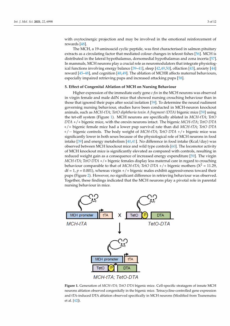
5. Effect of Congenital Ablation of MCH on Nursing Behaviour
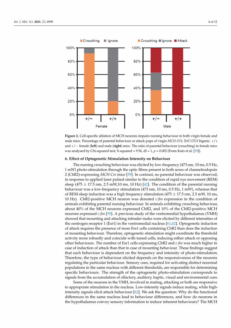
Higher expression of the immediate early gene c-fos in the MCH neurons was observedin virgin female and male ddN mice that showed nursing crouching behaviour than inthose that ignored their pups after social isolation [59]. To determine the neural rudimentgoverning nursing behaviour, studies have been conducted in MCH-neuron knockoutanimals, such as MCH-tTA; TetO diphtheria toxin A fragment (DTA) bigenic mice [59] usingthe tet-off system (Figure 1). MCH neurons are specifically ablated in MCH-tTA; TetODTA +/+ bigenic mice, with the orexin neurons intact. The bigenic MCH-tTA; TetO DTA+/+ bigenic female mice had a lower pup survival rate than did MCH-tTA; TetO DTA+/− bigenic controls. The body weight of MCH-tTA; TetO DTA +/+ bigenic mice wassignificantly lower in both sexes because of the physiological role of MCH neurons in foodintake [39] and energy metabolism [40,41]. No difference in food intake (Kcal/day) wasobserved between MCH knockout mice and wild type controls [60]. The locomotor activityof MCH knockout mice is significantly elevated as compared with controls, resulting inreduced weight gain as a consequence of increased energy expenditure [59]. The virginMCH-tTA; TetO DTA +/+ bigenic females display less maternal care in regard to crouchingbehaviour comparable to that of MCH-tTA; TetO DTA +/+ bigenic mothers (X2 = 11.29,df = 1, p = 0.001), whereas virgin +/+ bigenic males exhibit aggressiveness toward theirpups (Figure 2). However, no significant difference in retrieving behaviour was observed.Together, these findings indicated that the MCH neurons play a pivotal role in parentalnursing behaviour in mice.
Int. J. Mol. Sci. 2021, 22, x FOR PEER REVIEW 4 of 13
Figure 1. Generation of MCH-tTA; TetO DTA bigenic mice. Cell-specific stratagem of innate MCH neurons ablation observed congenitally in the bigenic mice. Tetracycline-controlled gene expression and tTA-induced DTA ablation observed specifically in MCH neurons (Modified from Tsunematsu et al. [42]).
Figure 2. Cell-specific ablation of MCH neurons impairs nursing behaviour in both virgin female and male mice. Percentage of parental behaviour or attack pups of virgin MCH-tTA; TetO DTA bi-genic. +/+ and +/− female (left) and male (right) mice. The ratio of parental behaviour (crouching) in female mice was analysed by Chi-squared test; X-squared = 9.56, df = 1, p = 0.002 (From Kato et al. [59]).
Figure 1. Generation of MCH-tTA; TetO DTA bigenic mice. Cell-specific stratagem of innate MCHneurons ablation observed congenitally in the bigenic mice. Tetracycline-controlled gene expressionand tTA-induced DTA ablation observed specifically in MCH neurons (Modified from Tsunematsuet al. [42]).
Int. J. Mol. Sci. 2021, 22, 6998 4 of 12
Int. J. Mol. Sci. 2021, 22, x FOR PEER REVIEW 4 of 13
Figure 1. Generation of MCH-tTA; TetO DTA bigenic mice. Cell-specific stratagem of innate MCH neurons ablation observed congenitally in the bigenic mice. Tetracycline-controlled gene expression and tTA-induced DTA ablation observed specifically in MCH neurons (Modified from Tsunematsu et al. [42]).
Figure 2. Cell-specific ablation of MCH neurons impairs nursing behaviour in both virgin female and male mice. Percentage of parental behaviour or attack pups of virgin MCH-tTA; TetO DTA bi-genic. +/+ and +/− female (left) and male (right) mice. The ratio of parental behaviour (crouching) in female mice was analysed by Chi-squared test; X-squared = 9.56, df = 1, p = 0.002 (From Kato et al. [59]).
Figure 2. Cell-specific ablation of MCH neurons impairs nursing behaviour in both virgin female andmale mice. Percentage of parental behaviour or attack pups of virgin MCH-tTA; TetO DTA bigenic. +/+and +/− female (left) and male (right) mice. The ratio of parental behaviour (crouching) in female micewas analysed by Chi-squared test; X-squared = 9.56, df = 1, p = 0.002 (From Kato et al. [59]).
6. Effect of Optogenetic Stimulation Intensity on Behaviour
The nursing crouching behaviour was elicited by low-frequency (473 nm, 10 ms, 0.5 Hz,1 mW) photo-stimulation through the optic fibres present in both sexes of channelrodopsin2 (ChR2)-expressing MCH-Cre mice [59]. In contrast, no parental behaviour was observed,in response to applied laser pulsed similar to the condition of rapid eye movement (REM)sleep (475 ± 17.5 nm, 2.5 mW,10 ms, 10 Hz) [42]. The condition of the parental nursingbehaviour was a low-frequency stimulation (473 nm, 10 ms, 0.5 Hz, 1 mW), whereas thatof REM sleep induction was a high frequency stimulation (475 ± 17.5 nm, 2.5 mW, 10 ms,10 Hz). ChR2-positive MCH neuron was denoted c-fos expression in the condition ofanimals exhibiting parental nursing behaviour. In animals exhibiting crouching behaviour,about 40% of the MCH neurons expressed ChR2, and 10% of the ChR2-positive MCHneurons expressed c-fos [59]. A previous study of the ventromedial hypothalamus (VMH)showed that mounting and attacking intruder males were elicited by different intensities ofthe oestrogen receptor 1 (Esr1) in the ventromedial nucleus [61,62]. Optogenetic inductionof attack requires the presence of more Ers1 cells containing ChR2 than does the inductionof mounting behaviour. Therefore, optogenetic stimulation might coordinate the thresholdactivity more robustly and coincide with tuned cells, inducing either attack or opposingother behaviours. The number of Esr1 cells expressing ChR2 and c-fos was much higher incase of induction of attack than that in case of mounting behaviour. These findings suggestthat each behaviour is dependent on the frequency and intensity of photo-stimulation.Therefore, the type of behaviour elicited depends on the responsiveness of the neuronsregulating the particular behaviour. Sensory cues, required for activating distinct neuronalpopulations in the same nucleus with different thresholds, are responsible for determiningspecific behaviours. The strength of the optogenetic photo-stimulation corresponds tosignals from the accumulation of olfactory, auditory, haptic, visual and environmental cues.
Some of the neurons in the VMH, involved in mating, attacking or both are responsiveto appropriate stimulation in the nucleus. Low-intensity signals induce mating, while high-intensity signals elicit attack behaviour [62]. We ask the question: Why do the functionaldifferences in the same nucleus lead to behaviour differences, and how do neurons inthe hypothalamus convey sensory information to induce inherent behaviours? The MCH
Int. J. Mol. Sci. 2021, 22, 6998 5 of 12
neuronal state for behaviour is variably induced by photo-stimulation: Low stimulationelicits nursing, and high stimulation elicits REM sleep. The neural circuit responsiblefor this MCH-induced behaviour could communicate with other brain areas to integrateeach behaviour. The extraordinary event of the behaviour must be induced by differentreactivities of the responsible neurons.
7. MCH Neural Relay in PVN Oxytocin Neurons Is Involved in Nursing Behaviour
Further evidence shows that MCH neurons are regulated by OT neurons in PVN thatproject anatomically posterior to LHA [38]. OT, a neurotransmitter synthesised in both thePVN and supraoptic nuclei of the hypothalamus [63], regulates peripheral reproductive-related functions and central actions in the brain. OT secretion from the posterior pituitarygland induces uterine contractions during parturition and also acts on the muscle in themammary gland trabeculae to induce milk ejection during lactation [63]. Oxytocinergicneurons are involved in a variety of central nervous system functions. Centrally andperipherally secreted OT acts through the OT receptor. This receptor is distributed inthe ventral tegmental area (VTA) and NAc and is involved in feeding, sexual behaviourand reward properties of social interactions and the formation of social bonds. OT hasbeen shown to facilitate the onset of maternal behaviour in rodents [31,32]. The possiblemechanism responsible for parental nursing behaviour is involved in the neural relay forthe LHA-PVN within the hypothalamus. Studies have shown that the periaqueductal grey(PAG) in the midbrain is implicated in reproductive behaviour such as the females’ lordosisbehaviour and the maternal arched back crouching behaviour, whereas no effects wererecorded in the pup grooming behaviour [64–66].
Alternatively, the stimulation of the projection to the PAG from the galanin neuronsresults in pup grooming, albite with no effect on crouching in both sexes [30]. Maternalbehaviour could be regulated by the LHA–MCH neuronal input to the PAG. GABAergicneurons in the LHA-to-PAG projection precipitate in predatory hunting in mice [66].Whereas, the role of the MCH receptor in the PAG is yet to be determined. However, themethod whereby the neural circuit for these diverse parenting behaviours govern each ofthe behavioural contents is still controversial.
8. OT Enhances the Neural Circuits of Rewarding from Pups
Previous studies have identified the mPOA as a critical region in the regulation ofparental behaviour [1–3,30,67–70]. OT neurons act on mPOA, VTA and NAc to promptparental behaviour. OT neurons in PVN receive the projection from the LHA [59]. In ourprevious study, MCH-tTA; TetO DTA bigenic (+/+) female mice with the complete innateablation of MCH neurons displayed less attention towards pups and less maternal carethan MCH-tTA; TetO DTA +/− bigenic controls, which was similar to MCH-tTA; TetO DTA+/+ bigenic mothers, that display significantly lowered crouching than the +/− controls.Moreover, the virgin +/+ bigenic females showed significantly lowered crouching thanMCH-tTA; TetO DTA +/− bigenic controls. MCH neurons are ablated partially using Crerecombinase-dependent DTA, which abolishes approximately 73% of MCH neurons invirgin females. Virgin females with partially ablated MCH neurons exhibit crouchingbehaviour for less time than green florescent protein controls. The MCHR expression isnecessary to the reward circuitry of the NAc as is the anatomic integrity of the oxytocinergicprojection of the mesolimbic system; these findings indicate a possible alliance betweenthese factors in the emotional reinforcement of rewards for parenting. The MCHR isexpressed in the olfactory regions, neocortex, hippocampus, NAc, amygdala, ventromedialhypothalamic nucleus and locus coeruleus. OT receptor expression [71], coupled withMCHR in the NAc has a synergistic effect on inherent rewarding, contributing to theexecution of parental behaviour [72]. Therefore, MCH neural networks along with OTsignalling in reward circuitry facilitate pup survival.
Maternal rewards system may contribute to maternal nursing systems. Relationshipsbetween maternal depression and OT levels were demonstrated previously [73]. MCH–
Int. J. Mol. Sci. 2021, 22, 6998 6 of 12
LHA projects closely to OT–PVN, which is the stimulation that induces parental behaviouralong with increasing plasma OT levels [59]. The recurrent process between the PVNprojection to LHA and the LHA projection to PVN are assumed to be tuning propertiesfor continuous parenting crouching behaviour. Almost all MCH neurons expressed OTreceptor mRNA; however, OT neurons faintly expressed the MCH receptor [38,50].
9. Social Isolation Modifies GABAergic Transmission
Extended periods of social isolation can affect parental behaviour by inducing neu-roanatomical changes. The expression of the immediate early gene c-fos in the MCHneurons increased during parental nursing behaviour of mice after social isolation [59].MCH neurons in the LHA contain and release γ-aminobutyric acid (GABA) [59,74] as wellas express GABA-synthesising enzymes GAD65 and GAD67 [59,75,76]. MCH neuronsalso contain and release glutamate in the lateral septum [77,78]. In the amygdala neuralcircuits, GABAergic and glutamatergic neurons in the VTA specifically tune each rewardingand aversive motivational predicaments [79]. mPOA, which is indicative of GABAergicneurotransmission, governs parental behaviour, whereas glutamatergic neurons in thesame nucleus are associated with anxiety-like behaviour [80]. Glutamatergic neurons in themPOA regulate anxiety-like behaviour, while GABAergic neurons contribute to anxiolyticeffects, i.e., parental behaviour, indicating that the mPOA in the same nucleus plays acrucial role in reconciliation of opposite behaviours. Moreover, the mPOA projections tothe neurons in midbrain reward circuits may prompt parental behavior with accommo-dation for dopamine release [80]. Therefore, GABAergic and glutamatergic neurons playa prominent role in opposing effects on the social behaviour. These results suggest thatthe same nucleus governs opposite positive or negative behaviours by discriminating theneurotransmission of the nucleus. Social isolation, which prompts parental behaviour forseveral resting weeks, could change the statement of the brain neurotransmission. In fact,conditions, such as accrues to excitatory neurotransmission of GABA neurons result in aprofound depolarising shift in magnocellular neurosecretory cells that secrete OT in thePVN [81]. Our previous study has shown that MCH fibre expressing enhanced yellowfluorescent protein projected close proximity to the OT neuron in the PVN. The MCHneuron was revealed to express GABA, which innervates as a neurotransmitter and formssynapses with OT neurons [59]. Moreover, GABA agonist musicimol injected into thePVN increases c-fos in the OT neurons. More c-fos expressing OT neurons were observedin the socially isolated female and male mice than the co-habituated female and malemice [59], indicating that the MCH neuron could regulate excitatory OT neurons in thePVN. Although GABA principally functions as an inhibitory neurotransmitter in the brain,excitatory GABAergic activity is identified in MCH neurons during development [82].In mature neurons, but the excitatory action of GABA under stress conditions has beenelucidated [83–86]. Social isolation might be presumed to be a validated stressor and toelicit changes in the synaptic organisation action in the rodent brain [16,28].
Therefore, social isolation stress may change the mode of GABAergic excitation. Theprojection from LHA to PVN under social, reward-context associations are responsible forthe LHA–PVN-evoked OT releases implicated in parenting augmentation. Therefore, socialisolation may change the mechanisms underlying the modality of GABAergic excitation.Abolishing MCH neurons may induce the superiority of glutamatergic circuit, thereby,stimulating broad area commitment to infanticide in the brain. Moreover, the impairmentof OT neurons showed acceleration aggression. The balance between GABA and glutamateutilisation in the MCH neurons in some aspects of parental and opposing behavioursremains to be elucidated.
10. Aggressive Behaviour towards Pups
Pheromonal signals are received by neurons in the vomeronasal organ (VNO) [87] andthe main olfactory epithelium within the nasal cavity [88]. In rodents, olfaction is known tobe important for the identification of conspecifics and sex differentiation. The excision of the
Int. J. Mol. Sci. 2021, 22, 6998 7 of 12
olfactory bulb results in defects in aggressive behaviour, indicating that olfactory perceptionis involved in dictating aggressiveness. A body of evidence implicates neural networksin the governing of aggressive behaviour, including a social behaviour circuit involvingthe mPOA, medial amygdala (MeA), bed nucleus of the stria terminalis (BNST) [89],lateral septum, anterior hypothalamus, VMH and PAG [65]. Social signals detected by theolfactory bulb are subsequently transmitted to specific brain regions: MeA and then to theVMH or BNST [90]. The VMH is downstream of the MeA, which in turn disinhibits theVMHvl glutamatergic neuron induction of aggressive behaviour; for example, this circuitryguides the behaviour in which male mice attack male intruders but not females [91,92].Distinguishing between females and males is also accomplished through the detection ofsemiochemicals, some of which are major urinary proteins [93,94]. These chemicals aredetected by sensory neurons in the VNO in a sex-dependent manner [95–97]. For example,during infanticide by virgin male mice, pheromonal signals from pups via the VNO are sentto the accessory olfactory bulb, then to the MeA and relayed to the anterior hypothalamicarea/VMH and BNST. The VNO neurons presumably are involved in the detection ofpheromonal signals related to parental care [3]. Knockout of cation channel subfamily Cmember 2 (Trpc2) in male mice causes impaired VNO-input signalling, resulting in reducedattacking of pups, indicating that VNO signalling elicits the attacking of pups [98–100].Our previous study reports that Trpc2 KO mice spent more time licking the pups andcrouching after social isolation. However, retrieval behaviour increased only in response tosocial isolation and was not affected by Trpc2 KO. These results indicate that not all thesocial signals are transmitted by the VNO [101].
The MCH-tTA; TetO DTA +/+ bigenic virgin males with ablated MCH neurons weremore aggressive toward the pups. In the resident-intruder test, the MCH-tTA; TetO DTA+/+ bigenic virgin mice, exhibited more male–male aggression than did MCH-tTA; TetODTA +/− bigenic controls [59]. Ablation of MCH neurons also leads MCH-tTA; TetO DTA+/+ bigenic male mice to exhibit more aggressiveness against other male mice and pups,suggesting that MCH neurons disinhibit the olfactory circuit and sensory integration fromthe olfactory bulb. This result is similar to that observed with neural circuit modulationthat results in male attack on pups and intermale aggressive behaviour [102]. The MeA isevidenced as an inhibitor of maternal behaviour [103–107]. Moreover, MCHR in the MeAis assumed to be implicated in maternal aggression towards intruder male [107]. The VNOand main olfactory bulb may be substantially involved in male–male aggressive behaviourprompted by pheromones [100] and play decisive roles in conferring infanticide in mice.The MCH-tTA; TetO DTA +/+ bigenic virgin mice were able to mate because of their abilityto discriminate sex due to their preserved VNO function. Neural circuits for attacking pupsmay be involved in relaying from MCH neurons in the LHA (MCH–LHA) to OT neuronsin the PVN (OT–PVN) in rodents [59].
The ablation of MCH neuron excitability to PVN may prevent the induction of OTsecretion. In contrast, MCH-tTA; TetO DTA +/+ bigenic female mice ignore the pups,indicating that the effect of abolishing MCH neurons differed in some degree betweenfemales and males. OT transmits signals involved in social interactions, such as parentaland pair bonding; abolishing OT facilitates aggressive behaviour [108]. Presumably, OTacts on the mPOA, which induces parental behaviour in rats [109,110].
Another proposed mechanism is effects on OT, which acts to regulate the salience ofexternal social cue rather than affiliative behaviours [111–113], which is a critical role of OTin the event of the discriminate mode of an emotional action in a conspecific [114].
11. Parental Licking Behaviour
Optogenetically-evoked crouching behaviour requires around 10% of the ChR2-expressing and MCH cells that express c-fos. The photo-stimulation of ChR2 MCH neuronssignificantly increased crouching behaviour, but did not affect licking behaviour [59].Therefore, the relative contribution of MCH neurons to licking behaviour may be minimal.
Int. J. Mol. Sci. 2021, 22, 6998 8 of 12
Mice contact pups at first and pup-licking is assumed to be dictated by emotive excitationor apprehension [115,116].
Optogenetic stimulation of galanin neurons in the mPOA induces retrieving andpup grooming rather decreasing attacking pups [30] and had no effect on other parentalbehaviours. Genetical ablation of galanin neurons in the mPOA induces pup attackingin virgin females but not in mating-experienced females and males. Therefore, the braincentre for pup retrieving and grooming behaviour is in the mPOA, and galanin is oneof the molecules involved in parenting pup grooming. Pup grooming behaviour is alsoaffected by GABAergic neurons in the posterodorsal (MeApd) in females [78]. The effectof photo-stimulation on retrieving pups and crouching is less than that on pup grooming.Higher GABAergic neuron activity in the MeApd induces the attacking of pups, whilelow activity of these neurons prompts parenting in male mice. Opposing behaviours, suchas parenting and aggression, are centred in different regions of the brain. For example,MeApd facilitates parenting behaviour, while aggression is colinear with the quantitativeresponses of GABAergic neurons in the brain.
12. Conclusions
Parental behaviour is composed of sequential behaviour events induced by an associ-ated nucleus for each event that is stimulated by a social cue. In this chapter, I proposedthat the key brain regions and molecules involved in regulating parental behaviour residein the POA, the focus of much research on this topic. Genetic ablation of MCH neurons intransgenic, MCH-tTA; TetO DTA +/+ mice results in impaired nursing parental behaviour.Virgin MCH-tTA; TetO DTA +/+ bigenic males engaged in infanticide toward the pups,while females ignored pups. A neural circuit from the LHA–MCH to PVN–OT was re-vealed in this study, and MCH reward neural circuitry, together with OT signalling, is arequisite for parental behaviours that promote pup survival.
Funding: This work was funded, in part, by grants-in-aid for scientific research from the JapaneseMinistry of Education, Science, Sports and Culture [19K12738] (C.O.).
Institutional Review Board Statement: Not applicable.
Informed Consent Statement: Not applicable.
Data Availability Statement: Not applicable.
Acknowledgments: The author thanks A. Inutsuka, Y. Kato, S. Minami, T. Onaka, A. Yamanaka andH. Katsumata for their helpful advice and technical assistance.
Conflicts of Interest: The author declares no conflict of interest.
References1. Numan, M. The Parental Behavior: Mechanisms, Development, and Evolution; Oxford University Press: Oxford, UK, 2020.2. Tachikawa, K.S.; Yoshihara, Y.; Kuroda, K.O. Behavioral transition from attack to parenting in male Mice: A crucial role of the
vomeronasal system. J. Neurosci. 2013, 33, 5120–5126. [CrossRef]3. Wu, Z.; Autry, A.E.; Bergan, J.F.; Watabe-Uchida, M.; Dulac, C.G. Galanin neurons in the medial preoptic area govern parental
behaviour. Nature 2014, 509, 325–330. [CrossRef]4. Marrocco, J.; McEwen, B.S. Sex in the brain: Hormones and sex differences. Dialogues Clin. Neurosci. 2016, 18, 373–383. [PubMed]5. Ubuka, T.; Trudeau, V.L.; Parhar, I. Editorial: Steroids and the Brain. Front. Endocrinol. 2020, 11, 366. [CrossRef]6. Blakemore, J.; Naftolin, F. Aromatase: Contributions to Physiology and Disease in Women and Men. Physiology 2016, 31, 258–269.
[CrossRef]7. McEwen, B.S. Hormones and behavior and the integration of brain-body science. Horm. Behav. 2020, 119, 104619. [CrossRef]8. Arnold, A.P. A general theory of sexual differentiation. J. Neurosci. Res. 2017, 95, 291–300. [CrossRef]9. Bakker, J. The Sexual Differentiation of the Human Brain: Role of Sex Hormones Versus Sex Chromosomes. In Neuroendocrine
Regulation of Behavior; Springer: Cham, Switzerland, 2018; Volume 43, pp. 45–67.10. McCarthy, M.M.; Wright, C.L. Convergence of Sex Differences and the Neuroimmune System in Autism Spectrum Disorder. Biol.
Psychiatry 2017, 81, 402–410. [CrossRef]11. Forger, N.G. Epigenetic mechanisms in sexual differentiation of the brain and behaviour. Philos. Trans. R. Soc. B Biol. Sci. 2016,
371, 20150114. [CrossRef]
Int. J. Mol. Sci. 2021, 22, 6998 9 of 12
12. Döhler, K.D.; Coquelin, A.; Davis, F.; Hines, M.; Shryne, J.E.; Gorski, R.A. Differentiation of the sexually dimorphic nucleus in thepreoptic area of the rat brain is determined by the perinatal hormone environment. Neurosci. Lett. 1982, 33, 295–298. [CrossRef]
13. Mhaouty-Kodja, S.; Naulé, L.; Capela, D. Sexual Behavior: From Hormonal Regulation to Endocrine Disruption. Neuroendocrinol-ogy 2018, 107, 400–416. [CrossRef]
14. Ribeiro, A.C.; Musatov, S.; Shteyler, A.; Simanduyev, S.; Arrieta-Cruz, I.; Ogawa, S.; Pfaff, D.W. siRNA silencing of estrogenreceptor-α expression specifically in medial preoptic area neurons abolishes maternal care in female mice. Proc. Natl. Acad. Sci.USA 2012, 109, 16324–16329. [CrossRef] [PubMed]
15. Orikasa, C.; Nagaoka, K.; Katsumata, H.; Sato, M.; Kondo, Y.; Minami, S.; Sakuma, Y. Social isolation prompts maternal behaviorin sexually naïve male ddN mice. Physiol. Behav. 2015, 151, 9–15. [CrossRef]
16. Mumtaz, F.; Khan, M.I.; Zubair, M.; Dehpour, A.R. Neurobiology and consequences of social isolation stress in animal model-Acomprehensive review. Biomed. Pharm. 2018, 105, 1205–1222. [CrossRef]
17. Buwalda, B.; Geerdink, M.; Vidal, J.; Koolhaas, J.M. Social behavior and social stress in adolescence: A focus on animal models.Neurosci. Biobehav. Rev. 2011, 35, 1713–1721. [CrossRef]
18. Farbstein, D.; Hollander, N.; Peled, O.; Apter, A.; Fennig, S.; Haberman, Y.; Gitman, H.; Yaniv, I.; Shkalim, V.; Pick, C.G.; et al.Social isolation in mice: Behavior, immunity, and tumor growth. Stress 2021, 24, 229–238. [CrossRef] [PubMed]
19. Takeda, A.; Tamano, H.; Kan, F.; Hanajima, T.; Yamada, K.; Oku, N. Enhancement of social isolation-induced aggressive behaviorof young mice by zinc deficiency. Life Sci. 2008, 82, 909–914. [CrossRef]
20. O’Keefe, L.M.; Doran, S.J.; Mwilambwe-Tshilobo, L.; Conti, L.H.; Venna, V.R.; McCullough, L.D. Social isolation after stroke leadsto depressive-like behavior and decreased BDNF levels in mice. Behav. Brain Res. 2014, 260, 162–170. [CrossRef]
21. Koike, H.; Ibi, D.; Mizoguchi, H.; Nagai, T.; Nitta, A.; Takuma, K.; Nabeshima, T.; Yoneda, Y.; Yamada, K. Behavioral abnormalityand pharmacologic response in social isolation reared mice. Behav. Brain Res. 2009, 202, 114–121. [CrossRef]
22. Kozhemyakina, R.V.; Shikhevich, S.G.; Konoshenko, M.Y.; Gulevich, R.G. Adolescent oxytocin treatment affects resident behaviorin aggressive but not tame adult rats. Physiol. Behav. 2020, 224, 113046. [CrossRef]
23. Tan, O.; Musullulu, H.; Raymond, J.S.; Wilson, B.; Langguth, M.; Bowen, M.T. Oxytocin and vasopressin inhibit hyper-aggressivebehaviour in socially isolated mice. Neuropharmacology. 2019, 156, 107573. [CrossRef] [PubMed]
24. Stevenson, J.R.; McMahon, E.K.; Boner, W.; Haussmann, M.F. Oxytocin administration prevents cellular aging caused by socialisolation. Psychoneuroendocrinology 2019, 103, 52–60. [CrossRef]
25. Ferdman, N.; Murmu, R.P.; Bock, J.; Braun, K.; Leshem, M. Weaning age, social isolation, and gender, interact to determine adultexplorative and social behavior, and dendritic and spine morphology in prefrontal cortex of rats. Behav. Brain Res. 2007, 180,174–182. [CrossRef]
26. Du Preez, A.; Onorato, D.; Eiben, I.; Musaelyan, K.; Egeland, M.; Zunszain, P.A.; Fernandes, C.; Thuret, S.; Pariante, C.M. Chronicstress followed by social isolation promotes depressive-like behaviour, alters microglial and astrocyte biology and reduceshippocampal neurogenesis in male mice. Brain Behav. Immun. 2021, 91, 24–47. [CrossRef] [PubMed]
27. Deguchi, Y.; Harada, M.; Shinohara, R.; Lazarus, M.; Cherasse, Y.; Urade, Y.; Yamada, D.; Sekiguchi, M.; Watanabe, D.; Furuyashiki,T.; et al. mDia and ROCK Mediate Actin-Dependent Presynaptic Remodeling Regulating Synaptic Efficacy and Anxiety. Cell Rep.2016, 17, 2405–2417. [CrossRef]
28. Matthews, G.A.; Edward, H.; Nieh, E.H.; Weele, C.M.V.; Halbert, S.A.; Pradhan, R.V.; Yosafat, A.S.; Glober, G.F.; Izadmehr, E.M.;Thomas, R.E.; et al. Dorsal Raphe Dopamine Neurons Represent the Experience of Social Isolation. Cell 2016, 164, 617–631.[CrossRef]
29. Scott, N.; Prigge, M.; Yizhar, O.; Kimchi, T. A sexually dimorphic hypothalamic circuit controls maternal care and oxytocinsecretion. Nature 2015, 525, 519–522. [CrossRef] [PubMed]
30. Kohl, J.; Babayan, B.M.; Rubinstein, N.D.; Autry, A.E.; Marin-Rodriguez, B.; Kapoor, V.; Miyamishi, K.; Zweifel, L.S.; Luo, L.;Uchida, N.; et al. Functional circuit architecture underlying parental behaviour. Nature 2018, 556, 326–331. [CrossRef] [PubMed]
31. Marlin, B.J.; Mitre, M.; D’amour, J.A.; Chao, M.V.; Froemke, R.C. Oxytocin enables maternal behaviour by balancing corticalinhibition. Nature 2015, 520, 499–504. [CrossRef] [PubMed]
32. Watarai, A.; Tsutaki, S.; Nishimori, K.; Okuyama, T.; Mogi, K.; Kikusui, T. The blockade of oxytocin receptors in the paraventricularthalamus reduces maternal crouching behavior over pups in lactating mice. Neurosci. Lett. 2020, 720, 134761. [CrossRef] [PubMed]
33. Yoshihar, C.; Numan, M.; Kuroda, K.O. Oxytocin and Parental Behaviors. Curr. Top. Behav. Neurosci. 2018, 35, 119–153.34. Rassovsky, Y.; Harwood, A.; Zagoory-Sharon, O.; Feldman, R. Martial arts increase oxytocin production. Sci. Rep. 2019, 9, 12980.
[CrossRef]35. Tse, W.S.; Siu, A.F.Y.; Zhang, Q.; Chan, H.Y.E. Maternal oxytocin responsiveness improves specificity of positive social memory
recall. Psychoneuroendocrinology 2018, 98, 148–152. [CrossRef]36. Bartz, J.A.; Zaki, J.; Ochsner, K.N.; Bolger, N.; Kolevzon, A.; Ludwig, N.; Lydon, J.E. Effects of oxytocin on recollections of
maternal care and closeness. Proc. Natl. Acad. Sci. USA 2010, 107, 21371–21375. [CrossRef]37. Brill-Maoz, N.; Maroun, M. Extinction of fear is facilitated by social presence: Synergism with prefrontal oxytocin. Psychoneuroen-
docrinology 2016, 66, 75–81. [CrossRef] [PubMed]38. Yao, Y.; Fu, L.Y.; Zhang, X.; van den Pol, A.N. Vasopressin and oxytocin excite MCH neurons, but not other lateral hypothalamic
GABA neurons. Am. J. Physiol. Regul. Integr. Comp. Physiol. 2012, 302, R815–R824. [CrossRef]
Int. J. Mol. Sci. 2021, 22, 6998 10 of 12
39. Qu, D.; Ludwig, D.S.; Gammeltoft, S.; Piper, M.; Pelleymounter, M.A.; Cullen, M.J.; Mathes, W.F.; Przypek, R.; Kanarek, R.;Maratos-Flier, E. A role for melanin-concentrating hormone in the central regulation of feeding behaviour. Nature 1996, 380,243–247. [CrossRef] [PubMed]
40. Shimada, M.; Tritos, N.A.; Lowell, B.B.; Flier, J.S.; Maratos-Flier, E. Mice lacking melanin-concentrating hormone are hypophagicand lean. Nature 1998, 396, 670–674. [CrossRef]
41. Marsh, D.J.; Weingarth, D.T.; Novi, D.E.; Chen, H.Y.; Trumbauer, M.E.; Chen, A.S.; Guan, X.M.; Jiang, M.M.; Feng, Y.; Camacho,R.E.; et al. Melanin-concentrating hormone 1 receptor-deficient mice are social isolation stress rodent lean, hyperactive, andhyperphagic and have altered metabolism. Proc. Natl. Acad. Sci. USA 2002, 99, 3240–3245. [CrossRef] [PubMed]
42. Tsunematsu, T.; Ueno, T.; Tabuchi, S.; Inutsuka, A.; Tanaka, K.F.; Hasuwa, H.; Kilduff, T.S.; Terao, A.; Yamanaka, A. Optogeneticmanipulation of activity and temporally controlled cell-specific ablation reveal a role for MCH meurons in sleep/wake regulation.J. Neurosci. 2014, 34, 6896–6909. [CrossRef]
43. Alhassen, L.; Phan, A.; Alhassen, W.; Nguyen, P.; Lo, A.; Shaharuddin, H.; Sanathara, N.; Civelli, O.; Alachkar, A. The role ofOlfaction in MCH-regulated spontaneous maternal responses. Brain Res. 2019, 1719, 71–76. [CrossRef]
44. Concetti, C.; Bracey, E.F.; Peleg-Raibstein, D.; Burdakov, D. Control of fear extinction by hypothalamic melanin-concentratinghormone-expressing neurons. Proc. Natl. Acad. Sci. USA 2020, 117, 22514–22521. [CrossRef]
45. García-Fuster, M.J.; Parks, G.S.; Clinton, S.M.; Watson, S.J.; Akil, H.; Civelli, O. The melanin-concentrating hormone (MCH)system in an animal model of depression-like behavior. Eur. Neuropsychopharmacol. 2012, 22, 607–613. [CrossRef]
46. Blouin, A.M.; Fried, I.; Wilson, C.L.; Staba, R.J.; Behnke, E.J.; Lam, H.A.; Maidment, N.T.; Karlsson, K.Æ.; Lapierre, J.L.; Siegel, J.M.Human hypocretin and melanin-concentrating hormone levels are linked to emotion and social interaction. Nat. Commun. 2013,4, 1547. [CrossRef]
47. Roy, M.; David, N.; Cueva, M.; Giorgetti, M. A study of the involvement of melanin-concentrating hormone receptor 1 (MCHR1)in murine models of depression. Biol. Psychiatry. 2007, 61, 174–180. [CrossRef]
48. Sherwood, A.; Wosiski-Kuhn, M.; Nguyen, T.; Holland, P.C.; Lakaye, B.; Adamantidis, A.; Johnson, A.W. The role of melanin-concentrating hormone in conditioned reward learning. Eur. J. Neurosci. 2012, 36, 3126–3133. [CrossRef]
49. Izawa, S.; Chowdhury, S.; Miyazaki, T.; Mukai, Y.; Ono, D.; Inoue, R.; Ohmura, Y.; Mizoguchi, H.; Kimura, K.; Yoshioka, M.; et al.REM sleep-active MCH neurons are involved in forgetting hippocampus-dependent memories. Science 2019, 365, 1308–1313.[CrossRef]
50. Komagata, N.; Latifi, B.; Rusterholz, T.; Bassetti, C.L.A.; Adamantidis, A.; Schmidt, M.H. Dynamic REM Sleep Modulation byAmbient Temperature and the Critical Role of the Melanin-Concentrating Hormone System. Curr. Biol. 2019, 29, 1976–1987.e4.[CrossRef]
51. Saito, Y.; Cheng, M.; Leslie, F.M.; Civelli, O. Expression of the melanin-concentrating hormone (MCH) receptor mRNA in the ratbrain. J. Comp. Neurol. 2001, 435, 26–40. [CrossRef] [PubMed]
52. Jang, J.H.; Park, J.Y.; Oh, J.Y.; Bae, S.J.; Jang, H.; Jeon, S.; Kim, J.; Park, H.J. Novel analgesic effects of melanin-concentratinghormone on persistent neuropathic and inflammatory pain in mice. Sci. Rep. 2018, 8, 707. [CrossRef]
53. Teixeira, P.D.S.; Wasinski, F.; Lima, L.B.; Frazão, R.; Bittencourt, J.C.; Donato, J., Jr. Regulation and neurochemical identity ofmelanin-concentrating hormone neurones in the preoptic area of lactating mice. J. Neuroendocrinol. 2020, 32, e12818. [CrossRef][PubMed]
54. Kawata, Y.; Okuda, S.; Hotta, N.; Igawa, H.; Takahashi, M.; Ikoma, M.; Kasai, S.; Ando, A.; Satomi, Y.; Nishida, M.; et al. A noveland selective melanin-concentrating hormone receptor 1 antagonist ameliorates obesity and hepatic steatosis in diet-inducedobese rodent models. Eur. J. Pharmacol. 2017, 796, 45–53. [CrossRef] [PubMed]
55. Sanathara, N.M.; Garau, C.; Alachkar, A.; Wang, L.; Wang, Z.; Nishimori, K.; Xu, X.; Civelli, O. Melanin concentrating hormonemodulates oxytocin-mediated marble burying. Neuropharmacology 2018, 128, 22–32. [CrossRef] [PubMed]
56. Kawauchi, H.; Kawazoe, I.; Tsubokawa, M.; Kishida, M.; Baker, B.I. Characterization of melanin-concentrating hormone in chumsalmon pituitaries. Nature 1983, 305, 321–323. [CrossRef]
57. Bittencourt, J.C.; Presse, F.; Arias, C.; Peto, C.; Vaughan, J.; Nahon, J.L.; Vale, W.; Sawchenko, P.E. The melanin-concentratinghormone system of the rat brain: An immuno- and hybridization histochemical characterization. J. Comp. Neurol. 1992, 319,218–245. [CrossRef]
58. Alachkar, A.; Alhassen, L.; Wang, Z.; Wang, L.; Onouye, K.; Sanathara, N.; Civelli, O. Inactivation of the melanin concentratinghormone system impairs maternal behavior. Eur. Neuropsychopharmacol. 2016, 26, 1826–1835. [CrossRef]
59. Kato, Y.; Katsumata, H.; Inutsuka, A.; Yamanaka, A.; Onaka, T.; Minami, S.; Orikasa, C. Involvement of MCH-oxytocin neuralrelay within the hypothalamus in murine nursing behavior. Sci. Rep. 2021, 11, 3348. [CrossRef]
60. Kokkotou, E.; Jeon, J.Y.; Wang, X.; Marino, F.E.; Carlson, M.; Trombly, D.J.; Maratos-Flier, E. Mice with MCH ablation resistdiet-induced obesity through strain-specific mechanisms. Am. J. Physiol. Regul. Integr. Comp. Physiol. 2005, 289, R117–R124.[CrossRef]
61. Remedios, R.; Kennedy, A.; Zelikowsky, M.; Grewe, B.F.; Schnitzer, M.J.; Anderson, D.J. Social behaviour shapes hypothalamicneural ensemble representations of conspecific sex. Nature 2017, 550, 388–392. [CrossRef]
62. Lee, H.; Kim, D.-W.; Remedios, R.; Anthony, T.E.; Chang, A.; Madisen, L.; Hongkui, Z.; Anderson, D.J. Scalable control ofmounting and attack by Esr1+ neurons in the ventromedial hypothalamus. Nature 2014, 509, 627–632. [CrossRef]
Int. J. Mol. Sci. 2021, 22, 6998 11 of 12
63. Costa, H.C.; Da-Silva, J.M.; Diniz, G.B.; Motta-Teixeira, L.C.; Da-Silva, R.J.; Battagello, D.S.; Sita, L.V.; de Moraes, M.C.; Horta-Júnior, J.A.C.; Bittencourt, J.C. Characterisation and origins of melanin-concentrating hormone immunoreactive fibres of theposterior lobe of the pituitary and median eminence during lactation in the Long-Evans rat. J. Neuroendocrinol. 2019, 31, e12723.[CrossRef]
64. Lonstein, J.S.; Stern, J.M. Site and behavioral specificity of periaqueductal gray lesions on postpartum sexual, maternal, andag-gressive behaviors in rats. Brain Res. 1998, 804, 21–35. [CrossRef]
65. Salzberg, H.C.; Lonstein, J.S.; Stern, J.M. GABA(A) receptor regulation of kyphotic nursing and female sexual behavior inthecaudal ventrolateral periaqueductal gray of postpartum rats. Neuroscience 2002, 114, 675–687. [CrossRef]
66. Rossier, D.; Franca, V.L.; Salemi, T.; Natale, S.; Gross, C.T. A neural circuit for competing approach and defense underlying preycapture. Proc. Natl. Acad. Sci. USA 2021, 118, e2013411118. [CrossRef]
67. Tsuneoka, Y.; Maruyama, T.; Yoshida, S.; Nishimori, K.; Kato, T.; Numan, M.; Kuroda, K.O. Functional, anatomical, andneurochemical differentiation of medial preoptic area subregions in relation to maternal behavior in the mouse. J. Comp. Neurol.2013, 521, 1633–1663. [CrossRef]
68. Fang, Y.Y.; Yamaguchi, T.; Song, S.C.; Tritsch, N.X.; Lin, D.A. Hypothalamic Midbrain Pathway Essential for Driving MaternalBehaviors. Neuron 2018, 98, 192–207.e10. [CrossRef]
69. Wei, Y.-C.; Wang, S.-R.; Jiao, Z.-L.; Zhang, W.; Lin, J.-K.; Li, X.-Y.; Li, S.-S.; Zhang, X.; Xu, X.-H. Medial preoptic area in mice iscapable of mediating sexually dimorphic behaviors regardless of gender. Nat. Commun. 2018, 18, 279. [CrossRef]
70. Numan, M. Medial preoptic area and maternal behavior in the female rat. J. Comp. Physiol. Psychol. 1974, 87, 746–759. [CrossRef]71. Young, L.J.; Wang, Z. The neurobiology of pair bonding. Nat. Neurosci. 2004, 7, 1048–1054. [CrossRef]72. Kelly, A.M.; Hiura, L.C.; Saunders, A.G.; Ophir, A.G. Oxytocin Neurons Exhibit Extensive Functional Plasticity Due To Offspring
Age in Mothers and Fathers. Integr. Comp. Biol. 2017, 57, 603–618. [CrossRef]73. Caitlin Post, C.; Leuner, B. The Maternal Reward System in Postpartum Depression. Arch. Womens Ment. Health 2019, 22, 417–429.
[CrossRef] [PubMed]74. Del Cid-Pellitero, E.; Jones, B.E. Immunohistochemical evidence for synaptic release of GABA from melanin-concentrating
hormone containing varicosities in the locus coeruleus. Neuroscience 2012, 223, 269–276. [CrossRef] [PubMed]75. Elias, C.F.; Lee, C.E.; Kelly, J.F.; Ahima, R.S.; Kuhar, M.; Saper, C.B.; Elmquist, J.K. Characterization of CART neurons in the rat
and human hypothalamus. J. Comp. Neurol. 2001, 432, 1–19. [CrossRef] [PubMed]76. Harthoorn, L.F.; Sañé, A.; Nethe, M.; Heerikhuize, J.J.V. Multi-transcriptional profiling of melanin-concentrating hormone and
orexin-containing neurons. Cell Mol. Neurobiol. 2005, 25, 1209–1223. [CrossRef]77. Abrahamson, E.E.; Leak, R.K.; Moore, R.Y. The suprachiasmatic nucleus projects to posterior hypothalamic arousal systems.
Neuroreport 2001, 12, 435–440. [CrossRef]78. Chee, M.J.S.; Arrigoni, E.; Maratos-Flier, E. Melanin-concentrating hormone neurons release glutamate for feedforward inhibition
of the lateral septum. J. Neurosci. 2015, 35, 3644–3651. [CrossRef] [PubMed]79. Jennings, J.H.; Sparta, D.R.; Stamatakis, A.M.; Ung, R.L.; Pleil, K.E.; Kash, T.L.; Stuber, G.D. Distinct extended amygdala circuits
for divergent motivational states. Nature 2013, 496, 224–228. [CrossRef]80. McHenry, J.A.; Otis, J.M.; Rossi, M.A.; Robinson, J.E.; Kosyk, O.; Miller, N.W.; McElligott, Z.A.; Budygin, E.A.; Rubinow, D.R.;
Stuber, G.D. Hormonal gain control of a medial preoptic area social reward circuit. Nat. Neurosci. 2017, 20, 449–458. [CrossRef][PubMed]
81. Gies, U.; Theodosis, D.T. Synaptic plasticity in the rat supraoptic nucleus during lactation involves GABA innervation andoxytocin neurons: A quantitative immunocytochemical analysis. J. Neurosci. 1994, 14 Pt 1, 2861–2869. [CrossRef]
82. Li, Y.; Gao, X.B.; Sakurai, T.; van den Pol, A.N. Hypocretin/orexin excites hypocretin neurons via a local glutamate neuron-apotential mechanism for orchestrating the hypothalamic arousal system. Neuron 2002, 36, 1169–1181. [CrossRef]
83. Marty, A.; Llano, I. Excitatory effects of GABA in established brain networks. Trends Neurosci. 2005, 28, 284–289. [CrossRef]84. Kim, J.S.; Kim, W.B.; Kim, Y.-B.; Lee, Y.; Kim, Y.S.; Shen, F.-Y.; Lee, S.W.; Park, D.; Choi, H.-J.; Hur, J.; et al. Chronic hyperosmotic
stress converts GABAergic inhibition into excitation in vasopressin and oxytocin neurons in the rat. J. Neurosci. 2011, 31,13312–13322. [CrossRef]
85. Lee, S.W.; Kim, Y.B.; Kim, J.S.; Kim, W.B.; Kim, Y.S.; Han, H.C.; Colwell, C.S.; Cho, Y.W.; Kim, Y.I. GABAergic inhibition isweakened or converted into excitation in the oxytocin and vasopressin neurons of the lactating rat. Mol. Brain 2015, 8, 34.[CrossRef]
86. Choi, K.; Lee, Y.; Lee, C.; Hong, S.; Lee, S.; Kang, S.J.; Shin, K.S. Optogenetic activation of septal GABAergic afferents entrainsneuronal firing in the medial habenula. Sci. Rep. 2016, 6, 34800. [CrossRef]
87. Tirindelli, R.; Dibattista, M.; Pifferi, S.; Menini, A. From pheromones to behavior. Physiol. Rev. 2009, 89, 921–956. [CrossRef]88. Brennan, P.A.; Zufall, F. Pheromonal communication in vertebrates. Nature 2006, 444, 308–315. [CrossRef]89. Gutiérrez-Castellanos, N.; Martínez-Marcos, A.; Martínez-García, F.; Lanuza, E. Chemosensory function of the amygdala. Vitam.
Horm. 2010, 83, 165–196.90. Trainor, B.C.; Rowland, M.R.; Nelson, R.J. Photoperiod affects estrogen receptor alpha, estrogen receptor beta and aggressive
behavior. Eur. J. Neurosci. 2007, 26, 207–218. [CrossRef]91. Xu, X.; Coats, J.K.; Yang, C.F.; Wang, A.; Ahmed, O.M.; Alvarado, M.; Izumi, T.; Shah, N.M. Modular genetic control of sexually
dimorphic behaviors. Cell 2012, 148, 596–607. [CrossRef]
Int. J. Mol. Sci. 2021, 22, 6998 12 of 12
92. Trouillet, A.-C.; Keller, M.; Jan Weiss, J.; Leinders-Zufall, T.; Birnbaumer, L.; Frank Zufall, F.; Pablo Chamero, P. Central role of Gprotein Gαi2 and Gαi2 + vomeronasal neurons in balancing territorial and infant-directed aggression of male mice. Proc. Natl.Acad. Sci. USA 2019, 116, 5135–5143. [CrossRef]
93. Chen, A.-X.; Yan, J.-J.; Zhang, W.; Wang, L.; Yu, Z.-X.; Ding, X.-J.; Wang, D.-Y.; Zhang, M.; Zhang, Y.-L.; Song, N.; et al. SpecificHypothalamic Neurons Required for Sensing Conspecific Male Cues Relevant to Inter-male Aggression. Neuron 2020, 108,763–774.e6. [CrossRef] [PubMed]
94. Armstrong, S.D.; Robertson, D.H.L.; Cheetham, S.A.; Hurst, J.L.; Beynon, R.J. Structural and functional differences in isoforms ofmouse major urinary proteins: A male-specific protein that preferentially binds a male pheromone. Biochem. J. 2005, 391 Pt 2,343–350. [CrossRef]
95. Tolokh, I.I.; Fu, X.; Holy, T.E. Reliable sex and strain discrimination in the mouse vomeronasal organ and accessory olfactory bulb.J. Neurosci. 2013, 33, 13903–13913. [CrossRef]
96. Isoga, Y.; Si, S.; Pont-Lezica, L.; Tan, T.; Kapoor, V.; Murthy, V.N.; Dulac, C. Molecular organization of vomeronasal chemoreception.Nature 2011, 478, 241–245. [CrossRef] [PubMed]
97. Kimoto, H.; Sato, K.; Nodari, F.; Haga, S.; Holy, T.E.; Touhara, K. Sex- and strain-specific expression and vomeronasal activity ofmouse ESP family peptides. Curr. Biol. 2007, 21, 1879–1884. [CrossRef] [PubMed]
98. Kumar, A.; Dudley, C.A.; Moss, R.L. Functional dichotomy within the vomeronasal system: Distinct zones of neuronal activity inthe accessory olfactory bulb correlate with sex-specific behaviors. J. Neurosci. 1999, 19, RC32. [CrossRef]
99. Leypold, B.G.; Yu, C.R.; Leinders-Zufall, T.; Kim, M.M.; Zufall, F.; Axel, R. Altered sexual and social behaviors in trp2 mutantmice. Proc. Natl. Acad. Sci. USA 2002, 99, 6376–6381. [CrossRef] [PubMed]
100. Stowers, L.; Holy, T.E.; Meister, M.; Dulac, C.; Koentges, G. Loss of sex discrimination and male-male aggression in mice deficientfor TRP2. Science 2002, 295, 1493–1500. [CrossRef]
101. Orikasa, C.; Kondo, Y.; Katsumata, H.; Terada, M.; Akimoto, T.; Sakuma, Y.; Minami, S. Vomeronasal signal deficiency enhancesparental behavior in socially isolated male mice. Physiol. Behav. 2017, 168, 98–102. [CrossRef]
102. Stolzenberg, D.S.; Mayer, H.S. Experience-dependent mechanisms in the regulation of parental care. Front. Neuroendocrinol. 2019,54, 100745. [CrossRef] [PubMed]
103. Fleming, A.S.; Vaccarino, F.; Luebke, C. Amygdaloid inhibition of maternal behavior in the nulliparous female rat. Physiol. Behav.1980, 25, 731–743. [CrossRef]
104. Numan, M.; Numan, M.J.; English, J.B. Excitotoxic amino acid injections into the medial amygdala facilitate maternal behavior invirgin female rats. Horm. Behav. 1993, 27, 56–81. [CrossRef]
105. Sheehan, T.; Paul, M.; Amaral, E.; Numan, M.J.; Numan, M. Evidence that the medial amygdala projects to the ante-rior/ventromedial hypothalamic nuclei to inhibit maternal behavior in rats. Neuroscience 2001, 106, 341–356. [CrossRef]
106. Numan, M.; Young, L.J. Neural mechanisms of mother-infant bonding and pair bonding: Similarities, differences, and broaderimplications. Horm. Behav. 2016, 77, 98–112. [CrossRef] [PubMed]
107. Niu, J.G.; Yokota, S.; Tsumori, T.; Oka, T.; Yasui, Y. Projections from the anterior basomedial and anteriorcortical amygdaloidnuclei to melanin-concentrating hormone-containing neurons in the lateral hypothalamus of the rat. Brain Res. 2012, 1479, 31–43.[CrossRef]
108. De Jong, T.R.; Neumann, I.D. Oxytocin and Aggression. Curr. Top. Behav. Neurosci. 2018, 35, 175–192.109. Pedersen, C.A.; Caldwell, J.D.; Walker, C.; Ayers, G.; Mason, G.A. Oxytocin activates the postpartum onset of rat maternal
behavior in the ventral tegmental and medial preoptic areas. Behav. Neurosci. 1994, 108, 1163–1171. [CrossRef]110. Zhang, G.-W.; Shen, L.; Tao, C.; Jung, A.-H.; Peng, B.; Li, Z.; Zhang, L.I.; Tao, H.W. Medial preoptic area antagonistically mediates
stress-induced anxiety and parental behavior. Nat. Neurosci. 2021, 24, 516–528. [CrossRef]111. Anpilov, S.; Shemesh, Y.; Eren, N.; Harony-Nicolas, H.; Benjamin, A.; Dine, J.; Oliveira, V.E.M.; Forkosh, O.; Karamihalev, S.;
Hüttl, R.E.; et al. Wireless Optogenetic Stimulation of Oxytocin Neurons in a Semi-natural Setup Dynamically Elevates BothPro-social and Agonistic Behaviors. Neuron 2020, 107, 644–655.e7. [CrossRef]
112. Shamay-Tsoory, S.G.; Abu-Akel, A. The Social Salience Hypothesis of Oxytocin. Biol. Psychiatry 2016, 79, 194–202. [CrossRef][PubMed]
113. Steinman, M.Q.; Duque-Wilckens, N.; Trainor, B.C. Complementary Neural Circuits for Divergent Effects of Oxytocin: SocialApproach Versus Social Anxiety. Biol. Psychiatry 2019, 85, 792–801. [CrossRef] [PubMed]
114. Ferretti, V.; Maltese, F.; Contarini, G.; Nigro, M.; Bonavia, A.; Huang, H.; Gigliucci, V.; Morelli, G.; Scheggia, D.; Managò, F.;et al. Oxytocin Signaling in the Central Amygdala Modulates Emotion Discrimination in Mice. Curr. Biol. 2019, 29, 1938–1953.e6.[CrossRef] [PubMed]
115. Caldji, C.; Diorio, J.; Meaney, M.J. Variations in maternal care in infancy regulate the development of stress reactivity. Biol.Psychiatry 2000, 48, 1164–1174. [CrossRef]
116. Francis, D.D.; Szegda, K.; Campbell, G.; Martin, W.D.; Insel, T.R. Epigenetic sources of behavioral differences in mice. Nat.Neurosci. 2003, 6, 445–446. [CrossRef] [PubMed]


