Embed Size (px)
Citation preview
Neurobiology of Disease
Monocarboxylate Transporter 2 and Stroke Severity in aRodent Model of Sleep Apnea
Yang Wang,1 Shang Z. Guo,2 Arend Bonen,3 Richard C. Li,1 Leila Kheirandish-Gozal,1 Shelley X. L. Zhang,1
Kenneth R. Brittian,2 and David Gozal1
1Department of Pediatrics, University of Chicago, Chicago, Illinois 60637, 2Department of Pediatrics, University of Louisville School of Medicine, Louisville,Kentucky 40202, and 3Department of Human Health and Nutritional Sciences, University of Guelph, Guelph, Ontario, Canada N1G 2W1
Stroke is not only more prevalent but is also associated with more severe adverse functional outcomes among patients with sleep apnea.Monocarboxylate transporters (MCT) are important regulators of cellular bioenergetics, have been implicated in brain susceptibility toacute severe hypoxia (ASH), and could underlie the unfavorable prognosis of cerebrovascular accidents in sleep apnea patients. Rodentswere exposed to either intermittent hypoxia (IH) during sleep, a characteristic feature of sleep apnea, or to sustained hypoxia (SH), andexpression of MCT1 and MCT2 was assessed. In addition, the functional recovery to middle cerebral artery occlusion (MCAO) in rats andhMCT2 transgenic mice and of hippocampal slices subjected to ASH was assessed, as well as the effects of MCT blocker and MCT2antisense oligonucleotides and siRNAs. IH, but not SH, induced significant reductions in MCT2 expression over time at both the mRNAand protein levels and in the functional recovery of hippocampal slices subjected to ASH. Similarly, MCAO-induced infarcts weresignificantly greater in IH-exposed rats and mice, and overexpression of hMCT2 in mice markedly attenuated the adverse effects of IH.Exogenous pyruvate treatment reduced infarct volumes in normoxic rats but not in IH-exposed rats. Administration of the MCT2 blocker4CN, but not the MCT1 antagonist p-chloromercuribenzene sulfonate, increased infarct size. Thus, prolonged exposures to IH mimickingsleep apnea are associated with increased CNS vulnerability to ischemia that is mediated, at least in part, by concomitant decreases in theexpression and function of MCT2. Efforts to develop agonists of MCT2 should provide opportunities to ameliorate the overall outcome ofstroke.
IntroductionObstructive sleep apnea (OSA), which affects 4 – 6% of the adultmiddle-age population, is characterized by repeated episodes ofhypoxia and hypercapnia during sleep as well as with sleep dis-continuity attributable to frequent arousals aiming to reestablishupper airway patency. The major deleterious consequences ofuntreated OSA can be partitioned into two major groups, namelycardiovascular and neural morbidities. From the cardiovascularstandpoint, OSA, via repeated arousals and/or intermittent hyp-oxia (IH), may play a causative role in hypertension, which inturn could accelerate the onset or aggravate the clinical course ofcardiovascular disease and increase the risk for sudden death(Somers et al., 2008). The major neurocognitive manifestationsof OSA include excessive daytime sleepiness, personality and psy-
chosocial maladjustment patterns, and mental impairment interms of thinking, perception, memory, communication, or theability to learn new information (Beebe et al., 2003; Kheirandishand Gozal, 2006; Nowak et al., 2006). Similarly, an increasedprevalence of stroke among OSA patients has been repeatedlyreported (Sahlin et al., 2008; Valham et al., 2008), whereby OSA isa risk factor for stroke as well as a consequence of stroke (Mino-guchi et al., 2007; Ali and Avidan, 2008). Furthermore, the sever-ity and anatomical extent of the stroke appears to be exacerbatedin the presence of OSA (Parra et al., 2000), with both the short-term and long-term outcomes of stroke being worse in patientssuffering from OSA (Spriggs et al., 1992; Good et al., 1996; Bas-setti and Aldrich, 1999; Ryan et al., 2011).
Sustained hypoxia (SH) confers neural protection in a rodentmodel of acute hypoxia–ischemia, possibly via a coordinated reg-ulation of multiple gene networks (Ratan et al., 2007). However,these findings apparently contradict those of increased strokeseverity among patients with OSA. These disparities suggest thatIH and SH may greatly differ in their effects on gene regulationand on their ability to mount protective neural survival mecha-nisms after acute ischemia. The mechanisms mediating the in-creased brain vulnerability to acute ischemia when IH is presentduring sleep are unknown.
Lactate accumulates in the brain not only during oxygen de-privation but also during normal cerebral stimulation in nor-moxic conditions (Fellows et al., 1993). Moreover, lactate cansupport synaptic function in vitro in the absence of glucose as the
Received March 22, 2011; revised May 12, 2011; accepted May 18, 2011.Author contributions: Y.W. and D.G. designed research; Y.W., S.Z.G., A.B., R.C.L., L.K.-G., S.X.L.Z., K.R.B., and D.G.
performed research; D.G. analyzed data; Y.W. and D.G. wrote the paper.The authors declare no competing financial interests.This study was supported by National Institutes of Health (NIH) Grant HL-086662 and NIH Specialized Centers of
Research Grant 2P50HL-60296 (D.G.), American Heart Association Grant AHA-0930129N (R.C.L.), and the NaturalSciences and Engineering Research Council of Canada and the Canada Research Chair Program (A.B.). We are gratefulto Drs. Luc Pelerin and Pierre Magistretti for graciously providing access to some of the MCT antibodies used in thisstudy.
Correspondence should be addressed to David Gozal, Department of Pediatrics, Pritzker School of Medicine,Comer Children’s Hospital, The University of Chicago, 5721 South Maryland Avenue, MC 8000, Suite K-160, Chicago,IL 60637. E-mail: [email protected].
DOI:10.1523/JNEUROSCI.1462-11.2011Copyright © 2011 the authors 0270-6474/11/3110241-08$15.00/0
The Journal of Neuroscience, July 13, 2011 • 31(28):10241–10248 • 10241
sole energy substrate (Schurr et al., 1988). In acute severe hypoxia(ASH), glycolysis becomes essential for ATP production, and theconcentration of monocarboxylates significantly rises in neuraltissue (Schurr et al., 1997a). The protective role of such lactateincrease during ASH is further supported by the observationsthat lactate is preferred over glucose and is an obligatory energysubstrate during functional neural recovery after ASH (Schurr etal., 1997b). The coupling of glutamatergic neuronal activity in thebrain and glucose and subsequent lactate utilization is explainedby the existence of an astrocyte–neuron lactate shuttle (Pellerinand Magistretti, 1994). Under such mechanism, lactate is releasedfrom astrocytes via monocarboxylate transporter 1 (MCT1) andundergoes uptake by neurons through MCT2, in which it is usedto fulfill the energetic needs of these cells. If lactate transport isimpaired, lactic acid will accumulate, resulting in a decrease inpH, an inhibition of glycolysis, a decrease in cellular ATP, andultimately leading to cell death in the astrocytes and to energydeprivation and cell dysfunction or death in neurons (Schurr andRigor, 1998; Berthet et al., 2009; Allaman et al., 2011; Suzuki et al.,2011). Thus, the increased neural tissue vulnerability to ASH inOSA could result from maladaptive changes in the expression oflactate transporters MCT1 and MCT2, leading to an imbalancebetween lactate utilization and transport and compromising theability of neurons to uptake monocarboxylates as obligatory en-ergy substrates for functional recovery from ASH.
Materials and MethodsAnimalsMale Sprague Dawley rats (200 –225 g; Charles River) and C57BL/6 (TheJackson Laboratory) and transgenic mice overexpressing the humanMCT2 (hMCT2 Tg) were used for experiments. The experimental pro-tocols were approved by the Institutional Animal Use and Care Commit-tee and are in close agreement with the National Institutes of HealthGuide for the Care and Use of Laboratory Animals. All efforts were made tominimize animal suffering, to reduce the number of animals used, and touse alternatives to in vivo techniques. hMCT2 Tg mice were generatedafter cloning the open-reading frame of hMCT2 using a PCR-based strat-egy and subsequently generating a ubiquitin C promoter-based hMCT2overexpression vector that also possessed a 3� V5–His tag for easy detec-tion of the fusion protein. DNA injections were performed using eggsderived from a C57BL/6�DBA2 (50 � 50%) hybrid strain, followed bybreeding the hMCT2 founders to identify nonchimeric, germ-line-transmitting F1 mice and backcrossing them with the C57BL/6 strain forat least 10 generations.
Intermittent or sustained hypoxia exposuresAnimals sojourned in commercially designed chambers (Oxycyclermodel A44XO; Reming Bioinstruments) that were operated under a 12 hlight/dark cycle (6:00 A.M. to 6:00 P.M.). Gas was circulated around eachof the chambers, attached tubing, and other units at 60 L/min (i.e., onecomplete change per 10 –30 s). The O2 concentration was continuouslymeasured by an O2 analyzer and was changed throughout daylight hoursby a computerized system controlling the gas valve outlets, such that themoment-to-moment desired oxygen concentration of the chamber wasprogrammed and adjusted automatically. Deviations from the desiredconcentration were met by addition of N2 or O2 through solenoid valves.For the remaining 12 h of nighttime, oxygen concentrations were kept at21%. Ambient CO2 in the chamber was periodically monitored andmaintained at �0.01% by adjusting overall chamber basal ventilation.Humidity was measured and maintained at 40 –50% by circulating thegas through a freezer and silica gel. Ambient temperature was kept at24°C for rats and 28°C for mice. The IH profile for rats consisted ofalternating room air (RA) and 10% oxygen every 90 s during daylighthours for the desired duration (Gozal et al., 2001), whereas SH consistedof constant exposure to 10% oxygen. For mice, a similar cycle durationwas used, but FIO2 nadir was 5.7%. The frequency and duration of the IHcycles was aimed to reproduce the oxyhemoglobin desaturation patterns
frequently found in patients with OSA or moderate severity. Time-point-matched control animals were exposed to circulating normoxic gas inone of the four chambers.
Preparation of hippocampal slicesAnimals were anesthetized with ether and perfused through the heartwith 60 ml of cold (4°C) bathing medium containing the following (inmM): 129 NaCl, 3.5 KCl, 2 MgSO4 1 NaH2PO4, 2.7 CaCl2, 26 NaHCO3,and 10 glucose (Kreisman et al., 2000). The rats were decapitated, thebrain was removed, and the tissue was placed immediately in ice-coldbathing medium. Transverse slices (300-�m-thick) were cut with a me-chanical tissue chopper (Stoelting) from the middle one-third of thehippocampus to avoid septotemporal gradients of excitability. Sliceswere then placed in the wells of a holding chamber on filter paper thor-oughly wetted with bathing medium and gassed with humidified 95%O2–5% CO2. The incubation medium was maintained at room temper-ature (23–24°C) and was replaced with fresh medium at 45 min intervals.After 90 –120 min of preincubation, slices were transferred two each timeto the nylon mesh of superfused-style recording chambers. The temper-ature in the recording chamber was maintained at 33–34°C, and the slicewas superfused with standard bathing medium flowing at 0.6 ml/min.Warmed, humidified 95% O2–5% CO2 superfused the slice at a rate of480 ml/min. Osmolarity of the standard bathing medium was 295–300mOsm/L.
Electrical field stimulation and recordingViability of the CA1 region was tested by stimulating the Schaffer collat-erals with constant-current pulses (400 mA, 0.2 ms) using glass micropi-pettes filled with 150 mM NaCl (tip resistance, 5–20 mV). Only stablerecordings of population spikes with a minimum amplitude of 3 mVwere acceptable. fEPSPs were recorded extracellularly in CA1 stratumradiatum in response to stimulating the Schaffer collaterals withconstant-current pulses (50 –100 mA, 1 ms) to produce a response thatwas 50 –75% of maximum amplitude. Extracellular direct current levelswere recorded continuously on a strip-chart recorder, and evoked re-sponses were digitally acquired.
Induction of in vitro hypoxiaASH was induced by switching the gas mixture superfusing the slicesfrom 95% O2 –5% CO2 to 95% N2–5% CO2. In several experiments, PO2
in the bathing medium was measured polarographically, by using aplatinum electrode polarized to 20.7 V relative to an Ag–AgCl wireconnected to ground. The mean PO2 values in the upper 1 mm of thebath during normoxia and hypoxia were 443 � 19 and 20 � 3 mmHg,respectively (n � 12). The duration of hypoxia was restricted to 10 or12 min, followed by reoxygenation. Functional recovery was assessedby reappearance of fEPSP within 30 min, provided that the amplitudeof fEPSP was �40% from baseline. In preliminary experiments,�85% of hippocampal slices derived from control rats displayedfunctional recovery after being exposed to 10 min ASH, whereas only55% recovered after 12 min. Therefore, a 10 min ASH duration wasretained for all subsequent experiments.
Middle cerebral artery occlusion(1) In rats under general anesthesia, a craniectomy was performed, andsurgical exposure of the right MCA was conducted under direct visual-ization using surgical microscope. The artery was occluded using a sur-gical string at its entrance into the lateral base of the skull for 65 min.Reperfusion was then allowed by releasing the occlusion, and reestablish-ment of blood flow to preligation levels was allowed and verified usinglaser Doppler flowmetry (Periflux 5000 System; Perimed). After 72 h,animals were killed, and the brain was extracted and stained using 2,3,5-triphenyltetrazolium chloride or green methionine stain. (2) In anothersubset of rats, recovery was allowed for 14 d, after which a water mazeprotocol was conducted, and the animals were killed and processed asabove. Infarct volume was determined from images obtained from serialsections of perfused brains stained with vital stain and examined by ablinded investigator. For controls, a craniectomy was conducted, and thedura were opened for vessel exposure but no occlusion was done (sham).(3) In a third subset of rats, MCAO was conducted, and within 1 h from
10242 • J. Neurosci., July 13, 2011 • 31(28):10241–10248 Wang et al. • MCT and Stroke in Sleep Apnea
reperfusion, either pyruvate (500 mg/kg) or vehicle was given intraperi-toneally. Animals were allowed to recover for 3 d, after which they werekilled and processed as above for assessment of infarct size. (4) In a fourthgroup, anesthetized animals underwent placement of a cannula in the leftlateral ventricle attached to an osmotic pump (Alzet model 1002 andbrain infusion kit) filled with the MCT blocker cyano-4-hydroxy-cinnamate (4-CN) (2 mM; Sigma), the MCT1 blocker p-chloromercuri-benzene sulfonate (pCMBS) (2 mM; Sigma), or vehicle. In addition, 10 �lof 2 mM 4-CN, pCMBS, or vehicle were given intracerebroventricularly atthe end of surgery. The concentration of 4-CN was established frompreliminary experiments designed to achieve an effective concentrationof the compound within the 0.5 mM range in vitro (Schurr et al., 2001). Inaddition, this concentration has been established as preferentially block-ing the activity of MCT2. Two days after recovery, MCAO was conductedas described above, and infarct size was determined after 3 d. (5) Anantisense oligonucleotide (ON) specifically targeted against MCT2 andvalidated in preliminary in vitro experiments and scrambled oligonucle-otide were administered intracerebroventricularly using an osmoticpump for 72 h, followed by MCAO. (6) Finally, chemically synthesizedsiRNAs (500 �g in 100 �l) were initially injected into the left lateralventricle in 10 min via the implanted cannula. This initial dose wasfollowed by an intracerebroventricular infusion of the same siRNAs at arate of 2.5 �g/h with the use of the osmotic pump for 5 d. Three days afterthe initial dose, MCAO was conducted as above, and, 2 d later, infarct sizewas determined. Two siRNAs specific for rat MCT2 were shown to beeffective in silencing the gene in cultured cells and were used in combi-nation (1:1) for intracerebroventricular applications (rMCT2/1066, 5�-AAGTAAGGTTGGCTCAAGACA-3�; rMCT2/1360, 5�AATCCGTCC-ACGAATCCAGTA-3�).
Antisense oligonucleotide approachA MCT2 knockdown strategy using phosphorothioate antisense ONswas developed to complement pharmacological inhibition of MCT2. Weidentified a 14 nt sequence (5�-TGGCATTTCTGAGC-3�) surroundingthe translation initiation site of the rat MCT2 gene and found that thisregion shared relatively low degree of homology with other members ofthe MCT gene family. For example, it is 59 and 43% homologous with therat MCT1 and MCT4 genes, respectively. The specificity and effective-ness of this antisense ON in inhibiting MCT2 protein synthesis wereconfirmed in a transient transfection assay in PC12 cells that consti-tutively express MCT2 and further verified in vivo before MCAOexperiments. A scrambled ON (5�-CTGAGTGTCATGCT-3�) wasused as control.
Transient middle cerebral artery occlusion in mousehMCT2 Tg and wild-type mice (10-week-old) were anesthetized by anintraperitoneal injection of 8 mg/kg xylazine and 100 mg/kg ketamine.
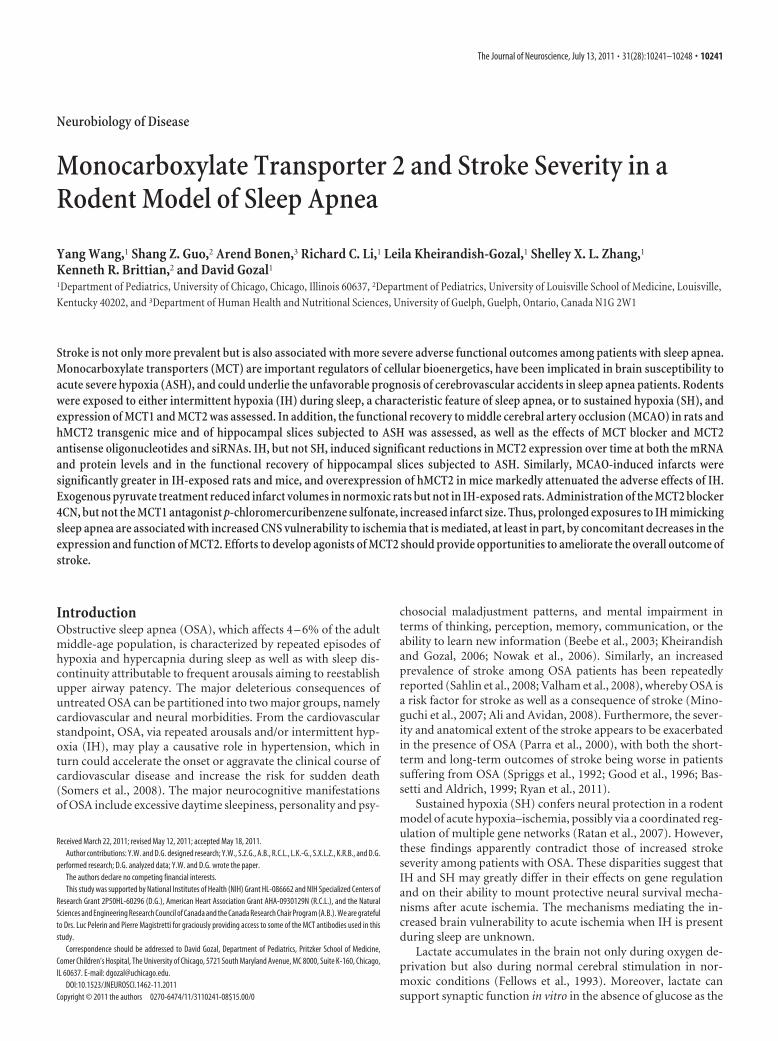
Figure 1. Hippocampal slices were exposed to ASH for 10 min after which reemergence offEPSPs was assessed. No differences in ASH susceptibility emerged in SH-exposed rats after 3, 7,or 14 d exposures (red columns; n � 24 slices from 6 different rats per group) when comparedwith RA-exposed rats (black column at time 0; n � 48 slices from 12 different rats per group). Incontrast, although no changes occurred after 3 d of IH, the percentage of slices harvested fromrats exposed to either 7 or 14 d IH showing functional recovery was reduced (*p � 0.0001 vstime 0; n � 24 slices from 6 different rats per group).
Figure 2. Infarct size after MCAO in adult rats after either 3 or 14 d (n � 12/group). Infarctvolume was significantly larger at both time points after IH exposures when compared withnormoxic controls (RA; *p � 0.001).
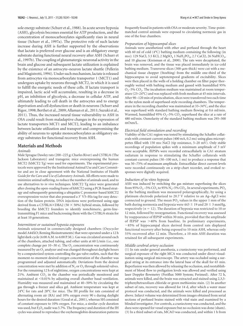
Figure 3. Behavioral test performances in rats subjected to MCAO (filled symbols) or sham(open symbols) after IH (square symbols) or RA (normoxic controls; triangle symbols). A,Eighteen-point neurological scores were significantly lower in IH rats subjected to MCAO (*p �0.01, IH vs RA; n � 12). B, Whisker stimulation and paw placement test showed decreasedresponses in IH-exposed rats after MCAO compared with RA controls ( #p � 0.04, ANOVA). C,Foot fault test performance tests showed increased number of misplacements in IH-exposedrats after MCAO ( #p � 0.04, ANOVA). D, Rotarod test performances showed that rats subjectedto IH exposures and MCAO had significantly shorter latencies than normoxic controls (*p �0.01vs RA).
Figure 4. Quantitative RT-PCR for MCT2 in rat cortical lysates harvested from animals ex-posed to SH (black columns) or IH (red columns) for 1–30 d. Significant downregulation of MCT2mRNA expression emerged at 7 d and continued to progress until 30 d of IH; however, nosignificant changes in MCT2 expression occurred in SH-exposed animals (*p � 0.01; n �6/time point). All values are reported as ratios between condition (SH or IH) and time-matchedroom air controls.
Wang et al. • MCT and Stroke in Sleep Apnea J. Neurosci., July 13, 2011 • 31(28):10241–10248 • 10243
Ischemia was induced by inserting an 11 mm silicone-coated 8-0 fila-ment through the left common carotid artery into the internal carotidartery. The filament was withdrawn after 30 min, allowing reperfusion.Regional cerebral blood flow was measured by laser Doppler flowmetry(Periflux 5000; Perimed) with a flexible probe fixed on the skull, 1 mmposteriorly and 6 mm laterally from bregma. The mice were killed 48 hafter the onset of focal ischemia, and 20-�m-thick, 720-�m-apart, cor-onal cryostat sections were stained with cresyl violet for histologic deter-mination of lesion size. A digitalized image of the Nissl-stained tissuewas obtained under a light stereomicroscope, and the lesion area onstained sections was determined by an examiner blinded for the treat-ment group using NIH ImageJ software (http://rsb.info.nih.gov/ij/).Direct infarct volume was calculated by multiplying the sum of theinfarct areas on each section by the spacing distance. To avoid biasattributable to edema, an indirect lesion size was calculated as follows:Indirect lesion � volumecontralateral � (volume ipsilateral � direct in-farct volume) as described previously by Swanson et al. (1990).
Behavioral testsEighteen-point composite neurological score. The composite neuroscorecomprises six different neurological tests: (1) spontaneous activity, (2)symmetry in limb movement, (3) forepaw outstretching, (4) climbing,(5) body proprioception, and (6) response to vibrissae touch. Each test isscored with a maximum of three points basedon a set of predetermined criteria as describedpreviously (Garcia et al., 1995). The scores foreach test were summed for a highest possiblescore of 18, indicating no neurological deficit,and the lowest score of 3, for animals with themost severe impairment.
Foot fault test. The foot fault (Columbus In-struments) is used to measure the coordinationof an animal walking across a horizontal ladder(Colle et al., 1986). The ladder width is 6.5inches, with 0.3125-inch beams 1 inch apart.The animal is trained to walk across a ladder 0.25inches above a steel plate. The plate is thencharged once during training so that, if the ani-mal’s paw comes in contact with the plate, theanimal is lightly shocked. The animal learns con-sequently to avoid the steel plate by keeping itspaws on the ladder beams during each run. Footmisplacements during the run and time to com-plete each run are counted. The average perfor-mance of three consecutive runs is reported foreach animal.
Vibrissae-evoked forelimb placing. The fore-limb placing test assesses the ability of rats tosense tactile stimulus from the vibrissae andsubsequently elicit a motor response of fore-limb placing (Schallert et al., 2003). In this test,animal body was held gently such that the pawswere suspended off the experimenter’s hand.The animal was then brought laterally to a tabletop while the vibrissae brush against the edge ofthe table, allowing the animal to reach the edgewith its forepaw. This procedure was repeated10 times on each side. The number of success-ful paw placements on to the edge of the tablewas recorded. A cross-midline variation to thetest was also performed. Here, the animal washeld on its side, and successful placements of the opposite paw to thestimulated whisker were recorded.
Rotarod motor test. In the rotarod motor test, only animals capable ofremaining on the rotarod cylinder for �300 s were used for experiments.The rotarod cylinder was accelerated from 0 to 10 rpm within 1 min, andthe time that each animal remained on the rotarod was measured with acutoff time of 300 s. Data are presented as the mean duration from threetrials.
Real-time PCR. Determinations of MCT1 mRNA and MCT2 mRNAwere determined using real-time PCR procedures, Total RNA was ex-tracted using RNeasy kit (Qiagen). Aliquots of total RNA (1 �g) werereverse transcribed to cDNA using random primers and Superscript IIReverse Transcriptase. cDNA equivalent to 100 ng of total RNA weresubjected to real-time PCR analysis. The cycling conditions consisted ofone cycle at 95°C for 10 min and 40 three-segment cycles (95°C for 15 s,59°C for 1 min, and 72°C for 30 s).
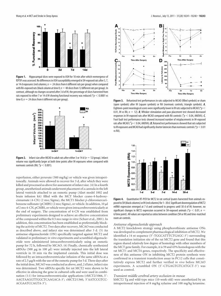
Figure 5. A, Representative immunoblots for MCT1 and MCT2 of cortical lysates after eitherIH or SH. B, MCT2 protein expression changes over time in rat cortex after IH or SH exposuresexpressed as ratios against corresponding �-actin densitometric values (n � 6/group; *p �0.01, SH vs RA; #p � 0.01, IH vs RA).
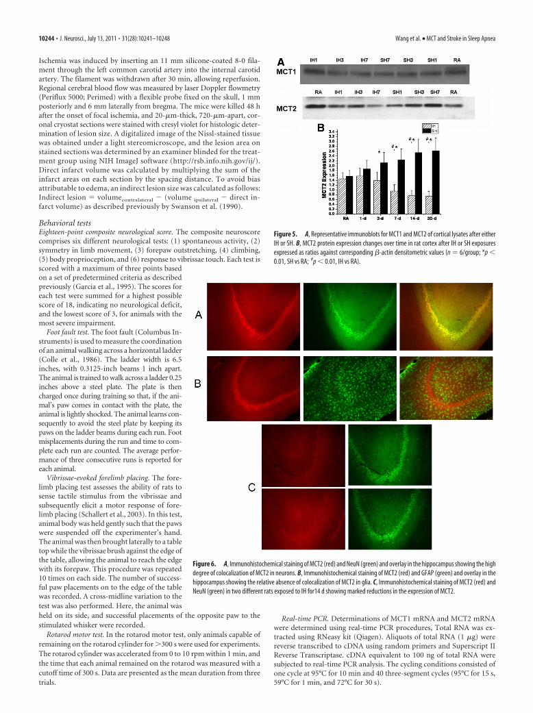
Figure 6. A, Immunohistochemical staining of MCT2 (red) and NeuN (green) and overlay in the hippocampus showing the highdegree of colocalization of MCT2 in neurons. B, Immunohistochemical staining of MCT2 (red) and GFAP (green) and overlay in thehippocampus showing the relative absence of colocalization of MCT2 in glia. C, Immunohistochemical staining of MCT2 (red) andNeuN (green) in two different rats exposed to IH for14 d showing marked reductions in the expression of MCT2.
10244 • J. Neurosci., July 13, 2011 • 31(28):10241–10248 Wang et al. • MCT and Stroke in Sleep Apnea
Immunoblot analyses. Rats were exposed to 0, 1, 3, 7, 14, or 30 d of IHor SH and were killed with a pentobarbital overdose. The skull was rap-idly opened, and the brain was extracted, immediately placed on dry ice,and dissected under surgical microscopy. A portion of parietofrontalcortex and the CA1 region of the hippocampus were carefully removed.Tissues from two to three animals were pooled and homogenized at 0°Cwith a tissue blender in 20 mM Tris-HCl buffer, pH 7.5, containing 2 mM
EDTA, 0.5 mM EGTA, 25 �g/ml leupeptin, 25 �g/ml aprotinin, and 1 mM
PMSF. The homogenate was centrifuged for 10 min at 1000 � g at 4°C toremove cell debris. Protein content was measured in each soluble fractionusing the Bradford method (DC Bio-Rad protein assay), and sampleswere frozen at �70°C until analysis. Homogenate proteins (50 �g) wereheated for 10 min at 90°C, loaded onto 8% PAGE gels, then transferredelectrophoretically onto nitrocellulose membranes. Membranes were in-cubated overnight at 4°C with antibodies to MCT1 (1:2000) and MCT2(1:2000) isoforms (Pierre et al., 2002; Pellerin et al., 2005), The samemembranes were also blotted with �-actin antibody (Sigma), and blotswere then normalized to �-actin. Densitometric analysis was performedwith a gel scanning densitometer (Molecular Dynamics). In initial exper-iments, a control lysate was included.
Immunohistochemistry. Animals were deeply anesthetized and per-fused intracardially with 4% phosphate-buffered paraformaldehyde. Se-rial sections were cut on a microtome. The free-floating sections wereincubated with anti-MCT2 (1:200 dilution) and either anti-NeuN (1:1000) or anti-GFAP antibodies (1:5000). Immunostained sections werefurther visualized with FITC-conjugated or rhodamine-conjugated sec-ond antibody. Sections were assessed using a Nikon Ellipse E800 micro-scope, and images were acquired using a SPOT digital camera.
Data analysisValues are reported as mean � SD unless indicated otherwise. Two-wayANOVA for repeated measures, followed by the Newman–Keuls or Bon-ferroni’s post hoc tests or Student’s t tests were used to compare acrossexperimental conditions as appropriate. A p value of �0.05 was consid-ered to achieve statistical significance.
ResultsIH, but not SH, increases hippocampal slice vulnerability toacute severe hypoxiaNo differences in ASH susceptibility emerged in SH-exposed ratsat 3, 7, or 14 d exposures when compared with normoxic rats(Fig. 1). In contrast, although no changes occurred after 3 d of IH,slices harvested from rats exposed to either 7 or 14 d of IH weresignificantly more susceptible to ASH as evidenced by the pro-portion of slices showing reemergence of fEPSP within 30 min(Fig. 1) ( p � 0.0001). Of note, lactate tissue levels were similarimmediately after ASH in all conditions (6.7 � 1.4 mM after roomair; 7.2 � 1.7 mM after SH, and 6.8 � 1.6 mM after IH; p � 0.05).
Infarct volume after MCAO and functional recovery inIH-exposed ratsBased on the increased susceptibility to ASH after IH exposures,rats were exposed to 14 d of IH or normoxia (RA). Figure 2 showsinfarct size after the MCAO procedure at 3 and 14 d of recovery.All IH-exposed rats had significantly larger infarcts comparedwith RA controls ( p � 0.001). Indeed, mean infarct volume 3 dafter MCAO was 318 � 43 mm 3 in IH-exposed rats (n � 12),whereas mean infarct volume was 223 � 38 mm 3 in the controlgroup (Fig. 2) (n � 12; p � 0.001), and no infarct was present insham-operated animals (n � 6 in IH and n � 4 in normoxia)(data not shown). Although infarct size was smaller at 14 d ofrecovery, the extent of the infarct remained larger in IH-exposedanimals (Fig. 2). In addition, the overall performance on thevarious behavioral tasks was significantly worse in IH-exposedrats after either a 3, 7, or 14 d recovery period after MCAO whencompared with RA-exposed rats (Fig. 3) (n � 12/group). Indeed,
the overall time-dependent trajectory in the 18-point neurologi-cal scale was significantly worse in IH-exposed animals duringthe acute phase (i.e., 3 d after MCAO and subsequent assess-ments) (Fig. 3A). Similar significant differences in the temporalchanges in the right whisker to right paw responses occurred,albeit to a lesser extent (Fig. 3B). The overall number of footmisplacements was significantly higher (Fig. 3C), and the latencyto fall in the rotarod task was significantly shorter in the IH-exposed rats after MCAO and was followed by a slower recovery(Fig. 3D).
MCT expressionOverall MCT1 mRNA expression in cortex and hippocampus(data not shown) was unchanged by SH or IH over time. In
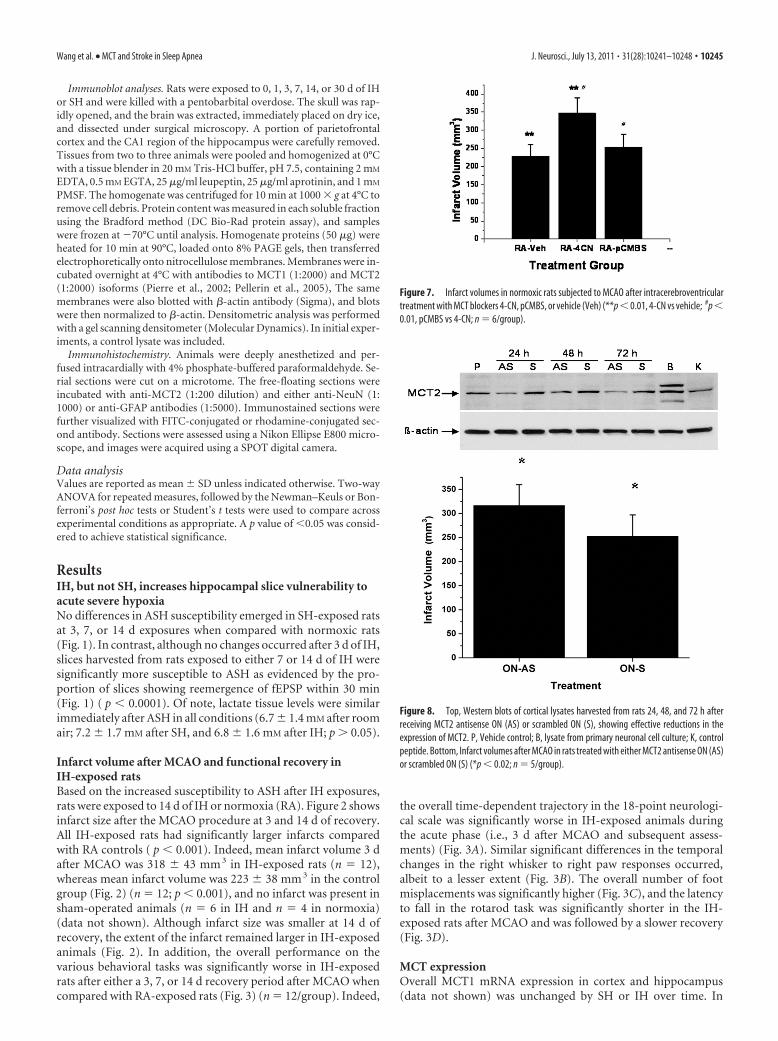
Figure 7. Infarct volumes in normoxic rats subjected to MCAO after intracerebroventriculartreatment with MCT blockers 4-CN, pCMBS, or vehicle (Veh) (**p � 0.01, 4-CN vs vehicle; #p �0.01, pCMBS vs 4-CN; n � 6/group).
Figure 8. Top, Western blots of cortical lysates harvested from rats 24, 48, and 72 h afterreceiving MCT2 antisense ON (AS) or scrambled ON (S), showing effective reductions in theexpression of MCT2. P, Vehicle control; B, lysate from primary neuronal cell culture; K, controlpeptide. Bottom, Infarct volumes after MCAO in rats treated with either MCT2 antisense ON (AS)or scrambled ON (S) (*p � 0.02; n � 5/group).
Wang et al. • MCT and Stroke in Sleep Apnea J. Neurosci., July 13, 2011 • 31(28):10241–10248 • 10245
contrast, increased expression of MCT2occurred over time after SH, whereas pro-gressive reductions in MCT2 gene expres-sion were apparent after 7 d of IH exposureswhen compared with normoxic time-matched controls (Fig. 4).
Western blots of cortical lysates furtherconfirmed the absence of any significantchanges in MCT1 protein expression,whereas increases in MCT2 expression afterlong-term SH and reduced MCT2 expres-sion after IH emerged (Fig. 5). Of note,MCT2 expression remained lower immedi-ately 30–60 min after MCAO in IH-exposed rats (n � 3/group; p � 0.04).
Immunohistochemical analysis ofhippocampal sections showed that MCT2expression was preferentially circumscribedto neurons (NeuN-positive cells) (Fig. 6A)and that GFAP positively labeled cells didnot exhibit intense staining (Fig. 6B). Fur-thermore, IH exposures for 14 d resulted inmarked decreases in hippocampal expres-sion of MCT2 in neurons (Fig. 6A,C).
MCT pharmacological experiments and MCT2 anti-senseoligonucleotides and siRNANormoxic rats implanted with 4-CN-containing osmotic pumpsand subjected to MCAO showed significant increases in infarctsize after 3 d when compared with rats receiving vehicle (Fig. 7)(n � 8/group; p � 0.001). However, administration of the MCT1blocker pCMBS, which does not affect the activity of MCT2 (Car-penter and Halestrap, 1994; Halestrap and Price, 1999; ManningFox et al., 2000), failed to modify the size of the infarct afterMCAO (Fig. 7) (n � 6/group).
Similarly, MCT2 antisense ON administration resulted in an-ticipated reduced expression of MCT2 protein and increased in-farct volumes after MCAO (Fig. 8) (n � 5; p � 0.02). In addition,selective targeting of MCT2 using siRNAs strategies resulted inincreased infarct size after MCAO (Fig. 9).
To examine whether exogenously administered pyruvate im-proved infarct size after MCAO, rats exposed to either IH or RAwere administered intraperitoneal 500 mg/kg pyruvate or vehiclewithin 1 h from reperfusion after MCAO. Pyruvate treatment wasassociated with significant reductions in infarct size in RA-exposed rats but failed to alter the extent of the infarct in IH-exposed animals (Fig. 10).
Infarct size in transgenic mice overexpressing hMCT2hMCT2 Tg mice and wild-type littermates were exposed to 14 dof IH or RA, after which they underwent MCAO procedures.Figure 10 shows that hMCT2 Tg mice were protected and thattheir infarct volumes in the cortex, striatum, or overall weresmaller even after IH exposures when compared with wild-typemice (Fig. 11) ( p � 0.001; n � 6/group).
DiscussionThe principal findings of this study include increased neuronalsusceptibility to ASH in animals undergoing chronic exposuresto intermittent hypoxia during sleep, mimicking the highly prev-alent disorder of sleep apnea. Furthermore, we now show thatthis increased susceptibility is mediated, at least in part, bychronic IH-induced decreases in the neuronal expression of
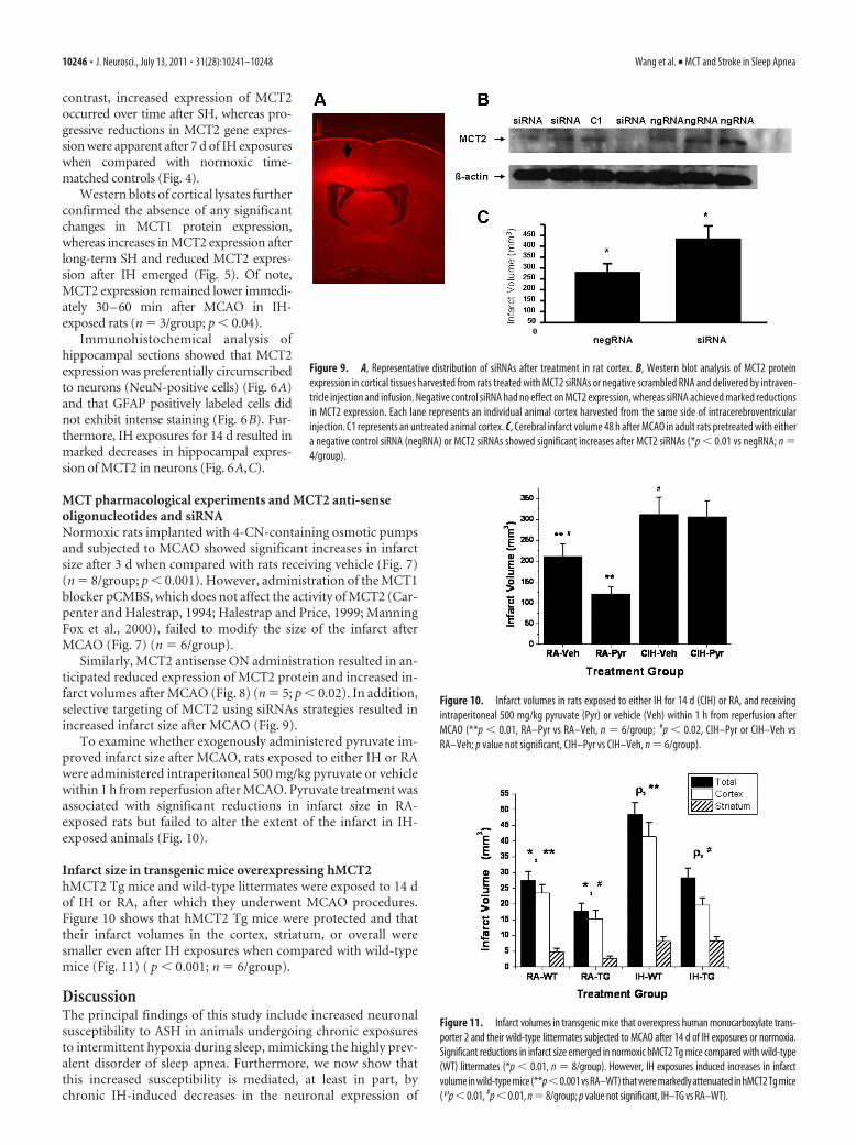
Figure 9. A, Representative distribution of siRNAs after treatment in rat cortex. B, Western blot analysis of MCT2 proteinexpression in cortical tissues harvested from rats treated with MCT2 siRNAs or negative scrambled RNA and delivered by intraven-tricle injection and infusion. Negative control siRNA had no effect on MCT2 expression, whereas siRNA achieved marked reductionsin MCT2 expression. Each lane represents an individual animal cortex harvested from the same side of intracerebroventricularinjection. C1 represents an untreated animal cortex. C, Cerebral infarct volume 48 h after MCAO in adult rats pretreated with eithera negative control siRNA (negRNA) or MCT2 siRNAs showed significant increases after MCT2 siRNAs (*p � 0.01 vs negRNA; n �4/group).
Figure 10. Infarct volumes in rats exposed to either IH for 14 d (CIH) or RA, and receivingintraperitoneal 500 mg/kg pyruvate (Pyr) or vehicle (Veh) within 1 h from reperfusion afterMCAO (**p � 0.01, RA–Pyr vs RA–Veh, n � 6/group; #p � 0.02, CIH–Pyr or CIH–Veh vsRA–Veh; p value not significant, CIH–Pyr vs CIH–Veh, n � 6/group).
Figure 11. Infarct volumes in transgenic mice that overexpress human monocarboxylate trans-porter 2 and their wild-type littermates subjected to MCAO after 14 d of IH exposures or normoxia.Significant reductions in infarct size emerged in normoxic hMCT2 Tg mice compared with wild-type(WT) littermates (*p � 0.01, n � 8/group). However, IH exposures induced increases in infarctvolume in wild-type mice (**p�0.001 vs RA–WT) that were markedly attenuated in hMCT2 Tg mice( �p � 0.01, #p � 0.01, n � 8/group; p value not significant, IH–TG vs RA–WT).
10246 • J. Neurosci., July 13, 2011 • 31(28):10241–10248 Wang et al. • MCT and Stroke in Sleep Apnea
MCT2, such that alternative sources of energy to neurons duringand after ASH cannot be effectively delivered. Together, theseresults present opportunities to further understand the mecha-nism underlying the dysregulation of MCT2 in the context ofchronic IH and the development of effective strategies aiming atpromoting MCT2 expression in patients with sleep apnea whomay be at risk for cerebrovascular ischemic events (Minoguchi etal., 2007).
The conceptual framework of a monocarboxylate-dependentenergy source alternative to neuronal function and survival hasbeen the subject of substantial debate over the past 2 decades.Notwithstanding, the overall evidence would support the exis-tence of a lactate shuttle between astrocytes and neurons thatplays a critical role during conditions such as acute ischemia–reperfusion injury (Schurr et al., 1988), as well as during physio-logical conditions such as learning and memory (Prichard et al.,1991; Allaman et al., 2011; Suzuki et al., 2011). As part of thisshuttle, MCT2 has emerged as a critical constituent of neuronalbioenergetic pathways. MCT2 cannot only be expressed in theplasma membrane, in which it governs the transport of mono-carboxylates into the cell, but is also expressed in mitochondria,in which it is associated with lactate dehydrogenase and cyto-chrome oxidase, thereby further supporting the existence of aneuronal mitochondrial monocarboxylate oxidation complexshuttle and that of a cell– cell-based monocarboxylate shuttle,similar to those identified previously in other non-CNS tissues(Brooks, 2002; Hashimoto et al., 2008). The neuronal monocar-boxylate oxidation complex described recently by Brooks andcolleagues would thus allow neurons to increase cell monocar-boxylate functional availability and to oxidize available monocar-boxylates, regardless of their sources (i.e., glycolytic pathways inthe same neuron, apposing astrocytes, or from the microvascula-ture). Indeed, Moreira et al. (2009) have shown recently that bothMCT1 and MCT2 are markedly upregulated in microglia, sug-gesting that these cells may also play a role in the recovery processfrom ischemic stroke. Accordingly, and consistent with previousfindings (Pellerin and Magistretti, 1994), such observationswould permit operation of both shuttles in brain, thereby ac-counting for their critical role in both physiological and patho-logical conditions.
Despite such obvious implications, it remains essentially un-known, however, how MCTs are regulated under conditions suchas hypoxia/ischemia, in which enhanced lactate metabolism isrequired for survival (McClelland and Brooks, 2002; Py et al.,2005). Preliminary evidence would suggest that the presence ofhypoxia would enhance the expression of MCTs, and indeed atleast one of the genes coding for MCTs, namely MCT4, was up-regulated by hypoxia at the transcriptional level via a hypoxia–inducible factor 1-dependent mechanism (Ullah et al., 2006). Wehave recently shown that the transcription of the MCT2 gene isinitiated from five distinct variants of exon 1, likely under thecontrol of five alternative promoters, and that a large number ofmature mRNA are produced from these pre-mRNA throughalternative splicing of multiple internal exons in the 5�-UTR re-gion. These findings would suggest that both alternative pro-moter usage and alternative splicing are key elements in thecellular- and subcellular-specific expression patterns of MCT2, aswell as in the gene expression responses to environmental stres-sors (Zhang et al., 2007).
From a functional standpoint, we found discrepant changes inhippocampal slice vulnerability to acute ischemia only after ani-mals had been exposed to IH, and yet exposures to a similarmagnitude of hypoxia in which the stimulus was sustained was
void of any adverse consequences after acute ischemia. Similarly,the structural and functional outcomes of MCAO were adverselyaffected by preceding IH. Such findings would suggest differen-tial regulation of MCT expression in IH and SH, despite the factthat the magnitude of hypoxia was the same. Confirmation ofsuch assumption emerged from the reported expression studies,whereby IH and SH clearly had discrepant effects on MCT2 ex-pression levels in brain, with significant reduction in MCT2 at themRNA and protein levels after IH. Of note, the expression pat-terns of MCT2 in control animals were analogous to those re-ported previously (Koehler-Stec et al., 1998; Pierre et al., 2002).We would also emphasize that the differences in infarct size wereconfirmed in wild-type mice exposed to IH and were attenuatedby overexpression of MCT2 in mice even after exposures to IH.
The use of pyruvate or lactate as alternative energy sourcesduring ASH has been repeatedly advocated as potentially confer-ring substantial neuronal protection in various model of stroke.Indeed, Lee et al. (2001) showed that exogenous administrationof pyruvate at doses similar to those used herein was associatedwith substantial neuroprotection in a forebrain stroke model inrats. Similar findings have been reported recently by Alano et al.(2010), whereby bypassing glycolytic inhibition associated withASH with pyruvate prevented mitochondrial failure and neuro-nal cell death. In our model, pyruvate was effective in affordingneuroprotection in normoxic animals but did not confer anyincremental benefit in IH-exposed rats. Such observations wouldbe commensurate with the reduction in MCT2 expression, whichwould then restrict the ability to activate the putative neuronalmitochondrial monocarboxylate oxidation complex and that of acell– cell-based monocarboxylate shuttle to enable improvedavailability of cellular ATP. It remains unclear, however, whethersuch mechanisms would also be able to favorably regulate mito-chondrial oxidative stress in the context of ASH (Fiskum et al.,2004). Such assumption may not be far-fetched considering thelife-prolonging effect of increased mitochondrial pyruvate avail-ability in a Caenorhabditis elegans model (Mouchiroud et al.,2011).
In summary, prolonged exposures to the intermittent hypoxiaduring sleep, which characterizes the highly prevalent conditionof sleep apnea, result in increased brain susceptibility to acuteischemic events aiming to reproduce stroke in a rodent. The en-hanced vulnerability of the neural substrate in this context ap-pears to be accounted for by concomitant reductions in MCT2neuronal expression. Thus, efforts to promote expression andefficiency of neuronal monocarboxylate transporters may ame-liorate the adverse outcomes of stroke in patients with underlyingsleep apnea.
ReferencesAlano CC, Garnier P, Ying W, Higashi Y, Kauppinen TM, Swanson RA
(2010) NAD depletion is necessary and sufficient for poly(ADP-ribose)polymerase-1-mediated neuronal death. J Neurosci 30:2967–2978.
Ali LK, Avidan AY (2008) Sleep-disordered breathing and stroke. Rev Neu-rol Dis 5:191–198.
Allaman I, Belanger M, Magistretti PJ (2011) Astrocyte-neuron metabolicrelationships: for better and for worse. Trends Neurosci 34:76 – 87.
Bassetti C, Aldrich MS (1999) Sleep apnea in acute cerebrovascular diseases:final report on 128 patients. Sleep 22:217–223.
Beebe DW, Groesz L, Wells C, Nichols A, McGee K (2003) The neuropsy-chological effects of obstructive sleep apnea: a meta-analysis of norm-referenced and case-controlled data. Sleep 26:298 –307.
Berthet C, Lei H, Thevenet J, Gruetter R, Magistretti PJ, Hirt L (2009) Neu-roprotective role of lactate after cerebral ischemia. J Cereb Blood FlowMetab 29:1780 –1789.
Wang et al. • MCT and Stroke in Sleep Apnea J. Neurosci., July 13, 2011 • 31(28):10241–10248 • 10247
Brooks GA (2002) Lactate shuttles in nature. Biochem Soc Trans 30:258 –264.
Carpenter L, Halestrap AP (1994) The kinetics, substrate and inhibitorspecificity of the lactate transporter of Ehrlich-Lettre tumour cells studiedwith the intracellular pH indicator BCECF. Biochem J 304:751–760.
Colle LM, Holmes LJ, Pappius HM (1986) Correlation between behavioralstatus and cerebral glucose utilization in rats following freezing lesion.Brain Res 397:27–36.
Fellows LK, Boutelle MG, Fillenz M (1993) Physiological stimulation in-creases nonoxidative glucose metabolism in the brain of the freely movingrat. J Neurochem 60:1258 –1263.
Fiskum G, Rosenthal RE, Vereczki V, Martin E, Hoffman GE, Chinopoulos C,Kowaltowski A (2004) Protection against ischemic brain injury by inhibi-tion of mitochondrial oxidative stress. J Bioenerg Biomembr 36:347–352.
Garcia JH, Wagner S, Liu KF, Hu XJ (1995) Neurological deficit and extentof neuronal necrosis attributable to middle cerebral artery occlusion inrats. Statistical validation. Stroke 26:627– 634; discussion 635.
Good DC, Henkle JQ, Gelber D, Welsh J, Verhulst S (1996) Sleep-disorderedbreathing and poor functional outcome after stroke. Stroke 27:252–259.
Gozal D, Daniel JM, Dohanich GP (2001) Behavioral and anatomical cor-relates of chronic episodic hypoxia during sleep in the rat. J Neurosci21:2442–2450.
Halestrap AP, Price NT (1999) The proton-linked monocarboxylate trans-porter (MCT) family: structure, function and regulation. Biochem J343:281–299.
Hashimoto T, Hussien R, Cho HS, Kaufer D, Brooks GA (2008) Evidencefor the mitochondrial lactate oxidation complex in rat neurons: demon-stration of an essential component of brain lactate shuttles. PLoS One3:e2915.
Kheirandish L, Gozal D (2006) Neurocognitive dysfunction in children withsleep disorders. Dev Sci 9:388 –399.
Koehler-Stec EM, Simpson IA, Vannucci SJ, Landschulz KT, Landschulz WH(1998) Monocarboxylate transporter expression in mouse brain. Am JPhysiol 275:E516 –E524.
Kreisman NR, Soliman S, Gozal D (2000) Regional differences in hypoxicdepolarization and swelling in hippocampal slices. J Neurophysiol 83:1031–1038.
Lee JY, Kim YH, Koh JY (2001) Protection by pyruvate against transientforebrain ischemia in rats. J Neurosci 21:RC171(1– 6).
Manning Fox JE, Meredith D, Halestrap AP (2000) Characterisation of hu-man monocarboxylate transporter 4 substantiates its role in lactic acidefflux from skeletal muscle. J Physiol 529:285–293.
McClelland GB, Brooks GA (2002) Changes in MCT 1, MCT 4, and LDHexpression are tissue specific in rats after long-term hypobaric hypoxia.J Appl Physiol 92:1573–1584.
Minoguchi K, Yokoe T, Tazaki T, Minoguchi H, Oda N, Tanaka A,Yamamoto M, Ohta S, O’Donnell CP, Adachi M (2007) Silent braininfarction and platelet activation in obstructive sleep apnea. Am J RespirCrit Care Med 175:612– 617.
Moreira TJ, Pierre K, Maekawa F, Repond C, Cebere A, Liljequist S, Pellerin L(2009) Enhanced cerebral expression of MCT1 and MCT2 in a rat isch-emia model occurs in activated microglial cells. J Cereb Blood Flow Metab29:1273–1283.
Mouchiroud L, Molin L, Kasturi P, Triba MN, Dumas ME, Wilson MC,Halestrap AP, Roussel D, Masse I, Dalliere N, Segalat L, Billaud M, SolariF (2011) Pyruvate imbalance mediates metabolic reprogramming andmimics lifespan extension by dietary restriction in Caenorhabditis elegans.Aging Cell 10:39 –54.
Nowak M, Kornhuber J, Meyrer R (2006) Daytime impairment and neuro-degeneration in OSAS. Sleep 29:1521–1530.
Parra O, Arboix A, Bechich S, García-Eroles L, Montserrat JM, Lopez JA,Ballester E, Guerra JM, Sopena JJ (2000) Time course of sleep-relatedbreathing disorders in first-ever stroke or transient ischemic attack. Am JRespir Crit Care Med 161:375–380.
Pellerin L, Magistretti PJ (1994) Glutamate uptake into astrocytes stimu-lates aerobic glycolysis: a mechanism coupling neuronal activity to glu-cose utilization. Proc Natl Acad Sci U S A 91:10625–10629.
Pellerin L, Bergersen LH, Halestrap AP, Pierre K (2005) Cellular and sub-cellular distribution of monocarboxylate transporters in cultured braincells and in the adult brain. J Neurosci Res 79:55– 64.
Pierre K, Magistretti PJ, Pellerin L (2002) MCT2 is a major neuronal mono-carboxylate transporter in the adult mouse brain. J Cereb Blood FlowMetab 22:586 –595.
Prichard J, Rothman D, Novotny E, Petroff O, Kuwabara T, Avison M,Howseman A, Hanstock C, Shulman R (1991) Lactate rise detected by1H NMR in human visual cortex during physiologic stimulation. ProcNatl Acad Sci U S A 88:5829 –5831.
Py G, Eydoux N, Lambert K, Chapot R, Koulmann N, Sanchez H, Bahi L,Peinnequin A, Mercier J, Bigard AX (2005) Role of hypoxia-inducedanorexia and right ventricular hypertrophy on lactate transport and MCTexpression in rat muscle. Metabolism 54:634 – 644.
Ratan RR, Siddiq A, Smirnova N, Karpisheva K, Haskew-Layton R, McCo-noughey S, Langley B, Estevez A, Huerta PT, Volpe B, Roy S, Sen CK,Gazaryan I, Cho S, Fink M, LaManna J (2007) Harnessing hypoxic ad-aptation to prevent, treat, and repair stroke. J Mol Med 85:1331–1338.
Ryan CM, Bayley M, Green R, Murray BJ, Bradley TD (2011) Influence ofcontinuous positive airway pressure on outcomes of rehabilitation instroke patients with obstructive sleep apnea. Stroke 42:1062–1067.
Sahlin C, Sandberg O, Gustafson Y, Bucht G, Carlberg B, Stenlund H, Frank-lin KA (2008) Obstructive sleep apnea is a risk factor for death in pa-tients with stroke: a 10-year follow-up. Arch Intern Med 168:297–301.
Schallert T, Woodlee MT, Fleming SM (2003) Experimental focal ischemicinjury: behavior-brain interactions and issues of animal handling andhousing. ILAR J 44:130 –143.
Schurr A, Rigor BM (1998) Brain anaerobic lactate production: a suicidenote or a survival kit? Dev Neurosci 20:348 –357.
Schurr A, West CA, Rigor BM (1988) Lactate-supported synaptic functionin the rat hippocampal slice preparation. Science 240:1326 –1328.
Schurr A, Payne RS, Miller JJ, Rigor BM (1997a) Brain lactate is an obliga-tory aerobic energy substrate for functional recovery after hypoxia: fur-ther in vitro validation. J Neurochem 69:423– 426.
Schurr A, Payne RS, Miller JJ, Rigor BM (1997b) Brain lactate, not glucose,fuels the recovery of synaptic function from hypoxia upon reoxygenation:an in vitro study. Brain Res 744:105–111.
Schurr A, Payne RS, Tseng MT, Gozal E, Gozal D (2001) Excitotoxic pre-conditioning elicited by both glutamate and hypoxia and abolished bylactate transport inhibition in rat hippocampal slices. Neurosci Lett307:151–154.
Somers VK, White DP, Amin R, Abraham WT, Costa F, Culebras A, DanielsS, Floras JS, Hunt CE, Olson LJ, Pickering TG, Russell R, Woo M, YoungT; American Heart Association Council for High Blood Pressure ResearchProfessional Education Committee, Council on Clinical Cardiology;American Heart Association Stroke Council; American Heart AssociationCouncil on Cardiovascular Nursing; American College of CardiologyFoundation (2008) Sleep apnea and cardiovascular disease: an Ameri-can Heart Association/American College Of Cardiology Foundation Sci-entific Statement from the American Heart Association Council for HighBlood Pressure Research Professional Education Committee, Council onClinical Cardiology, Stroke Council, and Council On CardiovascularNursing. In collaboration with the National Heart, Lung, and Blood In-stitute National Center on Sleep Disorders Research (National Institutesof Health). Circulation 118:1080 –1111.
Spriggs DA, French JM, Murdy JM, Curless RH, Bates D, James OF (1992)Snoring increases the risk of stroke and adversely affects prognosis. QJ Med 83:555–562.
Suzuki A, Stern SA, Bozdagi O, Huntley GW, Walker RH, Magistretti PJ,Alberini CM (2011) Astrocyte-neuron lactate transport is required forlong-term memory formation. Cell 144:810 – 823.
Swanson RA, Morton MT, Tsao-Wu G, Savalos RA, Davidson C, Sharp FR(1990) A semiautomated method for measuring brain infarct volume.J Cereb Blood Flow Metab 10:290 –293.
Ullah MS, Davies AJ, Halestrap AP (2006) The plasma membrane lactatetransporter MCT4, but not MCT1, is up-regulated by hypoxia through aHIF-1alpha-dependent mechanism. J Biol Chem 281:9030 –9037.
Valham F, Mooe T, Rabben T, Stenlund H, Wiklund U, Franklin KA (2008)Increased risk of stroke in patients with coronary artery disease and sleepapnea: a 10-year follow-up. Circulation 118:955–960.
Zhang SX, Searcy TR, Wu Y, Gozal D, Wang Y (2007) Alternative promoterusage and alternative splicing contribute to mRNA heterogeneity ofmouse monocarboxylate transporter 2. Physiol Genomics 32:95–104.
10248 • J. Neurosci., July 13, 2011 • 31(28):10241–10248 Wang et al. • MCT and Stroke in Sleep Apnea


