Embed Size (px)
Citation preview

Neuropathogenesis of herpesvirus papio 2 in miceparallels infection with Cercopithecine herpesvirus1 (B virus) in humans
Kristin M. Rogers, Jerry W. Ritchey, Mark Payton, Darla H. Blackand R. Eberle
Correspondence
Kristin M. Rogers
Department of Veterinary Pathobiology, Center for Veterinary Health Sciences, Oklahoma StateUniversity, 250 McElroy Hall, Stillwater, OK 74078-2007, USA
Received 31 August 2005
Accepted 30 September 2005
Cercopithecine herpesvirus 1 (monkey B virus; BV) produces extremely severe and usually fatal
infections when transmitted from macaque monkeys to humans. Cercopithecine herpesvirus 16
(herpesvirus papio 2; HVP2) is very closely related to BV, yet cases of human HVP2 infection are
unknown. However, following intramuscular inoculation of mice, HVP2 rapidly invades the
peripheral nervous system and ascends the central nervous system (CNS) resulting in death, very
much like human BV infections. In this study, the neurovirulence of HVP2 in mice was further
evaluated as a potential model system for human BV infections. HVP2 was consistently
neurovirulent when administered by epidermal scarification, intracranial inoculation and an eye
splash. Quantitative real-time PCR, histopathology and immunohistochemistry were used to follow
the temporal spread of virus following skin scarification and to compare the pathogenesis of
neurovirulent and apathogenic isolates of HVP2. Apathogenic isolates were found to be capable of
reaching the CNS but were extremely inefficient at replicating within the CNS. It is concluded
that neurovirulent strains of HVP2 exhibit a pathogenesis in mice that parallels that observed in
human BV infections and that this model system may prove useful in dissecting the viral
determinants underlying the extreme severity of zoonotic BV infections.
INTRODUCTION
Cercopithecine herpesvirus 1 (monkey B virus; BV) is aubiquitous pathogen of macaques (Macaca spp.). In captiveanimals, BV is most often acquired as an oral infection byinfants and juveniles or as a genital infection in sexuallymature animals. In many respects the biology of BV iscomparable to that of herpes simplex virus (HSV) inhumans and Cercopithecine herpesvirus 16 (herpesviruspapio 2; HVP2) in baboons (Papio spp.). While there havebeen no reported cases of human HVP2 infection, BV is wellknown for its extreme neuropathogenicity when transmittedto non-macaque primates, including humans (Davidson &Hummeler, 1960; Weigler, 1992; Whitley & Hilliard, 2001).Although zoonotic BV infections are rare, the extremeseverity of these infections has resulted in BV being classifiedas the only biosafety level 4 (BSL-4) herpesvirus. Further-more, the US Government classifies BV as a ‘select agent’due to concern regarding its potential use as a bioterrorismweapon.
Due to the hazardous nature of BV and the requirementfor special biocontainment facilities, relatively little researchhas been done on the virus. An additional difficulty has beenthe lack of a small animal system which faithfully andreproducibly portrays zoonotic BV infections. Although
rabbits are quite sensitive to BV infection, their size and costand the lack of analytical reagents make this a less than idealmodel system. The development of a small animal modelthat accurately depicts human BV infections will be of usenot only in studying zoonotic BV infections but also forinvestigating the more general question regarding zoonoses:what mechanisms cause some viruses to be so extremelyneurovirulent when introduced into a non-natural hostspecies?
Several studies have examined themouse as a potential smallanimal model system for studying BV neurovirulence(Gosztonyi et al., 1992; Ritchey et al., 2005). Althoughvery young animals are quite sensitive to BV infection(Gosztonyi et al., 1992), a number of concerns regarding theuse of a murine model to study cross-species BV infectionshave been identified. Firstly, the pathogenicity of differentstrains of BV in mice varies across the full range of virulencephenotypes from completely apathogenic to extremelyneurovirulent (Ritchey et al., 2005). Secondly, the virulenceof individual BV isolates in mice is variable dependingon the route of inoculation (J. W. Ritchey, D. H. Black andR. Eberle, unpublished data). Finally, BV infections in miceare not strictly dose-dependent, so calculation of infectiousand lethal doses can be difficult (Ritchey et al., 2005).
0008-1476 G 2006 SGM Printed in Great Britain 267
Journal of General Virology (2006), 87, 267–276 DOI 10.1099/vir.0.81476-0

Therefore, while a BV/mouse model may be appropriatefor examining certain aspects of BV infections, this systemmay not be suitable for molecular and genetic analysesaimed at examining the viral mechanisms which determinethe outcome of a cross-species BV infection.
In contrast to the wide range of pathogenic phenotypes ofBV observed in mice, intramuscular (i.m.) inoculation ofmice with multiple isolates of HVP2 revealed quite a differ-ent picture in that all isolates tested fell into one oftwo distinct subtypes, highly neurovirulent (HVP2nv) orapathogenic (HVP2ap) (Rogers et al., 2003). While allHVP2nv isolates tested produced fulminant, fatal, centralnervous system (CNS) infections equivalent to thoseinduced by the most pathogenic strains of BV, HVP2ap-inoculated mice never showed clinical signs of infection(Rogers et al., 2003). In addition, despite the very highdegree of genetic relatedness between BV and HVP2, HVP2is a BSL-2 pathogen. Thus, all indications are that anHVP2nv/mouse model system may be a safe, consistentand reproducible model for human BV infections.
Initial experiments with HVP2 inmice used i.m. inoculationto simulate infection via a monkey bite. While a number ofhuman BV infections have resulted from bites, several otherroutes of transmission have been implicated, including eyesplashes, scratches from macaques or contaminated equip-ment and needle sticks. To validate fully the usefulness ofthe HVP2nv/mouse system as a model for zoonotic BVinfections, it was necessary to assess whether the pathogenicphenotypes observed after i.m. inoculation ofmice remainedconsistent using additional routes of infection. Further, itwas important to characterize both the temporal and spatialdistribution of HVP2 infection in the mouse CNS. Finally,it was necessary to distinguish between the neuroinvasiveand neurovirulent capacity ofHVP2ap to determinewhetherthe differences between HVP2nv and HVP2ap were due tothe inability of HVP2ap to enter and/or replicate within theCNS.
METHODS
Viruses and cells. Vero cells were cultured in complete Dulbecco’smodified Eagle’s medium (DMEM) containing 10% fetal bovineserum (FBS) and maintained in DMEM containing 2% FBS. Theorigin of all HVP2 isolates as well as the purification of viral DNAhave been described previously (Eberle et al., 1995, 1997, 1998;Rogers et al., 2003).
Mouse inoculations. Female BALB/c mice, weighing 10–12 g(intra-cranial and eye splash inoculation) or 12–14 g (skin scarifica-tion) were obtained from Charles River Laboratories. For inocula-tion, mice were immobilized by anaesthetic inhalation (IsoFlo;Abbott Laboratories). Once infected, mice were observed twice dailyfor clinical signs of infection. All mice were humanely euthanized byCO2 inhalation when clinical signs of infection became severe or atthe termination of the experiment. Sterile PBS was used as diluentfor all viral dilutions and as inoculum for negative-control mice.
For skin scarification (s.s.), groups of eight mice were shaved and an8 mm circle was drawn on the left rear flank to identify the inoculation
site. A 22-gauge needle was used to scarify the skin superficially insidethe circle and 10 ml inoculum containing tenfold dilutions of HVP2nvisolate X313 or HVP2ap isolate OU2-5 ranging from 102 to 106 p.f.u.was applied to the site with a micropipettor.
For eye splash (e.s.) inoculation, 5 ml inoculum containing tenfolddilutions of HVP2nv isolate OU1-76 ranging from 101 to 106 p.f.u. orHVP2ap isolate OU2-5 ranging from 104 to 106 p.f.u. was placeddirectly into the left eye of mice in groups of five using a micropipettor.
For temporal studies, groups of 72 mice were inoculated by s.s. with105 p.f.u. HVP2nv isolate X313, HVP2ap isolate OU2-5 or diluent asdescribed above. Six mice from each group were euthanized daily ondays 1–9 post-infection (p.i.). Skin from the inoculation site, sectionsof lumbar and thoracic spinal cord and brainstem tissue were harvestedfrom three mice in each group and stored at 280 uC. The remainingthree mice from each group were processed for histopathologicalexamination.
For intracranial (i.c.) inoculation, groups of five mice were inoculatedusing a 50 ml syringe (model 705LT; Hamilton) with a 27-gauge needleto deliver 10 ml inoculum containing 102–106 p.f.u. HVP2ap isolatesA951 orOU2-5, 100–106 p.f.u. HVP2nv isolate X313 or diluent into thecerebrum.
Sample preparation. To obtain template DNA for the real-timePCR assay, total DNA was extracted from tissue samples using theDNAeasy Tissue kit (Qiagen) according to the manufacturer’s pro-tocol except that the amount of proteinase K was doubled, DNAwas eluted in a 100 ml final volume and 15 ml 0?25% linear poly-acrylamide was added to all spinal cord samples prior to ethanolprecipitation of the DNA (Gaillard & Strauss, 1990). DNA sampleswere dehydrated in an Eppendorf Vacufuge and stored at 220 uC.All DNA samples were resuspended in 10 mM Tris/HCl pH 8?0(brain and inoculation site epithelial DNA in 200 ml, spinal cordtissue DNA in 35 ml) and quantified by spectroscopy.
Real-time quantitative PCR assay. To avoid potential problemswith the real-time assay due to subtype-specific sequence variation,HVP2-specific primers and probe were designed from an alignmentof the HVP2 UL41 open reading frame (ORF) sequence of fourHVP2ap isolates and four HVP2nv isolates using the PRIMEREXPRESS
software (version 2.0; Applied Biosystems). The sequence of theforward primer (59-TGCGCCAACCTCTACCA-39) and reverseprimer (59-TGTCGGTCGTGTGGACGT-39) as well as a TaqManMGB probe (59-CCAACACCGTCGCG-39) labelled at the 59 endwith FAM (6-carboxyfluorescein) and at the 39 end with a non-fluorescing quencher were all designed from the nucleotide align-ment of the eight HVP2 sequences. Primers/probes and all real-timePCR reagents, disposables and equipment were purchased fromApplied Biosystems. PCRs were performed in a 96-well plate andcontained 16 TaqMan Universal PCR master mix, 2?0 mM eachprimer, 10 nM probe and 2?0 ml sample DNA in a final volume of25 ml. The TaqMan pre-designed assay for 18S rRNA was used asdirected by the manufacturer. PCR amplification and detectionwere performed on an ABI Prism 7000 Sequence Detection Systemusing the following cycling conditions: 50 uC for 2 min, 95 uC for10 min and 40 two-step cycles of 95 uC for 15 s and 60 uC for1 min. All PCRs were carried out in triplicate with appropriate con-trols run on each plate.
HVP2 DNA and cellular DNA standards. For use as a viral stan-dard in the PCR assay, an 800 bp segment of the HVP2 UL41 ORFwas amplified by standard PCR using the forward primer HVP2-492(59-GCATGTTGGAGAAGGCGGAGCTGG-39) and the reverse primerHVP2-493 (59-GACTGGTGCGGAGGGGAGGTTGG-39) (Sigma-Genosys). The Invitrogen TOPO TA cloning kit was used to clonethe PCR product and the fidelity of the insert was confirmed by
268 Journal of General Virology 87
K. M. Rogers and others
DNA sequencing. Purified plasmid DNA was isolated using theQiagen Plasmid maxi kit and quantified by spectroscopy. Theplasmid standard was used as described on the Applied Biosystemswebsite (http://www.appliedbiosystems.com/support/tutorials/pdf/quant_pcr.pdf). The dynamic range for detection of the HVP2plasmid standard by the real-time PCR assay was 100–105 viralgenome copies (VGC) per reaction.
To correlate 18S rRNA CT values with mouse cell numbers as a meansof normalizing for variable tissue mass, two 25 cm2 flasks of sub-confluent mouse L929 cells at different cell densities were countedusing a haematocytometer. Suspensions containing 1?36106 and3?16106 cells were centrifuged at 300 g for 5 min and each cell pelletwas resuspended in 500 ml sterile PBS for DNA extraction. The 18SrRNA assay was used to detect the number of 18S rRNA genes in atenfold dilution series of the two DNA samples. The 18S CT valuesshowed a linear decrease with increasing L929 cell DNA over a dynamicrange of <100 to 1?36104 cells. Cellular 18S CT values were plottedagainst the number of 18S genes per reaction to generate a standardcurve using Microsoft Excel 2003. Regression analysis was used tocalculate the number of mouse cells present in unknown DNA samplesbased on the 18S CT values.
Correlation between viral genome copies and infectiousvirus. To validate the use of real-time PCR for quantifying HVP2 intissue samples, it was necessary to correlate the number of VGC tothe number of infectious virions (p.f.u.). Mice in groups of twowere inoculated by s.s. with 105 p.f.u. HVP2nv isolate OU1-76 orHVP2ap isolate OU2-5. Mice were euthanized at 7 days p.i. andtissue samples were collected from the site of inoculation, spinalcord, brainstem and spleen. After the tissue samples were homoge-nized in 500 ml DMEM with 2% FBS (750 ml for inoculation-siteskin samples), 450 ml of the liquid was removed, divided equallyinto two sterile microcentrifuge tubes and placed at 280 uC. TheQiagen QIAamp 96 DNA Blood kit was used to extract total DNAfrom one aliquot for use as template in quantitative real-time PCRassay. The second aliquot was used to quantify infectious virus usinga standard plaque assay (Eberle & Hilliard, 1984).
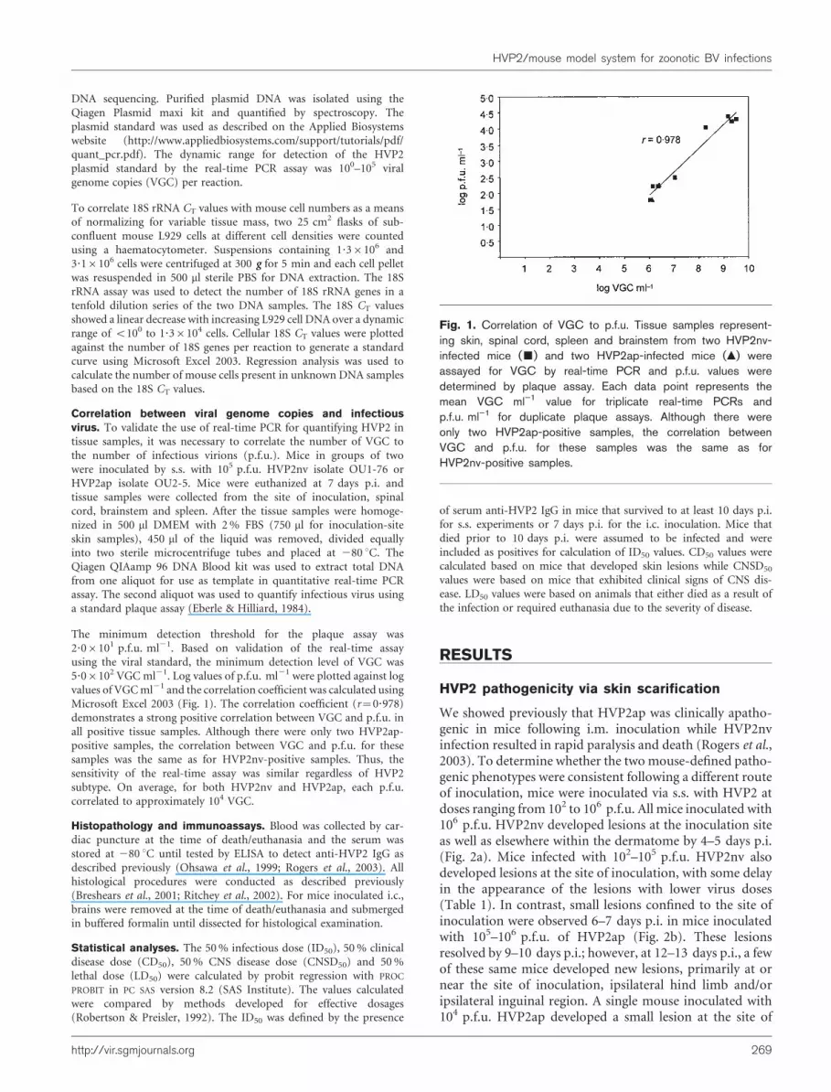
The minimum detection threshold for the plaque assay was2?06101 p.f.u. ml21. Based on validation of the real-time assayusing the viral standard, the minimum detection level of VGC was5?06102 VGCml21. Log values of p.f.u. ml21 were plotted against logvalues of VGCml21 and the correlation coefficient was calculated usingMicrosoft Excel 2003 (Fig. 1). The correlation coefficient (r=0?978)demonstrates a strong positive correlation between VGC and p.f.u. inall positive tissue samples. Although there were only two HVP2ap-positive samples, the correlation between VGC and p.f.u. for thesesamples was the same as for HVP2nv-positive samples. Thus, thesensitivity of the real-time assay was similar regardless of HVP2subtype. On average, for both HVP2nv and HVP2ap, each p.f.u.correlated to approximately 104 VGC.
Histopathology and immunoassays. Blood was collected by car-diac puncture at the time of death/euthanasia and the serum wasstored at 280 uC until tested by ELISA to detect anti-HVP2 IgG asdescribed previously (Ohsawa et al., 1999; Rogers et al., 2003). Allhistological procedures were conducted as described previously(Breshears et al., 2001; Ritchey et al., 2002). For mice inoculated i.c.,brains were removed at the time of death/euthanasia and submergedin buffered formalin until dissected for histological examination.
Statistical analyses. The 50% infectious dose (ID50), 50% clinicaldisease dose (CD50), 50% CNS disease dose (CNSD50) and 50%lethal dose (LD50) were calculated by probit regression with PROC
PROBIT in PC SAS version 8.2 (SAS Institute). The values calculatedwere compared by methods developed for effective dosages(Robertson & Preisler, 1992). The ID50 was defined by the presence
of serum anti-HVP2 IgG in mice that survived to at least 10 days p.i.for s.s. experiments or 7 days p.i. for the i.c. inoculation. Mice thatdied prior to 10 days p.i. were assumed to be infected and wereincluded as positives for calculation of ID50 values. CD50 values werecalculated based on mice that developed skin lesions while CNSD50
values were based on mice that exhibited clinical signs of CNS dis-ease. LD50 values were based on animals that either died as a result ofthe infection or required euthanasia due to the severity of disease.
RESULTS
HVP2 pathogenicity via skin scarification
We showed previously that HVP2ap was clinically apatho-genic in mice following i.m. inoculation while HVP2nvinfection resulted in rapid paralysis and death (Rogers et al.,2003). To determine whether the twomouse-defined patho-genic phenotypes were consistent following a different routeof inoculation, mice were inoculated via s.s. with HVP2 atdoses ranging from 102 to 106 p.f.u. All mice inoculated with106 p.f.u. HVP2nv developed lesions at the inoculation siteas well as elsewhere within the dermatome by 4–5 days p.i.(Fig. 2a). Mice infected with 102–105 p.f.u. HVP2nv alsodeveloped lesions at the site of inoculation, with some delayin the appearance of the lesions with lower virus doses(Table 1). In contrast, small lesions confined to the site ofinoculation were observed 6–7 days p.i. in mice inoculatedwith 105–106 p.f.u. of HVP2ap (Fig. 2b). These lesionsresolved by 9–10 days p.i.; however, at 12–13 days p.i., a fewof these same mice developed new lesions, primarily at ornear the site of inoculation, ipsilateral hind limb and/oripsilateral inguinal region. A single mouse inoculated with104 p.f.u. HVP2ap developed a small lesion at the site of
Fig. 1. Correlation of VGC to p.f.u. Tissue samples represent-ing skin, spinal cord, spleen and brainstem from two HVP2nv-infected mice (&) and two HVP2ap-infected mice (m) wereassayed for VGC by real-time PCR and p.f.u. values weredetermined by plaque assay. Each data point represents themean VGC ml”1 value for triplicate real-time PCRs andp.f.u. ml”1 for duplicate plaque assays. Although there wereonly two HVP2ap-positive samples, the correlation betweenVGC and p.f.u. for these samples was the same as forHVP2nv-positive samples.
http://vir.sgmjournals.org 269
HVP2/mouse model system for zoonotic BV infections

inoculation at 13 days p.i. Mice inoculated with doses lowerthan 104 p.f.u. HVP2ap never showed any clinical signs ofdisease. Based on these data, the CD50 values were determinedto be 102?7 p.f.u. for HVP2nv and 105?8 p.f.u. for HVP2ap.
Based on the presence of serum anti-HVP2 IgG, the ID50
value for HVP2ap-infected mice was 103?9 p.f.u. (Table 1).All HVP2nv-infected mice inoculated by s.s. that exhibitedsigns of CNS disease succumbed to the infection, resulting inidentical ID50, CNSD50 and LD50 values of 102?9 p.f.u. Incontrast, CNSD50 and LD50 values for HVP2ap by s.s. were>106 p.f.u., with no HVP2ap-infected mice requiringeuthanasia.
Temporal and spatial distribution of HVP2infection in mice
Mice were inoculated by s.s. with 105 p.f.u. HVP2 for severalpurposes: (i) to determine the time required for HVP2nv to
enter the CNS, (ii) to evaluate the anatomical distributionof HVP2 infection within the mouse CNS and (iii) toquantify the spread of HVP2nv compared with HVP2ap.At various time points, skin from the inoculation site,lumbar spinal cord, thoracic spinal cord and brainstemwere harvested, DNA was extracted and VGC per cell wasquantified by real-time PCR. As shown in Fig. 3(a), DNAfrom both HVP2 subtypes was detected at all time pointsfrom skin at the site of inoculation; however, HVP2nv DNAwas present at significantly higher levels than HVP2ap onfour of the seven days. HVP2nv was detected in both thelumbar and thoracic regions of the spinal cord by 4 and5 days p.i., respectively, with the amount of virus in bothregions increasing between 5 and 7 days p.i. (Fig. 3b, c). Incontrast, HVP2ap DNA was not detected in the spinal corduntil 5–6 days p.i. and at significantly lower levels thanHVP2nv DNA. Viral DNA was also detected at significantlevels in the brainstem of all HVP2nv-infected mice between6 and 7 days p.i. (Fig. 3d), while HVP2apwas never detectedin the brainstem throughout the experiment.
Following s.s. inoculation, detectable histological lesionswere restricted to the skin, spinal cord and brainstem. By3 days p.i., the epidermis of HVP2nv-infected mice exhi-bited hyperplasia as well as necrosis of epithelial cells,intranuclear inclusion bodies and viral antigen immuno-reactivity within epithelial cells of the epidermis and hairfollicles (Fig. 4a, b). At 5 days p.i., these lesions had pro-gressed to full-thickness necrosis of the epidermal andfollicular epithelium, formation of serocellular crusts andintense infiltrates of neutrophilic and mononuclear inflam-matory cells within the dermis and subcutis. Evidence ofviral infection within the epithelium of HVP2ap-infectedmice was evident by 5 days p.i. and was characterized byoccasional scant necrosis of epithelial cells, neutrophilic andmononuclear dermal infiltrate and the formation of surfacecrusts; however, full-thickness necrosis of the epitheliumand herpetic inclusion bodies were not conspicuous. Byimmunohistochemistry (IHC), positive staining was locatedmostly within the epidermis and hair follicles in HVP2ap-infected mice (Fig. 4c, d).
Despite the lack of clinical signs of CNS infection inHVP2ap-infected mice, the development of CNS lesionswas similar for the two HVP2 subtypes, differing primarilyin the severity of tissue destruction and intensity of viralantigen detected by IHC. In HVP2nv-infected mice, lesionscharacterized by swelling and necrosis of dorsal rootganglion (DRG) cells and a mild infiltration by mono-nuclear inflammatory cells as well as viral antigen werefirst detected at 4 days p.i. In HVP2ap-infected mice, alevel of involvement comparable to HVP2nv was not seenin the DRG until 6 days p.i. While lesions in the DRG ofHVP2nv-infected mice progressed with significant inflam-mation and loss of ganglion cells, DRG lesions in HVP2ap-infected mice did not progress over time. Further, theintensity of IHC staining was always greater in HVP2nv-infected tissues.
(a)
(b)
Fig. 2. Skin lesions at 6 days p.i. in mice infected s.s. with106 p.f.u. HVP2. In addition to more severe lesions at the siteof inoculation, HVP2nv-infected mice (a) displayed ipsilateralflaccid hind limb paralysis by 6 days p.i., which was not notedin any HVP2ap-infected mice (b) throughout the experiment.
270 Journal of General Virology 87
K. M. Rogers and others

Table 1. HVP2 pathogenesis in mice following s.s. inoculation
Entries for clinical signs are number of mice showing clinical signs/number in group. Mean times of
death are shown in parentheses in days p.i. For the ELISA, absorbance shows the mean A490 for positive
sera (positive cut-off ¢0?100) for anti-HVP2 IgG in serum from mice collected 10–19 days p.i.
Numbers in parentheses are the range of A490 values of positive sera. Seropositivity shows the number
of positive sera/total number of sera tested. Sera were not tested for mice that died prior to 10 days
p.i.; NSA, no sera available for testing.
Dose (p.f.u.) Clinical signs ELISA
Skin lesions CNS signs Death Absorbance Seropositivity
HVP2nv
106 7/7 7/7 7/7 (6) – NSA
105 8/8 8/8 8/8 (6) – NSA
104 8/8 8/8 8/8 (7) – NSA
103 5/8 4/8 4/8 (8?25) – 0/4
102 1/8 1/8 1/8 (14) 0?403 1/8
HVP2ap
106 5/8 0/8 0/8 0?382 (0?235–0?513) 8/8
105 1/8 0/8 0/8 0?321 (0?154–0?558) 8/8
104 1/8 0/8 0/8 0?436 (0?170–0?594) 6/8
103 0/8 0/8 0/8 – 0/8
102 0/8 0/8 0/8 – 0/8
Fig. 3. Real-time PCR quantification of VGC per cell in HVP2-infected mouse tissues 1–7 days p.i. Filled symbols (&, m, $)represent individual HVP2ap-infected mice and open symbols (%, n, #, +, ,) identify individual HVP2nv-infected mice.Tissues examined were skin from the site of inoculation (a), lumbar spinal cord (b), thoracic spinal cord (c) and brainstem (d).
http://vir.sgmjournals.org 271
HVP2/mouse model system for zoonotic BV infections

At 5 days p.i., spinal cord lesions were similar between miceinfected with the twoHVP2 subtypes, being characterized bymild infiltrates of mononuclear cells within the ipsilateraldorsal funiculus and overlying meninges of the lumbarspinal cord. By day 7 p.i., HVP2nv-infected mice exhibitedsevere inflammation and spongiosis in all regions of thethoracic and lumbar spinal cord (Fig. 4e), while spinal cordlesions in HVP2ap-infected mice (Fig. 4g) were restricted tothe ipsilateral dorsal funiculus of the lumbar and thoracicspinal cord and never developed to the level of severityobserved in HVP2nv-infected mice. By IHC, viral antigendistribution correlated with histological lesions, the inten-sity of viral antigen staining being greater in HVP2nv-infected mice than in HVP2ap-infected animals (Fig. 4f, h).
Histopathological lesions within the brainstem were seenat 8 days p.i. in one animal each from the HVP2nv- andHVP2ap-infected groups. While the lesions were nearlyidentical in character (Fig. 4i, k), viral antigen was markedly
conspicuous in the HVP2nv-infected mouse but scant in theHVP2ap-infected mouse (Fig. 4j, l).
HVP2 pathogenicity via i.c. inoculation
Inoculation of high doses of HVP2ap by s.s. resulted in aproductive infection, despite a lack of clinical signs of CNSinfection, evidenced by both seroconversion and CNSlesions. These data suggested two possible reasons for theapathogenicity of HVP2ap: (i) inefficient virus replicationor control of the virus by the host innate immune system atthe site of inoculation does not allow generation of sufficientvirus for efficient CNS invasion or (ii) HVP2ap is deficientfor replication within tissues of the CNS. To test this secondpossibility, groups of five mice were inoculated i.c. withdoses of HVP2nv ranging from 100 to 106 p.f.u. or with102–106 p.f.u. HVP2ap. The ID50 value for HVP2nvfollowing i.c. inoculation was <101 p.f.u., while the twoHVP2ap isolates had ID50 values of 105?1 (A951) and
Ski
nS
pina
l cor
dB
rain
stem
HVP2nv HVP2ap
(a) (b) (c) (d)
(e) (f) (g) (h)
(i) ( j ) (k) (l)
Fig. 4. Histological lesions present in mice infected with HVP2. (a–d) Skin. HVP2nv-infected mice exhibited a dermatitis withepidermal hyperplasia and necrosis of both epidermal and follicular epithelium (arrow; a). Viral antigen was conspicuous withinepidermal and follicular epithelial cells (arrows; b). Non-specific staining labelled dermal mast cells (arrowheads, b). HVP2ap-infected mice showed focal effacement of the epidermis at the scarification site (arrow, c) with scant viral antigenpredominantly in epidermal epithelial cells (arrow, d). (e–h) Spinal cord. HVP2nv-infected mice exhibited severe inflammation,spongiosis and necrosis in both ipsilateral and contralateral regions of the spinal cord (e) accompanied by widely distributedviral antigen by IHC (f). In HVP2ap-infected mice, lesions were restricted to mild mononuclear inflammation and subtlespongiosis of the ipsilateral dorsal funiculus (arrow, g). Viral antigen detected by IHC was predominantly seen in the lesionalipsilateral dorsal funiculus (arrows, h) with scant isolated staining near the central canal (arrowhead, h). (i–l) Brainstem.Lesions consisting primarily of perivascular cuffing (arrows; i, k) by mononuclear inflammatory cells were present in one mouseeach (8 days p.i.) from the HVP2nv-infected (i) and HVP2ap-infected (k) mice. By IHC staining, viral antigen was distributedthroughout the brainstem of HVP2nv-infected mice (arrows; j), but was not detectable in the brainstem of HVP2ap-infectedmice (l). Sections were prepared by H&E stain (a, c, e, g, i, k) or by IHC with Mayer’s haematoxylin counterstain (b, d, f, h, j, l).Bars, approx. 180 mm (a–d, i–l) and 250 mm (e–h).
272 Journal of General Virology 87
K. M. Rogers and others

104?3 p.f.u. (OU2-5) (Table 2). Clinical signs of diseasefollowing i.c. inoculation included circling, hyperaesthesia,photophobia and ataxia demonstrated by incoordinationand tip-toe walking. All mice that developed clinical signsrequired euthanasia, so that the CNSD50 and LD50 valuesfor HVP2nv were approximately 1 p.f.u. In contrast, theHVP2ap isolates A951 and OU2-5 had CNSD50 and LD50
values of 106?1 p.f.u. and 105?9 p.f.u., respectively.
Histopathological staining of brains from mice inoculatedwith HVP2ap revealed that lesions were restricted to a non-suppurative meningitis and subependymal accumulationsof granular material that was positive for viral antigen byIHC (Fig. 5a, b). Apart from these focal deposits that wereaccompanied by microglia, there were no inflammatorylesions or distribution of viral antigen distant from the siteof inoculation noted within the brain. In contrast, micethat received HVP2nv exhibited inflammation, neuronalnecrosis and viral antigen within the cerebrum as well as
regions distant from the inoculation site, including thecerebellum and brainstem (Fig. 5c, d).
HVP2 pathogenicity via e.s. inoculation
Mice were inoculated with HVP2 by eye splash to determinewhether HVP2 could enter the CNS through neural circuitswithout injury to the eye (corneal scarification), similar towhat is thought to have occurred in a case of human BVinfection (Anonymous, 1998). By 3 days p.i., all mice inocu-lated with 106 p.f.u. and one mouse inoculated with105 p.f.u. HVP2nv had pronounced swelling and rednessof the conjunctiva in the infected eye. Between 8 and 11 daysp.i., all mice infected with 106 p.f.u. and one mouse eachfrom the HVP2nv 105 and 103 p.f.u. groups either died orrequired euthanasia due to signs of severe CNS disease.Clinical signs of CNS disease were similar to those observedfor mice infected i.c. A single mouse inoculated with105 p.f.u. HVP2ap developed conjunctivitis at 6 days p.i.
Table 2. HVP2 pathogenesis in mice following i.c. inoculation
Values for death are the numbers of mice in each group that died or were humanely euthanized prior
to 14 days p.i. The mean survival time for groups of mice following i.c. inoculation is given in days p.i.
Numbers in parentheses represent the range of survival times for mice in each group; ND, no death. For
the ELISA, absorbance shows the mean A490 for positive sera (positive cut-off ¢0?100) for anti-HVP2
IgG in serum from mice collected 7–19 days p.i. Numbers in parentheses are the range of A490 values
of positive sera. Seropositivity shows the number of positive sera/total number of sera tested. Sera were
not tested for mice that died prior to 7 days p.i.; NSA, no sera available for testing.
Dose (p.f.u.) Death Mean survival time
(days p.i.)
ELISA
Absorbance Seropositivity
HVP2nv
106 4/4* 1?39 (1?0–2?0) – NSA
105 5/5 1?42 (1?27–2?0) – NSA
104 5/5 4?20 (4?0–5?0) – NSA
103 5/5 5?60 (5?0–7?0) – NSA
102 5/5 5?80 (5?0–6?0) – NSA
101 4/5 6?75 (6?0–7?0) 0?283 (0?228–0?340) 4/5
100 2/5 8?50 (6?0–11?0) 0?641 (0?151–1?130) 2/3D
HVP2ap OU2-5
106 4/5 1?45 (1?27–2?0) – NSA
105 0/5 ND 0?142 (0?112–0?168) 4/5
104 0/5 ND 0?186 (0?140–0?231) 2/5
103 0/5 ND – 0/5
102 0/5 ND – 0/5
HVP2ap A951
106 1/5 2?00 0?140 4/4
105 0/4 ND 0?113 1/4
104 0/5 ND – 0/5
103 0/5 ND – 0/5
102 0/5 ND – 0/5
*One mouse survived to 7 days p.i. The large degree of variation from the group mean time of death
suggested experimental error and the mouse was dropped from the experiment.
DBoth mice that died were found dead in the morning; serum was available only from the three survivors.
http://vir.sgmjournals.org 273
HVP2/mouse model system for zoonotic BV infections

similar to that observed in the HVP2nv-infected mice.However, the infection resolved by 10 days p.i. and themouse remained healthy throughout the experiment.
DISCUSSION
While human BV infections are rare, the high rate ofmortality associated with these infections is particularlystartling. The availability of a safe, consistent and repro-ducible small animal model system amenable to molecularanalysis and experimental studies would greatly facilitateelucidation of viral determinants responsible for the dicho-tomous behaviour of viruses in their natural versus aberranthost. HVP2nv produces infections in mice which closelyparallel human BV infections in many ways and has theadded benefit of being a safer, more convenient agent towork with experimentally. This study was undertaken toassess more fully the appropriateness of the HVP2/mousesystem as an accurate model of zoonotic BV infections.
One alarming characteristic of human BV infections is thatthis virus readily invades the CNS regardless of the mode ofinoculation. The first set of experiments was designed toevaluate the efficiency of HVP2 infection in mice usingepidermal scarification to mimic what occurs during zoo-notic transmission of BV via a scratch. For HVP2nv, skinlesions at the site of inoculation were significantly moresevere following s.s compared with i.m., suggesting thatHVP2nv replicates more efficiently in the skin than inmuscle tissue. Although the LD50 value for HVP2nv wassimilar by s.s. and i.m. inoculation (Rogers et al., 2003), allHVP2nv-infected mice inoculated with doses as high as105 p.f.u. by s.s. were dead by 8 days p.i., while 2/8 miceinoculated i.m. with this same dose survived to 11 and16 days p.i. and two additional mice survived to the termi-nation of the experiment. Thus, inoculation of HVP2nv intothe dermis results in more efficient invasion of the CNS anda more severe CNS infection. Weeks et al. (2000) showedthat both HSV-1 and HSV-2 produced more severe pri-mary and secondary lesions following flank scarification
(a) (b)
(c) (d)
Fig. 5. Brain tissue from mice inoculated i.c. with HVP2. (a, b) Lesions in HVP2ap-infected mice included a mild to moderatenon-suppurative meningitis and subependymal deposits of granular basophilic material (arrows; a) that were positive for viralantigen by IHC (arrows; b). (c, d) HVP2nv-infected mice exhibited evidence of dissemination of viral infection characterized byinflammation, necrosis and viral antigen distributed as far as the cerebellum and brainstem. In the cerebellum, the Purkinje cellswere especially affected by necrosis (arrows; c) with confirmation of viral antigen by IHC (arrows; d). Sections were preparedby H&E stain (a, c) or by IHC stain with Mayer’s haematoxylin counterstain (b, d). Bars, 180 mm.
274 Journal of General Virology 87
K. M. Rogers and others

compared with intradermal inoculation. Similarly, recentwork with Saimiriine herpesvirus 1 (a related a-herpesvirusof squirrel monkeys) demonstrated that more severe andconsistent infections resulted in mice following epidermalinoculation comparedwith i.m. inoculation (Breshears et al.,2005). Epidermal scarification permits efficient viral accessto numerous free sensory nerve endings located above thebasement membrane for ascension to neuronal cell bodiesin the DRG. Once in the DRG, virus must again replicateand travel back down afferent neurons to the dermatomesurrounding the site of inoculation to produce secondarylesions (zosteriform spread) and/or proceed cranially intothe CNS. The temporal and spatial distribution of HVP2nvin the mouse CNS as well as the appearance of skin lesions atthe site of inoculation that correlated with a rapid onset ofCNS signs and death are both consistent with this scenario.
One interesting observation was the ‘all-or-none’ infectionprocess in mice inoculated s.s. with HVP2nv: mice eitherdeveloped a rapidly fatal CNS infection prior to the appear-ance of an HVP2-specific IgG response or they did notbecome infected as evidenced by a lack of seroconversion insurviving mice. The single exception was one mouse in the102 p.f.u. dosage group that survived to 14 days p.i. and wasseropositive at death. Experiments where virus was inocu-lated directly into the brain produced comparable results,indicating that once HVP2nv invades the nervous systemthe infection inexorably progresses to death. In contrast,both mice that survived i.m. inoculation with 105 p.f.u.HVP2nv displayed mild CNS signs which resolved over thecourse of the experiment, and others that survived at least10 days p.i. were seropositive, even those that did notdisplay clinical signs of CNS infection. Following i.m.inoculation, virus deposited into muscle tissue would beable to elicit an immune response, even if it was unable togain entry into the CNS to cause overt disease. Since s.s.inoculation gives the virus direct access to free sensory nerveendings present in the dermis, it is easier for the virus toenter into these sensory nerves for transport to the DRG andentry into the CNS. If the virus is unable to gain entry intothe nerves for some reason (e.g. low inoculum dose), thevirus may be controlled by the innate immune system suchthat a specific immune response is not induced and noclinical disease develops. If in humans BV also either invadesthe nervous system, resulting in an extremely severe andgenerally fatal infection, or does not successfully enter thenervous system and is controlled by a local immune res-ponse, this could explain the lack of any concrete evidenceof asymptomatic BV infections in humans (Freifeld et al.,1995).
Althoughmost human BV infections have been attributed tomacaque bites or scratches, needle sticks or abrasive contactwith contaminated fomites, there is a single reported caseof a human infection resulting from contaminated fluidentering the CNS through an eye splash (Anonymous,1998). The results of the HVP2 e.s. inoculation of micedemonstrate that, while HVP2nv replicates within the eye,
only very high doses consistently produce a severe, fatalCNS infection. The relative inefficiency of HVP2nv entryinto the CNS through an uninjured eye may reflect a similarsituation in human BV infections, thus explaining thedearth of documented cases attributed to an eye splash.Further, while the eye splash is an ineffective route of entryfor HVP2 into the CNS, the fact that only HVP2nv causedCNS disease further strengthens the conclusion that thepathogenic phenotypes of the two HVP2 subtypes areconsistent regardless of route of inoculation.
One distinct asset of the HVP2nv/mouse system is theexistence of HVP2ap isolates that provide a ‘ready-to-use’,naturally occurring apathogenic form of HVP2nv. The factthat the majority of HVP2 isolates characterized to daterepresent the HVP2nv subtype suggests that HVP2nvrepresents the wild-type while HVP2ap is a somewhat lesssuccessful mutant. As a first step in determining howHVP2nv and HVP2ap differ within the mouse model, thetemporal progression of HVP2ap was compared with thatof HVP2nv following s.s. inoculation of mice. Regardlessof the peripheral replication of HVP2ap at the site ofinoculation and its ability to invade the CNS, no clinicalsigns of CNS disease were noted in any HVP2ap-infectedmice following s.s. infection with doses as high as 106 p.f.u.In addition, IHC staining of brain tissue samples from miceinoculated i.c. with HVP2ap revealed that HVP2ap waseffectively sequestered at the site of inoculation, suggestingthat HVP2ap is not competent for spread within the brain.These results suggest that a lack of neurovirulence and not alack of neuroinvasiveness is a major reason for the differ-ences observed between HVP2ap and HVP2nv in mice. Thisinformation will be useful in discerning which virus genesdiffer between the two HVP2 subtypes and may account forthe dichotomous pathogenicity in mice.
In conclusion, HVP2nv appears to behave in mice verysimilarly to BV in humans. Irrespective of the route ofinoculation, HVP2nv readily invades the CNS and producesa fulminant ascending encephalomyelitis which proves fatalonce virus reaches the brainstem. This neuropathogenicbehaviour is observed for all HVP2nv strains tested and thusappears to be an inherent property of the virus and notpeculiar to a single isolate. The degree of genetic relatednessbetween HVP2 and BV and their biological similarities(pathogenicity inmice, resistance to anti-HSV drugs, in vitroreplication, etc.) coupled with a preponderance of evidencedemonstrating just how closely HVP2 infection in miceparallels human BV infections all support the appropriate-ness of the HVP2nv/mouse system as a model for investi-gating zoonotic BV infections.
ACKNOWLEDGEMENTS
The authors thank Drs Roger Panciera, Melanie Breshears and Jeand’Offay for valuable discussions and insight, Ms Amy Jacobs and MsCari Ritchey for expert technical assistance andMsMonicaMattmullerand Ms Sandra Horton, College of Veterinary Medicine, North
http://vir.sgmjournals.org 275
HVP2/mouse model system for zoonotic BV infections

Carolina State University, for assistance with immunohistochemicalstains. This study was supported by PHS grants P40 RR12317 and R01RR07849.
REFERENCES
Anonymous (1998). Fatal Cercopithecine herpesvirus 1 (B virus)infection following a mucocutaneous exposure and interim recom-mendations for worker protection. MMWR Morb Mortal Wkly Rep47, 1073–1076, 1083.
Breshears, M. A., Eberle, R. & Ritchey, J. W. (2001). Characterizationof gross and histological lesions in Balb/C mice experimentallyinfected with Herpesvirus saimiri 1 (HVS1). J Comp Pathol 125,25–33.
Breshears, M. A., Eberle, R. & Ritchey, J. W. (2005). Temporalprogression of viral replication and gross and histological lesions inBalb/C mice inoculated epidermally with Saimiriine herpesvirus 1(SaHV-1). J Comp Pathol 133, 103–113.
Davidson, W. L. & Hummeler, K. (1960). B virus infection in man.Ann N Y Acad Sci 85, 970–979.
Eberle, R. & Hilliard, J. K. (1984). Replication of simian herpesvirusSA8 and identification of viral polypeptides in infected cells. J Virol50, 316–324.
Eberle, R., Black, D. H., Lipper, S. & Hilliard, J. K. (1995). Herpesviruspapio 2, an SA8-like a-herpesvirus of baboons. Arch Virol 140,529–545.
Eberle, R., Black, D. H., Blewett, E. L. & White, G. L. (1997).Prevalence of Herpesvirus papio 2 in baboons and identification ofimmunogenic viral polypeptides. Lab Anim Sci 47, 256–262.
Eberle, R., Black, D. H., Lehenbauer, T. W. & White, G. L. (1998).Shedding and transmission of baboon Herpesvirus papio 2 (HVP2) ina breeding colony. Lab Anim Sci 48, 23–28.
Freifeld, A. G., Hilliard, J., Southers, J., Murray, M., Savarese, B.,Schmitt, J. M. & Straus, S. E. (1995). A controlled seroprevalencesurvey of primate handlers for evidence of asymptomatic herpes Bvirus infection. J Infect Dis 171, 1031–1034.
Gaillard, C. & Strauss, F. (1990). Ethanol precipitation of DNA withlinear polyacrylamide as carrier. Nucleic Acids Res 18, 378.
Gosztonyi, G., Falke, D. & Ludwig, H. (1992). Axonal and trans-synaptic (transneuronal) spread of herpesvirus simiae (B virus) inexperimentally infected mice. Histol Histopathol 7, 63–74.
Ohsawa, K., Lehenbauer, T. W. & Eberle, R. (1999). Herpesviruspapio 2: alternative antigen for use in monkey B virus diagnosticassays. Lab Anim Sci 49, 605–616.
Ritchey, J. W., Ealey, K. A., Payton, M. E. & Eberle, R. (2002).Comparative pathology of infections with baboon and African greenmonkey alpha-herpesviruses in mice. J Comp Pathol 127, 150–161.
Ritchey, J. W., Payton, M. E. & Eberle, R. (2005). Clinicopathologicalcharacterization of monkey B virus (Cercopithecine herpesvirus 1)infection in mice. J Comp Pathol 132, 202–217.
Robertson, J. L. & Preisler, H. K. (1992). Pesticide Bioassays withArthropods, 2nd edn. Boca Raton, FL: CRC Press.
Rogers, K. M., Ealey, K. A., Ritchey, J. W., Black, D. H. & Eberle, R.(2003). Pathogenicity of different baboon Herpesvirus papio 2 isolatesis characterized by either extreme neurovirulence or completeapathogenicity. J Virol 77, 10731–10739.
Weeks, B. S., Ramchandran, R. S., Hopkins, J. J. & Friedman, H. M.(2000). Herpes simplex virus type-1 and -2 pathogenesis is restrictedby the epidermal basement membrane. Arch Virol 145, 385–396.
Weigler, B. J. (1992). Biology of B virus in macaque and humanhosts: a review. Clin Infect Dis 14, 555–567.
Whitley, R. J. & Hilliard, J. K. (2001). Cercopithecine herpesvirus (Bvirus). In Fields Virology, 4th edn, vol. 2, pp. 2835–2848. Edited byP. M. Howley & D. M. Knipe. Philadelphia: Lippincott Williams &Wilkins.
276 Journal of General Virology 87
K. M. Rogers and others


