Embed Size (px)
Citation preview

Neuroprotective Effects of IGF-I against TNF�-Induced Neuronal Damagein HIV-Associated Dementia
Jin Ying Wang, Francesca Peruzzi, Adam Lassak, Luis Del Valle, Sujahata Radhakrishnan, Jay Rappaport,Kamel Khalili, Shohreh Amini, and Krzysztof Reiss1
Center for Neurovirology and Cancer Biology, College of Science and Technology, Temple University, 1900 North 12th Street,Biology Life Science Building, Room 238, Philadelphia, Pennsylvania 19122
Received March 28, 2002; returned to author for revision June 11, 2002; accepted August 16, 2002
Human immunodeficiency virus type 1 (HIV-1) infection often results in disorders of the central nervous system, includingHIV-associated dementia (HAD). It is suspected that tumor necrosis factor-� (TNF�) released by activated and/or infectedmacrophages/microglia plays a role in the process of neuronal damage seen in AIDS patients. In light of earlier studiesshowing that the activation of the insulin-like growth factor I receptor (IGF-IR) exerts a strong neuroprotective effect, weinvestigated the ability of IGF-I to protect neuronal cells from HIV-infected macrophages. Our results demonstrate that theconditioned medium from HIV-1-infected macrophages, HIV/CM, causes loss of neuronal processes in differentiated PC12and P19 neurons and that these neurodegenerative effects are associated with the presence of TNF�. Furthermore, wedemonstrate that IGF-I rescues differentiated neurons from both HIV/CM and TNF�-induced damage and that IGF-I-mediatedneuroprotection is strongly enhanced by overexpression of the wt IGF-IR cDNA and attenuated by the antisense IGF-IRcDNA. Finally, IGF-I-mediated antiapoptotic pathways are continuously functional in differentiated neurons exposed toHIV/CM and are likely supported by TNF�-mediated phosphorylation of I�B. All together these results suggest that thebalance between TNF� and IGF-IR signaling pathways may control the extent of neuronal injury in this HIV-related
Key Words: HIV associated dementia; neuronal damage
INTRODUCTION
Human immunodeficiency virus (HIV) infection ofteninvolves clinical manifestations including cognitive motordisorders as well as behavioral and cognitive abnormal-ities, which are observed in at least two-thirds of AIDSpatients (Gabuzda and Hirsch, 1987). The brains at au-topsy often exhibit microglial nodules containingmultinucleated giant cells, increased numbers ofperivascular macrophages, and activated macrophages/microglia in brain parenchyma (Budka et al., 1987). Al-though monocyte and T cell infiltration of the centralnervous system appears relatively early during HIV in-fection, and the initial invasion is usually cleared, neu-rological changes occur much later during the course ofthe disease. Therefore, the presence of HIV-infectedcells within the brain tissue may not be the sole cause ofneuronal cell dysfunction and death (Price, 1988; Price etal., 1988). During the course of HIV infection, macro-phages/microglia, as well as other cells in the brain, canbe exposed to viral proteins such as gp120 and Tat. Thisin turn may activate expression and secretion of severalcytokines, including tumor necrosis factor-� (TNF�),
004© 20All r
nal survival; IGF-I; TNF�.
which via auto- or paracrine mechanisms may dysregu-late even further metabolism in the brain tissue (Rieck-mann et al., 1991; Rimaniol et al., 1997). TNF� mRNA andprotein expression are increased in HIV-induced neuro-logical disorders (Kaul and Lipton, 1999; Sippy et al.,1995; Tan et al., 1996). In addition to the deleteriouseffects of TNF� production, neuronal injury may be fur-ther promoted by parallel decreases in neuroprotectivefactors. In HIV patients with wasting syndrome and inchildren with Failure to Thrive (FTT), reduced insulin-likegrowth factor I (IGF-I) serum levels have been reported(Jain et al., 1998; Laue et al., 1990).The receptor for IGF-I (IGF-IR) is a membrane-associ-
ated multifunctional tyrosine kinase with activities inmany cell types, including cells from the central nervoussystem. IGF-IR signaling protects neurons from ischemicinjuries (Gluckman et al., 1992), inhibits low potassium-induced apoptosis of cerebellar granule neurons(D’Mello et al., 1993), and rescues brain tumor cell linesfrom apoptosis induced by anchorage-independent cul-ture conditions (Wang et al., 2001). At least three path-ways may contribute to the IGF-IR-mediated cell protec-tion from apoptotic death. Two of these pathways de-pend upon Akt activation and may result in Bad (Peruzzi
experimental setting. © 2002 Elsevier Science (USA)
1 To whom correspondence and reprint requests should be ad-dressed. Fax: (215) 204-0679. E-mail: [email protected].
Virology 305, 66–76 (2003)doi:10.1006/viro.2002.1690
et al., 2001; Zha et al., 1996) and/or procaspase 9 phos-phorylation (Cardone et al., 1998), events that are thought
2-6822/02 $35.0002 Elsevier Science (USA)ights reserved.
66
; neuro

to counteract apoptosis. The third pathway, in contrast,appears to be Akt-independent and results in Raf phos-phorylation and its translocation to the mitochondrialcompartment (Peruzzi et al., 2001).Similar to IGF-I, TNF� induces a broad range of bio-
logical events (Locksley et al., 2001). TNF� is an inflam-matory cytokine produced by activated macrophagesand other cell types, including lymphocytes, hepatocytes,and fibroblasts (Natoli et al., 1998; Wallach, 1997). It bindswith high affinity and activates two receptors, TNFR1 andTNFR2, which belong to a large superfamily includingmore than 20 surface proteins involved in a variety ofbiological responses including host defense, inflamma-tion, apoptosis, and differentiation (Locksley et al., 2001).Although TNFR2 is able to activate some cellular re-sponses mostly in hematopoietic cells, the TNFR1 isthought to be the major receptor responsible for TNF�signaling in the majority of cells including neurons (Garyet al., 1998; Natoli et al., 1998). The receptor does notreveal any known enzymatic activities; however, it en-gages into signaling pathways by recruiting other adap-tor proteins. Following ligand binding, receptor mole-cules trimerize creating internal complexes with signal-ing proteins such as TNFR-associated death domaincontaining protein (TRADD), TNFR-associated factor 2(TRAF2) and/or Fas-associated death domain protein(FADD) (Natoli et al., 1998). Present understanding of thesignaling events includes TRADD recruitment into C-terminus of TNFR1 and subsequent binding of FADD,TRAF2, and/or receptor interacting protein (RIP) into thecomplex. Depending on the circumstances, which arenot well defined and could depend on the activation ofother growth factor receptors and/or availability of thesubstrate(s), the TNFR1 receptor may send contradictorysignals. The first one is antiapoptotic and involves pref-erential binding of TRAF2 into ligand-activated receptor.Two signaling branches can diverge from here: NF�Btranslocation to the nucleus (Lieberson et al., 2001) andJNK/p38-mediated phosphorylation of transcription fac-tors including AP-1, ATF-2, and Elk (Boone et al., 1998;Yuasa et al., 1998). Both pathways lead to the rearrange-ment of the transcriptional activity of the cell, which inturn could render resistance to apoptosis via expressionof antiapoptotic molecules such as FLIPs (Micheau et al.,2001) and IAPs (Furusu et al., 2001). The second one ispurely proapoptotic and involves predominant recruit-ment of FADD, its direct association with the CARD(caspase recruitment domain) of caspase 8 or caspase10 (initiators), and subsequent cleavage and activation ofexecutioner caspases 3, 6, and 7, leading to apoptoticdeath of the cell (Boone et al., 2000; Kubota et al., 2001).Since IGF-IR and TNFR1 may affect neuronal function
and survival by engaging opposing signaling pathways,here we evaluate the contribution of TNF� and IGF-I indetermining the fate of differentiated neurons upon ex-posure to the conditioned medium from HIV-infected
macrophages. Our results demonstrate that in the ab-sence of IGF-I stimulation, TNF� strongly contributes tothe neuronal damage inflicted by the conditioned me-dium from HIV-infected macrophages (HIV/CM). IGF-Itreatment rescues differentiated neurons from bothHIV/CM and TNF�-induced damage, and the neuropro-tection is proportional to the level of IGF-IR expression inthe affected neurons. In the presence of IGF-I differenti-ated neurons retain the ability to survive the treatmentwith HIV/CM by activating antiapoptotic molecules fromthe IGF-IR.
RESULTS
Neurotoxic effect of the conditioned medium fromHIV-infected macrophages (HIV/CM)
We have utilized differentiated cultures of PC12 andP19 neurons to evaluate potential toxic effects of theconditioned medium collected from HIV-infected macro-phages (HIV/CM). P19 mouse teratocarcinoma cells pro-liferate in the absence of the differentiation signal andcan be efficiently transduced with retroviral expressionvectors. Treatment of the cells with retinoic acid resultsin the expression of neuronal cell markers and the de-velopment of axon-like and dendrite-like cellular pro-cesses (Jones-Villeneuve et al., 1982). Treatment of dif-ferentiated P19 or PC 12 neurons with HIV/CM caused adrastic difference in the morphology of the cells. Shownin Fig. 1 are representative morphological changes indifferentiated cultures of P19 cells before (A and B) andat 72 h following treatment with either the HIV/CM (D) orwith the control conditioned medium (CCM) (C). Note thatimages in Figs. 1A and 1B and images in Figs. 1C and 1Dwere taken from the same microscopic fields at twodifferent time points, respectively. Figure 1 also demon-strates representative alterations in PC 12 cells upon thetreatment with CCM (compare Figs. 1E and 1G) orHIV/CM (compare Figs. 1F and 1H). The quantitativeassessment of the observed differences for PC 12 andP19 cells is shown in Figs. 1I and 1J, respectively. Wehave determined the baseline for the number of theneurite-bearing cells present in three randomly chosenmicroscopic fields per plate at 3 days after NGF treat-ment (TO). The number of neurite-bearing cells in thesame microscopic fields was monitored at 24 and 96 hafter the HIV/CM treatment. In the absence of the con-ditioned media (No CM), the number of PC 12 cellsbearing cellular processes increased with time. In con-trast, a dose- and time-dependent decrease in the num-ber of differentiated neurons was observed following thetreatment with increasing concentrations of the HIV/CM.Although control conditioned medium (CCM) collectedfrom uninfected macrophages also affected the numberof neurite-bearing cells, the effects were much less pro-nounced than those seen upon exposure of the cells toHIV/CM. Since the observed difference between HIV/CM
67IGF-I-MEDIATED NEUROPROTECTION IN HIV DEMENTIA

FIG
.1.Effect
oftheHIV/CM
onthepreservationof
neuronal
processes.
Phase-contrastimages
takenfrom
P19
cells
5days
followingtheretinoicacid
treatment(A
andB),andfrom
PC12
cells
3days
followingNGFtreatment(E
andF).D
ifferentiatedculturesof
P19
andPC12
cells
weresubseq
uentlytreated
with
thecontrol-conditionedmed
ium(CCM)(CandG,respe
ctively);orwith
theHIV/CM
(DandH,respe
ctively).The
HIV/CM
incubationlasted
72hforP19
cells,
and96
hforPC12
cells.O
riginalmagnification,
�20.IandJillustrateinqu
antitativemannereffectsof
theHIV/CM
onretinoicaciddifferentiatedP19
cells
and
NGF-differentiatedPC12
cells,respe
ctively.Differentiatedneuronsweretreatedwith
aserie
sofdilutions
oftheHIV/CM
infreshculture
med
ium,and
theeffects
onneuronalprocesseswereevaluatedat24,72,and96
hfollowingtheHIV/CM
treatment.ForP
C12,resultsareexpressedas
percentage
change
inthenumbe
rof
neuritis-be
aringcells.S
ince
P19
cells
form
extrem
elylong
neuronal
processes,theresults
reflect
ape
rcentage
change
intheaverageleng
thof
neuronal
processespe
rfield.T
hemeasurements
oftheleng
thof
neuron-like
processesweretakenfrom
aserie
sof
pictures
ofcorrespo
ndingmicroscop
icfieldsand
reflect
anaverageleng
thof
neuron-like
processesde
tected
inaparticular
microscop
icfield.For
simplicity,results
forthecontrol-conditionedmed
ium
(CCM)
aregivenonlyforonedilution(1
volofthe
HIV/CM
and5volofthe
freshculture
med
ium,1:5).Resultsrepresenta
naverageof
twoexpe
rimentsconsistingof
two35-m
mplates
perpo
intfrom
which
measurements
weretakenin
threeseparate
microscop
icfields(n
�12
),andwith
anaverageof
atleast500cells
counted.Anasteriskindicatesvalues
that
arestatisticallysign
ificantlydifferent,P
�0.00
1.Com
parisonsweremadebe
tweenHIV/CM
(1/5)a
ndCCM
(1/5)a
t24
(**)and72
h(*)forP19
cells;and
at24
(**)and96
h(*)forPC12
cells.S
tatisticalsign
ificanceforcomparison
betweengroups
ofdata
was
determ
ined
byusingtheunpaire
d,two-tailedStude
nt’sttest.
68 WANG ET AL.

and CCM at the 1:5 dilution (1 vol of the HIV/CM or CCMand 4 vol of the fresh culture medium) was highly signif-icant (P � 0.001), this condition was used throughoutthe next series of experiments. Similar to PC 12, a mas-sive neuronal loss was detected in differentiated cul-tures of P19 cells upon treatment with HIV/CM. Thesecells appear to be particularly sensitive to HIV/CM sincea substantial loss of the neuronal processes was noticedas early as 24 h after the treatment, and virtually alldifferentiated P19 cells were lost within 72 h (Fig. 1J). Incontrast, examination of the effects of HIV/CM on undif-ferentiated monolayer cultures of PC 12 and P19 cellsrevealed no significant effects on the cell death, asdetermined by the TUNEL assay and by counting of thecell number. Following the treatment, HIV/CM 1:5 dilutionfor 48 h, only 0.27% (�0.04) of P19 cells and 0.15% (�0.05)
of PC12 cells show a positive labeling for DNA strandbreaks. In control conditions, C/CM, 1:5 dilution for 48 h,the values were similar, showing 0.22% (�0.01) and 0.1%(�0.02) of apoptotic cells, respectively.
Opposing effects of TNF� and IGF-I onHIV/CM-mediated neuronal damage
To evaluate a potential contribution of TNF� in HIV-mediated neuronal cell damage, we utilized the ap-proach in which the conditioned medium (HIV/CM) waspretreated with the antibody against TNF�. As shown inFig. 2A, preincubation with anti-TNF� neutralizing anti-body at 0.1 �g/ml for 2 h efficiently neutralized in a dosedependent manner toxicity of the HIV/CM. Conversely,addition of the recombinant TNF�, 20 ng/ml, into the
FIG. 2. Contribution of TNF� and IGF-I to HIV/CM-mediated neuronal damage. (A) Differentiated P19 cells were exposed either to HIV/CM or tocontrol conditioned medium (CCM) both at 1:5 dilution with the fresh culture medium. In some samples, HIV/CM was modified by 2 h preincubationwith increasing concentrations (from 0.01 to 0.5 �g/ml) of the anti-TNF� neutralizing antibody (R&D Systems; AF-210-NA). To imitate neurotoxic actionof the HIV/CM, differentiated P19 cells were also treated with the recombinant TNF� (20 ng/ml). All other parameters of the experiment are describedin the legend to Fig. 1. (B) P19 cells differentiated by retinoic acid were treated with IGF-I (50 ng/ml), TNF� (20 ng/ml), and the combination of eitherIGF-I and TNF� or IGF-I and HIV/CM (1:5 dilution). The results represent percentage change in an average length of neuronal processes incomparison to measurements taken before the treatment. This is an average of two experiments consisting of two 35-mm plates per data point, fromwhich measurements were taken in three separate microscopic fields (n � 12).
69IGF-I-MEDIATED NEUROPROTECTION IN HIV DEMENTIA

fresh medium markedly enhanced loss of neuronal pro-cesses in differentiated cultures of P19 cells, furthersupporting a role of this inflammatory cytokine in theHIV-mediated neuronal damage. To determine if activa-tion of the IGF-I receptor restores and/or promotes for-mation of neuronal processes, P19 cells were cultured inmedia containing IGF-I (50 ng/ml). As shown in Fig. 2B, itis evident that IGF-I facilitates retinoic acid induced for-mation of neuronal processes in P19 cells and that theneurotoxic effects of HIV/CM and TNF� are substantiallyreduced in cells treated with IGF-I.In the next set of experiments, we evaluated the ability
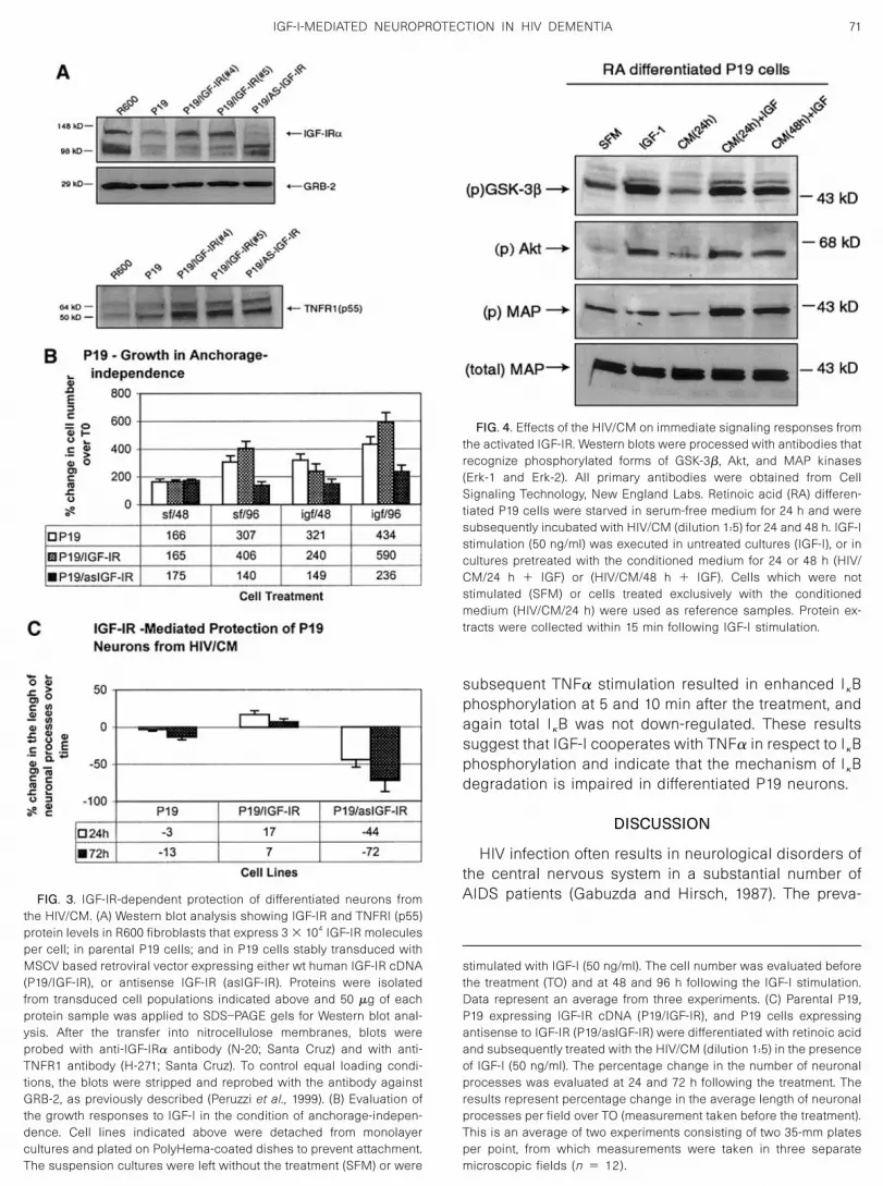
of IGF-IR to enhance IGF-I-mediated neuronal cell pro-tection. In parallel, we have evaluated the ability of anti-sense IGF-IR cDNA to suppress IGF-I-dependent neuro-nal outgrowth and to sensitize cells to neurodegenera-tive effects of the HIV/CM. We have generated stable celllines by transducing P19 cells with retrovirus vectorsexpressing human IGF-R cDNA in sense or antisenseorientations. To avoid clonal variability, mixed popula-tions of transduced cells were collected and utilized inthe study. Figure 3A indicates the levels of IGF-IR (top)and TNFR1 (bottom) in parental P19 cells, in P19 cellsstably transduced with the wild-type IGF-IR cDNA (P19/IGF-IR), in cells expressing antisense IGF-IR cDNA (P19/aslGF-IR), and in control R600 fibroblasts, which express3 � 104 IGF-IR molecules per cell (Reiss et al., 1998).Densitometric assessment revealed that P19/IGF-IR cellsare characterized by a threefold increase in the IGF-IRprotein level when compared with the parental P19 cells.Conversely, more than 50% decrease in the level ofIGF-IR was detected upon transducing cells with a ret-rovirus vector expressing antisense IGF-IR. The sameblot was utilized to examine the level of housekeepinggene product, GRB-2, to verify equal loading of the pro-teins in various samples. The observed differences in thelevels of IGF-IR in transduced cell populations correlatedwith the growth responses of the cells to IGF-I, as de-termined by anchorage-independence assays. This as-say is useful for evaluating the protective effects of IGF-Iunder conditions where the ability of cells to interact withextracellular matrix is restricted (Valentinis et al., 1998).Under these conditions parental P19 cells exhibited amodest ability to proliferate in response to IGF-I (Fig. 3B).As expected, P19/IGF-IR cell proliferation was noticeablyimproved in anchorage-independence, and the cells inwhich the endogenous IGF-IR level was reduced due tothe expression of antisense IGF-IR cDNA lost the abilityto respond with cell proliferation following the IGF-I stim-ulation. The cells, which were utilized in the above ex-periments, were further analyzed for the protective ef-fects of IGF-I. As mentioned above, differentiated P19cells lose neuronal processes after the treatment withHIV/CM (Fig. 1), and the addition of IGF-I substantiallyprotected their neuron-like phenotype (Fig. 2B). As illus-trated in Fig. 3C, sensitivity of the cells to HIV/CM is
greatly increased upon the down-regulation of the en-dogenous IGF-IR with the antisense cDNA (P19/asIGF-IRcells). In contrast, P19 cells which ectopically overex-press IGF-IR become resistant to the deleterious effectsof the HIV/CM upon the treatment with IGF-I (50 ng/ml).Altogether, these observations strongly support the hy-pothesis that activated IGF-I system confers protectionagainst neurotoxins present in the conditioned mediumof HIV-infected macrophages.
Effects of the HIV/CM on immediate signalingresponses from the IGF-IR
To evaluate whether the IGF-IR signaling pathways areaffected by HIV/CM, we investigated phosphorylationlevels for three signaling molecules (Akt, ERKs, GSK-3�)that are known to be activated by IGF-I stimulation and,further, suggested to play a role in neuronal differentia-tion and neuronal survival (Dudek et al., 1997; Yuan andYankner, 2000; Zheng et al., 2000). In these studies, IGF-Istimulation was applied to retinoic acid differentiatedP19 cells. Results in Fig. 4 demonstrate that in serum-free medium (SFM), P19 cells exhibit basal levels ofphosphorylation of GSK-3p, Akt, and Erk-2. Under thesame culture conditions, Erk-1 phosphorylation was notdetected. Within 15 min following IGF-I stimulation (IGF-I), both phospho-GSK-3� and phospho-Akt were signifi-cantly increased; phospho-Erk-2 was not affected andphospho-Erk-1 remained undetectable. Having estab-lished basal and induced levels of phosphorylation inresponse to IGF-I in differentiated P19 cells, we exam-ined whether pretreatment of the cells with the HIV/CMaffects the phosphorylation levels of these signaling mol-ecules. First of all HIV/CM by itself did not affect basallevels of the phosphorylation (CM/24 h). Preincubation ofP19 cells with the HIV/CM did not affect IGF-I-mediatedphosphorylation of GSK-3� and Akt at both 24 and 48 htime intervals. The only detectable change induced byHIV/CM was the enhancement of Erk-2 phosphorylationafter the IGF-I treatment. Altogether, these results indi-cate that HIV/CM is not affecting both basal and IGF-I-stimulated levels of Akt, ERKs, and GSK-3� phosphory-lation in differentiated 19 cells.The I�B protein binds to and prevents nuclear translo-
cation of the transcription factor Rel/NF�B (Baeuerle andBaltimore, 1988). The mechanism by which TNFR1 sig-naling activates NF�B involves I�B phosphorylation byIKKs, and subsequent proteosomal degradation of I�B(Zandi et al., 1998). Figure 5 shows that serum-starved,differentiated P19 cells have detectable levels of phos-phorylated I�B. Following TNF� stimulation, levels ofphosphorylated I�B increase and remain elevated for atleast 3 h. The same blot probed with antitotal I�B anti-body confirmed lack of expected down-regulation of I�Bat later time points following TNF� stimulation. WhenP19 cells were pretreated for 24 h with IGF-I (50 ng/ml),
70 WANG ET AL.
subsequent TNF� stimulation resulted in enhanced I�Bphosphorylation at 5 and 10 min after the treatment, andagain total I�B was not down-regulated. These resultssuggest that IGF-I cooperates with TNF� in respect to I�Bphosphorylation and indicate that the mechanism of I�Bdegradation is impaired in differentiated P19 neurons.
DISCUSSION
HIV infection often results in neurological disorders ofthe central nervous system in a substantial number ofAIDS patients (Gabuzda and Hirsch, 1987). The preva-
stimulated with IGF-I (50 ng/ml). The cell number was evaluated beforethe treatment (TO) and at 48 and 96 h following the IGF-I stimulation.Data represent an average from three experiments. (C) Parental P19,P19 expressing IGF-IR cDNA (P19/IGF-IR), and P19 cells expressingantisense to IGF-IR (P19/aslGF-IR) were differentiated with retinoic acidand subsequently treated with the HIV/CM (dilution 1:5) in the presenceof IGF-I (50 ng/ml). The percentage change in the number of neuronalprocesses was evaluated at 24 and 72 h following the treatment. Theresults represent percentage change in the average length of neuronalprocesses per field over TO (measurement taken before the treatment).This is an average of two experiments consisting of two 35-mm platesper point, from which measurements were taken in three separatemicroscopic fields (n � 12).
FIG. 3. IGF-IR-dependent protection of differentiated neurons fromthe HIV/CM. (A) Western blot analysis showing IGF-IR and TNFRI (p55)protein levels in R600 fibroblasts that express 3 � 104 IGF-IR moleculesper cell; in parental P19 cells; and in P19 cells stably transduced withMSCV based retroviral vector expressing either wt human IGF-IR cDNA(P19/IGF-IR), or antisense IGF-IR (asIGF-IR). Proteins were isolatedfrom transduced cell populations indicated above and 50 �g of eachprotein sample was applied to SDS–PAGE gels for Western blot anal-ysis. After the transfer into nitrocellulose membranes, blots wereprobed with anti-IGF-IR� antibody (N-20; Santa Cruz) and with anti-TNFR1 antibody (H-271; Santa Cruz). To control equal loading condi-tions, the blots were stripped and reprobed with the antibody againstGRB-2, as previously described (Peruzzi et al., 1999). (B) Evaluation ofthe growth responses to IGF-I in the condition of anchorage-indepen-dence. Cell lines indicated above were detached from monolayercultures and plated on PolyHema-coated dishes to prevent attachment.The suspension cultures were left without the treatment (SFM) or were
FIG. 4. Effects of the HIV/CM on immediate signaling responses fromthe activated IGF-IR. Western blots were processed with antibodies thatrecognize phosphorylated forms of GSK-3�, Akt, and MAP kinases(Erk-1 and Erk-2). All primary antibodies were obtained from CellSignaling Technology, New England Labs. Retinoic acid (RA) differen-tiated P19 cells were starved in serum-free medium for 24 h and weresubsequently incubated with HIV/CM (dilution 1:5) for 24 and 48 h. IGF-Istimulation (50 ng/ml) was executed in untreated cultures (IGF-I), or incultures pretreated with the conditioned medium for 24 or 48 h (HIV/CM/24 h � IGF) or (HIV/CM/48 h � IGF). Cells which were notstimulated (SFM) or cells treated exclusively with the conditionedmedium (HIV/CM/24 h) were used as reference samples. Protein ex-tracts were collected within 15 min following IGF-I stimulation.
71IGF-I-MEDIATED NEUROPROTECTION IN HIV DEMENTIA

lence of HIV associated dementia (HAD) has been esti-mated in the early 1990s to be as high as 20–30% ofthose patients with advanced HIV disease. It has beenspeculated that HAD remains the most common cause ofdementia worldwide among people at ages 40 or lessand is considered a significant independent risk factorfor death due to AIDS (Glass and Johnson, 1996; Price,1988; Sharer, 1992). The pathogenesis of HAD is believedto involve the release of toxic substances in the brain byactivated and/or infected mononuclear phagocytes, lead-ing to neuronal and astrocytic dysfunction and death(Price, 1988; Price et al., 1988; Thompson et al., 2001).The proinflammatory cytokine, TNF�, released by acti-vated and/or HIV-1-infected brain macrophages and mi-croglia, appears to provide a substantial contribution toneuronal cell damage (Rieckmann et al., 1991; Rimaniolet al., 1997). In addition to factors promoting neuronalinjury, a decrease in levels/activity of neuronal protectivemechanisms may also contribute to the overall injuryprocess. In support of this notion, several studies sug-gest that IGF-I-mediated responses are impaired during
the course of HIV infection (Jain et al., 1998; Laue et al.,1990), leaving neuronal cells with attenuated defensemechanism against proapoptotic cytokines and toxicmetabolites. Reduced levels of serum IGF-I have beenobserved in HIV-infected patients, particularly those withwasting syndrome and in children with failure to thrive(Jain et al., 1998; Laue et al., 1990). As IGF-I is a principalmediator of the action of human growth hormone, its rolein anabolic effects has prompted studies of IGF-I levelsin HIV-infected patients and the use of both IGF-I andgrowth hormone in the treatment of cachectic patients(Frost et al., 1996; Lo et al., 2001; Mynarcik et al., 1999,2000). Although some improvements in body mass havebeen noted, these studies also suggest partial resis-tance to the effects of growth hormone and/or IGF-I in thesetting of HIV-wasting syndrome (Jain et al., 1998). De-creased levels of IGF-I in the central nervous system maypromote neuronal apoptosis in HIV infection, or alterna-tively, by mechanisms which contribute to IGF-I resis-tance. The resistance may occur at the level of thereceptor (low expression of the IGF-IR or its mutation), or
FIG. 5. Effects of TNF� and IGF-I on I�B phosphorylation. (A) Western blots processed with antibodies that recognize phosphorylated (p) or total(total) I�B (Cell Signaling Technology, New England Labs). Retinoic acid (RA) differentiated P19 cells were starved in serum-free medium for 24 h (SFM)and were subsequently incubated with IGF-I (50 ng/ml) for 24 h (IGF-I 24 h), or were left without treatment (No IGF-I). Subsequently cells werestimulated with TNF� (20 ng/ml) and total proteins were isolated at indicated time points. (B) Densitometric evaluation of the blot depicted in A.
72 WANG ET AL.

by a specific attenuation of the critical signaling factorssuch as Akt. Some of these effects may be due to theaction of TNF�, which has been demonstrated to inhibitsome of the known activities of the IGF-IR and insulinreceptor (IR) in diabetes (Hotamisligil, 1999), and wasshown to inhibit IGF-I-stimulated protein synthesis inmyoblasts (Jain et al., 1998). These TNF�-mediated ef-fects have been suggested to involve serine phosphor-ylation and partial inactivation of two major signalingmolecules of the IGF system, IRS-I and IRS-2 (Peraldi etal., 1996; Venters et al., 1999).In the experimental setting proposed here, cellular
responses to HIV/CM have been initially tested in expo-nentially growing monolayer cultures of P19 and PC 12cells. Surprisingly, we observed no significant changesin the rate of cell proliferation and cell death, suggestingthat the undifferentiated neuronal cell lines are resistantto the secreted neurotoxic factors from the HIV/CM. Incontrast to the relative resistance of undifferentiatedcells, differentiated neuronal cultures were highly sensi-tive to the injuries inflicted by the HIV/CM. We observeda greater proportional decrease in the number of neurite-like processes compared to the modest decrease in thecell number. This may imply that cells which lost neuro-nal processes did not undergo apoptosis or that cellproliferation occurred despite of the treatment. To avoidpotentially complex issues related to the presence orabsence of apoptosis versus proliferation in neuronalcultures, we expressed our results in terms of the num-ber of neurite-bearing cells (PC 12 cells), or by the lengthof neurite-like processes per microscopic field (p19cells). Results presented in this article demonstrate thatdifferentiated neurons are not capable of maintainingneuronal processes when exposed to the conditionedmedium from HIV-I-infected macrophages (HIV/CM). Fur-ther, it is evident that TNF�-activating TNFR1 (p55) par-ticipates in the process of HIV/CM-induced neuronaldegeneration. This observation is in agreement with pre-vious reports showing neuronal death, evaluated by LDHrelease, only in differentiated cultures of NT2N neuronstreated with TNF� (Westmoreland et al., 1996).Since the IGF-IR efficiently rescues differentiated neu-
rons from both HIV/CM and TNF�-induced damage ofneuronal processes (Figs. 2 and 3), it is feasible that themechanism of its neuroprotective action involves, atleast partially, redirection of the signal from the TNFR1.Depending on the circumstances, which are not wellcharacterized, TNFR1 may send contradictory signals inrespect to cell survival: proapoptotic FADD-mediated re-cruitment of procaspase 8 or antiapoptotic Traf-2-medi-ated phosphorylation of I�B (Natoli et al., 1998). If theIGF-IR affects TNFR1 signaling on this level, the connec-tion to procaspase 8 should be attenuated and the acti-vation of NF�B should be enhanced. In this respect ourdata demonstrate a strong TNF�-mediated phosphoryla-tion of I�B in P19 cells preconditioned by the IGF-I (Fig. 5).
The IGF-IR could also switch TNFR1 into antiapoptoticmode by facilitating FLIP activity and subsequent inhibi-tion of caspase 8. Although this possibility has neverbeen shown for IGF-IR directly, signaling molecules thatare known to be activated by the IGF-IR could do so. Thisincludes Akt (Panka et al., 2001) and/or Raf activation(Kataoka et al., 2000). In addition, NF�B has been alsoshown to facilitate FLIP expression (Micheau et al., 2001;You et al., 2001), suggesting another possible level of thesynergy between IGF-IR and TNFR1. Importantly, the ma-jor signaling responses from the IGF-IR, including phos-phorylation of Akt, MAP kinases, and Gsk-3�, are notattenuated in the presence of HIV/CM. Therefore, neu-roprotective cross-talk between IGF-I and TNF� mayinclude, in addition to the sustained Akt, ERK2, andGSK3� phosphorylation, TNFR1-mediated activation ofNF�B in this HIV-related experimental setting. This alsoindicates that molecular manipulations to increase effec-tiveness of the IGF-IR system may represent a potentialtherapeutic approach to rescue differentiated neurons inHIV-associated dementia.
MATERIALS AND METHODS
Cell lines
Cell-culture conditions for growth and differentiation ofP19 mouse teratocarcinoma (ATCC No. CRL-1825), andPC12 rat pheochromocytoma (ATCC No. CRL-1721), werepreviously reported (Jones-Villeneuve et al., 1982; Teng etal., 1993). To induce neuronal differentiation, PC12 cellsplated on poly-D-lysine culture dishes were treated inserum-free medium with NGF (20 ng/ml). Neuronal pro-cesses began to appear within the first 24 h following thetreatment and could be observed in culture for at least 10days. The P19 neuronal differentiation was induced byretinoic acid (RA) (500 nM; Sigma). The response to RAtreatment required cell aggregation that was spontane-ously induced when the cells were forced to grow insuspension. The aggregates were collected, resus-pended in fresh culture medium (�-MEM � 7.5% CS �2.5% FBS), and treated with the RA for an additional 48 h.Subsequently, large cellular aggregates were dissoci-ated by trypsinization, and single cells and small cellularaggregates were plated on poly-D-lysine-coated culturedishes. Under these conditions, neuronal differentiationwas observed within 48 h after the final plating. Longneuronal processes were maintained in culture for 2weeks with the medium changed every second day. Toavoid overgrowth with nondifferentiated cells, RA-treatedcells were cultured either in serum-free medium (�-MEM � 0.1% BSA) or in serum-supplemented mediumcontaining Ara-C (1 �M) (cytosine �-D-arabino-furano-side, Sigma). R600 fibroblasts, which express 3 � 104 ofIGF-IR molecules/cell (Reiss et al., 1998), were used as areference sample to monitor the level of IGF-IR expres-
73IGF-I-MEDIATED NEUROPROTECTION IN HIV DEMENTIA

sion in parental P19 cells and in P19 cultures expressingIGF-IR cDNA in either sense or antisense orientation.
Preparation of conditioned media from HIV-I-infectedT-lymphocytes
Suspension cultures of supT1 cells were infected withHIV-1 strain SF162 (or JRFL) with a multiplicity of infection(m.o.i.) of 0.1. Briefly, the cells were incubated with thevirus inoculum for 2 h at 37°C in serum-free RPMI 1640.At the end of incubation period, the cells were centri-fuged at 1000 rpm for 5 min at 4°C. The cell pellet wassuspended in RPMI containing 2% fetal bovine serum,gentamicin (5 �g/ml), and incubated at 37°C. Four-fifthsof the cell supernatant was harvested as conditionedmedia on alternate days up to day 24 of infection andstored in 10 ml aliquots at �80°C. For generation ofvirus-free conditioned media, one aliquot of the storedsupernatant was thawed and then centrifuged at 3100rpm at 4°C for 45 min in a centrifugal filter device(Biomax 10K NMWL membrane, Millipore Corp., MA).The filtrate was used as virus-free (evaluated by p24assay) conditioned medium.
Growth in anchorage-independence
To determine this parameter, quiescent cells weredetached from a culture dish with 0.02% disodium ethyl-enediamine tetraacetate (EDTA) and seeded on dishescoated with poly(2-hydroxyethyle methacrylate) [poly-(HEMA)] (Aldrich, Milwaukee, WI), prepared according tothe methodology previously described (Reiss et al.,1998). Cells were seeded in SFM at 1 � 104 cells/cm2
and were treated either with 10% FBS or 50 ng/ml IGF-I orwere left untreated. Twenty-four hours later cell suspen-sions were collected and dissociated with 0.25% trypsin,and cells were counted in a Brightline Hemocytometer.Results represent an average of three experiments andare expressed as a percentage change in cell number.
Detection of DNA strand breaks
DNA strand breaks were labeled with biotinylateddUTP (Boehringer Mannheim), using exogenous terminaldeoxynucleotidyl transferase (TDT; Boehringer Mann-heim). Cells were fixed on ice with a buffered 10% meth-anol-free formaldehyde (Polyscience, Inc.) for 20 min,incubated with 70% ethanol for 1 h at �20°C, andwashed 3� with PBS. DNA strand breaks were detectedby in situ labeling of free 3�-OH ends on DNA. Fixed cellswere incubated with 50 �l of a reaction mixture (5 unitsTDT, 2.5 mM CoCl2, 0.2 M potassium cacodylate, 25 mMTris–HCl, 0.25% bovine serum albumin, and 0.5 nM bi-otin-16-dUTP) for 30 min at 37°C and incubated withstaining solution [5 �g/ml of fluorescein isothiocyanateextravidin (FITC), 4� SSC, 0.1% Triton X-100, and 5%nonfat dry milk] for 30 min at 37°C. DNA strand breakswere visualized under a fluorescent microscope
equipped with a set of excitation-emission filters forfluorescein (excitation blue, emission green).
Retroviral transduction
The MSCV.pac and MSCV.neoEB retroviral vectors,kindly provided by Dr. Hawley (University of Toronto,Canada), are described elsewhere (Hawley et al., 1994).The self-inactivating (SIN) version of the MSCV.pac wasengineered by deleting 299 bp in the 3� long terminalrepeat (LTR), as described elsewhere (Yu et al., 1986). Ageneral cloning strategy was based on PCR-amplifiedcDNA fragments of the wild-type IGF-IR cDNAs in thesense and antisense orientation. The PCR primers con-taining appropriate restriction sites in overhangs werepreviously described (Romano et al., 1999; Valentinis etal., 2000). Prior to cloning, each amplified fragment wasverified by sequencing (Nucleic Acid Facility at KimmelCancer Center, Thomas Jefferson University, Philadel-phia, PA).
Western blot analysis
To determine levels of phosphorylated forms of IGF-I-induced signaling molecules, differentiated cultures ofP19 cells were lysed on ice with 400 �l of lysis buffer [(50mM HEPES); pH 7.5; 150 mM NaCl; 1.5 mM MgCl2; 1 mMEGTA; 10% glycerol; 1% Triton X-100; 1 mM phenylmeth-ylsulfonyl fluoride (PMSF); 0.2 mM Na-orthovanadate,and 10 �g/ml aprotinin]. Protein concentration was de-termined by a Bio-Rad Protein Assay (Bio-Rad, Hercules,CA), and 50 �g of total proteins were separated on a4–15% gradient SDS–PAGE (Bio-Rad) and transferredonto nitrocellulose membranes. Blots were blocked with5% nonfat dry milk in TBST (10 mM Tris–HCl, pH 7.5, 150mM NaCl, 0.1% Tween 20) and were probed with theappropriate primary antibodies. Total and phosphory-lated forms of Akt/PKB, Erk1/Erk2, GSK-3�, and I�B weredetected by utilizing PhosphoPlus antibody kits obtainedfrom New England BioLabs, Beverly, MA.
ACKNOWLEDGMENT
This work is supported by Grant PO1 NS 36466 from NIH.
REFERENCES
Baeuerle, P. A., and Baltimore, D. (1988). I kappa B: A specific inhibitorof the NF-kappa B transcription factor. Science 242(4878), 540–546.
Boone, E., Vanden Berghe, T., Van Loo, G., De Wilde, G., De Wael, N.,Vercammen, D., Fiers, W., Haegeman, G., and Vandenabeele, P.(2000). Structure/function analysis of p55 tumor necrosis factor re-ceptor and fas-associated death domain. Effect on necrosis inL929sA cells. J. Biol. Chem. 275(48), 37596–37603.
Boone, E., Vandevoorde, V., De Wilde, G., and Haegeman, G. (1998).Activation of p42/p44 mitogen-activated protein kinases (MAPK) andp38 MAPK by tumor necrosis factor (TNF) is mediated through thedeath domain of the 55-kDa TNF receptor. FEBS Lett. 441(2), 275–280.
Budka, H., Costanzi, G., Cristina, S., Lechi, A., Parravicini, C., Trabattoni,
74 WANG ET AL.

R., and Vago, L. (1987). Brain pathology induced by infection with thehuman immunodeficiency virus (HIV). A histological, immunocyto-chemical, and electron microscopical study of 100 autopsy cases.Acta Neuropathol. (Berlin) 75(2), 185–198.
Cardone, M. H., Roy, N., Stennicke, H. R., Salvesen, G. S., Franke, T. F.,Stanbridge, E., Frisch, S., and Reed, J. C. (1998). Regulation of celldeath protease caspase-9 by phosphorylation. Science 282(5392),1318–1321.
D’Mello, S. R., Galli, C., Ciotti, T., and Calissano, P. (1993). Induction ofapoptosis in cerebellar granule neurons by low potassium: Inhibitionof death by insulin-like growth factor I and cAMP. Proc. Natl. Acad.Sci. USA 90(23), 10989–10993.
Dudek, H., Datta, S. R., Franke, T. F., Birnbaum, M. J., Yao, R., Cooper,G. M., Segal, R. A., Kaplan, D. R., and Greenberg, M. E. (1997).Regulation of neuronal survival by the serine-threonine protein ki-nase Akt. Science 275(5300), 661–665.
Frost, R. A., Fuhrer, J., Steigbigel, R., Mariuz, P., Lang, C. H., and Gelato,M. C. (1996). Wasting in the acquired immune deficiency syndrome isassociated with multiple defects in the serum insulin-like growthfactor system. Clin. Endocrinol. (Oxford) 44(5), 501–514.
Furusu, A., Nakayama, K., Xu, Q., Konta, T., Sugiyama, H., and Kitamura,M. (2001). Expression, regulation, and function of inhibitor of apopto-sis family genes in rat mesangial cells. Kidney Int. 60(2), 579–586.
Gabuzda, D. H., and Hirsch, M. S. (1987). Neurologic manifestations ofinfection with human immunodeficiency virus. Clinical features andpathogenesis. Ann. Intern. Med. 107(3), 383–391.
Gary, D. S., Bruce-Keller, A. J., Kindy, M. S., and Mattson, M. P. (1998).Ischemic and excitotoxic brain injury is enhanced in mice lacking thep55 tumor necrosis factor receptor. J. Cereb. Blood Flow Metab.18(12), 1283–1287.
Glass, J. D., and Johnson, R. T. (1996). Human immunodeficiency virusand the brain. Annu. Rev. Neurosci. 19, 1–26.
Gluckman, P., Klempt, N., Guan, J., Mallard, C., Sirimanne, E., Dragunow,M., Klempt, M., Singh, K., Williams, C., and Nikolics, K. (1992). A rolefor IGF-1 in the rescue of CNS neurons following hypoxic-ischemicinjury. Biochem. Biophys. Res. Commun. 182(2), 593–599.
Hawley, R. G., Lieu, F. H., Fong, A. Z., and Hawley, T. S. (1994). Versatileretroviral vectors for potential use in gene therapy. Gene Ther. 1(2),136–138.
Hotamisligil, G. S. (1999). The role of TNFalpha and TNF receptors inobesity and insulin resistance. J. Intern. Med. 245(6), 621–625.
Jain, S., Golde, D. W., Bailey, R., and Geffner, M. E. (1998). Insulin-likegrowth factor-I resistance. Endocr. Rev. 19(5), 625–646.
Jones-Villeneuve, E. M., McBurney, M. W., Rogers, K. A., and Kalnins, V. I.(1982). Retinoic acid induces embryonal carcinoma cells to differen-tiate into neurons and glial cells. J. Cell. Biol. 94(2), 253–262.
Kataoka, T., Budd, R. C., Holler, N., Thome, M., Martinon, F., Irmler, M.,Burns, K., Hahne, M., Kennedy, N., Kovacsovics, M., and Tschopp, J.(2000). The caspase-8 inhibitor FLIP promotes activation of NF-kappaB and Erk signaling pathways. Curr. Biol. 10(11), 640–648.
Kaul, M., and Lipton, S. A. (1999). Chemokines and activated macro-phages in HIV gp120-induced neuronal apoptosis. Proc. Natl. Acad.Sci. USA 96(14), 8212–8216.
Kubota, T., Miyagishima, M., Frye, C. S., Alber, S. M., Bounoutas, G. S.,Kadokami, T., Watkins, S. C., McTiernan, C. F., and Feldman, A. M.(2001). Overexpression of tumor necrosis factor-alpha activates bothanti- and pro-apoptotic pathways in the myocardium. J. Mol. CellCardiol. 33(7), 1331–1344.
Laue, L., Pizzo, P. A., Butler, K., and Cutler, G. B., Jr. (1990). Growth andneuroendocrine dysfunction in children with acquired immunodefi-ciency syndrome. J. Pediatr. 117(4), 541–545.
Lieberson, R., Mowen, K. A., McBride, K. D., Leautaud, V., Zhang, X.,Suh, W. K., Wu, L., and Glimcher, L. H. (2001). Tumor necrosis factorreceptor-associated factor (TRAF)2 represses the T helper cell type2 response through interaction with NFAT-interacting protein (NIP45).J. Exp. Med. 194(1), 89–98.
Lo, J. C., Mulligan, K., Noor, M. A., Schwarz, J. M., Halvorsen, R. A.,
Grunfeld, C., and Schambelan, M. (2001). The effects of recombinanthuman growth hormone on body composition and glucose metabo-lism in HIV-infected patients with fat accumulation. J. Clin. Endocr.Metab. 86(8), 3480–3487.
Locksley, R. M., Killeen, N., and Lenardo, M. J. (2001). The TNF and TNFreceptor superfamilies: Integrating mammalian biology. Cell 104(4),487–501.
Micheau, O., Lens, S., Gaide, O., Alevizopoulos, K., and Tschopp, J.(2001). NF-kappaB signals induce the expression of c-FLIP. Mol. CellBiol. 21(16), 5299–5305.
Mynarcik, D. C., Frost, R. A., Lang, C. H., DeCristofaro, K., McNurlan,M. A., Garlick, P. J., Steigbigel, R. T., Fuhrer, J., Ahnn, S., and Gelato,M. C. (1999). Insulin-like growth factor system in patients with HIVinfection: Effect of exogenous growth hormone administration. J.Acquir. Immune Defic. Syndr. 22(1), 49–55.
Mynarcik, D. C., McNurlan, M. A., Steigbigel, R. T., Fuhrer, J., and Gelato,M. C. (2000). Association of severe insulin resistance with both lossof limb fat and elevated serum tumor necrosis factor receptor levelsin HIV lipodystrophy. J. Acquir. Immune Defic. Syndr. 25(4), 312–321.
Natoli, G., Costanzo, A., Guido, F., Moretti, F., and Levrero, M. (1998).Apoptotic, non-apoptotic, and anti-apoptotic pathways of tumor ne-crosis factor signalling. Biochem. Pharmacol. 56(8), 915–920.
Panka, D. J., Mano, T., Suhara, T., Walsh, K., and Mier, J. W. (2001).Phosphatidylinositol 3-kinase/Akt activity regulates c-FLIP expres-sion in tumor cells. J. Biol. Chem. 276(10), 6893–6896.
Peraldi, P., Hotamisligil, G. S., Buurman, W. A., White, M. F., andSpiegelman, B. M. (1996). Tumor necrosis factor (TNF)-alpha inhibitsinsulin signaling through stimulation of the p55 TNF receptor andactivation of sphingomyelinase. J. Biol. Chem. 271(22), 13018–13022.
Peruzzi, F., Prisco, M., Dews, M., Salomoni, P., Grassilli, E., Romano, G.,Calabretta, B., and Baserga, R. (1999). Multiple signaling pathways ofthe insulin-like growth factor 1 receptor in protection from apoptosis.Mol. Cell Biol. 19(10), 7203–7215.
Peruzzi, F., Prisco, M., Morrione, A., Valentinis, B., and Baserga, R.(2001). Anti-apoptotic signaling of the IGF-I receptor through mito-chondrial translocation of c-Raf and Nedd4. J. Biol. Chem. 14, 14.
Price, R. W. (1988). Dementia associated with AIDS. Trans. Assoc. LifeInsur. Med. Dir. Am. 71, 235–240.
Price, R. W., Brew, B., Sidtis, J., Rosenblum, M., Scheck, A. C., andCleary, P. (1988). The brain in AIDS: Central nervous system HIV-1infection and AIDS dementia complex. Science 239(4840), 586–592.
Reiss, K., Valentinis, B., Tu, X., Xu, S. Q., and Baserga, R. (1998).Molecular markers of IGF-I-mediated mitogenesis. Exp. Cell Res.242(1), 361–372.
Rieckmann, P., Poli, G., Fox, C. H., Kehrl, J. H., and Fauci, A. S. (1991).Recombinant gp120 specifically enhances tumor necrosis factor-alpha production and Ig secretion in B lymphocytes from HIV-in-fected individuals but not from seronegative donors. J. Immunol.147(9), 2922–2927.
Rimaniol, A. C., Boussin, F. D., Dormont, D., Bach, J. F., and Zavala, F.(1997). Mechanisms of downmodulation and release of tumour ne-crosis factor receptor induced by human immunodeficiency virustype 1 in human monocytes. Cytokine 9(1), 9–18.
Romano, G., Prisco, M., Zanocco-Marani, T., Peruzzi, F., Valentinis, B.,and Baserga, R. (1999). Dissociation between resistance to apopto-sis and the transformed phenotype in IGF-I receptor signaling. J. Cell.Biochem. 72(2), 294–310.
Sharer, L. R. (1992). Pathology of HIV-1 infection of the central nervoussystem. A review. J. Neuropathol. Exp. Neurol. 51(1), 3–11.
Sippy, B. D., Hofman, F. M., Wallach, D., and Hinton, D. R. (1995).Increased expression of tumor necrosis factor-alpha receptors in thebrains of patients with AIDS. J. Acquir. Immune Defic. Syndr. Hum.Retrovirol. 10(5), 511–521.
Tan, S. V., Guiloff, R. J., Henderson, D. C., Gazzard, B. G., and Miller, R.(1996). AIDS-associated vacuolar myelopathy and tumor necrosisfactor-alpha (TNF alpha). J. Neurol. Sci. 138(1–2), 134–144.
Teng, K. K., Georgieff, I. S., Aletta, J. M., Nunez, J., Shelanski, M. L., and
75IGF-I-MEDIATED NEUROPROTECTION IN HIV DEMENTIA

Greene, L. A. (1993). Characterization of a PC12 cell sub-clone(PC12–C41) with enhanced neurite outgrowth capacity: Implicationsfor a modulatory role of high molecular weight tau in neuritogenesis.J. Cell. Sci. 106(Pt. 2), 611–626.
Thompson, K. A., McArthur, J. C., and Wesselingh, S. L. (2001). Corre-lation between neurological progression and astrocyte apoptosis inHIV-associated dementia. Ann. Neurol. 49(6), 745–752.
Valentinis, B., Navarro, M., Zanocco-Marani, T., Edmonds, P., McCor-mick, J., Morrione, A., Sacchi, A., Romano, G., Reiss, K., and Baserga,R. (2000). Insulin receptor substrate-1, p70S6K, and cell size intransformation and differentiation of hemopoietic cells. J. Biol. Chem.275(33), 25451–25459.
Valentinis, B., Reiss, K., and Baserga, R. (1998). Insulin-like growthfactor-I-mediated survival from anoikis: Role of cell aggregation andfocal adhesion kinase. J. Cell. Physiol. 176(3), 648–657.
Venters, H. D., Tang, Q., Liu, Q., VanHoy, R. W., Dantzer, R., and Kelley,K. W. (1999). A new mechanism of neurodegeneration: A proinflam-matory cytokine inhibits receptor signaling by a survival peptide.Proc. Natl. Acad. Sci. USA 96(17), 9879–9884.
Wallach, D. (1997). Cell death induction by TNF: A matter of self control.Trends Biochem. Sci. 22(4), 107–109.
Wang, J. Y., Del Valle, L., Gordon, J., Rubini, M., Romano, G., Croul, S.,Peruzzi, F., Khalili, K., and Reiss, K. (2001). Activation of the IGF-IRsystem contributes to malignant growth of human and mouse me-dulloblastomas. Oncogene 20(29), 3857–3868.
Westmoreland, S. V., Kolson, D., and Gonzalez-Scarano, F. (1996). Tox-icity of TNF alpha and platelet activating factor for human NT2N
neurons: A tissue culture model for human immunodeficiency virusdementia. J. Neurovirol. 2(2), 118–126.
You, Z., Ouyang, H., Lopatin, D., Polver, P. J., and Wang, C. Y. (2001).Nuclear factor-kappa B-inducible death effector domain-containingprotein suppresses tumor necrosis factor-mediated apoptosis byinhibiting caspase-8 activity. J. Biol. Chem. 276(28), 26398–26404.
Yu, S. F., von Ruden, T., Kantoff, P. W., Garber, C., Seiberg, M., Ruther, U.,Anderson, W. F., Wagner, E. F., and Gilboa, E. (1986). Self-inactivatingretroviral vectors designed for transfer of whole genes into mamma-lian cells. Proc. Natl. Acad. Sci. USA 83(10), 3194–3198.
Yuan, J., and Yankner, B. A. (2000). Apoptosis in the nervous system.Nature 407(6805), 802–809.
Yuasa, T., Ohno, S., Kehrl, J. H., and Kyriakis, J. M. (1998). Tumornecrosis factor signaling to stress-activated protein kinase (SAPK)/Jun NH2-terminal kinase (JNK) and p38. Germinal center kinasecouples TRAF2 to mitogen-activated protein kinase/ERK kinase ki-nase 1 and SAPK while receptor interacting protein associates witha mitogen-activated protein kinase kinase kinase upstream of MKK6and p38. J. Biol. Chem. 273(35), 22681–22692.
Zandi, E., Chen, Y., and Karin, M. (1998). Direct phosphorylation ofIkappaB by IKKalpha and IKKbeta: Discrimination between free andNF-kappaB-bound substrate. Science 281(5381), 1360–1363.
Zha, J., Harada, H., Yang, E., Jockel, J., and Korsmeyer, S. J. (1996).Serine phosphorylation of death agonist BAD in response to survivalfactor results in binding to 14-3-3 not BCL-X(L). Cell 87(4), 619–628.
Zheng, W. H., Kar, S., Dore, S., and Quirion, R. (2000). Insulin-like growthfactor-1 (IGF-1): A neuroprotective trophic factor acting via the Aktkinase pathway. J. Neural Transm. Suppl. (60), 261–272.
76 WANG ET AL.


