Embed Size (px)
Citation preview

A New Aryl Hydrocarbon Receptor Homology Model Targeted toImprove Docking Reliability
Ilaria Motto§, Annalisa Bordogna§, Anatoly A. Soshilov#, Michael S. Denison#, and LauraBonati§,*
Ilaria Motto: [email protected]; Annalisa Bordogna: [email protected]; Anatoly A. Soshilov:[email protected]; Michael S. Denison: [email protected]; Laura Bonati: [email protected]§Dipartimento di Scienze dell’Ambiente e del Territorio, Università degli Studi di Milano-Bicocca,Piazza della Scienza 1, 20126 Milano, Italy#Department of Environmental Toxicology, Meyer Hall, University of California, Davis, California95616
AbstractThe aryl hydrocarbon receptor (AhR) is a ligand-dependent, basic helix-loop-helix Per-ARNT-Sim (PAS) containing transcription factor that can bind and be activated by structurally diversechemicals, including the toxic environmental contaminant 2,3,7,8-tetrachlorodibenzo-p-dioxin(TCDD). As no experimentally determined structures of the AhR ligand binding domain (LBD)are available and previous homology models were only derived from apo template structures, wedeveloped a new model based on holo X-ray structures of the hypoxia-inducible factor 2α(HIF-2α) PAS B domain, targeted to improve the accuracy of the binding site for moleculardocking applications. We experimentally confirmed the ability of two HIF-2α crystallographicligands to bind to the mAhR with relatively high affinity and demonstrated that they are AhRagonists, thus justifying the use of the holo HIF-2α structures as templates. A specific modeling/docking approach was proposed to predict the binding modes of AhR ligands in the modeledLBD. It was validated by comparison of the calculated and the experimental binding affinities ofactive THS ligands and TCDD for the mAhR and by functional activity analysis using severalmAhR mutants generated on the basis of the modeling results. Finally the ability of the proposedapproach to reproduce the different affinities of TCDD for AhRs of different species wasconfirmed, and a first test of its reliability in virtual screening is carried out by analyzing thecorrelation between the calculated and experimental binding affinities of a set of 14 PCDDs.
IntroductionThe aryl hydrocarbon receptor (AhR)a is a basic helix-loop-helix (bHLH), Per-ARNT-Sim(PAS) containing ligand-dependent transcription factor that induces the expression of a largebattery of genes and produces diverse biological and toxic effects in a wide range of speciesand tissues.1–4 The best-characterized high affinity ligands include a variety of toxichalogenated aromatic hydrocarbons (HAHs), such as the polychlo-dibenzo-p-dioxins(PCDDs), dibenzofurans (PCDFs), and biphenyls (PCBs), and numerous polycyclic
*To whom correspondence should be addressed. Phone: (+39)0264482821. Fax: (+39)0264482839. [email protected] Information. Four additional Figures are provided as Supporting Information (SI):Figure SI1. Alignment of the LBD domains of mouse, human and rat AhRs against HIF-2α.Figure SI2. Cα-trace representation of the 100 mAhR homology models generated by MODELLER.Figure SI3. Results of ensemble docking of THS-017 and THS-020 in the mAhR homology models.Figure SI4. Results of PCDD ensemble docking in the rtAhR LBD homology models.This information is available free of charge via the Internet at http://pubs.acs.org/.
NIH Public AccessAuthor ManuscriptJ Chem Inf Model. Author manuscript; available in PMC 2012 November 28.
Published in final edited form as:J Chem Inf Model. 2011 November 28; 51(11): 2868–2881. doi:10.1021/ci2001617.
NIH
-PA Author Manuscript
NIH
-PA Author Manuscript
NIH
-PA Author Manuscript

aromatic hydrocarbons (PAHs) and PAH-like chemicals,5–7 all widespread classes ofenvironmental contaminants. Moreover, a number of natural, endogenous and synthetic AhRagonists and antagonists whose structure and physicochemical characteristics are differentfrom those of the prototypical HAH and PAH ligands have been identified as lower affinityligands and moderately potent inducers of AhR-dependent gene expression.7–11 Among thevarious protein domains responsible for the AhR functional activities, PAS B (one of thetwo structural repeats in the PAS domain) is the one responsible for ligand binding and it isalso involved in binding to the chaperone heat shock protein 90 (hsp90).12
Although the AhR signal transduction pathway has been studied for many years,1–4 severalunanswered questions remain. Major issues are the actual spectrum of ligands, how they canbind to the AhR, and how ligand binding to the ligand binding domain (LBD) results inactivation of the AhR and AhR-dependent gene expression. A molecular understanding ofthese events would require detailed structural information about the AhR PAS B LBD.However, neither X-ray nor NMR structures of the bound or unbound AhR have beendetermined to date.
Since the first crystal structures of distant homologous proteins belonging to the PASsuperfamily became available, we started developing theoretical models of the AhR LBD byhomology modeling techniques and the results provided an initial framework to makehypotheses on LBD characteristics and the mechanisms of AhR functionality.9,13,14 Thelatest model of the mouse AhR (mAhR) LBD we proposed14 was built using the NMRstructures of the PAS B domains of the human hypoxia-inducible factor 2α (HIF-2α)15 andof the human ARNT,16 both in the apo form, as templates. That was the most reliableamong the models developed in our group, since the template domains show the highestdegree of sequence identity and similarity with the AhR PAS B among all the PASstructures reported to date. Moreover, the full length template proteins are members of thebHLH/PAS family of transcriptional factors and functionally related to the AhR.4 On thebasis of model-driven site-directed mutagenesis and AhR functional analysis, the buriedcavity in the core of the domain was confirmed as the site involved in ligand binding.14
Moreover, analysis of the LBD models of several mammalian AhRs indicated that thephysico-chemical characteristics of the binding cavities are remarkably conserved in allAhRs with high affinity for the AhR ligand 2,3,7,8-tetrachlorodibenzo-p-dioxin (TCDD).17
Mutagenesis of the conserved residues, followed by AhR functional analysis, allowedidentification of the “TCDD-binding fingerprint”, i.e. the group of residues necessary foroptimal TCDD binding.17
On the basis of our findings, other authors recently reported the development of AhR LBDhomology models based on the same apo structure of HIF-2α and proposed the use of thesemodels for molecular docking applications.18–22 Although the use of homology models ofthe target protein, in addition to experimental structures, has greatly extended theapplicability of molecular docking approaches,23–25 the use of good quality models, inparticular of the binding site, is crucial for the prediction of reliable binding poses.24, 26–28
In fact, it was proven that the success of docking calculation decreases with the quality ofthe receptor structure and, in particular, with the decreasing ability of the structure to
aAbbreviations: AhR, aryl hydrocarbon receptor; mAhR, Mus Musculus (mouse) aryl hydrocarbon receptor; huAhR, Homo Sapiens(human) aryl hydrocarbon receptor; rtAhR, Rattus Norvegicus (rat) aryl hydrocarbon receptor; ARNT, aryl hydrocarbon receptornuclear translocator; HIF-2α, hypoxia-inducible factor 2α; hsp90, heat shock protein 90; bHLH, basic Helix-Loop-Helix; DRE, dioxinresponsive element; LBD, ligand binding domain; PAS, Per-ARNT-Sim; TCDD, 2,3,7,8-tetrachlorodibenzo-p-dioxin; HAH,halogenated aromatic hydrocarbons; PCDD, polychlorodibenzo-p-dioxin (MCDD, monochloro; DCDD, dichloro; TrCDD, trichloro;TCDD, tetrachloro; PeCDD, pentachloro; Hx, exachloro; O, octachloro); PCDF, polychlorodibenzofuran; PCB, polychlorobiphenyl;PAH, polycyclic aromatic hydrocarbons; PDB, Protein Data Bank; NMR, Nuclear Magnetic Resonance; RMSD, root mean squaredeviation; HM, homology model.
Motto et al. Page 2
J Chem Inf Model. Author manuscript; available in PMC 2012 November 28.
NIH
-PA Author Manuscript
NIH
-PA Author Manuscript
NIH
-PA Author Manuscript

reproduce the features of the active site that are important for ligand recognition. Modeledstructures, even those having high sequence identity to their template structure, can haveimproperly placed side chains in the binding site. This can be partially overcome by the useof holo template structures, that take into account the induced fit effects associated with thepresence of a ligand in the binding cavity, thus greatly improving the quality of the modeledcavity and the reliability of the predicted binding modes of similar ligands.26, 29 Since all ofthe AhR LBD models developed to date were derived from apo template structures, their usein molecular docking could have some limitations.
The recent depositions of new X-ray structures of the human HIF-2α:ARNT PAS Bheterodimer co-crystallized with artificial ligands30,31 provided us with an opportunity toimprove the quality of the modeled AhR binding site. With their studies, Gardner and co-workers demonstrated that other PAS domains, in addition to the AhR, are able to bindsmall-molecule ligands.30–32 Moreover, they highlighted that the crystal structures of theligand-bound HIF-2α PAS B domain show larger binding cavities and have importantconformational differences compared to the apo structures previously determined byNMR.30,31
Demonstration of the ability of these HIF-2α ligands to bind to the AhR LBD would suggestsimilarities in features of the two binding cavities that are involved in ligand recognition.This experimental finding would also justify the use of the new ligand-bound HIF-2α three-dimensional structures to develop a new homology model of the AhR LBD, specificallytargeted to improve the accuracy of the binding site for molecular docking applicationsaimed to reliably analyze the binding modes of the wide spectrum of chemicals that bind tothe AhR.
Here we demonstrate the binding and agonist activity of HIF-2α crystallographicligands30,31 to the AhR, confirming the choice of using the above mentioned ligand-boundHIF-2α complexes as templates for the AhR LBD modeling. Using this improved model, wepropose to predict the binding modes of AhR ligands in the modeled LBD by a specificallydeveloped docking approach. First, validation of the homology modeling/docking approachis performed by comparison of the calculated and the experimental binding affinities ofactive THS ligands and TCDD for the mAhR. Moreover, the predicted TCDD binding modewithin the binding cavity is confirmed by functional activity analysis using several mAhRmutants generated on the basis of the modeling results. Ligand binding to new homologymodels of the human AhR (huAhR) and the rat AhR (rtAhR) is also analyzed. With thisinformation, the ability of the proposed approach to reproduce the different affinities ofTCDD for AhRs of different species33 was verified and a first test of its reliability in virtualscreening applications is carried out by analyzing the correlation between the calculated andexperimental binding affinities of a set of 14 PCDDs for the rtAhR.34,35
Results and DiscussionTHS ligands can bind to and activate the mAhR LBD
A series of synthetic compounds that could bind to and be co-crystallized with arecombinant HIF-2α PAS B fragment31 has recently been reported. Considering that bothHIF-2α and AhR proteins belong to the same superfamily and have PAS B domains withrelatively high sequence identity and similarity, we hypothesized that these compoundsmight also bind to the AhR. Accordingly, we examined the ability of three of thesecompounds (THS-017, THS-020 and THS-044 (Figure 1)) to compete with [3H]TCDD forbinding to in vitro synthesized mAhR.36 Preliminary experiments demonstrated thatTHS-017 and THS-020, but not THS-044, could competitively inhibit the specific bindingof [3H]TCDD to the AhR (data not shown). Accordingly, to estimate the relative ligand
Motto et al. Page 3
J Chem Inf Model. Author manuscript; available in PMC 2012 November 28.
NIH
-PA Author Manuscript
NIH
-PA Author Manuscript
NIH
-PA Author Manuscript
binding affinity of these new AhR ligands, competitive hydroxyapatite binding assays within vitro synthesized mAhR was carried out with 2 nM [3H]TCDD in absence or presence ofincreasing concentrations of THS-017 and THS-020.36 These experiments revealed thatTHS-017 and THS-020 could competitively displace [3H]TCDD specific binding to the AhRin a concentration-dependent manner (Figure 2A). Relative binding affinity values (IC50) forTHS-017 (~0.6 μM) and THS-020 (~4.2 μM) were determined by non-linear regressionanalysis of the competitive binding curves and demonstrate that these ligands havesignificantly lower affinity than that of TCDD (1 nM).37,38 While binding analysis confirmsthe ability of the compounds to directly interact with the AhR, it does not provide anyinformation as to the functional activity of the ligand. To determine whether these ligandswere AhR agonists, we examined their ability to stimulate DNA binding of in vitrosynthesized mAhR (using gel retardation analysis) and AhR-dependent gene expression incells in culture (using transient transfection). Both THS-017 and THS-020 could stimulatemAhR DNA binding in a concentration-dependent manner (Figure 2B) and the amount ofinduced THS-017/THS-020:AhR:ARNT:DNA complex formed at each concentration frommultiple experiments was determined and expressed relative to that produced a maximalactivating concentration of TCDD (Figure 2C). The relative binding affinity of THS-017 andTHS-020 (Figure 2A) compares favourably with the relative potency of each to stimulatemAhR DNA binding (Figure 2B/C), with THS-017 having a slightly greater affinity andpotency. The ability of these compounds to stimulate AhR transformation and DNA bindingis consistent with their ability to act as AhR agonists. To confirm this, we examined theability of THS-017 and THS-020 to stimulate AhR-dependent luciferase reporter geneexpression. Cos-1 cells that had been transiently co-transfected with a mAhR expressionvector and an AhR-responsive luciferase plasmid (pGudLuc6.1)39 were incubated for 24hours with THS-017, THS-020 or several known AhR agonists (TCDD, 3-methylcholanthrene (3MC), indirubin or alpha-naphthoflavone (aNF)) and luciferase activitydetermined. These results clearly demonstrate the ability of both THS-017 and THS-020 tostimulate AhR-dependent gene expression and although they were less potent than the wellestablished AhR agonists TCDD, 3MC and indirubin, they were comparable to the lesserpotent AhR agonist aNF. Together, these results demonstrate that THS-017 and THS-020are AhR agonists with affinities and potencies comparable to many well-characterized AhRligands40 but they provide strong justification for use of the crystal structures of THS-boundHIF-2α as improved templates for modeling the AhR LBD.
mAhR homology modelingHomology modeling techniques were applied to predict the LBD structure (residues 278–384) of the mAhR that takes into account the presence of ligands in its binding site. In fact,an improved description of the binding cavity in the bound form makes the mAhR modelmore suitable for accurate docking calculations and virtual screening applications26 than themodels based on previously proposed apo template structures.14,17 This new homologymodel was derived by mono-template sequence alignment with the HIF-2α PAS B domain,for which multi-structural information exists. The availability of experimental structures ofthe HIF-2α PAS B domain co-crystallized with THS ligands (PDB ID: 3F1O,30 3H7W,31
3H8231) provides optimal template structures to improve the AhR PAS B homology modelfor docking applications. Their properties are summarized in Table 1. Despite ourexperiments demonstrated that only THS-017 and THS-020 act as AhR agonists, all thethree template structures were used in the homology modeling. In fact, while the ligandbinding site is highly conserved among the three crystallized THS:HIF-2α PAS B, thecombined use of the three structures does provide a better statistical representation of thepossible conformational variability of the whole protein in the bound form.
Motto et al. Page 4
J Chem Inf Model. Author manuscript; available in PMC 2012 November 28.
NIH
-PA Author Manuscript
NIH
-PA Author Manuscript
NIH
-PA Author Manuscript

The sequence alignment of the mAhR and the HIF-2α template previously proposed14,17
was employed for modeling. Analysis of the alignment (Figure SI1) demonstrates that themajor part of the AhR LBD can be modeled on the basis of all the three X-ray depositions ofthe template. A short region, corresponding to the Hβ/Iβ loop (the secondary structuresattributed by DSSPcont41 are shown in the Figure), is solved only in one template structure(PDB ID: 3H82).
The crystallographic structures of the three templates were prepared as described in theExperimental Section. One-hundred three-dimensional models for the mAhR LBD weregenerated using MODELLER42–45 and energetically ranked by the DOPE score46 (seeExperimental Section). The template-bound structures of the THS ligands (Figure 1) weretransferred into the mAhR homology models for the purpose of obtaining a binding cavitythat takes into account ligand induced-fit effects.
The first visual inspection of the resulting one-hundred mAhR homology models highlightedsome conformational variability in the Hβ/Iβ loop and around the inserted Gly 313 (FigureSI2). The flexibility of the Hβ/Iβ loop can be ignored as it doesn’t influence the dimensionof the binding site. In contrast, the conformational variability observed around Gly 313, thatlines the ligand binding cavity, was further analyzed since the dynamic behavior of thebinding site must be taken into account for docking applications. Based on theseobservations, a workflow for the selection of representative conformations of the mAhRbinding site was established and the implemented selection funnel is presented in Scheme 1.
Five conformational clusters for the AhR were identified and the almost regular membershipdistribution in clusters 1–4 confirmed a dynamic binding site behavior. Since only twoconformers populate cluster 5, it can be considered statistically less representative in thedescription of the mAhR conformational variability and thus it was not included insubsequent modeling analyses and applications.
PROCHECK47 validation of the selected representative conformations of each clusterindicated a good stereochemical quality, with 87 – 91% of residues belonging to the mostfavored areas of the Ramachandran plot and overall G-factors ranging from −0.26 to −0.19(this index ranges from −0.5 to 0.3 for structures solved at 1.5 Å resolution). Moreover theProSA z-scores48,49 values were between −4.14 and −3.87, within the range of valuestypical for native protein structures of similar size.
Comparison of the apo and holo homology modelsComparative analysis of the new mAhR homology models (holo mAhR) and the previouslypublished model (apo mAhR14,17) was carried out to elucidate the effects of using holotemplate structures and including ligands during the binding site modeling. The global andthe binding site RMSD of the holo mAhR representative conformations (in the followingalso named representative homology models, HM) were calculated relative to the apo mAhRmodel. The resulting values (Table 2) revealed that significant contributions to the globalRMSD appear to derive from differences in the conformations of the binding site residues.Focusing on the comparative analysis of residues within 5 Å of the THS ligands in the holomodel, several important differences between the apo and holo AhR LBD models appear.Particularly evident is the different conformation of the His 320 residue, whose side chain isprojected into the binding site in the apo model while it is projected outside in the holomodels. This directly results from the different structural depositions used in the homologymodeling. In fact, in the sequence alignment (Figure SI1), AhR His 320 corresponds to His282 in HIF-2α and it points away from the binding site in the utilized templates, while in theNMR apo HIF-2α structure (PDB ID: 1P9715), used in the previous homology modelingstudies,14,17 His 282 points inward. However, it is conceivable that this conformational
Motto et al. Page 5
J Chem Inf Model. Author manuscript; available in PMC 2012 November 28.
NIH
-PA Author Manuscript
NIH
-PA Author Manuscript
NIH
-PA Author Manuscript

effect is not related to a structural rearrangement of the binding site that occurs upon ligandbinding but to crystallographic packing, since His 282 adopts the same outwardconformation in all the available X-ray structures of HIF-2α, including the apo ones.30,31
Other conformational differences between the apo and holo template structures (i.e. sidechain and backbone shifts) are also observed for residues lining the binding pocket, and theyare clearly reflected in the respective mAhR homology models. In fact, the RMSD valuesbetween the residues identified as the “TCDD-binding fingerprint”17 in the holo and in theapo mAhR models (Table 2) indicate that significant conformational changes were inducedin these side-chains by the use of holo template structures.
Since the internal cavity volume is determined by the conformations assumed by the internalbinding site residues, the CASTp50 web server51 was used to calculate and compare theaccessible Connoly’s volumes52 for the main cavity of the HIF-2α templates, the publishedapo and the representative holo mAhR homology models. The results in Table 3 indicate aclear ligand induced-fit effect on the binding site extension, with inclusion of THS ligands inmAhR binding site almost doubling the cavity volume. Comparison of selected holo mAhRhomology models with the HIF-2α templates reveals a more extended “channel-like”binding cavity. Superimposition of these models with the templates shows an outward shiftof the Aβ strand that could explain the observed longer extension of the mAhR ligandbinding site.
Evaluation of the THS ligands binding mode in mAhRTHS compounds were rigidly copied from the HIF-2α structural templates into the mAhRbinding site during the homology modeling process. The representative conformationscomplexed with the HIF-2α ligands were subjected to energy minimization as described inthe Experimental Section. This permitted relaxation of THS ligands in their new proteinenvironment, in order to facilitate the evaluation of their possible binding mode in themAhR LBD.
All the energy minimized complexes were ranked using Glide extra precision (XP) scoringfunction53 and the representative complexes with the best scores (mAhR_HM79+THS-017,mAhR_HM70+THS-020, and mAhR_HM01+THS-044) were chosen for more focusedstructural analysis. An intramolecular hydrogen bond between the ligand aniline and nitrogroup, similar to the one described for the crystallographic HIF-2α/THS complexes,30,31
was observed also in the minimized complexes (Figure 3). This bond provides a highinternal stabilization and favors a stable bent conformation of the ligands. Polar interactionsof the amino and nitro groups with the His 285 and the Gln377 side chains anchor thecentral part of the ligand molecules. The trifluoromethyl group, that lies at the entrance ofthe cavity in a hydrophobic environment, is also subjected to some stabilizing polarinteractions with Ser 330 and Ser 359, whereas the aryl ring is packed with the side chainsof Phe 289 and Phe 345. As highlighted above, the mAhR LBD shows a more extendedbinding cavity than HIF-2α. Given the low calculated RMSD between the ligand structurespre- and post-energy minimization (Table 4), the binding conformations of the THS ligandsappear almost conserved after the energy minimization. However, as both THS-017 andTHS-020 contain a flexible spacer (methylene group), they can assume slightly moreextended conformations in mAhR than in HIF-2α by projecting their heterocyclic ringsdeeper into the mAhR cavity, in a highly hydrophobic region. As shown in Figure 3, thechemical properties and the binding conformations observed for the heterocycles ofTHS-017, THS-020 and THS-044 in this zone seem to correlate with the relative order oftheir experimental affinities. In fact, the thiophene ring in THS-017 is highly hydrophobicand thus would be particularly well stabilized in this environment by favorable Van derWaals contacts. The furan group in THS-020 would also prefer hydrophobic surroundingsbut the harder oxygen atom introduces some system polarity; in the derived model only the
Motto et al. Page 6
J Chem Inf Model. Author manuscript; available in PMC 2012 November 28.
NIH
-PA Author Manuscript
NIH
-PA Author Manuscript
NIH
-PA Author Manuscript

side chains of Thr 283, His 285 and Gln 377 could help in stabilizing this slightly polarsystem. In contrast, the placement of the polar and basic morpholine group of THS-044would not be favored in this binding site area and this destabilization could explain itsextremely low affinity for the AhR compared to the other THS ligands. Considering theexperimental affinity data we obtained for the THS compounds in mAhR, THS-044, thatdoes not bind mAhR, was excluded from further modeling investigations.
Ensemble docking approach to study ligand binding to the AhRAs discussed above, in mAhR homology models some conformational variability isobserved around Gly 313. This residue and its surroundings line the ligand binding pocketand its dynamic behavior could influence the dimensions of the cavity and consequentlyligand placement. For this reason, its dynamics should be taken into account during dockingcalculations. However, incorporating protein flexibility during molecular docking stillremains one of the major challenges of this method.54,55 Although numerous programsallow induced-fit docking, these calculations still remain computationally expensive, timeconsuming and not suitable for virtual screening. Accordingly, ensemble docking56 wasselected as the optimal approach to perform the “dynamic” docking of ligands in the mAhRhomology model, using the representative homology models selected by cluster analysis todescribe discrete conformational states of the binding site in which ligands could exploredifferent binding modes. The Glide program was used to perform an exploratory liganddocking and, in order to establish a general approach, we selected the Glide standardprecision (SP) protocol.57–59 The described approach was initially applied for the re-dockingof the two THS ligands that bind the mAhR, in order to test its ability in reproducing thereference binding modes derived from the HIF-2α templates and relaxed in the mAhRbinding site (Figure 3). The starting conformation of each THS ligand is the globalminimum conformer (obtained as described in the Experimental Section); in both cases itbelongs to the same conformational cluster of the crystallographic structure. To broadlyexplore the conformational variability of the flexible THS ligands, the first 10 docking posesfor each representative mAhR conformation were analyzed. Two clusters of poses wereobtained for both ligands and the most populated cluster (60% for THS-017, 73% for theTHS-020) reproduces the reference binding mode. The pose with the lowest RMSD to thereference obtained in each representative mAhR conformation was retained and subjected toenergy minimization, according to the protocol described in the Experimental Section.Finally, the energy minimized complexes were ranked on the basis of the RMSD to thereference. The best final poses (Figure SI3) reproduce well the ones obtained from thetemplates, with global RMSD values of 2.74 Å, for the THS-017, and 0.43 Å, for THS-020.Only a conformational rearrangement of the THS-017 thiophene ring, due to the flexibilityof the connecting methylene group, is observed. The network of stabilizing interactions withthe mAhR previously discussed (Figure 3) was maintained in these poses.
TCDD is the highest affinity and most potent AhR ligand.6,40 Considering its extremelyhigh potency and toxicity in many species, it has been widely studied in order to assess thestructural determinants that drive its strong association with the AhR. The TCDD best-ranked docking poses in the representative mAhR conformations, obtained applying ourdocking protocol, are shown in Figure 4a. Three clusters of TCDD placements within thecavity are observed: an “internal” pose (in mAhR_HM01), two “central” poses (inmAhR_HM70 and mAhR_HM77) and an “external” pose (in mAhR_HM79). Energyminimization of the complexes (see Experimental Section) permitted the relaxation of theTCDD molecule, its 5 Å shell of surrounding residues, and the residues that weexperimentally identified as the TCDD binding fingerprint (Thr 283, His 285, Phe 289, Tyr316, Ile 319, Phe 345 and Ala 375)17. After the energy minimization, almost all the docking
Motto et al. Page 7
J Chem Inf Model. Author manuscript; available in PMC 2012 November 28.
NIH
-PA Author Manuscript
NIH
-PA Author Manuscript
NIH
-PA Author Manuscript

poses converge in one conformational cluster that occupies the middle of the cavity,suggesting a favorable intermolecular interaction network in this zone.
To better investigate the observed positional convergence of TCDD in the representativemAhR homology models, we carried out combined Monte Carlo/Stochastic Dynamics (MC/SD) simulations.60 The MC/SD procedure differs from a normal dynamics simulation in thatit uses a mixture of Metropolis Monte Carlo61 and dynamics steps in order to greatlyincrease the rate at which a simulation explores conformational space. The “internal”(mAhR_HM01+TCDD) and “external” (mAhR_HM79+TCDD) poses obtained bymolecular docking were selected as starting conformations for 1.5 ns of MC/SD simulations.These simulations revealed that TCDD could move within the cavity by 0.4–4.0 Å of theRMSD, relative to its starting conformations. Almost 30% of the TCDD conformationalpopulation sampled during the MC/SD showed a hydrogen bond with the Thr 283 side chainand almost 70% forms a hydrogen bond with the Gln 377 side chain, thus confirming acertain degree of stabilization of the ligand in the middle part of the cavity. MC/SD resultsappear to correlate with both the observed docking poses and the energy minimizationresults and indicate that TCDD, a highly symmetric and flat molecule, can explore the cavityextension but seems better stabilized within the middle of the binding site where its oxygenatoms can form hydrogen bonds with Thr 283 and Gln 377 side chains.
The energy minimized mAhR/TCDD complexes were ranked using Glide extra precision(XP) scoring function53 in order to select a representative complex. The best scoringcomplex (mAhR_HM01+TCDD) is shown in Figure 4b. The mAhR binding site wasmapped using SiteMap program62 in order to verify matching between the ligand andprotein properties. An extended hydrophobic channel was identified at −0.5 kcal/moltogether with smaller hydrogen bond donor and acceptor globular volumes at −8 kcal/mol.TCDD fills the hydrophobic channel and orients one oxygen atom near the hydrogen bondacceptor spot (Figure 4c). Thus, the observed ligand binding placement seems to satisfy themAhR physico-chemical requirements. Superimposition of the best scored complexes forTCDD and the THS ligands allowed direct comparison of the binding mode for all theligands (Figure 5). TCDD and the THS ligands occupy almost the same region in thebinding site. However TCDD is projected slightly more into the cavity than the THSligands. In fact the former is a rigid and flat molecule while the latter ligands assume astable bent conformation driven by the intramolecular hydrogen bond within the amino andnitro groups.
Computational and experimental data correlation analysisThe Glide XP scores for the best mAhR/ligand complexes, both obtained by docking andderived from the templates, are summarized in Table 5. In both cases, the computationalranking is coherent with the differences observed in the experimental ligand bindingaffinities between the TCDD and the THS ligands. However, the scoring function applied isnot sensitive enough to clearly differentiate the higher intermolecular stabilization ofTHS-017 with respect to THS-020 in the binding site.
Extensive experimental mutagenesis of the mAhR LBD has been carried out over the lastfew years in order to identify and characterize the key residues involved in TCDD bindingand these experimental data were used to assess the binding pose of TCDD obtained bymolecular docking. We focused our attention specifically on the residues contained withinthe 5 Å shell around the docked ligand (Table 6) and indeed we found that these include allthe residues previously identified as the TCDD binding-fingerprint (Figure 4b), for whichmutagenesis resulted in the complete loss of TCDD binding and TCDD-induced DNAbinding.17 Other interesting residues that lie within the 5 Å shell around the docked TCDDwere identified by site-directed mutagenesis and functional analysis, carried out by our
Motto et al. Page 8
J Chem Inf Model. Author manuscript; available in PMC 2012 November 28.
NIH
-PA Author Manuscript
NIH
-PA Author Manuscript
NIH
-PA Author Manuscript

group and others, as important for TCDD binding. In particular: mutation of the residues Pro291, Phe 318 and Ala 361 reduced or eliminated TCDD binding;17,63,64 mutation of Cys 327to Ala partially reduced TCDD binding;17 Met 334 and Gln 377 mutagenesis had variableeffects, with Q377L63 and M334A17 having slightly or partially reduced TCDD binding,Q377A with a significantly greater reduction in binding, and M334E with no ligandbinding.17 Mutagenesis of Phe 281 did not appear to affect TCDD binding.63
Analysis of the new mAhR LBD homology model and of the TCDD docking also identifiedother interesting residues that have not been previously examined by mutagenesis andfunctional analysis (Table 6). Several of these residues (Leu 302, Leu 309, Leu 347 and Ser359) were selected for experimental mutagenesis analysis as they project their side chainstoward the two sides of the elongated binding cavity, with the three leucine residues liningthe inner cavity and Ser 359 present at the entrance of the cavity. Accordingly, we generateda series of mutant AhRs containing individual alanine substitutions at these specificpositions (L302A, L309A, L347A and S359A) and demonstrated that these specificmutations did not negatively affect expression levels of the AhRs as each had levels of the invitro expressed protein similar to that of wtAhR (Figure 6a). All the mutations producedsignificant effects on TCDD binding: AhRs containing L302A, L309A or L347A mutationsexhibited loss of [3H]TCDD binding or dramatically reduced ligand binding activity (to lessthan 50% of the wtAhR), and ligand binding to the AhR containing the S359A substitutionwas reduced by ~40% (Figure 6b). Interestingly, overall reductions in the amount of TCDD-inducible AhR DNA binding observed with each mutant AhR were very similar to thepercent loss of ligand binding observed with each (compare Figure 6b and 6c). These resultsstrongly suggest that the loss of ligand-dependent transformation and DNA binding is due toloss of ligand binding activity and is not due to alterations in other steps in ligand-dependentAhR transformation (i.e., hsp90 interactions, ARNT dimerization and/or DNA binding).Therefore, these new mutagenesis data further confirm the proposed binding mode of TCDDin the mAhR binding site, particularly highlighting the role of the hydrophobic environmentconferred by the three leucine residues in the inner part of the cavity in ligand stabilization.
Further validation of the reliability of the proposed approach was obtained by dockingTCDD into the human AhR (huAhR) model. We previously highlighted14,65 the critical roleof the unique internal residue of huAhR different from a residue of the mAhR, V381 insteadof A375 in mAhR (Figure SI1), in determining the lower TCDD binding affinity observedfor the huAhR.33 We proposed that the bigger valine side-chain reduces the internal spacerequired for accommodating the TCDD molecule, and confirmed this hypothesis byobserving the dramatically reduced TCDD binding to the A375V mutant mAhR.14,65 Thesefindings were also confirmed by molecular docking, carried out by our group and others,into the homology model of this mutant17,18 and of the huAhR.18
The huAhR homology model was built using the three holo HIF-2α structures as templates(alignment in Figure SI1) and four representative conformations were selected from the 100models generated by MODELLER (selection funnel reported in Scheme 1). The sameensemble docking and minimization procedure adopted for predicting the TCDD bindingpose in the mAhR was used (see Experimental Section). The comparison between the GlideXP best scoring pose of TCDD in the huAhR and in the mAhR shows that, even though themodeled cavity volumes were similar (803 Å3 in huAhR and 813 Å3 in mAhR), there aredifferences in the ligand orientation. In particular, the molecular plane of the TCDD isslightly rotated and shifted towards the entrance of the cavity in the huAhR, compared tothat in the mAhR, and this shift is due to the sterical hindrance of the Val 381 side-chain ofthe huAhR (results not shown). As a consequence, in the huAhR there are increaseddistances from the ligand to the side-chains for which highly stabilizing interactions werepredicted from the TCDD/mAhR pose (in particular Gln 383 and Thr 289, corresponding to
Motto et al. Page 9
J Chem Inf Model. Author manuscript; available in PMC 2012 November 28.
NIH
-PA Author Manuscript
NIH
-PA Author Manuscript
NIH
-PA Author Manuscript

Gln 377 and Thr 283 in mAhR). Accordingly, the best Glide XP score for the huAhR pose(−7.48) was higher (less favourable) than for the mAhR one (−8.70, in Table 5). Together,these results confirmed the ability of the proposed modeling-docking protocol to correctlyrationalize the binding affinity differences for the same ligand within the LBD of the AhRsfrom different species.
Virtual ligand screening of PCDDsTo verify the improvement resulting from the use of AhR LBD models derived from holoHIF-2α template structures in ligand docking, as well as to validate the proposed dockingprotocol, the virtual screening of a set of ligands with known experimental binding affinitydata was performed. PCDDs were selected as the ligands for these studies, as they are aclass of environmental contaminants that are known to bind the AhR with a wide range ofaffinities, from the highest known affinity ligand 2,3,7,8-TCDD to inactive compounds,depending on the chlorine substitution pattern on the aromatic rings.5–7 In particular, agroup of 14 PCDDs for which a homogeneous set of binding affinity data for the rat AhR isavailable34,35 were selected for these studies.
Given that the best available set of ligand binding data was derived using rtAhR, a rtAhRLBD homology model was developed using the same protocol described for the mAhR andthe huAhR models (the reference sequence alignment is reported in Figure SI1). Fourrepresentative models were selected and used for the ensemble docking of PCDDs and thefollowing refinement and rescoring steps (see Experimental Section). Moreover, the sameprocedure was also used to analyze PCDD binding to the rtAhR model we developedstarting from the apo NMR structures of the HIF-2α and ARNT PAS B,15,16 for comparativepurposes.
Similarly to the differences observed in the cavity space of the mAhR apo and holo models(Table 3), the obtained rtAhR binding cavities also have very different CASTp volumes,ranging from 344 Å3 to 773 Å3 for the four representative apo models and from 636 Å3 to1071 Å3 for the four representative holo models. The Glide XP best scoring poses obtainedat the end of the docking, minimization and rescoring procedure for the 14 PCDDs into theapo and holo models of the rtAhR are shown in Figure SI4. It can be observed that in theapo model (Figure SI4 a) favorable poses into the binding cavity were only found for alimited number of ligands, whereas others were excluded, irrespective of theirexperimentally established binding affinity. In particular, the poses of the lowest chlorinatedPCDDs (1-MCDD and 2,8-DCDD) were predicted in the center of the cavity, while those ofsix other PCDDs (1,2,3,7,8-PeCDD, 1,3,7,8-TCDD, 2,3,6-TrCDD, 2,3,6,7-TCDD, 2,3,7-TrCDD and 2,3,7,8-TCDD) were placed at one side of the cavity. The hierarchical series offilters used by the Glide program57,58,66 to search for possible locations of the ligandexcluded the other PCDDs from the binding site, probably due to their sterical hindrance.This group includes OCDD and all the PCDDs that have a bulky substituted aromatic ringdue to chlorination at both the ortho positions relative to the oxygen atoms (1,2,3,4-TCDD,1,2,3,4,7-PeCDD, 1,2,3,4,7,8-HxCDD, 1,2,4-TrCDD, 1,2,4,7,8-PeCDD).
In contrast, binding poses were found in the cavity of the holo model for all the PCDDs,except OCDD (Figure SI4 b). As expected from the observation that the rtAhR and mAhRmodeled binding cavities have similar volumes and share the same internal residues,17 thepredicted 2,3,7,8-TCDD pose lies in the same central region as in the mAhR model andinteracts with the same surrounding residues, whose role in TCDD binding to the mAhR wasvalidated by mutagenesis analysis. The other PCDDs were docked in the same zone but withdifferent orientations of the molecular planes and intermolecular interactions; in particular,the poses of the PCDDs that were excluded from the cavity of the apo model were placedwith the bulky aromatic ring close to the large entrance of the holo cavity. It is conceivable
Motto et al. Page 10
J Chem Inf Model. Author manuscript; available in PMC 2012 November 28.
NIH
-PA Author Manuscript
NIH
-PA Author Manuscript
NIH
-PA Author Manuscript

that the successful results reported in docking of ligands into the reduced cavity of AhRmodels derived from the HIF-2α apo structure18,21 is due to the use of different dockingprotocols having more tolerant intermolecular potentials or explicit treatment of the receptorflexibility.
It is known that docking scoring functions suffer limitations in predicting the bindingenergies,59 and a rescoring procedure is often required to obtain a better ranking of thebinding poses. Indeed, the Glide XP scoring function was not able to correctly evaluate theobserved differences in the PCDD poses in the holo rtAhR model and failed to reproduce theexperimentally determined ranking of their binding affinities. Therefore, a rescoring of theobtained poses was performed with the molecular mechanics generalized Born/surface area(MM-GBSA) protocol67 (see Experimental Section). Such calculations are largely used inlead optimization for rescoring poses unfavorably ranked by docking programs,21,68,69 andthis method has been proven to perform best for congeneric series of ligands.21
The resulting correlation between the MM-GBSA binding free energy and the experimentalpEC50 values34,35 for the PCDD set (Figure 7) was very good, with a correlation coefficientR2 = 0.81. Lower correlations with the experimental binding affinities were obtained byother authors, by using similar docking and rescoring programs, for the docking of PCDDsand other classical AhR ligands to an AhR LBD model derived from the apo HIF-2αtemplate.21
ConclusionsWhile an accurate structural description of the ligand binding domain of the AhR wouldcontribute to our understanding of key questions related to ligand binding and ligand-dependent AhR activation, no experimentally determined structures are currently available.Moreover, since all previous homology models of the AhR LBD were derived from apotemplate structures,13,14,17–21 it is expected that the description of the binding site theyprovide has some limitations in ligand binding prediction based on molecular dockingapproaches,26 particularly for virtual screening applications for which incorporating proteinflexibility is computationally too expensive. However, the recent determination of holo X-ray structures of the template HIF-2α PAS B domain, complexed with several synthetic THSligands,30,31 gave us an opportunity to develop a new homology model of the AhR LBDspecifically aimed at improving docking reliability.
The AhR functional analysis experiments reported here not only confirmed the ability of twoHIF-2α crystallographic ligands to bind to the mAhR with relatively high affinity, but theydemonstrated that THS-017 and THS-020 were AhR agonists. Together, these results justifythe use of the holo HIF-2α structures as templates. Indeed, comparative analysis of the holomAhR homology model obtained in this work with the apo model developed previously14
confirmed a significant ligand induced-fit effect that produces a considerable enlargement ofthe binding cavity. Moreover, the putative binding modes of the THS ligands within themAhR LBD, evaluated by energy minimization of the mAhR/THS complexes derived fromthe holo model, resulted in good agreement with the mAhR binding site properties.
On the basis of these positive results a specific approach was developed for ligand dockingusing the new mAhR homology model. It includes ensemble docking to take into accountthe conformational flexibility of the binding domain and a refinement step consisting ofpost-docking energy minimization of the obtained complexes. The approach was applied fordocking both the THS ligands and TCDD and a good coherence between the virtual rankingof the complexes, obtained by the Glide XP score, and the experimental ranking based onmeasured ligand binding affinities was obtained. Moreover the obtained TCDD docking
Motto et al. Page 11
J Chem Inf Model. Author manuscript; available in PMC 2012 November 28.
NIH
-PA Author Manuscript
NIH
-PA Author Manuscript
NIH
-PA Author Manuscript
poses were consistent with the mutagenesis and mAhR functional analysis data previouslygenerated by our group and others.14,17,63,64 Additional mutagenesis analyses were based onTCDD docking poses in the new homology model, and the results further confirmed thebinding mode obtained for the TCDD. Further validation of the proposed approach wasobtained from TCDD docking poses into the mAhR and the huAhR, that correctlyreproduced the experimental evidence for different ligand affinities. Finally, a verysatisfactory correlation was obtained between the pEC50 values for a set of 14 PCDDs forthe rtAhR and the binding free energy of their docking poses, as determined by the MM-GBSA rescoring protocol.
Overall, the agreement between the modeling and experimental results obtained in thesestudies supports the use of the proposed computational workflow for molecular docking intothe new AhR homology model to predict and study the binding modes of structurally diverseligands in the AhR LBD. This protocol can also be automated by setting up an operativepipeline with KNIME70 program, in order to perform virtual screening of large collectionsof compounds.
Experimental SectionExpression plasmids, site-directed mutagenesis and in vitro protein expression
C57BL AhR-expressing plasmid βAhR/pcDNA3 and ARNT-expressing plasmid βARNT/pcDNA3 have been previously described.12,37,71 AhR point mutants were generated usingthe QuikChange Multi Lightning site-directed mutagenesis kit (Promega) and mutationsverified by sequencing. Wild-type and mutant AhRs and ARNT were synthesized in vitro inthe presence of L-[35S]methionine (Perkin-Elmer) or unlabeled L-methionine using the TNTQuick coupled transcription/translation rabbit reticulocyte lysate kit (Promega) as previouslydescribed.12
Ligand binding analysis[3H]TCDD (13 Ci/mmole) was obtained from Dr. Safe (Texas A&M University).[3H]TCDD specific binding and competitive binding analysis for determination of relativeligand binding affinity was carried out using the hydroxyapatite binding assay as previouslydescribed for in vitro synthesized AhR.12 The relative binding affinity of THS-017 andTHS-022 for the AhR (i.e. IC50s) was determined from non-linear regression analysis(Sigma Plot) of the competitive binding curves. Expression levels of the in vitro synthesizedwt and mutant AhR was determined by synthesizing them as 35S-labeled proteins anresolving the denatured proteins by SDS-polyacrylamide gel electrophoresis as described.14
Gel retardation analysisAnnealed double-stranded oligonucleotides containing the AhR:ARNT DNA binding site(Dioxin Resposive Element 3, DRE3) from the murine CYP1A1 upstream regulatorysequence were 32P-labeled and utilized for gel retardation analysis as previouslydescribed12,17,36 with the following changes. In vitro synthesized wild type and mutantmAhR and mARNT reactions were incubated in the absence or presence of 10 nM TCDDfor 2 h at room temperature and an aliquot of the reaction was mixed with buffer containingpoly dI•dC (final buffer concentrations were: 25 mM Hepes, pH 7.5, 5 mMethylenediaminetetraacetic acid, 5 mM dithiotreitol, 10% [v/v] glycerol, 200 mM KCl and121.9 ng dI•dC) and incubated for 15 min at room temperature followed by addition of 32P-labeled DRE-containing DNA and further incubation at room temperature.TCDD:AhR:ARNT:DRE complexes were resolved by gel retardation analysis as previouslydescribed14,17 and quantitated using a Fujifilm FLA9000 imaging system and MultiGaugesoftware (Fujifilm).
Motto et al. Page 12
J Chem Inf Model. Author manuscript; available in PMC 2012 November 28.
NIH
-PA Author Manuscript
NIH
-PA Author Manuscript
NIH
-PA Author Manuscript

Transient TransfectionCOS-1 cells were obtained from the American Type Culture Collection (Manassas, VA) andwere maintained in minimum essential medium (αMEM) (Invitrogen) under 5% CO2 at37°C. Transient transfections were performed using lipofectamine 2000 (Invitrogen), with0.8 μg DNA and 2 μl lipofectamine 2000 added to each well in 24 well plates. Cells weretransfected with the following plasmids, per well: 40 ng βAhR/pcDNA3, 200 ngpGudLuc6.1,39 40 ng pRL-TK (Promega) and 520 ng of pcDNA3.1+ (Invitrogen) as carrierDNA. After 24 h, cells were incubated for 24 h with indicated concentrations of TCDD(obtained from Dr. S. Safe), and 3-methylcholanthrene (3MC; Sigma-Aldrich), indirubin(AmplaChem, Inc), alpha-naphthoflavone (aNF; Sigma-Aldrich) or indicated THScompounds. Cells were lyzed and dual luciferase activity measured using the Dual reportersystem (Promega) and Orion 1 luminometer (Berthold).
Templates preparationX-ray complexes 3F1O, 3H7W and 3H82 were downloaded by the Protein Data Bank.72
Chain A, including the HIF-2α/THS ligand complex, has been isolated removing chain B(ARNT protein) and all the crystallographic water molecules.
Protein Preparation Wizard included in Maestro 9v073 was used to check structural defectsof the raw structural data and to adjust them. Pre-processing step was carried out using allthe default values. THS ligands connectivity was corrected as it was improper. Thehydrogen bond optimization step was carried out in exhaustive way and the “Imprefoptimization” was performed only on the hydrogen with OPLS2005 force field andconverging to a RMSD of 0.30 Å.
The corrected and refined HIF-2α/THS ligand structures have then been used for all thesubsequent modeling activities.
Homology modelingMODELLER version 9v742–45 was used to predict mAhR three-dimensional structure. Onehundred individual models were obtained by random generation of the starting structure.The “copy ligand” option was activated in order to transfer all the THS ligands from thetemplates to the final homology models. DOPE scoring46 was selected to perform themodels ranking.
The quality of the obtained models was assessed by the PROCHECK program,47 thatprovides information about the stereo-chemical quality, and by the ProSA validationmethod,48,49 that evaluates model accuracy and statistical significance with a knowledge-based potential.
THS ligands conformational search in the free stateConformational search of the THS ligands in the free state has been performed usingMacroModel74 program included in Maestro 9v073 with the following parameters: Amber*force field,75 implicit (Generalized Born/Solvent Accessible, GB/SA) water solvation,76
automatic set up of the conformational freedom degrees, Monte Carlo Multiple Minimum(MC/MM) random search algorithm with 1000 MC steps for each ligand torsion andTruncated Newton Conjugate Gradient (TNCG) minimization algorithm77 with a maximumnumber of iterations of 1500, convergence on a gradient threshold of 0.05 kJ*mol−1* Å−1.
Motto et al. Page 13
J Chem Inf Model. Author manuscript; available in PMC 2012 November 28.
NIH
-PA Author Manuscript
NIH
-PA Author Manuscript
NIH
-PA Author Manuscript

Molecular mechanics energy minimization of the complexesEnergy minimization of the complexes was carried out using MacroModel74 programincluded in Maestro 9v073 with the following parameters: Amber* force field,75 implicit(Generalized Born/Solvent Accessible, GB/SA) water solvation,76 Truncated NewtonConjugate Gradient (TNCG) minimization algorithm77 with a maximum number ofiterations of 1500, convergence on a gradient threshold of 0.05 kJ*mol−1*Å−1.
A substructure defining different degrees of system flexibility was prepared for mAhR/THSligands complexes. THS ligands and the side chains of the residues shell within 5 Å from theTHS ligands were defined as free to move; the backbones of the residues shell within 5 Åfrom the THS ligands were constrained with a force constant of 200 kJ*mol−1* Å−2; theresidues within 5–7 Å from the THS ligands were constrained with a force constant of 500kJ*mol−1* Å−2 and, finally, all the remaining residues were frozen.
For mAhR/TCDD a slightly different substructure was defined to include in the flexibleshell also the fingerprint residues previously identified.17 The TCDD structure, the sidechains of the residues shell within 5 Å from the ligand and the side chains of the fingerprintresidues (Thr 283, His 285, Phe 289, Tyr 316, Ile 319, Phe 345, Ala 375) were defined asfree to move; the backbone of the residues shell within 5 Å from the ligand and thebackbone of the fingerprint residues were constrained with a force constant of 200kJ*mol−1* Å−2; the residues within 5–7 Å from the ligand were constrained with a forceconstant of 500 kJ*mol−1* Å−2 and, finally, all the remaining residues were frozen.
Molecular dockingMolecular docking was carried out using Glide program57,58,66 included in Maestro 9v0.73
Glide uses a hierarchical series of filters to search for possible locations of the ligand in theactive-site region of the receptor. The shape and properties of the receptor are represented ona grid by several different sets of fields that provide progressively more accurate scoring ofthe ligand poses. Ligand conformational flexibility is handled in Glide by an extensiveconformational search, augmented by a heuristic screen that rapidly eliminates unsuitableconformations. The final scoring of the poses is carried out using Schrödinger’s proprietaryGlideScore multi-ligand scoring function.
Grids for mAhR homology models were set up using default parameters. The binding boxwas centered in the averaged X, Y, Z coordinates of the three THS ligands centroids with 12Å sides length. Flexible ligand docking was carried out in standard precision (SP) approachsaving only one final pose. All the other parameters are the default ones.
The rescoring of the energy minimized complexes was performed using Glide extraprecision (XP) scoring function.53 All the other parameters are the default ones.
The rescoring of the PCDD docking poses into the rtAhR model was performed bymolecular mechanics generalized Born/surface area (MM-GBSA), which uses MDsimulations of the free ligand, free protein, and their complex as a basis for calculating thebinding free energy of protein-ligand complexes. This calculation was performed usingPrime MM-GBSA,67 excluding entropic terms, with a flexible receptor shell within 8 Åfrom the ligand.
Molecular dynamic simulationsMonte Carlo/Stochastic Dynamics (MC/SD)60 performs constant temperature calculationsthat take advantage of the strengths of Monte Carlo methods for quickly introducing largechanges in a few degrees of freedom, and stochastic dynamics for its effective local
Motto et al. Page 14
J Chem Inf Model. Author manuscript; available in PMC 2012 November 28.
NIH
-PA Author Manuscript
NIH
-PA Author Manuscript
NIH
-PA Author Manuscript
sampling of collective motions. For MC/SD simulations torsions to be rotated and, if there ismore than one molecule in the system, molecules to be translated and rotated must bespecified.
MC/SD calculations were carried out using MacroModel74 program included in Maestro9v073 MC/SD workflow starts with an initial energy minimization of the complex that wascarried out with the following parameters: Amber* force field,75 implicit (Generalized Born/Solvent Accessible, GB/SA) water solvation,76 Truncated Newton Conjugate Gradient(TNCG)77 minimization algorithm with a maximum number of iterations of 1500,convergence on a gradient threshold of 0.05 kJ*mol−1*Å−1.
Stochastic dynamics was performed with the following parameters: Amber* force field,75
implicit (Generalized Born/Solvent Accessible, GB/SA) water solvation,76 no shake,simulation temperature 300 K, time step 1.5 fs, equilibration time 10 ps, simulation time1500 ps, 1000 structure sampled. The snapshot structures sampled during the simulationwere not energy minimized.
TCDD is a rigid molecule and thus no torsions were varied during the Monte Carlo step thatwas used to allow its translation (0–5 Å range) and rotation (0°–180° range). The ratio of SDto MC steps was set to 1.
A substructure defining different degrees of system flexibility was prepared and applied inall the steps of the MC/SD: TCDD, the side chains of the residues shell within 5 Å from theligand and the side chains of the fingerprint residues17 Thr 283, His 285, Phe 289, Tyr 316,Ile 319, Phe 345, Ala 375 were defined as free to move; the backbones of the residues shellwithin 5 Å from the ligand and the backbones of the fingerprint residues were constrainedwith a force constant of 200 kJ*mol−1*Å−2; the residues within 5–7 Å from the ligand wereconstrained with a force constant of 500 kJ*mol−1*Å−2 and, finally, all the remainingresidues were frozen.
Supplementary MaterialRefer to Web version on PubMed Central for supplementary material.
AcknowledgmentsWe thank Dr. Kevin H. Gardner (University of Texas Southwestern) for providing us with the THS ligands and Dr.Steven Safe (Texas A&M University) for TCDD and [3H]TCDD. This work was supported by the NationalInstitutes of Environmental Health Sciences (ES007685 and Superfund Research Grant ES004699) and theCalifornia Agricultural Experiment Station.
References1. Schmidt JV, Bradfield CA. Ah receptor signaling pathways. Annu Rev Cell Dev Biol. 1996; 12:55–
89. [PubMed: 8970722]2. Denison, MS.; Elferink, CF.; Phelan, D. The Ah receptor signal transduction pathway. In: Denison,
MS.; Helferich, WG., editors. Toxicant-Receptor Interactions in the Modulation of SignalTransduction and Gene Expression. Taylor and Francis; Bristol, PA: 1998. p. 3-33.
3. Ma Q. Induction of CYP1A1. The AhR/DRE paradigm: Transcription, receptor regulation, andexpanding biological roles. Curr Drug Metab. 2001; 2:149–164. [PubMed: 11469723]
4. Kewley RJ, Whitelaw ML, Chapman-Smith A. The mammalian basic helix-loop-helix/PAS familyof transcriptional regulators. Int J Biochem Cell Biol. 2004; 36:189–204. [PubMed: 14643885]
5. Poland A, Knutson JC. 2,3,7,8-Tetrachlorodibenzo-p-dioxin and related halogenated aromatichydrocarbons: examination of the mechanism of toxicity. Annu Rev Pharmacol Toxicol. 1982;22:517–542. [PubMed: 6282188]
Motto et al. Page 15
J Chem Inf Model. Author manuscript; available in PMC 2012 November 28.
NIH
-PA Author Manuscript
NIH
-PA Author Manuscript
NIH
-PA Author Manuscript

6. Safe S. Polychlorinated biphenyls (PCBs), dibenzo-p-dioxins (PCDDs), dibenzofurans (PCDFs),and related compounds: environmental and mechanistic considerations which support thedevelopment of toxic equivalency factors (TEFs). Crit Rev Toxicol. 1990; 21:51–88. [PubMed:2124811]
7. Denison, MS.; Seidel, SD.; Rogers, WJ.; Ziccardi, M.; Winter, GM.; Heath-Pagliuso, S. Natural andsynthetic ligands for the Ah receptor. In: Puga, A.; Wallace, KB., editors. Molecular BiologyApproaches to Toxicology. Taylor & Francis; Philadelphia, PA: 1998. p. 393-410.
8. Denison MS, Heath-Pagliuso S. The Ah receptor: A regulator of the biochemical and toxicologicalactions of structurally diverse chemicals. Bull Environ Contam Toxicol. 1998; 61:557–568.[PubMed: 9841714]
9. Denison MS, Pandini A, Nagy SR, Baldwin EP, Bonati L. Ligand binding and activation of the Ahreceptor. Chem Biol Interact. 2002; 141:3–24. [PubMed: 12213382]
10. Denison MS, Nagy SR. Activation of the aryl hydrocarbon receptor by structurally diverseexogenous and endogenous chemicals. Annu Rev Pharmacol Toxicol. 2003; 43:309–334.[PubMed: 12540743]
11. Nguyen LP, Bradfield CA. The search for endogenous activators of the aryl hydrocarbon receptor.Chem Res Toxicol. 2008; 21:102–106. [PubMed: 18076143]
12. Soshilov AA, Denison MS. Role of the Per/Arnt/Sim domains in ligand-dependent transformationof the aryl hydrocarbon receptor. J Biol Chem. 2008; 283:32995–32305. [PubMed: 18806268]
13. Procopio M, Lahm A, Tramontano A, Bonati L, Pitea D. A model for recognition ofpolychlorinated dibenzo-p-dioxins by the aryl hydrocarbon receptor. Eur J Biochem. 2002;269:13–18. [PubMed: 11784293]
14. Pandini A, Denison MS, Song Y, Soshilov AA, Bonati L. Structural and functionalcharacterization of the aryl hydrocarbon receptor ligand binding domain by homology modelingand mutational analysis. Biochemistry. 2007; 46:696–708. [PubMed: 17223691]
15. Erbel PJ, Card PB, Karakuzu O, Bruick RK, Gardner KH. Structural basis for PAS domainheterodimerization in the basic helix-loop-helix-PAS transcription factor hypoxia-inducible factor.Proc Natl Acad Sci USA. 2003; 100:15504–15509. [PubMed: 14668441]
16. Card PB, Erbel PJ, Gardner KH. Structural basis of Arnt PAS-B dimerization: use of a commonbeta-sheet interface for hetero- and homodimerization. J Mol Biol. 2005; 353:664–678. [PubMed:16181639]
17. Pandini A, Soshilov AA, Song Y, Zhao J, Bonati L, Denison MS. Detection of the TCDD binding-fingerprint within the Ah receptor ligand binding domain by structurally driven mutagenesis andfunctional analysis. Biochemistry. 2009; 48:5972–5983. [PubMed: 19456125]
18. Bisson WH, Koch DC, O’Donnell EF, Khalil SM, Kerkvliet NI, Tanguay RL, Abagyan R, KolluriSK. Modeling of the aryl hydrocarbon receptor (AhR) ligand binding domain and its utility invirtual ligand screening to predict new AhR ligands. J Med Chem. 2009; 52:5635–5641. [PubMed:19719119]
19. Wu B, Zhang Y, Kong J, Zhang X, Cheng S. In silico predication of nuclear hormone receptors fororganic pollutants by homology modeling and molecular docking. Toxicol Lett. 2009; 191:69–73.[PubMed: 19683564]
20. Yoshikawa E, Miyagi S, Dedachi K, Ishihara-Sugano M, Itoh S, Kurita N. Specific interactionsbetween aryl hydrocarbon receptor and dioxin congeners: Ab initio fragment molecular orbitalcalculations. J Mol Graph Model. 2010; 29:197–205. [PubMed: 20655778]
21. Jogalekar AS, Reiling S, Vaz RJ. Identification of optimum computational protocols for modelingthe aryl hydrocarbon receptor (AhR) and its interaction with ligands. Bioorg Med Chem Lett.2010; 20:6616–9. [PubMed: 20875740]
22. Murray IA, Flaveny CA, Chiaro CR, Sharma AK, Tanos RS, Schroeder JC, Amin SG, Bisson WH,Kolluri SK, Perdew GH. Suppression of cytokine-mediated complement factor gene expressionthrough selective activation of the Ah receptor with 3′,4′-dimethoxy-α-naphthoflavone. MolPharmacol. 2011; 79:508–19. [PubMed: 21127131]
23. Hillisch A, Pineda LF, Hilgenfeld R. Utility of homology models in the drug discovery process.Drug Discov Today. 2004; 9:659–669. [PubMed: 15279849]
Motto et al. Page 16
J Chem Inf Model. Author manuscript; available in PMC 2012 November 28.
NIH
-PA Author Manuscript
NIH
-PA Author Manuscript
NIH
-PA Author Manuscript

24. Ferrara P, Jacoby E. Evaluation of the utility of homology models in high throughput docking. JMol Model. 2007; 8:897–905. [PubMed: 17487515]
25. Cavasotto CN, Phatak SS. Homology modeling in drug discovery: current trends and applications.Drug Discov Today. 2009; 14:676–683. [PubMed: 19422931]
26. McGovern SL, Shoichet BK. Information decay in molecular docking screens against holo, apo,and modeled conformations of enzymes. J Med Chem. 2003; 46:2895–2907. [PubMed: 12825931]
27. Fan H, Irwin JJ, Webb BM, Klebe G, Shoichet BK, Sali A. Molecular docking screens usingcomparative models of proteins. J Chem Inf Model. 2009; 49:2512–2527. [PubMed: 19845314]
28. Bordogna A, Pandini A, Bonati L. Predicting the accuracy of protein-ligand docking on homologymodels. J Comput Chem. 2011; 32:81–98. [PubMed: 20607693]
29. Rockey WM, Elcock AH. Structure selection for protein kinase docking and virtual screening:homology models or crystal structures? Curr Protein Pept Sci. 2006; 7:437–457. [PubMed:17073695]
30. Scheuermann TH, Tomchick DR, Machius M, Guo Y, Bruick RK, Gardner KH. Artificial ligandbinding within the HIF2α transcription factor. Proc Natl Acad Sci USA. 2009; 106:450–455.[PubMed: 19129502]
31. Key J, Scheuermann TH, Anderson PC, Daggett V, Gardner KH. Principles of ligand bindingwithin a completely buried cavity in HIF2α PAS-B. J Am Chem Soc. 2009; 131:17647–17654.[PubMed: 19950993]
32. Amezcua CA, Harper SM, Rutter J, Gardner KH. Structure and interactions of PAS kinase N-terminal PAS domain: Model for intramolecular kinase regulation. Structure. 2002; 10:1349–1361.[PubMed: 12377121]
33. Ema M, Ohe N, Suzuki M, Mimura J, Sogawa K, Ikawan S, Fujii-Kuriyama Y. Dioxin bindingactivities of polymorphic forms of mouse and human arylhydrocarbon receptors. J Biol Chem.1994; 269:27337–27343. [PubMed: 7961644]
34. Mason G, Farrell K, Keys B, Piskorska-Pliszczynska J, Safe L, Safe S. Polychlorinated dibenzo-p-dioxins: quantitative in vitro and in vivo structure-activity relationships. Toxicology. 1986; 41:21–31. [PubMed: 3750336]
35. Safe SH. Comparative toxicology and mechanism of action of polychlorinated dibenzo-p-dioxinsand dibenzofurans. Annu Rev Pharmacol Toxicol. 1986; 26:371–399. [PubMed: 3013079]
36. Denison, MS.; Rogers, JM.; Rushing, SR.; Jones, CL.; Tetangco, SC.; Heath-Pagliuso, S. Analysisof the Ah Receptor Signal Transduction Pathway. In: Maines, M.; Costa, LG.; Reed, DJ.; Sassa,S.; Sipes, IG., editors. Current Protocols in Toxicology. John Wiley and Sons; New York, NY:2002. p. (4.8)1-(4.8)45.
37. Poland A, Glover E, Kende A. Stereospecific high affinity binding of 2, 3, 7, 8-tetrachlorodibenzo-p-dioxin by hepatic cytosol. Evidence that the binding species is receptor for induction of arylhydrocarbon hydroxylase. J Biol Chem. 1976; 251:4936–4946. [PubMed: 956169]
38. Denison MS, Wilkinson CF, Okey AB. Ah receptor for 2,3,7,8-tetrachlorodibenzo-p-dioxin:comparative studies in mammalian and nonmammalian species. Chemosphere. 1986; 15:1665–1672.
39. Han DH, Nagy SR, Denison MS. Comparison of recombinant cell bioassays for the detection ofAh receptor agonists. Biofactors. 2004; 20:11–22. [PubMed: 15096657]
40. Petkov PI, Rowlands JC, Budinsky R, Zhao B, Denison MS, Mekenyan O. Mechanism basedcommon reactivity pattern (COREPA) modeling of AhR binding affinity. SAR QSAR EnvironRes. 2010; 21:187–214. [PubMed: 20373220]
41. Andersen CA, Palmer AG, Brunak S, Rost B. Continuum secondary structure captures proteinflexibility. Structure. 2002; 10:175–184. [PubMed: 11839303]
42. Sali A, Blundell TL. Comparative protein modeling by satisfaction of spatial restraints. J Mol Biol.1993; 234:779–815. [PubMed: 8254673]
43. Marti-Renom MA, Stuart A, Fiser A, Sanchez R, Melo F, Sali A. Comparative protein structuremodeling of genes and genomes. Annu Rev Biophys Biomol Struct. 2002; 29:291–325. [PubMed:10940251]
44. Fiser A, Do RK, Sali A. Modeling of loops in protein structures. Protein Sci. 2000; 9:1753–1773.[PubMed: 11045621]
Motto et al. Page 17
J Chem Inf Model. Author manuscript; available in PMC 2012 November 28.
NIH
-PA Author Manuscript
NIH
-PA Author Manuscript
NIH
-PA Author Manuscript

45. [accessed Nov 8, 2010] Modeller. Program for Comparative Protein Structure Modelling bySatisfaction of Spatial Restraints. http://www.salilab.org/modeller/
46. Shen M, Sali A. Statistical potential for assessment and prediction of protein structures. ProteinSci. 2006; 15:2507–2524. [PubMed: 17075131]
47. Laskowski RA, MacArthur MW, Moss DS, Thornton JM. PROCHECK: a program to check thestereochemical quality of protein structures. J Appl Crystallogr. 1993; 26:283–291.
48. Sippl MJ. Recognition of errors in three-dimensional structures of proteins. Proteins. 1993;17:355–362. [PubMed: 8108378]
49. Wiederstein M, Sippl MJ. ProSA-web: Interactive web service for the recognition of errors inthree-dimensional structures of proteins. Nucleic Acids Res. 2007; 35:W407–W410. [PubMed:17517781]
50. Dundas J, Ouyang Z, Tseng J, Binkowski A, Turpaz Y, Liang J. CASTp: Computed atlas ofsurface topography of proteins with structural and topographical mapping of functionallyannotated residues. Nucleic Acids Res. 2006; 34:W116–W118. [PubMed: 16844972]
51. CASTp. [accessed Nov 8, 2010] Computed Atlas of Surface Topography of proteins.http://sts.bioengr.uic.edu/castp/
52. Connolly ML. Analytical molecular surface calculation. J Appl Crystallogr. 1983; 16:548–558.53. Friesner RA, Murphy RB, Repasky MP, Frye LL, Greenwood JR, Halgren TA, Sanschagrin PC,
Mainz DT. Extra precision Glide: Docking and scoring incorporating a model of hydrophobicenclosure for protein-ligand complexes. J Med Chem. 2006; 49:6177–6196. [PubMed: 17034125]
54. Huang SY, Zou X. Advances and challenges in protein-ligand docking. Int J Mol Sci. 2010;11:3016–3034. [PubMed: 21152288]
55. B-Rao C, Subramanian J, Sharma SD. Managing protein flexibility in docking and its applications.Drug Discov Today. 2009; 14:394–400. [PubMed: 19185058]
56. Novoa EM, de Pouplana LR, Barril X, Orozco M. Ensemble docking from homology models. JChem Theory Comput. 2010; 6:2547–2557.
57. Friesner RA, Banks JL, Murphy RB, Halgren TA, Klicic JJ, Mainz DT, Repasky MP, Knoll EH,Shaw DE, Shelley M, Perry JK, Francis P, Shenkin PS. Glide: A new approach for rapid, accuratedocking and scoring. 1. Method and assessment of docking accuracy. J Med Chem. 2004;47:1739–1749. [PubMed: 15027865]
58. Halgren TA, Murphy RB, Friesner RA, Beard HS, Frye LL, Pollard WT, Banks JL. Glide: A newapproach for rapid, accurate docking and scoring. 2. Enrichment factors in database screening. JMed Chem. 2004; 47:1750–1759. [PubMed: 15027866]
59. Warren GL, Andrews CW, Capelli AM, Clarke B, LaLonde J, Lambert MH, Lindvall M, NevinsN, Semus SF, Senger S, Tedesco G, Wall ID, Woolven JM, Peishoff CE, Head MS. A criticalassessment of docking programs and scoring functions. J Med Chem. 2006; 49:5912–5931.[PubMed: 17004707]
60. Guarnieri F, Still WC. A rapidly convergent simulation method: Mixed Monte Carlo/StochasticDynamics. J Comput Chem. 1994; 15:1302–1310.
61. Metropolis N, Rosenbluth AW, Rosenbluth MN, Teller AH, Teller E. Equation of statecalculations by fast computing machines. J Chem Phys. 1953; 21:1087–1093.
62. SiteMap, version 2.3. Schrödinger, LLC; New York, NY: 2009.63. Henry EC, Gasiewicz TA. Molecular determinants of species-specific agonist antagonist activity of
a substituted flavone towards the aryl hydrocarbon receptor. Arch Biochem Biophys. 2008;472:77–88. [PubMed: 18294953]
64. Goryo K, Suzuki A, Del Carpio CA, Siizaki K, Kuriyama E, Mikami Y, Kinoshita K, Yasumoto K,Rannug A, Miyamoto A, Fujii-Kuriyama Y, Sogawa K. Identification of amino acid residues inthe Ah receptor involved in ligand binding. Biochem Biophys Res Commun. 2007; 354:396–402.[PubMed: 17227672]
65. Murray IA, Reen RK, Leathery N, Ramadoss P, Bonati L, Gonzalez FJ, Peters JM, Perdew GH.Evidence that ligand binding is a key determinant of Ah receptor mediated transcriptional activity.Arch Biochem Biophys. 2005; 442:59–71. [PubMed: 16137638]
66. Glide, version 5.5. Schrödinger, LLC; New York, NY: 2009.
Motto et al. Page 18
J Chem Inf Model. Author manuscript; available in PMC 2012 November 28.
NIH
-PA Author Manuscript
NIH
-PA Author Manuscript
NIH
-PA Author Manuscript

67. Prime, version 2.2. Schrödinger, LLC; New York, NY: 2010.68. Graves AP, Shivakumar DM, Boyce SE, Jacobson MP, Case DA, Shoichet BK. Rescoring docking
hit lists for model cavity sites: predictions and experimental testing. J Mol Biol. 2008; 377:914–934. [PubMed: 18280498]
69. Guimarães CR, Cardozo M. MM-GB/SA rescoring of docking poses in structure-based leadoptimization. J Chem Inf Model. 2008; 48:958–970. [PubMed: 18422307]
70. [(accessed Mar 14, 2011)] KNIME. http://www.knime.org/71. Fukunaga BN, Hankinson O. Identification of a novel domain in the aryl hydrocarbon receptor
required for DNA binding. J Biol Chem. 1996; 283:3743–3749. [PubMed: 8631989]72. RCSB, PDB. [(accessed Oct 11, 2010)] http://www.rcsb.org/pdb/home/home.do73. Maestro, version 9.0. Schrödinger, LLC; New York, NY: 2009.74. MacroModel, version 9.7. Schrödinger, LLC; New York, NY: 2009.75. McDonald DQ, Still WC. AMBER torsional parameters for the peptide backbone. Tetrahedron
Lett. 1992; 33:7743–7746.76. Still WC, Tempczyk A, Hawlely RC, Hendrickson TA. General treatment of solvation for
molecular mechanics. J Am Chem Soc. 1990; 112:6127–6129.77. Leach, AR. Molecular Modelling: Principles and Applications. 2. Pearson Prentice Hall; Harlow,
England: 2001.
Motto et al. Page 19
J Chem Inf Model. Author manuscript; available in PMC 2012 November 28.
NIH
-PA Author Manuscript
NIH
-PA Author Manuscript
NIH
-PA Author Manuscript

Figure 1.THS compounds used in these studies. The structure of (a) THS-044, (b) THS-017, (c)THS-020.
Motto et al. Page 20
J Chem Inf Model. Author manuscript; available in PMC 2012 November 28.
NIH
-PA Author Manuscript
NIH
-PA Author Manuscript
NIH
-PA Author Manuscript

Figure 2.THS-017 and THS-020 bind to and activate the AhR. A. THS compounds displace[3H]TCDD from the in vitro synthesized AhR in concentration-dependent manner. In vitrosynthesized AhR was diluted in MEDG buffer at 8:92 ratio and incubated with 2 nM[3H]TCDD and increasing concentrations of THS-017 or THS-020 for 30 min at roomtemperature. Ligand binding was measured by the hydroxyapatite assay. Values arepresented as the means +/− standard deviations of three independent experiments. Theresulting IC50 values from these competitive curves were 0.6 μM for THS-017 and 4.2 μMfor THS-020. B, C. THS compounds stimulate AhR DNA binding. In vitro synthesized AhRand ARNT were diluted in MEDGK buffer (MEDG supplemented with 0.15 M KCl) at1.5:1.5:7 ratio and incubated in the presence of 10 nM TCDD or increasing concentrationsof THS compounds (or 1% v/v DMSO) for 2.5 h at room temperature, and DNA binding byactivated AhR:ARNT complex was analyzed by gel retardation assay. Gels were visualizedwith FLA9000 (Fujifilm). C. Specific bands were quantitated in MultiGauge (Fujifilm).Values are presented as the means +/− standard deviations of three independentexperiments. D. THS compounds activate AhR-dependent gene expression. COS-1 cellswere transiently transfected with the AhR expression vector, AhR-dependent fireflyluciferase reporter and Renilla luciferase internal control. In 24 h post transfection, cellswere treated with 0.1% (v/v) DMSO, 10 nM TCDD, 1 μM 3MC or indirubin, 10 μM aNF, or20 μM THS-017 or THS-020 for 24 h, lyzed and analyzed for dual luciferase reporteractivity. Values are presented as the means +/− standard deviations of three independentexperiments. All compounds activated reporter gene expression at values that werestatistically higher than that of DMSO (solvent control) at P<0.05 as determined by theStudent’s t-test. A–D. Results are representative of two or three independent experiments.
Motto et al. Page 21
J Chem Inf Model. Author manuscript; available in PMC 2012 November 28.
NIH
-PA Author Manuscript
NIH
-PA Author Manuscript
NIH
-PA Author Manuscript

Figure 3.Post-energy minimization conformations of the mAhR_HM79+THS-017 (blue),mAhR_HM70+THS-020 (magenta) and mAh_HMR01+THS-044 (orange) complexes. Themost interesting interacting residues are shown as sticks. Secondary structures wereattributed by DSSPcont. 41
Motto et al. Page 22
J Chem Inf Model. Author manuscript; available in PMC 2012 November 28.
NIH
-PA Author Manuscript
NIH
-PA Author Manuscript
NIH
-PA Author Manuscript

Figure 4.TCDD ensemble docking in mAhR homology models (HM01+TCDD: green,HM70+TCDD: magenta, HM77+TCDD: yellow, HM79+TCDD: blue): (a) Glide SPdocking poses and (b) Glide XP best scoring post-energy minimization complex(HM01+TCDD, green). The fingerprint residues for TCDD17 are shown as stick. (c) mAhRbinding site mapping (yellow: hydrophobic site at −0.5 kcal/mol; blue: hydrogen bonddonor site at −8 kcal/mol; red: hydrogen bond acceptor site at −8 kcal/mol). Glide XP bestscoring pose for TCDD is shown.
Motto et al. Page 23
J Chem Inf Model. Author manuscript; available in PMC 2012 November 28.
NIH
-PA Author Manuscript
NIH
-PA Author Manuscript
NIH
-PA Author Manuscript

Figure 5.Superimposition of the Glide XP best scoring docking pose of the TCDD (HM01+TCDD:green) and the binding modes derived from the HIF-2α templates for the THS-017(HM79+THS-017: blue) and THS-020 (HM70+THS-020: magenta).
Motto et al. Page 24
J Chem Inf Model. Author manuscript; available in PMC 2012 November 28.
NIH
-PA Author Manuscript
NIH
-PA Author Manuscript
NIH
-PA Author Manuscript

Figure 6.Effect of mutation of key residues in the mAhR ligand binding site and TCDD ligandbinding and ligand-dependent DNA binding. (a) Relative expression levels of in vitrosynthesized wt and mutant AhRs, (b) ligand binding and (c) DNA binding. (b), (c): Resultsare presented as the means +/− standard deviations of three independent reactions andresults are representative of two independent experiments.
Motto et al. Page 25
J Chem Inf Model. Author manuscript; available in PMC 2012 November 28.
NIH
-PA Author Manuscript
NIH
-PA Author Manuscript
NIH
-PA Author Manuscript

Figure 7.Plot of the calculated ΔGbinding (MM-GBSA) for the docking poses of the 14 PCDDs intothe holo rtAhR homology model versus their experimental pEC50values for the receptorbinding.34,35
Motto et al. Page 26
J Chem Inf Model. Author manuscript; available in PMC 2012 November 28.
NIH
-PA Author Manuscript
NIH
-PA Author Manuscript
NIH
-PA Author Manuscript
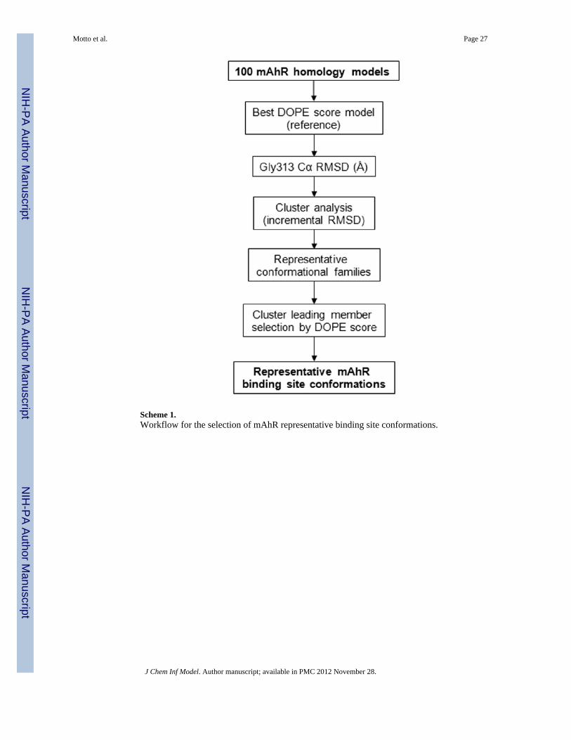
Scheme 1.Workflow for the selection of mAhR representative binding site conformations.
Motto et al. Page 27
J Chem Inf Model. Author manuscript; available in PMC 2012 November 28.
NIH
-PA Author Manuscript
NIH
-PA Author Manuscript
NIH
-PA Author Manuscript

NIH
-PA Author Manuscript
NIH
-PA Author Manuscript
NIH
-PA Author Manuscript
Motto et al. Page 28
Table 1
PDB structural information available on HIF-2α/THS ligand complexes.
PDB ID Chain Resolution (Å) Missing residues Ligand name
3F1O A 1.60 329–333 THS-044
3H7W A 1.65 328–333 THS-017
3H82 A 1.50 None THS-020
J Chem Inf Model. Author manuscript; available in PMC 2012 November 28.

NIH
-PA Author Manuscript
NIH
-PA Author Manuscript
NIH
-PA Author Manuscript
Motto et al. Page 29
Tabl
e 2
RM
SD b
etw
een
the
mA
hR a
po a
nd th
e re
pres
enta
tive
holo
hom
olog
y m
odel
s (H
M01
, HM
70, H
M77
and
HM
79).
Glo
bal R
MSD
(Å)
Bin
ding
site
a R
MSD
(Å)
TC
DD
-bin
ding
fing
erpr
intb
RM
SD (Å
)
mA
hRTo
tal
Back
bone
Side
Cha
inTo
tal
Back
bone
Side
Cha
inTo
tal
Back
bone
Side
Cha
in
HM
012.
271.
432.
865.
933.
635.
762.
011.
312.
42
HM
702.
31.
322.
965.
693.
605.
511.
901.
312.
26
HM
772.
371.
433.
015.
753.
635.
762.
191.
302.
70
HM
792.
291.
342.
935.
633.
625.
662.
001.
312.
40
a Res
idue
s in
a 5
Å sh
ell a
roun
d th
e TH
S lig
ands
are
con
side
red
to d
efin
e th
e bi
ndin
g si
te.
b The
grou
p of
resi
dues
nec
essa
ry fo
r opt
imal
TC
DD
bin
ding
, as d
eter
min
ed b
y m
utag
enes
is a
nd fu
nctio
nal a
naly
sis1
7 ar
e co
nsid
ered
.
J Chem Inf Model. Author manuscript; available in PMC 2012 November 28.
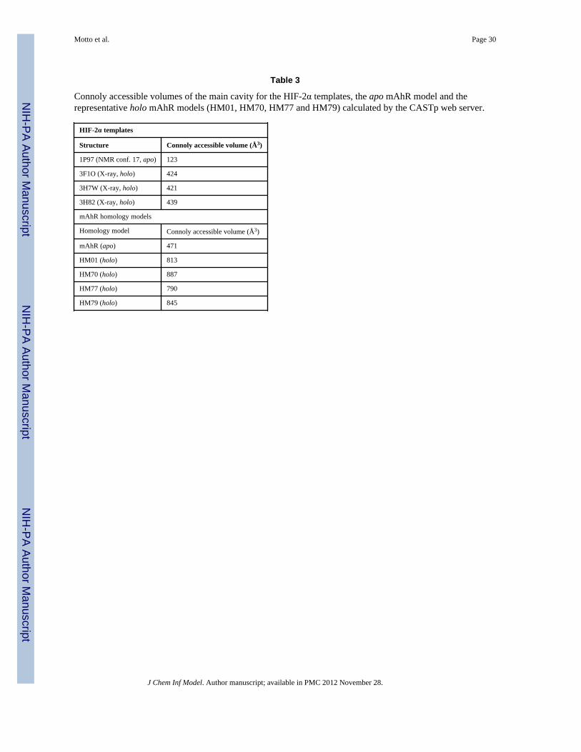
NIH
-PA Author Manuscript
NIH
-PA Author Manuscript
NIH
-PA Author Manuscript
Motto et al. Page 30
Table 3
Connoly accessible volumes of the main cavity for the HIF-2α templates, the apo mAhR model and therepresentative holo mAhR models (HM01, HM70, HM77 and HM79) calculated by the CASTp web server.
HIF-2α templates
Structure Connoly accessible volume (Å3)
1P97 (NMR conf. 17, apo) 123
3F1O (X-ray, holo) 424
3H7W (X-ray, holo) 421
3H82 (X-ray, holo) 439
mAhR homology models
Homology model Connoly accessible volume (Å3)
mAhR (apo) 471
HM01 (holo) 813
HM70 (holo) 887
HM77 (holo) 790
HM79 (holo) 845
J Chem Inf Model. Author manuscript; available in PMC 2012 November 28.

NIH
-PA Author Manuscript
NIH
-PA Author Manuscript
NIH
-PA Author Manuscript
Motto et al. Page 31
Table 4
Calculated pre/post-energy minimization RMSD for the THS ligands in the representative holo mAhRhomology models (HM01, HM70, HM77 and HM79)
mAhR THS-017 RMSD (Å) THS-020 RMSD (Å) THS-044 RMSD (Å)
HM01 1.77 1.05 0.82
HM70 1.70 1.93 1.25
HM77 1.10 1.12 1.42
HM79 1.42 0.80 1.13
J Chem Inf Model. Author manuscript; available in PMC 2012 November 28.

NIH
-PA Author Manuscript
NIH
-PA Author Manuscript
NIH
-PA Author Manuscript
Motto et al. Page 32
Table 5
Glide XP best scoring complexes for the holo mAhR models bound to TCDD and the THS ligands, comparedto the experimental relative affinity (IC50) values obtained with in vitro synthesized mAhR
Ligand Glide XP score Experimental IC50 (M), in vitro synthesized mAhR
complex derived from the templates complex obtained by docking
TCDD −8.70 1×10⁻⁹ a
THS-017 −6.71 −6.78 0.63×10⁻⁶
THS-020 −6.56 −6.68 4.2×10⁻⁶
aC57 mAhR TCDD binding affinity.37,38
J Chem Inf Model. Author manuscript; available in PMC 2012 November 28.

NIH
-PA Author Manuscript
NIH
-PA Author Manuscript
NIH
-PA Author Manuscript
Motto et al. Page 33
Table 6
Effects of mAhR mutagenesis on TCDD binding. Residues in the 5 Å shell from the TCDD of the selectedmAhR model (mAhR_HM01+TCDD) are listed. All mutant AhR analysis results, except those indicated witha reference number, were from the studies described here or our previously mutational analysis.14,17
Residue Effect of Indicated Mutation on TCDD Binding
Reduced to less than 50% Reduced to 50–60% Little or no effect
F281 F281Wb
T283a T283E, T283M
H285a H285F, H285A
F289a F289A, F289L, F289Y
P291 P291F
C294
L302 L302A
Y304
L309 L309A
G315
Y316a Y316A, Y316F
F318 F318A, F318Wc, F318Lc F318Y
I319a I319A, I319Y
C327 C327A
S330
H331
M334 M334E M334A
M342
F345a F345A, F345L
L347 L347A
L348
S359 S359A
A361 A361Lb, A361Vb
I373
A375a A375L, A375V
Q377 Q377A Q377Lb
aFingerprint residues identified by experimental mutagenesis.17
bResults from reference 63.
cResults from reference 64.
J Chem Inf Model. Author manuscript; available in PMC 2012 November 28.


![Protein structure prediction using homology modeling [ Edit ]](https://img.pdfslide.net/doc/110x75/6333b05ba6138719eb0aba9d/protein-structure-prediction-using-homology-modeling-edit-.jpg)


