Embed Size (px)
Citation preview

Mario D. GalignianaMarc B. Cox, Graciela Piwien-Pilipuk and María Fernanda Camisay, Vanina Fontana,Gisela I. Mazaira, Alejandro M. Molinari, Alejandra G. Erlejman, Sonia A. De Leo, ACTIVITYPEPTIDYL-PROLYL ISOMERASE FKBP51 and FKBP52: A ROLE FORModulated by FK506-binding Proteins
B Transcriptional Activity IsκNF-Signal Transduction:
doi: 10.1074/jbc.M114.582882 originally published online August 7, 20142014, 289:26263-26276.J. Biol. Chem.
10.1074/jbc.M114.582882Access the most updated version of this article at doi:
.JBC Affinity SitesFind articles, minireviews, Reflections and Classics on similar topics on the
Alerts:
When a correction for this article is posted•
When this article is cited•
to choose from all of JBC's e-mail alertsClick here
http://www.jbc.org/content/289/38/26263.full.html#ref-list-1
This article cites 80 references, 26 of which can be accessed free at
by Mario G
aligniana on October 20, 2014
http://ww
w.jbc.org/
Dow
nloaded from
by Mario G
aligniana on October 20, 2014
http://ww
w.jbc.org/
Dow
nloaded from

NF-�B Transcriptional Activity Is Modulated by FK506-binding Proteins FKBP51 and FKBP52A ROLE FOR PEPTIDYL-PROLYL ISOMERASE ACTIVITY*
Received for publication, May 19, 2014, and in revised form, July 30, 2014 Published, JBC Papers in Press, August 7, 2014, DOI 10.1074/jbc.M114.582882
Alejandra G. Erlejman‡, Sonia A. De Leo‡, Gisela I. Mazaira‡, Alejandro M. Molinari‡, María Fernanda Camisay‡,Vanina Fontana‡, Marc B. Cox§, Graciela Piwien-Pilipuk¶, and Mario D. Galigniana‡�1
From the ‡Departamento de Química Biológica, Facultad de Ciencias Exactas y Naturales, Universidad de Buenos Aires andInstituto de Quımica Biologica de Ciencias Exactas y Naturales (IQUIBICEN)/Consejo Nacional de Investigaciones Científicas yTécnicas (CONICET), Buenos Aires, C1428ADN Argentina, §Border Biomedical Research Center and Department of BiologicalSciences, University of Texas, El Paso, Texas 79968, ¶Laboratorio de Arquitectura Nuclear, Instituto de Biología y MedicinaExperimental/CONICET, Buenos Aires C1428ADN, Argentina, and �Laboratorio de Receptores Nucleares, Instituto de Biología yMedicina Experimental/CONICET, Buenos Aires C1428ADN, Argentina
Background: Hsp90 binding immunophilins may be regulators of the NF-�B mechanism of action.Results: FKBP51 and FKBP52 show antagonistic properties on the nuclear accumulation and transcriptional activity of NF-�B.Conclusion: Both immunophilins modulate NF-�B trafficking and NF-�B transcription when they are recruited to the pro-moter regions of target genes.Significance: The competitive effect between both immunophilins in different cell types may explain the pleiotropic actions ofNF-�B.
Hsp90 binding immunophilins FKBP51 and FKBP52 modu-late steroid receptor trafficking and hormone-dependent bio-logical responses. With the purpose to expand this model toother nuclear factors that are also subject to nuclear-cytoplas-mic shuttling, we analyzed whether these immunophilins mod-ulate NF-�B signaling. It is demonstrated that FKBP51 impairsboth the nuclear translocation rate of NF-�B and its transcrip-tional activity. The inhibitory action of FKBP51 requires neitherthe peptidylprolyl-isomerase activity of the immunophilin norits association with Hsp90. The TPR domain of FKBP51 isessential. On the other hand, FKBP52 favors the nuclear reten-tion time of RelA, its association to a DNA consensus bindingsequence, and NF-�B transcriptional activity, the latter effectbeing strongly dependent on the peptidylprolyl-isomeraseactivity and also on the TPR domain of FKBP52, but its interac-tion with Hsp90 is not required. In unstimulated cells, FKBP51forms endogenous complexes with cytoplasmic RelA. Upon cellstimulation with phorbol ester, the NF-�B soluble complexexchanges FKBP51 for FKBP52, and the NF-�B biological effectis triggered. Importantly, FKBP52 is functionally recruited tothe promoter region of NF-�B target genes, whereas FKBP51 isreleased. Competition assays demonstrated that both immuno-philins antagonize one another, and binding assays with puri-
fied proteins suggest that the association of RelA and immuno-philins could be direct. These observations suggest that thebiological action of NF-�B in different cell types could be posi-tively regulated by a high FKBP52/FKBP51 expression ratio byfavoring NF-�B nuclear retention, recruitment to the promoterregions of target genes, and transcriptional activity.
Because NF-�B2 was discovered in 1986 as a transcriptionfactor able to bind to the enhancer element of the immunoglob-ulin � light chain of activated B cells (1), it became clear that inaddition to having a crucial role in innate immunity, it is alsoable to regulate many other basic functions of the cell such asinflammatory responses, cell development, programmed celldeath, proliferation control, and tumorigenesis (see Refs. 2– 4)for recent comprehensive updates). NF-�B is actually regardedas a family of structurally related homologues that in mammalscomprises p50 (NF-�B1), p52 (NF-�B2), p65 (RelA), RelB, andc-Rel. All of these proteins share a highly conserved DNA bind-ing and dimerization domain known as RHD (Rel homologydomain) and could potentially form up to 15 types of dimers.Nonetheless, the physiological existence of all of these dimershas not been demonstrated to date, the p50�RelA heterodimer(the p65/RelA subunit will be named RelA onward) beingunquestionably the most abundant in all cell types (5). On theother hand, the NF-�B family may be divided from the tran-scriptional perspective into two groups based on the occur-
* This work was supported, in whole or in part, by National Institutes of HealthGrants 5 G12 RR008124 (Border Biomedical Research Center from theNational Center for Research Resources) and 8 G12 MD007592 (NationalInstitute on Minority Health and Health Disparities (to M. B. C.). This workwas also supported by Agencia Nacional de Promoción Científica y Tec-nológica de Argentina Grants PICT-2010-1170 and PICT-2011-1715 (toM. D. G.), PICT-2010-2215 (to A. G. E.), and PICT-2012-2612 (to G. P.-P.) andUniversity of Buenos Aires Grants UBACYT-2011/14-GC (to M. D. G.), UBA-CYT 2011/13-IJ, and UBACYT 2013/15-IF (to A. G. E.).
1 To whom correspondence should be addressed: IBYME/CONICET, Vueltade Obligado 2490, Rm. 304, Buenos Aires C1428ADN, Argentina.Tel.: 54-11-4783-2869 (ext. 304); Fax: 54-11-4786 –2564; E-mail:[email protected].
2 The abbreviations used are: NF-�B, nuclear factor-�B; I�B, inhibitor of �B;IKK, I�B kinase; Hsp90, heat-shock protein of 90 kDa; FKBP51, FK506-bind-ing protein of 51-kDa; FKBP52, FK506-binding protein of 52-kDa; TPR, tet-ratricopeptide repeats; PPIase, peptidylprolyl-isomerase; PMA, phorbol12-myristate 13-acetate; MMP, matrix metallopeptidase; GR, glucocorti-coid receptor; mineralocorticoid receptor; PR, progesterone receptor.
THE JOURNAL OF BIOLOGICAL CHEMISTRY VOL. 289, NO. 38, pp. 26263–26276, September 19, 2014© 2014 by The American Society for Biochemistry and Molecular Biology, Inc. Published in the U.S.A.
SEPTEMBER 19, 2014 • VOLUME 289 • NUMBER 38 JOURNAL OF BIOLOGICAL CHEMISTRY 26263
by Mario G
aligniana on October 20, 2014
http://ww
w.jbc.org/
Dow
nloaded from

rence of the CT-transactivation domains, which are only pres-ent in RelA, RelB, and c-Rel (5).
All Rel factors form homodimers or heterodimers with thesole exception of RelB, which forms only heterodimers. Therelative abundance of different NF-�B proteins may vary in dif-ferent tissues and cell types, whereas the p50�RelA heterodimeris highly ubiquitous and the most frequently detected complexin most cell types of all tissues (6).
In unstimulated cells p50�RelA heterodimers are normallyretained in the cytosol in an inactive form due to their associa-tion with the inhibitory factor I�B through an ankyrin repeatdomain (7). Upon cell stimulation, I�B is phosphorylated by thekinase IKK, dissociated from the complex with NF-�B proteins,and targeted to proteasomal degradation. This allows thenuclear translocation of the NF-�B heterodimer (8). Nonethe-less, a dynamic nuclear-cytoplasmic shuttling of NF-�B com-plexes always takes place (9 –11) allowing a low basal transcrip-tional activity of NF-�B given the fact that the I�B�NF-�Bcomplex is also subject to dynamic dissociation/reassociationevents. The nuclear-cytoplasmic shuttling of NF-�B resemblesthat observed of steroid receptors where the inactive cytoplas-mic form of these ligand-dependent transcription factors musttranslocate to the nucleus upon stimulation with steroid hor-mones (12–14).
In previous studies our laboratory and others have reportedthat the 51- and 52-kDa FK506-binding proteins FKBP51 andFKBP52 are responsible in a mutually exclusive fashion for theretrotransport mechanism of GR (15, 16) and mineralocorti-coid receptor (17, 18). Both FKBPs are also regulators of theligand-dependent transcriptional activity for those receptors(18 –20) and other members of the family such as PR (21, 22),AR (23, 24), and to a minor degree, estrogen receptor � (22, 25).These Hsp90 binding immunophilins are highly homologousand share 60% homology and 75% similarity (26). They arestructurally characterized by the presence of two key sequenc-es: the TPR domain, through which they bind to Hsp90, and thepeptidyl-prolyl isomerase (PPIase) domain (27), where themacrolide FK506 and also the dynein�dynactin motor complexbind. Both domains are essential for the retrotransport mecha-nism of steroid receptors (14, 28), although the enzymatic activ-ity of the PPIase domain does not appear to be essential. Uponsteroid binding, FKBP51 is released from the receptor�Hsp90heterocomplex and is replaced by FKBP52, which recruitsdynein�dynactin motor proteins favoring the transport of thereceptor to the nucleus on microtubule tracks (29).
Inasmuch as both types of transcription factors, steroidreceptors and NF-�B, show similar requirements for subcellu-lar redistribution upon the onset of a given activating stimulus,we asked whether NF-�B proteins could also be regulated byimmunophilins in a similar fashion as GR and mineralocorti-coid receptor. Therefore, we investigated the potential associ-ation of FKBP51 and FKBP52 with RelA, whether such complexaffects the nuclear translocation of RelA, and how these immu-nophilins influence the transcriptional activity of NF-�B.
EXPERIMENTAL PROCEDURES
Cell Culture and Treatments—Human embryonic kidneyHEK 293-T cells were grown at 37 °C in a humidified atmo-
sphere of 5% CO2, 95% O2 in Dulbecco’s modified Eagle’smedium (DMEM) supplemented with 10% (v/v) fetal bovineserum and antibiotics (50 units/ml penicillin and 50 mg/mlstreptomycin). Human placenta choriocarcinoma BeWo cellswere grown in DMEM/F-12 (1:1) medium supplemented with10% fetal bovine serum and antibiotics. Transcriptional activitywas measured in cells transfected with pNF-�B cis-reporterplasmid (Stratagene, La Jolla, CA, catalog no. 219078-51) asdescribed previously (18). After 24 h post-transfection, cellswere washed, and the culture medium was replaced by serum-free DMEM. Cells were stimulated with 100 ng/ml phorbol12-myristate 13-acetate (PMA) (Sigma) for 7 h. Results wereexpressed as the percentage of NF-�B activation after discount-ing the basal activity and normalized to the �-galactosidaseexpression. Overexpression of FKBP proteins (wild type ormutants) was achieved by transfection of 1 �g of pCI-neo-hFKBPs or empty vector per well of 35 mm containing 1 ml ofmedium following the calcium phosphate precipitation stan-dard method (18). The HEK-FKBP51 cell line stably transfectedwith hFKBP51 (termed 51�) was obtained by transfection ofHEK 293 cells with pCI-neo-hFKBP51 followed by selection in400 �g/ml Geneticin (G418; Invitrogen)-containing mediumfor a period of 30 days. The stably transfected cells were usuallycultured in DMEM supplemented with 10% fetal bovine serumand 200 �g/ml G418.
Zymographies—Gelatin zymography assays for PMA-stimu-lated BeWo cells were achieved following a standard methoddescribed in the literature (30). Briefly, the assays were per-formed running a 10% SDS-PAGE with 1 mg/ml gelatin poly-merized in the gel (0.75-mm thickness). Gels were loaded withsamples diluted (1:1) in non-reducing buffer (12.5% 0.5 M Tris-HCl, pH 6.8, 10% glycerol, 4% SDS, and 0.05% bromphenolblue). After electrophoresis, gels were washed twice with 2.5%Triton X-100 in PBS for 15 min, incubated with the reactionbuffer (50 mM Tris-HCl, 30 mM CaCl2, 0,15 mM NaCl, pH 7.6)for 18 h at 37 °C, stained with 0.5% Coomassie Brilliant BlueR-250 solution (containing 30% methanol and 10% acetic acid)for 30 min under gentle shaking, and destained twice for 20 minwith a solution containing 5% ethanol and 8% acetic acid. Gelswere scanned with a charge-coupled device (Fuji Film) and ana-lyzed by densitometry with National Institutes of Health Scion-Image Software.
IL-6 Expression—BeWo cells were plated in 24-well plates ata concentration of 3 � 103 cells per well. The next day cells weretransfected with the proper pCI-neo plasmid encoding for theproper immunophilin (or empty vector), and 24 h after the cul-ture, medium was replaced by serum-free DMEM-F-12 andincubated with or without 100 ng/ml PMA for 24 or 48 h or withIL-1� (0.5 ng/ml). The concentration of IL-6 en the culturesupernatant was measured using commercial ELISA kit fromRoche Applied Science according to the manufacturer’s guide-lines. Results were normalized by the total amount of protein ofeach well.
Indirect Immunofluorescence Assays—Cells grown on poly-lysine-coated coverslips were fixed with 3% p-formaldehyde for30 min and permeabilized with 0.1% Triton X-100 as described(31). Indirect immunofluorescence was performed using 1/100dilutions of the following primary antibodies: rabbit monoclo-
NF-�B Regulation by FKBP51 and FKBP52
26264 JOURNAL OF BIOLOGICAL CHEMISTRY VOLUME 289 • NUMBER 38 • SEPTEMBER 19, 2014
by Mario G
aligniana on October 20, 2014
http://ww
w.jbc.org/
Dow
nloaded from

nal IgG anti-FKBP51 (Affinity BioReagents, Golden, CO),MG19 mouse monoclonal IgG anti-FKBP51 (32), UP30 rabbitantiserum against FKBP52 (33), and anti-HA or anti-RelAmouse monoclonal antibodies (Santa Cruz Biotechnology, Dal-las, TX) to detect transfected RelA protein or endogenous RelAprotein, respectively. These incubations were followed by asubsequent incubation with 1/200 dilutions of the properAlexa-labeled secondary antibody (Molecular Probes, Eugene,OR). The subcellular distribution of these proteins was moni-tored by confocal microscopy using an Olympus Fluoview 1100microscope. Image analyses for co-immunolocalization ofFKBPs and RelA were carried out using the colocalizationthreshold plug-in of the Image-J program (v.1.45) from theNational Institutes of Health as described (31).
Immunoprecipitation Assays—After the indicated treatment,cells were washed 3 times with phosphate-buffered saline at pH7.3, scrapped, and homogenized in HEM buffer (10 mM Hepesbuffer at pH 7.4, 1 mM EDTA, and 20 mM Na2MoO4). After acentrifugation for 30 min at 4 °C at 120,000 � g, RelA wasimmunoprecipitated from 300 �l of cytosol (�4 mg/ml pro-tein) with 2 �l of anti-RelA (or anti-FKBP51) antibody and 30 �lof 50% g/v protein A-Sepharose (Sigma) under rotation for 2 hat 4 °C. Pellets were washed 5 times with 1 ml of HEM buffer,and proteins were resolved by Western blotting (0.05%v/v dilu-tion for primary antibodies and 0.02% v/v dilution for horserad-ish peroxidase-labeled secondary antibodies) and visualized byenhanced chemiluminescence.
Electrophoretic Mobility Shift Assays (EMSA)—Nuclear frac-tions of HEK 293-T cells were isolated as previously described(34). An oligonucleotide containing the consensus sequenceof NF-�B (AGTTGAGGGGACTTTCCCAGG; catalog no.E3292; Promega, San Luis Obispo, CA) was end-labeled with[�-32P]ATP (PerkinElmer Life Sciences) using T4 polynucle-otide kinase (Promega, Madison, WI) and purified usingChromaSpin-10 columns (Sigma). Samples were incubated for20 min at room temperature in binding buffer (10 mM Tris-HClbuffer, pH 7.5, 20% (v/v) glycerol, 1 mM MgCl2, 0.5 mM EDTA,0.5mMdithiothreitol,50mMNaCl,and0.05mg/mlpoly(dI-dC))withlabeled oligonucleotide (2–3 � 105 cpm). For supershift assays,samples were incubated in the presence of mouse antibodiesanti-RelA, anti-FLAG (clone M2, Sigma), anti-HA, or mousenon immune IgG (Santa Cruz Biotechnology., Dallas, TX)before the addition of labeled oligonucleotide. The productswere separated by electrophoresis in a 5% (w/v) non-denaturingpolyacrylamide gel using 45 mM Tris borate, 1 mM EDTA as therunning buffer. Gels were dried, the radioactivity was revealedwith Kodak x-ray film, and images were scanned in a Fuji FilmIntelligent Dark Box II with a Fuji film LAS-1000 digital camera(Stanford, CA). The specificity of the shifted complexes wasassessed after a pretreatment of 30 min with a 1–100-fold molarexcess of cold competitor (�B) and by using non-immuneantibodies.
Chromatin Immunoprecipitation (ChIP)—ChIP was per-formed following a modification of a previously describedmethod (35). Briefly, proteins and DNA were cross-linked afterHEK 293-T cell treatment with 1% formaldehyde for 10 min at37 °C and neutralized with glycine added at a final concentra-tion of 0.125 M. Cells were rinsed twice with cold PBS, scraped,
and centrifuged at 800 � g for 5 min at 4 °C. Cells were washedwith PBS and homogenized in SDS lysis buffer (1% (w/v) SDS,10 mM EDTA, 50 mM Tris-HCl buffer, pH 8.1) containing pro-tease inhibitors (protease inhibitor mixture, Roche Applied Sci-ence). Chromatin was fragmented in 500 – 800-bp fragments bysonication, lysates were centrifuged at 12,000 rpm for 10 min at4 °C, and the supernatants were diluted 10-fold in ChIP dilutionbuffer (1% (v/v) Triton X-100, 2 mM EDTA, 150 mM NaCl, 20mM Tris-HCl buffer, pH 8.1). Extracts were precleared withprotein A-agarose 50% slurry (antibody binding beads, Diag-enode, Denville, NJ 07834) for 30 min at 4 °C. Immunoprecipi-tations were performed overnight at 4 °C using the UP30 rabbitantiserum against FKBP52 or the MG19 mouse monoclonalIgG against FKBP51. Samples were incubated with proteinA-agarose for 1 h, and the immune complexes were collected bycentrifugation (1000 rpm at 4 °C), washed, and extracted with1% SDS, 0.1 M NaHCO3. The cross-linking was reversed byheating with 5 M NaCl at 65 °C for 4 h. Chromatin-associatedproteins were digested with proteinase K, and the samples wereextracted with phenol/chloroform followed by precipitationwith ethanol. The pellets were lysed in nuclease-free water andsubjected to real-time PCR. The MMP9 primers used for PCRwere synthesized by GenBiotech (Buenos Aires, Argentina), andtheir sequences are CCCTCCCTCCCTTTCATACAGTTC(forward) and GCTTACACCACCTCCTCCTCTC (reverse).
Statistics—Data were analyzed by one-way analysis of vari-ance using Statview 5.0 (SAS Institute Inc., Cary, NC). Fisher’sleast significance difference test was used to examine differ-ences between group means. A p � 0.05 was considered statis-tically significant. Values are shown as the means � S.E.
RESULTS
FKBP51 and FKBP52 Affect NF-�B Nuclear Translocation—In previous studies we showed that FKBP52 favors the nucleartranslocation of steroid receptors (15, 17, 36), p53 mutant iso-forms (37), and RAC3 (38). This is achieved by linking the cor-responding client protein�Hsp90 complex to the dynein�dynactin motor complex. On the other hand, it is regarded thatFKBP51 impairs this mechanism (18, 19, 39). Recently, it hasbeen reported that the RelA subunit of NF-�B is also associatedto components of the dynein�dynactin complex (11, 40). Inas-much as all of these transcription factors (steroid receptors,p53, RAC3, and NF-�B) are primarily localized in the cytoplasmand must be transported to the nucleus upon proper stimula-tion, we hypothesized that NF-�B nuclear translocation couldalso be modulated by Hsp90 binding immunophilins in a simi-lar fashion.
To test that premise we used HEK 293-T cells as they notonly show high efficiency for transfections but also a relativelylow level of expression of endogenous FKBP51 and FKBP52compared with other cell types, making these cells a highly sen-sitive system to evidence changes in the biological responsesdue to variations in the expression level of these immunophi-lins. Cells were cotransfected with pcDNA3-HA-RelA andpCI-neo empty vector, pCI-neo-FLAG-hFKBP51, or pCI-neo-hFKBP52. After 24 h the cells were stimulated with a time-course of PMA (0 – 60 min), and the nuclear localization ofRelA was analyzed by confocal microscopy and scored (Fig. 1A).
NF-�B Regulation by FKBP51 and FKBP52
SEPTEMBER 19, 2014 • VOLUME 289 • NUMBER 38 JOURNAL OF BIOLOGICAL CHEMISTRY 26265
by Mario G
aligniana on October 20, 2014
http://ww
w.jbc.org/
Dow
nloaded from

Although RelA reaches a maximal nuclear accumulation after30 min of stimulation with PMA and cycles back to cytoplasmvery rapidly (Fig. 1A, lower panel), the overexpression ofFKBP51 strongly delays nuclear accumulation such that RelAremains for longer periods of time in the cytoplasm (Fig. 1A,middle panel). On the other hand, the overexpression ofFKBP52 does not affect the nuclear import rate of RelA butsignificantly increases the nuclear retention time of the tran-scription factor. The rapid cycling to the cytoplasm of NF-�Bobserved in control cells after 30 min agrees with previouslyreported data (9, 34, 41, 42), whereas both effects of theimmunophilins, i.e. the nuclear translocation rate andnuclear retention time of the associated cargo, are quite sim-ilar to those previously reported for steroid receptors (14).This observation suggests a similar regulatory mechanism by
both TPR-domain immunophilins for both unrelated tran-scription factors.
Fig. 1, B–E, show indirect immunofluorescence images byconfocal microscopy of the RelA subunit of NF-�B (red) andeither immunophilin FKBP51 or FKBP52 (green). The far rightimages of all panels show a colocalization analysis performedwith the ImageJ program to analyze the actual colocalizationmask between both proteins (shown in white). Note the pooraccumulation of RelA in the nucleus of the cell that overex-presses FKBP51 (Fig. 1, B and D, white arrows) compared withthe other untransfected cells of the field. Interestingly, bothimmunophilins increased the respective nuclear pool colocal-izing with NF-�B upon cell stimulation with either stimulus,PMA, or TNF� (Fig. 1, C and E, yellow arrows), suggesting thepotential existence of nuclear interactions.
FIGURE 1. FKBP51 and FKBP52 influence NF-�B nuclear translocation. A, RelA nuclear translocation rate. HEK 293-T cells were transfected withpcDNA3-HA-RelA and pCI-neo (black circles), pcDNA3-HA-RelA and pCI-neo-FLAG-hFKBP51 (green circles), or pcDNA3-HA-RelA and pCI-neo-hFKBP52 (redsquares). After 24 h, cells were incubated with PMA (100 ng/ml), fixed at the indicated times, and scored for the nuclear and cytoplasmic localization of RelA afterperforming an indirect immunofluorescence by confocal microscopy. Data are the mean � S.E. after scoring 100 cells in each experiment (n � 4). The upperpanel shows cell images that are an example of the score assigned according to the distribution of HA-RelA. The cytosolic fraction depicted in the middle panelrepresents the percentage of transfected cell showing a score equal to 0 (i.e. cytosolic distribution only), and the RelA nuclear fraction depicted in the lowerpanel represents the percentage of transfected cell showing a score equal to 4 (nuclear distribution only). *, Significantly different compared with the group ofcells not transected with the immunophilin. B, endogenous NF-�B colocalizes with FKBPs. HEK 293-T cells transfected with pCI-neo-FLAG-hFKBP51 (panels Band D) or pCI-neo-hFKBP52 (panels C and E) were incubated for 30 min with (�) or without (�) 100 ng/ml PMA (panels B and C) or 10 ng/ml TNF� (panels D andE). An indirect immunofluorescence was performed using anti-FKBP51 or anti-FKBP52 antibodies (green) and anti-RelA antibody (red). DAPI was used fornuclear staining (cyan). The extreme right images correspond to a colocalization mask (c.mask) analysis between the immunophilins and RelA fluorescentsignals by using the colocalization threshold plug-in of the ImageJ program (v.1.45) from the National Institutes of Health. Bar scale, 10 �m. Arrows are pointingto those cells described in the text.
NF-�B Regulation by FKBP51 and FKBP52
26266 JOURNAL OF BIOLOGICAL CHEMISTRY VOLUME 289 • NUMBER 38 • SEPTEMBER 19, 2014
by Mario G
aligniana on October 20, 2014
http://ww
w.jbc.org/
Dow
nloaded from

FKBP51 and FKBP52 Form Complexes with NF-�B—In viewof the previous observations we asked whether both immuno-philins form complexes with NF-�B. In this sense, we notedsome differences in the subcellular localization of endoge-nously and exogenously expressed FKBP51. Fig. 2A shows aco-immunoprecipitation assay of endogenous FKBP51 withendogenous RelA in unstimulated cells (�PMA). The lack ofsignificant concentration of FKBP51 in the nucleus observed byconfocal microscopy in Fig. 1 was confirmed by cell fraction-ation (Fig. 2B), demonstrating that RelA�FKBP51 complexes aremostly cytoplasmic unless FKBP51 is highly expressed, a situa-tion where a significant fraction of FKBP51 is also recovered inthe nuclear fractions of stimulated cells. This observationresembles the well known action of this immunophilin on thesubcellular distribution of steroid receptors (17, 18, 43). Con-sistent with those images observed in Fig. 1 and the cell frac-tionation shown in Fig. 2B, Fig. 2C also shows that higher levelsof expression of FKBP51 make the immunophilin stay moreassociated to NF-�B in PMA-stimulated cells than in unstimu-lated cells (compare with Fig. 2A for endogenous FKBP51). Fig.2D confirms the interaction by reverse co-immunoprecipita-tion, i.e. RelA was co-immunoadsorbed with FKBP51. As
expected, Hsp90 is also co-immunoadsorbed as the immuno-philin forms several complexes with Hsp90. However, it is note-worthy that the interaction of FKBP51 with the transcriptionfactor is Hsp90-independent (Fig. 2A). This is a structural dif-ference regarding the steroid receptors complexes, where theassociation of TPR-domain immunophilins with the transcrip-tion factor is entirely dependent on Hsp90. Fig. 2E also demon-strates that endogenous Hsp70 is part of an endogenous com-plex with RelA, as was recently suggested in similar assays withoverexpressed proteins (44).
Inasmuch as FKBP51 is associated with steroid receptors in aligand-free medium and is exchanged by FKBP52 upon steroidbinding (16 –18), whether or not this is the case for NF-�Bactivation was investigated. Fig. 2F shows that FKBP52 isrecruited to RelA in an Hsp90-independent manner when cellswere incubated with PMA. Because FKBP51 is no longer recov-ered with RelA in the presence of PMA (Fig. 2A), it is likely thatFKBP51 is swapped with FKBP52 as was previously describedfor steroid receptors. In contrast to the case for steroid recep-tors, the binding of immunophilins to NF-�B does not dependon the presence of Hsp90. Therefore, it was speculated thatFKBP52 could bind directly to RelA. This is supported by the
FIGURE 2. FKBP51 and FKBP52 form complexes with NF-�B. A, HEK 293-T cells were incubated with (�) or without (�) 100 ng/ml PMA for 30 min, andendogenous RelA was immunoadsorbed with a specific IgG (I) or a non-immune IgG (NI). IP, immunoprecipitate. B, HEK 293-T cell transfected with pCI-neoempty vector (�) or pCI-neo-FLAG-hFKBP51 (�) were incubated with 100 ng/ml PMA for 30 min (�PMA) and then subjected to subcellular fractionation intocytosolic (CF) and nuclear (NF) fractions as described in a previous work (34). C, RelA was immunoprecipitated with a specific IgG (I) or a non-immune IgG (NI)from whole extracts of cells co-transfected with pcDNA3-HA-RelA and pCI-neo-FLAG-hFKBP51. D, FKBP51 was immunoprecipitated from whole extracts of theHEK 293-FKBP51 cell line that constitutively overexpresses FKBP51. E, endogenous RelA was immunoprecipitated from whole cell extracts of HEK 293-T andhsp70 was revealed by Western blot. F, HEK 293-T cells were incubated with (�) or without (�) 100 ng/ml PMA for 30 min, and RelA was immunoprecipitatedwith a specific IgG (I) or a non-immune IgG (NI). F, HEK 293-T cells were incubated with (�) or without (�) 100 ng/ml PMA for 30 min. Endogenous RelA wasimmunoadsorbed, and proteins were resolved by Western blot. G, direct interaction of FKBP52 with RelA. HA-tagged RelA was immunopurified, stripped ofassociated proteins with RIPA buffer/1 M NaCl, and incubated with GST-FKBP52 recombinant protein purified as described in a previous work (17).
NF-�B Regulation by FKBP51 and FKBP52
SEPTEMBER 19, 2014 • VOLUME 289 • NUMBER 38 JOURNAL OF BIOLOGICAL CHEMISTRY 26267
by Mario G
aligniana on October 20, 2014
http://ww
w.jbc.org/
Dow
nloaded from
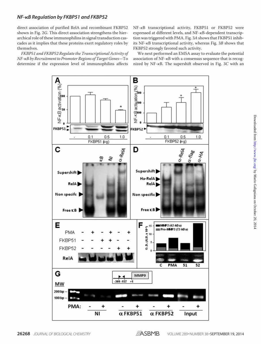
direct association of purified RelA and recombinant FKBP52shown in Fig. 2G. This direct association strengthens the hier-archical role of these immunophilins in signal transduction cas-cades as it implies that these proteins exert regulatory roles bythemselves.
FKBP51 and FKBP52 Regulate the Transcriptional Activity ofNF-�B by Recruitment to Promoter Regions of Target Genes—Todetermine if the expression level of immunophilins affects
NF-�B transcriptional activity, FKBP51 or FKBP52 wereexpressed at different levels, and NF-�B-dependent transcrip-tion was triggered with PMA. Fig. 3A shows that FKBP51 inhib-its NF-�B transcriptional activity, whereas Fig. 3B shows thatFKBP52 strongly favored such activity.
We next performed an EMSA assay to evaluate the potentialassociation of NF-�B with a consensus sequence that is recog-nized by NF-�B. The supershift observed in Fig. 3C with an
NF-�B Regulation by FKBP51 and FKBP52
26268 JOURNAL OF BIOLOGICAL CHEMISTRY VOLUME 289 • NUMBER 38 • SEPTEMBER 19, 2014
by Mario G
aligniana on October 20, 2014
http://ww
w.jbc.org/
Dow
nloaded from

anti-RelA antibody demonstrates the specificity of signal forendogenous RelA. Fig. 3D shows loss of signal for HA-RelAwith both antibodies, anti-FLAG and anti-HA, in samplesderived from FLAG-FKBP51 and HA-RelA co-transfectedcells. Fig. 3E shows the increased recruitment of RelA to theDNA in nuclear cell extracts treated with PMA, a property thatis impaired by FKBP51 and favored by FKBP52.
To assay the biological response of an endogenous gene,BeWo cells were used. This placenta choriocarcinoma cell lineproduces high amounts of matrix metallopeptidase 2 (MMP2)(30) and responds to PMA stimulation via NF-�B activation(45). In agreement with the results described above, the zymog-raphies shown in Fig. 3F demonstrate that a high level ofexpression of FKBP51 reduced the release of MMP2 to themedium, whereas the opposite effect was observed for FKBP52.Importantly, all these observations related to the role of immu-nophilins on the biological response of NF-�B target genes wereconfirmed by ChIP assays for endogenous immunophilins (Fig.3G). Consistent with the endogenous immunophilin swappingobserved in Fig. 2, A and F, this ChIP assay demonstrates thatendogenous immunophilins exchange on the promotersequence of the matrix metallopeptidase 9 (MMP9) gene, aknown classic target of NF-�B (46). In contrast to other MMPsthat are cell-specific, MMP9 is widely expressed in various celltypes (47, 48). Therefore, the regulatory mechanisms for thisMMP should be more complex than for others. These resultssuggest that the expression balance of FKBP51 and FKBP52may be part of such still poorly understood mechanism ofregulation.
FKBP51 and FKBP52 Regulate Endogenous NF-�B-depen-dent Biological Responses Triggered by Various Stimuli—Because the experiments shown in Fig. 3, F and G, demonstratethat FKBP52 is a strong activator of the NF-�B-dependent bio-logical response, we asked whether the ligand macrolide FK506could affect that response. The zymography shown in Fig. 4Ademonstrates that the endogenous biological response is fullyabrogated by preincubation of the cells with the inhibitory mac-rolide FK506, suggesting an apparent dependence of the biolog-ical action of the immunophilin with its PPIase enzymatic activ-ity. FKBP51 was included as an internal control. In a separatezymography assay, FK506 was also used to prevent the inhibi-tory action of FKBP51, but no differences with the control wereobserved (data not shown), suggesting that at variance ofFKBP52, the PPIase activity appears not to be required forFKBP51.
To determine if immunophilins affect NF-�B transcriptionalactivity triggered by other stimuli than PMA, TNF� also wasassayed. HEK 293T cells were treated with 10 ng/ml TNF� for6 h. Luciferase activity induction confirmed that FKBP51 hasinhibitory action, and FKBP52 is a strong enhancer of NF-�Bactivity (Fig. 4B).
It has been previously reported that BeWo cells produce IL-6via NF-�B upon stimulation with phorbol esters or IL-1� (49,50). Therefore, these cells were stimulated with PMA for 24 hand 48 h (Fig. 4C) or IL-1� (Fig. 4D), and IL-6 expression wasmeasured confirming the roles of both immunophilins on dif-ferent types of NF-�B target genes.
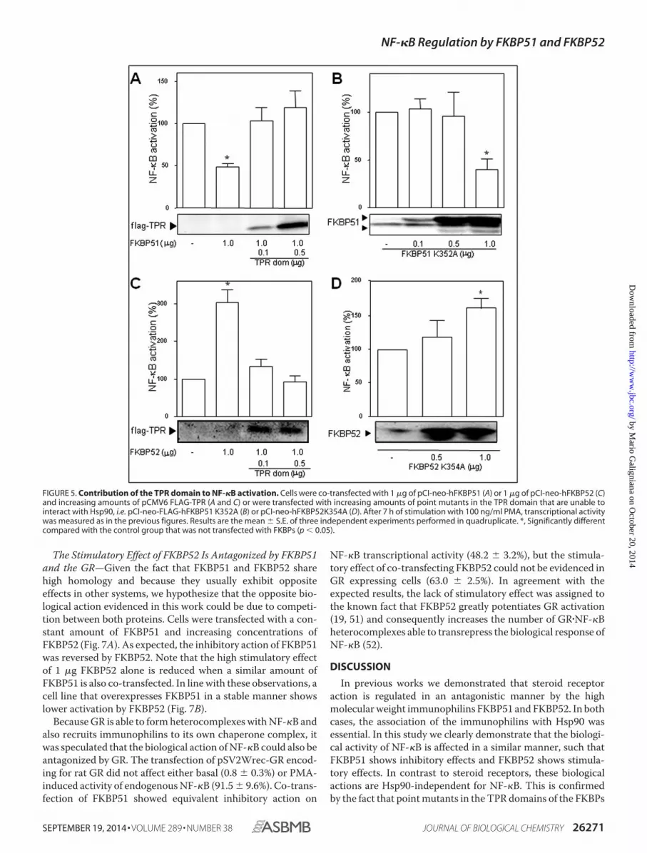
Role of the FKBP51 and FKBP52 TPR Domains—Usually,FKBP51 and FKBP52 form heterocomplexes with Hsp90 viatheir TPR domains, such that both immunophilins form a func-tional complex with this essential chaperone. Nonetheless, theco-immunoprecipitation assays shown in Fig. 2 demonstratethat the complexes of these FKBPs with NF-�B are Hsp90-in-dependent. We next analyzed such unexpected property fromthe functional perspective. The potential involvement of theTPR domain of FKBP51 and FKBP52 on the regulation of thetranscriptional activity of NF-�B was assessed by competitionwith the TPR domain. Both the inhibitory action of FKBP51(Fig. 5A) and the stimulatory effect of FKBP52 (Fig. 5C) werereversed by the increased expression of the TPR peptide. Toanalyze whether this effect is due to the ability of these immu-nophilins to interact with Hsp90 in some other point of thesignaling cascade, point mutants in the TPR domain that areunable to interact with Hsp90 were assayed. The inhibitoryaction of FKBP51 was not modified (Fig. 5B) compared withcontrols transfected with wild type FKBP51, whereas the stim-ulatory action of FKBP52 was still observed (Fig. 5D). Thisobservation agrees with the functional recruitment of FKBP52shown in the co-immunoprecipitation assays of Fig. 2. Takentogether, these results suggest that the regulatory action of bothimmunophilins may involve their TPR-domains but not due toassociation with Hsp90.
FKBP52 Regulatory Action Is PPIase Activity-dependent—Because the PPIase activity can affect the conformation andorientation of small peptides and the helixes of larger proteins,we analyzed the potential role of such enzymatic activity in theregulation of the biological response of NF-�B. The firstapproach was to analyze the transcriptional activity of NF-�B incells stimulated with PMA in the presence of FK506, a pharma-cological inhibitor of the PPIase activity of FKBP proteins.
FIGURE 3. FKBP51 and FKBP52 modulate NF-�B transcriptional activity by recruitment to promoter regions of target genes. HEK 293-T cells weretransfected with increasing amounts of pCI-neo-FLAG-hFKBP51 (A) or pCI-neo-hFKBP52 plasmid (B); the cells were stimulated with 100 ng/ml PMA for 7 h, andNF-�B transcriptional activity was measured with a luciferase reporter-gene and normalized by �-galactosidase activity. The results are expressed as thepercentage of NF-�B activation after subtracting the basal activity (mean � S.E. of five independent experiments). *, Significantly different compared with thecontrol group transected with empty vector (p � 0.01). C, RelA binding to an NF-�B consensus sequence of DNA was analyzed by EMSA using the nuclearfractions of HEK 293-T cells stimulated with 100 ng/ml PMA for 60 min. The specificity of the shifted complexes detected by EMSA was evaluated after apretreatment of 30 min with an excess of cold competitors (�B), nonspecific immunoglobulin (NI), and a specific antibody anti-RelA (�-RelA). D, HEK 293-T cellswere co-transfected with pcDNA3-HA-RelA and pCI-neo-FLAG-hFKBP51 and incubated with 100 ng/ml PMA for 60 min, and the extracts were pretreated witha specific antibody anti-RelA (�-RelA), anti-FLAG tag antibody (�-FLAG), and anti-hemagglutinin tag antibody (�-HA). Arrowheads show specific DNA-proteincomplexes, antibody-DNA-protein complexes (supershift), or free �B consensus sequences. E, profiles of RelA binding to DNA in cells overexpressing FKBP51or FKBP52 that were stimulated (or not) with PMA. F, zimography for MMP2 in BeWo cells. C, control cells; PMA, cells treated with 100 ng/ml PMA; 51 and 52, cellstransfected with 4 �g of pCI-neo-hFKBP51 or pCI-neo-hFKBP52 and then treated with PMA. The bar graph represents the optical density for both bands MMP2full-length (black) and pro-MMP2 (gray). G, ChIP using the NF-�B promoter site of MMP-9 in HEK 293-T cells treated (�) or not (�) with 100 ng/ml PMA for 30min. ChIPs were performed using antibodies against both immunophilins or a nonspecific immunoglobulin (NI), followed by quantitative real-time PCR. Inputchromatin (1%) was removed from samples before immunoprecipitation and also subjected to quantitative real-time PCR to control any potential variation ofthe starting material.
NF-�B Regulation by FKBP51 and FKBP52
SEPTEMBER 19, 2014 • VOLUME 289 • NUMBER 38 JOURNAL OF BIOLOGICAL CHEMISTRY 26269
by Mario G
aligniana on October 20, 2014
http://ww
w.jbc.org/
Dow
nloaded from

Although the overexpression of FKBP51 reduced the transcrip-tional activity of NF-�B to 48.7 � 4% compared with controlstransfected with empty vector, the presence of 1 �M FK506 didnot modify that effect of the immunophilin (47.2 � 7.5%), sug-gesting that the inhibitory action of FKBP51 does not require itsPPIase activity. This result agrees with the lack of effect of thisdrug on MMP2 release upon stimulation of BeWo cells withPMA as discussed before. Nonetheless, this is a pharmacologi-cal approach. A conclusive confirmatory experiment was thetransfection of an FKBP51 mutant devoid of PPIase activity(FD67/68DV) (Fig. 6A). Therefore, it is concluded that theeffect of FKBP51 on NF-�B transcriptional activity depends on
the presence of the immunophilin itself and does not lay on itsPPIase activity.
On the other hand the highly stimulatory action of FKBP52(303.7 � 34.7%) was almost fully abolished by FK506 (31.5 �15.0%). Similarly, the overexpression of two PPIase inactivemutants of FKBP52 (F67Y and F1300Y) also decreased the tran-scriptional activity of NF-�B to a similar extent (Fig. 6B). Thisdemonstrates a relevant role for the PPIase activity of FKBP52on the stimulatory action of NF-�B. As expected, the co-trans-fection of increasing amounts of wild type FKBP52 partiallyreversed the inhibitory action of the mutants to the levels mea-sured for controls.
FIGURE 4. FKBP51 and FKBP52 regulate the expression and activity of endogenous NF-�B target genes in a stimulus-independent manner. A, a gelatinzymography assay performed in BeWo cells incubated with PMA as in Fig. 3F shows that the stimulatory action of FKBP52 on NF-�B action is impaired by cellpreincubation of cells with 1 �M FK506 for 30 min. Bar graphs are the optical densities scanned for four independent gels (mean � S.E.). Results are thepercentage of induced MMP activity after subtracting the basal activity. *, Significantly different compared with the control group (p � 0.003). B, HEK 293T cellswere stimulated with 10 ng/ml TNF� for 6 h, and NF-�B activation was assessed by the induction of a luciferase reporter genes. IL-6 expression was measuredafter treatment of BeWo cells with 100 ng/ml PMA for 24 or 48 h (C) or with 0.5 ng/ml IL-1� for 24 h (D). All the transfections with immunophilins or empty vectorused 1 �g of DNA. Results of panels B, C, and D represent the mean � S.E. of three independent experiments. *, Significantly different from controls at p � 0.05.
NF-�B Regulation by FKBP51 and FKBP52
26270 JOURNAL OF BIOLOGICAL CHEMISTRY VOLUME 289 • NUMBER 38 • SEPTEMBER 19, 2014
by Mario G
aligniana on October 20, 2014
http://ww
w.jbc.org/
Dow
nloaded from
The Stimulatory Effect of FKBP52 Is Antagonized by FKBP51and the GR—Given the fact that FKBP51 and FKBP52 sharehigh homology and because they usually exhibit oppositeeffects in other systems, we hypothesize that the opposite bio-logical action evidenced in this work could be due to competi-tion between both proteins. Cells were transfected with a con-stant amount of FKBP51 and increasing concentrations ofFKBP52 (Fig. 7A). As expected, the inhibitory action of FKBP51was reversed by FKBP52. Note that the high stimulatory effectof 1 �g FKBP52 alone is reduced when a similar amount ofFKBP51 is also co-transfected. In line with these observations, acell line that overexpresses FKBP51 in a stable manner showslower activation by FKBP52 (Fig. 7B).
Because GR is able to form heterocomplexes with NF-�B andalso recruits immunophilins to its own chaperone complex, itwas speculated that the biological action of NF-�B could also beantagonized by GR. The transfection of pSV2Wrec-GR encod-ing for rat GR did not affect either basal (0.8 � 0.3%) or PMA-induced activity of endogenous NF-�B (91.5 � 9.6%). Co-trans-fection of FKBP51 showed equivalent inhibitory action on
NF-�B transcriptional activity (48.2 � 3.2%), but the stimula-tory effect of co-transfecting FKBP52 could not be evidenced inGR expressing cells (63.0 � 2.5%). In agreement with theexpected results, the lack of stimulatory effect was assigned tothe known fact that FKBP52 greatly potentiates GR activation(19, 51) and consequently increases the number of GR�NF-�Bheterocomplexes able to transrepress the biological response ofNF-�B (52).
DISCUSSION
In previous works we demonstrated that steroid receptoraction is regulated in an antagonistic manner by the highmolecular weight immunophilins FKBP51 and FKBP52. In bothcases, the association of the immunophilins with Hsp90 wasessential. In this study we clearly demonstrate that the biologi-cal activity of NF-�B is affected in a similar manner, such thatFKBP51 shows inhibitory effects and FKBP52 shows stimula-tory effects. In contrast to steroid receptors, these biologicalactions are Hsp90-independent for NF-�B. This is confirmedby the fact that point mutants in the TPR domains of the FKBPs
FIGURE 5. Contribution of the TPR domain to NF-�B activation. Cells were co-transfected with 1 �g of pCI-neo-hFKBP51 (A) or 1 �g of pCI-neo-hFKBP52 (C)and increasing amounts of pCMV6 FLAG-TPR (A and C) or were transfected with increasing amounts of point mutants in the TPR domain that are unable tointeract with Hsp90, i.e. pCI-neo-FLAG-hFKBP51 K352A (B) or pCI-neo-hFKBP52K354A (D). After 7 h of stimulation with 100 ng/ml PMA, transcriptional activitywas measured as in the previous figures. Results are the mean � S.E. of three independent experiments performed in quadruplicate. *, Significantly differentcompared with the control group that was not transfected with FKBPs (p � 0.05).
NF-�B Regulation by FKBP51 and FKBP52
SEPTEMBER 19, 2014 • VOLUME 289 • NUMBER 38 JOURNAL OF BIOLOGICAL CHEMISTRY 26271
by Mario G
aligniana on October 20, 2014
http://ww
w.jbc.org/
Dow
nloaded from

unable to bind Hsp90 show similar effects as the wild typeimmunophilins. Nevertheless, the overexpression of a TPRpeptide inhibits the biological action of both proteins, suggest-ing that the domain itself is required at some point in the mech-anism of action, perhaps for interacting with other regulatoryfactors such as cofactors (53). On the other hand, the PPIaseactivity of FKBP52 is very important for its stimulatory action,although this enzymatic activity is not required for the FKBP51inhibitory action. A similar independent effect on the PPIaseactivity was described for the regulation of the GR by FKBP51(19, 54), whereas a PPIase-dependent mechanism is implicatedfor FKBP52 (19, 51).
One interesting extrapolation of these effects is that the bio-logical action of NF-�B may be regulated in different tissues andcell types by the overall expression balance of both immunophi-lins, which could contribute in part to the pleiotropic actions ofNF-�B. Moreover, our assays showed that Hsp70 is also a RelA-interactor, which is in agreement with a very recent report inneurons (44), where the nuclear translocation of both RelA andHsp70 was postulated to occur as a protein-protein complex.Interestingly, the up-regulation of Hsp70 was also reported toinduce nuclear translocation of RelA in rat liver cells (55).
Upon phosphorylation of the NF-�B inhibitory protein I�Bby a kinase located upstream, the IKK complex, NF-�B dimersare transported to the nucleus. Even so, a dynamic nuclear-cytoplasmic shuttling of NF-�B complexes always takes place(9 –11) such that a basal transcriptional activity of NF-�B isalways present because the I�B�NF-�B complex is also subjectto dynamic dissociation/reassociation events. Experimentswith IKK� knock-out mice (56) demonstrated defective cellproliferation and differentiation and have also shown that IKK�
FIGURE 6. Contribution of the respective PPIase domains to NF-�B acti-vation. Cells were transfected with pCI-neo-hFKBP51 or pCI-neo-hFKBP51FD67/68DV (A) or with pCI-neo-hFKBP52 or either plasmid pCI-neo-hFKBP52F67Y or pCI-neo-hFKBP52F130Y encoding for PPIase inactive FKBP52 (B).After 7 h of stimulation with 100 ng/ml PMA, transcriptional activity wasmeasured as in the previous figures. Results are expressed as the percentageof NF-�B activation after subtracting the basal activity (mean � S.E. of fourindependent experiments performed in quadruplicate). *, Significantly differ-ent with regard to the group that was not transfected with FKBPs (p � 0.05). �,Significantly different versus the FKBP52 transfected group (p � 0.05). #, Sig-nificantly different versus the FKBP51 transfected group (p � 0.01).
FIGURE 7. Competition of FKBP51 and FKBP52 regulates NF-�B activa-tion. A, transcriptional activity measured in cells treated with PMA that werepreviously co-transfected with 1 �g of pCI-neo-FLAG-hFKBP51 and increasedamounts of pCI-neo-hFKBP52 (mean � S.E. of four independent experi-ments). *, Significantly different versus the control group not transfected withFKBP51 or FKBP52 (p � 0.05). #, Significantly different versus the FKBP51 trans-fected group (p � 0.01). B, inhibition of the stimulatory action of FKBP52 inHEK 293 (no T) fibroblasts that constitutively overexpress FKBP51. TheFKBP52-dependent potentiation of NF-�B transcriptional activity measuredin HEK no T wild type cells (HEK 293 wt cells) stimulated with PMA is signifi-cantly attenuated in the same cell type overexpressing FKBP51 (HEK 293–51�
cells). *, Significantly different from untreated control groups (p � 0.01).
NF-�B Regulation by FKBP51 and FKBP52
26272 JOURNAL OF BIOLOGICAL CHEMISTRY VOLUME 289 • NUMBER 38 • SEPTEMBER 19, 2014
by Mario G
aligniana on October 20, 2014
http://ww
w.jbc.org/
Dow
nloaded from

is dispensable for I�B degradation. Moreover IKK� has beenreported to be required for the termination of NF-�B activation(57). A physical interaction between FKBP51 and the IKK com-plex has been demonstrated, most likely via the IKK� subunitbound to Hsp90 (58, 59), but the biological function of FKBP51on IKK signaling is still unclear (59). Although down-modula-tion of Hsp90� and Hsp90� likewise resulted in reduced kinaseactivity, it has been shown that FKBP51 is not a constitutivelyassociated component of the IKK complex (59), and its down-modulation interfered with neither TNF�-induced IKK activitynor I�B� degradation and RelA translocation. Actually, theprevailing complex for the IKK�Hsp90 complex to generate anactivated state is the one that recruits Cdc37 rather thanFKBP51 (59), both factors being transiently associated withNF-�B. Interestingly, it was reported that TNF� is unable tomodify the association of IKK with those interacting factors(59). In short, the role of FKBP51 on NF-�B signaling cascaderemains still elusive. Our study demonstrates an inhibitoryaction for this immunophilin on NF-�B signaling and, there-fore, suggests the possibility that NF-�B biological activitycould be mitigated in those tissues where the expression ofFKBP51 is constitutively high or induced by stimuli. The oppo-site reasoning is equally valid for tissues that express differentamounts of FKBP52, a clear stimulating factor of NF-�B activ-ity. In line with these concepts, it should be emphasized thatboth the subcellular distribution and transcriptional activity ofsteroid receptors is modulated by the expression level of these
two immunophilins (17, 18, 43). Consequently, based on theexperimental data shown in this work, we would like to proposea novel mechanism of regulation of NF-�B by FKBPs thataffects NF-�B activation at different levels, i.e. nuclear trans-port, nuclear retention, and transcriptional activity (Fig. 8).
To the best of our knowledge this work is the first that showssuch antagonistic activity of high molecular weight immuno-philins on NF-�B biological action and also shows the first evi-dence that both endogenous immunophilins are dynamicallyrecruited to the promoter sites of target genes. In line with ourfindings where endogenous proteins were analyzed, a recentreport by Romano et al. (60) has also shown that overexpressedFLAG-tagged FKBP51 forms part of the transcriptional com-plexes of ABCG2 (ATP binding cassette transporters) in mela-noma cells.
A crucial nuclear mechanism for gene expression is the mod-ification of the chromatin environment of the respective genes.It has been shown that when NF-�B is activated, histone phos-phorylation can be mediated by nuclear IKK� that is recruitedto the promoter sites of NF-�B-regulated genes (61, 62).Among a number of chromatin remodelers is the PPIase pro-tein Pin1 (63), which is an immunophilin that targets RelA (64).Because PPIase-induced conformational changes have func-tional effects on target proteins, the action of Pin1 on RelA isreflected in a more efficient nuclear accumulation of RelA andalso a greater stability by preventing its ubiquitin-mediatedproteolysis (64). In certain types of cancer cells, Pin1 is usually
FIGURE 8. Model for the regulation of the biological action of NF-�B by FKBPs. The p50�RelA dimer is associated to FKBP51 in its inactive cytoplasmic state(Fig. 2A). Hsp70 is also part of the heterocomplex (Fig. 2E). Upon cell stimulation, the kinase activity of IKK is activated by phosphorylation via the cdc37�(Hsp90)2interacting complex (59). This results in I�B phosphorylation, its dissociation from the NF-�B complex, and the subsequent degradation of I�B via proteasome.Active NF-�B replaces FKBP51 by FKBP52 (Fig. 2F), which is able to interact with dynein�dynactin motors proteins (15), favoring both the retrotransport of NF-�B(11, 40) and its interaction with the nuclear sites of action. FKBP52 greatly favors (���) NF-�B biological action (Fig. 3B) in a PPIase-dependent manner(inhibited by FK506) when the immunophilin is recruited to the promoter sites of NF-�B target genes (Fig. 3G). On the other hand, the recruitment of FKBP51to those promoters inhibits the NF-�B biological response (Fig. 3A). Both immunophilins compete one another (7) and can hamper the original effect of theother (Fig. 7). The steroid-dependent activation of the GR, which is also improved by FKBP52 (19, 51), also prevents NF-�B effects via its known mechanism oftans-repression (52).
NF-�B Regulation by FKBP51 and FKBP52
SEPTEMBER 19, 2014 • VOLUME 289 • NUMBER 38 JOURNAL OF BIOLOGICAL CHEMISTRY 26273
by Mario G
aligniana on October 20, 2014
http://ww
w.jbc.org/
Dow
nloaded from
up-regulated (65– 67), whereas the E3-ubiquitin-ligase of RelA,SOCS1, is down-regulated (68 –70) or mutated (71), all ofwhich may contribute to the constitutive activation of NF-�B inthose cancers. A similar mechanism can be proposed herefor the expression balance of FKBP51 and FKBP52, in particularfor the latter immunophilin, which shows an important stimu-latory action dependent on its PPIase activity.
Given the role played in the initiation and progression ofcancer, the NF-�B signaling pathway is a potent node of phar-macological interference in the clinic. Because NF-�B is also anessential protein in the immunological response against cancer,there has been a reluctance to use NF-�B-targeting inhibitorsfor the treatment of such malignancies. Nevertheless, combin-ing classical chemotherapeutics with inhibitors of NF-�B acti-vation seems to result in a promising synergistic strategy (72,73). Elevated NF-�B activity and/or higher half-life persistencein the nucleus of cancer cells (like that evidenced with greateramounts of FKBP52 in Fig. 1A) provide a survival mechanismby up-regulating anti-apoptotic genes, thereby representing amajor causative factor for drug resistance (74). In line with thisconcept, it has been shown that 0.1 �M rapamycin abolishesdoxorubicin-induced activation of NF-�B and therebyenhances drug-induced apoptosis, an effect that was firstassigned to FKBP51 inhibition (75). However, that drug con-centration cannot guarantee specificity of action as rapamycincan equally interact with FKBP51 (Ki � 5 nM) (76, 77) andFKBP52 (Ki 8 nM) (78, 79). Even though FKBP51 can interactwith IKK (58), an up-stream regulator of the NF-�B signalingcascade, it has been demonstrated that FKBP51 is not a consti-tutive member of the IKK complex (59), and the biological con-sequence of such association (when it takes place) is unknownat this point. Nonetheless, we cannot rule out the possibilitythat differences among cell types may exist such that NF-�B isregulated in different manner.
Here, we demonstrate that in the biological systems weassayed, FKBP51 shows inhibitory rather stimulatory action onthe overall activity of NF-�B and that this effect is not depen-dent on the PPIase activity (which is inhibited by rapamycin orFK506). Because FKBP52 also shows a stimulatory action onNF-�B action in a PPIase-dependent manner, we propose that alikely explanation for all these experimental facts, ours andthose reported by others, is the PPIase-dependent (drug-inhib-ited) stimulatory role of FKBP52 on NF-�B activation. Accord-ingly, zymographies like those shown in Fig. 3F performed inthe presence of FK506 fully abolished the production of MMP2in BeWo cells stimulated with PMA (Fig. 4A).
The development of immunophilin ligands appears to havepromising perspectives in the coming years (80). Thus, the abil-ity to regulate the functions of a specific protein using cell-permeable small molecules like those that bind FKBPs is anunquestionably powerful method, not only to study biologicalsystems, but also a desired alternative to be used in therapeutictreatments. Ideally, the biological function of certain nuclearfactors could be regulated if we can influence the mechanismsby which they reach their sites of action. In this sense, becauseNF-�B is active in many cancer cells and its persistent localiza-tion in the nucleus strengthens or directly leads to tumor devel-opment, we propose that targeting specifically the PPIase activ-
ity of FKBP52 could be a novel regulatory approach to inhibitNF-�B activity.
Acknowledgments—We are indebted to Dr. Paola De Luca and Dr.Adriana De Siervi for the helpful advance on ChIP assay and to Dr.William B. Pratt from the University of Michigan for the kind gift ofthe UP30 antibody against FKBP52.
REFERENCES1. Sen, R., and Baltimore, D. (1986) Multiple nuclear factors interact with the
immunoglobulin enhancer sequences. Cell 46, 705–7162. Hoesel, B., and Schmid, J. A. (2013) The complexity of NF-�B signaling in
inflammation and cancer. Mol. Cancer 12, 863. Diamant, G., and Dikstein, R. (2013) Transcriptional control by NF-�B:
elongation in focus. Biochim. Biophys. Acta 1829, 937–9454. Tornatore, L., Thotakura, A. K., Bennett, J., Moretti, M., and Franzoso, G.
(2012) The nuclear factor �B signaling pathway: integrating metabolismwith inflammation. Trends Cell Biol. 22, 557–566
5. Oeckinghaus, A., and Ghosh, S. (2009) The NF-�B family of transcriptionfactors and its regulation. Cold Spring Harb. Perspect. Biol. 1, a000034
6. Hoffmann, A., Natoli, G., and Ghosh, G. (2006) Transcriptional regulationvia the NF-�B signaling module. Oncogene 25, 6706 – 6716
7. Ciechanover, A., Gonen, H., Bercovich, B., Cohen, S., Fajerman, I., Israël,A., Mercurio, F., Kahana, C., Schwartz, A. L., Iwai, K., and Orian, A. (2001)Mechanisms of ubiquitin-mediated, limited processing of the NF-�B1precursor protein p105. Biochimie 83, 341–349
8. Gilmore, T. D. (1999) The Rel/NF-�B signal transduction pathway: intro-duction. Oncogene 18, 6842– 6844
9. Birbach, A., Gold, P., Binder, B. R., Hofer, E., de Martin, R., and Schmid,J. A. (2002) Signaling molecules of the NF-� B pathway shuttle constitu-tively between cytoplasm and nucleus. J. Biol. Chem. 277, 10842–10851
10. Huang, T. T., Kudo, N., Yoshida, M., and Miyamoto, S. (2000) A nuclearexport signal in the N-terminal regulatory domain of I�B� controls cyto-plasmic localization of inactive NF-�B/I�B� complexes. Proc. Natl. Acad.Sci. U.S.A. 97, 1014 –1019
11. Mikenberg, I., Widera, D., Kaus, A., Kaltschmidt, B., and Kaltschmidt, C.(2007) Transcription factor NF-�B is transported to the nucleus via cyto-plasmic dynein/dynactin motor complex in hippocampal neurons. PLoSONE 2, e589
12. Elbi, C., Walker, D. A., Romero, G., Sullivan, W. P., Toft, D. O., Hager,G. L., and DeFranco, D. B. (2004) Molecular chaperones function as ste-roid receptor nuclear mobility factors. Proc. Natl. Acad. Sci. U.S.A. 101,2876 –2881
13. Madan, A. P., and DeFranco, D. B. (1993) Bidirectional transport of glu-cocorticoid receptors across the nuclear envelope. Proc. Natl. Acad. Sci.U.S.A. 90, 3588 –3592
14. Galigniana, M. D., Echeverría, P. C., Erlejman, A. G., and Piwien-Pilipuk,G. (2010) Role of molecular chaperones and TPR-domain proteins in thecytoplasmic transport of steroid receptors and their passage through thenuclear pore. Nucleus 1, 299 –308
15. Galigniana, M. D., Radanyi, C., Renoir, J. M., Housley, P. R., and Pratt,W. B. (2001) Evidence that the peptidylprolyl isomerase domain of thehsp90-binding immunophilin FKBP52 is involved in both dynein interac-tion and glucocorticoid receptor movement to the nucleus. J. Biol. Chem.276, 14884 –14889
16. Davies, T. H., Ning, Y. M., and Sánchez, E. R. (2002) A new first step inactivation of steroid receptors: hormone-induced switching of FKBP51and FKBP52 immunophilins. J. Biol. Chem. 277, 4597– 4600
17. Galigniana, M. D., Erlejman, A. G., Monte, M., Gomez-Sanchez, C., andPiwien-Pilipuk, G. (2010) The hsp90-FKBP52 complex links the mineralo-corticoid receptor to motor proteins and persists bound to the receptor inearly nuclear events. Mol. Cell. Biol. 30, 1285–1298
18. Gallo, L. I., Ghini, A. A., Piwien Pilipuk, G., and Galigniana, M. D. (2007)Differential recruitment of tetratricorpeptide repeat domain immunophi-lins to the mineralocorticoid receptor influences both heat-shock protein90-dependent retrotransport and hormone-dependent transcriptional ac-
NF-�B Regulation by FKBP51 and FKBP52
26274 JOURNAL OF BIOLOGICAL CHEMISTRY VOLUME 289 • NUMBER 38 • SEPTEMBER 19, 2014
by Mario G
aligniana on October 20, 2014
http://ww
w.jbc.org/
Dow
nloaded from
tivity. Biochemistry 46, 14044 –1405719. Wochnik, G. M., Rüegg, J., Abel, G. A., Schmidt, U., Holsboer, F., and Rein,
T. (2005) FK506-binding proteins 51 and 52 differentially regulate dyneininteraction and nuclear translocation of the glucocorticoid receptor inmammalian cells. J. Biol. Chem. 280, 4609 – 4616
20. Riggs, D. L., Roberts, P. J., Chirillo, S. C., Cheung-Flynn, J., Prapapanich, V.,Ratajczak, T., Gaber, R., Picard, D., and Smith, D. F. (2003) The Hsp90binding peptidylprolyl isomerase FKBP52 potentiates glucocorticoid sig-naling in vivo. EMBO J. 22, 1158 –1167
21. Tranguch, S., Cheung-Flynn, J., Daikoku, T., Prapapanich, V., Cox, M. B.,Xie, H., Wang, H., Das, S. K., Smith, D. F., and Dey, S. K. (2005) Cochap-erone immunophilin FKBP52 is critical to uterine receptivity for embryoimplantation. Proc. Natl. Acad. Sci. U.S.A. 102, 14326 –14331
22. Yang, Z., Wolf, I. M., Chen, H., Periyasamy, S., Chen, Z., Yong, W., Shi, S.,Zhao, W., Xu, J., Srivastava, A., Sánchez, E. R., and Shou, W. (2006) FK506-binding protein 52 is essential to uterine reproductive physiology con-trolled by the progesterone receptor A isoform. Mol. Endocrinol. 20,2682–2694
23. Cheung-Flynn, J., Prapapanich, V., Cox, M. B., Riggs, D. L., Suarez-Quian,C., and Smith, D. F. (2005) Physiological role for the cochaperone FKBP52in androgen receptor signaling. Mol. Endocrinol. 19, 1654 –1666
24. Periyasamy, S., Hinds, T., Jr., Shemshedini, L., Shou, W., and Sanchez, E. R.(2010) FKBP51 and Cyp40 are positive regulators of androgen-dependentprostate cancer cell growth and the targets of FK506 and cyclosporin A.Oncogene 29, 1691–1701
25. Schülke, J. P., Wochnik, G. M., Lang-Rollin, I., Gassen, N. C., Knapp, R. T.,Berning, B., Yassouridis, A., and Rein, T. (2010) Differential impact oftetratricopeptide repeat proteins on the steroid hormone receptors. PLoSONE 5, e11717
26. Cioffi, D. L., Hubler, T. R., and Scammell, J. G. (2011) Organization andfunction of the FKBP52 and FKBP51 genes. Curr. Opin. Pharmacol. 11,308 –313
27. Sivils, J. C., Storer, C. L., Galigniana, M. D., and Cox, M. B. (2011) Regu-lation of steroid hormone receptor function by the 52-kDa FK506-bindingprotein (FKBP52). Curr. Opin. Pharmacol. 11, 314 –319
28. Storer, C. L., Dickey, C. A., Galigniana, M. D., Rein, T., and Cox, M. B.(2011) FKBP51 and FKBP52 in signaling and disease. Trends Endocrinol.Metab. 22, 481– 490
29. Galigniana, M. D. (2012) Steroid receptor coupling becomes nuclear.Chem. Biol. 19, 662– 663
30. Fontana, V., Coll, T. A., Sobarzo, C. M., Tito, L. P., Calvo, J. C., and Cebral,E. (2012) Matrix metalloproteinase expression and activity in trophoblast-decidual tissues at organogenesis in CF-1 mouse. J. Mol. Histol. 43,487– 496
31. Quintá, H. R., Maschi, D., Gomez-Sanchez, C., Piwien-Pilipuk, G., andGaligniana, M. D. (2010) Subcellular rearrangement of hsp90-binding im-munophilins accompanies neuronal differentiation and neurite out-growth. J. Neurochem. 115, 716 –734
32. Gallo, L. I., Lagadari, M., Piwien-Pilipuk, G., and Galigniana, M. D. (2011)The 90-kDa heat-shock protein (Hsp90)-binding immunophilin FKBP51is a mitochondrial protein that translocates to the nucleus to protect cellsagainst oxidative stress. J. Biol. Chem. 286, 30152–30160
33. Quintá, H. R., and Galigniana, M. D. (2012) The neuroregenerative mech-anism mediated by the Hsp90 binding immunophilin FKBP52 resemblesthe early steps of neuronal differentiation. Br. J. Pharmacol. 166, 637– 649
34. Erlejman, A. G., Jaggers, G., Fraga, C. G., and Oteiza, P. I. (2008) TNF�-induced NF-�B activation and cell oxidant production are modulated byhexameric procyanidins in Caco-2 cells. Arch. Biochem. Biophys. 476,186 –195
35. Susperreguy, S., Prendes, L. P., Desbats, M. A., Charó, N. L., Brown, K.,MacDougald, O. A., Kerppola, T., Schwartz, J., and Piwien-Pilipuk, G.(2011) Visualization by BiFC of different C/EBP� dimers and their inter-action with HP1� reveals a differential subnuclear distribution of com-plexes in living cells. Exp. Cell Res. 317, 706 –723
36. Piwien Pilipuk, G., Vinson, G. P., Sanchez, C. G., and Galigniana, M. D.(2007) Evidence for NL1-independent nuclear translocation of the min-eralocorticoid receptor. Biochemistry 46, 1389 –1397
37. Galigniana, M. D., Harrell, J. M., O’Hagen, H. M., Ljungman, M., and Pratt,
W. B. (2004) Hsp90 binding immunophilins link p53 to dynein during p53transport to the nucleus. J. Biol. Chem. 279, 22483–22489
38. Colo, G. P., Rubio, M. F., Nojek, I. M., Werbajh, S. E., Echeverría, P. C.,Alvarado, C. V., Nahmod, V. E., Galigniana, M. D., and Costas, M. A.(2008) The p160 nuclear receptor co-activator RAC3 exerts an anti-apo-ptotic role through a cytoplasmatic action. Oncogene 27, 2430 –2444
39. Sanchez, E. R. (2012) Chaperoning steroidal physiology: lessons frommouse genetic models of Hsp90 and its cochaperones. Biochim. Biophys.Acta 1823, 722–729
40. Mackenzie, G. G., Keen, C. L., and Oteiza, P. I. (2006) Microtubules arerequired for NF-�B nuclear translocation in neuroblastoma IMR-32 cells:modulation by zinc. J. Neurochem. 99, 402– 415
41. Nelson, G., Paraoan, L., Spiller, D. G., Wilde, G. J., Browne, M. A., Djali,P. K., Unitt, J. F., Sullivan, E., Floettmann, E., and White, M. R. (2002)Multi-parameter analysis of the kinetics of NF-�B signalling and tran-scription in single living cells. J. Cell Sci. 115, 1137–1148
42. Hohmann, H. P., Remy, R., Scheidereit, C., and van Loon, A. P. (1991)Maintenance of NF-� B activity is dependent on protein synthesis and thecontinuous presence of external stimuli. Mol. Cell. Biol. 11, 259 –266
43. Banerjee, A., Periyasamy, S., Wolf, I. M., Hinds, T. D., Jr., Yong, W., Shou,W., and Sanchez, E. R. (2008) Control of glucocorticoid and progesteronereceptor subcellular localization by the ligand-binding domain is medi-ated by distinct interactions with tetratricopeptide repeat proteins. Bio-chemistry 47, 10471–10480
44. Klenke, C., Widera, D., Engelen, T., Müller, J., Noll, T., Niehaus, K.,Schmitz, M. L., Kaltschmidt, B., and Kaltschmidt, C. (2013) Hsc70 is anovel interactor of NF-�B p65 in living hippocampal neurons. PLoS ONE8, e65280
45. Yura, S., Sagawa, N., Ogawa, Y., Masuzaki, H., Mise, H., Matsumoto, T.,Ebihara, K., Fujii, S., and Nakao, K. (1998) Augmentation of leptin synthe-sis and secretion through activation of protein kinases A and C in culturedhuman trophoblastic cells. J. Clin. Endocrinol. Metab. 83, 3609 –3614
46. Suh, J., and Rabson, A. B. (2004) NF-�B activation in human prostatecancer: important mediator or epiphenomenon? J. Cell. Biochem. 91,100 –117
47. Nissinen, L., and Kähäri, V. M. (2014) Matrix metalloproteinases in in-flammation. Biochim. Biophys. Acta 1840, 2571–2580
48. Li, Y. F., Xu, X. B., Chen, X. H., Wei, G., He, B., and Wang, J. D. (2012) Thenuclear factor-�B pathway is involved in matrix metalloproteinase-9 ex-pression in RU486-induced endometrium breakdown in mice. Hum. Re-prod. 27, 2096 –2106
49. Tsukihara, S., Harada, T., Deura, I., Mitsunari, M., Yoshida, S., Iwabe, T.,and Terakawa, N. (2004) Interleukin-1�-induced expression of IL-6 andproduction of human chorionic gonadotropin in human trophoblast cellsvia nuclear factor-�B activation. Am. J. Reprod. Immunol. 52, 218 –223
50. Fujisawa, K., Nasu, K., Arima, K., Sugano, T., Narahara, H., and Miyakawa,I. (2000) Production of interleukin (IL)-6 and IL-8 by a choriocarcinomacell line, BeWo. Placenta 21, 354 –360
51. Riggs, D. L., Cox, M. B., Cheung-Flynn, J., Prapapanich, V., Carrigan, P. E.,and Smith, D. F. (2004) Functional specificity of co-chaperone interac-tions with Hsp90 client proteins. Crit. Rev. Biochem. Mol. Biol. 39,279 –295
52. Scheinman, R. I., Gualberto, A., Jewell, C. M., Cidlowski, J. A., and Bald-win, A. S., Jr. (1995) Characterization of mechanisms involved in transre-pression of NF-�B by activated glucocorticoid receptors. Mol. Cell. Biol.15, 943–953
53. Erlejman, A. G., Lagadari, M., Toneatto, J., Piwien-Pilipuk, G., and Galig-niana, M. D. (2014) Regulatory role of the 90-kDa heat-shock protein(Hsp90) and associated factors on gene expression. Biochim. Biophys. Acta1839, 71– 87
54. Cluning, C., Ward, B. K., Rea, S. L., Arulpragasam, A., Fuller, P. J., andRatajczak, T. (2013) The helix 1–3 loop in the glucocorticoid receptorLBD is a regulatory element for FKBP cochaperones. Mol. Endocrinol. 27,1020 –1035
55. Dokladny, K., Lobb, R., Wharton, W., Ma, T. Y., and Moseley, P. L. (2010)LPS-induced cytokine levels are repressed by elevated expression ofHSP70 in rats: possible role of NF-�B. Cell Stress Chaperones 15, 153–163
56. Sil, A. K., Maeda, S., Sano, Y., Roop, D. R., and Karin, M. (2004) I�B
NF-�B Regulation by FKBP51 and FKBP52
SEPTEMBER 19, 2014 • VOLUME 289 • NUMBER 38 JOURNAL OF BIOLOGICAL CHEMISTRY 26275
by Mario G
aligniana on October 20, 2014
http://ww
w.jbc.org/
Dow
nloaded from

kinase-� acts in the epidermis to control skeletal and craniofacial mor-phogenesis. Nature 428, 660 – 664
57. Lawrence, T., Bebien, M., Liu, G. Y., Nizet, V., and Karin, M. (2005) IKK�limits macrophage NF-�B activation and contributes to the resolution ofinflammation. Nature 434, 1138 –1143
58. Bouwmeester, T., Bauch, A., Ruffner, H., Angrand, P. O., Bergamini, G.,Croughton, K., Cruciat, C., Eberhard, D., Gagneur, J., Ghidelli, S., Hopf, C.,Huhse, B., Mangano, R., Michon, A. M., Schirle, M., Schlegl, J., Schwab,M., Stein, M. A., Bauer, A., Casari, G., Drewes, G., Gavin, A. C., Jackson,D. B., Joberty, G., Neubauer, G., Rick, J., Kuster, B., and Superti-Furga, G.(2004) A physical and functional map of the human TNF-�/NF-� B signaltransduction pathway. Nat. Cell Biol. 6, 97–105
59. Hinz, M., Broemer, M., Arslan, S. C., Otto, A., Mueller, E. C., Dettmer, R.,and Scheidereit, C. (2007) Signal responsiveness of I�B kinases is deter-mined by Cdc37-assisted transient interaction with Hsp90. J. Biol. Chem.282, 32311–32319
60. Romano, S., Staibano, S., Greco, A., Brunetti, A., Nappo, G., Ilardi, G.,Martinelli, R., Sorrentino, A., Di Pace, A., Mascolo, M., Bisogni, R., Scal-venzi, M., Alfano, B., and Romano, M. F. (2013) FK506 binding protein 51positively regulates melanoma stemness and metastatic potential. CellDeath Dis. 4, e578
61. Park, K. J., Krishnan, V., O’Malley, B. W., Yamamoto, Y., and Gaynor, R. B.(2005) Formation of an IKK�-dependent transcription complex is re-quired for estrogen receptor-mediated gene activation. Mol. Cell 18,71– 82
62. Park, G. Y., Wang, X., Hu, N., Pedchenko, T. V., Blackwell, T. S., andChristman, J. W. (2006) NIK is involved in nucleosomal regulation byenhancing histone H3 phosphorylation by IKK�. J. Biol. Chem. 281,18684 –18690
63. Raghuram, N., Strickfaden, H., McDonald, D., Williams, K., Fang, H., Miz-zen, C., Hayes, J. J., Th’ng, J., and Hendzel, M. J. (2013) Pin1 promoteshistone H1 dephosphorylation and stabilizes its binding to chromatin.J. Cell Biol. 203, 57–71
64. Ryo, A., Suizu, F., Yoshida, Y., Perrem, K., Liou, Y. C., Wulf, G., Rottapel,R., Yamaoka, S., and Lu, K. P. (2003) Regulation of NF-�B signaling byPin1-dependent prolyl isomerization and ubiquitin-mediated proteolysisof p65/RelA. Mol. Cell 12, 1413–1426
65. Pang, R. W., Lee, T. K., Man, K., Poon, R. T., Fan, S. T., Kwong, Y. L., andTse, E. (2006) PIN1 expression contributes to hepatic carcinogenesis.J. Pathol. 210, 19 –25
66. Lu, K. P., Liou, Y. C., and Zhou, X. Z. (2002) Pinning down proline-di-rected phosphorylation signaling. Trends Cell Biol. 12, 164 –172
67. Wulf, G. M., Ryo, A., Wulf, G. G., Lee, S. W., Niu, T., Petkova, V., and Lu,K. P. (2001) Pin1 is overexpressed in breast cancer and cooperates with Rassignaling in increasing the transcriptional activity of c-Jun towards cyclinD1. EMBO J. 20, 3459 –3472
68. Oshimo, Y., Kuraoka, K., Nakayama, H., Kitadai, Y., Yoshida, K., Chayama,K., and Yasui, W. (2004) Epigenetic inactivation of SOCS-1 by CpG island
hypermethylation in human gastric carcinoma. Int. J. Cancer 112,1003–1009
69. Oh, J., Kim, S. H., Ahn, S., and Lee, C. E. (2012) Suppressors of cytokinesignaling promote Fas-induced apoptosis through down-regulation ofNF-�B and mitochondrial Bfl-1 in leukemic T cells. J. Immunol. 189,5561–5571
70. Zhao, X. D., Zhang, W., Liang, H. J., and Ji, W. Y. (2013) Overexpression ofmiR �155 promotes proliferation and invasion of human laryngeal squa-mous cell carcinoma via targeting SOCS1 and STAT3. PLoS ONE 8,e56395
71. Schif, B., Lennerz, J. K., Kohler, C. W., Bentink, S., Kreuz, M., Melzner, I.,Ritz, O., Trümper, L., Loeffler, M., Spang, R., and Möller, P. (2013) SOCS1mutation subtypes predict divergent outcomes in diffuse large B-cell lym-phoma (DLBCL) patients. Oncotarget 4, 35– 47
72. Fuchs, O. (2010) Transcription factor NF-�B inhibitors as single thera-peutic agents or in combination with classical chemotherapeutic agentsfor the treatment of hematologic malignancies. Curr. Mol. Pharmacol. 3,98 –122
73. Fabre, C., Mimura, N., Bobb, K., Kong, S. Y., Gorgun, G., Cirstea, D., Hu,Y., Minami, J., Ohguchi, H., Zhang, J., Meshulam, J., Carrasco, R. D., Tai,Y. T., Richardson, P. G., Hideshima, T., and Anderson, K. C. (2012) Dualinhibition of canonical and noncanonical NF-�B pathways demonstratessignificant antitumor activities in multiple myeloma. Clin. Cancer Res. 18,4669 – 4681
74. Salem, K., Brown, C. O., Schibler, J., and Goel, A. (2013) Combinationchemotherapy increases cytotoxicity of multiple myeloma cells by modi-fication of nuclear factor (NF)-�B activity. Exp. Hematol. 41, 209 –218
75. Romano, M. F., Avellino, R., Petrella, A., Bisogni, R., Romano, S., andVenuta, S. (2004) Rapamycin inhibits doxorubicin-induced NF-�B/Relnuclear activity and enhances the apoptosis of melanoma cells. Eur. J.Cancer 40, 2829 –2836
76. Yeh, W. C., Bierer, B. E., and McKnight, S. L. (1995) Rapamycin inhibitsclonal expansion and adipogenic differentiation of 3T3-L1 cells. Proc.Natl. Acad. Sci. U.S.A. 92, 11086 –11090
77. Sinars, C. R., Cheung-Flynn, J., Rimerman, R. A., Scammell, J. G., Smith,D. F., and Clardy, J. (2003) Structure of the large FK506-binding proteinFKBP51, an Hsp90 binding protein, and a component of steroid receptorcomplexes. Proc. Natl. Acad. Sci. U.S.A. 100, 868 – 873
78. Galat, A. (1993) Peptidylproline cis-trans-isomerases: immunophilins.Eur. J. Biochem. 216, 689 –707
79. Peattie, D. A., Harding, M. W., Fleming, M. A., DeCenzo, M. T., Lippke,J. A., Livingston, D. J., and Benasutti, M. (1992) Expression and character-ization of human FKBP52, an immunophilin that associates with the 90-kDa heat shock protein and is a component of steroid receptor complexes.Proc. Natl. Acad. Sci. U.S.A. 89, 10974 –10978
80. Gaali, S., Gopalakrishnan, R., Wang, Y., Kozany, C., and Hausch, F. (2011)The chemical biology of immunophilin ligands. Curr. Med. Chem. 18,5355–5379
NF-�B Regulation by FKBP51 and FKBP52
26276 JOURNAL OF BIOLOGICAL CHEMISTRY VOLUME 289 • NUMBER 38 • SEPTEMBER 19, 2014
by Mario G
aligniana on October 20, 2014
http://ww
w.jbc.org/
Dow
nloaded from


