Embed Size (px)
Citation preview
International Journal of Systematic Bacteriology (1 998), 48, 1049-1 055 Printed in Great Britain
NOTE ldentif ication of Staphylococcus species by 16s-23s rDNA intergenic spacer PCR analysis
Marcos Mendoza, H d h e Meugnier, Mich&le Bes, Jerome Etienne and Jean Freney
Author for correspondence: Jean Freney. Tel : + 33 4 72 11 07 62. Fax: + 33 4 72 1 1 07 64. e-mail : [email protected] 1 .fr
Centre National de Reference des Staphylocoques, UPRES EA 1655, Laboratoire de BactCriologie, FacultC de Medecin R.T.H. Laennec, rue Guillaume Paradin, 69372 Lyon cedex, France
~ ~~~~~
To investigate whether 165-235 rDNA (rDNA) spacer region length polymorphisms are suitable for the identification of Staphylococcus strains, the 16S-235 rDNA intergenic spacer region lengths of 221 strains belonging to 31 species were studied by using a PCR-based method. Each species presented a specific 165-235 pattern made of 1-8 fragments ranging from 104 to 771 bp, with the exception of the species Staphylococcus warneri, Staphylococcus caprae and Staphylococcus piscifermentans, which presented larger or smaller fragments. Very few species showed more than one pattern, Staphylococcus saprophyticus subsp. saprophyticus and Staphylococcus aureus being the most heterogeneous species (five different patterns for eight strains). Five clinical strains that could not be identified at the species level by phenotypical tests were finally identified using this method. Discrimination between some species that showed close patterns (Staphylococcus cohniilStaphylococcus chromogeneslStaphylococcus equomm, Staphylococcus aureuslStaphylococcus in termedius, Staphylococcus sciurilStaphylococcus pasteurilStaphylococcus gallinarum, Staphylococcus delphinilStaphylococcus felis, Staphylococcus wituluslStaphylococcus auricularis) was further achieved after Dral digestion of the PCR products. Although it does not allow discrimination of subspecies, the use of 16s-235 spacer region length data determined by PCR-mediated amplification is suitable for the identification of the 31 Staphylococcus species tested in th is study. The method is rapid, easy and may be a useful tool for the identification of Staphylococcus species in the clinical microbiology laboratory.
Keywords: Staphylococcus, 16s-23s intergenic spacer PCR analysis, ITS-PCR, identification
Staphylococci remain among the most important pathogens isolated in the clinical microbiology lab- oratory (13). Phenotypic methods, including bio- chemical systems (9, 11, 17, 18, 19) and GLC analysis of cellular fatty acids (20) have been evaluated as identification methods for staphylococcus species. However, a large number of isolates are still incorrectly or not identified by these methods, and supplementary tests are often required for complete identification (9, 19). Molecular methods that enable identification of staphylococci to the species level include ribotyping analysis (12), DNA PFGE (1 6), total genomic DNA- DNA hybridization, and genomic DNA hybridization to labelled probes specific to hypervariable sequences
........................................................................................................................ . .......... .... ...... ... Abbreviation : ITS-PCR, intergenic spacer PCR analysis.
(5). However, these methods are fairly laborious, the patterns may be difficult to interpret, and inter- laboratory reproducibility and standardization still need improvement. There is therefore a need for accurate genotyping systems that can be easily applied for the identification of Staphylococcus species in clinical microbiology laboratories. The genes encoding rRNA are highly conserved in bacteria and other kingdoms. With some exceptions, the rRNA operon is transcribed into one pre-rRNA transcript that con- tains the following sequences (in the order 5’ + 3’) : 16S, internal transcribed spacer, tRNA, internal trans- cribed spacer, 23S, internal transcribed spacer and 5s rRNA. Although the 16s and 23s rRNA genes have been used extensively to study the evolution and differentiation of bacterial species, the sequences of 16s and 23s rRNA genes are very similar from one
~~ ~~
00696 0 1998 IUMS 1049
Notes
species to another within a genus, making it necessary to find other species-specific sequences. The sequence of the intergenic spacer region between these two genes, however, is more variable than the sequence of the genes themselves and has led some authors to propose the study of this 16s-23s spacer for bacterial identification (2, 8). Many bacteria have multiple copies of the rRNA operon per genome which has raised the possibility of using this method for the epidemiological typing of Burkholderia cepacia (1 4) and Clostridiurn dzficile (6). The method has also been proposed for the identification and typing of many other bacteria (1, 8), and has been used previously to type Staphylococcus aureus isolates (3, 7, 15). In the present study, we tested 16s-23s rDNA intergenic spacer PCR analysis (ITS-PCR) as a tool to identify Staphylococcus isolates at the species level.
The study was carried out with 221 Staphylococcus strains, comprising 153 human clinical isolates, 30 animal isolates and 38 reference strains (Table 1). The human and animal strains were collected and pheno- typically identified as described previously (4) by the Centre National de Rkference des Staphylocoques (Lyon, France) between 1985 and 1997. Most of the clinical and animal strains were also identified by ribotyping, and some of them were identified by DNA-DNA hybridization (1 9). The oligonucleotide primers used for PCR amplification of the 16s-23s intergenic spacer regions have been described pre- viously (1 0). Primer G 1 (5'-GAAGTCGTAACAA- GG-3') (Eurogentec), which was selected from a highly conserved region in the 16s RNA gene, is located about 30-40 nucleotides upstream from the spacer region. Primer L 1 (5'-CAAGGCATCCACCGT-3') contained the most conserved 23s sequence immedi- ately following the spacer and is located about 20 nucleotides downstream from the spacer boundary. Primers for both the 16s and 23s regions were limited to a length of 15 nucleotides because of variations in sequence beyond these highly conserved regions. Strains were grown for 48 h at 37 "C on trypticase soy agar plates containing 5 % horse blood (TSAB) (bioMkrieux). A 10 pl loopful of bacteria was then resuspended in 10 ml distilled water. The bacterial suspension was centrifuged for 3 min at 3000g. One microlitre of the pellet was disrupted by incubation in 55 p1 of a 75 pg ml-l lysostaphin solution (Sigma). This mixture was incubated for 30 min at 37 "C, then 30 min at 95 "C, and finally centrifuged for 10 min at 13 000 g. PCR amplification was carried out in a 100 p1 (total volume) mixture containing 5 p1 of the super- natant and 95 pl of a solution which contained 0.5 pM primer GI, 0.5 pM primer L1, 200 pM each dNTP (Pharmacia Biotech), lop1 Gene Amp l o x PCR buffer (Perkin-Elmer) and 2.5 U AmpliTaq DNA polymerase (Perkin-Elmer). PCR amplification was performed with an OmniGene TR3 CM220 thermal cycler (Hybaid). After an initial denaturation step of 2 min at 94 "C, a total of 25 cycles of amplification was performed using the following thermal profile: 1 min
at 94 "C (denaturation), 7 min at 47 "C (annealing) and 2 min at 72 "C (elongation). The last cycle was followed by a 7 min extension step at 72 "C. To check on possible contamination, a negative control con- taining distilled water instead of template DNA was included in each experiment. The 16s-23s rDNA intergenic spacer regions were amplified twice for each Staphylococcus strain included in this study. To in- vestigate the reproducibility of the band pattern obtained by PCR amplification, ten independent PCR amplifications were performed for Staphylococcus capitis subsp. capitis CCM 2734T, and the amplicons were included on ten different gels. Gel-to-gel variation of PCR fragment migration was assessed by comparing S. capitis subsp. capitis CCM 2734T profiles in these gels. The mean size and standard deviation of each fragment were calculated and compared. Genomic stability was tested by passaging single colonies of S. capitis subsp. capitis CCM 2734T from TSAB three times a week over 5 weeks. The amplified products were analysed by electrophoresis with a GNA-200 apparatus (Pharmacia Biotech) at a constant 4-5 V cm-l 4 h in 3 YO agarose gels (Metaphor; FMC) containing 2 pg ethidium bromide ml-l (BioProbe System) in 0.5 x TBE buffer. The PCR products were visualized by UV fluorescence. To calculate the lengths of fragments, DNA Molecular Weight Marker VI (Boehringer Mannhein) was included on each gel every eight lanes. The gels were photographed under UV light with an MP-4 camera (Polaroid) using a Wratten 2A filter with a Polaroid type 55/pn film, and then scanned. The lengths of the PCR products of each strain were calculated automatically by using the RestrictoScan and RestrictoTyper programs (Taxo- lab, Institut Pasteur, Paris, France). The similar- ity of the strains was calculated using the Dice coefficient, and hierachical clustering was performed using the unweighted pair group method with averages (UPGMA). For some groups of species that could not be differentiated because they showed very little variation in their patterns, the amplified products were digested with 20 U DraI or HinfI, as instructed by the manufacturer (Oncor Appligene). The digested PCR products and the amplicons of the same strain without digestion were run side by side on the same gel.
All the strains except S. aureus CCM 885T were typeable. The mean size and the standard deviation of S. capitis subsp. capitis CCM 2734T amplicons were calculated from the results of ten separate PCR runs. The standard deviation did not exceed 10 bp, indi- cating good reproducibility. The level of uncertainty in the calculated sizes of amplification products did not exceed 2-6 YO. Fifteen subcultures (three per week) of the same strain were amplified to test genomic stability. We did not observe a variation in the number of fragments within the 5 week test period. The standard deviation of fragment size with time did not exceed 4 bp for each of the fragments when run on the same gel. In some cases, weaker and unreproducible ampli- fication products were observed and were not con-
1050 International Journal of Systematic Bacteriology 48
Notes
Table 1. Strains used in this study
a*b'c*d.eyfStrains kindly provided by W. E. Kloos, P. E. Varaldo, S. Igimi, N. El Solh, S . Tanasupawat and V. Hajek, respectively. Type strains (') were obtained from the American Type Culture Collection (ATCC) (Rockville, MD, USA), the Czechoslovak Collection of Microorganisms (CCM) (Brno, Czech Republic), the Deutsche Sammlung von Mikroorganismen (DSM) (Gottingen, Germany) and the Japan Collection of Microorganisms (JCM) (RIKEN, Saitama, Japan). S. aureus CCM 885' was not typeable by ITS-PCR.
Taxon as determined by ITS-PCR (after restriction if necessary) strains or molecular methods
No. of Reference strains and Staphylococcus isolates as identified by phenotypical
Staphylococcus arlettae Staphylococcus aureus
Staphylococcus auricularis Staphylococcus capitis
Staphylococcus capitis
Staphylococcus caprae Staphylococcus carnosus Staphylococcus caseolyticus Staphylococcus chromogenes Staphylococcus cohnii
Staphylococcus cohnii
Staphylococcus delphini Staphylococcus epidermidis
Staphylococcus equorum
Staphylococcus felis Staphylococcus gallinarum Staphylococcus haemolyticus
Staphylococcus hominis
Staphylococcus hyicus Staphylococcus intermedius
Staphylococcus kloosii Staphylococcus lentus Staphylococcus lugdunensis
Staphylococcus muscae Staphylococcus pasteuri
Staphylococcus piscifermentans Staphylococcus saprophyticus
Staphylococcus saprophyticus
Staphylococcus schleiferi Staphylococcus schleiferi
Staphylococcus sciuri Staphylococcus simulans Staphylococcus vitulus Staphylococcus warneri
Staphylococcus xylosus
2 9
3 10
10
3 1 3 3 9
8
2 18
12
3 3
13
10
3 10
1 3
12
1 13
3 5
10
3 10
3 3 1 9
9
S. arlettae DSM 20672T, N910254 S. aureus CCM WT, N950351, N950382, N950448, N950522, N950534,
S. auricularis ATCC 3753T, N910215, N910217 S. capitis subsp. capitis CCM 2734T, N880027, N880193, N900125,
S. capitis subsp. ureolyticus ATCC 49326T, ATCC 49325, ATCC 49324,
S. caprae CCM 3573T, N920116t, N920117t S. carnosus DSM 20501T S. caseolyticus ATCC 13548', N8800667, N880071t S. chromogenes CCM 3387T, N900352?, N930152t S. cohnii subsp. cohnii CCM 2736T, N920077, N870198, N900274, N920270,
S. cohnii subsp. urealyticum ATCC 49330T, N920176, N880225, N910033,
S. delphini DSM 20771T, Nonobt S. epidermidis CCM 2124T, N860069, N880212, N930084, N930131, N9 10 102, N9 101 60, N910 19 1, N9 1023 1, N9 10249, N9 10275, N9 10280, N910288, N910308, N920102*, N930332*, N940056*, N940057*
S. equorum DSM 20674T, N880062t, N8800637, N940280t, N940282t, N940284t, N940298t, N940300t, N940301 t, N94046 1 t, N940462t, N940465t
N950565, N950603, N950619
N910309, N920161, N930202, N930252, N930315, N940269
ATCC 49327, N970267*, 3600-4", 3740-4", 4147-1", 5394-1", 5520-1"
N930144, N930306, N940210, N940379
N910058, N930190, N930253, N950054
S. felis ATCC 4916gT, S14lC, GD104' S. gallinarum CCM 3572T, N880497t, N910325t S. haemolyticus CCM 2737T, N850435, N870222, N910076, N910248, N910287, N910291, N920160, N920186, N930100, N930168, N930185, N930208
N930317, N930329, N930341, N940139 S. hominis DSM 2032gT, N850447, N850456, N930160, N930207, N930305,
S. hyicus CCM 236gT, N9003507, N900354f S. intermedius CCM 5739T, N870338, N900260, N900269, N910201,
S. kloosii DSM 20676T S. lentus ATCC 29070T, N850542t, N870235t S. lugdunensis ATCC 43809T, N880214, N880325, N920143, N930432, N940025, N940084, N940113, N940135, N940164, N950232, N950646
S. muscae CCM 4175T S. pasteuri ATCC 51129T, BM 10426d, BM 10507d, N910099, N910242, N910243, N910259, N920288, N930200, N930231, N930248, N930314, N950027
N930300, N940276, N940453, N940457, N950082
S. piscifermentans JCM 6057T, SK 14et, SK 16et S. saprophyticus subsp. bovis CCM 4410T, KV 19'7, KV 2 6 t , KV 3 6 t , KV
S. saprophyticus subsp. saprophyticus CCM 883T, N850206, N930040, N9 10122, N910128, N91013 1, N9 10132, N920147, N940065, N940472
S. schleiferi subsp. coagulans JCM 7470T, GA64', N920247 S. schleiferi subsp. schleiferi ATCC 4380gT, N860173, N880218, N910093,
S. sciuri ATCC 29062T, N920212, N860392 S. simulans ATCC 2784gT, N900050, N850124 S. vitulus ATCC 51 145T S. warneri CCM 2730T, N850183, N860227, N890412, N910159, N910224,
S. xylosus ATCC 29971T, N850267, N930157, N930173, N930189, N930239,
5 6 t
N910245, N920053, N920068, N920162, N920172, N920260
N920234, N920275, N930014
N930249, N930408, N930410
* Clinical strains which could not be identified by phenotypical methods. t Animal strain. The strains without indication, except type strains, are clinical strains.
International Journal of Systematic Bacteriology 48 1051
Notes
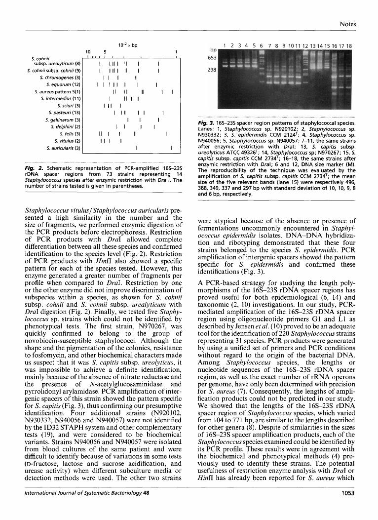
5. cohnii subsp. cohnii (9) 5. chromogenes (3)
5. equorum (12) 5. saprophyticus subsp. bovis (5)
5. saprophyticus su bsp. saprophyticus pattern 1 (9) 5. saprophyticus subsp. saprophyticus pattern 2” (1)
5. lentus (3) 5. xylosus (9)
5. aureus pattern 1b ( I ) 5. aureus pattern 2 (1) S. aureus pattern 3 (1) 5. aureus pattern 4 (4) 5. aureus pattern 5 (1)
5. intermedius (1 1 ) 5. sciuri (3 )
5. pasteuri (1 3 ) 5. gallinarum ( 3 )
5. schleiferi subsp. schleiferi (1 0) 5. schleiferi subsp. coagulans (3)
5. simulans (3) 5. muscae (1)
5. hominis(10) 5. kloosii (1) .--
5. fe/is (3) 5. delphini (2)
5. hyicus (3) 5. epidermidis (1 4) 4
N920102C N930332C -- N940057C’$II
x bp 20 10 5 1
I I I I I I I I I I I I I l l I I I I l l II I I I I I I II II I I
I 1 1 I I I I I l l I l l I I II I I I I I II I I I
I l l I I I I I I I I I I I I I I I I I I I I I
-------- I I I I I
-------- -------- I I I I A’ I I I I -5 I I I I I
-------- I I I ---------- I I
I I I
-5 II II , I I I ---s II II I I I I
I I
5. cohnii subsp. urealyticum (8) 1 ’ ” ’ ’ ’ II ‘ I ’ I
N940056C 5. warneri(9) 5. c v a e (3)
5. capitis subsp. capitis (10) N970267C
5. capitis su bsp. ureolyticus (9) 5. haemolyticus (1 3)
5. caseolyticus (3) 5. vitulus ( I )
5. auricularis (3) 5. carnosus (1)
- 3 1 I I I I I I ’/ I l l I I I II I I I I I I
// I I II I -/ I I II I -/ I/ I I I I
I I I I -/ I I I
111 II
’/ I // I I I -/ I
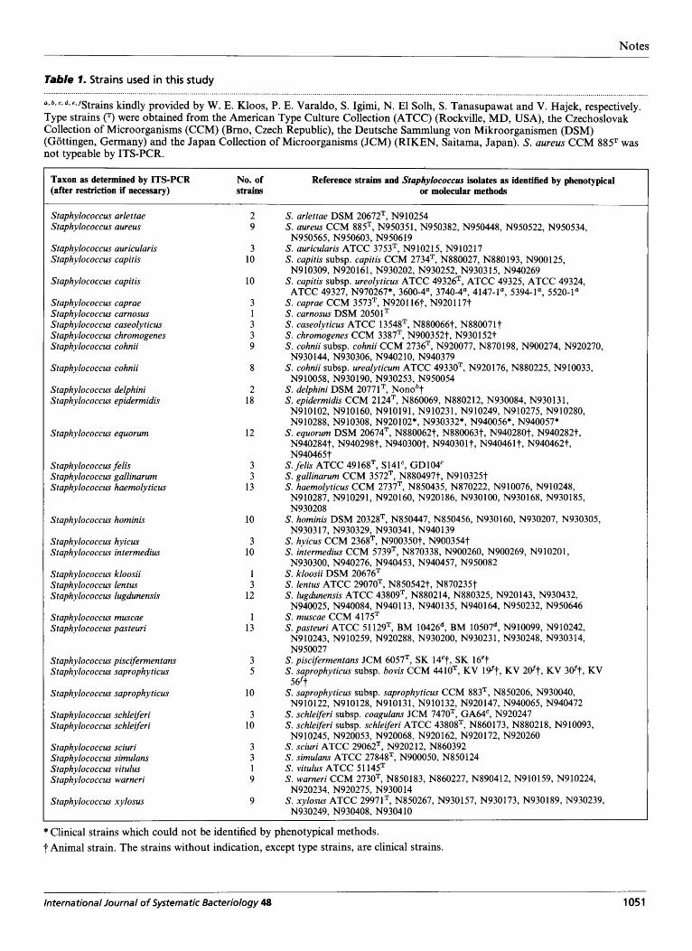
....................... I ...................................................................................................................................................................................... , ...................... . ................... I . . . . . ........ . ................ . ......... .... .......... . Fig. 1. Schematic representation of PCR-amplified 165-235 rDNA spacer region from 220 Staphylococcus strains representing 31 species. The number of strains tested is given in parentheses. ”Pattern 2 was presented by 5. saprophyticus subsp. saprophyticus N910222, pattern 1 was presented by the nine other strains of the same species cited in Table 1. bPatterns 1, 2, 3, 4 and 5 were presented respectively by 5. aureus N950351/N950448/N950619/N950603, N950534, N950522, N950382 and N950565. %rains not identified by biochemical characteristics.
sidered when comparing amplifications results. With- out enzyme digestion, each species presented a specific PCR pattern made of 1-8 fragments whose sizes ranged from 297 to 771 bp, with the exception of three species: Staphylococcus warneri which presented three larger fragments (828, 1146 and 1445 bp), Staphylococcus caprae which showed five smaller fragments (148, 185, 205, 227 and 273 bp), and Staphylococcus piscifermentans which presented special patterns with only two small fragments (104 and 205 bp) (Fig. 1). Staphylococcus carnosus and Staphylococcus arlettae also showed special patterns with only one fragment of 314 and 375 bp, respectively. When several strains of the same species were tested, the pattern showed very little variation within a species, with the exception of S. aureus and Staphylococcus saprophyticus subsp. sapro- phyticus which showed more than one pattern. Five
different patterns were observed for S. aureus, the most heterogeneous species. Nevertheless, the S. aureus patterns shared a core of common fragments: 569 bp (patterns 1-5), 457 bp (patterns 1, 3, 4, 5) and 437 bp (patterns 1-4) which allowed discrimination from other Staphylococcus species (Fig. 1). S. saprophyticus subsp. saprophyticus strains presented two patterns differing by two large fragments (368 and 360 bp). Pattern 2 was represented by only one strain (Fig. 1). The method did not allow discrimination of subspecies within a species (Fig. 1).
Since the species Staphylococcus cohniilStaphy- lococcus chromogeneslStaphylococcus equorum, S . aureuslStaphylococcus intermedius, Staphylococcus sciurilStaphylococcus pasteurilStaphylococcus gallin- arumlStaphylococcus delphinilStaphylococcus felis,
1052 International Journal of Systematic Bacteriology 48
Notes
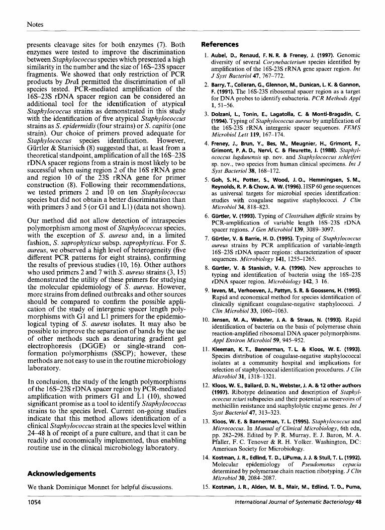
S. cohnii subsp. urealyticum (8)
S. cohnii subsp. cohnii (9) 5. chromogenes (3)
S. equorum (12) 5. aureus pattern 5(1)
5. intermedius (1 1 )
5. sciuri (3) S. pasteuri (1 3)
5. gallinarum (3) 5, delphini (2)
5. felis (3) 5. vitulus (2)
5. auricularis (3)
x bp 10 5 1
I 1 1 I I I I 1 I I
I I I I I I I I I I I I I I I I I I
I I I I I I I I I
I I I I I
I I I II
II I I II I I
I I I I I I I I I I
I I I I I I I
I l l I II I I I I I
I I
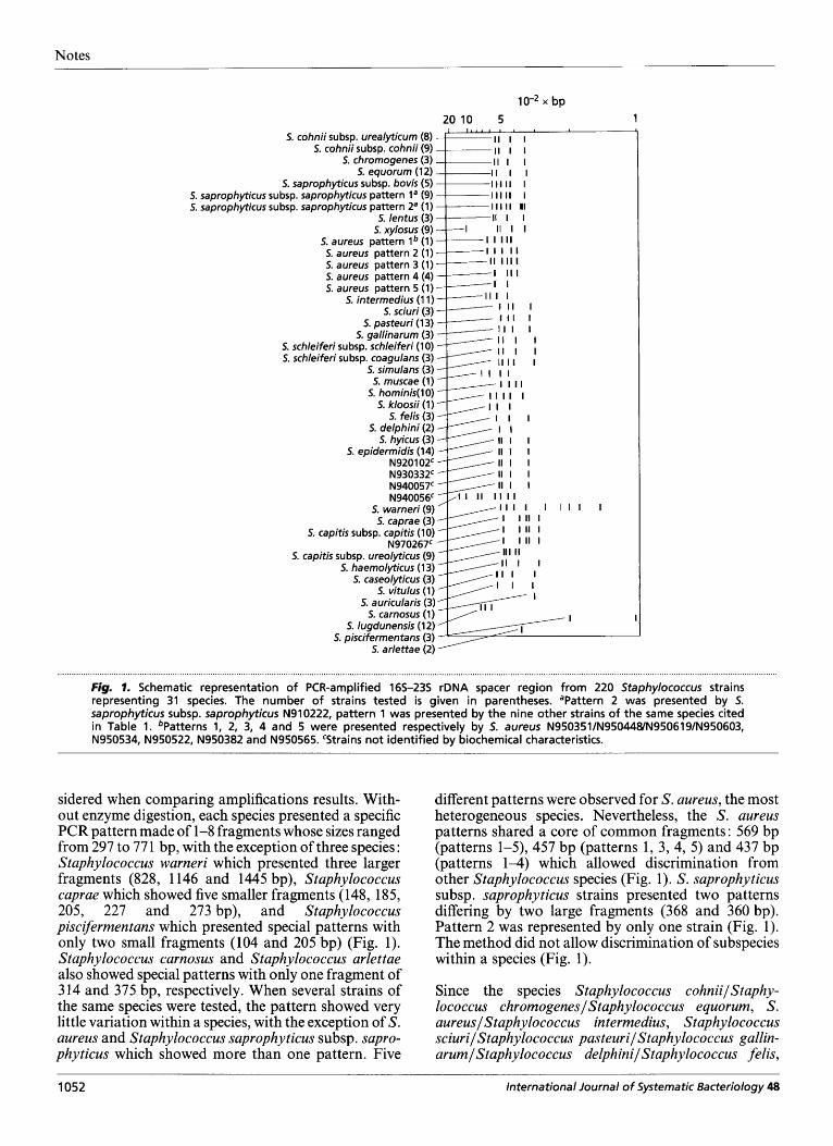
. . . . . . . . . . . . . . . . . . . . . . . . . . . . , . . . . . . . . . . . . . . . . . . . , , , , . . . , . . , . . . , . . , . , . . . . . . . . . . . . . . . . . . . . . . . . . . . . . . . . . . . . . . . . . . . . . . . . . . . . . , . , . . . . . . . . . . . . . . . . . . . . . Fig. 2. Schematic representation of PCR-amplified 165-23s rDNA spacer regions from 73 strains representing 14 Staphylococcus species after enzymic restriction with Dra 1. The number of strains tested is given in parentheses.
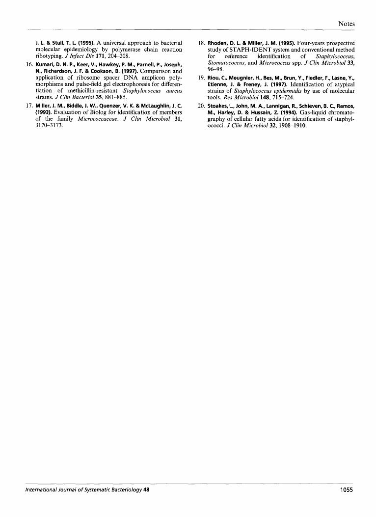
Fig. 3. 165-23s spacer region patterns of staphylococcal species. Lanes: 1, Staphylococcus sp. N920102; 2, Staphylococcus sp. N930332; 3, 5. epidermidis CCM 21 24T; 4, Staphylococcus sp. N940056; 5, Staphylococcus sp. N940057; 7-1 1, the same strains after enzymic restriction with Drat; 13, 5. capitis subsp. ureoly-ticus ATCC 49326T; 14, 5taphylococcus sp; N970267; 15, 5. capitis subsp. capitis CCM 2734T; 16-18, the same strains after enzymic restriction with Dral; 6 and 12, DNA size marker (M). The reproducibility of the technique was evaluated by the amplification of 5. capitis subsp. capitis CCM 2734T; the mean size of the five relevant bands (lane 15) were respectively 496, 388, 349, 337 and 297 bp with standard deviation of 10, 10, 9, 8 and 6 bp, respectively.
Staphylococcus vitulusl Staphylococcus auricularis pre- sented a high similarity in the number and the size of fragments, we performed enzymic digestion of the PCR products before electrophoresis. Restriction of PCR products with DraI allowed complete differentiation between all these species and confirmed identification to the species level (Fig. 2). Restriction of PCR products with Hinff also showed a specific pattern for each of the species tested. However, this enzyme generated a greater number of fragments per profile when compared to DraI. Restriction by one or the other enzyme did not improve discrimination of subspecies within a species, as shown for S. cohnii subsp. cohnii and S. cohnii subsp. urealyticurn with DraI digestion (Fig. 2). Finally, we tested five Staphy- lococcus sp. strains which could not be identified by phenotypical tests. The first strain, N970267, was quickly confirmed to belong to the group of novobiocin-susceptible staphylococci. Although the shape and the pigmentation of the colonies, resistance to fosfomycin, and other biochemical characters made us suspect that it was S. capitis subsp. ureolyticus, it was impossible to achieve a definite identification, mainly because of the absence of nitrate reductase and the presence of N-acetylglucosaminidase and pyrrolidonyl arylamidase. PCR amplification of inter- genic spacers of this strain showed the pattern specific for S. capitis (Fig. 3), thus confirming our presumptive identification. Four additional strains (N920 102, N930332, N940056 and N940057) were not identified by the ID32 STAPH system and other complementary tests (19), and were considered to be biochemical variants. Strains N940056 and N940057 were isolated from blood cultures of the same patient and were difficult to identify because of variations in some tests (D-fructose, lactose and sucrose acidification, and urease activity) when different subculture media or detection methods were used. The other two strains
were atypical because of the absence or presence of fermentations uncommonly encountered in Staphyl- ococcus epiderrnidis isolates. DNA-DNA hybridiza- tion and ribotyping demonstrated that these four strains belonged to the species S. epidermidis. PCR amplification of intergenic spacers showed the pattern specific for S. epiderrnidis and confirmed these identifications (Fig. 3).
A PCR-based strategy for studying the length poly- morphisms of the 16s-23s rDNA spacer regions has proved useful for both epidemiological (6, 14) and taxonomic (2, 10) investigations. In our study, PCR- mediated amplification of the 16s-23s rDNA spacer region using oligonucleotide primers G1 and L1 as described by Jensen et al. (10) proved to be an adequate tool for the identification of 220 Staphylococcus strains representing 3 1 species. PCR products were generated by using a unified set of primers and PCR conditions without regard to the origin of the bacterial DNA. Among Staphylococcus species, the lengths or nucleotide sequences of the 16s-23s rDNA spacer region, as well as the exact number of rRNA operons per genome, have only been determined with precision for S. aureus (7). Consequently, the lengths of ampli- fication products could not be predicted in our study. We showed that the lengths of the 16s-23s rDNA spacer region of Staphylococcus species, which varied from 104 to 771 bp, are similar to the lengths described for other genera (8). Despite of similarities in the sizes of 16s-23s spacer amplification products, each of the Staphy lococcus species examined could be identified by its PCR profile. These results were in agreement with the biochemical and phenotypical methods (4) pre- viously used to identify these strains. The potential usefulness of restriction enzyme analysis with DraI or Hinfl has already been reported for S. aureus which
International Journal of Systematic Bacteriology 48 1053
Notes
presents cleavage sites for both enzymes (7). Both enzymes were tested to improve the discrimination between Staphylococcus species which presented a high similarity in the number and the size of 16s-23s spacer fragments. We showed that only restriction of PCR products by DraI permitted the discrimination of all species tested. PCR-mediated amplification of the 16s-23s rDNA spacer region can be considered an additional tool for the identification of atypical Staphylococcus strains as demonstrated in this study with the identification of five atypical Staphylococcus strains as S. epidermidis (four strains) or S. capitis (one strain). Our choice of primers proved adequate for Staphylococcus species identification. However, Gurtler & Stanisich (8) suggested that, at least from a theoretical standpoint, amplification of all the 16S23S rDNA spacer regions from a strain is most likely to be successful when using region 2 of the 16s rRNA gene and region 10 of the 23s rRNA gene for primer construction (8). Following their recommendations, we tested primers 2 and 10 on ten Staphylococcus species but did not obtain a better discrimination than with primers 3 and 5 (or GI and Ll) (data not shown).
Our method did not allow detection of intraspecies polymorphism among most of Staphylococcus species, with the exception of S. aureus and, in a limited fashion, S. saprophyticus subsp. saprophyticus. For S. aureus, we observed a high level of heterogeneity (five different PCR patterns for eight strains), confirming the results of previous studies (10, 16). Other authors who used primers 2 and 7 with S. aureus strains (3, 15) demonstrated the utility of these primers for studying the molecular epidemiology of S. aureus. However, more strains from defined outbreaks and other sources should be compared to confirm the possible appli- cation of the study of intergenic spacer length poly- morphisms with G1 and L1 primers for the epidemio- logical typing of S . aureus isolates. It may also be possible to improve the separation of bands by the use of other methods such as denaturing gradient gel electrophoresis (DGGE) or single-strand con- formation polymorphisms (SSCP) ; however, these methods are not easy to use in the routine microbiology laboratory.
In conclusion, the study of the length polymorphisms of the 16s-23s rDNA spacer region by PCR-mediated amplification with primers GI and L1 (lo), showed significant promise as a tool to identify Staphylococcus strains to the species level. Current on-going studies indicate that this method allows identification of a clinical Staphylococcus strain at the species level within 24-48 h of receipt of a pure culture, and that it can be readily and economically implemented, thus enabling routine use in the clinical microbiology laboratory.
Acknowledgements
We thank Dominique Monnet for helpful discussions.
References 1. Aubel, D., Renaud, F. N. R. & Freney, 1. (1997). Genomic
diversity of several Corynebacterium species identified by amplification of the 16s-23s rRNA gene spacer region. Int J Syst Bacteriol47, 767-772.
2. Barry, T., Colleran, G., Glennon, M., Dunican, L. K. & Gannon, F. (1991). The 16s-23s ribosomal spacer region as a target for DNA probes to identify eubacteria. P CR Methods Appl
3. Dolzani, L., Tonin, E., Lagatolla, C. & Monti-Bragadin, C. (1 994). Typing of Staphylococcus aureus by amplification o f the 16s-23s rRNA intergenic spacer sequences. FEMS Microbiol Lett 119, 167-1 74.
4. Freney, J., Brun, Y., Bes, M., Meugnier, H., Grimont, F., Grimont, P. A. D., Nervi, C. & Fleurette, J. (1988). Staphyl- ococcus lugdunensis sp. nov. and Staphylococcus schleiferi sp. nov., two species from human clinical specimens. Int J Syst Bacteriol38, 168-1 72.
5 . Goh, S.H., Potter, S., Wood, 1. O., Hemmingsen, 5. M., Reynolds, R. P. & Chow, A. W. (1996). HSP 60 gene sequences as universal targets for microbial species identification : studies with coagulase negative staphylococci. J Clin Microbiol34, 8 18-823.
6. GUrtler, V. (1993). Typing of Clostridium dzficile strains by PCR-amplification of variable length 16S-23S rDNA spacer regions. J Gen Microbioll39, 3089-3097.
7. GUrtler, V. & Barrie, H. D. (1995). Typing of Staphylococcus aureus strains by PCR amplification of variable-length 16S-23S rDNA spacer regions : characterization of spacer sequences. Microbiology 141, 1255-1 265.
8. GUrtler, V. & Stanisich, V. A. (1996). New approaches to typing and identification of bacteria using the 16s-23s rDNA spacer region. Microbiology 142, 3-16.
9. leven, M., Verhoeven, J., Pattyn, 5. R. & Goossens, H. (1995). Rapid and economical method for species identification of clinically significant coagulase-negative staphylococci. J Clin Microbiol33, 1060-1063.
10. Jensen, M. A., Webster, 1. A. & Straus, N. (1993). Rapid identification of bacteria on the basis of polymerase chain reaction-amplified ribosomal DNA spacer polymorphisms. Appl Environ Microbiol59, 945-952.
11. Kleeman, K. T., Bannerman, T. L. & Kloos, W. E. (1993). Species distribution of coagulase-negative staphylococcal isolates at a community hospital and implications for selection of staphylococcal identification procedures. J Clin MicrobioE31, 13 18-1 32 1.
12. Kloos, W. E., Ballard, D. N., Webster, 1. A. & 12 other authors (1997). Ribotype delineation and description of Staphyl- ococcus sciuri subspecies and their potential as reservoirs of methicillin resistance and staphylolytic enzyme genes. Int J Syst Bacteriol47, 3 13-323.
13. Kloos, W. E. & Bannerman, T. L. (1995). Staphylococcus and Micrococcus. In Manual of Clinical Microbiology, 6th edn, pp. 282-298. Edited by P. R. Murray, E. J. Baron, M. A. Pfaller, F. C. Tenover & R. H. Yolker. Washington, DC: American Society for Microbiology.
14. Kostman, 1. R., Edlind, T. D., LiPuma, J. 1. & Stull,T. L. (1992). Molecular epidemiology of Pseudomonas cepacia determined by polymerase chain reaction ribotyping. J Clin Microbiol30, 2084-2087.
15. Kostman, J. R., Alden, M. B., Mair, M., Edlind, T. D., Puma,
1, 51-56.
1054 International Journal of Systematic Bacteriology 48
Notes
16.
17.
1. L. & Stull, T. L. (1995). A universal approach to bacterial molecular epidemiology by polymerase chain reaction ribotyping. J Infect Dis 171, 204-208. Kumari, D. N. P., Keer, V., Hawkey, P. M., Parnell, P., Joseph, N., Richardson, J. F. & Cookson, B. (1997). Comparison and application of ribosome spacer DNA amplicon poly- 19. morphisms and pulse-field gel electrophoresis for differen- tiation of methicillin-resistant Staphylococcus aureus strains. J Clin Bacteriol35, 881-885. Miller, 1. M., Biddle, J. W., Quenzer, V. K. & McLaughlin, 1. C. (1993). Evaluation of Biolog for identification of members of the family Micrococcaceae. J Clin Microbiol 31, 3 1 70-3 1 73.
18.
20.
Rhoden, D. L. & Miller, J. M. (1995). Four-years prospective study of STAPH-IDENT system and conventional method for reference identification of Staphylococcus, Stomatococcus, and Micrococcus spp. J Clin Microbiol33, 9698. Riou, C., Meugnier, H., Bes, M., Brun, Y., Fiedler, F., Lasne, Y., Etienne, J. & Freney, J. (1997). Identification of atypical strains of Staphylococcus epidermidis by use of molecular tools. Res Microbioll48, 71 5-724. Stoakes, L., John, M. A., Lannigan, R., Schieven, B. C., Ramos, M., Harley, D. & Hussain, 2. (1994). Gas-liquid chromato- graphy of cellular fatty acids for identification of staphyl- ococci. J Clin Microbiol32, 1908-1910.
International Journal of Systematic Bacteriology 48 1055


