Embed Size (px)
Citation preview

T
S§ACa
Dddfvndmd
Developmental Biology 210, 15–29 (1999)Article ID dbio.1999.9261, available online at http://www.idealibrary.com on
Notochord-Dependent Expression of MFH1 andPAX1 Cooperates to Maintain the Proliferationof Sclerotome Cells during the VertebralColumn Development
Taka-aki Furumoto,*,†,‡ Naoyuki Miura,§ Takeshi Akasaka,*,†Yoko Mizutani-Koseki,*,† Hidefumi Sudo,*,†,‡ Katsuyuki Fukuda,†Mamiko Maekawa,¶ Shigeki Yuasa,¶ Yan Fu,§ Hideshige Moriya,‡Masaru Taniguchi,*,| Kenji Imai,** Edger Dahl,** Rudi Balling,**Maria Pavlova,†† Achim Gossler,†† and Haruhiko Koseki*,†,1
*CREST (Core Research for Evolutionary Science and Technology) of Japan Science andechnology Corp., †Department of Molecular Embryology, ‡Department of Orthopediatric
Surgery, ¶Department of Anatomy, and \Department of Molecular Immunology, Graduatechool of Medicine, Chiba University, 1-8-1 Inohana, Chuo-ku, Chiba 260-8670, Japan;Department of Biochemistry, Akita University School of Medicine, 1-1-1 Hondo,kita 010-8543, Japan; **Institute of Mammalian Genetics, GSF-National Researchenter for Environmental and Health, 85764 Neuherberg, Germany;nd ††The Jackson Laboratory, Bar Harbor, Maine 04609
uring axial skeleton development, the notochord is essential for the induction of the sclerotome and for the subsequentifferentiation of cartilage forming the vertebral bodies and intervertebral discs. These functions are mainly mediated by theiffusible signaling molecule Sonic hedgehog. The products of the paired-box-containing Pax1 and the mesenchymeorkhead-1 (Mfh1) genes are expressed in the developing sclerotome and are essential for the normal development of theertebral column. Here, we demonstrate that Mfh1 like Pax1 expression is dependent on Sonic hedgehog signals from theotochord, and Mfh1 and Pax1 act synergistically to generate the vertebral column. In Mfh1/Pax1 double mutants,orsomedial structures of the vertebrae are missing, resulting in extreme spina bifida accompanied by subcutaneousyelomeningocoele, and the vertebral bodies and intervertebral discs are missing. The morphological defects in Mfh1/Pax1
ouble mutants strongly correlate with the reduction of the mitotic rate of sclerotome cells. Thus, both the Mfh1 and thePax1 gene products cooperate to mediate Sonic hedgehog-dependent proliferation of sclerotome cells. © 1999 Academic Press
Key Words: mouse; embryo; somite; vertebral column; development; notochord; Mfh1; Pax1; spina bifida;
myelomeningocoele.bnartqs
INTRODUCTION
The vertebral column is composed of alternating verte-brae and intervertebral discs that are metamerically ar-ranged along the anterior–posterior body axis. Each vertebrais subdivided into several distinct domains along the dor-soventral axis. The ventral region is composed of vertebral
ls
1 To whom correspondence should be addressed. Fax: 181-43-227-1498. E-mail: [email protected].
0012-1606/99 $30.00Copyright © 1999 by Academic PressAll rights of reproduction in any form reserved.
odies and the underlying intervertebral discs. Dorsally, theeural arches, which are subdivided into ventral pediclesnd dorsal laminae, enclose the spinal cord. In the thoracicegion, ribs are associated with vertebrae. All these struc-ures are derived from the somitic mesoderm as a conse-uence of interactions between the paraxial mesoderm andurrounding tissues (Christ and Ordahl, 1995).After the segmentation of the paraxial mesoderm, epithe-
ial somites differentiate into the ventral mesenchymalclerotome and the dorsal epithelial dermomyotome. Sonic
15

16 Furumoto et al.
FIG. 1. Expression of Mfh1 and Pax1 in developing somites. Expression of Mfh1 (A, B, C, D, E, F, G) and Pax1 (H, I, J, K, L, M, N) mRNAin 9.5- (A, D, E, H, L, K), 10.5- (B, F, I, M), and 11.5- (C, G, J, N) dpc embryos analyzed by in situ hybridization, and expression of MFH1and PAX1 proteins in 11.5-dpc embryos analyzed by indirect immunohistochemistry (O, P, Q, R). (A, D, E) Mfh1 expression in 9.5-dpcembryos. (A) Mfh1 expression in the caudal region of the unsegmented paraxial mesoderm and in the somites along the entire axis. Thearrowhead indicates the boundary between the presomitic mesoderm and first epithelial somite. The line indicates the level of the sectionin E. (D) Higher magnification of the tail-bud region. Boundaries between the presomitic mesoderm and first and second somites areindicated. (E) Mfh1 expression in the sclerotome of differentiating somites and in mesenchymal cells surrounding the dorsal aorta. Thedermomyotome is indicated by an arrow. (B, F) Mfh1 expression in 10.5-dpc embryos. (B) Mfh1 expression in the presomitic mesoderm,somites, and posterior region of the sclerotome. The line indicates the level of the section in F. (F) Mfh1 expression in the sclerotome. Notethe strong expression in the ventrolateral regions. (C, G) Mfh1 expression in 11.5-dpc embryos. (C) Strong Mfh1 expression in the posteriorregion of the sclerotome. The white arrowhead indicates the strong expression in the distal tips of rib anlagen. Fore- and hindlimbs areindicated by arrows. The line indicates the level of the sections in G and O. (G) Mfh1 expression in the sclerotome. Note the strong Mfh1expression in the ventrolateral regions. (H, K, L) Pax1 expression in 9.5-dpc embryos. (H) Pax1 expression in the newly segmented somites
and in posterior regions of the sclerotome. Note that Pax1 expression is confined to the ventral region while Mfh1 expression extendsdorsally. The arrowhead indicates the boundary between unsegmented paraxial mesoderm and epithelial somite. The line indicates theCopyright © 1999 by Academic Press. All rights of reproduction in any form reserved.

P
tsw
g
17Interactions in Mouse Vertebral Column Development
hedgehog (SHH) signals emanating from the notochord andthe ventral neural tube are essential for sclerotome differ-entiation and the subsequent formation of the vertebralcolumn (Fan et al., 1994; Chiang et al., 1996; Teillet et al.,1998). In shh-deficient mice, axial skeleton development isseverely impaired due to the extensive degeneration ofsclerotome cells (Chiang et al., 1996). Culture of thesomitic mesoderm and microsurgical approaches in avianembryos revealed that SHH is essential not only for theestablishment of the dorsoventral axis of the somitic me-soderm but also for cell proliferation and for the preventionof cell death (Fan et al., 1994; Teillet et al., 1998). Thepleiotropic biological effects of SHH are thought to bemediated by a series of transcriptional regulators, includingmembers of the Pax and MyoD gene families, that areexpressed in the paraxial mesoderm (Dahl et al., 1997;Munsterberg et al., 1995).
PAX1 is a paired-box-containing transcriptional activa-tor, whose expression in the sclerotome depends on SHHemanating from the notochord or the floor plate of theneural tube (Chalepakis et al., 1991; Fan et al., 1994;Dietrich et al., 1997). Phenotypic analysis of an allelicseries of Pax1 mutations revealed that sclerotomal Pax1expression is essential for the appropriate generation ofvertebral bodies and intervertebral discs by allowing scle-rotome cells to accumulate around the notochord(Gruneberg, 1954; Wallin et al., 1994; Dietrich et al., 1995;Wilm et al., 1998). Thus, Pax1 is one of the mediators ofnotochordal signals required to generate ventral structuresof vertebral column from mesenchymal sclerotome cells(Koseki et al., 1993; Dietrich and Gruss, 1993).
Mesenchymal forkhead-1 (MFH1), a member of thewinged helix/forkhead family of proteins, is also expressedin the developing sclerotome and is required for normaldevelopment of the axial skeleton (Miura et al., 1993;Kaestner et al., 1996; Winnier et al., 1997; Iida et al., 1997).In Mfh1-deficient mice, defects of the vertebral bodies andneural arches resulting in spina bifida occulta are observedin addition to impaired aortic arch formation. The similarphenotypes in the ventral moiety of Mfh1- and Pax1-deficient mice suggest that Mfh1 and Pax1 have commonfunctions during sclerotome development.
level of the section in L. (K) Higher magnification of the tail-bud rsecond somites are indicated. Note that Pax1 expression appeared ahe ventral region of the sclerotome. (I, M) Pax1 expression inclerotome. The line indicates the level of the section in M. (M) Pahile Mfh1 expression extends dorsally. (J, N) Pax1 expression in
sclerotome. The line indicates the level of the section in N. (N) Pax(O) Expression of MFH1 protein in 11.5-dpc embryos. The MFH1 pthe Mfh1 transcript. Higher magnification views of the boxed regiprotein expression in the ventrolateral region of the sclerotome. Tview of PAX1 protein expression in the same section as is shownof P and Q. Cells expressing both MFH1 and PAX1 are indicated by y
and PAX1 proteins while dorsal cells express MFH1 but not PAX1. Scanglion; (fb) forelimb bud; (hb) hindlimb bud; (nc) notochord; (nt) neurCopyright © 1999 by Academic Press. All right
In this study, we investigate the importance of noto-chordal signals for Mfh1 expression as well as the geneticinteraction between Mfh1 and Pax1. Mfh1 expression wassignificantly reduced in Danforth’s short-tail (Sd) mutantembryos whose notochords degenerate (Gruneberg, 1953;Koseki et al., 1993; Dietrich and Gruss, 1993; Maatman etal., 1997), and ectopic grafts of a notochord or of SHH-expressing cells induced Mfh1 expression, indicating thatsignals from the notochord are necessary and sufficient tomaintain Mfh1 expression in the somitic mesoderm. Theobservation that the expression of Mfh1 and Pax1 dependson notochordal signals prompted us to investigate theskeletal phenotypes of mice lacking both Mfh1 and Pax1 touncover potential overlapping functions of both gene prod-ucts during vertebral column development. Embryosdouble mutant for Mfh1 and Pax1 showed extreme spinabifida associated with subcutaneous myelomeningocoeleand completely lacked vertebral bodies and intervertebraldiscs, due to a synergistic reduction of the mitotic activityof sclerotome cells. Our results demonstrate that the Mfh1and Pax1 gene products cooperate to maintain the SHH-dependent proliferation of sclerotome cells, but both genesare dispensable for the formation of sclerotome and itssurvival.
MATERIALS AND METHODS
Mice
Mice lacking a functional Pax1 gene were generated by homolo-gous recombination using J7 ES cells, such that the Escherichia colib-galactosidase gene was introduced into the XhoI site within thepaired-box-encoding exon, resulting in the premature terminationof the Pax1 gene product (K. Imai, E. Dahl, T. Akasaka, H. Koseki,and R. Balling, unpublished results). The skeletal phenotypes ofthis allele, designated Pax1lacZ, resembled those of the Pax1un-ex and
ax1null alleles (Wallin et al., 1994; Wilm et al., 1998). Heterozy-gous mutants were backcrossed onto C57Bl/6 three to four genera-tions and homozygotes were generated by matings between het-erozygous mutants. Mfh1 mutants (Iida et al., 1997) werebackcrossed onto the C57Bl/6 background several times. Pax1/Mfh1 double heterozygotes were generated by crossing Pax1lacZ
homozygotes and Mfh1 heterozygotes. Double homozygous em-
. Boundaries between the presomitic mesoderm and the first andthe segmentation of the paraxial mesoderm. (L) Pax1 expression indpc embryos. (I) Pax1 expression in the posterior region of thepression is restricted to the ventrolateral region of the sclerotome
5-dpc embryos. (J) Pax1 expression in the posterior region of thepression is restricted to the ventrolateral region of the sclerotome.
was found in the entire sclerotome and strongly overlapped withre shown in P, Q, and R. (P) Higher magnification view of MFH1ntral root is indicated by an arrowhead. (Q) Higher magnificationThe ventral root is indicated by an arrowhead. (R) Overlap imagecolor. Note that most ventral sclerotomal cells express the MFH1
egionfter10.5-x1 ex
11.1 ex
roteinon ahe vein P.ellow
ale bars represent 50 mm. (dm) dermomyotome; (drg) dorsal rootal tube; (sc) sclerotome.
s of reproduction in any form reserved.

La
18 Furumoto et al.
FIG. 2. Mfh1 expression in day 10.5 wild-type and Sd homozygous embryos and after notochord graft and ectopic SHH expression.ateral views (A, B, E, F) and transverse frozen (C, D, G, H) sections of wild-type (A, B, C, D) and Sd homozygous (E, F, G, H) embryosfter whole-mount in situ hybridization. Mfh1 expression in the prospective sacrocaudal region of the somitic mesoderm was
significantly affected in Sd homozygotes, whereas expression in the more cranial region was unaffected (E), consistent with theimpaired notochord formation in the caudal body region of Sd homozygotes (Koseki et al., 1993). (B, F) Higher magnification viewsof the caudal region. Note that Mfh1 expression in the segmented region was significantly reduced in Sd homozygotes while theexpression in the unsegmented paraxial mesoderm was not. Arrows designated as C, D, G, and H indicate the levels of the transversesections shown in C, D, G, and H, respectively. (C) Mfh1 expression in wild-type embryos is strong in the unsegmented paraxialmesoderm but absent from the neural tube, notochord, hindgut, and mesenchymal cells in the ventromedial region (indicated by abracket) or from the surface ectoderm. The arrow indicates the notochord. (D) In differentiating somites Mfh1 expression is strong inthe mesenchymal sclerotome but is reduced in the dermomyotome. The arrow and arrowhead indicate the notochord and floor plate,respectively. (G) In the presomitic mesoderm of Sd homozygous embryos Mfh1 is ectopically expressed in the hindgut and inmesenchymal cells in the ventromedial region of the tail bud (indicated by a bracket). (H) In differentiating somites sclerotomalexpression of Mfh1 is significantly reduced in Sd homozygotes. Floor-plate formation (arrowhead) is apparently impaired in Sdhomozygotes. Arrows in (G, H) indicate the notochord. (I) Schematic depicting the position of grafts (red dot) of notochord ofSHH-expressing cell pellets between the neural tube and the unsegmented paraxial mesoderm. Ectopic expression of Mfh1 (arrow-
heads) induced by (J) the ectopic notochord (arrow) or by (K) a graft of cell pellet expressing SHH (arrow). (dm) dermomyotome; (fb)forelimb bud; (h) hindgut; (nt) neural tube; (p) unsegmented paraxial mesoderm; (sc) sclerotome.Copyright © 1999 by Academic Press. All rights of reproduction in any form reserved.
es(t(((
19Interactions in Mouse Vertebral Column Development
bryos were derived from matings between double heterozygotesand genotyped using the following oligonucleotides: for detectionof the Pax1 wild-type allele, Pax1 common (59-GTAGGCAACT-GCGGGTCTCTC-39) and Pax1 wild (59-CGGATCTCCCAGGC-AAAGA-39); for detection of the mutant Pax1 allele, Pax1 commonand Pax1 KO (59-GCGAAAGGGGGATGTGCT-39). Oligonucleo-tides used to genotype Mfh1 mutants were previously described(Iida et al., 1997). Double mutants were genotyped by PCR analysisof tail, kidney, or yolk sac DNA.
The Sd mice used to collect embryos were kept on a mixedgenetic background with the wild-type allele of Sd derived fromCAST/Ei. Day (d) 10.5 postcoitus (pc) embryos from Sd/1 (CAST/Ei) 3 Sd/1 (CAST/Ei) matings were collected in RNase-freephosphate-buffered saline (PBS). Embryos were typed for Sd byanalyzing yolk sac DNA with two MIT markers flanking Sd:D2Mit362 (about 0.15 cM proximal to Sd) and D2Mit416 (about 0.6cM distal to Sd) as described (Maatman et al., 1997).
Grafting Experiments
Fertilized eggs of the White Leghorn chick were purchased fromlocal farms and were incubated at 38°C for about 2.5 days to reachthe 18- to 25-somite stages (stages 14–18; Hamburger and Hamil-ton, 1951). Surgical manipulations were performed as describedpreviously (Aoyama and Asamoto, 1988).
A 2.5-kb fragment encoding the full-length mouse shh cDNA(kindly provided by Dr. A. McMahon) was subcloned into theRCASBP(A) vector (Echelard et al., 1993; Hughs et al., 1987).Retroviral vector DNA was introduced into chicken embryonicfibroblasts by calcium phosphate precipitation. Retroviral particlessecreted into the culture medium supplemented with 0.5% fetalcalf serum were concentrated more than 20 times by ultracentrifu-gation. QT6 cells were infected by retrovirus overnight and pelletsof infected QT6 were transplanted into chick embryos resistant toviral infection.
Chicken Mfh1 riboprobes were generated from a 1.9-kb full-length cDNA (H.K., unpublished results). The GenBank accessionnumber of chicken Mfh1 is U95823.
Skeletal Analysis
Skeletal preparations were made from perinatal mice and clearedskeletons were analyzed stereomicroscopically as previously de-scribed (Kessel and Gruss, 1991).
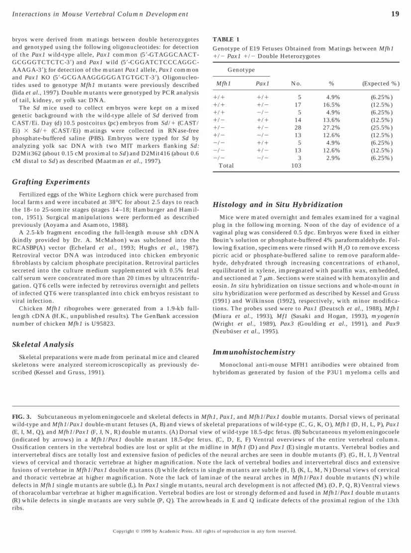
FIG. 3. Subcutaneous myelomeningocoele and skeletal defects inwild-type and Mfh1/Pax1 double-mutant fetuses (A, B) and views o(E, I, M, Q), and Mfh1/Pax1 (F, J, N, R) double mutants. (A) Dorsal(indicated by arrows) in a Mfh1/Pax1 double mutant 18.5-dpc fOssification centers in the vertebral bodies are lost or split at theintervertebral discs are totally lost and extensive fusion of pediclesviews of cervical and thoracic vertebrae at higher magnification. Nfusions of vertebrae in Mfh1/Pax1 double mutants (J) while defectsand thoracic vertebrae at higher magnification. Note the lack ofdefects in Mfh1 single mutants are subtle (L). In Pax1 single mutantof thoracolumbar vertebrae at higher magnification. Vertebral bodie
(R) while defects in single mutants are very subtle (P, Q). The arrowheribs.Copyright © 1999 by Academic Press. All right
Histology and in Situ Hybridization
Mice were mated overnight and females examined for a vaginalplug in the following morning. Noon of the day of evidence of avaginal plug was considered 0.5 dpc. Embryos were fixed in eitherBouin’s solution or phosphate-buffered 4% paraformaldehyde. Fol-lowing fixation, specimens were rinsed with H2O to remove excesspicric acid or phosphate-buffered saline to remove paraformalde-hyde, dehydrated through increasing concentrations of ethanol,equilibrated in xylene, impregnated with paraffin wax, embedded,and sectioned at 7 mm. Sections were stained with hematoxylin andosin. In situ hybridization on tissue sections and whole-mount initu hybridization were performed as described by Kessel and Gruss1991) and Wilkinson (1992), respectively, with minor modifica-ions. The probes used were to Pax1 (Deutsch et al., 1988), Mfh1Miura et al., 1993), Mf1 (Sasaki and Hogan, 1993), myogeninWright et al., 1989), Pax3 (Goulding et al., 1991), and Pax9Neubuser et al., 1995).
Immunohistochemistry
Monoclonal anti-mouse MFH1 antibodies were obtained fromhybridomas generated by fusion of the P3U1 myeloma cells and
TABLE 1Genotype of E19 Fetuses Obtained from Matings between Mfh11/2 Pax1 1/2 Double Heterozygotes
Genotype
No. % (Expected %)Mfh1 Pax1
1/1 1/1 5 4.9% (6.25%)1/1 1/2 17 16.5% (12.5%)1/1 2/2 5 4.9% (6.25%)1/2 1/1 14 13.6% (12.5%)1/2 1/2 28 27.2% (25.5%)1/2 2/2 13 12.6% (12.5%)2/2 1/1 5 4.9% (6.25%)2/2 1/2 13 12.6% (12.5%)2/2 2/2 3 2.9% (6.25%)
Total 103
1, Pax1, and Mfh1/Pax1 double mutants. Dorsal views of perinatalletal preparations of wild-type (C, G, K, O), Mfh1 (D, H, L, P), Pax1of wild-type 18.5-dpc fetus. (B) Subcutaneous myelomeningocoele(C, D, E, F) Ventral overviews of the entire vertebral column.
ine in Mfh1 (D) and Pax1 (E) single mutants. Vertebral bodies ande neural arches are seen in double mutants (F). (G, H, I, J) Ventral
the lack of vertebral bodies and intervertebral discs and extensivengle mutants are subtle (H, I). (K, L, M, N) Dorsal views of cervicalae of the neural arches in Mfh1/Pax1 double mutants (N) whileral arch development is not affected (M). (O, P, Q, R) Ventral viewslost or strongly deformed and fused in Mfh1/Pax1 double mutants
Mfhf skeviewetus.midlof thotein silamins, neus are
ads in E and Q indicate defects of the proximal region of the 13th
s of reproduction in any form reserved.

20

21

acsioaigf2s
aewR
22 Furumoto et al.
spleen cells prepared from a Wistar rat immunized with recombi-nant GST/mouse MFH1 (amino acid 290–494) fusion protein (Y.Fu, X-L. Yang, and N. Miura, unpublished result). Since theantibodies are directed against a region of the MFH1 proteinoutside the forkhead domain which is not conserved betweenMFH1 and MF1, this monoclonal antibody is considered to bespecific for MFH1. The generation and purification of anti-PAX1antibodies was performed as described previously (Chalepakis etl., 1991; Wallin et al., 1994). Embryos were embedded in OCTompound immediately after dissection and sectioned on a cryo-tat. Sections were fixed in ice-cold acetone for 20 min and washedn 10% goat serum in PBS for 1 h. Tissue sections were incubatedvernight at 4°C with rat monoclonal anti-mouse MFH1 and rabbitnti-PAX1 antibodies, washed in PBS/10% goat serum as above,ncubated with a fluorescein-conjugated goat anti-rat immuno-lobulin and a rhodamine-conjugated anti-rabbit immunoglobulinor 1 h at room temperature, washed, mounted in 75% glycerol/5% PBS, and observed under a Zeiss LSM 410 invert confocal lasercan microscope.
Detection of Apoptosis
Day 11.5 pc embryos were dissected, fixed for 6 h in 4%paraformaldehyde in PBS, and embedded in paraffin wax. Sectionswere cut at 7 mm and placed onto 3-aminopropryltriethoxysilane-coated slides. The in situ TdT-mediated dUTP-fluorescein-labelednick end labeling (TUNEL) was performed using the cell deathdetection kit (Boehringer Mannheim) with the TUNEL reactionmixture being diluted threefold with 30 mM Tris (pH 7.2), 140 mMcacodylic acid, and 1 mM cobalt chloride.
Cell Proliferation
Fifty micrograms of bromodeoxyuridine (BrdU; Sigma) per gramof body weight was injected intraperitoneally into pregnant mice1.5 h before embryo collection. Embryos were fixed in 4% parafor-maldehyde for 4 h and embedded in paraffin wax, sectioned at 7 mmthickness, dewaxed, and rehydrated and BrdU incorporation wasdetected with anti-BrdU antibodies (clone B44; Becton–Dickinson)as described previously (Wallin et al., 1994). To determine the rateof cell proliferation in the sclerotome, photomicrographs weretaken at 203 magnification, and labeled and unlabeled cells in fourdjacent sections from four independent wild-type and mutantmbryos were counted. The overall percentage of labeled nucleias determined and statistically analyzed by Student’s t test.esults were considered significant at a P value , 0.05.
FIG. 4. Vertebral morphology in Mfh1, Pax1, and Mfh1/Pax1 dou(L2) vertebrae from wild type (A, E, I, M, Q) and Mfh1 (B, F, J, N, RDefects of the dorsal atlas (arrowheads) in Mfh1 (B) and in Mfh1/Paxin Mfh1/Pax1 double mutants. The arrow in (D) indicates the fusioMfh1 (F) or Pax1 (G) single, mutants the odontoid process (arrowhof the neural arches (filled arrowheads), unilateral defect of the fvertebral body (arrow) of vertebra C5 in Mfh1/Pax1 double, but noneural arches (arrowheads) and absence of the vertebral body (arrow
single, mutants. Note the missing articulation of the ribs in (O) and (P).in Mfh1 (R) and Mfh1/Pax1 double (T) mutants and defects of the verteCopyright © 1999 by Academic Press. All right
RESULTS
Notochord-Dependent Expression of Mfh1 andPax1 in the Paraxial Mesoderm
The expression of Mfh1 and Pax1 in developing somiticmesoderm was comparatively investigated in 9.5, 10.5, and11.5-dpc embryos. In 9.5-dpc embryos, Mfh1 expressionfirst appeared in the caudal region of the unsegmentedparaxial mesoderm and was seen ubiquitously within thesomitic mesoderm before the deepithelialization of epithe-lial somites (Figs. 1A, 1D, and 2C). Immediately after thedeepithelialization of the somite, Mfh1 expression wasrestricted to the sclerotome (Figs. 1E and 2D). Initial Pax1expression in the somitic mesoderm was seen in the first orsecond newly segmented somite, indicating that Pax1 isexpressed later than Mfh1 in the paraxial mesoderm (Figs.1H and 1K; Koseki et al., 1993). Pax1 expression in thesclerotome was restricted to the ventral region (Fig. 1L). In10.5- and 11.5-dpc embryos, Mfh1 and Pax1 expression wasseen in the sclerotome along the entire axis (Figs. 1B, 1C, 1I,and 1J). Mfh1 expression was seen in the dorsal and poste-rior region of the sclerotome demarcating the neural archanlagen (Figs. 1B and 1C). Expression of Mfh1 in the neuralarch anlagen was confirmed by in situ hybridization oftransverse sections (Figs. 1F and 1G). Pax1 expression wasfound in the ventral and posterior, but not in the dorsal,region of the sclerotome (Figs. 1I, 1J, 1M, and 1N). MFH1protein in 11.5-dpc embryos as monitored immunohisto-chemically with the anti-MFH1 monoclonal antibody waslocalized in the developing sclerotome and mesenchymaltissues around the aorta, consistent with the results of thein situ hybridizations (Fig. 1O). Simultaneous detection ofMFH1 and PAX1 by confocal laser microscopy demon-strated that the proteins are coexpressed in sclerotome cellsin the ventrolateral region (Figs. 1P, 1Q, and 1R).
Since the expression of Pax1 in the sclerotome dependson notochordal signals mainly mediated by SHH (Koseki etal., 1993; Dietrich et al., 1993; Fan and Tessier-Levigne,1994), we investigated the requirement of the notochord forMfh1 expression. The expression of Mfh1 was examined inDanforth’s short-tail mutant embryos, which in the poste-rior region have no or a severely reduced notochord (Figs.2C and 2G; Gruneberg, 1953; Koseki et al., 1993; Dietrich et
utants. Dissected cervical (C1, C2, C5), thoracic (T), and lumbarx1 (C, G, K, O, S), and Mfh1/Pax1 double mutants (D, H, L, P, T).uble (D), but not in Pax1 (C), mutants. Note the increased severityodontoid process to the atlas. (H) In Mfh1/Pax1 double, but not infailed to fuse with vertebra C2. (L) Complete lack of the laminaeen for the vertebral artery (open arrowhead), and absence of theMfh1 (J) or Pax1 (K) single, mutants. (P) Defective laminae of thevertebra T8 in Mfh1/Pax1 double, but not in Mfh1 (N) or Pax1 (O)
ble m), Pa1 don ofeads)oramt in) of
Defective laminae of the neural arches (arrowheads) of vertebra L2bral body (arrows) in Pax1 (S) and Mfh1/Pax1 double (T) mutants.
s of reproduction in any form reserved.

mdHsd
fidacimnSpMts
wdhaDhdat(boa
t
23Interactions in Mouse Vertebral Column Development
al., 1993). As a consequence of the poorly developed noto-chord, the paraxial mesoderm was fused across the midline(Figs. 2C, 2D, 2G, and 2H). Expression of Mfh1 in theunsegmented paraxial mesoderm was not significantly af-fected although ectopic expression of Mfh1 was seen in the
ost ventral mesenchymal cells and in the abnormallyeveloped hindgut (Figs. 2A, 2B, 2C, 2E, 2F, and 2G).owever, the strong expression of Mfh1 in the segmented
omitic mesoderm of wild-type embryo was markedly re-uced in Sd homozygotes (Figs. 2D and 2H).The dependence of Mfh1 expression on the notochord was
urther investigated by grafting experiments. A surgicallysolated piece of a chicken notochord was transplanted imme-iately beneath the surface ectoderm between the neural tubend the unsegmented paraxial mesoderm of stage 14–18hicken embryos as shown in Fig. 2I. The notochord graftnduced ectopic expression of chicken Mfh1 in the paraxial
esoderm (Fig. 2J), consistent with the requirement of theotochord for Mfh1 expression seen in Sd mutant embryos.imilarly, a cell pellet secreting SHH grafted to the dorsalresomitic mesoderm induced ectopic expression of chickenfh1 (Fig. 2K). These results indicated that SHH signals from
he notochord are necessary and sufficient to maintain theclerotomal expression of Mfh1.
Axial Skeleton Defects in Mfh1/Pax1 DoubleMutants
The MFH1 and PAX1 gene products are coexpressed inpart of the developing sclerotome and required for thenormal development of the vertebral column. To analyze
FIG. 5. Histological analysis of the developing vertebral columTransverse sections of wild-type (A, E, I) and Mfh1 (B, F, J), Pax1 (Transverse sections through the cervical region. In Mfh1 (B) and Paxhe annuli fibrosi are not generated normally. In Mfh1/Pax1 double
are completely lost. The fusion of connective tissue surroundsubcutaneous tissue is significantly expanded in double mutants. TF, G, H) Transverse sections through the thoracic region. Note tharches are totally impaired in Mfh1/Pax1 double mutants (H). (I, J, Ktube extrudes unilaterally into the subcutaneous tissue (arrowheadMfh1/Pax1 double mutants. Scale bars represent 500 mm.FIG. 6. Histological analysis of somite development in Mfh1/Pax1wild-type (A, B, C, D, E) and Mfh1/Pax1 double-mutant (F, G, H, I,9.0-dpc embryos. The deepithelization of somites is not affected incervical region of 9.5-dpc embryos. The sclerotome size appears to bis less affected. The neural tube appears compressed vertically and tof the dermomyotome and dorsal aorta are indicated by arrows andcervicothoracic region of 10.5-dpc embryos. (H) The reduction of thmutants are more prominent. The dorsal root ganglia are dislocateregion. The dorsal edges of the dermomyotome and dorsal rootTransverse sections of the prospective thoracic region of 11.5-dpmagnification in (E) and (J). The dorsal root ganglia are indicated bMfh1/Pax1 double mutant. (E, J) Higher magnification views of the
accumulation of sclerotome cells around the notochord is strongly affrepresent 50 mm in (A, B, F, G) and 60 mm in (C, H, D, I).Copyright © 1999 by Academic Press. All right
hether MFH1 and PAX1 cooperate during sclerotomeevelopment, we have analyzed double mutants. Doubleeterozygous Mfh1- and Pax1-deficient mice were healthynd fertile, and the vertebral columns were not affected.ouble heterozygotes were intercrossed to generate doubleomozygotes. The frequency of homozygous Mfh1/Pax1ouble mutants recovered perinatally was only appropri-tely half of the expected (Table 1). Since the frequency ofhese mutants was not significantly reduced up to 11.5 dpc5.4%), the subsequent loss of double homozygotes mighte due to abnormal aortic arch formation caused by the lossf MFH1 function, which results in embryonic deathround 12.5 dpc in half of the Mfh1 mutant embryos (T.F.
and H.K., unpublished results; Iida et al., 1997). The fre-quency of Mfh1 2/2 mice (20%) in the present study wasslightly increased compared to the report by Iida et al.(16%), which could be due to differences in the geneticbackgrounds caused by introducing the Pax1 mutation.
Visual inspection of Mfh1/Pax1 double homozygous mu-tant 18.5-dpc fetuses revealed that the craniocaudal lengthwas around two-thirds of the wild-type littermates and thecaudal region was strongly shortened. In addition, a subcu-taneous myelomeningocoele was found in all Mfh1/Pax1double mutants at the thoracolumbar border, a phenotypenever observed in either single mutant (Figs. 3A and 3B).Skeletal malformations of the axial skeleton in Mfh1/Pax1double-mutant mice were clearly much stronger than ineither single mutant (Figs. 3C, 3D, 3E, and 3F). In Mfh1single mutants, the ossification centers of vertebral bodieswere absent in the cervical region and were irregularlyformed in the thoracolumbar region (Figs. 3D, 3H, 3L, 3P,
15.5-dpc Mfh1, Pax1, and Mfh1/Pax1 double-mutant fetuses., K), and Mfh1/Pax1 double mutant (D, H, L) fetuses. (A, B, C, D)) single mutants, vertebral bodies are smaller than in wild type andants (D), the vertebral bodies and the laminae of the neural archeshe developing back muscles (arrowhead) is also impaired andeural tube is slightly deformed as if it were extended vertically. (E,e formation of the vertebral bodies and the laminae of the neuralransverse sections through the lumbar region. Note that the neuralicating the formation of the subcutaneous myelomeningocoele in
ble-mutant fetuses between 9 and 11.5 dpc. Transverse sections ofbryos. (A, F) Transverse sections of prospective cervical region of1/Pax1 double mutants. (B, G) Transverse sections of prospectiveuced in Mfh1/Pax1 double mutants (G), while the dermomyotomelagen of the dorsal aorta are shifted dorsolaterally. The dorsal edgesheads, respectively. (C, H) Transverse sections of the presumptive
lerotome and deformation of the neural tube in Mfh1/Pax1 doublesally, presumably due to the lack of sclerotome cells in the dorsalion are indicated by arrows and arrowheads, respectively. (D, I)bryos. The regions indicated by open boxes are shown at higherowheads. Note that the dorsal root ganglia are shifted dorsally inhordal region. The arrow indicates the notochord. (J) Note that the
n inC, G1 (Cmut
ing the nat th, L) T), ind
douJ) emMfh
e redhe anarrowe sc
d dorganglc emy arrperic
ected in the Mfh1/Pax1 double mutant. (m) myotome. Scale bars
s of reproduction in any form reserved.

n(ma
l1ats
24 Furumoto et al.
4B, 4F, 4J, 4N, and 4R). The dorsal portion of the neural archwas affected in the atlas and thoracic vertebrae (Figs. 3L and4B; Iida et al., 1997; Winnier et al., 1997). The ribs were
ormal except for occasional fusions of the proximal partsFig. 3P; Iida et al., 1997; Winnier et al., 1997). In Pax1
utants, the ossification centers of vertebral bodies werebsent in the cervical region and split in the thoracic,
rv
Copyright © 1999 by Academic Press. All right
umbar, and sacral regions, and the proximal region of the3th rib was lacking (Figs. 3E, 3I, 3M, 3Q, 4C, 4G, 4K, 4Ond 4S). The vertebral columns of Mfh1/Pax1 double mu-ants were much shorter than those of either single mutant,uggesting that the craniocaudal length of the vertebrae was
educed (Fig. 3F). Ventral as well as dorsal structures of theertebrae were lacking or strongly reduced in the vertebrals of reproduction in any form reserved.

25Interactions in Mouse Vertebral Column Development
Copyright © 1999 by Academic Press. All rights of reproduction in any form reserved.

v(etttoIwnm
ptdp6dcpcdeat(mwMsmeidwt7o7ddifad
F
26 Furumoto et al.
column of Mfh1/Pax1 double mutants (Fig. 3F, 3J, 3N, 3R,4D, 4H, 4L, 4P, and 4T), and only the pedicles of the neuralarches were less severely affected (Figs. 4L, 4P, and 4T).Extensive fusions of deformed vertebrae were observed inthe cervical, thoracic, and lumbar regions. In most cases,the ribs failed to articulate with the vertebral bodies and theproximal ribs were occasionally fused (Figs. 3F, 3R, and 4P).Synergistic effects on development of the scapula or neuro-cranium were not seen in Mfh1/Pax1 double mutantsalthough Mfh1 and Pax1 expressions overlap in these re-gions (Deutsch et al., 1988; Timmons et al., 1994; Winnieret al., 1997; Iida et al., 1997).
The defects of axial structures in Mfh1/Pax1 double mu-tants were further investigated histologically in 15.5-dpc fe-tuses. In Mfh1 and Pax1 single mutants, vertebral bodies werepoorly developed and occasionally split, and the annuli fibrosiwere not generated normally at all examined axial levels.Impaired neural arch formation or back muscle developmentwas not prominent (Figs. 5A, 5B, 5C, 5E, 5F, 5G, 5I, 5J, and 5K).In Mfh1/Pax1 double mutants, the cartilaginous primordia ofentral and dorsal structures of vertebrae were not generatedFigs. 5D, 5H, and 5L). The subdermal connective tissue wasxpanded compared to single mutants but no brown adiposeissue was apparent. In the cervical region, the connectiveissue surrounding the back musculature failed to fuse athe dorsal midline and concomitantly the medial portionsf back musculature were not generated normally (Fig. 5D).n the lumbar region, the presumptive subarachinoid cavityas significantly narrower than in single mutants, and theeural tissue extruded dorsally leading to subcutaneousyelomeningocoele (Fig. 5L).
Reduction of Sclerotome Cells in Mfh1/Pax1Double Mutants
Early defects of vertebral column development were inves-tigated histologically. In 9.0-dpc double-mutant embryos,deepithelialization of somites was not affected (Figs. 6A and6F). In the prospective cervical region of double mutants at 9.5dpc, the expansion of the sclerotome was significantly im-
FIG. 7. Expression of marker genes in the prospective cervical regD, I, J, K, L) and bright-field (E, F, G, H, M, N, O, P) views of transve(A, B, C, D, E, F, G, H) and Mfh1/Pax1 double-mutant (I, J, K, L, Mof Mf1 (I), myogenin (J), Pax3 (K), and Pax9 (L) is essentially normIG. 8. Proportion of replicating cells in the sclerotome of Mfh1,
and 11.5. (A) Overview showing the sclerotome regions in wiBrdU-labeled cells. The prospective neural arches are labeled wiperichordal region with “c”. (B) Higher magnification views of the(right) and hematoxylin–eosin-stained adjacent sections (left) are pucounted in each region of the sclerotome of wild-type and Mfh1,significant differences in BrdU incorporation were observed for thefor the dorsal (“a”) or ventromedial (“c”) region. (D) Bar diagrams
sclerotome of wild-type and Mfh1, Pax1, and Mfh1/Pax1 double-mutanreduced in each analyzed region.Copyright © 1999 by Academic Press. All right
aired and the dorsal aorta and dorsal tips of the dermomyo-ome were shifted dorsally. The neural tube was deformed byorsoventral compression, presumably due to imbalancedroliferation in the sclerotome and neural tube (Figs. 6B andG). In the prospective cervicothoracic region of Mfh1/Pax1ouble-mutant 10.5-dpc embryos, the reduction of sclerotomeells and the deformations of the neural tube were morerominent (Figs. 6C and 6H). The dorsal mass of sclerotomeells surrounding the dorsal root ganglion was strongly re-uced in double mutants. The dermomyotome was relativelynlarged compared to the sclerotome and shifted dorsomedi-lly. At 11.5 dpc, the accumulation of sclerotome cells aroundhe notochord was impaired in Mfh1/Pax1 double mutantFigs. 6D, 6E, 6I, and 6J). Early differentiation of the somitic
esoderm in Mfh1/Pax1 double-mutant 9.5-dpc embryosas investigated by in situ hybridization analysis usingf1, myogenin, Pax3, and Pax9 probes. Mf1 was expressed
trongly in the entire sclerotome and weakly in the dermo-yotome both in the wild-type and in the double-mutant
mbryos (Figs. 7A, 7E, 7I, and 7M). Myogenin expressionndicated the normal differentiation of myotome cells inouble mutants (Figs. 7B, 7F, 7J, and 7N). Pax3 expressionas found in the dorsal neural tube and to the dermomyo-
ome in double mutants, similar to wild-type embryos (Figs.C, 7G, 7K, and 7O). Pax9 expression in the ventral moietyf the sclerotome was not affected in double mutants (Figs.D, 7H, 7L, and 7P). These results demonstrate that theifferentiation of somites into sclerotome, myotome, andermatome occurs in Mfh1/Pax1 double mutants, indicat-ng that the MFH1 or PAX1 gene products are not essentialor sclerotome formation per se and suggesting that thexial skeletal defects seen in Mfh1/Pax1 double mutants areue to insufficient expansion of sclerotome cells.
Synergistic Reduction of Mitotic Cells inVentrolateral Region of the Sclerotomeof Mfh1/Pax1 Double Mutant
Since the significant reduction of sclerotome cells inMfh1/Pax1 double mutants could be due to an increase of
f Mfh1/Pax1 double-mutant 9.5-dpc embryos. Dark-field (A, B, C,ctions through the prospective cervical region of 9.5-dpc wild-typeO, P) embryos after mRNA in situ hybridization. The expressionMfh1/Pax1 double mutants. Scale bars represent 50 mm., and Mfh1/Pax1 double-mutant embryos on embryonic days 10.5pe and double-mutant embryos (black rectangles) analyzed for”, the ventrolateral region of the sclerotome with “b”, and therotomal areas indicated in (A). Microphotographs of BrdU-labelede by side. (C) Bar diagrams of the proportions of BrdU-labeled cells1, and Mfh1/Pax1 double-mutant day 11.5 embryos. Statisticallytrolateral region (“b”) of wild type and of double mutants, but note proportions of BrdU-labeled cells counted in each region of the
ion orse se, N,
al inPax1ld-tyth “a
sclet sidPaxvenof th
t day 10.5 embryos. The proportions of mitotic cells were slightly
s of reproduction in any form reserved.

e
bitefamdTst
et
27Interactions in Mouse Vertebral Column Development
apoptotic cell death or a decrease of cell proliferation, thesepossibilities were examined. We first compared the propor-tions of sclerotome cells undergoing apoptosis in wild-typeand Mfh1/Pax1 double-mutant 10.5- and 11.5-dpc embryosby TUNEL labeling. A few apoptotic cells were observed inthe perichordal region and ventrolateral regions of thesclerotome. However, no significant differences were seenbetween wild type and Mfh1/Pax1 double mutants (T.F.,T.A., and H.K., unpublished result).
The proportion of mitotic sclerotome cells in the prospec-tive thoracic region of wild-type and Mfh1, Pax1, andMfh1/Pax1 double-mutant 11.5-dpc embryos was comparedafter labeling cells in S phase with BrdU. This analysisrevealed a marked reduction of proliferating cells in Mfh1/Pax1 double-mutant embryos (Figs. 8A, 8B, and 8C). Inwild-type embryos, the Mfh1- and Pax1-coexpressing ven-trolateral region of the sclerotome was mitotically veryactive, with about 70% of cells being labeled by BrdU,whereas sclerotome cells giving rise to the prospectiveneural arch and perichordal tube were less mitoticallyactive (Figs. 8A, 8B, and 8C). We could not detect asignificant difference in BrdU incorporation between thecranial and the caudal moieties of the ventral sclerotome at11.5 dpc (T.F. and H.K., unpublished result). Intriguingly,the most mitotic subpopulation of the sclerotome wassignificantly reduced in Mfh1 and Pax1 single mutants andin Mfh1/Pax1 double mutants, while the mitotic activity inthe prospective neural arch and perichordal tube was notaffected (Figs. 8A, 8B, and 8C). Cell proliferation in theventrolateral region of the sclerotome was more severelyaffected in Mfh1/Pax1 double mutants than in either of thesingle mutants, correlating the severity of the axial skel-eton defects with the mitotic activity of sclerotome cells.Thus, the MFH1 and PAX1 proteins appear to cooperativelymaintain the mitotic activity of sclerotome cells. Sincesclerotome development was already affected in 10.5-dpcMfh1/Pax1 double-mutant embryos, we have investigatedthe frequency of mitotic cells in 9.5- and 10.5-dpc embryos.In 9.5-dpc embryos, we could not find a significant differ-ence between wild type and double mutants (T.F. and H.K.,unpublished result). In day 10.5 embryos, BrdU incorpora-tion was slightly, however, significantly, reduced in theventral but not in the dorsal region of the sclerotome inMfh1 and Pax1 single as well as double mutants (Fig. 8D).
DISCUSSION
MFH1 and PAX1 as Mediators for SHH-DependentProliferation of the Sclerotome
The expression pattern of Mfh1 in homozygous Sd em-bryos suggests that the induction of Mfh1 expression in thepresomitic mesoderm either requires only weak noto-chordal signals or is independent of such signals, while themaintenance of Mfh1 expression in the somitic mesoderm
depends on signals from the notochord. Grafting an addi-tional notochord or a cell pellet expressing SHH in avianMa
Copyright © 1999 by Academic Press. All right
mbryos induces ectopic expression of Mfh1 in the paraxialmesoderm. Thus, both Mfh1 and Pax1 appear to be down-stream effectors of SHH in the somitic mesoderm. How-ever, the sclerotomal Mfh1 expression domain is muchwider than the Pax1 domain. Since Pax1 expression appearsto be repressed by bone morphogenetic proteins (BMP),particularly by BMP-4 derived from the dorsal neural tubeor the lateral plate mesoderm (Pourquie et al., 1996;Monsoro-Burq et al., 1996; Watanabe et al., 1998), the widerexpression domain could indicate that Mfh1 expression isless sensitive to signals counteracting SHH. SHH emanat-ing from the notochord is a diffusible factor essential foraxial skeleton development by establishing the dorsoven-tral axis of the somitic mesoderm and by promoting cellcycle progression and preventing apoptosis of the medialmoiety of the somitic mesoderm (Fan et al., 1994; Chiang etal., 1996; Teillet et al., 1998). Since the proliferation ofsclerotome cells is more severely affected in Mfh1/Pax1double mutants than in either single mutant, and MFH1and PAX1 are present in the same cells in the sclerotome,MFH1 and PAX1 are likely to act cooperatively and mediateSHH-dependent sclerotomal cell proliferation (Winnier etal., 1997). This idea is supported by the observations thatexclusively axial structures derived from the medial portionof the somites are affected in shh and Mfh1/Pax1 doublemutants, while structures derived from the lateral portionsincluding the distal parts of the ribs are unaffected (Chianget al., 1996). In contrast, neither MFH1 nor PAX1 appears tobe required for sclerotome formation per se or for theprevention of cell death in the somitic mesoderm. Thus,other molecules induced by SHH such as PAX9 or MF2might be responsible for sclerotome formation or for theprevention of apoptosis, or they might compensate forPAX1 or MFH1 functions in these processes (Neubusser etal., 1996; Wu et al., 1998).
Cellular Basis for the Interaction between Mfh1and Pax1 Mutations
The subtle spina bifida observed in the cervical andthoracic regions of Mfh1 mutants is significantly enhancedy the Pax1 mutation although Pax1 itself is not expressedn the dorsal region of the sclerotome and is not required forhe neural arch development (Wallin et al., 1994; Dietricht al., 1995; Winnier et al., 1997; Iida et al., 1997). Theormation of ventral structures of the vertebral column islso much more severely affected in Mfh1/Pax1 doubleutants, and extensive fusions of the vertebrae are seen in
ouble mutants but not in either of the single mutants.hus, the Mfh1 and Pax1 mutations appear to affect axialkeleton development synergistically rather than addi-ively.
Since a sufficient density of precursor cells in the blast-ma is known to be necessary for cartilaginous condensa-ion (Hall and Miyake, 1992), the extreme spina bifida in
fh1/Pax1 double mutants may result from an insufficientccumulation of mesenchymal cells in the dorsal region of
s of reproduction in any form reserved.

bcsp
efarrricnbid
motS
bg1
C
C
C
C
D
D
D
D
D
E
F
G
28 Furumoto et al.
the sclerotome. The impaired expansion of the sclerotomein 9.5-, 10.5-, and 11.5-dpc Mfh1/Pax1 double-mutant em-ryos supports this argument. Thus, the insufficient allo-ation of sclerotome cells in the dorsomedial region of theclerotome could be the basis for the novel synergistichenotype in Mfh1/Pax1 double mutants.Alternatively, since interactions between the surface
ctoderm, neural tube, and mesenchymal cells are essentialor cartilage differentiation in the dorsal region of the neuralrch (Takahashi et al., 1992; Monsoro-Burq et al., 1996), theesponsiveness of sclerotomal cells to inductive signalsequired for neural arch formation could be synergisticallyeduced by the Mfh1 and Pax1 mutations. This possibilitys supported by observations by Winnier et al. (1997) thathondrogenic differentiation of somitic mesoderm couldot be stimulated by transforming growth factor-b or fibro-last growth factor in Mfh1 mutants. The profound geneticnteraction between the Pax1 mutation and Patch, whicheletes the PDGF-receptor a chain, results in extreme spina
bifida and suggests the involvement of the Pax1 geneproduct in mediating PDGF signals during the neural archdevelopment (Helwig et al., 1995).
The formation of ventral structures of the vertebralcolumn is also much more severely affected in Mfh1/Pax1double mutants than in either single mutant. The accumu-lation of sclerotome cells around the notochord generatingthe perichordal tube is an essential prerequisite for theformation of the cartilaginous condensations of the verte-bral bodies and is impaired in a series of Pax1 mutantalleles, in Mfh1 mutants, and more strongly in doublemutants (Wallin et al., 1994; Dietrich et al., 1995; Winnieret al., 1997; Iida et al., 1997). Defects of proliferation ormigration of sclerotome cells toward the notochord mightbe synergistically enhanced in Mfh1/Pax1 double mutants.
Etiology and Pathogenesis of the SubcutaneousMyelomeningocoele
The interaction between Mfh1 and Pax1 leads to extremespina bifida associated with subcutaneous myelomeningo-coele in the thoracolumbar transition. Since neural tubeclosure and the proliferation of neural cells in the mantlelayer are not affected in Mfh1/Pax1 double mutants, andneither Mfh1 nor Pax1 is expressed in the developing neuraltube, neural tube development per se is unlikely be dis-turbed in double mutants. Therefore it is possible thatimbalances in the proliferation rates in the sclerotome andneural tube cause the extrusion of the neural tube andresult in the subcutaneous myelomeningocoele. In clinicalobservations, myelomeningocoele is frequently associatedwith defects in the ventral region, radiologically termedhemivertebrae and block vertebrae (Roth, 1971), which arealso characteristic for Mfh1 single and Mfh1/Pax1 double
utants. Since similar defects of the vertebral column arebserved in mice lacking Gli2 and Gli3, other likely media-
ors of SHH signals, the genetic cascade downstream ofHH involving Mfh1, Pax1, Gli2, or Gli3 could be impairedCopyright © 1999 by Academic Press. All right
y environmental and/or genetic factors during the patho-enesis of subcutaneous myelomeningocoele (Mo et al.,997).
ACKNOWLEDGMENTS
We thank Drs. P. Gruss, A. McMahon, Y. Nabeshima, and H.Sasaki for Pax3, shh, myogenin, and mf1 cDNA clones, respec-tively. We also thank Dr. H. Mossman, Dr. M. Amano, Ms. HirokoTanabe, Ms. Yoko Sawano, Ms. Kazuko Higashino, Ms. MisaoUchida, and Mr. Shozo Sugimori for their help in many aspects ofthis project. This project is supported by grants from the Ministryof Education, Science, and Culture (H.K.), from the Agency ofScience and Technology (H.K.), The Japan Spina Bifida and Hydro-cephalus Research Foundation (H.K.), and from the Ministry ofHealth and Welfare (M.T.) and by NIH Grant NS34960 (A.G.).
REFERENCES
Aoyama, H., and Asamoto, K. (1988). Determination of somitecells: Independence of cell differentiation and morphogenesis.Development 104, 15–28.halepakis, G., Fritsch, R., Fickenscher, H., Deutsch, U., Goulding,M., and Gruss, P. (1991). The molecular basis of the undulated/Pax1 mutation. Cell 66, 875–884.halepakis, G., Jones, F. S., Edelman, G. M., and Gruss, P. (1994).Pax-3 contains domains for transcription activation and tran-scription inhibition. Proc. Natl. Acad. Sci. USA 91, 12745–12749.hiang, C., Litingtung, Y., Lee, E., Young, K. E., Corden, J. L.,Westphal, H., and Beachy, P. A. (1996). Cyclopia and defectiveaxial patterning in mice lacking Sonic hedgehog gene function.Nature 383, 407–413.hrist, B., and Ordahl, C. P. (1995). Early stages of chick somitedevelopment. Anat. Embryol. 191, 381–396.ahl, E., Koseki, H., and Balling, R. (1997). Pax genes and organo-genesis. BioEssays 19, 755–765.eutsch, U., Dressler, G. R., and Gruss, P. (1988). Pax1, a memberof a paired box homologous murine gene family, is expressed insegmented structures during development. Cell 53, 617–625.ietrich, S., Schubert, F. R., and Gruss, P. (1993). Altered Pax geneexpression in murine notochord mutants: The notochord isrequired to initiate and maintain ventral identity in the somite.Mech. Dev. 44, 189–207.ietrich, S., and Gruss, P. (1995). Undurated phenotypes suggest arole of Pax1 for the development of vertebral and extravertebralstructures. Dev. Biol. 167, 529–548.ietrich, S., Schubert, F. R., and Lumsden, A. (1997). Control ofdorsoventral pattern in the chick paraxial mesoderm. Develop-ment 124, 3895–3908.
chelard, Y., Epstein, D. J., St-Jacques, B., Shen, L., Mohler, J.,McMahon, J. A., and McMahon, A. P. (1993). Sonic hedgehog, amember of a family of putative signaling molecules, is impli-cated in the regulation of CNS polarity. Cell 75, 1417–1430.
an, C. M., and Tessier-Lavigne, M. (1994). Patterning of mamma-lian somites by surface ectoderm and notochord: Evidence forsclerotome induction by a hedgehog homolog. Cell 79, 1175–1186.oulding, M. D., Chalepakis, G., Deutsch, U., Erselius, J. R., and
Gruss, P. (1991). Pax-3, a novel murine DNA binding proteinexpressed during early neurogenesis. EMBO J. 10, 1135–1147.s of reproduction in any form reserved.

G
H
H
I
K
K
K
M
M
M
M
M
M
N
P
R
S
T
T
T
W
W
W
W
W
W
W
29Interactions in Mouse Vertebral Column Development
Gruneberg, H. (1953). Genetical studies on the skeleton of themouse. VI. Danforth’s shot tail. J. Genet. 51, 317–326.runeberg, H. (1954). Genetical studies on the skeleton of themouse. XII. The development of undulated. J. Genet. 52, 441–455.all, B. K., and Miyake, T. (1992). The membranous skeleton: Therole of cell condensations in vertebrate skeletogenesis. Anat.Embryol. 186, 107–124.
Hamburger, V., and Hamilton, H. L. (1951). A series of normalstages in the development of the chick embryo. J. Morphol. 88,49–91.
Helwig, U., Imai, K., Schmahl, W., Thomas, B. E., Varnum, D. S.,Nadeau, J. H., and Balling, R. (1995). Interaction between undu-lated and Patch leads to an extreme form of spina bifida indouble-mutant mice. Nat. Genet. 11, 60–63.ughes, S. H., Greenhouse, J. J., Petropoulous, C. J., and Sutrave, P.(1987). Adaptor plasmids simplify the insertion of foreign DNAinto helper-independent retroviral vectors. J. Virol. 61, 3004–3012.
ida, K., Koseki, H., Kakinuma, H., Kato, N., Mizutani-Koseki, Y.,Ohuchi, H., Yoshioka, N., Noji, S., Kawamura, K., Kataoka, Y.,Ueno, F., Taniguchi, M., Yoshida, N., Sugiyama, T., and Miura,N. (1997). Essential roles of the winged helix transcription factorMFH1 in aortic arch patterning and skeletogenesis. Develop-ment 124, 4627–4638.aestner, K. H., Bleckman, S. C., Monaghan, A. P., Scholondorff, J.,Mincheva, A., Lichter, P., and Schutz, G. (1996). Clusteredarrangement of winged helix genes fkh-6 and MFH1: Possibleimplications for mesoderm development. Development 122,1751–1758.essel, M., and Gruss, P. (1991). Homeotic transformations ofmurine vertebrae and concomitant alteration of Hox codesinduced by retinotic acid. Cell 67, 89–104.oseki, H., Wallin, J., Wilting, J., Mizutani, Y., Kispert, A.,Ebensperger, C., Herrmann, B. G., Christ, B., and Balling, R.(1993). A role for Pax1 as a mediator of notochordal signals duringthe dorsoventral specification of vertebrae. Development 119,649–660.aatman, R., Zachgo, J., and Gossler, A. (1997). The Danforth’sshort tail mutation acts cell autonomously in notochord cellsand ventral hindgut endoderm. Development 124, 4019–4028.aulbecker, C. C., and Gruss, P. (1993). The oncogenic potential ofPax genes. EMBO J. 12, 2361–2367.iura, N., Wanaka, A., Tohyama, M., and Tanaka, K. (1993).MFH1, a new member of the fork head domain family, isexpressed in developing mesenchyme. FEBS Lett. 326, 171–176.o, R., Freer, A. M., Zinyk, D. L., Crackower, M. A., Michaud, J.,Heng, H. H., Chik, K. W., Shi, X. M., Tsui, L. C., Cheng, S. H.,Joyner, A. L., and Hui, C. (1997). Specific and redundant func-tions of Gli2 and Gli3 zinc finger genes in skeletal patterning anddevelopment. Development 124, 113–123.onsoro-Burq, A. H., Duprez, D., Watanabe, Y., Bontoux, M.,Vincent, C., Brickell, P., and Le Douarin, N. (1996). The role ofbone morphogenetic proteins in vertebral development. Devel-opment 122, 3607–3616.
unsterberg, A. E., Kitajewski, J., Bumcrot, D. A., McMahorn,A. P., and Lassar, A. B. (1995). Combinatorial signaling by SonicCopyright © 1999 by Academic Press. All right
hedgehog and Wnt family members induces myogenic bHLHgene expression in the somite. Genes Dev. 9, 2911–2922.eubuser, A., Koseki, H., and Balling, R. (1995). Characterizationand developmental expression of Pax9, a paired-box-containinggene related to Pax1. Dev. Biol. 170, 701–716.
ourquie, O., Fan, C. M., Coltey, M., Hirsinger, E., Watanabe, Y.,Breant, C., Francis-West, P., Brickell, P., Tessier-Lavigne, M., andLe Douarin, N. M. (1996). Lateral and axial signals involved inavian somite patterning: A role for BMP4. Cell 84, 461–471.
oth, K. (1971). Spinal deformities in myelomeningocoele: Aradiologic assessment. In “Scoliosis” (Paul R. Harrington, Ed.),pp. 1–29.
asaki, H., and Hogan, B. L. (1993). Differential expression ofmultiple fork head related genes during gastrulation and axialpattern formation in the mouse embryo. Development 118,47–59.akahashi, Y., Monsoro-Burq, A. H., Bontoux, M., and Le Douarin,N. M. (1992). A role for Quox-8 in the establishment of thedorsoventral pattern during vertebrate development. Proc. Natl.Acad. Sci. USA 89, 10237–10241.
eillet, M., Watanabe, Y., Jeffs, P., Duprez, D., Lapointe, F., and LeDouarin, N. M. (1998). Sonic hedgehog is required for survival ofboth myogenic and chondrogenic somitic lineages. Development125, 2019–2030.immons, P. M., Wallin, J., Rigby, P. W., and Balling, R. (1994).Expression and function of Pax1 during development of thepectoral girdle. Development 120, 2773–2785.allin, J., Wilting, J., Koseki, H., Fritsch, R., Christ, B., and Balling,R. (1994). The role of Pax1 in axial skeleton development.Development 120, 1109–1121.atanabe, Y., Duprez, D., Monsoro-Burq, A. H., Vincent, C., and LeDouarin, N. M. (1998). Two domains in vertebral development:Antagonistic regulation by SHH and BMP4 proteins. Develop-ment 125, 2631–2639.ilkinson, D. G. (1992). “In Situ Hybridization: A Practical Ap-proach.” Oxford Univ. Press, Oxford.ilm, B., Dahl, E., Peters, H., Balling, R., and Imai, K. (1998).Targeted disruption of Pax1 defines its null phenotype andproves haploinsufficiency. Proc. Natl. Acad. Sci. USA 95,8692– 8697.inner, G. E., Hargett, L., and Hogan, B. L. (1997). The wingedhelix transcription factor MFH1 is required for proliferation andpatterning of paraxial mesoderm in the mouse embryo. GenesDev. 11, 926–940.right, W. E., Sassoon, D. A., and Lin, V. K. (1989). Myogenin, afactor regulating myogenesis, has a domain homologous toMyoD. Cell 56, 607–617.u, S. C., Grindley, J., Winnier, G. E., Hargett, L., and Hogan, B. L.(1998). Mouse Mesenchyme forkhead 2 (Mf2): Expression, DNAbinding and induction by sonic hedgehog during somitogenesis.Mech. Dev. 70, 3–13.
Received for publication December 7, 1998
Revised February 19, 1999Accepted March 3, 1999
s of reproduction in any form reserved.


