Embed Size (px)
Citation preview

Journal of Neuroendocrinology, 2014, 26, 557–572
REVIEW ARTICLE © 2014 British Society for Neuroendocrinology
Novel Aspects of Glucocorticoid ActionsE. T. Uchoa*, G. Aguilera†, J. P. Herman‡, J. L. Fiedler§, T. Deak¶ and M. B. C. de Sousa**
*Department of Physiology, School of Medicine of Ribeirao Preto, University of Sao Paulo, Ribeirao, Preto, SP, Brazil.
†Section on Endocrine Physiology, National Institute of Child Health and Human Development, Bethesda, MD, USA.
‡Department of Psychiatry and Behavioural Neuroscience, Metabolic Diseases Institute, University of Cincinnati, Cincinnati, OH, USA.
§Department of Biochemistry and Molecular Biology, Faculty of Chemical and Pharmaceutical Sciences, Universidad de Chile, Santiago, Chile.
¶Department of Psychology, Binghamton University, Binghamton, NY, USA.
**Brain Institute, Universidade Federal do Rio Grande do Norte, Natal, RN, Brazil.
Journal ofNeuroendocrinology
Correspondence to:
E. T. Uchoa, Avenida Bandeirantes
3900, 14049-900 Ribeirao Preto, Sao
Paulo, Brazil (e-mail:
Normal hypothalamic-pituitary-adrenal (HPA) axis activity leading to the rhythmic and episodic
release of adrenal glucocorticoids (GCs) is essential for body homeostasis and survival during
stress. Acting through specific intracellular receptors in the brain and periphery, GCs regulate
behaviour, as well as metabolic, cardiovascular, immune and neuroendocrine activities. By con-
trast to chronic elevated levels, circadian and acute stress-induced increases in GCs are neces-
sary for hippocampal neuronal survival and memory acquisition and consolidation, as a result of
the inhibition of apoptosis, the facilitation of glutamatergic neurotransmission and the forma-
tion of excitatory synapses, and the induction of immediate early genes and dendritic spine for-
mation. In addition to metabolic actions leading to increased energy availability, GCs have
profound effects on feeding behaviour, mainly via the modulation of orexigenic and anorixegen-
ic neuropeptides. Evidence is also emerging that, in addition to the recognised immune suppres-
sive actions of GCs by counteracting adrenergic pro-inflammatory actions, circadian elevations
have priming effects in the immune system, potentiating acute defensive responses. In addition,
negative-feedback by GCs involves multiple mechanisms leading to limited HPA axis activation
and prevention of the deleterious effects of excessive GC production. Adequate GC secretion to
meet body demands is tightly regulated by a complex neural circuitry controlling hypothalamic
corticotrophin-releasing hormone (CRH) and vasopressin secretion, which are the main regula-
tors of pituitary adrenocorticotrophic hormone (ACTH). Rapid feedback mechanisms, likely
involving nongenomic actions of GCs, mediate the immediate inhibition of hypothalamic CRH
and ACTH secretion, whereas intermediate and delayed mechanisms mediated by genomic
actions involve the modulation of limbic circuitry and peripheral metabolic messengers. Consis-
tent with their key adaptive roles, HPA axis components are evolutionarily conserved, being
present in the earliest vertebrates. An understanding of these basic mechanisms may lead to
novel approaches for the development of diagnostic and therapeutic tools for disorders related
to stress and alterations of GC secretion.
Key words: glucocorticoids, feedback, HPA axis, neuroplasticity, immune system, food intake
doi: 10.1111/jne.12157
Living organisms are constantly faced with external and internal
challenges or stressors threatening the internal environment equi-
librium or homeostasis (1). Adaptation to this changing environ-
ment requires the coordinated activation of multiple
neuroendocrine responses, prominently including activation of the
hypothalamic pituitary adrenal (HPA) axis (2). Control of HPA
axis activity is complex, involving sensors conveying signals to
corticotrophin-releasing hormone (CRH) neurones in the hypotha-
lamic paraventricular nucleus (PVN), either through direct neural
connections, or indirectly through pathways relaying on limbic
structures such as the amygdala, frontal cortex, bed nucleus of the
stria terminalis and hippocampus (2). CRH and vasopressin (AVP),

released from parvocellular neurones of the PVN into the pituitary
portal circulation, increase the secretion of adrenocorticotrophic
hormone (ACTH) from pituitary corticotrophs, which in turn stimu-
lates glucocorticoid (GC) secretion from the adrenal. The HPA axis
is highly conserved through evolution, with its components being
present in early vertebrates. The secretion of GCs (i.e. cortisol in
humans, corticosterone in rats and mice) is episodic, following cir-
cadian (daily) and ultradian (hourly) rhythms, and shows marked
but transient increases after exposure to stressors. This episodic
nature of GC secretion is critical for the transcriptional activity of
the steroid (3).
Acting through intracellular receptors of the nuclear receptor
family, GCs are essential for stress adaptation by controlling energy
supply through the stimulation of glycolysis, gluconeogenesis and
lipolysis, as well as inducing proteolysis to supply amino acids as
substrates for gluconeogenesis. In addition, GCs modulate the
immune system, as well as the synthesis and action of a number of
hormones. By acting directly in neurones or by modulating the syn-
thesis, secretion or/and activity of neuropeptides and neurotrans-
mitters, GCs affect memory acquisition and consolidation, and
impact upon the function of other neuroendocrine systems, such as
the central control of metabolic activity, feeding behaviour and
reproductive activity. Given these wide ranging effects of GCs,
either GC deficiency or a failure to limit HPA axis activation will
have profound consequences on the well-being of an organism. An
important mechanism for maintaining episodic HPA axis activation
and for limiting HPA axis activity comprises negative-feedback by
GCs, resulting in the inhibition of expression and secretion of hypo-
thalamic CRH and AVP, as well as pituitary ACTH.
This review covers novel aspects concerning the regulation and
function of GCs, including the critical importance of adequate GC
regulation on synaptic remodelling, interactions between the HPA
axis and inflammatory processes, and other neuroendocrine sys-
tems, such as the control of appetite, as well as molecular mecha-
nisms, brain and peripheral circuitries involved in HPA axis
regulation and GC feedback.
GC receptors and mineralocorticoid receptors (MRs)
The action of GCs involves binding evolutionary conserved intracel-
lular receptors belonging to the nuclear receptor family, GC recep-
tors type 1 or MR, and type 2 or GC receptor (GR) (4). The genes of
MRs and GRs are considered to have arisen from a common ances-
tor, the corticoid receptor, before the evolution of aldosterone (5).
These receptors are ligand activated transcription factors, which,
upon ligand binding, translocate to the nucleus and interact with
responsive elements in gene promoters or interact with other tran-
scription factors, inducing transcriptional activation or repression.
Under basal conditions, GRs and MRs are located in the cytoplasm
and are associated with the chaperone proteins, heat shock proteins
(HSP) 90 and 70, and co-chaperones such as the immunophilin,
FK506 binding protein 5. HSP90 and other chaperone and co-chap-
erone proteins are part of the mechanism of receptor activation
and translocation, as well as the subsequent GR transactivation
effects (6).
In addition to these genomic actions regulating gene transcrip-
tion, there is clear evidence that GRs and MRs also mediate nonge-
nomic responses of their ligands. Some biological actions of GCs
are too fast to be mediated by genomic actions (which require pro-
tein synthesis) and they are assumed to depend on membrane
receptors (7,8). As indicated above, GCs are critical for body homeo-
stasis and act through GRs and MRs; thus, the well-adjusted acti-
vation of these receptors is crucial for maintaining homeostasis.
GC receptors are widely expressed in the central nervous system,
including the hypothalamus and hippocampus. Under normal condi-
tions, GRs are essentially ligand-free at the nadir of the circadian
rhythm and largely occupied by peak plasma levels of GCs. Simi-
larly, GRs are occupied in situations of stress, mediating negative-
feedback, which regulates HPA responsiveness (9).
MRs are expressed in the kidney, where they mediate sodium re-
absorption, as well as other epithelial and non-epithelial tissues, in
which the function of MRs remains to be clearly determined (10).
Although MRs have equally high affinity to GCs and mineralocortic-
oids, the presence of 11-b-hydroxysteroid dehydrogenase type 2
(11b-HSD2) in mineralocorticoid target tissues protects MR from
the much higher circulating levels of GCs, by converting GCs to
inactive 11-keto steroids (10). MRs are also expressed in the central
nervous system (CNS), mostly in limbic sites. The hippocampus is
the main limbic structure that expresses MRs and, in conjunction
with other regions of the CNS, expresses the type 1 isozyme of
11b-HSD, which regenerates active GCs from the circulating inert
11-keto steroids. Consequently, at this site, MRs bind to GCs with a
10-fold higher affinity than GRs. Because of the high affinity for
GCs, and the higher circulating concentrations of GCs compared to
mineralocorticoids, MR in the hippocampus are occupied by basal
and low concentrations of GCs. MRs of the hippocampus plays an
important role in the GC-mediated feedback control of the HPA axis
and it is assumed that they are involved in the maintenance of the
basal HPA activity, mainly at the nadir of the circadian rhythm
when hippocampal MRs are significantly occupied (11). The type 2
isozyme of 11b-HSD is not detectable at limbic sites, and co-locali-
sation of 11b-HSD2 and MRs has been identified only in the
nucleus of the solitary tract (NTS), an area related to cardiovascular
regulation and sodium appetite (12). Thus, although the NTS
appears to be the only mineralocorticoid dependent site in the
brain, activation of MRs in other brain areas are mediated by GCs
and not mineralocorticoids.
Overall, in the context of the regulation of the HPA axis, the
available evidence indicates that MRs are involved in the feedback
of GCs during the phase of the nadir of the circadian rhythm,
whereas increasing levels of GCs recruit GRs during the reactive
feedback and during stressful episodes.
Molecular mechanisms of GC feedback
Inhibitory feedback by GCs at the central and pituitary levels plays
a major role in reducing HPA axis responses to stress. As discussed
below, an important mechanism for GC feedback involves the mod-
ulation of direct and indirect circuitry controlling CRH neurone
activity. GCs inhibit CRH expression (13) and secretion (14) in the
© 2014 British Society for Neuroendocrinology Journal of Neuroendocrinology, 2014, 26, 557–572
558 E. T. Uchoa et al.

PVN, and inhibit both ACTH output (15,16) and the transcription of
the precursor protein pro-opiomelanocortin (POMC) (17,18) in the
anterior pituitary corticotroph. Concerning transcriptional regula-
tion, it is clear that inhibition of POMC transcription by GCs is a
genomic effect dependent on the interaction of GR with the POMC
promoter (17,18). However, the mechanism by which GCs inhibit
CRH transcription is less obvious (19). In addition, there is evidence
that GCs may influence translational activity and mRNA stability of
both POMC and CRH mRNA (20,21).
In vitro studies performed in primary cultures of rat anterior
pituitary or hypothalamic neurones have shown marked differences
between the effects of GCs on CRH and POMC transcription. In
these experiments, transcription rates were assessed by measuring
levels of primary transcript or heteronuclear RNA (hnRNA). As
shown in Fig. 1(A), preincubation of primary cultures of rat anterior
pituitary cells with 100 nM corticosterone for 30 min before the
addition of 1 nM CRH completely prevented the stimulatory effect
of CRH on POMC hnRNA. Full inhibition of CRH-stimulated POMC
transcription persisted 18 h after addition of corticosterone. By
contrast, exposure of primary cultures of foetal rat hypothalamic
neurones to corticosterone had only minor effects of cyclic AMP-
stimulated CRH hnRNA production. In these experiments, 7-day
neuronal cultures maintained for 48 h in steroid-free culture med-
ium were exposed to 100 nM corticosterone before incubation with
the adenylate cyclase stimulator, forskolin, for an additional
45 min. As seen in Fig. 1(B), corticosterone tended to inhibit forsko-
lin-stimulated CRH hnRNA in cells preincubated with corticosterone
for 30 min, an inhibition that was statistically significant only after
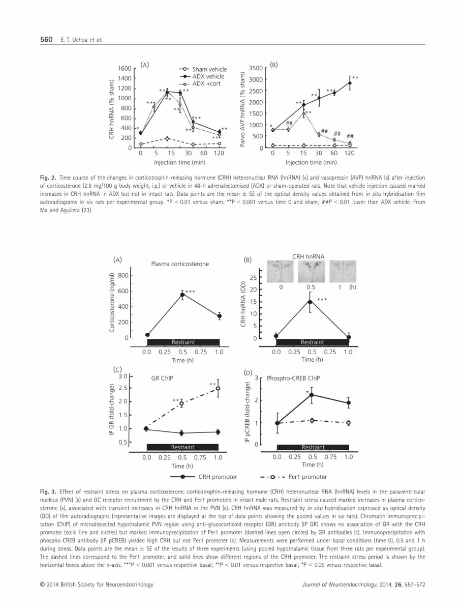
log transformation of the data (22). Similarly, the administration of
corticosterone doses that increased the plasma concentration to
100-fold stress levels in adrenalectomised rats did not affect the
magnitude or duration of the increase in CRH hnRNA in the PVN in
response to a mild stress (22,23) (Fig. 2A). In the same rats, the
injection of corticosterone markedly attenuated stress-induced AVP
hnRNA levels in parvocellular neurones (Fig. 2B) (23). Similarly, Kov-
acs and Sawchenko (24) showed that the injection of corticosterone
10 min before ether stress in rats failed to inhibit stress induced
increases in CRH hnRNA.
Although the above studies suggest that GCs have little effect on
CRH transcription, GRs are present in the CRH neurone (25,26) and
there is clear evidence that GCs negatively regulate CRH mRNA lev-
els. For example, removal of endogenous GCs by adrenalectomy
markedly increased CRH mRNA and peptide content in the PVN
(27,28), and also potentiated the stimulatory effect of stress on CRH
transcription (29). GC administration, systemic or directly, in the
PVN region had the converse effect (13,30). Also, it is clear from in
situ hybridisation studies that chronic GC administration inhibits
basal and stress-stimulated CRH transcription (30). However, from
the above evidence, it is not clear whether GCs repress CRH tran-
scription directly through interaction of GR with the CRH promoter.
Several experiments have been performed to clarify the mecha-
nisms underlying GC suppression of CRH transcription. Indeed, no
classical GC response element in the CRH promoter has been
reported in the literature but, by using in vitro systems (e.g. repor-
ter gene assays, gel shift assays), Malkoski and Dorin (31,32) char-
acterised a conserved sequence located closely upstream of the
essential cyclic AMP response element (CRE) of the CRH promoter,
capable of mediating repression of promoter activity by GCs. How-
ever, other studies have not confirmed the functional activity of
this site and show that the repressor effect of GCs requires the
CRH promoter CRE (33), suggesting that the effect is mediated by
protein–protein interactions. Because the interpretation of data
based on reporter gene assays or gel shift analyses can be ques-
tionable as a result of the lack of context of construct DNA with
the chromatin landscape, studies were also conducted aiming to
examine the interaction of GR with the proximal CRH promoter
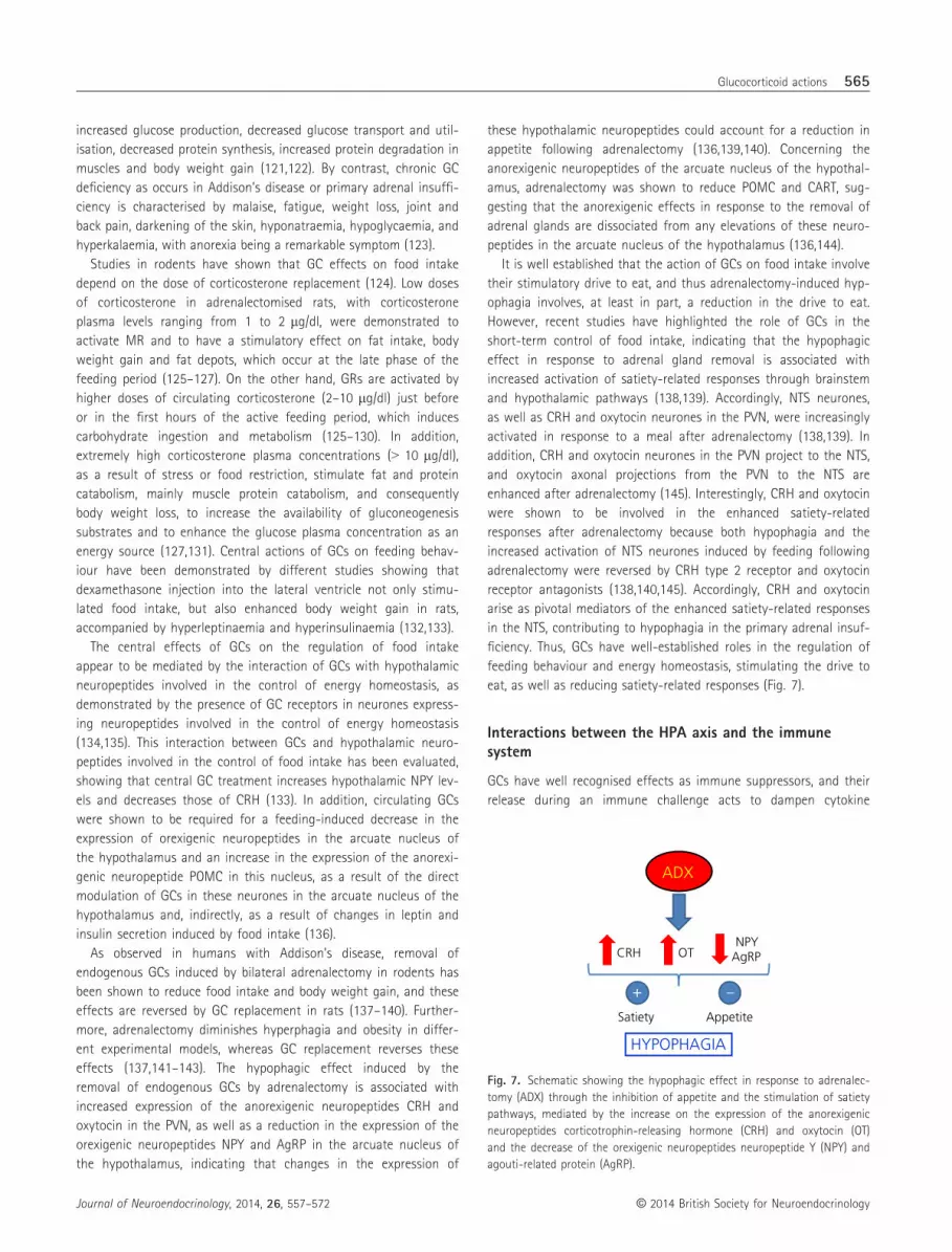
during physiological changes in circulating GCs (22). Restraint
stress in rats causes marked increases in circulating corticosterone
(Fig. 3A), which is associated with transient increases in CRH hnRNA
in the PVN (29,34–36) (Fig. 3B), suggesting that the declining phase
0
1
2
3
4
POM
C h
nRN
A
(fol
d-ch
ange
)
0 –0.5 –18
BasalCRH (30 pM)
Time of corticosterone addition (h)
5Pituitary cell cultures
0
1
2
3
4
5
6
7
8
(fol
d-ch
ange
)C
RH h
nRN
A
0 –0.5 –18Time of corticosterone addition (h)
BasalFSK (3 µM)
Hypothalamic neuronal cultures
******
***
***
#
& &
(A)
(B)
Fig. 1. Effect of corticosterone on corticotrophin-releasing hormone (CRH)-
stimulated pro-opiomelanocortin (POMC) heteronuclear RNA (hnRNA) in pri-
mary cultures of rat anterior pituitary cells (A) and forskolin-stimulated CRH
hnRNA in primary cultures of hypothalamic neurones (B). Three-day cultured
trypsin-dispersed anterior pituitary cells, maintained in stripped serum for
36 h were exposed to 100 nM corticosterone for 18 or 30 min before the
addition of CRH 30 pM for an additional 30 min. Bars represent the mean
and SE of POMC hnRNA levels determined by a quantitative reverse trnas-
criptase-polymerase chain reaction in three cell preparations. In (B), 10-day
cultured foetal rat hypothalamic neuronal cultures were exposed to 100 nM
corticosterone for 18 h or 30 min before addition of forskolin (FSK) for an
additional 45 min before RNA preparation. Data points are the mean and SE
of CRH hnRNA levels, normalised to GAPDH mRNA in four experiments.
***P < 0.001 compared to basal; #P < 0.05 lower than Fsk at 0 min after
log transformation of the data. &P < 0.001 versus CRH at time 0. The hori-
zontal dashed lined represent the SE of maximal stimulated values in the
absence of corticosterone.
© 2014 British Society for NeuroendocrinologyJournal of Neuroendocrinology, 2014, 26, 557–572
Glucocorticoid actions 559
Sham vehicleADX vehicleADX +cort
0
500
1000
1500
2000
2500
3000
3500
Parv
o A
VP
hnRN
A (%
sha
m)
0 5 15
Injection time (min)
30 60 120
**
*
****
**
#### ## ##
**
0
200
400
600
800
1000
1200
1400
CRH
hnR
NA
(% s
ham
)
0 5 15
Injection time (min)
30 60 120
1600
**
*
****
**
**
**
**
****
(A) (B)
Fig. 2. Time course of the changes in corticotrophin-releasing hormone (CRH) heteronuclear RNA (hnRNA) (A) and vasopressin (AVP) hnRNA (B) after injection
of corticosterone (2.8 mg/100 g body weight, i.p.) or vehicle in 48-h adrenalectomised (ADX) or sham-operated rats. Note that vehicle injection caused marked
increases in CRH hnRNA in ADX but not in intact rats. Data points are the mean � SE of the optical density values obtained from in situ hybridisation film
autoradiograms in six rats per experimental group. *P < 0.01 versus sham; **P < 0.001 versus time 0 and sham; ##P < 0.01 lower than ADX vehicle. From
Ma and Aguilera (23).
CRH promoter Per1 promoter
0.5
1.0
1.5
2.0
2.5
3.0
IP G
R (f
old-
chan
ge)
**
**
Time (h)0.0 0.25 0.5 0.75 1.0
GR ChIP
Restraint
Cor
ticos
tero
ne (n
g/m
l)
0
200
400
600
800
Time (h)0.0 0.25 0.5 0.75 1.0
***
Plasma corticosterone
Restraint
3
0
Time (h)0.0 0.25 0.5 0.75 1.0
IP p
CRE
B (f
old-
chan
ge)
1
2*
Phospho-CREB ChIP
Restraint
CRH
hnR
NA
(OD
)
0
5
10
15
20 0 0.5 1
CRH hnRNA
25
Time (h)0.0 0.25 0.5 0.75 1.0
***
(h)
Restraint
(A) (B)
(C) (D)
Fig. 3. Effect of restraint stress on plasma corticosterone, corticotrophin-releasing hormone (CRH) heteronuclear RNA (hnRNA) levels in the paraventricular
nucleus (PVN) (B) and GC receptor recruitment by the CRH and Per1 promoters in intact male rats. Restraint stress caused marked increases in plasma cortico-
sterone (A), associated with transient increases in CRH hnRNA in the PVN (B). CRH hnRNA was measured by in situ hybridisation expressed as optical density
(OD) of film autoradiographs (representative images are displayed at the top of data points showing the pooled values in six rats). Chromatin immunoprecipi-
tation (ChIP) of microdissected hypothalamic PVN region using anti-glucocorticoid receptor (GR) antibody (IP GR) shows no association of GR with the CRH
promoter (solid line and circles) but marked immunoprecipitation of Per1 promoter (dashed lines open circles) by GR antibodies (C). Immunoprecipitation with
phospho-CREB antibody (IP pCREB) yielded high CRH but not Per1 promoter (D). Measurements were performed under basal conditions (time 0), 0.5 and 1 h
during stress. Data points are the mean � SE of the results of three experiments (using pooled hypothalamic tissue from three rats per experimental group).
The dashed lines correspond to the Per1 promoter, and solid lines show different regions of the CRH promoter. The restraint stress period is shown by the
horizontal boxes above the x-axis. ***P < 0.001 versus respective basal; **P < 0.01 versus respective basal; *P < 0.05 versus respective basal.
© 2014 British Society for Neuroendocrinology Journal of Neuroendocrinology, 2014, 26, 557–572
560 E. T. Uchoa et al.

of transcription is a result of repression by elevated GCs. However,
chromatin immunoprecipitation assays failed to identify changes in
CRH promoter in chromatin pulled down by a GR antibody cocktail,
at the same time as detecting marked increases in period 1 (Per1)
promoter, a recognised GC dependent gene (22) (Fig. 3C). By con-
trast, immunoprecipitation with phospho-CREB antibody yielded the
expected increases in CRH promoter at 30 min during restraint
stress (22) (Fig. 3D). A similar lack of change in CRH promoter
immunoprecipitation was observed using quantitative reverse trans-
criptase-polymerase chain reaction (qRT-PCR) primers targeting
regions up to 2 kb upstream from the transcription start site (22).
In the same study, immunoprecipitation of hypothalamic chromatin
from intact or adrenalectomised rats subjected to corticosterone
injections showed minor association of the proximal CRH promoter
with GR compared to phospho-CREB (22). The above data suggest
that repression of CRH transcription by GCs is not mediated by
interaction of GR with the proximal CRH promoter. However, a pos-
sible interaction of GR with another protein in the transcriptional
complex cannot be ruled out because a short-arm cross-linker,
formaldehyde, was used to stabilise GR–DNA interactions before
sonication and immunoprecipitation. Also, interaction of GR with
sites upstream of the 2000 bp that was scanned could lead to
changes in chromatin configuration and interaction with the CRE
region in the proximal promoter, as has been described for a num-
ber of genes (37–39).
CRH transcription depends upon activation and binding to the
CRH promoter CRE of phospho-CREB and its co-activator, transducer
of regulated CREB activity (TORC) (40,41), and it is also possible that
GCs interfere with the activation and binding of these proteins to the
CRH promoter. In this regard, GC administration to rats or mice abol-
ishes stress-induced increases in phospho-CREB immunoreactivity in
the PVN and prevents nuclear translocation of TORC (42). However,
this effect could reflect the inhibition of afferent pathways to the
PVN, rather than direct effects on the CRH neurone, because several
studies show that GCs do not inhibit nuclear accumulation of phos-
pho-CREB in vitro (22,43,44). A recent study reported that corticoste-
rone has no effect on the activation and nuclear translocation of
TORC in the hypothalamic cell line 4B (22), suggesting that the inhibi-
tion of TORC translocation by GCs reported in mice depends on mod-
ulation of afferent inputs to the PVN neurone.
Although it is generally assumed that GCs repress CRH transcrip-
tion by interacting with the putative negative GC response element
in the proximal promoter, accumulating evidence suggests that
such an interaction does not occur during physiological elevations
of the natural GC, corticosterone. The minor effects of GCs on CRH
transcription compared to the marked transcriptional repressor
activity on AVP in parvocellular neurones and pituitary POMC
strongly suggest that the major mechanism by which GCs repress
CRH transcription is indirect, as a result of the modulation of neu-
ral pathways controlling CRH neurone function.
Novel mechanisms of GC feedback signalling
As already noted, the HPA axis is reliably activated by psychogenic or
systemic stressors, and the largely catabolic actions of high GC levels
mandate mechanisms to limit overexposure. This is accomplished by
negative-feedback inhibition of hormone release, which is regulated
in large part by the GR (as a result of its binding capacity). Whereas
the genomic effects of GCs contribute to long-term inhibition of
ACTH release (so-called ‘delayed feedback’), the minute-to-minute
control needed to constrain HPA axis function is likely to require
nongenomic mechanisms. Consequently, GC feedback appears to be
modulated by multiple processes that converge to limit activation of
the HPA axis by inhibition of PVN neurones driving ACTH release.
Here, we discuss three recently-delineated feedback processes that
highlight the richness of the biology of GC signalling, including rapid
PVN feedback mediated by retrograde messengers, a brainstem feed-
back pathway that appears to involve modulation of RNA stability,
and peripheral mechanisms that capitalise on the interaction of GCs
with metabolic effector pathways (Fig. 4).
GC fast feedback
Rapid effects of GCs were observed as early as the 1960s, primarily
in the form of rapid feedback inhibition of GC release following
stress (45). Rapid GC feedback inhibition is nongenomic in nature,
and occurs at time delays consistent with possible membrane
actions. Work by Tasker and colleagues has demonstrated that GCs
rapidly inhibit PVN by way of a membrane-associated receptor
(46,47). The mechanism of rapid inhibitory action is accomplished
by postsynaptic G-protein-mediated release of endocannabinoids
(46). Anandamide and 2-arachidonoylglycerol are rapidly synthes-
ised following GC exposure or acute stress in the PVN, and act as
GLP-1
FFAs
Mechanism 1
Mechanism 2
Glu
ECs
GCsGCs
GCsEnhanced adipocyte
lipolysis
Mechanism 3
Fig. 4. Proposed ‘nontraditional’ glucocorticoid (GC) feedback mechanisms.
Mechanism 1: GCs act via nongenomic mechanisms to inhibit paraventricu-
lar nucleus corticotrophin-releasing hormone (CRH) neurones, acting via
membrane GC receptors (pentagon) to mobilise endocannabinoids (ECs),
which bind to CB1 receptors and inhibit presaynaptic glutamate release.
Mechanism 2: GCs act to destabilise preproglucagon mRNA, reducing the
magnitude of glucacon-like peptide 1 (GLP-1) excitation of CRH neurones.
Mechanism 3: GCs act in the periphery (possibly at adipocytes) to generate
inhibitory messengers, such as free fatty acids (FFAs), which can inhibit
hypothalamic-pituitary-adrenal (HPA) axis activation secondarily.
© 2014 British Society for NeuroendocrinologyJournal of Neuroendocrinology, 2014, 26, 557–572
Glucocorticoid actions 561

retrograde messengers by binding to presynaptic CB1 receptors
(46). A role for endocannabinoids in GC inhibition has been demon-
strated in vitro and in vivo because CB1 antagonism or knockout
(KO) leads to elevated CRH expression in the PVN and increased
plasma ACTH and corticosterone (48–51). Furthermore, bilateral PVN
injections of dexamethasone and a CB1 inverse agonist block the
suppression of HPA axis responses to acute stress seen following
dexamethasone administration alone (52).
Although the receptor mechanism mediating GC signals at the
membrane has not been completely worked out, most of the evi-
dence suggests that the effects are associated with the ‘classical’
GR molecule. Pre-administration of the GR antagonist mifepristone
is not sufficient to completely block the inhibitory effects of dexa-
methasone in vitro (46). However, rapid GC feedback is blocked in
slices from mice with targeted deletion of the GR in PVN neurones
(generated by breeding transgenic mice with cre recombinase
expression driven by the simple-minded-1 promoter with mice engi-
neered to have exon 2 of the GR gene flanked by loxP sequences)
(53). Moreover, GR can be localised to neuronal postsynaptic mem-
branes, supporting membrane localisation of the so-called ‘nuclear’
receptor (54–57). Finally, PVN administration of dexamethasone-
bovine serum albumin conjugate, which should not cross the cell
membrane, inhibits stress-induced HPA axis activation in vivo (52).
Intermediate time-frame: rapid GC inhibition of ascendingstress effector pathways
The medial parvocellular PVN receives synaptic innervation from
ascending stress-regulatory neurones in the caudal medulla and
locus coeruleus (58,59). Most of the innervation of the CRH-con-
taining subregion of the PVN comes from the NTS (58,59). There is
a strong body of evidence suggesting that NTS norepinephrine–
epinephrine neurones are involved in excitation of PVN neurones
controlling HPA axis responses to stress. Stress causes norepinephrine
release in the PVN (60) and local norepinephrine administration is
sufficient to cause ACTH release and enhance the expression of crh
gene transcription (61,62). Moreover, local a-adrenergic receptor
blockade reduces stress-induced HPA axis activation (61). Recent
data suggest that HPA axis activation is also mediated by noncate-
cholaminergic projections from the NTS, emanating from neurones
expressing glucagon-like peptide 1 (GLP-1). These neurones send
GLP-1ergic projections to CRH neurones (63,64). Blockade of GLP-1
receptors reduces ACTH and corticosterone responses to acute
physiological or psychogenic stressors, and local infusion of GLP-1
into the PVN causes the release of corticosterone (65), emphasising
the need for GLP-1 in acute stress reactivity. Moreover, central
infusion of a GLP-1 receptor antagonist reduces the impact of
chronic stress exposure on HPA axis end points (66). Taken
together, the data suggest a prominent role for GLP-1 in stress
excitation at the level of the PVN.
Recent data suggest that the GLP-1 system is also a target for
GC feedback. Experiments were performed to assess the regulation
of NTS stress-excitatory pathways following exposure to acute stress
regimens. Surprisingly, a very rapid depletion of preproglucagon
(PPG; the GLP-1 precursor protein) mRNA is observed following
acute stress, with mRNA levels falling to less than 50% of basal
values within 20–30 min of stress onset (67). This rapid down-regu-
lation is mimicked by exogenous corticosterone administration.
Moreover, stress-induced down-regulation is blocked in adrenalecto-
mised animals clamped at basal circulating corticosterone levels
(67), indicating that PPG down-regulation was mediated by stress-
related GC release. Importantly, PPG hnRNA was substantially
increased by acute stress exposure, suggesting that changes in PPG
mRNA were not a result of decreased transcription. Reductions in
PPG mRNA are relevant at the level of the synapse because stress
causes a rapid decrement in PVN GLP-1 immunoreactivity (67), per-
haps as a consequence of synaptic release associated with the stres-
sor. Given the rapid timing of the stress-induced PPG decrement, we
hypothesise that the effects of stress are mediated by rapid, pre-
sumably nongenomic effects of GCs on PPG mRNA stability. GCs are
known to function in this capacity in cell-based systems (68,69)
and, in combination with the data reported above, suggest that GC-
mediated modulation of mRNA stability may participate in feedback
regulation of the HPA axis function at the level of the NTS.
The observed time-frame of gene turn off and loss of GLP-1
immunoreactivity implies a translational ‘pause’ that will not affect
the immediate release of neuropeptide, although it reduces PVN
excitability subsequently. Consequently, the excitatory impact of
GLP-1 on HPA axis activation will be reduced as a consequence of
down-regulation during the period of depletion of peptide in termi-
nals, approximately 30–60 min after stress (Fig. 5), predicting that
the HPA axis may be less ‘excitable’ during the immediate post-
stress period. This is indeed the case because numerous studies
report that imposition of a second stress 5–60 min after an initial
stressor causes a marked reduction in HPA axis responsiveness. The
stress-refractory period can also be observed if the initial stressor is
replaced with a bolus injection of GCs, suggesting that the inhibition
is GC-driven [referred to as ‘intermediate feedback’ by Keller-Wood
200
150mRNA hnRNA Peptide
50
100
–50
0
Time (post-stress)
0 20 40 60 80 100 120
% c
hang
e (r
elat
ive
to b
asel
ine)
–100
Fig. 5. Schematic comparing the time course of preproglucagon (PPG)
mRNA degradation (PPG mRNA) with PPG transcription (PPG mRNA) and
loss of paraventricular nucleus GLP-1 immunoreactive terminals (peptide).
Note that transcriptional effects do not correspond with a loss of mRNA,
suggesting the mRNA and peptide loss are likely linked to mRNA degrada-
tion or turnover. hnRNA, heteronuclear RNA.
© 2014 British Society for Neuroendocrinology Journal of Neuroendocrinology, 2014, 26, 557–572
562 E. T. Uchoa et al.

and Dallman (70)]. Our data suggest that at least part of the inter-
mediate feedback effect may be mediated by temporary destabilisa-
tion of PPG mRNA by stress-induced GC release.
Peripheral GC signalling and stress regulation
In addition to the actions of GCs in brain, it is important to consider
that GC receptors are abundantly expressed in the periphery, and
may exert indirect effects on the HPA axis via ascending vagal affer-
ents or hormonal messengers. Work by Laugero et al. (71) has dem-
onstrated that the pronounced HPA axis activation seen following
adrenalectomy (e.g. increased PVN CRH synthesis) could be reversed
by allowing the animals to drink sucrose, suggesting that GCs may
inhibit the HPA axis by modulating peripheral metabolic signals. These
data are supported by studies showing that central sucrose adminis-
tration is not able to reduce adrenalectomy-induced activation of the
HPA axis, implying a peripheral mechanism of action (72). Conversely,
increasing energy stores (in the form of fat depots) by voluntary
intake of a high-fat diet reduces HPA axis responses to stress, accom-
panied by elevated insulin secretion (73). The data suggest that GCs
act in the periphery to promote glucose and insulin release, which is
relayed to the hypothalamus to inhibit PVN activation.
Recent studies suggest the involvement of adipose-derived sig-
nals. Specific deletion of the GR in adipocytes enhances GC
responses to stress and attenuates GC feedback inhibition of the
HPA axis (de Kloet et al., unpublished observations). GCs increase
adipocyte lipolysis by activation of hormone sensitive lipase, which
increases circulating free fatty acids (74). Depletion of free fatty
acids, in particular palmitic acid (75,76), causes elevated plasma
ACTH and corticosterone, suggesting a role with respect to con-
straining the HPA axis. One of the major sensors of free fatty acids
and lipid messengers, peroxisome-proliferator activated receptor
gamma (PPARc), is localised to PVN neurones (77). Treatment with
a PPARc agonist rosiglitazone inhibits stress-induced PVN Fos
induction and corticosterone release (78), whereas direct infusion of
the PPARc antagonist GW9662 into the PVN increases ACTH
release. These data suggest that FFAs may provide a blood-borne
inhibitory signal at the level of the PVN, and are consistent with
GC-mediated increases in lipolysis in adipocytes.
GC feedback: a distributed process
The data reported above highlight the richness of the GC feedback
process, identifying a number of check-points whereby secretion
can be limited. These studies complement a rich literature citing
trans-synaptic feedback inhibition from neural sources, such as the
prefrontal cortex and hippocampus (79); genomic and nongenomic
actions at the level of the pituitary (80); and rich interactions with
the autonomic nervous system that can alter secretory profile at
the level of the adrenal (81).
GCs, stress and neuroplasticity
GCs in conjunction with a myriad of chemical mediators released
during stress response modify several aspects of brain function,
including learning and memory formation (82). Major brain areas
targeted by GCs and other stress hormones include the hippocam-
pal formation (mediating declarative memory), amygdala (mediating
fear response) and prefrontal cortex (mediating working memory)
(83). Morphological and biochemical studies have found that nor-
mal circadian or stress-induced variations in circulating GCs pro-
mote neuroplasticity in these structures, especially in the
hippocampus, with modifications of neurone morphology and
changes in neuronal excitability and synaptic efficacy (83).
In the hippocampus, GCs regulate neuronal turnover through
effects on both cell death, as well as proliferation of neural pro-
genitors. Adrenalectomy induces apoptosis of mature granule cells
of the hippocampal dentate gyrus, which is prevented by cortico-
sterone or MR agonist administration (84–88). The trophic influence
of adrenal steroids on granular neurone survival appears to be
related to the regulation of intrinsic apoptotic signalling pathways
(89). Evidence indicates that adrenalectomy increases mRNA
expression of the proapoptotic gene bax (89) and promotes the
activation of cysteine protease caspase-9 (90). This enzyme pro-
motes the proteolytic cleavage of executor caspases that subse-
quently destroy several proteins, culminating in apoptosis. In
addition, adrenalectomy induces an increase in the rate of prolifer-
ation of progenitor cells in the subgranular zone of the dentate
gyrus, an effect that is prevented by corticosterone administration
(91). On the other hand, the increase in GC secretion induced by
acute stress can act as a positive or negative modulator of learn-
ing, memory and retrieval (92). Several studies have suggested that
acute stress is associated with increased excitatory glutamatergic
neurotransmission in areas of the forebrain (93–95). Some studies
have demonstrated that adrenalectomy attenuates the stress-
induced release of glutamate in the hippocampus and prefrontal
cortex, suggesting a direct relationship between GCs and glutamate
release (96). Moreover, intrahippocampal perfusion of corticoste-
rone by retrodialysis produces a fast and reversible increase in the
release of glutamate in vivo (97). In addition, electrophysiological
studies have shown that application of corticosterone to hippo-
campal slices increases the frequency of excitatory postsynaptic
potentials in the CA1 area of the hippocampus, suggesting a fast
action of corticosteroids on glutamate release (98). This fast action
of corticosterone is likely to be nongenomic and appears to involve
the MR (99).
GC fluctuations can also modulate the learning processes
through modifications of the postsynaptic elements that receive
the majority of excitatory glutamatergic inputs in the CNS. These
postsynaptic elements are named spines, which are small actin-rich
protrusions formed by a head that is connected to the dendrite
shaft by a neck. Circadian GC peaks induce the formation and
development of dendritic spines in the mouse cortex after motor
skill learning and the circadian GC troughs are required for the
stabilisation of new spines (100). Furthermore, the improved asso-
ciative learning promoted by acute stress is accompanied by a rise
in the spine density of hippocampal neurones (101). In line with
these observations, brief exposure of hippocampal slices to dexa-
methasone, a GR agonist (55), or corticosterone (100–1000 nM)
(102) promotes an increase in spine density in pyramidal neurones
© 2014 British Society for NeuroendocrinologyJournal of Neuroendocrinology, 2014, 26, 557–572
Glucocorticoid actions 563

of the CA1 hippocampal area. Furthermore, the co-administration
of RU486, an antagonist of GR, abolished the effect of corticoste-
rone (102). It has been proposed that spinogenesis is modulated
by synaptic GRs and kinases, including protein kinase A, protein
kinase C (102) and extracellular signal-regulated protein kinases 1
and 2 (103). Although GRs have been localised to neuronal cell
bodies and dendrites, a recent study has shown that GRs are
localised to dendritic spine heads and to spine necks of CA1 pyra-
midal cells (103). Additionally, a recent study in KO mice for fragile
X mental retardation protein (FMRP) demonstrated a reduction in
GR levels in CA1 dendrites (104). FMRP acts as a protein that
transports a subset of neuronal mRNAs from the nucleus into
dendrites and spines (104,105). Furthermore, the bulk of FMRP is
associated with polyribosomes and represses the translation of
various mRNAs (104,105). In accordance with these findings, it has
been shown that spine GR levels increase rapidly by metabotropic
glutamate receptor (mGluR) activation, an effect not observed in
KO mice for FMRP (103). Thus, it has been proposed that synaptic
levels of GR in CA1 spines are regulated by local GR mRNA trans-
lation involving mGluR activity and a FMRP-dependent mechanism
(103).
It appears that GR binding also affects glutamatergic receptor
levels. It was shown that GR activation increases the surface
expression of a-amino-3-hydroxy-5-methyl-4-isoxazolepropionicacid glutamate receptor subunit 2 in primary hippocampal cul-
tures (106). This GC-induced increase in GluA2 subunit of AMPA
receptors at the hippocampal synaptic sites is involved in the
facilitation of learning during acute stress (107). The variation in
GluA2 levels is probably related to receptor trafficking mecha-
nisms rather than changes in GluA2 mRNAs levels and/or transla-
tion (107).
The mRNA encoding the immediate early gene, activity regu-
lated cytoskeletal-associated protein (Arc), is increased by activa-
tion of N-methyl-D-aspartate receptor. Arc mRNA traffics to
dendrites and its translation is controlled by FMRP and the activa-
tion of mGluR (108). Reduction of Arc protein levels in the hippo-
campus blocks both late-phase long-term potentiation and the
consolidation of some forms of memory (109). Furthermore, it has
been reported that Arc reduces the surface expression of GluA2/A3
receptors, promoting a decrease in AMPA receptor-mediated syn-
aptic currents (110). These findings suggest that Arc protein stabi-
lises the internal pool of AMPA receptors, which would
presumably increase or decrease the levels of these receptors in
synaptic sites depending on the initial stimulus (111). Because
activation of GRs produce variation in the synaptic levels of GluA2
(107), the effect of acute restraint stress on Arc protein levels was
evaluated. Figure 6 shows that 0.5 h of restraint stress promotes
an increase of Arc protein levels in the hippocampus, suggesting a
fast Arc mRNA translation. Thus, it is plausible that stress-induced
corticosterone secretion mediates mGluR activation, increasing Arc
mRNA translation, which in turn may regulate the surface expres-
sion of AMPA receptors. Overall, these findings suggest that stress
and GCs may modulate excitatory neurotransmission via increased
glutamate release, modified spine density and AMPA receptor traf-
ficking.
GCs in the control of food intake
Food intake is a basic behaviour regulated by multiple factors,
including the adiposity signals leptin and insulin, and satiety signals,
such as mechanical and chemical stimulation of the stomach and
small intestine, as well as hormones released during a meal, such as
cholecystokinin (112). The adiposity factors are involved in the long-
term control of energy balance and act primarily in hypothalamic
neurones expressing orexigenic or anorexigenic neuropeptides, which
are key mediators in the control of energy homeostasis (112). Neu-
ropeptide Y (NPY) and agouti-related protein (AgRP) in the arcuate
nucleus of the hypothalamus, and orexins and melanin-concentrat-
ing hormone in the lateral hypothalamic area comprise important
hypothalamic orexigenic neuropeptides (113–115). On the other
hand, POMC and cocaine- and amphetamine-regulated transcript
(CART) in the arcuate nucleus of the hypothalamus, as well as CRH
and oxytocin in the PVN, represent the main hypothalamic media-
tors involved in the inhibition of food intake (112,115). The satiety
signals, in turn, are implicated in the short-term control of food
intake and have their actions mediated by brainstem areas, mainly
the NTS, controlling the size of a meal (116).
In addition to their important role regulating metabolic activity in
the periphery, there are profound interactions between GCs and
appetite control (117). Feeding is a major synchroniser of HPA axis
rhythmicity (118), and the amount of food ingested is associated
with GC secretion (119). Reciprocally, increased food intake and body
weight gain in humans are associated with increases in circulating
GCs associated with the therapeutic administration of GCs or Cush-
ing’s disease (120). Excess GC in these cases is associated with
2.0*
2.5 18 24Time (h)
0
2.5
24–
0.5 2.50
0 0
1.5
1.0
STRESS0.5
ARC
/β-a
ctin
β-actin
Stress (h)
Post-Stress (h)
ARC
Fig. 6. Effect of acute restraint stress on the Arc (activity-regulated cyto-
skeleton-associated protein) protein levels in the hippocampus of rats. Acute
restraint stress induces an increase of Arc protein levels in rat hippocampus.
Sprague–Dawley male rats were restrained for 0.5 h or 2.5 h and immedi-
ately sacrificed or restrained for 2.5 h and sacrificed 24 h after the restraint.
(A) Representative immunoblots of homogenates from the hippocampus of
stressed rats using anti-Arc and b-actin antibodies. (B) Graph shows the
relative ratio of Arc levels relative to b-actin. Data represent the mean � SD
of n = 4 per experimental condition. *P < 0.05 compared to nonstressed
animals (i.e. time = 0).
© 2014 British Society for Neuroendocrinology Journal of Neuroendocrinology, 2014, 26, 557–572
564 E. T. Uchoa et al.
increased glucose production, decreased glucose transport and util-
isation, decreased protein synthesis, increased protein degradation in
muscles and body weight gain (121,122). By contrast, chronic GC
deficiency as occurs in Addison’s disease or primary adrenal insuffi-
ciency is characterised by malaise, fatigue, weight loss, joint and
back pain, darkening of the skin, hyponatraemia, hypoglycaemia, and
hyperkalaemia, with anorexia being a remarkable symptom (123).
Studies in rodents have shown that GC effects on food intake
depend on the dose of corticosterone replacement (124). Low doses
of corticosterone in adrenalectomised rats, with corticosterone
plasma levels ranging from 1 to 2 lg/dl, were demonstrated to
activate MR and to have a stimulatory effect on fat intake, body
weight gain and fat depots, which occur at the late phase of the
feeding period (125–127). On the other hand, GRs are activated by
higher doses of circulating corticosterone (2–10 lg/dl) just before
or in the first hours of the active feeding period, which induces
carbohydrate ingestion and metabolism (125–130). In addition,
extremely high corticosterone plasma concentrations (> 10 lg/dl),as a result of stress or food restriction, stimulate fat and protein
catabolism, mainly muscle protein catabolism, and consequently
body weight loss, to increase the availability of gluconeogenesis
substrates and to enhance the glucose plasma concentration as an
energy source (127,131). Central actions of GCs on feeding behav-
iour have been demonstrated by different studies showing that
dexamethasone injection into the lateral ventricle not only stimu-
lated food intake, but also enhanced body weight gain in rats,
accompanied by hyperleptinaemia and hyperinsulinaemia (132,133).
The central effects of GCs on the regulation of food intake
appear to be mediated by the interaction of GCs with hypothalamic
neuropeptides involved in the control of energy homeostasis, as
demonstrated by the presence of GC receptors in neurones express-
ing neuropeptides involved in the control of energy homeostasis
(134,135). This interaction between GCs and hypothalamic neuro-
peptides involved in the control of food intake has been evaluated,
showing that central GC treatment increases hypothalamic NPY lev-
els and decreases those of CRH (133). In addition, circulating GCs
were shown to be required for a feeding-induced decrease in the
expression of orexigenic neuropeptides in the arcuate nucleus of
the hypothalamus and an increase in the expression of the anorexi-
genic neuropeptide POMC in this nucleus, as a result of the direct
modulation of GCs in these neurones in the arcuate nucleus of the
hypothalamus and, indirectly, as a result of changes in leptin and
insulin secretion induced by food intake (136).
As observed in humans with Addison’s disease, removal of
endogenous GCs induced by bilateral adrenalectomy in rodents has
been shown to reduce food intake and body weight gain, and these
effects are reversed by GC replacement in rats (137–140). Further-
more, adrenalectomy diminishes hyperphagia and obesity in differ-
ent experimental models, whereas GC replacement reverses these
effects (137,141–143). The hypophagic effect induced by the
removal of endogenous GCs by adrenalectomy is associated with
increased expression of the anorexigenic neuropeptides CRH and
oxytocin in the PVN, as well as a reduction in the expression of the
orexigenic neuropeptides NPY and AgRP in the arcuate nucleus of
the hypothalamus, indicating that changes in the expression of
these hypothalamic neuropeptides could account for a reduction in
appetite following adrenalectomy (136,139,140). Concerning the
anorexigenic neuropeptides of the arcuate nucleus of the hypothal-
amus, adrenalectomy was shown to reduce POMC and CART, sug-
gesting that the anorexigenic effects in response to the removal of
adrenal glands are dissociated from any elevations of these neuro-
peptides in the arcuate nucleus of the hypothalamus (136,144).
It is well established that the action of GCs on food intake involve
their stimulatory drive to eat, and thus adrenalectomy-induced hyp-
ophagia involves, at least in part, a reduction in the drive to eat.
However, recent studies have highlighted the role of GCs in the
short-term control of food intake, indicating that the hypophagic
effect in response to adrenal gland removal is associated with
increased activation of satiety-related responses through brainstem
and hypothalamic pathways (138,139). Accordingly, NTS neurones,
as well as CRH and oxytocin neurones in the PVN, were increasingly
activated in response to a meal after adrenalectomy (138,139). In
addition, CRH and oxytocin neurones in the PVN project to the NTS,
and oxytocin axonal projections from the PVN to the NTS are
enhanced after adrenalectomy (145). Interestingly, CRH and oxytocin
were shown to be involved in the enhanced satiety-related
responses after adrenalectomy because both hypophagia and the
increased activation of NTS neurones induced by feeding following
adrenalectomy were reversed by CRH type 2 receptor and oxytocin
receptor antagonists (138,140,145). Accordingly, CRH and oxytocin
arise as pivotal mediators of the enhanced satiety-related responses
in the NTS, contributing to hypophagia in the primary adrenal insuf-
ficiency. Thus, GCs have well-established roles in the regulation of
feeding behaviour and energy homeostasis, stimulating the drive to
eat, as well as reducing satiety-related responses (Fig. 7).
Interactions between the HPA axis and the immunesystem
GCs have well recognised effects as immune suppressors, and their
release during an immune challenge acts to dampen cytokine
ADX
HYPOPHAGIA
Satiety Appetite
CRH OTNPY
AgRP
+ –
Fig. 7. Schematic showing the hypophagic effect in response to adrenalec-
tomy (ADX) through the inhibition of appetite and the stimulation of satiety
pathways, mediated by the increase on the expression of the anorexigenic
neuropeptides corticotrophin-releasing hormone (CRH) and oxytocin (OT)
and the decrease of the orexigenic neuropeptides neuropeptide Y (NPY) and
agouti-related protein (AgRP).
© 2014 British Society for NeuroendocrinologyJournal of Neuroendocrinology, 2014, 26, 557–572
Glucocorticoid actions 565

production and inflammatory responses. However, the HPA axis and
immune system show complex bidirectional interactions not only
under immune challenges, but also during exposure to non-immune
stressors. A growing body of literature suggests that stress signifi-
cantly impacts many facets of neuroimmune function. For example,
exposure to a variety of acute stress challenges increased the
expression of interleukin-1b (IL-1b) in the hypothalamus (146–148),
whereas increased prostaglandin activity has been observed
throughout the cortex in response to acute stress (149). Converging
lines of evidence suggest that these changes in neuroimmune sig-
nalling factors (cytokines, chemokines, prostaglandins, etc.) are
associated with other tell-tale signs of neuroinflammation, such as
priming (150), proliferation (151) and activation (152,153) of micro-
glia. Consistently, injection of the putative microglial inhibitor mi-
nocycline blocked cytokine expression evoked by stress (152,154).
Thus, a rich constellation of neuroimmune changes occur in
response to stress, which raises several critical issues, including: (i)
how do hormones classically associated with stress (nor/epinephrine
and GCs, in particular) influence cytokine expression and other
aspects of neuroimmune function; (ii) to what extent do neuroim-
mune signalling pathways serve as either moderators or mediators
of HPA axis output across natural diurnal rhythms and/or in
response to later stress challenges; and (iii) under what circum-
stances do neuroimmune consequences of stress either compromise
or protect against the development of pathological states of the
CNS?
Although the precise mechanisms controlling cytokine expression
during times of stress have not been fully delineated, several key
mechanisms have emerged. Previous work has shown that the
administration of b-adrenergic blockers inhibits or severely blunts
the expression of interleukin-1 evoked by stress, and the adminis-
tration of the b-adrenergic agonist isoproterenol both recapitulates
and potentiates the expression of IL-1 induced by stress
(152,154,155). By contrast to the dependence of central cytokine
responses on b-adrenergic receptor activation, plasma cytokine
responses to stress appear to be selectively mediated by a1-adren-ergic receptors (156). These findings are supported by lesion studies,
where complete lesions of central adrenergic systems incurred by
injection of the neurotoxin DSP4 completely blocked the IL-1
response produced by stress in several brain structures (155),
whereas more targeted lesions of the ventral noradrenergic bundle
only partially attenuated the IL-1 response to stress in the PVN
(156). These data fit well with strain differences in central cytokine
responses to stress demonstrating that hyperadrenergic Fisher 344
rats show much greater increases in central cytokine expression rel-
ative to their less reactive Sprague–Dawley comparators (157).
Indeed, the potentiation of cytokine responses to stress in hyper-
adrenergic, Fisher 344 rats is particularly noteworthy given that this
strain also evinces a potentiated HPA axis response relative to other
strains (158). Thus, norepinephrine appears to be a key driver of
stress-induced cytokine expression (Fig. 8). However, it should be
noted that other transmitter systems such as glutamate (151) and
other intermediaries such as danger associated molecular patterns
(159) may participate in various features of the neuroimmune
response to stressor exposure as well.
GCs, on the other hand, powerfully constrain the expression of
cytokines during times of stress. Evidence to support this is pro-
vided by studies showing that the removal of endogenous GCs via
adrenalectomy (160) and the administration of GC receptor antago-
nists (161) or the GC synthesis inhibitor metyrapone (152) mas-
sively potentiates the expression of cytokines evoked by stress.
These findings fit well with a range of molecular interactions
through which corticosteroid receptors squelch inflammatory sig-
nalling pathways (162–164). Very recently, however, a few studies
have begun to reveal effects that indicate priming-like actions of
GCs as well (165,166). However, it should be noted that these non-
canonical actions may be (i) unique to specific tissue types within
the body; (ii) occur at only low-dose or low-physiological concen-
trations of circulating GCs; or (iii) reflect a short-term, early
response to GC signalling. These intriguing findings suggest that
the relationship between corticosteroid signalling and neuroimmune
consequences of stress will require detailed consideration of
numerous key parameters (tissue, dose, timing, etc.). Nevertheless, it
is clear that in the absence of effective GC signalling, neuroimmune
consequences of stress are quite severe.
Moving beyond mechanisms, neuroimmune consequences of
stress (particularly increased cytokine expression) may play an
important role as moderators of HPA axis activation. For example,
IL-1 receptor 1 KO mice show blunted HPA axis response to mild
but not intense stress challenges (167). Previously stressed rats
showed a faster HPA axis response to later lipopolysaccharide injec-
tion, an effect that was reversed by IL-1 receptor antagonist (168).
Furthermore, it has been noted that dissociations between ACTH
and corticosterone are often observed under inflammatory condi-
tions (169,170), including after immune activation by lipopolysac-
charide (171,172), as well as in inflammatory disease states
(173,174). Thus, cytokines and other immune-related signalling
Stress
CytokinesHPA axis
IL-1, IL-6, TNF
Corticosteroids-- Inhibitory feedback on further cytokine expression
+
+ +
STOP
Fig. 8. Schematic showing the interaction between corticosteroids and
cytokine expression. Stress exposure directly induces the release of hypotha-
lamic-pituitary-adrenal (HPA) axis hormones and increases the expression of
cytokines. Although there is emerging evidence to suggest that corticoster-
oids may enhance cytokine expression under certain circumstances, a pri-
mary effect of corticosteroids is the suppression of cytokine expression.
Cytokines, on the other hand, directly stimulate activation of the HPA axis
via actions that are both intrinsic and extrinsic to the axis, and appear to
augment HPA axis sensitivity to later stress challenges. IL, interleukin; TNF,
tumour necrosis factor.
© 2014 British Society for Neuroendocrinology Journal of Neuroendocrinology, 2014, 26, 557–572
566 E. T. Uchoa et al.

factors have the capacity to induce (through actions extrinsic to
the axis) or augment (through intrinsic actions) HPA axis responses
to later stress challenges.
The impact of neuroimmune consequences of stress is not
restricted to the HPA axis. For example, the neuroimmune conse-
quences of stress appear to control certain behavioural conse-
quences of stress exposure (175), as well as impairments in
cognitive function precipitated by stress (176,177), and may be
involved in certain depressive-like consequences of stress such as
learned helplessness (178). Indeed, stress-related cytokines have
been argued as a potentially critical mechanistic bridge between
stressful experiences and the development of stress-related pathol-
ogy (179–181). Thus, with such far-reaching implications of stress-
related changes in neuroimmune function, the interaction between
classic stress-responsive systems (HPA axis, sympathetic nervous
system) and neuroimmune function provides an important starting
block for gaining a better understanding of the long-term, cumula-
tive impact of stress on animals and humans (180).
Concluding remarks
Important progress has been made during the last few years in
understanding the physiological actions of GCs and the mecha-
nisms of GC feedback regulation of the HPA axis. GCs have pleo-
tropic actions regulating metabolism and brain function and
modulating the function of most cells in the body. Growing evi-
dence indicates that normal GC-dependent regulation depends
upon the prevailing levels of the steroid, secretion pattern, as well
as interaction of GCs with neurotransmitters and neuropeptides.
This is partly illustrated by the concentration-dependent effects of
GCs in hippocampal neuroplasticity and immune function discussed
above. It is also clear that not all the effects of GCs are direct, with
a number of actions depending of changes in other regulatory fac-
tors. For example, the profound modulatory effects of GCs on food
intake largely depend on the opposing effects of GCs on orexigenic
and anorexigenic neuropeptides.
Negative GC feedback is essential for the fine control of HPA axis
activity to avoid deleterious consequences of excessive CRH and GC
production. New evidence indicates that GC feedback inhibits HPA
axis activity at a number of anatomical and molecular targets, rap-
idly shutting-off hypothalamic and pituitary responses at the cell
membrane level, controlling the intensity and duration of stress
responses at limbic sites, and inducing long-term inhibition by
modulating transcription and mRNA stability at the central and
pituitary levels.
Despite recent advances in the field of HPA axis regulation and
GC actions, a number of challenges still remain. This includes the
identification of peripheral metabolic signals and central sensors
impacting on the sensitivity of GC feedback and HPA axis activity,
and the exact identity of the membrane GC receptor and its
signalling mechanisms. Understanding the functions of GCs and
mechanisms of feedback regulation of HPA axis activity will
contribute to the development of new diagnostic and therapeutic
tools for disorders related to stress and alterations of GC
secretion.
Acknowledgements
This study was supported by FAPESP and CNPq to E.T.U.; Intramural
Research Program, National Institute of Child Health and Human Develop-
ment to G.A.; NIH grants MH049698 and MH069860 to J.P.H.; FONDECYT
1120528 to J.L.F.; National Science Foundation (NSF) grant 0822129 to T.D.;
and CNPq (Proc. No. 302592/2009-1) to M.B.C.S.
Received 20 January 2014,
revised 6 April 2014,
accepted 8 April 2014
References
1 Johnson EO, Kamilaris TC, Chrousos GP, Gold PW. Mechanisms of
stress: a dynamic overview of hormonal and behavioral homeostasis.
Neurosci Biobehav Rev 1992; 16: 115–130.
2 Smith SM, Vale WW. The role of the hypothalamic-pituitary-adrenal
axis in neuroendocrine responses to stress. Dialogues Clin Neurosci
2006; 8: 383–395.
3 Lightman SL, Wiles CC, Atkinson HC, Henley DE, Russell GM, Leendertz
JA, McKenna MA, Spiga F, Wood SA, Conway-Campbell BL. The signifi-
cance of glucocorticoid pulsatility. Eur J Pharmacol 2008; 583: 255–
262.
4 Jo€els M, de Kloet ER. Mineralocorticoid and glucocorticoid receptors in
the brain. Implications for ion permeability and transmitter systems.
Prog Neurobiol 1994; 43: 1–36.
5 Baker ME, Funder JW, Kattoula SR. Evolution of hormone selectivity in
glucocorticoid and mineralocorticoid receptors. J Steroid Biochem Mol
Biol 2013; 137: 57–70.
6 Grad I, Picard D. The glucocorticoid responses are shaped by molecular
chaperones. Mol Cell Endocrinol 2007; 275: 2–12.
7 Hill MN, Tasker JG. Endocannabinoid signaling, glucocorticoid-mediated
negative feedback, and regulation of the hypothalamic-pituitary-adre-
nal axis. Neuroscience 2012; 204: 5–16.
8 Evanson NK, Herman JP, Sakai RR, Krause EG. Nongenomic actions of
adrenal steroids in the central nervous system. J Neuroendocrinol
2010; 22: 846–861.
9 Herman JP, McKlveen JM, Solomon MB, Carvalho-Netto E, Myers B.
Neural regulation of the stress response: glucocorticoid feedback
mechanisms. Braz J Med Biol Res 2012; 45: 292–298.
10 Viengchareun S, Le Menuet D, Martinerie L, Munier M, Pascual-Le Tal-
lec L, Lomb�es M. The mineralocorticoid receptor: insights into its
molecular and (patho)physiological biology. Nucl Recept Signal 2007;
5: e012.
11 Berardelli R, Karamouzis I, D’Angelo V, Zichi C, Fussotto B, Giordano R,
Ghigo E, Arvat E. Role of mineralocorticoid receptors on the hypothala-
mus-pituitary-adrenal axis in humans. Endocrine 2013; 43: 51–58.
12 Geerling JC, Loewy AD. Aldosterone in the brain. Am J Physiol Renal
Physiol 2009; 297: F559–F576.
13 Sawchenko PE. Evidence for a local site of action for glucocorticoids in
inhibiting CRF and vasopressin expression in the paraventricular
nucleus. Brain Res 1987; 403: 213–224.
14 Plotsky PM, Otto S, Sapolsky RM. Inhibition of immunoreactive cortico-
tropin-releasing factor secretion into the hypophysial-portal circulation
by delayed glucocorticoid feedback. Endocrinology 1986; 119: 1126–
1130.
15 Plotsky PM, Sawchenko PE. Hypophysial-portal plasma levels, median
eminence content, and immunohistochemical staining of corticotropin-
releasing factor, arginine vasopressin, and oxytocin after pharmacologi-
cal adrenalectomy. Endocrinology 1987; 120: 1361–1369.
© 2014 British Society for NeuroendocrinologyJournal of Neuroendocrinology, 2014, 26, 557–572
Glucocorticoid actions 567

16 Dallman MF, Akana SF, Levin N, Walker CD, Bradbury MJ, Suemaru S,
Scribner KS. Corticosteroids and the control of function in the hypo-
thalamo-pituitary-adrenal (HPA) axis. Ann N Y Acad Sci 1994; 746:
22–28.
17 Gagner JP, Drouin J. Opposite regulation of pro-opiomelanocortin gene
transcription by glucocorticoids and CRH. Mol Cell Endocrinol 1985;
40: 25–32.
18 Eberwine JH, Jonassen JA, Evinger MJ, Roberts JL. Complex transcrip-
tional regulation by glucocorticoids and corticotropin-releasing hor-
mone of proopiomelanocortin gene expression in rat pituitary cultures.
DNA 1987; 6: 483–492.
19 Aguilera G, Liu Y. The molecular physiology of CRH neurons. Front
Neuroendocrinol 2012; 33: 17.
20 Ma XM, Camacho C, Aguilera G. Regulation of corticotropin-releasing
hormone (CRH) transcription and CRH mRNA stability by glucocortic-
oids. Cell Mol Neurobiol 2001; 21: 465–475.
21 Spencer CM, Eberwine J. Cytoplasmic proteins interact with a transla-
tional control element in the protein-coding region of proopiomelano-
cortin mRNA. DNA Cell Biol 1999; 18: 39–49.
22 Evans AN, Liu Y, Macgregor R, Huang V, Aguilera G. Regulation of
hypothalamic corticotropin releasing hormone transcription by elevated
glucocorticoids. Mol Endocrinol 2013; 27: 1796–1807.
23 Ma XM, Aguilera G. Differential regulation of corticotropin-releasing
hormone and vasopressin transcription by glucocorticoids. Endocrinol-
ogy 1999; 140: 5642–5650.
24 Kov�acs KJ, F€oldes A, Sawchenko PE. Glucocorticoid negative feedback
selectively targets vasopressin transcription in parvocellular neurose-
cretory neurons. J Neurosci 2000; 20: 3843–3852.
25 Liposits Z, Uht RM, Harrison RW, Gibbs FP, Paull WK, Bohn MC. Ultra-
structural localization of glucocorticoid receptor (GR) in hypothalamic
paraventricular neurons synthesizing corticotropin releasing factor
(CRF). Histochemistry 1987; 87: 407–412.
26 Fenoglio KA, Brunson KL, Avishai-Eliner S, Chen Y, Baram TZ. Region-
specific onset of handling-induced changes in corticotropin-releasing
factor and glucocorticoid receptor expression. Endocrinology 2004;
145: 2702–2706.
27 Harbuz MS, Lightman SL. Glucocorticoid inhibition of stress-induced
changes in hypothalamic corticotrophin-releasing factor messenger
RNA and proenkephalin A messenger RNA. Neuropeptides 1989; 14:
17–20.
28 Makino S, Smith MA, Gold PW. Increased expression of corticotropin-
releasing hormone and vasopressin messenger ribonucleic acid (mRNA)
in the hypothalamic paraventricular nucleus during repeated stress:
association with reduction in glucocorticoid receptor mRNA levels.
Endocrinology 1995; 136: 3299–3309.
29 Ma XM, Lightman SL, Aguilera G. Vasopressin and corticotropin-releas-
ing hormone gene responses to novel stress in rats adapted to
repeated restraint. Endocrinology 1999; 140: 3623–3632.
30 K�ovacs KJ, Makara GB. Corticosterone and dexamethasone act at dif-
ferent brain sites to inhibit adrenalectomy-induced adrenocorticotropin
hypersecretion. Brain Res 1988; 474: 205–210.
31 Malkoski SP, Handanos CM, Dorin RI. Localization of a negative gluco-
corticoid response element of the human corticotropin releasing hor-
mone gene. Mol Cell Endocrinol 1997; 127: 189–199.
32 Malkoski SP, Dorin RI. Composite glucocorticoid regulation at a func-
tionally defined negative glucocorticoid response element of the
human corticotropin-releasing hormone gene. Mol Endocrinol 1999;
13: 1629–1644.
33 Guardiola-Diaz HM, Kolinske JS, Gates LH, Seasholtz AF. Negative glu-
corticoid regulation of cyclic adenosine 30 , 50-monophosphate-stimu-lated corticotropin-releasing hormone-reporter expression in AtT-20
cells. Mol Endocrinol 1996; 10: 317–329.
34 Ma XM, Levy A, Lightman SL. Rapid changes of heteronuclear RNA for
arginine vasopressin but not for corticotropin releasing hormone in
response to acute corticosterone administration. J Neuroendocrinol
1997; 9: 723–728.
35 Shepard JD, Liu Y, Sassone-Corsi P, Aguilera G. Role of glucocorticoids
and cAMP-mediated repression in limiting corticotropin-releasing hor-
mone transcription during stress. J Neurosci 2005; 25: 4073–4081.
36 Liu Y, Poon V, Sanchez-Watts G, Watts AG, Takemori H, Aguilera G.
Salt-inducible kinase is involved in the regulation of corticotropin-
releasing hormone transcription in hypothalamic neurons in rats.
Endocrinology 2012; 153: 223–233.
37 So AY, Chaivorapol C, Bolton EC, Li H, Yamamoto KR. Determinants of
cell- and gene-specific transcriptional regulation by the glucocorticoid
receptor. PLoS Genet 2007; 3: e94.
38 Hakim O, John S, Ling JQ, Biddie SC, Hoffman AR, Hager GL. Glucocor-
ticoid receptor activation of the Ciz1-Lcn2 locus by long range interac-
tions. J Biol Chem 2009; 284: 6048–6052.
39 Paakinaho V, Makkonen H, J€a€askel€ainen T, Palvimo JJ. Glucocorticoid
receptor activates poised FKBP51 locus through long-distance interac-
tions. Mol Endocrinol 2010; 24: 511–525.
40 Liu Y, Coello AG, Grinevich V, Aguilera G. Involvement of transducer of
regulated cAMP response element-binding protein activity on cortico-
tropin releasing hormone transcription. Endocrinology 2010; 151:
1109–1118.
41 Liu Y, Knobloch HS, Grinevich V, Aguilera G. Stress induces nuclear
translocation of the CREB co-activator, transducer of regulated CREB
activity (TORC) in corticotropin releasing hormone neurons. J Neuroen-
docrinol 2011; 23: 216–223.
42 Jeanneteau FD, Lambert WM, Ismaili N, Bath KG, Lee FS, Garabedian
MJ, Chao MV. BDNF and glucocorticoids regulate corticotrophin-releas-
ing hormone (CRH) homeostasis in the hypothalamus. Proc Natl Acad
Sci 2012; 109: 1305–1310.
43 Adler GK, Smas CM, Majzoub JA. Expression and dexamethasone
regulation of the human corticotropin-releasing hormone gene in a
mouse anterior pituitary cell line. J Biol Chem 1988; 263: 5846–
5852.
44 Kageyama K, Akimoto K, Suda T. Corticotrophin-releasing factor gene
transcription is directly activated after deprivation of glucocorticoids in
hypothalamic cells. J Neuroendocrinol 2010; 22: 971–978.
45 Dallman MF, Yates FE. Dynamic asymmetries in the corticosteroid feed-
back path and distribution-metabolism-binding elements of the adre-
nocortical system. Ann N Y Acad Sci 1969; 156: 696–721.
46 Di S, Malcher-Lopes R, Halmos KC, Tasker JG. Nongenomic glucocorti-
coid inhibition via endocannabinoid release in the hypothalamus: a fast
feedback mechanism. J Neurosci 2003; 23: 4850–4857.
47 Malcher-Lopes R, Di S, Marcheselli VS, Weng FJ, Stuart CT, Bazan NG,
Tasker JG. Opposing crosstalk between leptin and glucocorticoids rap-
idly modulates synaptic excitation via endocannabinoid release. J Neu-
rosci 2006; 26: 6643–6650.
48 Patel S, Roelke CT, Rademacher DJ, Cullinan WE, Hillard CJ. Endocanna-
binoid signaling negatively modulates stress-induced activation of the
hypothalamic-pituitary-adrenal axis. Endocrinology 2004; 145: 5431–
5438.
49 Cota D, Steiner MA, Marsicano G, Cervino C, Herman JP, Grubler Y,
Stalla J, Pasquali R, Lutz B, Stalla GK, Pagotto U. Requirement of
cannabinoid receptor type 1 for the basal modulation of hypotha-
lamic-pituitary-adrenal axis function. Endocrinology 2007; 148:
1574–1581.
50 Ginsberg AB, Pecoraro NC, Warne JP, Horneman HF, Dallman MF. Rapid
alteration of stress-induced hypothalamic-pituitary-adrenal hormone
secretion in the rat: a comparison of glucocorticoids and cannabinoids.
Stress 2010; 13: 248–257.
© 2014 British Society for Neuroendocrinology Journal of Neuroendocrinology, 2014, 26, 557–572
568 E. T. Uchoa et al.

51 Hill MN, McEwen BS. Involvement of the endocannabinoid system in
the neurobehavioural effects of stress and glucocorticoids. Prog Neuro-
psychopharmacol Biol Psychiatry 2010; 34: 791–797.
52 Evanson NK, Tasker JG, Hill MN, Hillard CJ, Herman JP. Fast feedback
inhibition of the HPA axis by glucocorticoids is mediated by endocann-
abinoid signaling. Endocrinology 2010; 151: 4811–4819.
53 Hamm J, Halmos KC, Muglia LJ, Tasker JG. Rapid synaptic modulation
of hypothalamic neurons by glucocorticoids requires the glucocorticoid
receptor. 2010 Neuroscience Meeting Planner. San Diego, CA: Society
for Neuroscience, 2010; Program No. 389.19.
54 Johnson LR, Farb C, Morrison JH, McEwen BS, LeDoux JE. Localization
of glucocorticoid receptors at postsynaptic membranes in the lateral
amygdala. Neuroscience 2005; 136: 289–299.
55 Komatsuzaki Y, Murakami G, Tsurugizawa T, Mukai H, Tanabe N, Mi-
tsuhashi K, Kawata M, Kimoto T, Ooishi Y, Kawato S. Rapid spinogene-
sis of pyramidal neurons induced by activation of glucocorticoid
receptors in adult male rat hippocampus. Biochem Biophys Res Com-
mun 2005; 335: 1002–1007.
56 Wang CC, Wang SJ. Modulation of presynaptic glucocorticoid receptors
on glutamate release from rat hippocampal nerve terminals. Synapse
2009; 63: 745–751.
57 Prager EM, Brielmaier J, Bergstrom HC, McGuire J, Johnson LR. Locali-
zation of mineralocorticoid receptors at mammalian synapses. PLoS
One 2010; 5: e14344.
58 Cunningham ET Jr, Sawchenko PE. Anatomical specificity of noradren-
ergic inputs to the paraventricular and supraoptic nuclei of the rat
hypothalamus. J Comp Neurol 1988; 274: 60–76.
59 Cunningham ET Jr, Bohn MC, Sawchenko PE. Organization of adrener-
gic inputs to the paraventricular and supraoptic nuclei of the hypo-
thalamus in the rat. J Comp Neurol 1990; 292: 651–667.
60 Pacak K, Palkovits M, Kopin IJ, Goldstein DS. Stress-induced norepi-
nephrine release in the hypothalamic paraventricular nucleus and pitu-
itary-adrenocortical and sympathoadrenal activity: in vivo microdialysis
studies. Front Neuroendocrinol 1995; 16: 89–150.
61 Szafarczyk A, Malaval F, Laurent A, Gibaud R, Assenmacher I. Further
evidence for a central stimulatory action of catecholamines on adreno-
corticotropin release in the rat. Endocrinology 1987; 121: 883–892.
62 Itoi K, Suda T, Tozawa F, Dobashi I, Ohmori N, Sakai Y, Abe K, Demura
H. Microinjection of norepinephrine into the paraventricular nucleus of
the hypothalamus stimulates corticotropin-releasing factor gene
expression in conscious rats. Endocrinology 1994; 135: 2177–2182.
63 Sarkar S, Fekete C, Legradi G, Lechan RM. Glucagon like peptide-1 (7-
36) amide (GLP-1) nerve terminals densely innervate corticotropin-
releasing hormone neurons in the hypothalamic paraventricular
nucleus. Brain Res 2003; 985: 163–168.
64 Tauchi M, Zhang R, D’Alessio DA, Stern JE, Herman JP. Distribution of
glucagon-like peptide-1 immunoreactivity in the hypothalamic parav-
entricular and supraoptic nuclei. J Chem Neuroanat 2008a; 36: 144–
149.
65 Kinzig KP, D’Alessio DA, Herman JP, Sakai RR, Vahl TP, Figueiredo HF,
Murphy EK, Seeley RJ. CNS glucagon-like peptide-1 receptors mediate
endocrine and anxiety responses to interoceptive and psychogenic
stressors. J Neurosci 2003; 23: 6163–6170.
66 Tauchi M, Zhang R, D’Alessio DA, Seeley RJ, Herman JP. Role of central
glucagon-like peptide-1 in hypothalamo-pituitary-adrenocortical facili-
tation following chronic stress. Exp Neurol 2008b; 210: 458–466.
67 Zhang R, Packard BA, Tauchi M, D’Alessio DA, Herman JP. Glucocorti-
coid regulation of preproglucagon transcription and RNA stability dur-
ing stress. Proc Natl Acad Sci USA 2009; 106: 5913–5918.
68 Fossom LH, Sterling CR, Tank AW. Regulation of tyrosine hydroxylase
gene transcription rate and tyrosine hydroxylase mRNA stability by
cyclic AMP and glucocorticoid. Mol Pharmacol 1992; 42: 898–908.
69 Dhawan L, Liu B, Blaxall BC, Taubman MB. A novel role for the gluco-
corticoid receptor in the regulation of monocyte chemoattractant pro-
tein-1 mRNA stability. J Biol Chem 2007; 282: 10146–10152.
70 Keller-Wood M, Dallman MF. Corticosteroid inhibition of ACTH secre-
tion. Endocr Rev 1984; 5: 1–24.
71 Laugero KD, Bell ME, Bhatnagar S, Soriano L, Dallman MF. Sucrose
ingestion normalizes central expression of corticotropin-releasing-fac-
tor messenger ribonucleic acid and energy balance in adrenalectomized
rats: a glucocorticoid-metabolic-brain axis? Endocrinology 2001; 142:
2796–2804.
72 Dallman MF, Akana SF, Laugero KD, Gomez F, Manalo S, Bell ME,
Bhatnagar S. A spoonful of sugar: feedback signals of energy stores
and corticosterone regulate responses to chronic stress. Physiol Behav
2003; 79: 3–12.
73 la Fleur SE, Houshyar H, Roy M, Dallman MF. Choice of lard, but not
total lard calories, damps adrenocorticotropin responses to restraint.
Endocrinology 2005; 146: 2193–2199.
74 Campbell JE, Peckett AJ, D’Souza AM, Hawke TJ, Riddell MC. Adipogenic
and lipolytic effects of chronic glucocorticoid exposure. Am J Physiol
Cell Physiol 2011; 300: C198–C209.
75 Oh YT, Oh KS, Kang I, Youn JH. A fall in plasma free fatty acid (FFA)
level activates the hypothalamic-pituitary-adrenal axis independent of
plasma glucose: evidence for brain sensing of circulating FFA. Endocri-
nology 2012; 153: 3587–3592.
76 Oh YT, Kim J, Kang I, Youn JH. Regulation of hypothalamic-pituitary-
adrenal axis by circulating free fatty acids in male Wistar rats: role of
individual free fatty acids. Endocrinology 2014; 155: 923–931.
77 Sarruf DA, Yu F, Nguyen HT, Williams DL, Printz RL, Niswender KD,
Schwartz MW. Expression of peroxisome proliferator-activated recep-
tor-gamma in key neuronal subsets regulating glucose metabolism and
energy homeostasis. Endocrinology 2009; 150: 707–712.
78 Ryan KK, Grayson BE, Jones KR, Schneider AL, Woods SC, Seeley RJ,
Herman JP, Ulrich-Lai YM. Physiological responses to acute psychologi-
cal stress are reduced by the PPARgamma agonist rosiglitazone. Endo-
crinology 2012; 153: 1279–1287.
79 Ulrich-Lai YM, Herman JP. Neural regulation of endocrine and auto-
nomic stress responses. Nat Rev Neurosci 2009; 10: 397–409.
80 Buckingham JC, John CD, Solito E, Tierney T, Flower RJ, Christian H,
Morris J. Annexin 1, glucocorticoids, and the neuroendocrine-immune
interface. Ann N Y Acad Sci 2006; 1088: 396–409.
81 Jasper MS, Engeland WC. Splanchnicotomy increases adrenal sensitivity
to ACTH in nonstressed rats. Am J Physiol 1997; 273: E363–E368.
82 Joels M, Baram TZ. The neuro-symphony of stress. Nat Rev Neurosci
2009; 10: 459–466.
83 McEwen BS. Physiology and neurobiology of stress and adaptation:
central role of the brain. Physiol Rev 2007; 87: 873–904.
84 Bravo JA, Parra CS, Arancibia S, Andres S, Morales P, Herrera-Marschitz
M, Herrera L, Lara HE, Fiedler JL. Adrenalectomy promotes a permanent
decrease of plasma corticoid levels and a transient increase of apopto-
sis and the expression of transforming growth factor beta1 (TGF-beta1)
in hippocampus: effect of a TGF-beta1 oligo-antisense. BMC Neurosci
2006; 7: 40.
85 Greiner M, Cardenas S, Parra C, Bravo J, Avalos AM, Paredes A, Lara
HE, Fiedler JL. Adrenalectomy regulates apoptotic-associated genes in
rat hippocampus. Endocrine 2001; 15: 323–333.
86 Sloviter RS, Sollas AL, Dean E, Neubort S. Adrenalectomy-induced gran-
ule cell degeneration in the rat hippocampal dentate gyrus: character-
ization of an in vivo model of controlled neuronal death. J Comp
Neurol 1993; 330: 324–336.
87 Sloviter RS, Valiquette G, Abrams GM, Ronk EC, Sollas AL, Paul LA,
Neubort S. Selective loss of hippocampal granule cells in the mature
rat brain after adrenalectomy. Science 1989; 243: 535–538.
© 2014 British Society for NeuroendocrinologyJournal of Neuroendocrinology, 2014, 26, 557–572
Glucocorticoid actions 569
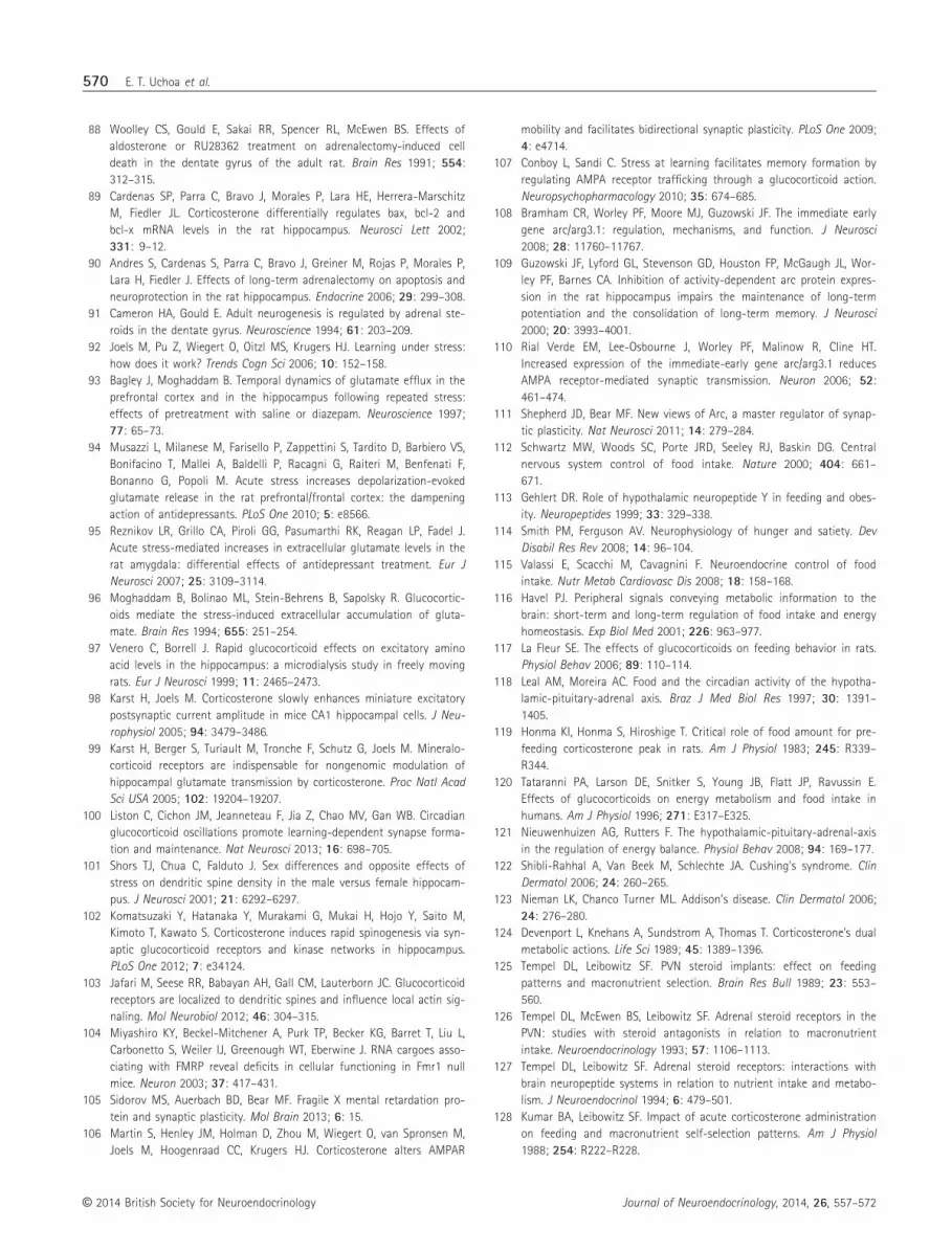
88 Woolley CS, Gould E, Sakai RR, Spencer RL, McEwen BS. Effects of
aldosterone or RU28362 treatment on adrenalectomy-induced cell
death in the dentate gyrus of the adult rat. Brain Res 1991; 554:
312–315.
89 Cardenas SP, Parra C, Bravo J, Morales P, Lara HE, Herrera-Marschitz
M, Fiedler JL. Corticosterone differentially regulates bax, bcl-2 and
bcl-x mRNA levels in the rat hippocampus. Neurosci Lett 2002;
331: 9–12.
90 Andres S, Cardenas S, Parra C, Bravo J, Greiner M, Rojas P, Morales P,
Lara H, Fiedler J. Effects of long-term adrenalectomy on apoptosis and
neuroprotection in the rat hippocampus. Endocrine 2006; 29: 299–308.
91 Cameron HA, Gould E. Adult neurogenesis is regulated by adrenal ste-
roids in the dentate gyrus. Neuroscience 1994; 61: 203–209.
92 Joels M, Pu Z, Wiegert O, Oitzl MS, Krugers HJ. Learning under stress:
how does it work? Trends Cogn Sci 2006; 10: 152–158.
93 Bagley J, Moghaddam B. Temporal dynamics of glutamate efflux in the
prefrontal cortex and in the hippocampus following repeated stress:
effects of pretreatment with saline or diazepam. Neuroscience 1997;
77: 65–73.
94 Musazzi L, Milanese M, Farisello P, Zappettini S, Tardito D, Barbiero VS,
Bonifacino T, Mallei A, Baldelli P, Racagni G, Raiteri M, Benfenati F,
Bonanno G, Popoli M. Acute stress increases depolarization-evoked
glutamate release in the rat prefrontal/frontal cortex: the dampening
action of antidepressants. PLoS One 2010; 5: e8566.
95 Reznikov LR, Grillo CA, Piroli GG, Pasumarthi RK, Reagan LP, Fadel J.
Acute stress-mediated increases in extracellular glutamate levels in the
rat amygdala: differential effects of antidepressant treatment. Eur J
Neurosci 2007; 25: 3109–3114.
96 Moghaddam B, Bolinao ML, Stein-Behrens B, Sapolsky R. Glucocortic-
oids mediate the stress-induced extracellular accumulation of gluta-
mate. Brain Res 1994; 655: 251–254.
97 Venero C, Borrell J. Rapid glucocorticoid effects on excitatory amino
acid levels in the hippocampus: a microdialysis study in freely moving
rats. Eur J Neurosci 1999; 11: 2465–2473.
98 Karst H, Joels M. Corticosterone slowly enhances miniature excitatory
postsynaptic current amplitude in mice CA1 hippocampal cells. J Neu-
rophysiol 2005; 94: 3479–3486.
99 Karst H, Berger S, Turiault M, Tronche F, Schutz G, Joels M. Mineralo-
corticoid receptors are indispensable for nongenomic modulation of
hippocampal glutamate transmission by corticosterone. Proc Natl Acad
Sci USA 2005; 102: 19204–19207.
100 Liston C, Cichon JM, Jeanneteau F, Jia Z, Chao MV, Gan WB. Circadian
glucocorticoid oscillations promote learning-dependent synapse forma-
tion and maintenance. Nat Neurosci 2013; 16: 698–705.
101 Shors TJ, Chua C, Falduto J. Sex differences and opposite effects of
stress on dendritic spine density in the male versus female hippocam-
pus. J Neurosci 2001; 21: 6292–6297.
102 Komatsuzaki Y, Hatanaka Y, Murakami G, Mukai H, Hojo Y, Saito M,
Kimoto T, Kawato S. Corticosterone induces rapid spinogenesis via syn-
aptic glucocorticoid receptors and kinase networks in hippocampus.
PLoS One 2012; 7: e34124.
103 Jafari M, Seese RR, Babayan AH, Gall CM, Lauterborn JC. Glucocorticoid
receptors are localized to dendritic spines and influence local actin sig-
naling. Mol Neurobiol 2012; 46: 304–315.
104 Miyashiro KY, Beckel-Mitchener A, Purk TP, Becker KG, Barret T, Liu L,
Carbonetto S, Weiler IJ, Greenough WT, Eberwine J. RNA cargoes asso-
ciating with FMRP reveal deficits in cellular functioning in Fmr1 null
mice. Neuron 2003; 37: 417–431.
105 Sidorov MS, Auerbach BD, Bear MF. Fragile X mental retardation pro-
tein and synaptic plasticity. Mol Brain 2013; 6: 15.
106 Martin S, Henley JM, Holman D, Zhou M, Wiegert O, van Spronsen M,
Joels M, Hoogenraad CC, Krugers HJ. Corticosterone alters AMPAR
mobility and facilitates bidirectional synaptic plasticity. PLoS One 2009;
4: e4714.
107 Conboy L, Sandi C. Stress at learning facilitates memory formation by
regulating AMPA receptor trafficking through a glucocorticoid action.
Neuropsychopharmacology 2010; 35: 674–685.
108 Bramham CR, Worley PF, Moore MJ, Guzowski JF. The immediate early
gene arc/arg3.1: regulation, mechanisms, and function. J Neurosci
2008; 28: 11760–11767.
109 Guzowski JF, Lyford GL, Stevenson GD, Houston FP, McGaugh JL, Wor-
ley PF, Barnes CA. Inhibition of activity-dependent arc protein expres-
sion in the rat hippocampus impairs the maintenance of long-term
potentiation and the consolidation of long-term memory. J Neurosci
2000; 20: 3993–4001.
110 Rial Verde EM, Lee-Osbourne J, Worley PF, Malinow R, Cline HT.
Increased expression of the immediate-early gene arc/arg3.1 reduces
AMPA receptor-mediated synaptic transmission. Neuron 2006; 52:
461–474.
111 Shepherd JD, Bear MF. New views of Arc, a master regulator of synap-
tic plasticity. Nat Neurosci 2011; 14: 279–284.
112 Schwartz MW, Woods SC, Porte JRD, Seeley RJ, Baskin DG. Central
nervous system control of food intake. Nature 2000; 404: 661–
671.
113 Gehlert DR. Role of hypothalamic neuropeptide Y in feeding and obes-
ity. Neuropeptides 1999; 33: 329–338.
114 Smith PM, Ferguson AV. Neurophysiology of hunger and satiety. Dev
Disabil Res Rev 2008; 14: 96–104.
115 Valassi E, Scacchi M, Cavagnini F. Neuroendocrine control of food
intake. Nutr Metab Cardiovasc Dis 2008; 18: 158–168.
116 Havel PJ. Peripheral signals conveying metabolic information to the
brain: short-term and long-term regulation of food intake and energy
homeostasis. Exp Biol Med 2001; 226: 963–977.
117 La Fleur SE. The effects of glucocorticoids on feeding behavior in rats.
Physiol Behav 2006; 89: 110–114.
118 Leal AM, Moreira AC. Food and the circadian activity of the hypotha-
lamic-pituitary-adrenal axis. Braz J Med Biol Res 1997; 30: 1391–
1405.
119 Honma KI, Honma S, Hiroshige T. Critical role of food amount for pre-
feeding corticosterone peak in rats. Am J Physiol 1983; 245: R339–
R344.
120 Tataranni PA, Larson DE, Snitker S, Young JB, Flatt JP, Ravussin E.
Effects of glucocorticoids on energy metabolism and food intake in
humans. Am J Physiol 1996; 271: E317–E325.
121 Nieuwenhuizen AG, Rutters F. The hypothalamic-pituitary-adrenal-axis
in the regulation of energy balance. Physiol Behav 2008; 94: 169–177.
122 Shibli-Rahhal A, Van Beek M, Schlechte JA. Cushing’s syndrome. Clin
Dermatol 2006; 24: 260–265.
123 Nieman LK, Chanco Turner ML. Addison’s disease. Clin Dermatol 2006;
24: 276–280.
124 Devenport L, Knehans A, Sundstrom A, Thomas T. Corticosterone’s dual
metabolic actions. Life Sci 1989; 45: 1389–1396.
125 Tempel DL, Leibowitz SF. PVN steroid implants: effect on feeding
patterns and macronutrient selection. Brain Res Bull 1989; 23: 553–
560.
126 Tempel DL, McEwen BS, Leibowitz SF. Adrenal steroid receptors in the
PVN: studies with steroid antagonists in relation to macronutrient
intake. Neuroendocrinology 1993; 57: 1106–1113.
127 Tempel DL, Leibowitz SF. Adrenal steroid receptors: interactions with
brain neuropeptide systems in relation to nutrient intake and metabo-
lism. J Neuroendocrinol 1994; 6: 479–501.
128 Kumar BA, Leibowitz SF. Impact of acute corticosterone administration
on feeding and macronutrient self-selection patterns. Am J Physiol
1988; 254: R222–R228.
© 2014 British Society for Neuroendocrinology Journal of Neuroendocrinology, 2014, 26, 557–572
570 E. T. Uchoa et al.

129 Kumar BA, Papamichael M, Leibowitz SF. Feeding and macronutrient
selection patterns in rats: adrenalectomy and chronic corticosterone
replacement. Physiol Behav 1988; 42: 581–589.
130 Goldstein RE, Wasserman DH, McGuinness OP, Lacy DB, Cherrington
AD, Abumrad NN. Effects of chronic elevation in plasma cortisol on
hepatic carbohydrate metabolism. Am J Physiol 1993; 264: E119–E127.
131 Tomas FM, Munro HN, Young VR. Effect of glucocorticoid administra-
tion on the rate of muscle protein breakdown in vivo in rats, as mea-
sured by urinary excretion of N tau-methylhistidine. Biochem J 1979;
178: 139–146.
132 Cusin I, Rouru J, Rohner-Jeanrenaud F. Intracerebroventricular gluco-
corticoid infusion in normal rats: induction of parasympathetic-medi-
ated obesity and insulin resistance. Obes Res 2001; 9: 401–406.
133 Zakrzewska KE, Cusin I, Stricker-Krongrad A, Boss O, Ricquier D, Jean-
renaud B, Rohner-Jeanrenaud F. Induction of obesity and hyperleptin-
emia by central glucocorticoid infusion in the rat. Diabetes 1999; 48:
365–370.
134 Aronsson M, Fuxe K, Dong Y, Agnati LF, Okret S, Gustafsson JA. Locali-
zation of glucocorticoid receptor mRNA in the male rat brain by in situ
hybridization. Proc Natl Acad Sci USA 1988; 85: 9331–9335.
135 Hisano S, Kagotani Y, Tsuruo Y, Daikoku S, Chihara K, Whitnall MH.
Localization of glucocorticoid receptor in neuropeptide Y-containing
neurons in the arcuate nucleus of the rat hypothalamus. Neurosci Lett
1988; 95: 13–18.
136 Uchoa ET, Silva LE, de Castro M, Antunes-Rodrigues J, Elias LL. Gluco-
corticoids are required for meal-induced changes in the expression of
hypothalamic neuropeptides. Neuropeptides 2012; 46: 119–124.
137 Freedman MR, Castonguay TW, Stern JS. Effect of adrenalectomy and
corticosterone replacement on meal patterns of Zucker rats. Am J
Physiol 1985; 249: R584–R594.
138 Uchoa ET, Sabino HA, Ruginsk SG, Antunes-Rodrigues J, Elias LL. Hypo-
phagia induced by glucocorticoid deficiency is associated with an
increased activation of satiety-related responses. J Appl Physiol 2009;
106: 596–604.
139 Uchoa ET, Silva LE, de Castro M, Antunes-Rodrigues J, Elias LL. Hypo-
thalamic oxytocin neurons modulate hypophagic effect induced by
adrenalectomy. Horm Behav 2009; 56: 532–538.
140 Uchoa ET, Silva LE, de Castro M, Antunes-Rodrigues J, Elias LL. Cortico-
trophin-releasing factor mediates hypophagia after adrenalectomy,
increasing meal-related satiety responses. Horm Behav 2010; 58: 714–
719.
141 Bruce BK, King BM, Phelps GR, Veitia MC. Effects of adrenalectomy
and corticosterone administration on hypothalamic obesity in rats. Am
J Physiol 1982; 243: E152–E157.
142 Dubuc PU, Wilden NJ. Adrenalectomy reduces but does not reverse
obesity in ob/ob mice. Int J Obes 1986; 10: 91–98.
143 Yukimura Y, Bray GA, Wolfsen AR. Some effects of adrenalectomy in
the fatty rat. Endocrinology 1978; 103: 1924–1928.
144 Germano CM, Castro M, Rorato R, Laguna MT, Antunes-Rodrigues J,
Elias CF, Elias LL. Time course effects of adrenalectomy and food intake
on cocaine- and amphetamine-regulated transcript expression in the
hypothalamus. Brain Res 2007; 1166: 55–64.
145 Uchoa ET, Zahm DS, de Carvalho Borges B, Rorato R, Antunes-Rodri-
gues J, Elias LL. Oxytocin projections to the nucleus of the solitary
tract contribute to the increased meal-related satiety responses in pri-
mary adrenal insufficiency. Exp Physiol 2013; 98: 1495–1504.
146 Deak T, Bordner K, McElderry N, Barnum C, Blandino P Jr, Deak M,
Tammariello S. Stress-induced increases in hypothalamic IL-1: a sys-
tematic analysis of multiple stressor paradigms. Brain Res Bull 2005;
64: 541–556.
147 Hueston CM, Barnum CJ, Eberle JA, Ferraioli FJ, Buck HM, Deak T.
Stress-dependent changes in neuroinflammatory markers observed
after common laboratory stressors are not seen following acute social
defeat of the Sprague Dawley rat. Physiol Behav 2011; 104: 187–198.
148 Hueston CM, Buck HM, Deak T. The inflamed axis: the interaction
between cytokines and HPA hormones in the stress response. Physiol
Behav 2014; 124: 77–91.
149 Garcia-Bueno B, Madrigal JL, Perez-Nievas BG, Leza JC. Stress media-
tors regulate brain prostaglandin synthesis and peroxisome prolifera-
tor-activated receptor-gamma activation after stress in rats.
Endocrinology 2008; 149: 1969–1978.
150 Frank MG, Baratta MV, Sprunger DB, Watkins LR, Maier SF. Microglia
serve as a neuroimmune substrate for stress-induced potentiation of
CNS pro-inflammatory cytokine responses. Brain Behav Immun 2007;
21: 47–59.
151 Nair A, Bonneau RH. Stress-induced elevation of glucocorticoids
increases microglia proliferation through NMDA receptor activation. J
Neuroimmunol 2006; 171: 72–85.
152 Blandino P Jr, Barnum CJ, Solomon LG, Larish Y, Lankow BS, Deak T.
Gene expression changes in the hypothalamus provide evidence for
regionally-selective changes in IL-1 and microglial markers after acute
stress. Brain Behav Immun 2009; 23: 958–968.
153 Sugama S, Fujita M, Hashimoto M, Conti B. Stress induced morphologi-
cal microglial activation in the rodent brain: involvement of interleu-
kin-18. Neuroscience 2007; 146: 1388–1399.
154 Blandino P Jr, Barnum CJ, Deak T. The involvement of norepinephrine
and microglia in hypothalamic and splenic IL-1beta responses to stress.
J Neuroimmunol 2006; 173: 87–95.
155 Johnson JD, Campisi J, Sharkey CM, Kennedy SL, Nickerson M, Green-
wood BN, Fleshner M. Catecholamines mediate stress-induced
increases in peripheral and central inflammatory cytokines. Neurosci-
ence 2005; 135: 1295–1307.
156 Blandino P Jr, Hueston CM, Barnum CJ, Bishop C, Deak T. The impact
of ventral noradrenergic bundle lesions on increased IL-1 in the PVN
and hormonal responses to stress in male sprague dawley rats. Endo-
crinology 2013; 154: 2489–2500.
157 Porterfield VM, Zimomra ZR, Caldwell EA, Camp RM, Gabella KM, John-
son JD. Rat strain differences in restraint stress-induced brain cyto-
kines. Neuroscience 2011; 188: 48–54.
158 Windle RJ, Wood SA, Lightman SL, Ingram CD. The pulsatile character-
istics of hypothalamo-pituitary-adrenal activity in female Lewis and
Fischer 344 rats and its relationship to differential stress responses.
Endocrinology 1998; 139: 4044–4052.
159 Maslanik T, Mahaffey L, Tannura K, Beninson L, Greenwood BN,
Fleshner M. The inflammasome and danger associated molecular
patterns (DAMPs) are implicated in cytokine and chemokine
responses following stressor exposure. Brain Behav Immun 2013;
28: 54–62.
160 Nguyen K, Deak T, Owens S, Kohno T, Fleshner M, Watkins L, Maier S.
Exposure ot acute stress induces brain interleukin-1 beta protein in the
rat. J Neurosci 1998; 18: 2239–2246.
161 Nguyen KT, Deak T, Will MJ, Hansen MK, Hunsaker BN, Fleshner M,
Watkins LR, Maier SF. Timecourse and corticosterone sensitivity of the
brain, pituitary, and serum interleukin-1b protein response to acute
stress. Brain Res 2000; 859: 193–201.
162 Busillo JM, Cidlowski JA. The five Rs of glucocorticoid action during
inflammation: ready, reinforce, repress, resolve, and restore. Trends
Endocrinol Metab 2013; 24: 109–119.
163 Necela BM, Cidlowski JA. Mechanisms of glucocorticoid receptor action
in noninflammatory and inflammatory cells. Proc Am Thorac Soc 2004;
1: 239–246.
164 Watkins LR, Hansen MK, Nguyen KT, Lee JE, Maier SF. Dynamic regula-
tion of the proinflammatory cytokine interleukin-1b: molecular biology
for non-molecular biologists. Life Sci 1999; 65: 449–481.
© 2014 British Society for NeuroendocrinologyJournal of Neuroendocrinology, 2014, 26, 557–572
Glucocorticoid actions 571

165 Frank MG, Watkins LR, Maier SF. Stress-induced glucocorticoids as a
neuroendocrine alarm signal of danger. Brain Behav Immun 2013; 33:
1–6.
166 Sorrells SF, Sapolsky RM. An inflammatory review of glucocorticoid
actions in the CNS. Brain Behav Immun 2007; 21: 259–272.
167 Goshen I, Kreisel T, Ben-Menachem-Zidon O, Licht T, Weidenfeld J,
Ben-Hur T, Yirmiya R. Brain interleukin-1 mediates chronic stress-
induced depression in mice via adrenocortical activation and hippo-
campal neurogenesis suppression. Mol Psychiatry 2008; 13: 717–728.
168 Johnson JD, O’Connor KA, Watkins LR, Maier SF. The role of IL-1beta in
stress-induced sensitization of proinflammatory cytokine and cortico-
sterone responses. Neuroscience 2004; 127: 569–577.
169 Bornstein SR, KZiegler CG, Krug AW, Kanczkowski W, Rettori V, McC-
ann SM, Wirth M, Zacharowski K. The role of toll-like receptors in the
immune-adrenal crosstalk. Ann N Y Acad Sci 2006; 1088: 307–318.
170 Deak T. From classic aspects of the stress response to neuroinflamma-
tion and sickness: implications for individuals and offspring of diverse
species. Int J Comp Psychol 2007; 20: 96–110.
171 Engstrom L, Rosen K, Angel A, Fyrberg A, Mackerlova L, Konsman JP,
Engblom D, Blomqvist A. Systemic immune challenge activates an
intrinsically regulated local inflammatory circuit in the adrenal gland.
Endocrinology 2008; 19: 1436–14350.
172 Mazzocchi G, Gottardo G, Nussdorfer GG. A local immuno-endocrine
interaction may mediate rat adrenal glucocorticoid response to bacte-
rial endotoxins. Life Sci 1998; 62: 1783–1787.
173 Beishuizen A, Thijs LG, Haanen C, Vermes I. Macrophage migration
inhibitory factor and hypothalamo-pituitary-adrenal function during
critical illness. J Clin Endocrinol Metab 2001; 86: 2811–2816.
174 Vermes I, Beishuizen A, Hampsink RM, Haanen C. Dissociation of
plasma adrenocorticotropin and cortisol levels in critically ill patients:
possible role of endothelin and atrial natriuretic hormone. J Clin Endo-
crinol Metab 1995; 80: 1238–1242.
175 Arakawa H, Blandino P Jr, Deak T. Central infusion of interleukin-1
receptor antagonist blocks the reduction in social behavior produced
by prior stressor exposure. Physiol Behav 2009; 98: 139–146.
176 Koo JW, Duman RS. Interleukin-1 receptor null mutant mice show
decreased anxiety-like behavior and enhanced fear memory. Neurosci
Lett 2009; 456: 39–43.
177 Pugh CR, Nguyen KT, Gonyea JL, Fleshner M, Watkins LR, Maier SF,
Rudy JW. Role of interleukin-1 beta in impairment of contextual fear
conditioning caused by social isolation. Behav Brain Res 1999; 106:
109–118.
178 Maier SF, Watkins LR. Intracerebroventricular interleukin-1 receptor
antagonist blocks the enhancement of fear conditioning and interfer-
ence with escape produced by inescapable shock. Brain Res 1995;
695: 279–282.
179 Barnum CJ, Tansey MG. Neuroinflammation and non-motor symptoms:
the dark passenger of Parkinson’s disease? Curr Neurol Neurosci Rep
2012; 12: 350–358.
180 Deak T. From hippocampus to dorsal horn: the pervasive impact of IL-
1 on learning and memory spans the length of the neuroaxis. Brain
Behav Immun 2007; 21: 746–747.
181 Wager-Smith K, Markou A. Depression: a repair response to stress-
induced neuronal microdamage that can grade into a chronic
neuroinflammatory condition? Neurosci Biobehav Rev 2011; 35:
742–764.
© 2014 British Society for Neuroendocrinology Journal of Neuroendocrinology, 2014, 26, 557–572
572 E. T. Uchoa et al.


