Embed Size (px)
Citation preview
Novel Bacteriophages Containing a Genome of AnotherBacteriophage within Their GenomesMaud M. Swanson1., Brian Reavy1.*, Kira S. Makarova2, Peter J. Cock1, David W. Hopkins1¤,
Lesley Torrance1, Eugene V. Koonin2, Michael Taliansky1
1 The James Hutton Institute, Invergowrie, Dundee, United Kingdom, 2 National Center for Biotechnology Information, National Library of Medicine, National Institutes of
Health, Bethesda, Maryland, United States of America
Abstract
A novel bacteriophage infecting Staphylococus pasteuri was isolated during a screen for phages in Antarctic soils. The phagenamed SpaA1 is morphologically similar to phages of the family Siphoviridae. The 42,784 bp genome of SpaA1 is a linear,double-stranded DNA molecule with 39 protruding cohesive ends. The SpaA1 genome encompasses 63 predicted protein-coding genes which cluster within three regions of the genome, each of apparently different origin, in a mosaic pattern. Intwo of these regions, the gene sets resemble those in prophages of Bacillus thuringiensis kurstaki str. T03a001 (genesinvolved in DNA replication/transcription, cell entry and exit) and B. cereus AH676 (additional regulatory and recombinationgenes), respectively. The third region represents an almost complete genome (except for the short terminal segments) of adistinct bacteriophage, MZTP02. Nearly the same gene module was identified in prophages of B. thuringiensis serovarmonterrey BGSC 4AJ1 and B. cereus Rock4-2. These findings suggest that MZTP02 can be shuttled between genomes ofother bacteriophages and prophages, leading to the formation of chimeric genomes. The presence of a complete phagegenome in the genome of other phages apparently has not been described previously and might represent a ‘fast track’route of virus evolution and horizontal gene transfer. Another phage (BceA1) nearly identical in sequence to SpaA1, and alsoincluding the almost complete MZTP02 genome within its own genome, was isolated from a bacterium of the B. cereus/B.thuringiensis group. Remarkably, both SpaA1 and BceA1 phages can infect B. cereus and B. thuringiensis, but only one ofthem, SpaA1, can infect S. pasteuri. This finding is best compatible with a scenario in which MZTP02 was originally containedin BceA1 infecting Bacillus spp, the common hosts for these two phages, followed by emergence of SpaA1 infecting S.pasteuri.
Citation: Swanson MM, Reavy B, Makarova KS, Cock PJ, Hopkins DW, et al. (2012) Novel Bacteriophages Containing a Genome of Another Bacteriophage withinTheir Genomes. PLoS ONE 7(7): e40683. doi:10.1371/journal.pone.0040683
Editor: Ramy K. Aziz, Cairo University, Egypt
Received March 1, 2012; Accepted June 14, 2012; Published July 17, 2012
Copyright: � 2012 Swanson et al. This is an open-access article distributed under the terms of the Creative Commons Attribution License, which permitsunrestricted use, distribution, and reproduction in any medium, provided the original author and source are credited.
Funding: The work of authors MMS, BR, PJC, DH, LT and MT was funded by the Scottish Government’s Rural and Environment Science and Analytical Services(RESAS) Division. KSM and EVK are supported by the intramural funds of the United States Department of Health and Human Services (National Library ofMedicine, National Institutes of Health). The funders had no role in study design, data collection and analysis, decision to publish, or preparation of themanuscript.
Competing Interests: The authors have declared that no competing interests exist.
* E-mail: [email protected]
. These authors contributed equally to this work.
¤ Current address: School of Life Sciences, Heriot-Watt University, Edinburgh, United Kingdom
Introduction
Viruses are the most abundant entities in the biosphere. In
marine and soil habitats, the number of virus particles exceeds the
number of cells by at least an order of magnitude [1–3]. Numerous
viruses infect organisms from all branches of cellular life. However,
virus research has traditionally focused on viruses that infect
humans, other vertebrates and plants due to the obvious medical
and agricultural importance of these viruses. In addition, viruses
infecting several model bacteria (bacteriophages) have been
studied in detail thanks primarily to their utility as tools of
molecular biology. Viruses from diverse environments are
incomparably less thoroughly characterized but recently environ-
mental genomics and metagenomics of viruses have become
rapidly growing research areas [4–7].
A total of about 2300 viruses are recognized by the Interna-
tional Committee on Taxonomy of Viruses [8] but this is likely to
be a gross underestimate because of the enormous diversity of
viruses in unsampled or poorly investigated habitats (see for
example, [9], [10]. Virus particles are abundant in air, water and
soils [1], [5], [11–15]. Recent metagenomic analyses have revealed
hitherto unknown diverse assemblages of viruses in these
environments [6], [9], [10], [16], [17]. For example, Fierer et al.
[10] reported that the majority of the 4577 virus-related nucleotide
sequences found in soils from different ecosystems showed no
similarity to previously described sequences. Analysis of metage-
nomic data suggests novel patterns of virus evolution and reveals
new groups of viruses providing unprecedented insights into the
composition and dynamics of the virus world [7]. Viruses, in
particular transducing bacteriophages, have been long known to
make major contributions to gene exchange between bacteria
[18]. Recently, a distinct class of defective bacteriophages, the
Gene Transfer Agents (GTAs) [19], have been characterized as
apparent dedicated vehicles for horizontal gene transfer that might
account for extensive gene flow in bacterial and archaeal
PLoS ONE | www.plosone.org 1 July 2012 | Volume 7 | Issue 7 | e40683
communities [19], [20]. Furthermore, viruses have emerged as a
major force shaping the geochemistry and ecology of diverse
environmental ecosystems [5], [21–23].
Tailed dsDNA bacteriophages account for 95% of all known
bacterial viruses, and possibly make up the majority of phages on
the planet [24]. They belong to the order Caudovirales which
consists of three families: Myoviridae (long rigid contractile tails),
Siphoviridae (long flexible non-contractile tails) and Podoviridae (short
contractile tails) [8], [25–27]. One of the key features of the
genomes of Caudovirales is their apparent mosaic architecture; in
essence, each genome is a unique set of modules with different
evolutionary histories that have been horizontally exchanged
among phages [28–30].
In this work we describe a novel phage genome architecture
where one phage genome nestles inside the genome of another
phage, similar to a ‘‘Russian Doll’’ arrangement. We show that
bacteriophages SpaA1 and BceA1, obtained from the bacterium
Staphylococus pasteuri and a bacterium belonging to the Bacillus
cereus/B. thuringiensis group respectively, and isolated from a soil
sample from the Garwood Valley, Southern Victoria Land,
Antarctica, harbor almost the complete sequence of the bacterio-
phage MZTP02 that had been identified previously in China [31].
Results
Isolation and Morphology of SpaA1A novel temperate bacteriophage, named SpaA1, was isolated
from Staphylococus pasteuri recovered from soils of the Garwood
Valley, Southern Victoria Land, Antarctica. Bacterial cultures
were grown from single colonies in liquid nutrient medium in the
presence of mitomycin C to induce prophages from lysogenic
bacteria. SpaA1 was isolated from the growth medium and
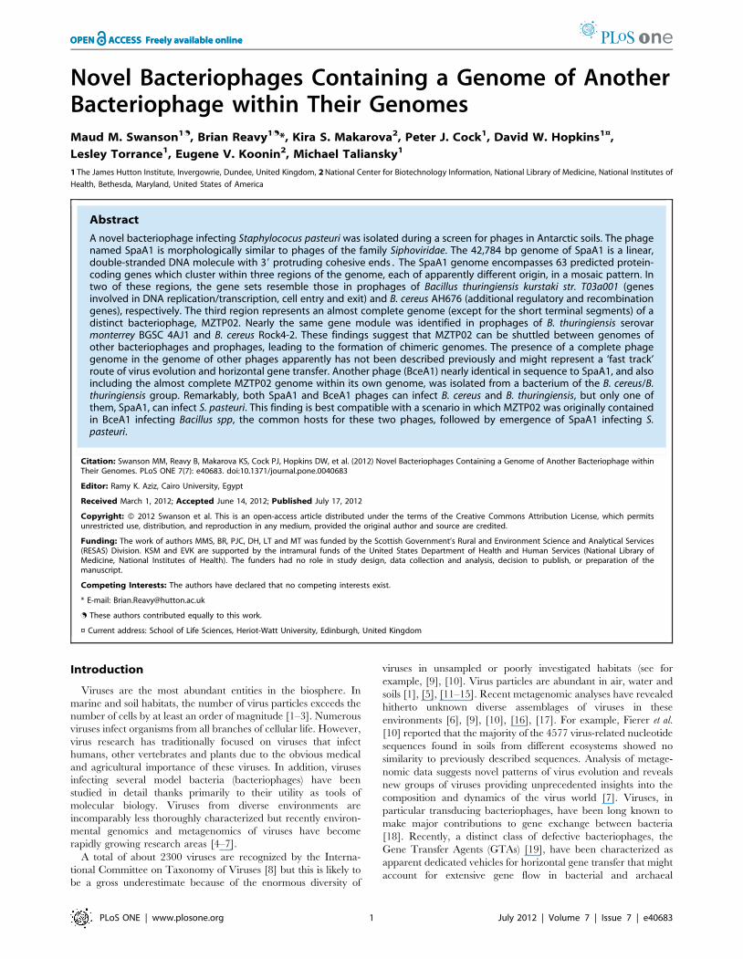
examined by transmission electron microscopy (TEM) (Figure 1A).
The morphology of SpaA1 is typical of the Siphoviridae family of
phages. SpaA1 virions have isometric heads (B1 morphotype) with
a diameter of ,63 nm. The virion tails are ,210 nm long and
appear to be flexible and non-contractile.
General Features of the SpaA1 GenomeThe genome of phage SpaA1 consists of 42,784 bp flanked by
complementary 9-bp single stranded cohesive (cos) ends (59-
…TGGAGGAGG -39 and 39-CCTCCTCCA…-59). Using Gen-
eMark.hmm [32], 63 open reading frames (ORFs) were identified
as probable protein-coding genes. The predicted proteins encoded
by these 63 ORFs were compared to the non-redundant protein
sequence database (National Center for Biotechnology Informa-
tion, NIH, Bethesda) using PSI-BLAST [33] and the Conserved
Domain Database using RPS-BLAST [34]. Analysis of the most
similar proteins (best hits) for all predicted gene products of SpaA1
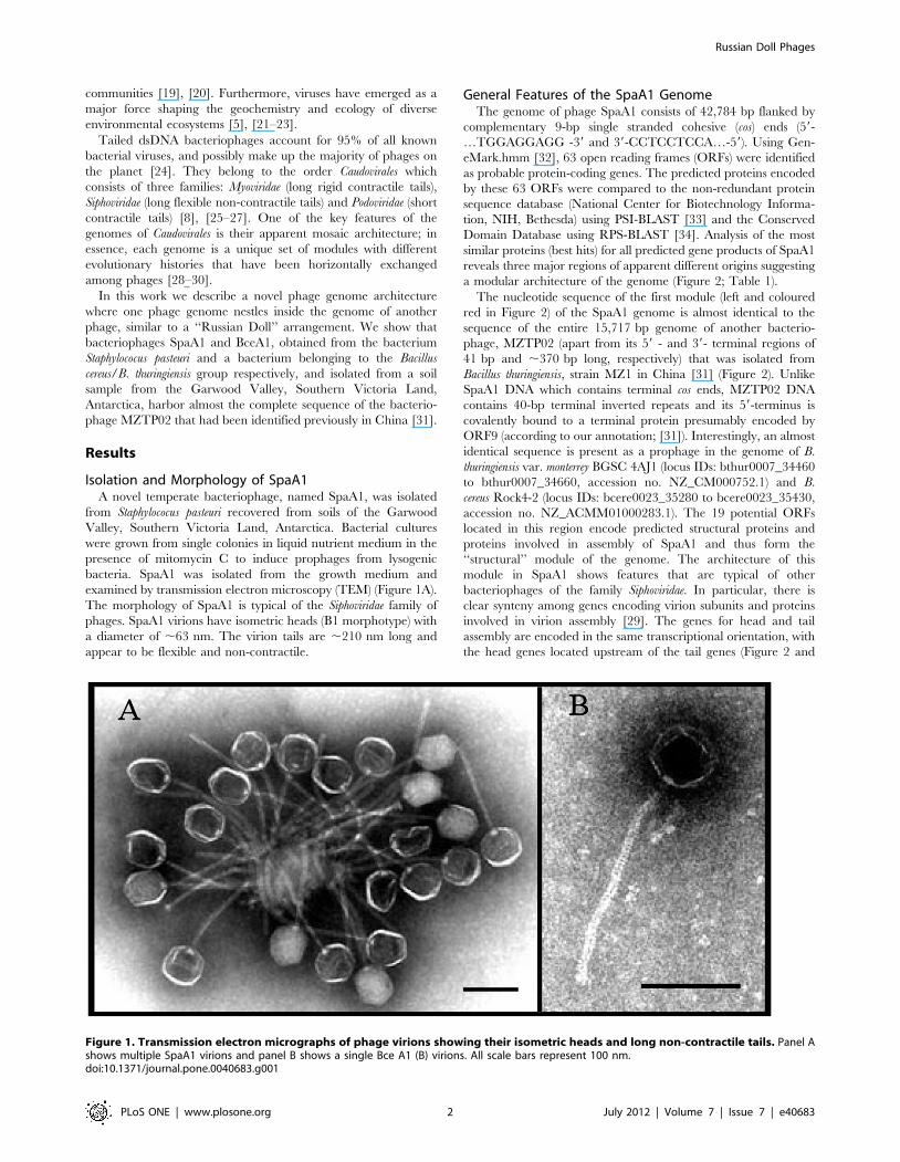
reveals three major regions of apparent different origins suggesting
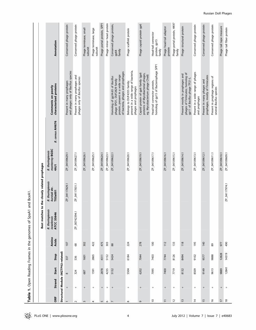
a modular architecture of the genome (Figure 2; Table 1).
The nucleotide sequence of the first module (left and coloured
red in Figure 2) of the SpaA1 genome is almost identical to the
sequence of the entire 15,717 bp genome of another bacterio-
phage, MZTP02 (apart from its 59 - and 39- terminal regions of
41 bp and ,370 bp long, respectively) that was isolated from
Bacillus thuringiensis, strain MZ1 in China [31] (Figure 2). Unlike
SpaA1 DNA which contains terminal cos ends, MZTP02 DNA
contains 40-bp terminal inverted repeats and its 59-terminus is
covalently bound to a terminal protein presumably encoded by
ORF9 (according to our annotation; [31]). Interestingly, an almost
identical sequence is present as a prophage in the genome of B.
thuringiensis var. monterrey BGSC 4AJ1 (locus IDs: bthur0007_34460
to bthur0007_34660, accession no. NZ_CM000752.1) and B.
cereus Rock4-2 (locus IDs: bcere0023_35280 to bcere0023_35430,
accession no. NZ_ACMM01000283.1). The 19 potential ORFs
located in this region encode predicted structural proteins and
proteins involved in assembly of SpaA1 and thus form the
‘‘structural’’ module of the genome. The architecture of this
module in SpaA1 shows features that are typical of other
bacteriophages of the family Siphoviridae. In particular, there is
clear synteny among genes encoding virion subunits and proteins
involved in virion assembly [29]. The genes for head and tail
assembly are encoded in the same transcriptional orientation, with
the head genes located upstream of the tail genes (Figure 2 and
Figure 1. Transmission electron micrographs of phage virions showing their isometric heads and long non-contractile tails. Panel Ashows multiple SpaA1 virions and panel B shows a single Bce A1 (B) virions. All scale bars represent 100 nm.doi:10.1371/journal.pone.0040683.g001
Russian Doll Phages
PLoS ONE | www.plosone.org 2 July 2012 | Volume 7 | Issue 7 | e40683
Table 1). The predicted head genes include the large and small
terminase subunits (ORF3 and ORF4, respectively), the portal
protein (ORF5), the minor capsid subunit (ORF6), the scaffold
protein (ORF8), gp-like tail connector (ORF1) and head-tail
adapter (ORF11); the tail genes include the major tail subunit
(ORF12) and the tape measure protein (ORF17), followed by the
tail fiber protein (ORF18) and the minor tail protein (ORF19)
(Table 1). The length of the tape measure protein gene
corresponds to the length of the phage tail and is thus commonly
the largest gene in the genome [29]. In SpaA1, however, the tape
measure protein (979 aa) is only the second largest protein, the
largest being the minor tail structural protein (1569 aa). Bacillus
phage TP21-L also has a minor structural protein that is larger
than the tape measure protein [35]. For most of the known phages,
the size of the tape measure protein corresponds to a fairly
constant 0.15 nm of tail length per amino acid residue [36].
However, the tail length-to-amino acid ratio for SpaA1 is
,0.20 nm per amino acid residue, suggesting that this protein
might be somewhat more extended than those in other known
phages.
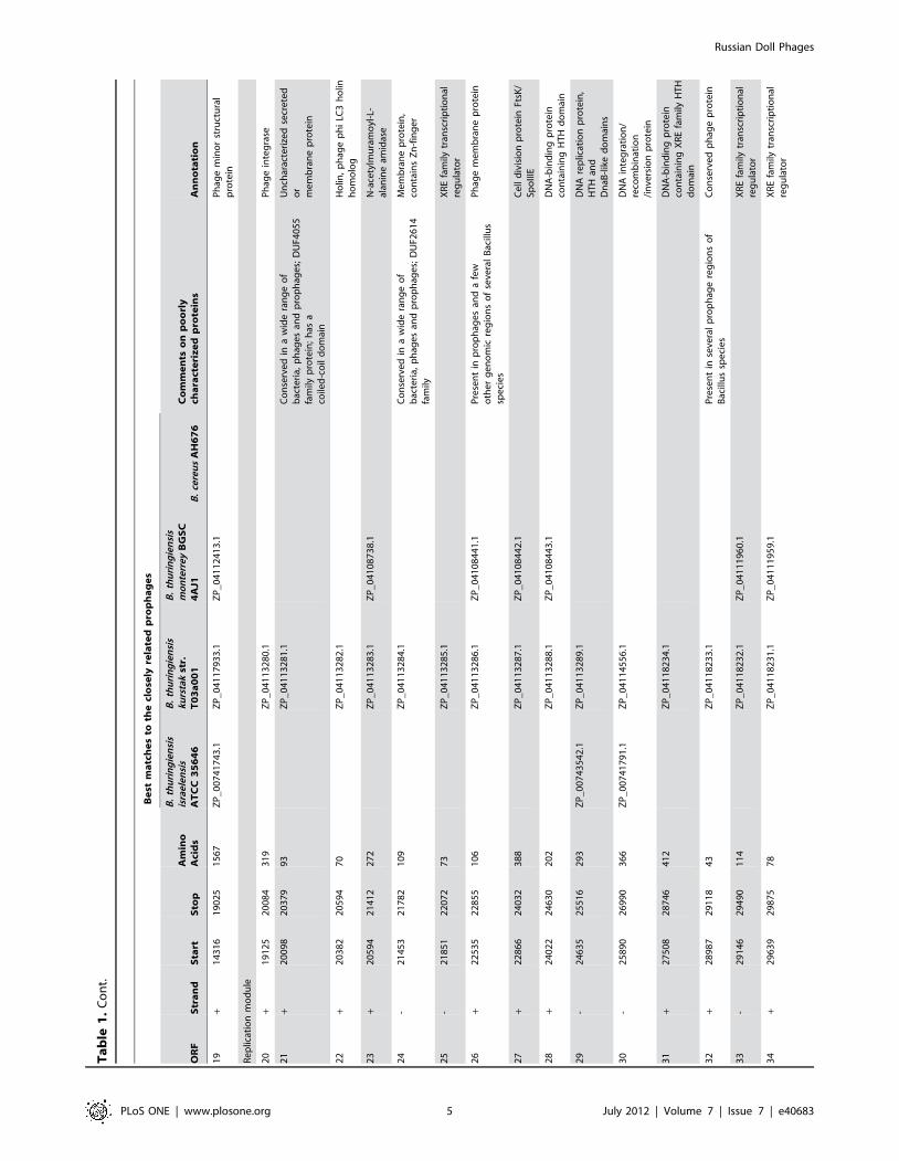
The gene arrangement in the second SpaA1 genome module
(coloured green in Figure 2), which consists of genes with functions
in DNA integration, replication, transcription, cell entry and exit
(ORF20–ORF46), and may be denoted the ‘replication module’, is
very similar to the organization of the corresponding regions in
several prophages of B. thuringiensis Kurstaki strain (Figure 2,
Table 1). The longest conserved gene array (locus_ID:
bthur0006_5910 to bthur0006_6000; accession no.
NZ_CM000751.1) contains the first 10 ORFs in this region. In
particular, the replication module encompasses five predicted
transcriptional regulators (ORFs 25, 33–35 and 45) and four
putative DNA-binding proteins (ORFs 24, 28, 31, and 46). Other
ORFs related to replication in this module include ones encoding
a FtsK/SpoIIIE- like protein (ORF27), and three proteins
containing HTH and DnaB domains (ORF29), a DnaD domain
(ORF41) and a predicted ATPase related to DnaC (ORF42). The
module also encodes an antirepressor (ORF37), two proteins
involved in cell lysis (ORFs 22 and 23) and two integrases, ORF20
which shows 95% amino acid sequence identity with the integrase
of prophage lamdaBa02 (accession number EEM54966.1), and
ORF30 which shows 80% amino acid sequence identity with an
integrase from B. thuringiensis (accession number EAO53934.1).
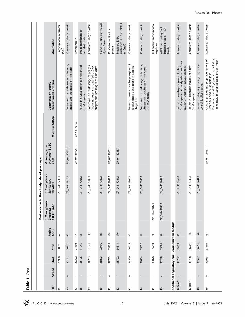
The third genomic module (coloured yellow in Figure 2) of
SpaA1 is similar to a portion of B.cereus AH676 prophage and
contains additional regulatory and recombination related genes
including a potential recombination protein U (ORF53) and a
potential DNA-binding protein (ORF54). ORFs 55 and 56 are
similar to the N-terminal and C-terminal parts of an RNA
polymerase sigma 70 factor, respectively. The last nucleotide of the
TAA termination codon of ORF55 is also the first nucleotide of
the ATG initiation codon of ORF56 within a TAATG sequence.
However, the reading frame of ORF56 extends 59 without an
Figure 2. Architectures of SpaA1, BceA1 and MZTP02 genomes: comparison with BLAST protein matches to phage proteins in fourBacillus genomes. The horizontal bars represent DNA sequences (all to scale) with annotated CDS on the forward (upper) or reverse (lower) strandshown as pointed boxes, generally in alternating blue and purple. The red, green and yellow shading indicates the three functional modules ofphages SpaA1 and BceA1 (center) which are 100% identical except for the area around ORF47 (bright red), and the 99% nucleotide identicalmatching region in module I with phage MZTP02 (second row from top). Rather than the original annotation for MZTP02, annotation based onSpaA1/BceA1 genome analysis (Table 1) is shown, with grey colouring for partial sequences (1 and 19), and genes with frame shifts (12, 13, 17, 18).The bottom three bars represent complete contigs from three separate Bacillus genomes, with red/yellow highlighting top BLAST matches fromSpaA1/BceA1 module I and III proteins, showing synteny visually. The top row of bars represents seven contigs from another Bacillus draft genomewith green highlighting for BLAST protein matches from SpaA1/BceA1 module II proteins. Three of these contigs have been truncated for display. Forclarity, additional BLAST matches to other contigs from these bacterial genomes are not shown (e.g. SpaA1/BceA1 ORF37 matches another contig inB. thuringiensis var. monterrey BGSC A4J1). This figure was drawn using GenomeDiagram [61] and Biopython [62].doi:10.1371/journal.pone.0040683.g002
Russian Doll Phages
PLoS ONE | www.plosone.org 3 July 2012 | Volume 7 | Issue 7 | e40683
Ta
ble
1.
Op
en
Re
adin
gFr
ame
sin
the
ge
no
me
so
fSp
aA1
and
Bce
A1
.
Be
stm
atc
he
sto
the
clo
sely
rela
ted
pro
ph
ag
es
OR
FS
tra
nd
Sta
rtS
top
Am
ino
Aci
ds
B.
thu
rin
gie
nsi
sis
rae
len
sis
AT
CC
35
64
6
B.
thu
rin
gie
nsi
sku
rsta
kst
r.T
03
a0
01
B.
thu
rin
gie
nsi
sm
on
terr
ey
BG
SC
4A
J1B
.ce
reu
sA
H6
76
Co
mm
en
tso
np
oo
rly
cha
ract
eri
ze
dp
rote
ins
An
no
tati
on
Str
uct
ura
lM
od
ule
(MZ
TP
02
-re
late
d)
1+
83
37
10
7Z
P_
04
11
78
29
.1Z
P_
04
10
96
29
.1P
rese
nt
inm
any
pro
ph
age
san
dp
hag
es
on
lyo
fB
acill
us
spe
cie
sC
on
serv
ed
ph
age
pro
tein
2+
32
45
36
68
ZP
_0
07
42
39
4.1
ZP
_0
41
17
83
1.1
ZP
_0
41
09
62
7.1
Pre
sen
tin
man
yp
rop
hag
es
and
ph
age
so
nly
of
Ba
cillu
ssp
eci
es
Co
nse
rve
dp
hag
ep
rote
in
3+
68
71
60
13
02
ZP
_0
41
09
62
6.1
Ph
age
term
inas
e,
smal
lsu
bu
nit
4+
15
91
28
65
42
2Z
P_
04
10
96
25
.1P
hag
ete
rmin
ase
,la
rge
sub
un
it
5+
28
78
43
11
47
5Z
P_
04
10
96
24
.1P
hag
ep
ort
alp
rote
in,
SPP
1
6+
42
35
51
52
30
3Z
P_
04
10
96
23
.1P
hag
em
ino
rh
ead
pro
tein
7+
51
52
54
24
88
ZP
_0
41
09
62
2.1
Ho
mo
log
of
gp
34
.65
of
Ba
cillu
sp
hag
eSP
O1
,D
UF2
82
9fa
mily
pro
tein
,p
rese
nt
ina
wid
era
ng
eo
fb
acte
ria,
ph
age
san
dp
rop
hag
es
Co
nse
rve
dp
hag
ep
rote
in,
gp
64
fam
ily
8+
55
04
61
84
22
4Z
P_
04
10
96
20
.1B
elo
ng
sto
DU
F43
55
fam
ily;
pre
sen
tin
aw
ide
ran
ge
of
bac
teri
a,p
hag
es
and
pro
ph
age
s
Ph
age
scaf
fold
pro
tein
9+
62
02
70
44
27
8Z
P_
04
10
96
19
.1C
apsi
dp
rote
ino
fg
p6
fam
ily(g
p6
pro
tein
so
fM
yco
bac
teri
alp
hag
es,
eg
.M
yco
bac
teri
um
ph
age
Ch
e8
)
Ph
age
cap
sid
pro
tein
gp
6
10
+7
09
57
40
31
00
ZP
_0
41
09
61
7.1
He
ad-t
ail
con
ne
cto
rp
rote
in,
ho
mo
log
of
gp
15
of
Bac
teri
op
hag
eSP
P1
He
ad-t
ail
con
ne
cto
rp
rote
in,
gp
15
fam
ily
11
+7
40
07
74
41
12
ZP
_0
41
09
61
6.1
Ph
age
he
ad-t
ail
adap
tor
pro
tein
12
+7
71
98
12
61
33
ZP
_0
41
09
61
5.1
Ph
age
po
rtal
pro
tein
,H
K9
7fa
mily
13
+8
13
28
49
41
18
ZP
_0
41
09
61
4.1
Pre
sen
tm
ost
lyin
pro
ph
age
san
dp
hag
es
of
Firm
icu
tes;
ho
mo
log
of
gp
11
of
Ba
cillu
sp
hag
eT
P2
1-L
Ph
age
stru
ctu
ral
pro
tein
14
+8
50
99
10
21
95
ZP
_0
41
09
61
3.1
Pre
sen
tin
aw
ide
ran
ge
of
ph
age
san
dp
rop
hag
es
Co
nse
rve
dp
hag
ep
rote
in
15
+9
14
99
57
71
40
ZP
_0
41
09
61
2.1
Pre
sen
tin
man
yp
hag
es
and
pro
ph
age
s,m
ost
lyo
fFi
rmic
ute
sC
on
serv
ed
ph
age
pro
tein
16
+9
61
39
88
88
9Z
P_
04
10
96
11
.1P
rese
nt
inp
rop
hag
ere
gio
ns
of
seve
ral
Ba
cillu
ssp
eci
es
Co
nse
rve
dp
hag
ep
rote
in
17
+9
88
91
28
28
97
7Z
P_
04
10
96
10
.1P
hag
eta
ilta
pe
me
asu
re
18
+1
28
41
14
31
94
90
ZP
_0
41
17
97
4.1
ZP
_0
41
09
60
9.1
Ph
age
tail
fib
er
pro
tein
Russian Doll Phages
PLoS ONE | www.plosone.org 4 July 2012 | Volume 7 | Issue 7 | e40683
Ta
ble
1.
Co
nt.
Be
stm
atc
he
sto
the
clo
sely
rela
ted
pro
ph
ag
es
OR
FS
tra
nd
Sta
rtS
top
Am
ino
Aci
ds
B.
thu
rin
gie
nsi
sis
rae
len
sis
AT
CC
35
64
6
B.
thu
rin
gie
nsi
sku
rsta
kst
r.T
03
a0
01
B.
thu
rin
gie
nsi
sm
on
terr
ey
BG
SC
4A
J1B
.ce
reu
sA
H6
76
Co
mm
en
tso
np
oo
rly
cha
ract
eri
ze
dp
rote
ins
An
no
tati
on
19
+1
43
16
19
02
51
56
7Z
P_
00
74
17
43
.1Z
P_
04
11
79
33
.1Z
P_
04
11
24
13
.1P
hag
em
ino
rst
ruct
ura
lp
rote
in
Re
plic
atio
nm
od
ule
20
+1
91
25
20
08
43
19
ZP
_0
41
13
28
0.1
Ph
age
inte
gra
se
21
+2
00
98
20
37
99
3Z
P_
04
11
32
81
.1C
on
serv
ed
ina
wid
era
ng
eo
fb
acte
ria,
ph
age
san
dp
rop
hag
es;
DU
F40
55
fam
ilyp
rote
in;
has
aco
iled
-co
ild
om
ain
Un
char
acte
rize
dse
cre
ted
or
me
mb
ran
ep
rote
in
22
+2
03
82
20
59
47
0Z
P_
04
11
32
82
.1H
olin
,p
hag
ep
hi
LC3
ho
linh
om
olo
g
23
+2
05
94
21
41
22
72
ZP
_0
41
13
28
3.1
ZP
_0
41
08
73
8.1
N-a
cety
lmu
ram
oyl
-L-
alan
ine
amid
ase
24
-2
14
53
21
78
21
09
ZP
_0
41
13
28
4.1
Co
nse
rve
din
aw
ide
ran
ge
of
bac
teri
a,p
hag
es
and
pro
ph
age
s;D
UF2
61
4fa
mily
Me
mb
ran
ep
rote
in,
con
tain
sZ
n-f
ing
er
25
-2
18
51
22
07
27
3Z
P_
04
11
32
85
.1X
RE
fam
ilytr
ansc
rip
tio
nal
reg
ula
tor
26
+2
25
35
22
85
51
06
ZP
_0
41
13
28
6.1
ZP
_0
41
08
44
1.1
Pre
sen
tin
pro
ph
age
san
da
few
oth
er
ge
no
mic
reg
ion
so
fse
vera
lB
acill
us
spe
cie
s
Ph
age
me
mb
ran
ep
rote
in
27
+2
28
66
24
03
23
88
ZP
_0
41
13
28
7.1
ZP
_0
41
08
44
2.1
Ce
lld
ivis
ion
pro
tein
FtsK
/Sp
oIII
E
28
+2
40
22
24
63
02
02
ZP
_0
41
13
28
8.1
ZP
_0
41
08
44
3.1
DN
A-b
ind
ing
pro
tein
con
tain
ing
HT
Hd
om
ain
29
-2
46
35
25
51
62
93
ZP
_0
07
43
54
2.1
ZP
_0
41
13
28
9.1
DN
Are
plic
atio
np
rote
in,
HT
Han
dD
naB
-lik
ed
om
ain
s
30
-2
58
90
26
99
03
66
ZP
_0
07
41
79
1.1
ZP
_0
41
14
55
6.1
DN
Ain
teg
rati
on
/re
com
bin
atio
n/i
nve
rsio
np
rote
in
31
+2
75
08
28
74
64
12
ZP
_0
41
18
23
4.1
DN
A-b
ind
ing
pro
tein
con
tain
ing
XR
Efa
mily
HT
Hd
om
ain
32
+2
89
87
29
11
84
3Z
P_
04
11
82
33
.1P
rese
nt
inse
vera
lp
rop
hag
ere
gio
ns
of
Bac
illu
ssp
eci
es
Co
nse
rve
dp
hag
ep
rote
in
33
-2
91
46
29
49
01
14
ZP
_0
41
18
23
2.1
ZP
_0
41
11
96
0.1
XR
Efa
mily
tran
scri
pti
on
alre
gu
lato
r
34
+2
96
39
29
87
57
8Z
P_
04
11
82
31
.1Z
P_
04
11
19
59
.1X
RE
fam
ilytr
ansc
rip
tio
nal
reg
ula
tor
Russian Doll Phages
PLoS ONE | www.plosone.org 5 July 2012 | Volume 7 | Issue 7 | e40683
Ta
ble
1.
Co
nt.
Be
stm
atc
he
sto
the
clo
sely
rela
ted
pro
ph
ag
es
OR
FS
tra
nd
Sta
rtS
top
Am
ino
Aci
ds
B.
thu
rin
gie
nsi
sis
rae
len
sis
AT
CC
35
64
6
B.
thu
rin
gie
nsi
sku
rsta
kst
r.T
03
a0
01
B.
thu
rin
gie
nsi
sm
on
terr
ey
BG
SC
4A
J1B
.ce
reu
sA
H6
76
Co
mm
en
tso
np
oo
rly
cha
ract
eri
ze
dp
rote
ins
An
no
tati
on
35
+2
99
08
30
09
66
2Z
P_
04
11
82
30
.1T
ran
scri
pti
on
alre
gu
lato
r,p
bsX
36
+3
01
21
30
27
66
3Z
P_
04
11
81
15
.1Z
P_
04
12
36
82
.1C
on
serv
ed
ina
wid
era
ng
eo
fb
acte
ria,
ph
age
san
dp
rop
hag
es
of
Firm
icu
tes
Co
nse
rve
dp
hag
ep
rote
in
37
+3
03
22
31
10
16
4Z
P_
04
11
19
06
.1Z
P_
04
19
51
92
.1A
nti
rep
ress
or
38
+3
11
26
31
24
26
5Z
P_
04
11
79
06
.1Fo
un
din
seve
ral
pro
ph
age
reg
ion
so
fB
aci
llus
spe
cie
sP
hag
em
em
bra
ne
or
secr
ete
dp
rote
in
39
+3
12
63
31
57
71
12
ZP
_0
41
17
90
5.1
Co
nse
rve
din
aw
ide
ran
ge
of
ph
age
s(i
ncl
ud
ing
seve
ral
un
clas
sifi
ed
dsD
NA
ph
age
s)an
dp
rop
hag
es
of
Firm
icu
tes
Co
nse
rve
dp
hag
ep
rote
in
40
+3
18
52
32
49
92
15
ZP
_0
41
17
90
9.1
Sig
ma7
0,
RN
Ap
oly
me
rase
sig
ma
fact
or
41
+3
27
23
33
73
93
38
ZP
_0
41
17
94
3.1
ZP
_0
41
12
66
1.1
Dn
aD-l
ike
rep
licat
ion
pro
tein
42
+3
37
02
34
51
42
70
ZP
_0
41
17
94
4.1
ZP
_0
41
12
58
7.1
Pre
dic
ted
DN
Are
plic
atio
nA
TP
ase
rela
ted
toD
naC
43
+3
45
56
34
82
28
8Z
P_
04
11
79
45
.1P
rese
nt
inse
vera
lp
rop
hag
ere
gio
ns
of
Bac
illu
ssp
eci
es
and
fou
nd
inB
acill
us
ph
age
IEB
H
Co
nse
rve
dp
hag
ep
rote
in
44
+3
48
94
35
05
85
4Z
P_
04
11
79
46
.1C
on
serv
ed
ina
wid
era
ng
eo
fb
acte
ria,
ph
age
san
dp
rop
hag
es
of
Firm
icu
tes;
DU
F39
54
fam
ily
Co
nse
rve
dp
hag
ep
rote
in
45
+3
50
76
35
29
17
1Z
P_
00
74
20
82
.1X
RE
fam
ilytr
ansc
rip
tio
nal
reg
ula
tor
46
-3
52
88
35
58
79
9Z
P_
00
74
20
83
.1Z
P_
04
11
79
47
.1w
HT
Hco
nta
inin
gD
NA
bin
din
gp
rote
in,
Yjc
Qfa
mily
Ad
dit
ion
al
Re
gu
lato
rya
nd
Re
com
bin
ati
on
Mo
du
le
47
SpaA
1+
35
73
73
59
91
84
ZP
_0
41
17
96
0.1
Pre
sen
tin
pro
ph
age
reg
ion
so
fa
few
Ba
cillu
ssp
eci
es,
dis
tan
tly
rela
ted
toG
p4
0p
rote
ino
fC
lost
rid
ium
ph
age
ph
i36
26
Co
nse
rve
dp
hag
ep
rote
in
47
Bce
A1
35
73
83
62
08
15
6Z
P_
04
11
29
32
.1P
rese
nt
inp
rop
hag
ere
gio
ns
of
afe
wB
aci
llus
spe
cie
sC
on
serv
ed
ph
age
pro
tein
48
+3
65
97
36
95
91
20
ZP
_0
41
17
74
1.1
Pre
sen
tin
ph
age
sp
rop
hag
ere
gio
ns
of
seve
ral
Bac
illu
ssp
eci
es
Co
nse
rve
dp
hag
ep
rote
in
49
+3
69
93
37
16
95
8Z
P_
04
10
96
37
.1Fo
un
din
ph
age
san
dp
rop
hag
ere
gio
ns
of
Firm
icu
tes,
seve
ral
ho
mo
log
sin
Stre
pto
cocc
us
and
Sta
ph
ylo
cocc
us,
incl
ud
ing
PH
10
_g
p2
2o
fSt
rep
toco
ccu
sp
hag
eP
H1
0
Co
nse
rve
dp
hag
ep
rote
in
Russian Doll Phages
PLoS ONE | www.plosone.org 6 July 2012 | Volume 7 | Issue 7 | e40683
Ta
ble
1.
Co
nt.
Be
stm
atc
he
sto
the
clo
sely
rela
ted
pro
ph
ag
es
OR
FS
tra
nd
Sta
rtS
top
Am
ino
Aci
ds
B.
thu
rin
gie
nsi
sis
rae
len
sis
AT
CC
35
64
6
B.
thu
rin
gie
nsi
sku
rsta
kst
r.T
03
a0
01
B.
thu
rin
gie
nsi
sm
on
terr
ey
BG
SC
4A
J1B
.ce
reu
sA
H6
76
Co
mm
en
tso
np
oo
rly
cha
ract
eri
ze
dp
rote
ins
An
no
tati
on
50
+3
72
05
37
40
26
5N
od
ete
ctab
leh
om
olo
gs
Hyp
oth
eti
cal
pro
tein
51
+3
73
99
37
67
79
2C
on
serv
ed
ina
wid
era
ng
eo
fb
acte
ria,
ph
age
san
dp
rop
hag
es
of
Firm
icu
tes;
Co
nse
rve
dp
hag
ep
rote
in
52
+3
77
97
38
18
31
28
Co
nse
rve
din
aw
ide
ran
ge
of
bac
teri
a,p
hag
es
and
pro
ph
age
sm
ost
lyo
fg
ram
-po
siti
veo
rgan
ism
s;St
ruct
ure
avai
lab
le(P
DB
:2O
X7
)
Co
nse
rve
dp
hag
ep
rote
in,
Yo
pX
sup
erf
amily
53
+3
82
14
38
74
41
76
ZP
_0
41
94
91
5.1
Re
com
bin
atio
np
rote
inU
54
+3
87
64
39
08
11
05
ZP
_0
41
94
91
6.1
Pre
sen
tin
aw
ide
ran
ge
of
pro
ph
age
reg
ion
so
fFi
rmic
ute
ssp
eci
es;
oft
en
asse
par
ate
lye
nco
de
dZ
n-f
ing
er
and
HT
Hd
om
ain
s
Zn
-fin
ge
ran
dH
TH
do
mai
nco
nta
inin
gp
rote
in
55
+3
91
08
39
44
61
10
ZP
_0
41
94
91
7.1
Sig
ma7
0,
RN
Ap
oly
me
rase
sig
ma
fact
or,
po
siti
veco
ntr
ol
fact
or
Xp
f
56
+3
94
46
39
73
09
4Z
P_
04
19
49
17
.1Si
gm
a70
,R
NA
po
lym
era
sesi
gm
afa
cto
r,p
osi
tive
con
tro
lfa
cto
rX
pf
57
+4
03
17
40
49
35
8N
od
ete
ctab
leh
om
olo
gs
Hyp
oth
eti
cal
pro
tein
58
+4
06
23
40
86
58
0Z
P_
04
19
49
20
.1O
ne
ho
mo
log
pre
sen
tin
the
pro
ph
age
reg
ion
of
Ba
cillu
sce
reu
sA
H6
76
Hyp
oth
eti
cal
pro
tein
59
+4
08
58
41
12
48
8Z
P_
04
11
78
14
.1Z
P_
04
19
49
21
.1Fo
un
din
man
yp
rop
hag
es
and
ph
age
so
nly
of
Bac
illu
ssp
eci
es
Co
nse
rve
dp
hag
ep
rote
in
60
+4
12
63
41
50
58
0Z
P_
00
74
18
08
.1H
om
olo
go
fg
p3
4.6
5o
fB
aci
llus
ph
age
SPO
1,
DU
F28
29
fam
ilyp
rote
in,
fou
nd
ina
wid
era
ng
eo
fb
acte
ria,
ph
age
san
dp
rop
hag
es
Co
nse
rve
dp
hag
ep
rote
in
61
+4
15
05
41
76
88
7Z
P_
00
74
18
08
.1H
om
olo
go
fg
p3
4.6
5o
fB
aci
llus
ph
age
SPO
1,
DU
F28
29
fam
ilyp
rote
in,
fou
nd
ina
wid
era
ng
eo
fb
acte
ria,
ph
age
san
dp
rop
hag
es
Co
nse
rve
dp
hag
ep
rote
in
62
+4
20
28
42
34
81
06
ZP
_0
41
12
46
7.1
ZP
_0
41
94
92
2.1
Pre
sen
tin
pro
ph
age
reg
ion
so
fse
vera
lB
aci
llus
spe
cie
sC
on
serv
ed
ph
age
pro
tein
63
+4
23
76
42
75
91
27
|N
od
ete
ctab
leh
om
olo
gs
Hyp
oth
eti
cal
pro
tein
Th
en
ucl
eo
tid
est
art
and
sto
pco
do
np
osi
tio
ns
for
the
SpaA
1O
RFs
are
ind
icat
ed
;fo
rth
eal
tern
ativ
eO
RF4
7,g
en
eco
ord
inat
es
of
Bce
A1
also
are
pro
vid
ed
.Be
stm
atch
es
are
sho
wn
for
fou
rB
aci
llus
ge
no
me
sth
atco
nta
inm
ost
sim
ilar
pro
ph
age
reg
ion
s.D
UF,
Do
mai
no
fU
nkn
ow
nFu
nct
ion
fam
ilyin
the
PFA
Md
atab
ase
.d
oi:1
0.1
37
1/j
ou
rnal
.po
ne
.00
40
68
3.t
00
1
Russian Doll Phages
PLoS ONE | www.plosone.org 7 July 2012 | Volume 7 | Issue 7 | e40683
initiation codon to nucleotide 39374 in SpaA1, and a -1 frameshift
in the region of nucleotides 39385–39390 during translation of
ORF55 could result in a single protein of 206 amino acids which is
similar to an intact RNA polymerase sigma factor from B. cereus
(accession number ACM16007.1). Interestingly, approximately
70% of dsDNA long-tailed phages including siphoviruses exploit
the programmed frameshift mechanism for gene expression and the
majority of frameshift candidates appear to use a -1 frameshift [37].
However, no canonical -1 frameshift signal has been detected by
KnotInFrame, a tool for the prediction of ribosomal frameshift
events [38]. Alternatively, ORF55 and ORF56 might encode two
distinct proteins possibly forming a two-subunit complex. ORF40 of
SpaA1 encodes a second RNA polymerase sigma 70 factor that is
not closely related to the ORF55/56 sigma factor and is most
similar to a homolog from B. thuringiensis (accession number
EEM99580.1). The longest region of synteny conservation between
SpaA1 and AH676 contains 6 ORFs (locus_ID: bcere0027_53380
to bcere0027_53450; accession no. NZ_CM000738.1).
Phage terminase genes can be used to construct phylogenetic
trees which correlate with the structure of the phage DNA termini
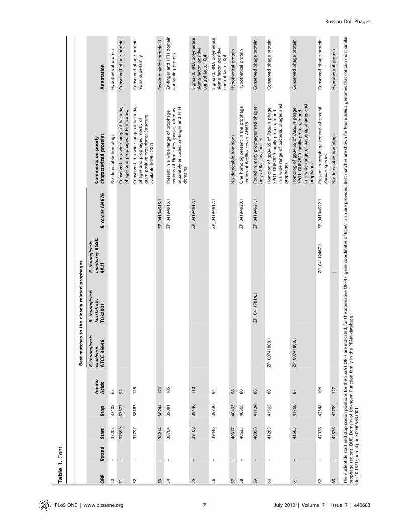
[39]. However, we have detected evidence of recombination in the
MZTP02 region that encompasses at least the gene for the large
terminase subunit of SpaA1. The majority of the ORFs within the
ORF1-ORF18 region (the MZTP02sequence) show best hits into
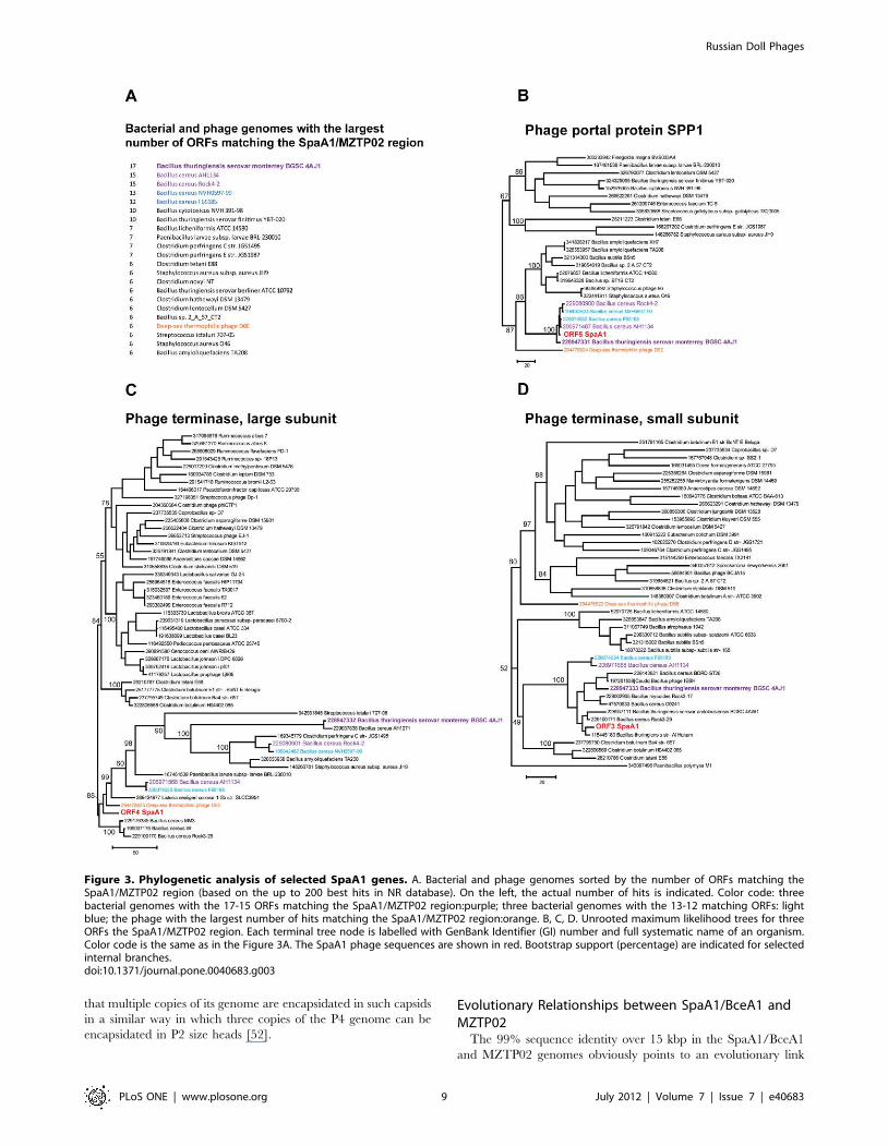
several Bacilli genomes (Figure 3A), and the tree for phage portal
protein SPP1, taken as a typical example, clearly demonstrates
clustering with sequences from these organisms (Figure 3B). In
contrast, the tree for ORF4, the large subunit of phage terminase,
shows very different topology (Figure 3C), suggesting that
notwithstanding the synteny in this region (Figure 2), ORF4
appears to have been acquired from a different, unknown source.
The topology of the tree for ORF3, the small subunit of phage
terminase, was compatible with the typical, SPP1-like topology
(Figure 3B and 3D). Thus, the large subunit gene apparently was
displaced via ‘in situ’ recombination [40], an observation that
further emphasizes the mosaicism in the phage genomes.
Neither the second nor the third genomic modules of SpaA1
completely match any known prophages or phages. Even with the
most closely related phages, such as Cherry [41], EJ [42],
phBC6A51 [43] and the deep-sea thermophilic phage D6E, [44]
there are only a few significantly similar predicted proteins
(Figure 3A and Table 1) indicating that SpaA1 represents a novel
group of tailed phages.
The overall G + C content of the phage is 35.63% strongly
resembling its host S. pasteuri (35%, [45]) as well as the host for
MZTP02 (B. thuringiensis, 35.3%, [46]). No significant differences
in the GC content were detected among the three genomic
modules of SpaA1.
The BceA1 BacteriophageA further search and characterization of bacteriophages from
Antarctic soils identified another temperate bacteriophage, named
BceA1, from a bacterium of the B. cereus/B. thuringiensis group. The
morphology of BceA1 is very similar to that of SpaA1 and hence is
typical of the Siphoviridae family. BceA1 virions also had isometric
heads with a diameter of ,63 nm and flexible tails of ,210 nm in
length (Figure 1B). The genome of phage BceA1 consists of
42,932 bp and like SpaA1 encompasses 63 ORFs. These two
phages are identical apart from ORF47 and the immediate
surrounding area; the SpaA1 ORF47 encodes a protein of 84 aa
and BceA1 a protein of 156 aa. These two proteins have non-
overlapping sets of homologs and hence appear to be unrelated
(Figure 2, Table 1 and data not shown). Although the functions of
both these proteins are unknown, it seems plausible that they are
directly involved in the control of the host range as both SpaA1
and BceA1 could infect B. cereus but only SpaA1 could infect S.
pasteuri (Table 2).
Host Ranges of SpaA1 and BceA1SpaA1 and BceA1 inocula were used to infect B. cereus and S.
pasteuri in a plaque assay. BceA1 produced plaques with a titre of
greater than 107 plaque forming units (pfu)/ml on both bacterial
species but SpaA1 produced plaques with a high titre only on S.
pasteuri (Table 2).
Discussion
The Entire MZTP02 Genome is a Potentially IndependentMobile Element
As pointed out above, the nucleotide sequence of the
‘‘structural’’ module of the SpaA1 and BceA1 genomes is 99%
identical to the sequence of the entire genome of another
bacteriophage, MZTP02 (apart from short 59 - and 39- terminal
regions) [31]; (Figure 2). SpaA1 and BceA1 are similar in this
respect to phage N15 which acquired a module encoding head
and tail protein genes from a lambda-like phage [47]. However
SpaA1 is the first finding of an almost complete phage genome
within the genome of another phage. The presence of similar
inserts in the genomes of B. thuringiensis var. monterrey BGSC 4AJ1
and B. cereus Rock4-2 in the form of a prophage (Figure 2) suggests
that the (nearly) complete MZTP02 genome can travel between
genomes as a distinct entity. The MZTP02 genome does not
contain any identifiable integrase genes so a question arises as to
how it became integrated into these genomes. It is possible that
MZTP02 does not integrate on its own but rather exists as a linear
prophage in the same way as GIL01 [48]. The MZTP02 and
GIL01 genomes are both ,15 kbp long and contain inverted
terminal repeats and 59 terminal genome-linked proteins [31],
[48]. MZTP02 could then possibly recombine with a separate co-
infecting phage and this could have led to the integration of the
resulting composite phage genomes into the bacterial chromo-
some. Alternatively, the integrase of a co-infecting phage could
facilitate integration of MZTP02. The MZTP02 genome encodes
only virion subunits as well as proteins involved in DNA packaging
and capsid assembly (Table 1). We hypothesise that MZTP02 is
likely to be a satellite virus as it does not encode proteins required
for DNA replication and transcription, and more importantly,
proteins involved in cell entry and exit. If this is the case then
MZTP02 probably is unable to infect and replicate in a host
bacterium by itself, but rather depends on co-infection of the host
with a helper virus that remains to be identified. MZTP02 infected
six different B. thuringiensis strains [31] suggesting that such a
putative helper phage must be fairly ubiquitous among B.
thuringiensis strains, possibly as an integrated prophage. A
thoroughly studied satellite bacteriophage is P4, also regarded as
a natural phasmid (phage-plasmid), which depends on phage P2
for reproduction in Escherichia coli [49]. However, in contrast to
MZTP02, P4 possesses genes essential for DNA replication, but
depends on P2 helper genes for the head and tail morphogenesis
and for lysis of the host cell [50]. The size of the head of SpaA1 is
,63 nm which contrasts with the size of 84 nm reported for
MZTP02 [31]. In the P2/P4 helper virus system, two different
capsid sizes are produced from proteins encoded by P2 and a size-
determining protein encoded by P4 produces smaller capsids to
package the smaller P4 DNA [51]. SpaA1 might encode an
unidentified size-determining protein that produces smaller
capsids. A capsid of ,84 nm in size might seem large to
encapsidate the 15.7 kb genome of MZTP02 but it is conceivable
Russian Doll Phages
PLoS ONE | www.plosone.org 8 July 2012 | Volume 7 | Issue 7 | e40683
that multiple copies of its genome are encapsidated in such capsids
in a similar way in which three copies of the P4 genome can be
encapsidated in P2 size heads [52].
Evolutionary Relationships between SpaA1/BceA1 andMZTP02
The 99% sequence identity over 15 kbp in the SpaA1/BceA1
and MZTP02 genomes obviously points to an evolutionary link
Figure 3. Phylogenetic analysis of selected SpaA1 genes. A. Bacterial and phage genomes sorted by the number of ORFs matching theSpaA1/MZTP02 region (based on the up to 200 best hits in NR database). On the left, the actual number of hits is indicated. Color code: threebacterial genomes with the 17-15 ORFs matching the SpaA1/MZTP02 region:purple; three bacterial genomes with the 13-12 matching ORFs: lightblue; the phage with the largest number of hits matching the SpaA1/MZTP02 region:orange. B, C, D. Unrooted maximum likelihood trees for threeORFs the SpaA1/MZTP02 region. Each terminal tree node is labelled with GenBank Identifier (GI) number and full systematic name of an organism.Color code is the same as in the Figure 3A. The SpaA1 phage sequences are shown in red. Bootstrap support (percentage) are indicated for selectedinternal branches.doi:10.1371/journal.pone.0040683.g003
Russian Doll Phages
PLoS ONE | www.plosone.org 9 July 2012 | Volume 7 | Issue 7 | e40683
between these bacteriophages. However, the precise nature of this
link remains unclear given that, firstly, these phages were isolated
from geographically distant regions; SpaA1 and BceA1 in
Antarctica and MZTP02 in China, and secondly, SpaA1 and
MZTP02 were isolated from different host species; Staphylococcus
and Bacillus, respectively. The presence of a sequence identical to
the nearly complete genome of MZTP02 in the genome of SpaA1
suggests the existence of a common host and a common habitat for
the two viruses in the recent past. It seems likely that this common
host is a bacterium of the genus Bacillus. Indeed, BceA1 which is
nearly identical in sequence to SpaA1 and also includes the almost
complete MZTP02 genome within its own genome, was isolated
from a bacterium of the B. cereus/B. thuringiensis group. The
discovery of identical phage sequences in habitats as geographi-
cally and ecologically distant as Antarctica and China might seem
puzzling. However, numerous studies have reported global
distribution of at least some bacteriophages [9], [53] and the
present results suggest that MZTP02 belongs to this class of
ubiquitous phages. There are two alternative evolutionary
scenarios to account for the relationship between MZTP02 and
SpaA1. Firstly, an ancestor of SpaA1 might have possessed a
structural module homologous to MZTP02, and MZTP02 arose
as a result of excision from the ancestral SpaA1/BceA1-like phage.
Alternatively MZTP02 might have evolved elsewhere with
subsequent recombination leading to the integration of MZTP02
into the genome of an ancestor of BceA1/SpaA1 and replacement
of the original structural module of that ancestral phage with the
structural module of MZTP02. Our experiments showed that both
SpaA1 and BceA1 phages can infect B. cereus, but only one of
them, SpaA1, is able to infect S. Pasteuri. These findings are best
compatible with a scenario in which MZTP02 and BceA1 first
evolved in Bacillus spp, the common hosts for these two phages,
whereas SpaA1 evolved later, after ORF47 was replaced in BceA1
by an unrelated gene.
The findings reported here indicate that MZTP02 is not only a
satellite phage but also an independent mobile module that occurs
in the genomes of phages and prophages, resulting in chimeric
viral genomes. To our knowledge, such nested architecture of a
phage genome has not been described previously and seems to
indicate that complete viral genomes could play an even greater
role in genetic exchanges in the prokaryote world than previously
suspected.
Materials and Methods
Ethics StatementAll necessary permits were obtained for the described field
studies. The Garwood Valley falls within the McMurdo dry valleys
Antarctic specially managed area (ASMA) no. 2 designated under
the Protocol on Environmental Protection to the International
Antarctic Treaty. Entry to and field operations in the ASMA
(including sampling and removal of soils, rocks, organisms and
water) for the research described here is regulated by a permit
issued to field party K052, which included D.W. Hopkins, by
Antarctica New Zealand, The International Antarctic Centre,
Orchard Road, Christchurch, New Zealand.
Isolation of Bacteria from Antarctic SoilA soil sample was collected in the Garwood Valley, Antarctica
(78901uS, 163953uE; Ross Dependency Ross Sea region; [54]) in
January 2006, at the site of a soil ecological experiment [55]. The
samples were transported to the UK frozen and stored at 4uC. 1 g
of soil was mixed with 100 ml sterile 0.016 nutrient broth (1022
dilution) and stirred at room temperature for 1 h. Serial dilutions
to 1025 were made in 0.016 nutrient broth and 200 ml of each
dilution was plated onto LB Agar plates and incubated at 20uC.
Bacterial colonies of different appearance were chosen and sub-
cultured three times on LB Agar plates.
Induction and Isolation of BacteriophagesA single colony of the bacterium was grown up overnight in
10 ml LB in a shaking incubator at 28uC. Cells were then
centrifuged for five minutes at 3,0006g; the cell pellet was drained
and resuspended in 2.5 ml 0.01 M Mg2SO4, and 20 ml of
mitomycin c (20 mg/ml) added. Cell suspensions were then shaken
at 28uC for 1 h and washed twice with 2.5 ml 0.01 M Mg2SO4.
Cells were finally resuspended in 10 ml LB and shaken at 28uCovernight. Bacteria were centrifuged as before and the supernatant
was filtered through 0.45 mm syringe filters (Millipore Corpora-
tion, Billerica, MA 01821). Filtrate was centrifuged through a CsCl
step gradient containing 1 ml of each of 1.3 g/ml, 1.5 g/ml and
1.7 g/ml CsCl in an SW41 rotor at 83,0006 g for two hours at
10uC in an OptimaTM L-80 XP ultracentrifuge (Beckman Coulter
Inc.). The middle density layer was collected, diluted at least 1:5 in
SM medium (0.05 M Tris-HCl pH 7.5, 0.1 M NaCl,
0,01 M MgSO4.7H2O) and centrifuged in an R90 Ti rotor for
1.5 hours at 214,0006 g. Pelleted bacteriophage particles were
resuspended in a small volume of SM medium.
Transmission Electron Microscopy (TEM)TEM analysis of virus particles was done as follows: carbon-
coated copper grids were floated for five minutes on 10 ml drops of
samples on wax slides. Grids were then removed from the drops
and excess sample was drained from the grids using filter paper.
Then 10 ml drops of 1% (w/v) phosphotungstic acid pH 6.0–7.0
were put on the grids, left for 30 seconds and then drained from
the grids using filter paper. Grids were examined in a Jeol 100 S
Electron Microscope at 80 kV. Measurements of virus particles
dimensions were done using Adobe Photoshop CS2.
Identification of Bacterial SpeciesBacterial hosts of isolated bacteriophages were identified by
amplifying their 16 S ribosomal RNA genes by PCR and comparing
these sequences to the GenBank database using the BLAST
program available at the National Center for Biotechnology
Information (http://www.ncbi.nlm.nih.gov). A single colony from
a plate was mixed with 50 ml dH2O and heated at 95uC for 4
minutes and 2 ml was then used for PCR. PCR was carried out using
Phusion DNA polymerase (Finnzymes) and primers 63F (CAG-
GCCTAACACATGCAAGTC) and 1387R (GGGCGGTGTG-
TACAAGGC). The PCR products were cut out from 1% agarose
gels and purified using QIAquick gel extraction kit (Qiagen) and
sequenced by Sanger capillary method using primers 63F, 1387R,
V2F (GAGTGGCGGACGGGTGAGTAAT), V3R (CGTAT-
TACCGCGGCTG), V6F (TCGATGCAACGCGAAGAA) and
Table 2. SpaA1 and BceA1 host specificities on S. pasteuriand B. cereus.
Bacterium SpaA1 titera BceA1 titera
S. pasteuri 1.46108 1.76107
B. cereus ,10 5.06107
aTiters are expressed in PFU per milliliter. Means were determined on the basisof the results of three different infections.doi:10.1371/journal.pone.0040683.t002
Russian Doll Phages
PLoS ONE | www.plosone.org 10 July 2012 | Volume 7 | Issue 7 | e40683
V7R (ACATTTCACAACACGAGCTGACGA). The bacterial
host of SpeA1 was identified as Staphylococcus pasteuri with which it
had a greater than 99% identity. The bacterial host of BceA1 was
identified as a member of the Bacillus cereus/Bacillus thuringiensis group
which share greater than 99% identity in the 16 S ribosomal RNA
gene.
Virus Host Range DeterminationThe SpaA1 and BceA1 phages were propagated in LB broth on
S. pasteuri and B. cereus, respectively. Phage preparations were
added to an equivalent volume of mid-log-phase bacteria and
incubated at 30uC with agitation for 24 h. Phage supernatants
were recovered, and this process was repeated until a sufficiently
high-titer phage stock was obtained (.109/ml). All phage
preparations were filter sterilized prior to use. 0.1-ml aliquots of
an overnight LB broth culture were added separately to 0.1 ml of
undiluted phage and each of three 100-fold serial dilutions, in four
sterile, 10-ml, round-bottom polypropylene tubes. After incuba-
tion at 37uC for 15 min, 3 ml of soft LB agar was added to each
tube, gently mixed by inversion, and poured over the surface of a
pre-warmed LB agar plate. Plates were incubated for 24 h at
30uC, and plaques were enumerated to determine the number of
PFU per milliliter.
Isolation of Nucleic Acid from Bacteriophage ParticlesSuspensions of bacteriophage particles were treated with DNase
(Promega) and RNase (Promega) and incubated at 37uC for 30
minutes. The reaction was stopped by adding Stop buffer (10% (v/
v) 0.02 M EGTA) and incubating at 65uC for 10 minutes. The
samples were then incubated with 1/10th volume of 2 M Tris-HCl
pH8.5, 0.2 M EDTA, 1/20th volume 0.5 M EDTA pH8 and an
equal volume of formamide at room temperature for 30 minutes.
Two volumes of 100% ethanol were then added and the samples
kept at 220uC overnight. Samples were then centrifuged at
13,0006 g 8uC in a bench-top Eppendorf 5415R for 20 minutes
and the pellets washed with 70% ethanol, air-dried and resuspended
in TE buffer (0.01 M Tris-HCl pH8, 0.001 M EDTA).
454 Sequencing of Nucleic AcidsRoche 454 sequencing was performed by GenePool (University
of Edinburgh) using 2/16 of a PicoTiterPlate for each phage. For
SpaA1 the FLX platform was used and gave 29338 reads with
median read length 247 bp and an approximate coverage of 1066The later sample for BceA1 used the ‘‘Titanium’’ upgrade and
gave 51597 reads with median read length 320 bp and an
approximate coverage of 186; however this was variable with
regions that had no coverage and gaps were filled in by Sanger
capillary sequencing (see below).
Assembly of 454 SequenceThe 454 reads for SpaA1 were initially assembled with Roche
‘‘Newbler’’ gsAssembler v1.1, later v2.0, however this required
manual intervention to cope with the high coverage. SpaA1 was
then assembled with MIRA v3.2 [56], additional Sanger capillary
sequencing done, and a hybrid assembly performed with MIRA.
This gave one large contig whose ends repeated, giving a
circularised sequence of approximately 43 kb, with no marked
coverage variation to suggest possible end points of the phage’s
linear form (visualized using Tablet, [57]). For BceA1, despite
having more 454 data, de novo assembly was unsuccessful as the
proportion of viral reads was lower. A MIRA reference guided
assembly using the completed SpaA1 sequence suggested the
phage were highly similar, and PCR primers were designed to
close the gaps with additional Sanger capillary sequencing to
confirm this. The final BceA1 assembly was completed manually.
Sequences of the viruses have been submitted to the EMBL
European Nucleotide Archive with accession numbers HE614281
(SpaA1) and HE614282 (BceA1).
Cohesive EndsTo determine the sequences of the SpaA1 genome termini,
PCR with primers annealing close to and directed towards
genome ends was performed using SpaA1 DNA as a template.
The appearance of a distinct PCR product was observed.
Sequence analysis of the PCR product and the SpeA1 genome
end sequences determined by primer walking revealed that the
PCR product contained nine extra base-pairs at the junction site
between the viral DNA ends. The presence of these extra base-
pairs indicates that the ends of the SpeA1 genome form cohesive
39 overhangs.
Annotation and Comparison of the Genomes andPhylogenetic Tree Reconstruction
An initial set of gene predictions was generated using Gene-
Mark.hmm [Version 2.8] [32]. These predictions were then refined
and annotated manually using results of searches against the non-
redundant protein sequence database (NCBI, NIH, Bethesda) using
PSI-BLAST [33] and the Conserved Domain Database using RPS-
BLAST [34]. For each ORF within the OFR1-ORF18 region, up to
200 best PSI-BLAST hits were collected and the taxonomic
distribution of the best hits was generated. The MUSCLE program
[58] was used for construction of multiple amino acid sequence
alignments. Maximum likelihood (ML) phylogenetic trees were
constructed using the MOLPHY program [59] with the JTT
substitution matrix to perform local rearrangement of an original
Fitch tree [60]. MOLPHY was used also to calculate bootstrap
probability which was estimated for each internal branch by using
the resampling of estimated log-likelihoods (RELL) method with
10,000 bootstrap replications. Figure 2 was drawn using Genome-
Diagram [61] and Biopython [62].
Acknowledgments
We thank G. Fraser for technical assistance.
Author Contributions
Conceived and designed the experiments: MMS BR DWH LT EVK MT.
Performed the experiments: MMS BR. Analyzed the data: MMS BE KSM
PJC EVK. Wrote the paper: MMS BR KSM PJC DWH LT EVK MT.
References
1. Suttle CA (2005) Viruses in the sea. Nature 437: 356–61.
2. Edwards RA, Rohwer F (2005) Viral metagenomics. Nat Rev Microbiol 3: 504–10.
3. Casas V, Rohwer F (2007) Phage metagenomics. Meth Enzymol 421: 259–68.
4. Angly FE, Felts B, Breitbart M, Salamon P, Edwards RA, et al. (2006) The
marine viromes of four oceanic regions. PLoS Biol 4: e368.
5. Suttle CA (2007) Marine viruses–major players in the global ecosystem. Nat Rev
Microbiol 5: 801–812.
6. Dinsdale EA, Edwards RA, Hall D, Angly F, Breitbart M, et al. (2008)
Functional metagenomic profiling of nine biomes. Nature 452: 629–632.
7. Kristensen DM, Mushegian AR, Dolja VV, Koonin EV (2010) New dimensions
of the virus world discovered through metagenomics. Trends Microbiol 18: 11–
19.
8. King AMQ, Adams MJ, Carstens EB, Lefkowitz EJ (Eds). (2011) Ninth Report
of the International Committee on Taxonomy of Viruses. San Diego: Elsevier.
Russian Doll Phages
PLoS ONE | www.plosone.org 11 July 2012 | Volume 7 | Issue 7 | e40683
9. Breitbart M, Rohwer F (2005) Here a virus, there a virus, everywhere the same
virus? Trends Microbiol 13: 278–284.10. Fierer N, Breitbart M, Nulton J, Salamon P, Lozupone C, et al. (2007)
Metagenomic and small-subunit RNA analyses reveal the high genetic diversity
of bacteria, archaea, fungi, and viruses in soil. Appl Environ Microbiol 73:7059–7066.
11. Ashelford KE, Day MJ, Fry JC (2003) Elevated Abundance of BacteriophageInfecting Bacteria in Soil. Appl Environ Microbiol 69: 285–289.
12. Williamson KE, Radosevich M, Wommack KE (2005) Abundance and Diversity
of Viruses in Six Delaware Soils. Appl Environ Microbiol 71: 3119–3125.13. Williamson SJ, Rusch DB, Yooseph S, Halpern AL, Heidelberg KB, et al. (2007)
The Sorcerer II Global Ocean Sampling Expedition: Metagenomic Character-ization of Viruses within Aquatic Microbial Samples. PLoS One 3: e1456.
14. Brussow H, Kutter E. (2005) Phage ecology. In: Kutter E, Sulakvelidse A,editors. Bacteriophages: Biology and Applications. Washington, DC: CRC Press.
129–163.
15. Wommack KE, Colwell RR (2000) Virioplankton: viruses in aquatic ecosystems.Microbiol Mol Biol Rev 64: 69–114.
16. Breitbart M, Salamon P, Andresen B, Mahaffy JM, Segall AM, et al. (2002)Genomic analysis of uncultured marine viral communities. Proc Natl Acad Sci
USA 99: 14350–14355.
17. Breitbart M, Felts B, Kelley S, Mahaffy JM, Nulton J, et al. (2004) Diversity andpopulation structure of a nearshore marine sediment viral community.
Proc R Soc Lond B. Biol Sci 271: 565–574.18. Frost LS, Leplae R, Summers AO, Toussaint A (2005) Mobile genetic elements:
the agents of open source evolution. Nat Rev Microbiol 9: 722–732.19. McDaniel LD, Young E, Delaney J, Ruhnau F, Ritchie KB, et al. (2010) High
frequency of horizontal gene transfer in the oceans. Science 330: 50.
20. Sobecky PA, Hazen TH (2009) Horizontal gene transfer and mobile geneticelements in marine systems. Methods Mol Biol 532: 435–453.
21. Fuhrman JA (1999) Marine viruses: biogeochemical and ecological effects.Nature 399: 541–548.
22. Haaber J, Middelboe M (2009) Viral lysis of Phaeocystis pouchetii: Implications for
algal population dynamics and heterotrophic C, N, and P cycling. ISME J 3:430–441.
23. Rohwer F, Thurber RV (2009) Viruses manipulate the marine environment.Nature 459: 207–212.
24. McGrath S, van Sinderen D (2007) Bacteriophage: Genetics and MolecularBiology. Norfolk, England: Caister Academic Press.
25. Ackermann H-W (2007) 5550 Phages examined in the electron microscope.
Arch Virol 152: 277–243.26. Calendar R (2006) The bacteriophages. Oxford University Press.
27. Kutter E, Sulakvelidze A (2005) Bacteriophages: biology and applications. BocaRaton, Florida: CRC Press.
28. Hendrix RW, Lawrence JG, Hatfull GF, Casiens S (2000) The origins and
ongoing evolution of viruses. Trends Microbiol 8: 504–508.29. Hatfull GF, Cresawn SG, Hendrix RW (2008) Comparative genomics of the
mycobacteriophages: insights into bacteriophage evolution. Res Microbiol 159:332–339.
30. Pope WH, Jacobs-Sera D, Russell DA, Peebles CL, Al-Atrache Z, et al. (2011)Expanding the diversity of mycobacteriophages: insights into genome architec-
ture and evolution. PLoS One 6: e16329.
31. Liao W, Song S, Sun F, Jia Y, Zeng W, et al. (2008) Isolation, characterizationand genome sequencing of phage MZTP02 from Bacillus thuringiensis MZ1. Arch
Virol 153: 1855–1865.32. Lukashin AV, Borodovsky M (1998) GeneMark.hmm: new solutions for gene
finding. Nuc Acids Res 26: 1107–1115.
33. Altschul SF, Madden TL, Schaffer AA, Zhang J, Zhang Z, et al. (1997) GappedBLAST and PSI-BLAST: a new generation of protein database search
programs. Nucleic Acids Res 25: 3389–402.34. Marchler-Bauer A, Anderson JB, Chitsaz F, Derbyshire MK, DeWeese-Scott C,
et al. (2009) CDD: specific functional annotation with the Conserved Domain
Database. Nucleic Acids Res 37 (Database issue), D205–210.35. Klumpp J, Calendar R, Loessner MJ (2010) Complete nucleotide sequence and
molecular characterization of Bacillus phage TP21 and its relatedness to otherphages with the same name. Viruses 2: 961–971.
36. Katsura I (1990) Mechanism of length determination in bacteriophage lambda
tails. Adv Biophys 26: 1–18.37. Xu J, Hendrix RW, Duda RL (2004) Conserved translational frameshift in
dsDNA bacteriophage tail assembly genes. Mol. Cell 16 11–21.
38. Theis C, Reeder J, Giegerich R (2008) KnotInFrame: prediction of 21ribosomal frameshift events. Nucleic Acids Re 36: 6013–6020.
39. Casjens SR, Gilcrease EB, Winn-Stapley DA, Schiklmaier P, Schmieger H, et al.(2005) The generalized transducing Salmonella bacteriophage ES18: complete
genome sequence and DNA packaging strategy. J Bacteriol 187: 1091–1104.
40. Omelchenko MV, Makarov KS, Wolf YI, Rogozin IB, Koonin EV (2003)Evolution of mosaic operons by horizontal gene transfer and gene displacement
in situ. Genome Biol 4: R55.41. Fouts DE, Rasko DA, Cer RZ, Jiang L, Fedorova NB, et al. (2006) Sequencing
Bacillus anthracis typing phages gamma and cherry reveals a common ancestry.J. Bacteriol 188: 3402–3408.
42. Romero P, Lopez R, Garcıa E (2004) Genomic organization and molecular
analysis of the inducible prophage EJ-1, a mosaic myovirus from an atypicalpneumococcus. Virology 322: 239–252.
43. Ivanova N, Sorokin A, Anderson I, Galleron N, Candelon B, et al.(2003)Genome sequence of Bacillus cereus and comparative analysis with Bacillus
anthracis. Nature 423: 87–91.
44. Wang Y, Zhang X (2010) Genome analysis of deep-sea thermophilic phageD6E. Appl Environ Microbiol 76: 7861–7866.
45. Chesneau O, Morvan A, Grimont F, Labischinski H, El Solh N (1993)Staphylococcus pasteuri sp. nov., isolation from human, animal, and food specimens.
Int J Syst Bacteriol 43: 237–244.46. He J, Shao X, Zheng H, Li M, Wang J, et al. (2010) Complete genome sequence
of Bacillus thuringiensis mutant strain BMB171. J Bacteriol 192: 4074–4075.
47. Ravin NV (2011) N15: The linear phage-plasmid. Plasmid 65: 102–109.48. Verheust C, Jensen G, Mahilon J (2003) pGIL01, a linear tectiviral plasmid
prophage originating from Bacillus thuringiensis serovar israelensis. Microbiology149: 2083–2092.
49. Christie GE, Calendar R (1990) Interactions between satellite bacteriophage P4
and its helpers. Ann Rev Genet 24: 465–90.50. Briani F, Deho G, Forti F, Ghisotti D (2001) The plasmid status of satellite
bacteriophage P4. Plasmid 45: 1–17.51. Shore D, Deho G, Tsipis J, Goldstein R (1978) Determination of capsid size by
satellite bacteriophage P4. Proc Natl Acad Sci (USA) 75: 400–404.52. Pruss G, Goldstein RN, Calendar R (1974) In vitro packaging of satellite phage
P4 DNA. Proc Natl Acad Sci (USA) 71: 2367–2371.
53. Thurber R (2009) Current insights into phage biodiversity and biogeography.Curr Opin Microbiol 12: 582–587.
54. Elberling B, Gregorich EG, Hopkins DW, Sparrow AD, Novis P, et al. (2006)Distribution and dynamics of soil organic matter in an Antarctic dry valley. Soil
Biol Biochem 38: 3095–3106.
55. Hopkins DW, Sparrow AD, Elberling B, Gregorich EG, Novis PM, et al. (2006)Carbon, nitrogen and temperature controls on microbial activity in soils from an
Antarctic dry valley. Soil Biol Biochem 38: 3130–3140.56. Chevreux B, Wetter T, Suhai S (1999) Genome sequence assembly using trace
signals and additional sequence information. Computer Science and Biology:Proceedings of the German Conference on Bioinformatics (GCB) 99: 45–56.
57. Milne I, Bayer M, Cardle L, Shaw P, Stephen G, et al. (2010) Tablet - next
generation sequence assembly visualization. Bioinformatics 26: 401–402.58. Edgar RC (2004) MUSCLE: multiple sequence alignment with high accuracy
and high throughput. Nucleic Acids Res 32: 1792–1797.59. Adachi J, Hasegawa M (1992) MOLPHY: programs for molecular phyloge-
netics. In Computer Science Monographs 27; Institute of Statistical Mathemat-
ics.60. Fitch WM, Margoliash E (1967) Construction of phylogenetic trees. Science 155:
279–284.61. Pritchard L, White JA, Birch PRJ, Toth IK (2006) GenomeDiagram: a python
package for the visualization of large-scale genomic data. Bioinformatics 22:
616–617.62. Cock PJ, Antao T, Chang JT, Chapman BA, Cox CJ, et al. (2009) Biopython:
freely available Python tools for computational molecular biology andbioinformatics. Bioinformatics 25: 1422–1423.
Russian Doll Phages
PLoS ONE | www.plosone.org 12 July 2012 | Volume 7 | Issue 7 | e40683