Embed Size (px)
Citation preview

Novel Lignan and Stilbenoid Mixture ShowsAnticarcinogenic Efficacy in Preclinical PC-3M-luc2Prostate Cancer ModelEmrah Yatkin1,2., Lauri Polari1,2., Teemu D. Laajala3, Annika Smeds4, Christer Eckerman4,
Bjarne Holmbom4, Niina M. Saarinen1,2, Tero Aittokallio3,5, Sari I. Makela1,2*
1 Functional Foods Forum, University of Turku, Turku, Finland, 2 Turku Center for Disease Modeling (TCDM), Institute of Biomedicine, University of Turku, Turku, Finland,
3 Department of Mathematics and Statistics, University of Turku, Turku, Finland, 4 Abo Akademi University, Process Chemistry Centre, Laboratory of Wood and Paper
Chemistry, Turku, Finland, 5 Institute for Molecular Medicine Finland (FIMM), University of Helsinki, Helsinki, Finland
Abstract
Prostate cancer is the most common cancer of men in the Western world, and novel approaches for prostate cancer riskreduction are needed. Plant-derived phenolic compounds attenuate prostate cancer growth in preclinical models by severalmechanisms, which is in line with epidemiological findings suggesting that consumption of plant-based diets is associatedwith low risk of prostate cancer. The objective of this study was to assess the effects of a novel lignan-stilbenoid mixture inPC-3M-luc2 human prostate cancer cells in vitro and in orthotopic xenografts. Lignan and stilbenoid –rich extract wasobtained from Scots pine (Pinus sylvestris) knots. Pine knot extract as well as stilbenoids (methyl pinosylvin and pinosylvin),and lignans (matairesinol and nortrachelogenin) present in pine knot extract showed antiproliferative and proapoptoticefficacy at $40 mM concentration in vitro. Furthermore, pine knot extract derived stilbenoids enhanced tumor necrosisfactor-related apoptosis-inducing ligand (TRAIL) induced apoptosis already at $10 mM concentrations. In orthotopic PC-3M-luc2 xenograft bearing immunocompromized mice, three-week peroral exposure to pine knot extract (52 mg of lignans andstilbenoids per kg of body weight) was well tolerated and showed anti-tumorigenic efficacy, demonstrated by multivariateanalysis combining essential markers of tumor growth (i.e. tumor volume, vascularization, and cell proliferation). Methylpinosylvin, pinosylvin, matairesinol, nortrachelogenin, as well as resveratrol, a metabolite of pinosylvin, were detected inserum at total concentration of 7273 mM, confirming the bioavailability of pine knot extract derived lignans and stilbenoids.In summary, our data indicates that pine knot extract is a novel and cost-effective source of resveratrol, methyl pinosylvinand other bioactive lignans and stilbenoids. Pine knot extract shows anticarcinogenic efficacy in preclinical prostate cancermodel, and our in vitro data suggests that compounds derived from the extract may have potential as novelchemosensitizers to TRAIL. These findings promote further research on health-related applications of wood biochemicals.
Citation: Yatkin E, Polari L, Laajala TD, Smeds A, Eckerman C, et al. (2014) Novel Lignan and Stilbenoid Mixture Shows Anticarcinogenic Efficacy in Preclinical PC-3M-luc2 Prostate Cancer Model. PLoS ONE 9(4): e93764. doi:10.1371/journal.pone.0093764
Editor: Rajesh Agarwal, University of Colorado Denver, United States of America
Received January 13, 2014; Accepted March 8, 2014; Published April 3, 2014
Copyright: � 2014 Yatkin et al. This is an open-access article distributed under the terms of the Creative Commons Attribution License, which permitsunrestricted use, distribution, and reproduction in any medium, provided the original author and source are credited.
Funding: This project was supported by the FIBIC Finnish Bioeconomy Cluster, the BioRefine program of Finnish Funding Agency for Technology and Innovation,and Academy of Finland. The funders had no role in study design, data collection and analysis, decision to publish, or preparation of the manuscript.
Competing Interests: The authors have declared that no competing interests exist.
* E-mail: [email protected]
. These authors contributed equally to this work.
Introduction
Prostate cancer (PCa) is the most common cancer of men in
North America, Western Europe, Eastern Europe, and Scandina-
via. The long natural history of PCa presents a relatively wide time
window for dietary or pharmacological interventions that could
manifest as reduction in incidence, recurrence, morbidity or
progression of the disease [1]. Proposed modifiable risk factors for
PCa that provide potential targets for prevention include
inflammation, steroid hormones and their receptors, obesity,
hypercholesterolemia and dietary factors [2]. Natural phenolic
compounds, possessing anticarcinogenic properties, may offer
interesting possibilities for the reduction of cancer burden [3,4].
However, more data on their efficacy and mechanisms of action is
needed from relevant preclinical models, in order to select the
optimal compounds or mixtures for clinical trials and product
development.
Trees are an abundant source of phenolic compounds,
structurally identical or similar to those in edible plants [5]. These
compounds can easily be isolated from wood by hydrophilic
extraction after removing the lipophilic extractives by hexane
extraction [6]. Wood-derived extracts are thus a cost-effective
source of natural phenolic compounds, and an attractive option
for the development of novel health-promoting products.
Plant phenolics, such as lignans and stilbenoids, modulate
several important biological processes in mammalian cells [7,8],
and show anticarcinogenic properties in preclinical PCa models.
Rye or flaxseed-containing diets, which have a high lignan
content, as well as a diet supplemented with a wood-derived plant
lignan, 7-hydroxymataresinol (HMR), inhibit the growth of PCa in
in vivo [9,10,11,12]. Similarly, plant-derived stilbenoids, resveratrol
PLOS ONE | www.plosone.org 1 April 2014 | Volume 9 | Issue 4 | e93764

(RV) and pterostilbene have been reported to suppress the growth
of PCa in vivo [13]. Several possible mechanisms, by which lignans
and stilbenoids may exert their anticarcinogenic actions in PCa
have been identified, including cell cycle arrest and induction of
apoptosis [14,15], inhibition of cell proliferation [16,17], tumor
vascularization [18], modulation of cytokine profile [19], and
sensitization of cancer cells to programmed cell death [20,21,22].
Tumor Necrosis Factor-Related Apoptosis-Inducing Ligand
(TRAIL) –mediated apoptosis has been recently identified as an
interesting target for lignans and stilbenoids. TRAIL is a protein
functioning as a ligand, which activates a death-signaling pathway
especially in cancer cells, with minimal toxicity to normal tissues
[23]. Resistance to TRAIL is common in PCa, and compounds
that sensitize PCa to TRAIL are currently under active
investigation. Interestingly, lignans, such as matairesinol (MR),
and nortrachelogenin (NTG), as well as RV enhance the
antitumor activity of TRAIL in PCa cells [21,22] or xenografts
[18].
Experimental studies thus suggest that natural lignans and
stilbenoids attenuate PCa growth via numerous mechanisms of
action. It is, therefore, plausible to propose that combination of the
two groups of phenolic compounds may offer additional benefits
over the use of single compounds. However, no published data
exists on the combined effects of lignans and stilbenoids in
controlled experimental settings. In this study, we demonstrated
growth-inhibitory effects of pine knotwood extract (PKE), rich in
lignans and stilbenoids, in PC-3M-luc2 human PCa cells in vitro
and in vivo. Furthermore, we confirmed the bioavailability of PKE-
derived phenolics, and identified the compounds likely to account
for the anticarcinogenic effects of PKE.
Materials and Methods
Ethics StatementAnimal care and use was conducted in accordance with the
Finnish Act on Animal Experimentation and EU laws, guidelines,
and recommendations. All studies were approved by the national
Animal Experiment Board in Finland (License number 1993/
04.10.03/2011).
Preparation and Chemical Characterization of PKEScots pine knots were obtained from an industrial pulp mill
process (so-called stone trap) (UPM Tervasaari, Valkeakoski,
Finland). The knots were ground, screened (,2 mm), freeze-dried,
and extracted sequentially with n-hexane at 90uC (365 min) and
ethanol-water (95:5 by vol.) at 100uC (365 min) using accelerated
solvent extraction. The PKE was chemically characterised by GC-
flame ionization detection (FID), GC-MS and by high-perfor-
mance size-exclusion chromatography with evaporative light-
scattering detection (HPSEC-ELSD). The GC-FID, GC-MS, and
HPSEC-ELSD analyses were performed as described previously
[24]. GC-FID was used for quantification of the components in
the extract that eluted from the GC column. The quantification
was based on the triplicate analyses of the same extract.
Heneicosanoic acid was used as internal standard, with the
response factors 1.00. The individual compounds detected by GC-
FID were identified by GC-MS, using commercial and the
laboratory’s own spectral libraries. The molar mass distribution of
the PKE components was determined by HPSEC-ELSD.
The major compound groups in the PKE were: lignans (16%),
stilbenoids (17%), oxidized resin acids (20%), resin acids (24%),
and higher-molar-mass compounds (550–4000 Da, 18%). The
concentrations (wt-% of dry extract) of the main GC-detectable
phenolics in the PKE were pinosylvin monomethyl ether (MePS,
10.2%), NTG (7.0%), pinosylvin (PS, 4.0%), MR (1.5%), abietic
acid (1.5%), and pinostilbene (0.4%). The GC-detectable resin
acids accounted for 21.8% of the dry weight of the PKE. Minor
amounts (,0.1–0.5%) of lignans HMR, conidendric acid,
secoisolariciresinol and todolactol A were found.
Reference and Test CompoundsThe preparation and purities of the lignans HMR, MR, NTG,
secoisolarici-, larici-, cyclolarici-, 7-oxomatai-, pino-, and medior-
esinol, a-conidendrin, enterolactone, 7-hydroxyenterolactone,
MR-d6, and enterolactone-d6 have been described previously
[25,26]. The stilbenes PS and MePS and the resin acid
dehydroabietic acid (purities .95%) were prepared in the
Laboratory of Wood and Paper Chemistry at Abo Akademi
University by isolation from wood and purification by flash
chromatography (Biotage Flash 40i) with silica columns. RV and
enterodiol were from Sigma-Aldrich Co, pinostilbene from TCl
Pharma Chem., Inc (Vaughan, ON, Canada) and O-methylpo-
docarpic acid, neoabietic acid and abietic acid from Helix Biotech
Corp. (CanSyn Chem Corp., Toronto, Canada).
In order to study the combined effects of pure PKE-derived
lignans and stilbenoids in vitro, we prepared a lignan-stilbene
mixture (LS mixture) containing the four most abundant PKE-
derived compounds in molar ratios similar to PKE (Table 1).
Cell Culture and Proliferation, Apoptosis and Cell CycleAssays
PC-3M-luc2 cells (Caliper Life Sciences, Hopkinton, MA) were
cultured in Dulbeccos’s phenol red-free modified Eagle medium
supplemented with 50 IU/ml penicillin, 50 mg/ml streptomycin,
2 mM L-glutamine, and 10% heat-inactivated fetal bovine serum
(Invitrogen, Paisley, UK). The cells were maintained in a
humidified 5% CO2/air atmosphere at 37uC.
Table 1. Concentrations of four main lignans and stilbenoids in pine knot extract (PKE) and composition of lignan-stilbenoid (LS)mixture.
Polyphenol PKE PKE (40 mg/l) LS mixture (40 mM)
(w/w %) (mM) (mM)
Methylpinosylvin 10 20 21.4
Nortrachelogenin 7 7 6.8
Pinosylvin 4 10 9.6
Matairesinol 1.5 2 2.2
doi:10.1371/journal.pone.0093764.t001
Lignans and Stilbenoids in Prostate Cancer
PLOS ONE | www.plosone.org 2 April 2014 | Volume 9 | Issue 4 | e93764

For cell proliferation assay, PC-3M-luc2 cells were seeded into a
96-well plate (1500 cells per well). Dimethyl sulfoxide stock
solutions of PKE (1–100 mg/l), purified compounds (1–100 mM)
or dimethyl sulfoxide controls prepared in culture medium were
added to wells (six replicates). After 48 hours, cell proliferation was
measured with Cell Proliferation ELISA BrdU immunoassay kit
(Roche Diagnostics, Mannheim, Germany) according to the
manufacturer’s instructions.
For apoptosis assay, PC-3M-luc2 cells were seeded into a 96-
well plate. When 60–80% confluent half of the medium was
removed and replaced with the medium containing the wood-
derived compounds (final concentrations 1–40 mg/l). After 48
hours, cells were detached with trypsin and disrupted with 0.4 M
citrate buffer in PBS containing 0.3% triton-X (Sigma Life
Sciences, St. Louis, MO). Nuclei and nuclear fragments were
labeled with propidium iodide (Sigma). To determine the fraction
of sub-G0/G1 events, samples were analyzed with flow cytometer
(BD LSR II, BD Biosciences, Franklin Lakes, NJ, USA). For
assessment of TRAIL sensitivity, cells were treated with combi-
nation of wood derived compounds (as above) and human
TRAIL/Apo2L (cat. no. C-63600, Promokine, Heidelberg,
Germany) at concentrations of 3, 25, 50, and 100 ng/ml.
Orthotopic PC-3M-luc2 Xenografts in Athymic MiceHsd:Athymic Nude – Foxn1nu male mice (5–6-week-old)
purchased from Harlan Laboratories (Horst, The Netherlands)
were housed in aseptic conditions in the Central Animal
Laboratory of the University of Turku. The mice were acclima-
tized for two weeks; provided with soy-free natural ingredient diet
(RM3, SDS, Essex, UK) and tap water ad libitum.
PC-3M-luc2 cells used in orthotopic xenograft experiment were
cultured in RPMI 1640 phenol red-free medium supplemented
with 50 IU/ml penicillin, 50 mg/ml streptomycin, 2 mM L-
glutamine, and 10% heat inactivated fetal bovine serum. The
Figure 1. Pine knot extract (PKE) and its polyphenols inhibit the proliferation of PC-3M-luc2 prostate cancer cells. A) Cells weretreated with PKE or a mixture of its main lignans and stilbenoids (LS mixture) for 48 hours. B) Cells were treated with individual PKE derivedcompounds for 48 hours. Cell proliferation rate expressed as percent of control was measured with BrdU assay. The data are means of threeindependent experiments 6 SEM. Statistical significance was determined by non-parametric t-test for each treatment in comparison to respectivevehicle treatment (Ctrl). *p,0.05, **p,0.01, ***p,0.001.doi:10.1371/journal.pone.0093764.g001
Lignans and Stilbenoids in Prostate Cancer
PLOS ONE | www.plosone.org 3 April 2014 | Volume 9 | Issue 4 | e93764

cells were maintained in a humidified 5% CO2/air atmosphere at
37uC. PC-3M-luc2 cells (16106 cells in 20 ml plain RPMI 1640
medium) were inoculated into the dorsolateral prostate through an
abdominal incision under isoflurane anesthesia. For pain relief,
mice were given buprenorphine (Temgesic, 0.05–0.1 mg/kg s.c.)
and carpofen (Rimadyl, 5 mg/kg s.c.) before and after the
operation, respectively. Tumor growth was followed by biolumi-
nescence imaging with IVIS lumina II, equipped with XGI-8 Gas
Anesthesia System (Caliper Life Sciences, Runcorn, UK). Mice
were anesthetized with isoflurane, injected i.p. with 150 mg/kg D-
luciferin (Xenogen cat. no. XR-1001, Oregon, USA) 10 min prior
to imaging, and bioluminescence was quantified using Living-
Image 4.09A Carbon software (Xenogen).
One week after inoculation, tumor-free animals were identified
by bioluminescence imaging and excluded from the study. Tumor
bearing mice were allocated into vehicle (control), low dose
(32 mg/kg) or high dose (160 mg/kg) PKE groups. Low and high
dose contained approximately 5.0 and 5.4, and 25 and 27 mg of
lignans and stilbenoids, respectively. PKE was dissolved in a
vehicle containing 10% (v/v) of absolute ethanol and 90% (v/v)
corn oil. Both vehicle and PKE were gavaged p.o. daily. Mice were
weighed weekly, and bioluminescence imaging was carried out
twice a week.
During the third week of treatment 24-h urine samples were
collected in metabolic cages (5–6 mice per cage). Urine was
collected in jars containing 0.15 M sodium azide and 0.56 M
ascorbic acid as preservatives. Urine samples were centrifuged and
stored in 220uC until analyzed.
After three-week treatment, the mice were sacrificed with CO2
suffocation followed by cervical dislocation. Blood was collected
via heart puncture, centrifuged, and the serum was separated and
Figure 2. PKE and its compounds induce A) apoptosis and B)G0/G1 cell cycle arrest of PC-3M-luc2 cells in vitro. Apoptosisrates and number of cells in G0/G1 phase are expressed relative tovehicle control (Ctrl = 1). The cells were treated with test compounds for48 hours and apoptosis rate and cell cycle phase was determined withflow cytometer. LS, mixture of main PKE lignans and stilbenes; MePS,pinosylvin monomethyl ether; NTG, nortrachelogenin; PS, pinosylvin;MR, matairesinol; Ab, abietic acid; RV, resveratrol. The data are means ofthree independent experiments 6 SEM. Statistical significance wasdetermined by non-parametric t-test for each treatment in comparisonto respective Ctrl. *p,0.05, **p,0.01, ***p,0.001.doi:10.1371/journal.pone.0093764.g002
Figure 3. PKE and its compounds sensitize PC-3M-luc2 cells toTRAIL -induced apoptosis in vitro. A) Cells treated with PKE incombination with increasing concentrations of human TRAIL (02100 ng/ml), values are percentages of nuclear fragmentation. B) Cellstreated with PKE, LS mixture, or with individual PKE compounds incombination with human TRAIL (25 ng/ml), values are relativeapoptosis rates (Ctrl = 1). The cells were treated with test compoundsfor 48 hours and nuclear fragmentation was determined with flowcytometer. LS, mixture of main PKE lignans and stilbenes; MePS,pinosylvin monomethyl ether; NTG, nortrachelogenin; PS, pinosylvin;MR, matairesinol; Ab, abietic acid; RV, resveratrol. The data are means ofthree independent experiments 6 SEM. Statistical significance wasdetermined by non-parametric t-test for each treatment in comparisonto respective vehicle treatment (Ctrl). *p,0.05, **p,0.01, ***p,0.001.doi:10.1371/journal.pone.0093764.g003
Lignans and Stilbenoids in Prostate Cancer
PLOS ONE | www.plosone.org 4 April 2014 | Volume 9 | Issue 4 | e93764
kept in 270uC until analyzed. The tumors were dissected and
weighed, and the tumor size was measured with a caliper in three
dimensions. The tumor volume was calculated using the formula:
volume (mm3) = (length 6 width 6 height) 6 p/6. The tumors
were fixed in 10% neutral buffered formalin and processed for
paraffin embedding and used for immunohistochemical analyses.
Analysis of Lignans and Stilbenoids Mouse Serum andUrine
Serum (50 ml) and urine (300 ml) samples were hydrolyzed and
solid-phase extracted as previously described with some modifica-
tions [25,27]. For hydrolysis, a freshly prepared b-glucuroni-
dase/2sulphatase in 10 mM of sodium acetate buffer (pH 5.0)
was added to serum and urine samples and incubated for 19 h at
37uC. After the hydrolysis, internal standards were added to the
samples: MR-d6 for plant lignans and stilbenes, EL-d6 for
enterolignans (final concentrations ,1 mg/ml), and O-methylpo-
docarpic acid for the resin acids (final concentration ,3 mg/ml).
The solid-phase extracted and evaporated serum and urine
samples [25] were redissolved in 150 ml and 500 ml methanol/
0.1% acetic acid 20:80 (v/v), respectively. The PKE components
and their metabolites were quantified using HPLC-MS/MS with
multiple ion monitoring in the negative ion mode as described
previously [24]. The optimized multiple ion monitoring transitions
were the deprotonated molecular ion and the following fragments
in MS2 (m/z); PS 169 18; MePS 182 20; RV 143, 22; pinostilbene
225. The resin acids formed no fragments in MS2. The used cone
voltages were 40 V for the stilbenes and 48 V for the resin acids.
The collision energies were around 20 eV for the stilbenes and
5.0 eV for the resin acids. The MS parameters for the analyzed
lignans have been described previously [26]. The quantifications
were carried out using standard solutions containing the internal
standards and six concentration levels of the analytes as described
previously [26].
Immunohistochemistry and Detection of Apoptotic Cellsin Tumor Samples
For von Willebrand factor (vWF, dilution: 1:4000, ab6994-100,
Abcam, Cambridge, U.K.) and phospho-Histone H3 (pH-H3,
dilution: 1:200, Ser10, Cell signaling) immunohistochemistry,
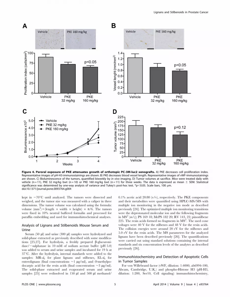
Figure 4. Peroral exposure of PKE attenuates growth of orthotopic PC-3M-luc2 xenografts. A) PKE decreases cell proliferation index.Representative images of pH-H3-immunostainings are shown. B) PKE decreases blood vessel length. Representative images of vWF-immunostainingsare shown. C) Bioluminescence of the tumors, quantified biweekly by in vivo imaging. D) Tumor volumes at sacrifice. Mice were treated daily withvehicle (n = 11), PKE 32 mg/kg bwt (n = 10) or PKE 160 mg/kg bwt (n = 11) for three weeks. The data is expressed as mean 6 SEM. Statisticalsignificance was determined by one-way analysis of variance and Tukey’s post-hoc test. *p,0.05. Scale bars, 100 mm.doi:10.1371/journal.pone.0093764.g004
Lignans and Stilbenoids in Prostate Cancer
PLOS ONE | www.plosone.org 5 April 2014 | Volume 9 | Issue 4 | e93764

tissue section were deparaffinized, rehydrated, and incubated in
10 mM Tris-EDTA pH 9 (for vWF staining) or in 10 mM sodium
citrate pH 6 (for pH-H3 staining) buffer, in a microwave oven for
antigen retrieval. The endogenous peroxidase activity was blocked
with 3% H2O2. After rinsing with PBS, the sections were
incubated with 3% bovine serum albumin (BSA) for 10 minutes,
and labeled with vWF or pH-H3 antibodies for 60 minutes at
room temperature (RT) or overnight at +4uC, respectively.
Horseradish peroxidase labeled polymer anti-rabbit (DAKO
envision system, Dako) was used as a secondary antibody.
Diaminobenzidine (Dako) was applied to the sections followed
by Mayer’s hematoxylin and mounting.
The terminal deoxynucleotidyl transferase biotin-dUTP nick
end labeling (TUNEL) method was used to determine apoptotic
cells in tumor sections. The analysis was performed using
ApopTag Peroxidase in situ Apoptosis Detection Kit (Chemicon
International, Hampshire, UK) according to the manufacturer’s
instructions. The sections were deparaffinized, rehydrated and
treated with 10 mM sodium citrate buffer in microwave for
antigen retrieval. The endogenous peroxidase activity was blocked
with 3% H2O2. For positive control, one section was incubated
with DNase I (Invitrogen) for 30 min at +37uC. For the TUNEL,
sections were incubated with 0.8 U/ml TdT reaction mix: TdT
buffer, 4 U/ml transferase, 25 mM CoCl2 (Terminal transferase,
Roche, Mannheim, Germany), biotin-16-dUTP (Roche) and
MilliQ water; and for the negative control, one section was
incubated with TUNEL reaction mix without transferase for one
hour at +37uC. TUNEL reaction was stopped with 300 mM
NaCl, 30 mM sodium citrate in dH2O (15 min at RT).
Nonspecific sites were blocked with 3% BSA/PBS for 30 min at
RT. ExtrAvidineH-Peroxidase 1:500 (Sigma) was applied in 1%
BSA/PBS for 30 min at +37uC, after which diaminobenzidine was
applied and the sections were stained with Mayer’s hematoxylin
and mounted.
Quantification of Proliferation and Apoptosis Indices, andBlood Vessel Density and Length
Stained sections were scanned with a virtual microscope (Digital
Virtual Microscope, Soft Imaging System, Olympus, Germany).
The number and length of vWF-positive vessels was quantified in
three or four selected areas by using the measurement tool of
dotSlide program (Soft Imaging System). The quantification of
pH-H3 and TUNEL-positive cell in tumor sections was carried
out with custom functions developed for tissue image analysis by
Quva Ltd (Tampere, Finland). First, the images obtained from
scanned slides were normalized for intensity and color variations.
Processing was continued by image thresholding, which detects
areas with suitable color to be considered as the positively stained
target cells. Finally, extraneous detections were filtered out by size
and shape information, and the resulting cells were visualized in
red spots over the original image.
Statistical AnalysesThe univariate analyses were performed using GraphPad Prism
version 4.00 for Windows (GraphPad Software Inc., San Diego,
Figure 5. Multivariate testing and visualization of the relationships among vascularization, proliferation and tumor volume A)Vehicle vs. low dose comparison, and B) Vehicle vs. high dose comparison (p = 0.0051) using MD (p = 0.4195). C) Tumor volume, vessellength and proliferation index were detected as the most important factors of differences in the multivariate analyses. The hyperplane indicates thattwo of the groups (vehicle in black and high dose in green) were linearly separable when the three factors were considered together (Hyperplane:100.55 = 0.1046Tumor volume +0.8956Proliferation index +21.16Vessel length).doi:10.1371/journal.pone.0093764.g005
Lignans and Stilbenoids in Prostate Cancer
PLOS ONE | www.plosone.org 6 April 2014 | Volume 9 | Issue 4 | e93764

CA). In normally distributed data, the one-way analysis of
variance and Tukey’s post-hoc test were used; otherwise, the
Kruskal-Wallis or non-parametric t-test was used. All data are
presented as group mean6SEM. Differences were considered
statistically significant at p,0.05.
Pairwise multivariate comparisons of group means (say m1 and
m2) were tested with the Mahalanobis distance (MD):
D(m1,m2)~(m1{m2)T S{1(m1{m2): ð1Þ
Mahalanobis distance is a well-known multivariate statistic,
which incorporates linear correlations of covariates through the
covariance-variance matrix S, with dimension equal to the number
of covariates [28]. By accumulating complementary information
from interconnected factors and having sensitivity to patterns
differing from the major trends [29], MD is a convenient statistic
for testing more subtle treatment effects potentially missed by the
univariate tests. In special cases, MD reduces to the standardized
Euclidean distance, if S is a diagonal matrix, and to the ordinary
Euclidean distance, if S is a unit matrix, and thus links closely to
the conventional t statistic testing of independent tumor growth
markers. Statistical significance of the distance D was evaluated by
random permutation of the class labels in the two-sample tests
(Vehicle vs. Low dose or Vehicle vs. High dose) [30]. The null
distributions were obtained by running 100,000 simulations of
such randomly generated distances and the empirical p-values
were defined as the proportion of random distances greater than
or equal to the observed distance D.
Results
PKE and its Components Inhibit PC-3M-luc2 CellProliferation and Induce Apoptosis in vitro
PKE significantly attenuated PC-3M-luc2 cell proliferation at $
40 mg/l (Fig. 1A), demonstrated by reduced incorporation of
BrdU. In line with this, the main constituents of PKE, stilbenoids
(MePS and PS), as well as lignans (MR and NTG) each
significantly inhibited cell proliferation at 40–100 mM concentra-
tion (Fig. 1B). Furthermore, combination of these four compounds
(MePS, PS, MR, and NTG), in the same ratio as present in PKE
(lignan-stilbene, LS, mixture), reduced BrdU incorporation in a
manner similar to the original extract (Fig. 1C). PKE (40 mg/l)
and LS mixture (40 mM), both increased apoptosis rate, demon-
strated by flow cytometric analysis (Fig. 2A). PKE induced cell
cycle arrest, i.e. increased the fraction of non-apoptotic cells in
G0/G1 (Fig. 2B), and, respectively, decreased the fraction of cells
in S and G2/M phases. Moreover, PKE (10 and 40 mg/ml)
sensitized PC-3M-luc2 cells to TRAIL-induced apoptosis (Fig. 3A).
LS mixture and all the tested PKE-derived compounds (MePS,
NTG, PS, MR, and RV), except abietic acid, significantly
enhanced TRAIL-mediated apoptosis (Fig. 3B), PS, MePS, and
MR showing the most pronounced effects.
Effect of PKE on Orthotopic PC-3M-luc2 XenograftVolume, Cell Proliferation and Angiogenesis
Three-week daily ingestion of PKE was well tolerated, and no
signs of adverse effects were observed at any time point. PKE
significantly reduced the proliferation index (Fig. 4A) and the
length of blood vessels (i.e. perimeter) in the PC-3M-luc2 tumors
(Fig. 4B). A trend to reduced bioluminescence and the PCa tumor
volume was observed in the higher PKE dose (160 mg/kg) group
(Fig. 4C and D). No significant changes were seen in the apoptosis
Ta
ble
2.
Co
nce
ntr
atio
ns
of
PK
E-d
eri
ved
ph
en
olic
com
po
un
ds
inse
rum
of
PC
-3M
-lu
c2o
rth
oto
pic
xen
og
raft
be
arin
gm
ice
.a
Lig
na
ns
an
dst
ilb
en
oid
sp
rese
nt
inP
KE
Me
tab
oli
tes
of
PK
E-d
eri
ve
dli
gn
an
sa
nd
stil
be
no
ids
nN
TG
Me
PS
PS
HM
RM
RP
ST
RV
EL
Ve
hic
le3
0.0
66
0.0
10
.06
0.0
0.1
46
0.0
90
.02
56
0.0
10
.056
0.0
10
.06
0.0
0.0
60
.00
.176
0.0
7
PK
E1
60
mg
/kg
bw
t6
3.2
61
.1**
*9
.76
4.7
***
2.2
60
.3**
*0
.46
0.2
***
0.8
60
.3**
*1
.26
0.4
**5
.86
4.0
**0
.366
0.0
6*
aT
um
or
be
arin
gm
ale
mic
ew
ere
gav
age
dw
ith
veh
icle
or
PK
Efo
r3
we
eks
.Te
rmin
alse
rum
sam
ple
sw
ere
colle
cte
d2
–3
ho
urs
afte
rth
ela
stg
avag
eo
fP
KE.
NT
G,n
ort
ach
elo
ge
nin
;Me
PS,
pin
osy
lvin
mo
no
me
thyl
eth
er;
PS,
pin
osy
lvin
;H
MR
,7
-hyd
roxy
mat
aire
sin
ol;
MR
,m
atai
resi
no
l;P
ST,
pin
ost
ilbe
ne
;R
V,
resv
era
tro
l;EL
,e
nte
rola
cto
ne
.C
on
cen
trat
ion
s(m
M)
are
exp
ress
ed
asm
ean
6SE
M.
*p,
0.0
5,
**p
,0
.01
,**
*p,
0.0
01
.d
oi:1
0.1
37
1/j
ou
rnal
.po
ne
.00
93
76
4.t
00
2
Lignans and Stilbenoids in Prostate Cancer
PLOS ONE | www.plosone.org 7 April 2014 | Volume 9 | Issue 4 | e93764

index, as quantified from TUNEL-stained tumor sections (data not
shown). The multivariate comparison of the excised tumor
volume, log-transformed tumor bioluminescence, apoptosis and
proliferation indices, and blood vessel density and length was
performed using the MD statistic (Eq. 1). When the response was
tested in terms of the combined effect of these 6 covariates (Fig.
S1), there was a significant difference between the vehicle and
high-dose PKE groups (p = 0.0051, Fig. 5A), while the vehicle and
the low-dose PKE groups were similar (p = 0.4195, Fig. 5B).
Interestingly, the combined evaluation of the covariates and their
interactions clearly separated the vehicle and high-dose PKE
groups (Fig. 5C, with the 3 most important features visualized).
The inference drawn from this combined analysis was profoundly
different from the independent univariate analyses (Fig. 4); for
instance, the multivariate approach detected that some tumors had
reduced vascularization (vessel length), while others had reduced size
(tumor volume), and hence gave a multidimensional view of the
treatment effects. The original tumor measurements are shown in
the supplementary material as bivariate (Fig. S1) and univariate
plots (Fig. S2). The calculated variance-covariance structure S,
after scaling to correlation matrix, is shown in Fig. S3. As
expected, the related factors, such as tumor volume and tumor
bioluminescence or vessel density and vessel length, were highly
correlated, and their distances were effectively grouped in the MD
analysis (Fig. S3).
In order to evaluate those factors that were most essential for
distinguishing the high-dose PKE group from the control group,
an exhaustive testing of the MD for different combinations of the 6
covariates was performed. The most prominent effects were
detected as a combination of tumor volume, proliferation index
and vessel length. Investigation of the interactions among these
three markers revealed that even if the individual covariates alone
were not informative enough for distinguishing the two groups,
when considered together, the vehicle and high-dose PKE groups
were linearly separable with a hyperplane (Fig. 5C). Furthermore,
albeit the vehicle and low-dose group difference was not
statistically significant, the overall trend across the three groups
suggested an increased antitumor effect with an increasing dose
level (as seen in the order of the black, red and green clouds in
Fig. 5C).
Concentrations of Phenolic Compounds in the Serumand Urine of Tumor Bearing Mice
Micromolar concentrations of NTG, and MePS were detected
in the serum of animals exposed to the higher dose of PKE
(Table 2). Resin acids that were present in the PKE were not
detected in the serum or urine samples. Interestingly, significant
concentrations of RV, which is the monohydroxylated metabolite
of PS, as well as pinostilbene, a metabolite of MePS, were detected
in the serum and urine from mice fed with the PKE (Table 2). In
addition, the concentrations of MR and HMR and their
metabolites enterolactone, enterodiol, and 7-hydroxyenterolactone
as well as concentrations of MePS, PS, NTG, and pinostilbene
significantly increased compared to the control (Table 2). Stilbene
and lignan metabolites were present in the serum 15 min23 h
after the last administration (data not shown), and the highest
concentrations were measured at 2 h (Table 2). The metabolite
profile in the urine (24 hour collection) was very similar to that in
serum (data not shown).
Discussion
Epidemiological and preclinical studies suggest an inverse
relationship between consumption of polyphenol-rich foods and
the incidence of chronic diseases including cancer [3,7,31]. In
particular, lignans and stilbenes, and their metabolites, are
attractive candidates for PCa risk reduction. In this study, we
have demonstrated anticarcinogenic effects of the Scots pine knot
extract (PKE), a mixture rich in lignans and stilbenoids, in vitro and
in vivo in an orthotopic PCa xenograft model.
Our results show that PKE, as well as its main lignan and
stilbenoid components are antiproliferative and proapoptotic in
PC-3M-luc2 cells in vitro. The effects of LS mixture, comprising of
the main components of PKE (MePS, NTG, PS and MR) in molar
ratios similar to PKE, are almost identical to those of PKE,
indicating that these four phenolics are, indeed, the main bioactive
components of PKE. Moreover, several of PKE-derived lignans
and stilbenoids inhibited PC-3M-luc2 cell proliferation and
induced apoptosis, when added alone. PKE and LS mixture and
majority of individual PKE-derived phenolics showed significant
effects at $40 mM, which is in the same range as has been
reported for similar compounds before [20,21,32]. The total
concentration range of bioactive PKE-derived phenolics (MePS,
PS, NTG, MR, and the metabolite RV) in the serum of animals
receiving the higher dose of PKE varied from 7 mM to 73 mM.
This indicates that PKE-derived lignans and stilbenoids and their
metabolites are bioavailable. Further, serum polyphenol concen-
trations were in the range found to reduce proliferation and
increase apoptosis in vitro suggesting that the PKE-derived
polyphenols may account for the anti-tumorigenic effect of PKE
in vivo.
Two doses of PKE (32 and 160 mg/kg bwt) were tested in
orthotopic PC-3M-luc2 xenograft model. It is known that
xenograft studies display high variation in tumor growth, which
sometimes makes it difficult to demonstrate the effects of dietary
interventions [33]. The combined multivariate analysis that was
employed in this study accounted for different patterns in the
subtle treatment effects and the MD statistic was found to be more
sensitive to detecting differences that were missed by the univariate
statistical approaches. The non-aggressive nature of the PKE
treatment, along with the interactions between the covariates and
the underlying heterogeneity in the response patterns may be
beyond the capability of the univariate analyses, but were
effectively taken into the account here through the covariance
structure S in MD and multidimensional comparisons of the group
means. According to our analyses, the treatment effect became
evident when interactions among multiple phenotypic readouts
were combined, including tumor volume, vessel length and
proliferation index, especially at a high enough dose of PKE.
The lower dose corresponds roughly to a 1 g daily intake of plant
phenolics, which, in humans, is achievable through a normal plant
rich diet [34]. Significant effects on orthotopic PC-3M-luc2
xenograft growth were, however, seen only in the high-dose
group, suggesting that supplementation of foods with plant
phenolics may offer additional benefits as regards cancer risk
reduction.
Furthermore, we demonstrated that PKE, LS mixture, and
PKE-derived phenolics chemosensitize PC-3M-luc2 cells to
TRAIL-mediated apoptosis in vitro. Resistance to TRAIL is
common in cancers and considered a major obstacle for
TRAIL-based clinical applications [35]. Sensitization to TRAIL-
induced apoptosis have been earlier reported for several natural
phenolic compounds, such as RV, fisetin, curcumin, MR, and
NTG [21,36,37,38], while this is the first study showing TRAIL-
enhancing activity for pinosylvins in vitro. LS mixture and PKE-
derived compounds, MePS, PS, MR and RV, were effective
already at $10 mM concentrations in vitro. Total concentration of
MePS, PS, MR and RV detected in the serum of PKE-fed animals
Lignans and Stilbenoids in Prostate Cancer
PLOS ONE | www.plosone.org 8 April 2014 | Volume 9 | Issue 4 | e93764

was over 25 mM, suggesting that PKE, or compounds derived
from PKE, may have potential as novel chemosensitizers to
TRAIL. However, further studies with PKE or its compounds are
warranted to demonstrate chemosensitization to TRAIL in vivo.
HPLC/GC analysis of urine and serum demonstrated that the
main compounds in PKE, i.e MePS, PS, NTG, HMR, and MR,
are bioavailable and excreted in urine, while resin acids, also
present in PKE, were not detected. Interestingly, we found a
number of novel as well as previously documented metabolites
derived from PKE in the serum and urine in significant
concentrations. These included pinostilbene (a novel identified
metabolite of MePS), RV (a known metabolite of PS) [39], and
enterolactone, 7-hydroxyenterolactone, and enterodiol (known
metabolites of HMR and MR, respectively) [40]. PS, MePS, NTG
and MR were present in the serum in high mM concentrations, i.e.
in concentrations found to be effective in vitro, suggesting that these
compounds may account for the observed anticancer effects also
in vivo. Both lignans and stilbenoids are thus likely to contribute to
the growth-inhibitory activity of PKE. Previous studies have
demonstrated that lignans present in PKE (i.e. NTG and MR) or
their metabolites (e.g. enterolactone) inhibit growth of PCa cells
in vitro [22,41], but it is not known if they are effective in PCa
in vivo. Stilbenoids, such as RV and pterostilbene attenuate the
growth of PCa xenografts [13,42,43], but there is no earlier
published data on PS or its analogs in PCa models in vitro or in vivo.
It was recently, however, reported that PS inhibits the growth of
human colorectal cancer cells by suppressing Src/ERK and GSK-
3/b-catenin signaling [32], and it is well possible that similar
mechanisms may also be operative in PCa.
The serum and urine of PKE-fed mice contained RV,
confirming earlier findings that PS is metabolized to RV in vivo
[39]. This is of special interest regarding the development of PKE-
based applications, as RV has been shown to inhibit PCa growth
[42,43], enhance antitumor activity of TRAIL in PCa in vivo [18],
and to have favorable metabolic effects [8,44]. There is a
considerable interest in RV as a health-promoting agent, but
development of RV-containing products and therapies has been
hampered by its rapid metabolism, thus requiring very high or
frequent dosing [4,45]. PS, the likely precursor of RV in our study,
constituted approximately 4% of the PKE, resulting in 6.4 mg/kg
exposure of PS per day in high-PKE group. The fact that RV was
not present in the PKE, but was found in the serum in micromolar
concentrations hours after PKE gavage, demonstrates that oral
administration of PKE is an attractive approach to reach bioactive
concentrations of RV in serum. Furthermore, we demonstrated
that MePS, which has not been studied before in PCa models, is
well bioavailable from PKE, and exerts highly interesting
biological effects at concentrations that can be reached in vivo.
In conclusion, we have demonstrated that orally administered
PKE is well tolerated and inhibits the growth of orthotopic PCa
xenografts. Based on our in vitro results, we suggest that PKE-
derived lignans and stilbenoids and their metabolites account for
the anticarcinogenic effect. We have identified a novel bioactive
and bioavailable stilbenoid, MePS, and demonstrated that PKE is
an abundant source on MePS, as well as other anticarcinogenic
plant phenolics. Furthermore, serum RV concentration increases
markedly after ingestion of PKE. Taken together, PKE is a novel
and cost-effective source of RV, MePS and other potentially
beneficial lignans and stilbenoids, promoting development of
health-related applications of wood biochemicals.
Supporting Information
Figure S1 Bivariate plots of the interactions between the tumor
growth markers. Increasing the dose appears to shift the means of
the groups to an expected direction. One measurement was
missing for vessel density and vessel length in the vehicle group.
(TIF)
Figure S2 Univariate plots of the 6 end-point covariates. Means
and medians are displayed in order to visualize the progressive
trend for increasing dosage.
(TIF)
Figure S3 Covariance-variance matrix S scaled to a correlation
matrix for visualization purposes. Agglomerative clustering of the
factors illustrates that tumor volume and bioluminescence or vessel
length and vessel density were correlated.
(TIF)
Acknowledgments
We thank Jenny Kallio, Heli Niittymaki and Nina Messner for skilful
technical assistance. Jarl Hemming is acknowledged for the GC-FID and
HPSEC-ELSD determinations and Markku Reunanen for the GC-MS
analyses.
Author Contributions
Conceived and designed the experiments: EY TDL TA NMS SIM.
Performed the experiments: EY LP TDL AS NMS. Analyzed the data: EY
LP TDL AS CE BH NMS TA SIM. Contributed reagents/materials/
analysis tools: CE AS TDL TA. Wrote the paper: EY LP TDL AS CE BH
NMS TA SIM. Design of the study: EY SIM. Final approval of submission:
EY LP TDL AS CE BH NMS TA SIM.
References
1. Lieberman R, Bermejo C, Akaza H, Greenwald P, Fair W, et al. (2001) Progress
in prostate cancer chemoprevention: modulators of promotion and progression.
Urology 58: 835–842.
2. Trottier G, Bostrom PJ, Lawrentschuk N, Fleshner NE (2010) Nutraceuticals
and prostate cancer prevention: a current review. Nat Rev Urol 7: 21–30.
3. Surh YJ (2003) Cancer chemoprevention with dietary phytochemicals. Nat Rev
Cancer 3: 768–780.
4. Baur JA, Sinclair DA (2006) Therapeutic potential of resveratrol: the in vivo
evidence. Nat Rev Drug Discov 5: 493–506.
5. Holmbom B, Eckerman C, Eklund P, Hemming J, Nisula L, et al. (2003) Knots
in trees – A new rich source of lignans. Phytochem Rev 2: 331–340.
6. Willfor S, Hemming J, Reunanen M, Holmbom B (2003) Phenolic and lipophilic
extractives in Scots pine knots and stemwood. Holzforschung 57: 359–372.
7. Adlercreutz H (2007) Lignans and human health. Crit Rev Clin Lab Sci 44:
483–525.
8. Vang O, Ahmad N, Baile CA, Baur JA, Brown K, et al. (2011) What is new for
an old molecule? Systematic review and recommendations on the use of
resveratrol. PloS One 6: e19881.
9. Landstrom M, Zhang JX, Hallmans G, Aman P, Bergh A, et al. (1998)
Inhibitory effects of soy and rye diets on the development of Dunning R3327
prostate adenocarcinoma in rats. Prostate 36: 151–161.
10. Bylund A, Zhang JX, Bergh A, Damber JE, Widmark A, et al. (2000) Rye bran
and soy protein delay growth and increase apoptosis of human LNCaP prostate
adenocarcinoma in nude mice. Prostate 42: 304–314.
11. Lin X, Gingrich JR, Bao W, Li J, Haroon ZA, et al. (2002) Effect of flaxseed
supplementation on prostatic carcinoma in transgenic mice. Urology 60: 919–
924.
12. Bylund A, Saarinen N, Zhang JX, Bergh A, Widmark A, et al. (2005) Anticancer
effects of a plant lignan 7-hydroxymatairesinol on a prostate cancer model
in vivo. Exp Biol Med 230: 217–223.
13. Li G, Rivas P, Bedolla R, Thapa D, Reddick RL, et al. (2012) Dietary
resveratrol prevents development of high-grade prostatic intraepithelial
neoplastic lesions: involvement of SIRT1/S6K axis. Cancer Prev Res 6: 27–39.
14. Horvath Z, Marihart-Fazekas S, Saiko P, Grusch M, Ozsuy M, et al. (2007)
Novel resveratrol derivatives induce apoptosis and cause cell cycle arrest in
prostate cancer cell lines. Anticancer Res 27: 3459–3464.
15. Wang TT, Schoene NW, Kim YS, Mizuno CS, Rimando AM (2010)
Differential effects of resveratrol and its naturally occurring methylether analogs
Lignans and Stilbenoids in Prostate Cancer
PLOS ONE | www.plosone.org 9 April 2014 | Volume 9 | Issue 4 | e93764

on cell cycle and apoptosis in human androgen-responsive LNCaP cancer cells.
Mol Nutr Food Res 54: 335–344.16. Lin X, Switzer BR, Demark-Wahnefried W (2001) Effect of mammalian lignans
on the growth of prostate cancer cell lines. Anticancer Res 21: 3995–3999.
17. Karna P, Gundala SR, Gupta MV, Shamsi SA, Pace RD (2011) Polyphenol-richsweet potato greens extract inhibits proliferation and induces apoptosis in
prostate cancer cells in vitro and in vivo. Carsinogenesis 32: 1872–1880.18. Ganapathy S, Chen Q, Singh KP, Shankar S, Srivastava RK (2010) Resveratrol
enhances antitumor activity of TRAIL in prostate cancer xenografts through
activation of FOXO transcription factor. PLoS One 5: e15627.19. Kwon GT, Jung JI, Song HR, Woo EY, Jun JG, et al. (2012) Piceatannol inhibits
migration and invasion of prostate cancer cells: possible mediation by decreasedinterleukin-6 signaling. J Nutr Biochem 23: 228–238.
20. Fulda S, Debatin K (2004) Sensitization for tumor necrosis factor-relatedapoptosis-inducing ligand-induced apoptosis by the chemopreventive agent
resveratrol. Cancer Res 64: 337–346.
21. Peuhu E, Rivero-Muller A, Stykki H, Torvaldson E, Holmbom T, et al. (2010)Inhibition of Akt signaling by the lignan matairesinol sensitizes prostate cancer
cells to TRAIL-induced apoptosis. Oncogene 29: 898–908.22. Peuhu E, Paul P, Remes M, Holmbom T, Eklund P, et al. (2013) The antitumor
lignan Nortrachelogenin sensitizes prostate cancer cells to TRAIL-induced cell
death by inhibition of the AKT pathway and growth factor signaling. BiochemPharmacol 86: 571–583.
23. Gonzalvez F, Ashkenazi A (2010) New insights into apoptosis signaling byApo2L/TRAIL. Oncogene 29: 4752–4765.
24. Smeds AI, Eklund P, Monogioudi E, Willfor SM (2012) Chemical character-ization of polymerized products formed in the reactions of matairesinol and
pinoresinol with the stable radical 2,2-diphenyl-1-picrylhydrazyl. Holzforschung
66: 283–294.25. Smeds AI, Hakala K, Hurmerinta TT, Kortela L, Saarinen N, et al. (2006)
Determination of plant and enterolignans in human serum by high-performanceliquid chromatography with tandem mass spectrometric detection. J Pharm
Biomed Anal 41: 898–905.
26. Smeds AI, Eklund PC, Sjoholm RE, Willfor SM, Nishibe S, et al. (2007)Quantification of a broad spectrum of lignans in cereals, oilseeds, and nuts.
J Agric Food Chem 55: 1337–1346.27. Smeds AI, Saarinen NM, Eklund PC, Sjoholm RE, Makela SI (2005) New
lignan metabolites in rat urine. J Chromatogr B Analyt Technol Biomed Life Sci816: 87–97.
28. Mahalanobis PC (1936) On the generalised distance in statistics. Proc Nat Instit
Sci India 12: 49–55.29. De Maesschalck R, Jouan-Rimbaud D, Massart DL (2000) The Mahalanobis
distance. Chemometr Intell Lab 50: 1–18.30. Hutz JE, Nelson T, Wu H, McAllister G, Moutsatsos I, et al. (2013) The
multidimensional perturbation value: a single metric to measure similarity and
activity of treatments in high-throughput multidimensional screens. J Biomol
Screen 18: 367–377.31. Venkateswaran V, Klotz LH (2010) Diet and prostate cancer: mechanisms of
action and implications for chemoprevention. Nat Rev Urol 7: 442–453.
32. Park EJ, Chung HJ, Park HJ, Kim GD, Ahn YH, et al. (2013) Suppression ofSrc/ERK and GSK-3/b-catenin signaling by pinosylvin inhibits the growth of
human colorectal cancer cells. Food Chem Toxicol 55: 424–433.33. Laajala TD, Corander J, Saarinen NM, Makela K, Savolainen S, et al. (2012)
Improved statistical modeling of tumor growth and treatment effect in
preclinical animal studies with highly heterogeneous responses in vivo. ClinCancer Res 18: 4385–4396.
34. Scalbert A, Williamson G (2000) Dietary Intake and Bioavailability ofPolyphenols. J Nutr 130: 2073S–2085S.
35. Wiezorek J, Holland P, Graves J (2010) Death receptor agonists as a targetedtherapy for cancer. Clin Cancer Res 16: 1701–1708.
36. Deeb DD, Jiang H, Gao X, Divine G, Dulchavsky SA, et al. (2005)
Chemosensitization of hormone-refractory prostate cancer cells by curcuminto TRAIL-induced apoptosis. J Exp Ther Oncol 5: 81–91.
37. Shankar S, Siddiqui I, Srivastava R (2007) Molecular mechanism of resveratrol(3,4,5-trihydroxy-trans-stilbene) and its interaction with TNF-related apoptosis
inducing ligand (TRAIL) in androgen-insensitive prostate cancer cells. Mol Cell
Biochem 304: 273–285.38. Szliszka E, Helewski KJ, Mizgala E, Krol W (2011) The dietary flavonol fisetin
enhances the apoptosis-inducing potential of TRAIL in prostate cancer cells.Int J Oncol 39: 771–779.
39. Roupe K, Halls S, Davies NM (2005) Determination and assay validation ofpinosylvin in rat serum: application to drug metabolism and pharmacokinetics.
J Pharm Biomed Anal 38: 148–154.
40. Smeds AI, Saarinen NM, Hurmerinta TT, Penttinen PE, Sjoholm RE, et al.(2004) Urinary excretion of lignans after administration of isolated plant lignans
to rats: the effect of single dose and ten-day exposures. J Chromatogr B AnalytTechnol Biomed Life Sci 813: 303–312.
41. McCann MJ, Gil IRC, Linton T, Berrar D, McGlynn H, et al. (2008)
Enterolactone restricts the proliferation of the LNCaP human prostate cancercell line in vitro. Mol Nut Food Res 52: 567–580.
42. Wang TTY, Hudson TS, Wang TC, Remsberg CM, Davies NM, et al. (2008)Differential effects of resveratrol on androgen-responsive LNCaP human
prostate cancer cells in vitro and in vivo. Carsinogenesis 29: 2001–2010.43. Harper CE, Patel BB, Wang J, Arabshahi A, Eltoum IA, et al. (2007) Resveratrol
suppresses prostate cancer progression in transgenic mice. Carsinogenesis 28:
1946–1953.44. Xu Q, Si LY (2012) Resveratrol role in cardiovascular and metabolic health and
potential mechanisms of action. Nutr Res 32: 648–658.45. Scott E, Steward WP, Gescher AJ, Brown K (2012) Resveratrol in human cancer
chemoprevention–choosing the ‘right’ dose. Mol Nutr Food Res 56: 7–13.
Lignans and Stilbenoids in Prostate Cancer
PLOS ONE | www.plosone.org 10 April 2014 | Volume 9 | Issue 4 | e93764


