Embed Size (px)
Citation preview
chemosensors
Review
Novel Prostate Cancer Biomarkers: Aetiology, ClinicalPerformance and Sensing Applications
Tomas Bertok 1,2 , Aniko Bertokova 1,2, Stefania Hroncekova 2, Erika Chocholova 2, Natalia Svecova 2,Lenka Lorencova 2, Peter Kasak 3 and Jan Tkac 1,2,*
Citation: Bertok, T.; Bertokova, A.;
Hroncekova, S.; Chocholova, E.;
Svecova, N.; Lorencova, L.; Kasak, P.;
Tkac, J. Novel Prostate Cancer
Biomarkers: Aetiology, Clinical
Performance and Sensing
Applications. Chemosensors 2021, 9,
205. https://doi.org/10.3390/
chemosensors9080205
Received: 29 April 2021
Accepted: 30 June 2021
Published: 4 August 2021
Publisher’s Note: MDPI stays neutral
with regard to jurisdictional claims in
published maps and institutional affil-
iations.
Copyright: © 2021 by the authors.
Licensee MDPI, Basel, Switzerland.
This article is an open access article
distributed under the terms and
conditions of the Creative Commons
Attribution (CC BY) license (https://
creativecommons.org/licenses/by/
4.0/).
1 Glycanostics Ltd., Dubravska Cesta 9, 845 38 Bratislava, Slovakia;[email protected] or [email protected] (T.B.);[email protected] or [email protected] (A.B.)
2 Institute of Chemistry, Slovak Academy of Sciences, Dubravska Cesta 9, 845 38 Bratislava, Slovakia;[email protected] (S.H.); [email protected] (E.C.); [email protected] (N.S.);[email protected] (L.L.)
3 Center for Advanced Materials, Qatar University, Doha 2713, Qatar; [email protected]* Correspondence: [email protected] or [email protected]
Abstract: The review initially provides a short introduction to prostate cancer (PCa) incidence,mortality, and diagnostics. Next, the need for novel biomarkers for PCa diagnostics is brieflydiscussed. The core of the review provides details about PCa aetiology, alternative biomarkersavailable for PCa diagnostics besides prostate specific antigen and their biosensing. In particular, lowmolecular mass biomolecules (ions and metabolites) and high molecular mass biomolecules (proteins,RNA, DNA, glycoproteins, enzymes) are discussed, along with clinical performance parameters.
Keywords: prostate cancer; biosensors; biomarkers; glycans; lectins; aptamers; molecularly im-printed polymers
1. Prostate Cancer
Prostate cancer (PCa) is the second most common type of cancer among men andthe fifth most common cause of male mortality globally with up to 1.28 million new casesreported in 2018 worldwide and with 358,989 associated deaths [1,2]. PCa disease isquite often indolent and only requires active surveillance and therefore, in order to avoidovertreatment, PCa screening is recommended for men of 55+ years [1–4]. Since the PCaincidence will further increase to 2.1 million by 2035, accurate early stage PCa diagnosticsis very important [5]. The traditional PCa biomarker, i.e., a level of prostate specific antigen(PSA), is not sufficiently reliable, affording high false negative and high false positiveresults. The analysis of PSA in serum provides the following diagnostic performance withAUC (area under the curve, i.e., receiver operating curve) of 0.68: (i) at a cut off valueof 4.1 ng mL−1: sensitivity 20%, specificity 94%; (ii) at a cut off value of 2.6 ng mL−1:sensitivity 40%, specificity 81% [5]. This is why, in order to detect PCa at an early stageand to avoid unnecessary biopsies, several liquid biopsy-based approaches have beendeveloped with a better clinical performance than PSA level analysis [6]. Small metabolites,mRNA, micro RNA (miRNA), tumour DNA, proteins and glycans as novel biomarkershave been used as promising biomarkers for PCa screening and disease management [7,8].
As already discussed elsewhere, serum levels of total PSA (tPSA) and any derivedmeasurements (fPSA%, PHI, 4K score, PCA3; fPSA% stands for percentage of free PSAform (fPSA) divided by tPSA; PHI stands for Prostate Health Index) [9] are commonlyapplied to PCa diagnostics. For example, the diagnostic performance of PHI to discriminatebetween PCa patients (tPSA of 2.6–40.6 ng mL−1) and benign prostatic hyperplasia (BPH)patients (tPSA of 3.9–14.5 ng mL−1) showed an AUC of 0.74; sensitivity 84% and specificity45% [5]. However, since PSA is more tissue-specific than cancer-specific, and increased
Chemosensors 2021, 9, 205. https://doi.org/10.3390/chemosensors9080205 https://www.mdpi.com/journal/chemosensors
Chemosensors 2021, 9, 205 2 of 33
levels are often associated with increased prostate volume, causing poor predictability ofPCa in older men suffering from BPH, markers with higher positive/negative predictivevalues are needed. At the same time, there is an ongoing search for markers with highdiscrimination power in the “grey zone” (tPSA = 4–10 ng mL−1), while some markers, suchas the neutrophil-to-lymphocyte ratio, are known to be elevated for clinically relevant PCawith tPSA levels ≥ 20 ng mL−1 [10,11].
This review provides an aetiological introduction to possible PCa biomarkers with afocus on detection of small metabolites and glycan-based biomarkers. Novel types of PCabiomarkers based on miRNA and proteins are also briefly discussed. The reader is advisedto read a review paper with a focus on protein analysis [12] and on clinical challenges inPCa diagnostics [13].
2. Ions and Small Molecules as PCa Biomarkers
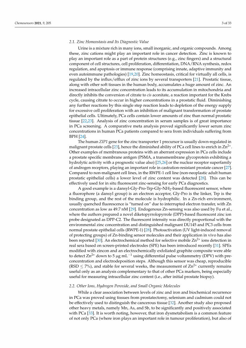
Although the most commonly used biomarker for the diagnostics and monitoring ofprostate conditions, including cancer, is PSA and its different forms, there are others whichmay significantly contribute to differential diagnosis. Age and ethnicity are common riskfactors for PCa, with 97% of incidence after 50 years of age and with the highest mortalityrates in black men (about 2.5 times higher chance than in white men) [14,15]. Usually, thequantification of PSA in serum is involved in diagnostics using immunoassay techniques,which are less affordable in developing countries. Different PCa markers have already beendescribed in blood, urine, semen, or tissue, with urine being the most sought-after matrixfor PCa diagnostics due to the non-invasive nature of its collection [16]. Standardisation ofprotein concentration in urine samples is almost impossible due to the biological variationin urine composition and its dependence on different factors. For example, proteinuria isdefined as a total protein concentration ≥150–300 mg L−1 [17]. Along with other proteins,PSA is also present in urine. Increased urinal PSA concentrations might be achieved after aprostate massage/DRE exam or due to disease progression. Very recently, the use of ionicliquid-based aqueous biphasic systems (biological Good’s buffers and K3C6H5O7 salt) hasbeen introduced as an effective pre-treatment of human urine (a less complex, non-invasivematrix compared to human serum) for PSA detection and quantification [18]. This reviewsummarises not only the clinical aspects of individual markers and the performance ofdevices and assays analysing these molecules, but also the biochemical pathways leadingto their occurrence (Figure 1). This may be considered the other side of the same coin,since knowing these pathways may lead to an explanation as to why, in some cases, thesemarkers offer only poor information about a particular disease, even though the diagnosticassay itself is quite reliable (or vice versa).
Chemosensors 2021, 9, x FOR PEER REVIEW 3 of 33
normal prostatic tissue [22,23]. Analysis of zinc concentration in serum samples is of great importance in PCa screening. A comparative meta analysis proved significantly lower se-rum zinc concentrations in human PCa patients compared to sera from individuals suf-fering from BPH [24].
Figure 1. Human genito-urinary system with the prostate located right below the bladder, making urine an accessible source of many precious (not just) prostate oncomarkers compared to more com-plex biological matrices, such as serum, plasma, or cell lysate to be used in sensing applications.
The human ZIP1 gene for the zinc transporter 1 precursor is usually down-regulated in malignant prostate cells [23], hence the diminished ability of PCa cell lines to enrich in Zn2+. Other examples of membranous proteins with an aberrant expression in PCa cells include a prostate specific membrane antigen (PSMA, a transmembrane glycoprotein ex-hibiting a hydrolytic activity with a prognostic value also) [25,26] or the nuclear receptor superfamily of androgen receptors, playing an important role in castration-resistant pros-tate cancer [27]. Compared to non-malignant cell lines, in the RWPE-1 cell line (non-neo-plastic adult human prostatic epithelial cells) a lower level of zinc content was detected [28]. This can be effectively used for in situ fluorescent zinc-sensing for early PCa diag-nostics.
A good example is a dansyl-Gly-Pro-Trp-Gly-NH2-based fluorescent sensor, where a fluorophore (a dansyl group) is an electron acceptor, Gly-Pro is the linker, Trp is the bind-ing group, and the rest of the molecule is hydrophilic. In a Zn-rich environment, usually quenched fluorescence is “turned on” due to interrupted electron transfer, with Zn con-centration as low as 49.7 nM [29]. Endogenous Zn-sensing was also used by Fu et al., where the authors prepared a novel diketopyrrolopyrrole (DPP)-based fluorescent zinc ion probe designated as DPP-C2. The fluorescent intensity was directly proportional with the environmental zinc concentration and distinguished malignant DU145 and PC3 cells from normal prostate epithelial cells (RWPE-1) [28]. Photoactivation (UV light-induced removal of protecting groups) of Zn-binding sensor molecules and their application in vivo has also been reported [30]. An electrochemical method for selective mobile Zn2+ ions detection in real sera based on screen-printed electrodes (SPE) has been introduced re-cently [31]. SPEs modified with zincon and an electrochemically exfoliated graphite com-posite were able to detect Zn2+ down to 5 μg mL−1 using differential pulse voltammetry (DPV) with pre-concentration and electrodeposition steps. Although this sensor was cheap, reproducible (RSD ≤ 7%), and stable for several weeks, the measurement of Zn2+ currently remains useful only as an analysis complementary to that of other PCa markers, being especially useful for measuring intracellular zinc content (i.e., after initial prostate biopsy).
Figure 1. Human genito-urinary system with the prostate located right below the bladder, makingurine an accessible source of many precious (not just) prostate oncomarkers compared to morecomplex biological matrices, such as serum, plasma, or cell lysate to be used in sensing applications.
Chemosensors 2021, 9, 205 3 of 33
2.1. Zinc Homeostasis and Its Diagnostic Value
Urine is a mixture rich in many ions, small inorganic, and organic compounds. Amongthese, zinc cations might play an important role in cancer detection. Zinc is known toplay an important role as a part of protein structures (e.g., zinc fingers) and a structuralcomponent of cell structures, cell proliferation, differentiation, DNA/RNA synthesis, redoxregulation, and apoptosis or immune response (comprising innate, adaptive immunity andeven autoimmune pathologies) [19,20]. Zinc homeostasis, critical for virtually all cells, isregulated by the influx/efflux of zinc ions by several transporters [21]. Prostatic tissue,along with other soft tissues in the human body, accumulates a huge amount of zinc. Anincreased intracellular zinc concentration leads to its accumulation in mitochondria anddirectly inhibits the conversion of citrate to cis aconitate, a reaction important for the Krebscycle, causing citrate to occur in higher concentrations in a prostatic fluid. Diminishingany further reactions by this single step reaction leads to depletion of the energy supplyfor excessive cell proliferation with an inhibition of malignant transformation of prostateepithelial cells. Ultimately, PCa cells contain lower amounts of zinc than normal prostatictissue [22,23]. Analysis of zinc concentration in serum samples is of great importancein PCa screening. A comparative meta analysis proved significantly lower serum zincconcentrations in human PCa patients compared to sera from individuals suffering fromBPH [24].
The human ZIP1 gene for the zinc transporter 1 precursor is usually down-regulated inmalignant prostate cells [23], hence the diminished ability of PCa cell lines to enrich in Zn2+.Other examples of membranous proteins with an aberrant expression in PCa cells includea prostate specific membrane antigen (PSMA, a transmembrane glycoprotein exhibiting ahydrolytic activity with a prognostic value also) [25,26] or the nuclear receptor superfamilyof androgen receptors, playing an important role in castration-resistant prostate cancer [27].Compared to non-malignant cell lines, in the RWPE-1 cell line (non-neoplastic adult humanprostatic epithelial cells) a lower level of zinc content was detected [28]. This can beeffectively used for in situ fluorescent zinc-sensing for early PCa diagnostics.
A good example is a dansyl-Gly-Pro-Trp-Gly-NH2-based fluorescent sensor, wherea fluorophore (a dansyl group) is an electron acceptor, Gly-Pro is the linker, Trp is thebinding group, and the rest of the molecule is hydrophilic. In a Zn-rich environment,usually quenched fluorescence is “turned on” due to interrupted electron transfer, with Znconcentration as low as 49.7 nM [29]. Endogenous Zn-sensing was also used by Fu et al.,where the authors prepared a novel diketopyrrolopyrrole (DPP)-based fluorescent zinc ionprobe designated as DPP-C2. The fluorescent intensity was directly proportional with theenvironmental zinc concentration and distinguished malignant DU145 and PC3 cells fromnormal prostate epithelial cells (RWPE-1) [28]. Photoactivation (UV light-induced removalof protecting groups) of Zn-binding sensor molecules and their application in vivo has alsobeen reported [30]. An electrochemical method for selective mobile Zn2+ ions detection inreal sera based on screen-printed electrodes (SPE) has been introduced recently [31]. SPEsmodified with zincon and an electrochemically exfoliated graphite composite were ableto detect Zn2+ down to 5 µg mL−1 using differential pulse voltammetry (DPV) with pre-concentration and electrodeposition steps. Although this sensor was cheap, reproducible(RSD ≤ 7%), and stable for several weeks, the measurement of Zn2+ currently remainsuseful only as an analysis complementary to that of other PCa markers, being especiallyuseful for measuring intracellular zinc content (i.e., after initial prostate biopsy).
2.2. Other Ions, Hydrogen Peroxide, and Small Organic Molecules
While a clear association between levels of zinc and iron and biochemical recurrencein PCa was proved using tissues from prostatectomy, selenium and cadmium could notbe effectively used to distinguish the cancerous tissue [32]. Another study also proposedother heavy metals, namely Mn, As, and Sb, to be significantly and positively associatedwith PCa [33]. It is worth noting, however, that iron dysmetabolism is a common featureof not only PCa (where iron plays an important role in tumour proliferation), but also of
Chemosensors 2021, 9, 205 4 of 33
different types of cancer cells in general, involving changes in expression of iron importers(transferrin receptors, TFRs), intracellular regulators (responsive element binding proteins,IRPs) or exporters (ferroportin, FPN), which makes these proteins also a therapeutictarget [34]. Divalent ions, along with glycosaminoglycans, play a crucial role in the centralprocesses of cell migration, angiogenesis, and extracellular matrix remodelling, thus beingassociated with a metastatic potential of various tumours [35].
Usually, the information about iron concentration is extracted from the measurementsof living cells or tissue samples, using for example an anthracene-based fluorescent probecontaining benzothiazole group (BFA), which interacted with Fe3+ and Cr3+ even in vivo(using PC-3 PCa cell line) down to 450 and 460 nM, respectively. Although some noveldevices have been developed to detect iron directly in serum samples, such as robust drysensor strips based on colour change upon chemical reaction and smartphone applica-tion [36] or graphene-based field-effect transistors with anti-ferritin antibodies (detectingferritin down to 10 fM, albeit measured in a buffer), these analyses are more suited to thediagnostics of nutritional disorders and iron deficiency [37].
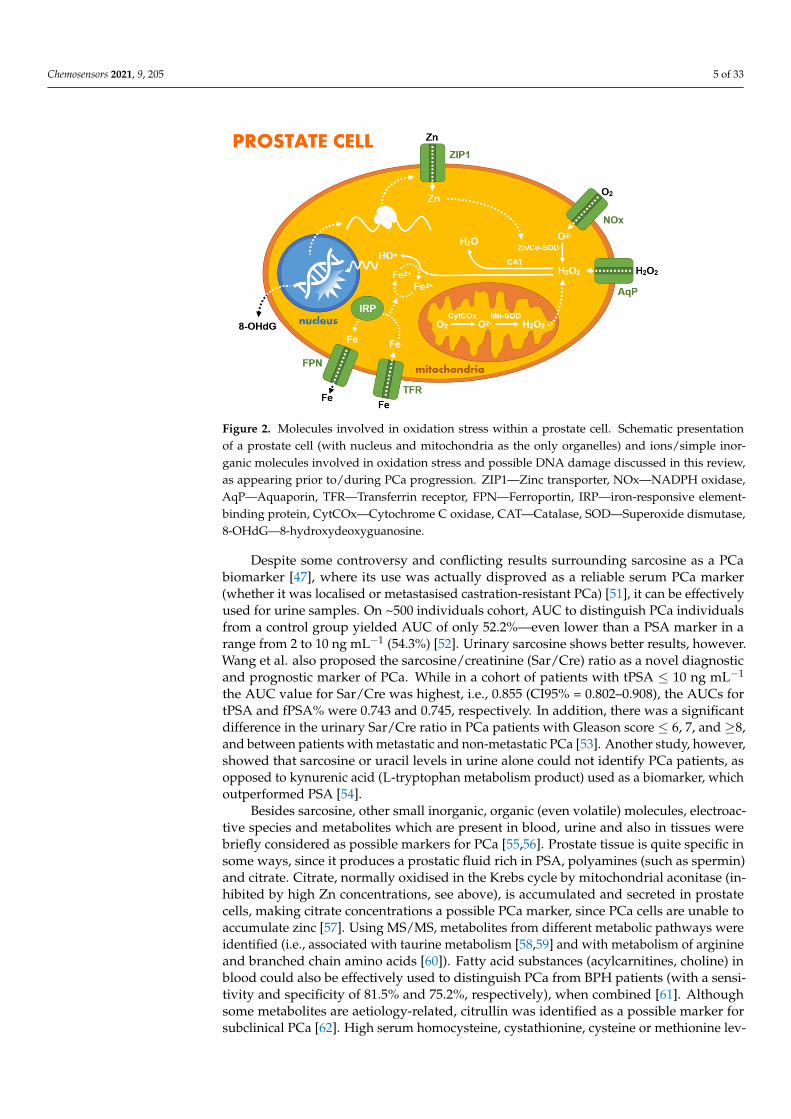
It is well known that PCa cells/samples (e.g., LNCaP cell line) often overproduceH2O2 and overexpress NADPH oxidase (NOx), resulting in increased intracellular reactiveoxygen species (ROS) levels [38]. Furthermore, increased ROS production is, similar to PCaitself, associated with increasing age, and thus ROS may help not just to initiate but also toadvance tumour growth [39]. Since supraphysiological levels of H2O2 exhibit cytotoxiceffects on cells, the results from a phase 1 clinical trial focusing on the effect of H2O2with radiation therapy on breast cancer were published recently, showing that H2O2 is awell-tolerated enhancer to radiotherapy itself [40].
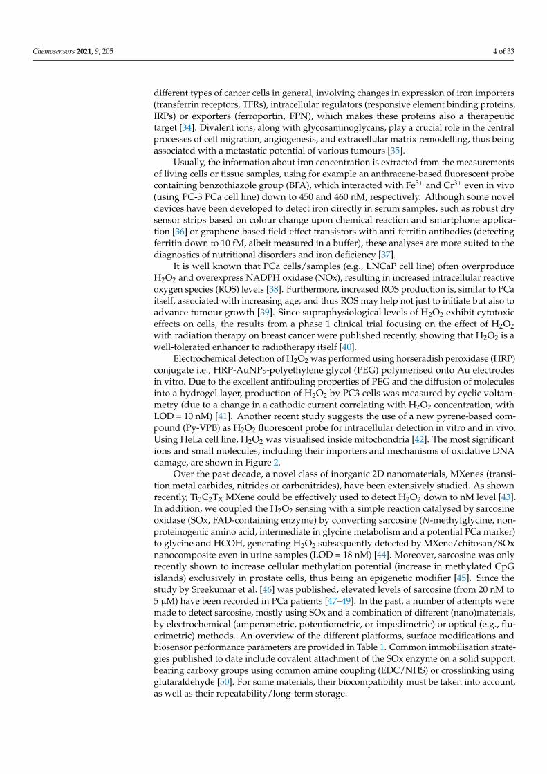
Electrochemical detection of H2O2 was performed using horseradish peroxidase (HRP)conjugate i.e., HRP-AuNPs-polyethylene glycol (PEG) polymerised onto Au electrodesin vitro. Due to the excellent antifouling properties of PEG and the diffusion of moleculesinto a hydrogel layer, production of H2O2 by PC3 cells was measured by cyclic voltam-metry (due to a change in a cathodic current correlating with H2O2 concentration, withLOD = 10 nM) [41]. Another recent study suggests the use of a new pyrene-based com-pound (Py-VPB) as H2O2 fluorescent probe for intracellular detection in vitro and in vivo.Using HeLa cell line, H2O2 was visualised inside mitochondria [42]. The most significantions and small molecules, including their importers and mechanisms of oxidative DNAdamage, are shown in Figure 2.
Over the past decade, a novel class of inorganic 2D nanomaterials, MXenes (transi-tion metal carbides, nitrides or carbonitrides), have been extensively studied. As shownrecently, Ti3C2TX MXene could be effectively used to detect H2O2 down to nM level [43].In addition, we coupled the H2O2 sensing with a simple reaction catalysed by sarcosineoxidase (SOx, FAD-containing enzyme) by converting sarcosine (N-methylglycine, non-proteinogenic amino acid, intermediate in glycine metabolism and a potential PCa marker)to glycine and HCOH, generating H2O2 subsequently detected by MXene/chitosan/SOxnanocomposite even in urine samples (LOD = 18 nM) [44]. Moreover, sarcosine was onlyrecently shown to increase cellular methylation potential (increase in methylated CpGislands) exclusively in prostate cells, thus being an epigenetic modifier [45]. Since thestudy by Sreekumar et al. [46] was published, elevated levels of sarcosine (from 20 nM to5 µM) have been recorded in PCa patients [47–49]. In the past, a number of attempts weremade to detect sarcosine, mostly using SOx and a combination of different (nano)materials,by electrochemical (amperometric, potentiometric, or impedimetric) or optical (e.g., flu-orimetric) methods. An overview of the different platforms, surface modifications andbiosensor performance parameters are provided in Table 1. Common immobilisation strate-gies published to date include covalent attachment of the SOx enzyme on a solid support,bearing carboxy groups using common amine coupling (EDC/NHS) or crosslinking usingglutaraldehyde [50]. For some materials, their biocompatibility must be taken into account,as well as their repeatability/long-term storage.
Chemosensors 2021, 9, 205 5 of 33
Chemosensors 2021, 9, x FOR PEER REVIEW 5 of 33
fluorimetric) methods. An overview of the different platforms, surface modifications and biosensor performance parameters are provided in Table 1. Common immobilisation strategies published to date include covalent attachment of the SOx enzyme on a solid support, bearing carboxy groups using common amine coupling (EDC/NHS) or crosslink-ing using glutaraldehyde [50]. For some materials, their biocompatibility must be taken into account, as well as their repeatability/long-term storage.
Figure 2. Molecules involved in oxidation stress within a prostate cell. Schematic presentation of a prostate cell (with nucleus and mitochondria as the only organelles) and ions/simple inorganic mol-ecules involved in oxidation stress and possible DNA damage discussed in this review, as appearing prior to/during PCa progression. ZIP1—Zinc transporter, NOx—NADPH oxidase, AqP—Aqua-porin, TFR—Transferrin receptor, FPN—Ferroportin, IRP—iron-responsive element-binding pro-tein, CytCOx—Cytochrome C oxidase, CAT—Catalase, SOD—Superoxide dismutase, 8-OHdG—8-hydroxydeoxyguanosine.
Despite some controversy and conflicting results surrounding sarcosine as a PCa bi-omarker [47], where its use was actually disproved as a reliable serum PCa marker (whether it was localised or metastasised castration-resistant PCa) [51], it can be effec-tively used for urine samples. On ~500 individuals cohort, AUC to distinguish PCa indi-viduals from a control group yielded AUC of only 52.2%—even lower than a PSA marker in a range from 2 to 10 ng mL−1 (54.3%) [52]. Urinary sarcosine shows better results, how-ever. Wang et al. also proposed the sarcosine/creatinine (Sar/Cre) ratio as a novel diagnos-tic and prognostic marker of PCa. While in a cohort of patients with tPSA ≤ 10 ng mL−1 the AUC value for Sar/Cre was highest, i.e., 0.855 (CI95% = 0.802–0.908), the AUCs for tPSA and fPSA% were 0.743 and 0.745, respectively. In addition, there was a significant differ-ence in the urinary Sar/Cre ratio in PCa patients with Gleason score ≤6, 7, and ≥8, and between patients with metastatic and non-metastatic PCa [53]. Another study, however, showed that sarcosine or uracil levels in urine alone could not identify PCa patients, as opposed to kynurenic acid (L-tryptophan metabolism product) used as a biomarker, which outperformed PSA [54].
Besides sarcosine, other small inorganic, organic (even volatile) molecules, electroac-tive species and metabolites which are present in blood, urine and also in tissues were briefly considered as possible markers for PCa [55,56]. Prostate tissue is quite specific in some ways, since it produces a prostatic fluid rich in PSA, polyamines (such as spermin) and citrate. Citrate, normally oxidised in the Krebs cycle by mitochondrial aconitase (in-hibited by high Zn concentrations, see above), is accumulated and secreted in prostate cells, making citrate concentrations a possible PCa marker, since PCa cells are unable to
Figure 2. Molecules involved in oxidation stress within a prostate cell. Schematic presentationof a prostate cell (with nucleus and mitochondria as the only organelles) and ions/simple inor-ganic molecules involved in oxidation stress and possible DNA damage discussed in this review,as appearing prior to/during PCa progression. ZIP1—Zinc transporter, NOx—NADPH oxidase,AqP—Aquaporin, TFR—Transferrin receptor, FPN—Ferroportin, IRP—iron-responsive element-binding protein, CytCOx—Cytochrome C oxidase, CAT—Catalase, SOD—Superoxide dismutase,8-OHdG—8-hydroxydeoxyguanosine.
Despite some controversy and conflicting results surrounding sarcosine as a PCabiomarker [47], where its use was actually disproved as a reliable serum PCa marker(whether it was localised or metastasised castration-resistant PCa) [51], it can be effectivelyused for urine samples. On ~500 individuals cohort, AUC to distinguish PCa individualsfrom a control group yielded AUC of only 52.2%—even lower than a PSA marker in arange from 2 to 10 ng mL−1 (54.3%) [52]. Urinary sarcosine shows better results, however.Wang et al. also proposed the sarcosine/creatinine (Sar/Cre) ratio as a novel diagnosticand prognostic marker of PCa. While in a cohort of patients with tPSA ≤ 10 ng mL−1
the AUC value for Sar/Cre was highest, i.e., 0.855 (CI95% = 0.802–0.908), the AUCs fortPSA and fPSA% were 0.743 and 0.745, respectively. In addition, there was a significantdifference in the urinary Sar/Cre ratio in PCa patients with Gleason score ≤ 6, 7, and ≥8,and between patients with metastatic and non-metastatic PCa [53]. Another study, however,showed that sarcosine or uracil levels in urine alone could not identify PCa patients, asopposed to kynurenic acid (L-tryptophan metabolism product) used as a biomarker, whichoutperformed PSA [54].
Besides sarcosine, other small inorganic, organic (even volatile) molecules, electroac-tive species and metabolites which are present in blood, urine and also in tissues werebriefly considered as possible markers for PCa [55,56]. Prostate tissue is quite specific insome ways, since it produces a prostatic fluid rich in PSA, polyamines (such as spermin)and citrate. Citrate, normally oxidised in the Krebs cycle by mitochondrial aconitase (in-hibited by high Zn concentrations, see above), is accumulated and secreted in prostatecells, making citrate concentrations a possible PCa marker, since PCa cells are unable toaccumulate zinc [57]. Using MS/MS, metabolites from different metabolic pathways wereidentified (i.e., associated with taurine metabolism [58,59] and with metabolism of arginineand branched chain amino acids [60]). Fatty acid substances (acylcarnitines, choline) inblood could also be effectively used to distinguish PCa from BPH patients (with a sensi-tivity and specificity of 81.5% and 75.2%, respectively), when combined [61]. Althoughsome metabolites are aetiology-related, citrullin was identified as a possible marker forsubclinical PCa [62]. High serum homocysteine, cystathionine, cysteine or methionine lev-
Chemosensors 2021, 9, 205 6 of 33
els independently predicted a risk of a disease recurrence and its aggressiveness [63], using11C-methionine as a radiotracer [64] in imaging techniques. A combined marker—ratio ofcholine + spermine (as the main polyamine) + creatine over citrate [(Cho+(Spm+)Cr)/Cit]was successfully used as a PCa biomarker in (1)H MR spectroscopic imaging ((1)H-MRSI)of the prostate [65].
Table 1. Basic operational parameters for sensors detecting sarcosine.
Detection Surface Modification LOD(nM)
Linear Range(µM)
RT(s) Stability (h) Application Refs.
Amper. PVA-Ag/AnNPs-pphTEOS-SOX/GE 500 0.5–7.5 17 - Aq. media [66]Amper. SOX/EDC/NHS/Au/ZnONPs/SPEs 16 0.01–0.1 - 60 d Synth. urine [50]Amper. SOX/CHIT/CuNPs/cMWCNT/AuE 0.0001 0.1–100 2 180 d Serum [67]Amper. SOXNPs/AuE 10 0.1–100 2 180 Urine [68]Amper. SOX/Pt@ZIF8/GCE 1060 5–30 - 3 Aq. media [69]Amper. Nafion-SOX/Pt/AAO 50 0.05–100 - - Aq. media [70]Amper. SOX/Pt/OIHMMP/GCE 130 1–70 - - Serum [71]Amper. SOX/PAA/GCE 0.4 0.001–0.05 - 15 d Urine [72]Amper. SOX/Pt-Fe3O4@C/GCE 430 0.5–60 - - Serum [73]Amper. SOX/chitosan/Ti3C2TX/GCE 18 0.036–7.8 2 - Synth. urine [44]Amper. Fe3O4@ZIF-8@MIP/AuE 0.0004 0.000001–0.0001 - 5 w Urine [74]Potent. MIP-based sensor 0.14 0.001–10 <120 >5 m Aq. media [75]Potent. GO based nanocomposite 3.3 0.01–100 60 3–4 m Aq. media [76]Potent. Non-GO based nanocomposite 0.005 0.001–10 60 3–4 m Aq. media [76]Imped. MIP/AuNPs/SPCE 8.5 0.011–17.9 - ~7 d Aq. media [77]Color. PdNPs based sensing platform 5.0 0.01–50 - - Urine [78]Color. NQS/GO/GCE 730 6.2–26.3 - - Aq. media [79]Fluor. Nanomaghemite/AuNPs/QD/peptide 0.05 0.005–0.05 - - Urine, cells [80]Fluor. ssDNA aptamer-based sensor 55 0.1–2 - - Urine [81]
Abbreviations: LOD—Limit of detection, RT—Response time, PVA—polyvinyl alcohol, pph-TEOS—partially pre-hydrolysed tetraethylorthosilicate, CHIT—chitosan, cMWCNT—carboxylated multi-walled carbon nanotubes, Pt@ZIF8—nanoplatinum-loaded porous zeoliticimidazolate framework-8, AAO—anodised aluminium oxide, Pt/OIHMMP—platinum-supported mesoporous organic-inorganic hybridmolybdenum phosphonate, MIP—molecularly imprinted polymer, GO—graphene oxide, GO-based nanocomposite: Ab-GO@graphite-powder@dibutyl phthalate-electrode; NQS—1,2-naphthoquinone-4-sulphonic acid sodium salt.
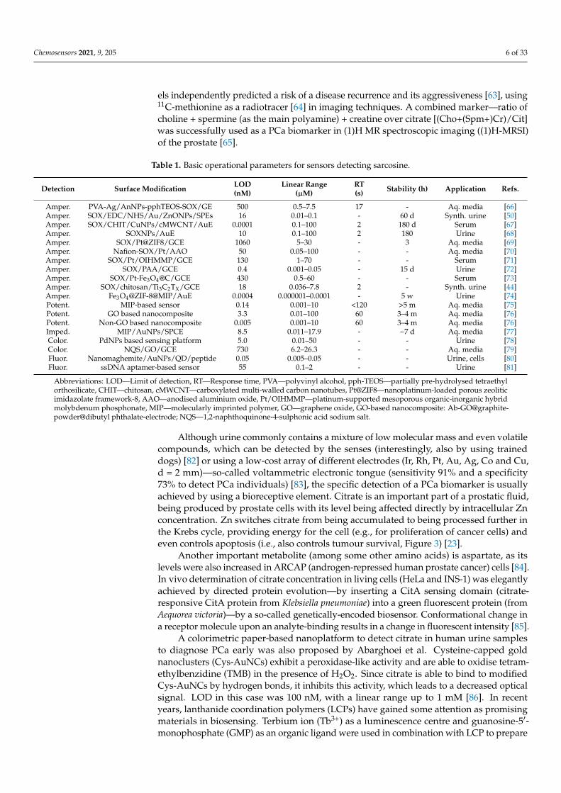
Although urine commonly contains a mixture of low molecular mass and even volatilecompounds, which can be detected by the senses (interestingly, also by using traineddogs) [82] or using a low-cost array of different electrodes (Ir, Rh, Pt, Au, Ag, Co and Cu,d = 2 mm)—so-called voltammetric electronic tongue (sensitivity 91% and a specificity73% to detect PCa individuals) [83], the specific detection of a PCa biomarker is usuallyachieved by using a bioreceptive element. Citrate is an important part of a prostatic fluid,being produced by prostate cells with its level being affected directly by intracellular Znconcentration. Zn switches citrate from being accumulated to being processed further inthe Krebs cycle, providing energy for the cell (e.g., for proliferation of cancer cells) andeven controls apoptosis (i.e., also controls tumour survival, Figure 3) [23].
Another important metabolite (among some other amino acids) is aspartate, as itslevels were also increased in ARCAP (androgen-repressed human prostate cancer) cells [84].In vivo determination of citrate concentration in living cells (HeLa and INS-1) was elegantlyachieved by directed protein evolution—by inserting a CitA sensing domain (citrate-responsive CitA protein from Klebsiella pneumoniae) into a green fluorescent protein (fromAequorea victoria)—by a so-called genetically-encoded biosensor. Conformational change ina receptor molecule upon an analyte-binding results in a change in fluorescent intensity [85].
A colorimetric paper-based nanoplatform to detect citrate in human urine samplesto diagnose PCa early was also proposed by Abarghoei et al. Cysteine-capped goldnanoclusters (Cys-AuNCs) exhibit a peroxidase-like activity and are able to oxidise tetram-ethylbenzidine (TMB) in the presence of H2O2. Since citrate is able to bind to modifiedCys-AuNCs by hydrogen bonds, it inhibits this activity, which leads to a decreased opticalsignal. LOD in this case was 100 nM, with a linear range up to 1 mM [86]. In recentyears, lanthanide coordination polymers (LCPs) have gained some attention as promisingmaterials in biosensing. Terbium ion (Tb3+) as a luminescence centre and guanosine-5′-monophosphate (GMP) as an organic ligand were used in combination with LCP to prepare
Chemosensors 2021, 9, 205 7 of 33
a sensing platform (Tb–GMP LCP-based biosensor) for citrate detection down to 4 µM.Upon citrate binding, a 6.5-fold enhancement in fluorescence was achieved, compared to aweak natural fluorescence of Tb-GMP [87]. Urinary polyamines were also investigated aspotential PCa diagnostic markers, where spermin was identified as the most promisingmarker (distinguishing PCa patients from BPH controls with AUC = 0.83) [88], which out-performed putrescine and spermidine, although spermidine dietary supplementation wasassociated with an increased survival among cancer patients at an early stage [89]. A hybridhydrogel matrix for the detection of spermin was described recently, with LOD = 6 µM. Adye-enriched agarose matrix increases its fluorescence in the presence of spermin 27-foldin real samples analysis (such as blood or urine), since such a sensor performance is notcompromised by interferents such as various metal ions, anions, monosaccharides and evenbasic amino acids such as histidine, arginine, lysine, ornithine, or glutamine [90]. Althoughthere are quite a few molecules currently identified as possible PCa biomarkers, each ofthese were tested only for PCa diagnostics and their involvement in other pathologicalconditions was not studied, which limits their clinical performance. A common example iscreatinine (a metabolic product of creatine phosphate, providing energy to muscles) andits serum levels are associated with a higher risk of PCa [91], while it commonly servesas an indicator of renal function after dialysis and is associated with some urinary tractdiseases symptoms, thyroid malfunction, and also muscle damage [92,93]. Thus, an alteredcreatinine level in itself cannot be used in accurate and robust PCa diagnostics.
Chemosensors 2021, 9, x FOR PEER REVIEW 7 of 33
Figure 3. Zinc regulating citrate levels and apoptotic events in PCa. Schematic presentation of a prostate epithelial cell introducing different roles of zinc ions in regulation of citrate levels in a pros-tatic fluid and even controlling apoptotic events in PCa tissues. In PCa, while the levels of Zn are decreased, Zn ions no longer inhibit aconitase enzyme in the Krebs cycle, so the cycle proceeds fur-ther and the cell gains energy necessary e.g., for proliferation. As a result, levels of citrate in PCa decrease in a prostatic fluid. Interstitial fluid is also depicted, as a rich source of low molecular mass Zn-ligands (Zn-citrate, Zn-aspartate, Zn-histidine, etc.). Abbreviations: ZIP1—Zinc transporter, SLC1—Membrane transport protein for some amino acids, GLUT1—Glucose transporter, Asp—As-partate, Glc—Glucose, Pyr—Pyruvate, AcCoA—Acetyl-CoA, Mal—Malate, Oxa—Oxalacetate, Cit—Citrate, IsoCit—Isocitrate, CytC—Cytochrome C, Cas9—Caspase 9, BAX (product of a BAX gene)—apoptosis regulator bcl-2-like protein 4. Image redrawn from ref. [23]. Copyright (2016) with permission from Elsevier.
Another important metabolite (among some other amino acids) is aspartate, as its levels were also increased in ARCAP (androgen-repressed human prostate cancer) cells [84]. In vivo determination of citrate concentration in living cells (HeLa and INS-1) was elegantly achieved by directed protein evolution—by inserting a CitA sensing domain (citrate-responsive CitA protein from Klebsiella pneumoniae) into a green fluorescent pro-tein (from Aequorea victoria)—by a so-called genetically-encoded biosensor. Conforma-tional change in a receptor molecule upon an analyte-binding results in a change in fluo-rescent intensity [85].
A colorimetric paper-based nanoplatform to detect citrate in human urine samples to diagnose PCa early was also proposed by Abarghoei et al. Cysteine-capped gold nanoclusters (Cys-AuNCs) exhibit a peroxidase-like activity and are able to oxidise tetra-methylbenzidine (TMB) in the presence of H2O2. Since citrate is able to bind to modified Cys-AuNCs by hydrogen bonds, it inhibits this activity, which leads to a decreased optical signal. LOD in this case was 100 nM, with a linear range up to 1 mM [86]. In recent years, lanthanide coordination polymers (LCPs) have gained some attention as promising mate-rials in biosensing. Terbium ion (Tb3+) as a luminescence centre and guanosine-5′-mono-phosphate (GMP) as an organic ligand were used in combination with LCP to prepare a sensing platform (Tb–GMP LCP-based biosensor) for citrate detection down to 4 μM. Upon citrate binding, a 6.5-fold enhancement in fluorescence was achieved, compared to a weak natural fluorescence of Tb-GMP [87]. Urinary polyamines were also investigated
Figure 3. Zinc regulating citrate levels and apoptotic events in PCa. Schematic presentation ofa prostate epithelial cell introducing different roles of zinc ions in regulation of citrate levels ina prostatic fluid and even controlling apoptotic events in PCa tissues. In PCa, while the levelsof Zn are decreased, Zn ions no longer inhibit aconitase enzyme in the Krebs cycle, so the cycleproceeds further and the cell gains energy necessary e.g., for proliferation. As a result, levelsof citrate in PCa decrease in a prostatic fluid. Interstitial fluid is also depicted, as a rich sourceof low molecular mass Zn-ligands (Zn-citrate, Zn-aspartate, Zn-histidine, etc.). Abbreviations:ZIP1—Zinc transporter, SLC1—Membrane transport protein for some amino acids, GLUT1—Glucosetransporter, Asp—Aspartate, Glc—Glucose, Pyr—Pyruvate, AcCoA—Acetyl-CoA, Mal—Malate,Oxa—Oxalacetate, Cit—Citrate, IsoCit—Isocitrate, CytC—Cytochrome C, Cas9—Caspase 9, BAX(product of a BAX gene)—apoptosis regulator bcl-2-like protein 4. Image redrawn from ref. [23].Copyright (2016) with permission from Elsevier.
Chemosensors 2021, 9, 205 8 of 33
3. Nucleic Acid-Based PCa Biomarkers
Having their origin in the cell nucleus, in this section we describe different types,mechanism of synthesis and detection principles for the analysis of genes with changedexpression profile, mutated genes, and aberrant gene products, as well as micro RNAsinvolved in PCa diagnostics and prognostics. Since the detection often relies on a relativelylow amount of the markers present in the original sample, a polymerase chain reaction(PCR) is usually introduced prior to analysis by a genosensor platform [94,95].
3.1. PCA3 Gene RNA Product
The PCA3 score test is one of the PCa biomarkers applied in clinical practice. Theproduct of a prostate cancer gene 3 (PCA3) is a non-coding RNA that can be collectednon-invasively from urine samples. In prostatic tissue, PCA3 is highly overexpressedcompared to other tissues, where it is not present at all, and its expression increases furtherin cancer cells (up to ~100×) [96]. It shows a higher diagnostic and also prognostic valuethan fPSA% and also correlates with the increasing Gleason’s score, also being a possibleprognostic marker [97]. Moreover, it has been shown that patients with high-grade prostateintraepithelial neoplasia (HG PIN) have a higher PCA3 score than a control group. ThePCA3 test should ideally be performed from the first-caught urine after a prostate massageduring a digital rectal examination for one of the following cases: (i) individuals withelevated PSA levels and a negative initial biopsy; (ii) individuals with elevated PSA levelsand prostatitis; or (iii) individuals undergoing active surveillance with low-grade tumouror presumed microfocal disease [96]. In the studies published, the PCA3 test outperformedthe tPSA or fPSA% tests, providing quite a wide range of assay sensitivities and specificities(58–94.9% and 41.8–72% [97–99], respectively, for a cut off value of 35), most likely due todifferent or unknown inclusion/exclusion criteria, as discussed in a recent meta analysisstudy [100]. Wang et al. proposed a combination of PCA3 and PSA RNA transcripts(PCA3/PSA RNA) to be a more reliable PCa biomarker, as suggested by the area underreceiver operating characteristic curves: AUC(PCA3) = 0.717, AUC(PSA) = 0.444 andAUC(PCA3/PSA) = 0.916 [101].
As for point-of-care diagnostics, an electrochemical genosensor was recently proposedfor the detection of PCA3 using a PCA3 single-stranded -NH2-containing DNA probeimmobilised via amine coupling on layer-by-layer (LbL) modified interdigitated goldelectrodes by chitosan and multi-walled carbon nanotubes [102]. LOD in this case wasas low as 0.128 nM [102]. Electrochemical impedance spectroscopy is known to be oneof the most sensitive label-free electrochemical techniques [103,104]. Modifying carbon-printed electrodes or quartz with layer-by-layer deposited films of gold nanoparticles(AuNPs) and chondroitin sulphate, PCA3 complementary DNA sequence as a probe couldbe immobilised on a surface, affording LOD as low as 83 pM (2000 pM in the event of usingcyclic voltammetry for the detection) [105].
Optical detection platforms have also become popular in low resource settings af-fording such excellent operational parameters as simplicity of use, low cost, and shortdetection time. Among others, lateral flow assay (LFA) biosensors are also a popular choicefor the possibility to easily expand to mass production. An impressive detection limit of3 fM of PCA3 mimic DNA (with detection range from 0.01 to 50,000 pM) was achieved bya SERS-based competitive LFA assay using malachite green isothiocyanate and AuNPs.In addition, no significant differences were observed when compared not just to a blankand a non-complementary strain but also to a single-base mismatched DNA, suggestinghigh selectivity [106]. For different optical platforms, nanoparticles (NPs) are often used to(i) provide the detectable signal; or for (ii) enrichment prior to any signal generation. Useof an up-conversion NPs-graphene sensor platform affords early diagnostics based on thespecific detection of oligonucleotide sequences in complex matrices (such as cell lysates orplasma) [107]. These lanthanide-doped NPs serve as a fluorescence donor—by absorbingtwo or more low energy photons, they emit fluorescence at a shorter wavelength. Upon thehybridisation of a probe immobilised on NPs, graphene oxide (GO) is added and, following
Chemosensors 2021, 9, 205 9 of 33
laser irradiation (λ = 980 nm), a fluorescent signature of NPs is observed. GO affords manyunique properties for this array, such as high surface area, water solubility, quenchingproperties and the fact that single-stranded (ss) oligonucleotides preferentially bind toGO via π-π interactions, while double-stranded (ds) do not exhibit such behaviour. Incontrast, magnetic NPs for target analyte enrichment have been used for an enzyme-linkedoligonucleotide assay to overcome any drawback of a common electrophoretic detectionof PCR products. Running a RT-PCR assay in a tube with forward and reverse primersattached to magnetic NPs and biotin, respectively, using HRP and a common TMB/H2O2system, a sensitive optical detection of PCA3 in urine was achieved. Moreover, PCa patientswere readily distinguishable, not just from healthy controls, but also from BPH patients,suggesting that PCA3 remains one of the most PCa-specific markers available to date [108].
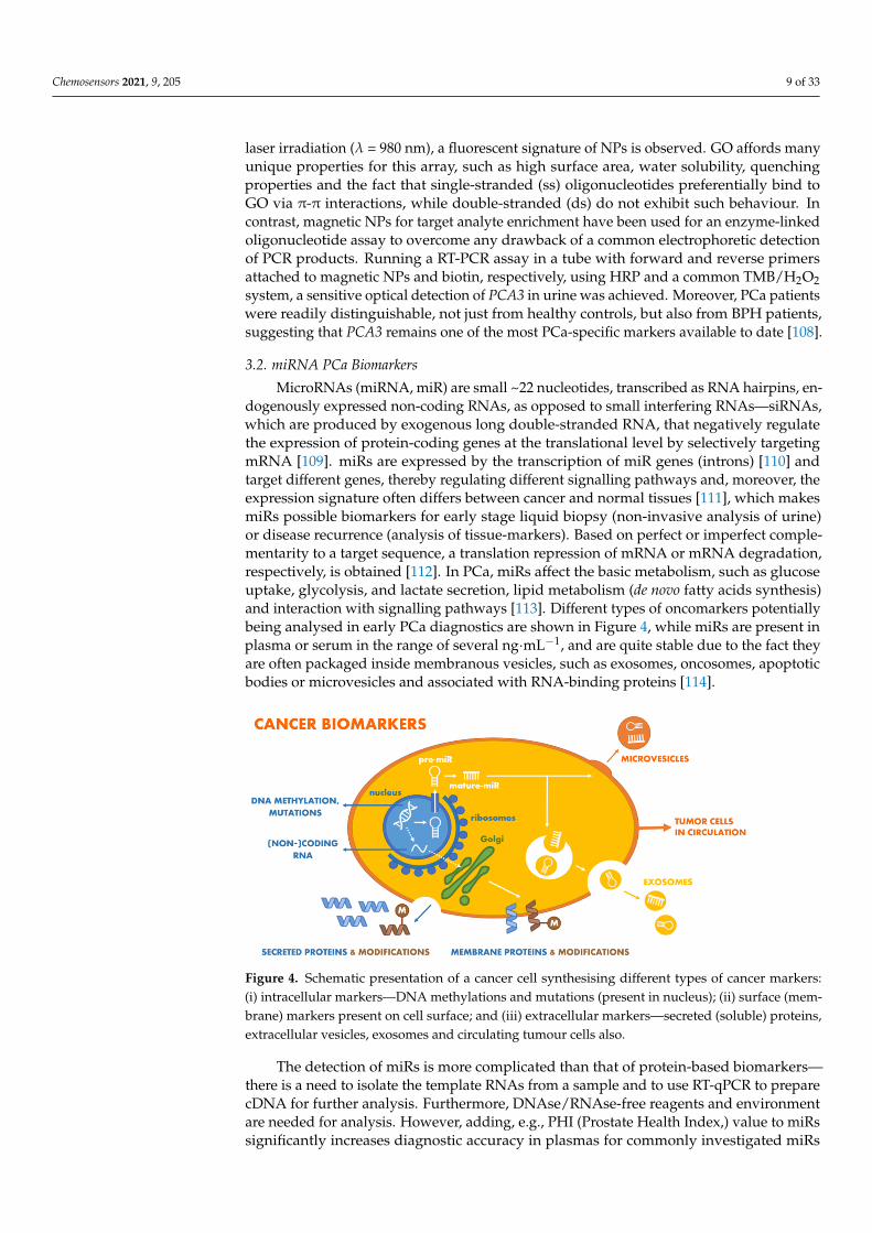
3.2. miRNA PCa Biomarkers
MicroRNAs (miRNA, miR) are small ~22 nucleotides, transcribed as RNA hairpins, en-dogenously expressed non-coding RNAs, as opposed to small interfering RNAs—siRNAs,which are produced by exogenous long double-stranded RNA, that negatively regulatethe expression of protein-coding genes at the translational level by selectively targetingmRNA [109]. miRs are expressed by the transcription of miR genes (introns) [110] andtarget different genes, thereby regulating different signalling pathways and, moreover, theexpression signature often differs between cancer and normal tissues [111], which makesmiRs possible biomarkers for early stage liquid biopsy (non-invasive analysis of urine)or disease recurrence (analysis of tissue-markers). Based on perfect or imperfect comple-mentarity to a target sequence, a translation repression of mRNA or mRNA degradation,respectively, is obtained [112]. In PCa, miRs affect the basic metabolism, such as glucoseuptake, glycolysis, and lactate secretion, lipid metabolism (de novo fatty acids synthesis)and interaction with signalling pathways [113]. Different types of oncomarkers potentiallybeing analysed in early PCa diagnostics are shown in Figure 4, while miRs are present inplasma or serum in the range of several ng·mL−1, and are quite stable due to the fact theyare often packaged inside membranous vesicles, such as exosomes, oncosomes, apoptoticbodies or microvesicles and associated with RNA-binding proteins [114].
Chemosensors 2021, 9, x FOR PEER REVIEW 10 of 33
Figure 4. Schematic presentation of a cancer cell synthesising different types of cancer markers: (i) intracellular markers—DNA methylations and mutations (present in nucleus); (ii) surface (mem-brane) markers present on cell surface; and (iii) extracellular markers—secreted (soluble) proteins, extracellular vesicles, exosomes and circulating tumour cells also.
The detection of miRs is more complicated than that of protein-based biomarkers—there is a need to isolate the template RNAs from a sample and to use RT-qPCR to prepare cDNA for further analysis. Furthermore, DNAse/RNAse-free reagents and environment are needed for analysis. However, adding, e.g., PHI (Prostate Health Index,) value to miRs significantly increases diagnostic accuracy in plasmas for commonly investigated miRs associated with PCa, such as miR-21 [115,116] (sensitivity ≤ 95% and specificity = 100% for miR-21 and miR-221 [117] in combination with PHI. For diagnostic and prognostic pur-poses, miR-182-5p and miR-375-3p were isolated from tissues and plasmas and subse-quently detected. Although miR-375-3p could not be reliably used for PCa diagnostics, the higher circulating levels of both the above miRs were associated with more patholog-ical stages and miR-375-3p could even be associated with patients more prone to develop a metastatic PCa [118]. In addition, miR-375-3p could be associated (significantly overex-pressed together with miRs 141 and 378*) with castration-resistant PCa, as shown in an-other study [119]. In the case of PCa itself, there are many miR-based tissue biomarkers with a diagnostic or a prognostic value. As reported in a microarray-based study of 470 human miRs, 10 of these miRs were downregulated, 5 upregulated, and some even corre-lated with a Gleason score parameter (miR-31, 96 and 205) [120]. On the basis of this study, the best miR biomarker for PCa was miR-205 (diagnostic accuracy of 72%, AUC = 0.82). Using all miRs and a logistic regression, accuracy increased to 82% and AUC value only slightly to 0.86 [120]. miR-141 and miR-375 usually detected in tissues (e.g., after radical prostatectomy in untreated PCa patients) are also present in serum samples in higher amounts in metastatic PCa patients (in microvesicles or exosomes), miR-107 and miR-574-3p were identified as potential urinary markers (with best AUC = 0.74 for miR-107 on cohorts, where PCA3 yielded an AUC = 0.61) [121]. Besides the two above-mentioned miRs, miR-21-5p, miR-141-3p, and miR-375 were most recently proposed for considera-tion as potential urinary biomarkers for non-invasive PCa diagnostics [122]. Except for a single biomarker approach, Fredsøe et al. showed an impressive performance of their three miR-based model for urinary detection of PCa (miR-222-3p*miR-24-3p/miR-30c-5p), distinguishable from a BPH cohort with an AUC = 0.89–0.95 [123]. Similarly, another model (miR-125b-5p*let-7a-5p/miR-151-5p) predicted time to biochemical recurrence after radical prostatectomy (distinguished low- and high-risk groups with high statistical sig-nificance, p = 0.0176) [123].
In discussing miRs detection using sensors, electrochemical detection is by far one of the most popular methods. In a recent review paper published by El Aamri et al., the authors proposed five different classes of electrochemical biosensors: (i) biosensors
Figure 4. Schematic presentation of a cancer cell synthesising different types of cancer markers:(i) intracellular markers—DNA methylations and mutations (present in nucleus); (ii) surface (mem-brane) markers present on cell surface; and (iii) extracellular markers—secreted (soluble) proteins,extracellular vesicles, exosomes and circulating tumour cells also.
The detection of miRs is more complicated than that of protein-based biomarkers—there is a need to isolate the template RNAs from a sample and to use RT-qPCR to preparecDNA for further analysis. Furthermore, DNAse/RNAse-free reagents and environmentare needed for analysis. However, adding, e.g., PHI (Prostate Health Index,) value to miRssignificantly increases diagnostic accuracy in plasmas for commonly investigated miRs
Chemosensors 2021, 9, 205 10 of 33
associated with PCa, such as miR-21 [115,116] (sensitivity ≤ 95% and specificity = 100%for miR-21 and miR-221 [117] in combination with PHI. For diagnostic and prognosticpurposes, miR-182-5p and miR-375-3p were isolated from tissues and plasmas and subse-quently detected. Although miR-375-3p could not be reliably used for PCa diagnostics, thehigher circulating levels of both the above miRs were associated with more pathologicalstages and miR-375-3p could even be associated with patients more prone to developa metastatic PCa [118]. In addition, miR-375-3p could be associated (significantly over-expressed together with miRs 141 and 378*) with castration-resistant PCa, as shown inanother study [119]. In the case of PCa itself, there are many miR-based tissue biomark-ers with a diagnostic or a prognostic value. As reported in a microarray-based study of470 human miRs, 10 of these miRs were downregulated, 5 upregulated, and some evencorrelated with a Gleason score parameter (miR-31, 96 and 205) [120]. On the basis ofthis study, the best miR biomarker for PCa was miR-205 (diagnostic accuracy of 72%,AUC = 0.82). Using all miRs and a logistic regression, accuracy increased to 82% and AUCvalue only slightly to 0.86 [120]. miR-141 and miR-375 usually detected in tissues (e.g.,after radical prostatectomy in untreated PCa patients) are also present in serum samples inhigher amounts in metastatic PCa patients (in microvesicles or exosomes), miR-107 andmiR-574-3p were identified as potential urinary markers (with best AUC = 0.74 for miR-107on cohorts, where PCA3 yielded an AUC = 0.61) [121]. Besides the two above-mentionedmiRs, miR-21-5p, miR-141-3p, and miR-375 were most recently proposed for considera-tion as potential urinary biomarkers for non-invasive PCa diagnostics [122]. Except fora single biomarker approach, Fredsøe et al. showed an impressive performance of theirthree miR-based model for urinary detection of PCa (miR-222-3p*miR-24-3p/miR-30c-5p),distinguishable from a BPH cohort with an AUC = 0.89–0.95 [123]. Similarly, another model(miR-125b-5p*let-7a-5p/miR-151-5p) predicted time to biochemical recurrence after radicalprostatectomy (distinguished low- and high-risk groups with high statistical significance,p = 0.0176) [123].
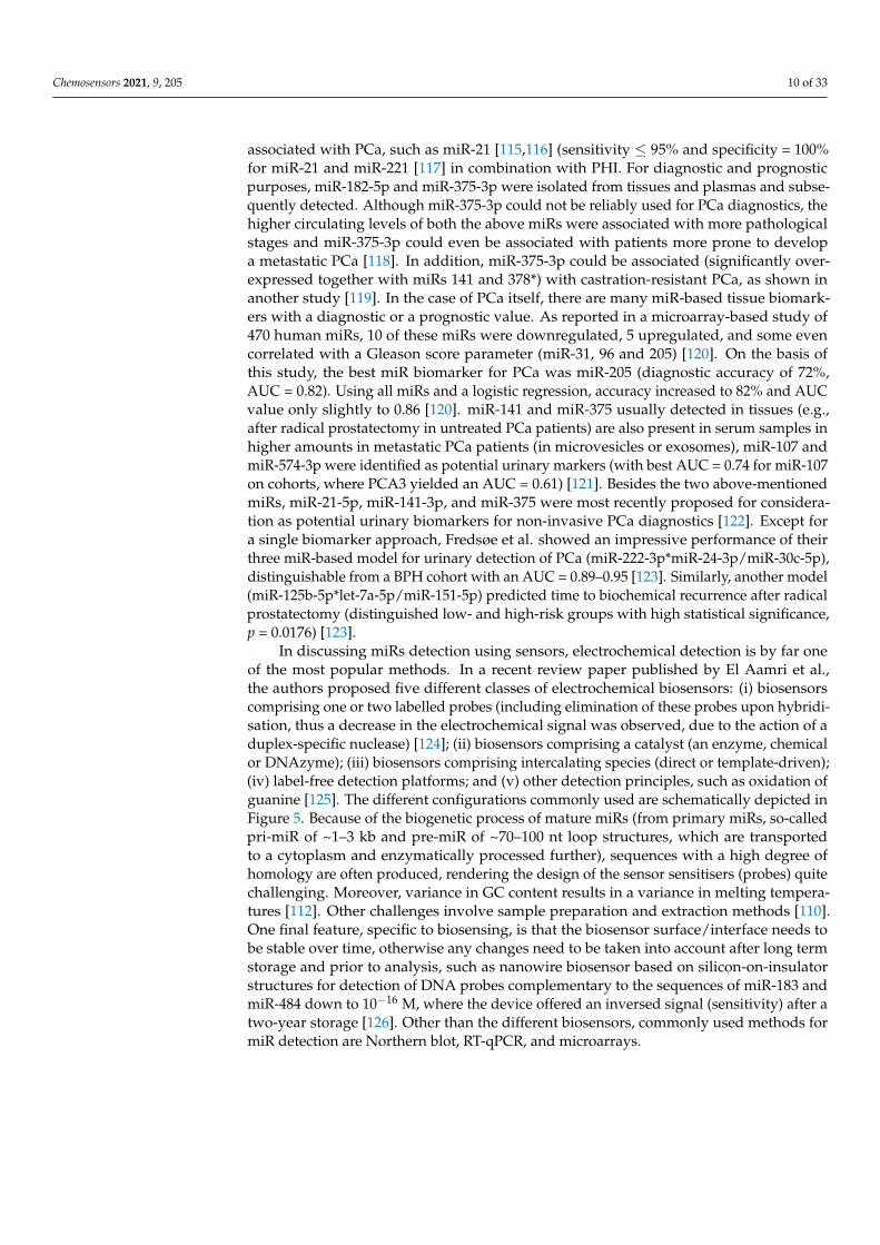
In discussing miRs detection using sensors, electrochemical detection is by far oneof the most popular methods. In a recent review paper published by El Aamri et al.,the authors proposed five different classes of electrochemical biosensors: (i) biosensorscomprising one or two labelled probes (including elimination of these probes upon hybridi-sation, thus a decrease in the electrochemical signal was observed, due to the action of aduplex-specific nuclease) [124]; (ii) biosensors comprising a catalyst (an enzyme, chemicalor DNAzyme); (iii) biosensors comprising intercalating species (direct or template-driven);(iv) label-free detection platforms; and (v) other detection principles, such as oxidation ofguanine [125]. The different configurations commonly used are schematically depicted inFigure 5. Because of the biogenetic process of mature miRs (from primary miRs, so-calledpri-miR of ~1–3 kb and pre-miR of ~70–100 nt loop structures, which are transportedto a cytoplasm and enzymatically processed further), sequences with a high degree ofhomology are often produced, rendering the design of the sensor sensitisers (probes) quitechallenging. Moreover, variance in GC content results in a variance in melting tempera-tures [112]. Other challenges involve sample preparation and extraction methods [110].One final feature, specific to biosensing, is that the biosensor surface/interface needs tobe stable over time, otherwise any changes need to be taken into account after long termstorage and prior to analysis, such as nanowire biosensor based on silicon-on-insulatorstructures for detection of DNA probes complementary to the sequences of miR-183 andmiR-484 down to 10−16 M, where the device offered an inversed signal (sensitivity) after atwo-year storage [126]. Other than the different biosensors, commonly used methods formiR detection are Northern blot, RT-qPCR, and microarrays.
Chemosensors 2021, 9, 205 11 of 33
Chemosensors 2021, 9, x FOR PEER REVIEW 11 of 33
comprising one or two labelled probes (including elimination of these probes upon hy-bridisation, thus a decrease in the electrochemical signal was observed, due to the action of a duplex-specific nuclease) [124]; (ii) biosensors comprising a catalyst (an enzyme, chemical or DNAzyme); (iii) biosensors comprising intercalating species (direct or tem-plate-driven); (iv) label-free detection platforms; and (v) other detection principles, such as oxidation of guanine [125]. The different configurations commonly used are schemati-cally depicted in Figure 5. Because of the biogenetic process of mature miRs (from primary miRs, so-called pri-miR of ~1–3 kb and pre-miR of ~70–100 nt loop structures, which are transported to a cytoplasm and enzymatically processed further), sequences with a high degree of homology are often produced, rendering the design of the sensor sensitisers (probes) quite challenging. Moreover, variance in GC content results in a variance in melt-ing temperatures [112]. Other challenges involve sample preparation and extraction meth-ods [110]. One final feature, specific to biosensing, is that the biosensor surface/interface needs to be stable over time, otherwise any changes need to be taken into account after long term storage and prior to analysis, such as nanowire biosensor based on silicon-on-insulator structures for detection of DNA probes complementary to the sequences of miR-183 and miR-484 down to 10−16 M, where the device offered an inversed signal (sensitivity) after a two-year storage [126]. Other than the different biosensors, commonly used meth-ods for miR detection are Northern blot, RT-qPCR, and microarrays.
Figure 5. Schematic presentation of different configurations of miR (orange) detection platforms, based on hybridization with a complementary sequence (probe, blue). (1) Interaction of miR analyte with a redox-labelled complementary probe on an electrode surface, while (a) probe is a hairpin which unfolds upon binding, leading to a decreased signal; (b) a product is specifically cleaved, leading to a decreased signal, (c) a secondary probe (green) is used for the detection, leading to a signal generation. (2) Hybridization of miR with an unlabelled probe leads to desorption of a non-covalently adsorbed probe from an electrode surface, leading to a decreased signal. (3) A signal is generated due to introduction of an additional component, e.g., by an intercalating species via (d) a direct or (e) a template-directed way; (f) a signal is generated using a catalyst molecule. (4) A label-free detection, using for example electrochemical impedance spectroscopy and a charge transfer resistance parameter as an evaluated signal; inset: Nyquist plots for a bioreceptive layer and after a biorecognition/hybridization took place. Redrawn with permission from ref. [127], Copyright (2015), Royal Society for Chemistry.
Electrochemical biosensors provide a platform readily transferable to an array for-mat, which is easy to miniaturise, but deals with some problems such as selectivity due to non-specific interactions and output signal inconsistencies. Jolly et al. proposed a dual mode platform based on gold nanoparticles and EIS/SWV measurements for the detection of miR-145 down to 0.37 fM. Moreover, two mismatches in a strain were not significantly different in their response compared to a blank, which makes this kind of platform highly selective toward specific sequences [128]. Furthermore, besides a Nyquist plot in a label-
Figure 5. Schematic presentation of different configurations of miR (orange) detection platforms,based on hybridization with a complementary sequence (probe, blue). (1) Interaction of miR analytewith a redox-labelled complementary probe on an electrode surface, while (a) probe is a hairpinwhich unfolds upon binding, leading to a decreased signal; (b) a product is specifically cleaved,leading to a decreased signal, (c) a secondary probe (green) is used for the detection, leading toa signal generation. (2) Hybridization of miR with an unlabelled probe leads to desorption of anon-covalently adsorbed probe from an electrode surface, leading to a decreased signal. (3) A signalis generated due to introduction of an additional component, e.g., by an intercalating species via(d) a direct or (e) a template-directed way; (f) a signal is generated using a catalyst molecule. (4) Alabel-free detection, using for example electrochemical impedance spectroscopy and a charge transferresistance parameter as an evaluated signal; inset: Nyquist plots for a bioreceptive layer and after abiorecognition/hybridization took place. Redrawn with permission from ref. [127], Copyright (2015),Royal Society for Chemistry.
Electrochemical biosensors provide a platform readily transferable to an array format,which is easy to miniaturise, but deals with some problems such as selectivity due tonon-specific interactions and output signal inconsistencies. Jolly et al. proposed a dualmode platform based on gold nanoparticles and EIS/SWV measurements for the detectionof miR-145 down to 0.37 fM. Moreover, two mismatches in a strain were not significantlydifferent in their response compared to a blank, which makes this kind of platform highlyselective toward specific sequences [128]. Furthermore, besides a Nyquist plot in a label-free format, a Cole-Cole (-C” vs. C′) plot might be used if an electrochemical probe isattached to the electrode surface, such as ferrocene derivatives.
3.3. Changes in DNA Level (DNA-Based and Derived Oncomarkers)
Although PCa is known to have a different epidemiology across different ethnicgroups, genetic/hereditary factors are also known to play an important role in the diseaseoccurrence. The Nordic Twin Study estimated the risk of the development of PCa due to in-heritability at 57% (CI95% = 51–63%), making it one of the most inheritable cancers togetherwith melanoma and non-melanoma skin cancers [129]. By means of genetic variations,different loci were identified (~170) as being more or less susceptible to changes due toPCa development, while more common genetic variants of some genes may confer a lowerrisk for PCa [130,131]. Examples involve mutations in tumour suppressor/DNA repairgenes, such as BRCA1, BRCA2, ATM, CHEK2, RAD51D or PALB2 [132] and also more rarevariations, such as HOXB13 (G84E variant, 1.4% in Europe, being more common for PCawith an early onset)—a transcription factor gene important in prostate development [133].Although commonly used as a marker for breast and ovarian cancer (with 85% and 63%lifetime risks of hereditary cancer in women, respectively) [134], recent efforts to detectBRCA1 changes involve mostly nanomaterial-supported electrochemical detection, e.g.,thermally reduced GO with functional groups still used to immobilise streptavidin andsubsequently to bind a PCR product with a biotin-labelled primer entering the reaction
Chemosensors 2021, 9, 205 12 of 33
(a biosensor response was measured in real time as a decrease in resistance in MΩ rangewith LOD = 0.2 nM) [135] or thiol-modified DNA tetrahedral-structured probe and AuNP-modified reporter DNA, creating a typical sandwich with BRCA1 sequence and generatingan electrochemical (amperometry, voltammetry) signal using HRP and TMB/H2O2 foranalyte concentrations much lower than in the previous case—down to 0.1 fM [136].
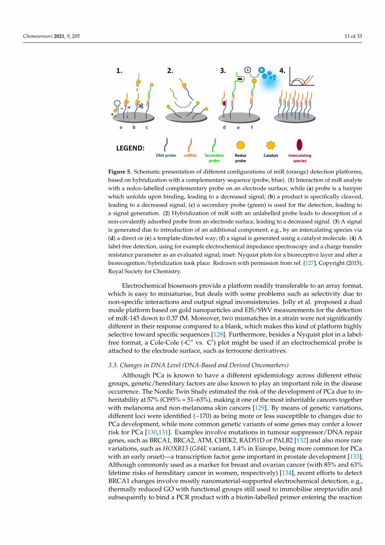
Different types of gene-associated changes in PCa, further discussed in this section,are depicted in Figure 6. Fusion of transmembrane protease serine 2:v-ets erythroblastosisvirus E26 oncogene homologue (TMPRSS2:ERG) is often associated with PCa. Further-more, through lentiviral-mediated gene-silencing, the mRNA product of ERG and proteinexpression was significantly reduced, which resulted in cell-cycle arrest at G0/G1 phase forVCaP cells (Vertebral-Cancer of the Prostate) [137]. Besides chromosomal rearrangementscreating fusion genes, RNA sequencing technology proved that fusion is also mediatedat a transcription level [138]. ERG gene rearrangements are quite common in prostateadenocarcinomas, while less data is available for small-cell prostate carcinomas (a rare neu-roendocrine tumour variant), which manifest themselves quite early (including metastases),are not accompanied by an increase in serum PSA and are resistant to androgen ablation. Inaddition, TMPRSS2:ERG and PCA3 were shown to have an independent positive predictivevalue to detect PCa [139]. A rapid, low-cost (~5 USD/test), and a robust (isothermal reversetranscriptase-recombinase polymerase amplification-based) approach has been developedfor the detection of the fusion gene in urine, detecting as low as 105 copies of transcripts.Naked eye detection was possible due to the formation of cDNA amplicons exclusively inthe presence of fusion genes in urine, providing a non-invasive matrix. These ampliconsthen bind to MPs and spontaneous flocculation occurs [140]. Another important geneassociated with PCa is glutathione S-transferase P1 (GSTP1), expressed in some humantissues. This enzyme detoxifies the cell from endogenous and exogenous toxic compoundsusing glutathione or by acting as a ligandin (also via protein-protein interactions) [141,142],exhibiting quite impressive substrate promiscuity. In PCa, the promoter of GSTP1 is hyper-methylated, which leads to a loss of expression levels and possibly to DNA damage due toincreased oxidative stress. Induction of GSTP1 activity in LNCaP (human prostate adeno-carcinoma cells derived from the left supraclavicular lymph node metastasis) cells with asilenced GSTP1 gene lowered endogenous (ROS) levels when exposed to H2O2 [143,144].Additionally, miRs 133-a/b, 144, 144*, 153-1, 590-3p and 590-5p were shown to regulateGSTP1, being another class of possible PCa markers [145]. Biosensors detecting GSTP1hypermethylation, which are stable for a long time, could be successfully designed basedon a hybridisation strategy with a probe immobilised on a single-use graphite carbonelectrode or multi-walled carbon nanotubes screen printed electrodes. In both cases, LODsdown to pM level were achieved based on changes in guanine oxidations using differentialpulse voltammetry (DPV) [146,147].
If oxidative stress (in the form of a ROS attack) is not prevented, it might directly dam-age cellular DNA. Predominantly, bases in DNA structure are hydroxylated with guaninebeing the most prone to such an oxidation. Hence, for several decades, the most studiedand abundant lesion is most likely 8-hydroxydeoxyguanosine (8-OHdG), which occurs inurine as a product of mismatch repair mechanism [148]. Moreover, 8-OHdG concentration(controlled by local antioxidant capacity) in urine was shown to refer not only to increasedoxidative damage but also to be associated with some diseases, including breast, bladderand prostate cancers [149]—which could even be distinguished from benign hyperplasiausing a tissue-staining microarray (10 adenocarcinoma patients vs. 70 controls) [150]. 8-OHdG might be directly electrochemically oxidised on modified surfaces with sufficientelectrocatalytic activities (on carbon surfaces, 8-OHdG exhibits a 2-electron transfer reac-tion), such as electrochemically reduced GO and multi-walled carbon nanotubes-modifiedglassy carbon electrode, where a limit of detection of 35 nM was achieved (with linearrange from 3 to 75 µM), as well as analysis in the presence of common interferents (ascorbicand uric acid, xanthine, and hypoxanthine) and in urine [151]. Similarly, LOD = 28 nM(with linear range from 0.5 to 100 µM) was achieved at the edge plane surface of a py-
Chemosensors 2021, 9, 205 13 of 33
rolytic graphite electrode [152]. A molecularly-imprinted sensor has also been fabricatedusing edge plane pyrolytic graphite and glutaraldehyde/poly-1,5-diaminonaphthalenebioreceptive interface; however LOD = 3 nM and a linear range from 20 nM up to 3 µMwas not significantly different from the less complicated assays mentioned above [153].For point-of-care diagnostics, an interesting concept using 8-OHdG oxidation on carbonink-modified paper substrate coupled with DPV readings was used [154]. LOD in this casewas once again in nM range (~50 nM, i.e., 14.4 ng mL−1). Since this device might be readilyminiaturised and mass-produced at a low price with a performance comparable with previ-ous devices, it is an ideal candidate for in situ testing of biological samples. The parametersof this paper-based sensor could be further tailored using different additives in the car-bon ink, such as (carboxylated) MWCNTs or PEDOT (poly(3,4-ethylenedioxythiophene))nanoparticles. Mohd Azmi et al. [155] developed a biosensor employing silicon nanowire(SiNW) for investigating 8-hydroxydeoxyguanosine (8-OHdG) as a potential biomarker.The functionalisation of the SiNW surface with antibodies was performed by electrochemi-cal diazotisation grafting in such a way that nitro-phenyl was attached and, after reductionof the nitro group to an amine, aniline was attached. The measurable decrease in the SiNWchannel resistance after attachment of the 8-OHdG biomarker to the SiNW-bound antibodywas evaluated and resulted in the LOD of 1 ng mL−1 (3.5 nM) in a linear range of 1 to40 ng mL−1 [155].
Chemosensors 2021, 9, x FOR PEER REVIEW 13 of 33
[145]. Biosensors detecting GSTP1 hypermethylation, which are stable for a long time, could be successfully designed based on a hybridisation strategy with a probe immobi-lised on a single-use graphite carbon electrode or multi-walled carbon nanotubes screen printed electrodes. In both cases, LODs down to pM level were achieved based on changes in guanine oxidations using differential pulse voltammetry (DPV) [146,147].
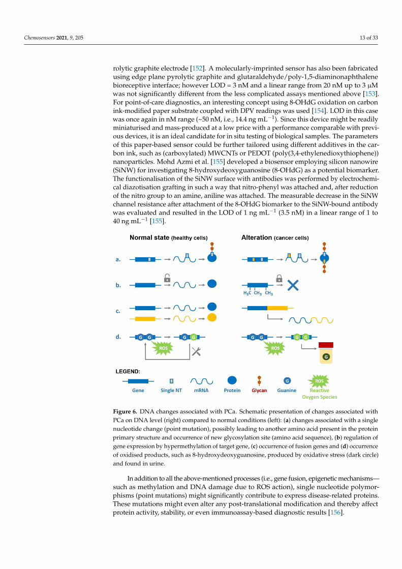
Figure 6. DNA changes associated with PCa. Schematic presentation of changes associated with PCa on DNA level (right) compared to normal conditions (left): (a) changes associated with a single nu-cleotide change (point mutation), possibly leading to another amino acid present in the protein pri-mary structure and occurrence of new glycosylation site (amino acid sequence), (b) regulation of gene expression by hypermethylation of target gene, (c) occurrence of fusion genes and (d) occur-rence of oxidised products, such as 8-hydroxydeoxyguanosine, produced by oxidative stress (dark circle) and found in urine.
If oxidative stress (in the form of a ROS attack) is not prevented, it might directly damage cellular DNA. Predominantly, bases in DNA structure are hydroxylated with guanine being the most prone to such an oxidation. Hence, for several decades, the most studied and abundant lesion is most likely 8-hydroxydeoxyguanosine (8-OHdG), which occurs in urine as a product of mismatch repair mechanism [148]. Moreover, 8-OHdG concentration (controlled by local antioxidant capacity) in urine was shown to refer not only to increased oxidative damage but also to be associated with some diseases, includ-ing breast, bladder and prostate cancers [149]—which could even be distinguished from benign hyperplasia using a tissue-staining microarray (10 adenocarcinoma patients vs. 70 controls) [150]. 8-OHdG might be directly electrochemically oxidised on modified sur-faces with sufficient electrocatalytic activities (on carbon surfaces, 8-OHdG exhibits a 2-electron transfer reaction), such as electrochemically reduced GO and multi-walled car-bon nanotubes-modified glassy carbon electrode, where a limit of detection of 35 nM was achieved (with linear range from 3 to 75 μM), as well as analysis in the presence of com-mon interferents (ascorbic and uric acid, xanthine, and hypoxanthine) and in urine [151]. Similarly, LOD = 28 nM (with linear range from 0.5 to 100 μM) was achieved at the edge plane surface of a pyrolytic graphite electrode [152]. A molecularly-imprinted sensor has also been fabricated using edge plane pyrolytic graphite and glutaraldehyde/poly-1,5-di-aminonaphthalene bioreceptive interface; however LOD = 3 nM and a linear range from 20 nM up to 3 μM was not significantly different from the less complicated assays
Figure 6. DNA changes associated with PCa. Schematic presentation of changes associated withPCa on DNA level (right) compared to normal conditions (left): (a) changes associated with a singlenucleotide change (point mutation), possibly leading to another amino acid present in the proteinprimary structure and occurrence of new glycosylation site (amino acid sequence), (b) regulation ofgene expression by hypermethylation of target gene, (c) occurrence of fusion genes and (d) occurrenceof oxidised products, such as 8-hydroxydeoxyguanosine, produced by oxidative stress (dark circle)and found in urine.
In addition to all the above-mentioned processes (i.e., gene fusion, epigenetic mechanisms—such as methylation and DNA damage due to ROS action), single nucleotide polymor-phisms (point mutations) might significantly contribute to express disease-related proteins.These mutations might even alter any post-translational modification and thereby affectprotein activity, stability, or even immunoassay-based diagnostic results [156].
Chemosensors 2021, 9, 205 14 of 33
4. Protein-Based PCa Biomarkers4.1. Osteopontin
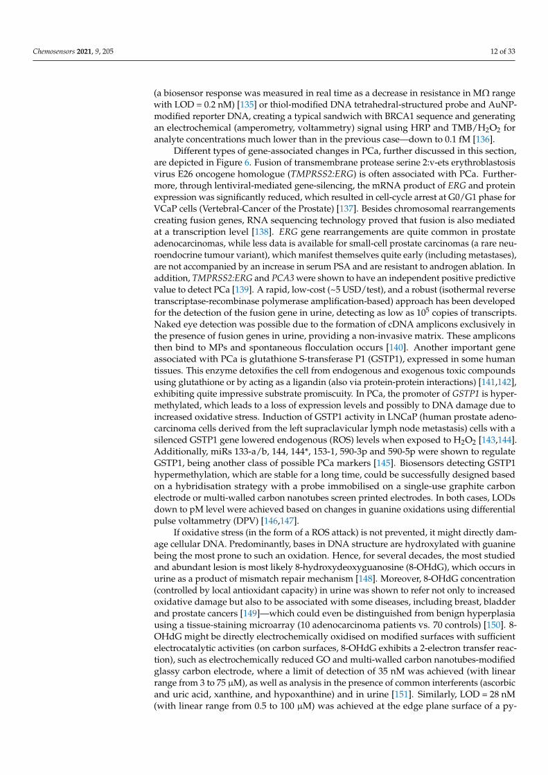
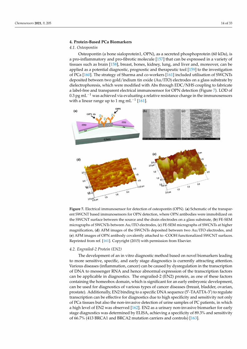
Osteopontin (a bone sialoprotein1, OPN), as a secreted phosphoprotein (60 kDa), isa pro-inflammatory and pro-fibrotic molecule [157] that can be expressed in a variety oftissues such as brain [158], breast, bones, kidney, lung, and liver and, moreover, can beapplied as a potential diagnostic, prognostic and therapeutic tool [159] to the investigationof PCa [160]. The strategy of Sharma and co-workers [161] included utilisation of SWCNTsdeposited between two gold/indium tin oxide (Au/ITO) electrodes on a glass substrate bydielectrophoresis, which were modified with Abs through EDC/NHS coupling to fabricatea label-free and transparent electrical immunosensor for OPN detection (Figure 7). LOD of0.3 pg mL−1 was achieved via evaluating a relative resistance change in the immunosensorswith a linear range up to 1 mg mL−1 [161].
Chemosensors 2021, 9, x FOR PEER REVIEW 14 of 33
mentioned above [153]. For point-of-care diagnostics, an interesting concept using 8-OHdG oxidation on carbon ink-modified paper substrate coupled with DPV readings was used [154]. LOD in this case was once again in nM range (~50 nM, i.e., 14.4 ng mL−1). Since this device might be readily miniaturised and mass-produced at a low price with a per-formance comparable with previous devices, it is an ideal candidate for in situ testing of biological samples. The parameters of this paper-based sensor could be further tailored using different additives in the carbon ink, such as (carboxylated) MWCNTs or PEDOT (poly(3,4-ethylenedioxythiophene)) nanoparticles. Mohd Azmi et al. [155] developed a bi-osensor employing silicon nanowire (SiNW) for investigating 8-hydroxydeoxyguanosine (8-OHdG) as a potential biomarker. The functionalisation of the SiNW surface with anti-bodies was performed by electrochemical diazotisation grafting in such a way that nitro-phenyl was attached and, after reduction of the nitro group to an amine, aniline was at-tached. The measurable decrease in the SiNW channel resistance after attachment of the 8-OHdG biomarker to the SiNW-bound antibody was evaluated and resulted in the LOD of 1 ng mL−1 (3.5 nM) in a linear range of 1 to 40 ng mL−1 [155].
In addition to all the above-mentioned processes (i.e., gene fusion, epigenetic mech-anisms—such as methylation and DNA damage due to ROS action), single nucleotide polymorphisms (point mutations) might significantly contribute to express disease-re-lated proteins. These mutations might even alter any post-translational modification and thereby affect protein activity, stability, or even immunoassay-based diagnostic results [156].
4. Protein-Based PCa Biomarkers 4.1. Osteopontin
Osteopontin (a bone sialoprotein1, OPN), as a secreted phosphoprotein (60 kDa), is a pro-inflammatory and pro-fibrotic molecule [157] that can be expressed in a variety of tissues such as brain [158], breast, bones, kidney, lung, and liver and, moreover, can be applied as a potential diagnostic, prognostic and therapeutic tool [159] to the investigation of PCa [160]. The strategy of Sharma and co-workers [161] included utilisation of SWCNTs deposited between two gold/indium tin oxide (Au/ITO) electrodes on a glass substrate by dielectrophoresis, which were modified with Abs through EDC/NHS coupling to fabricate a label-free and transparent electrical immunosensor for OPN detection (Figure 7). LOD of 0.3 pg mL−1 was achieved via evaluating a relative resistance change in the immunosen-sors with a linear range up to 1 mg mL−1 [161].
Figure 7. Electrical immunosensor for detection of osteopontin (OPN). (a) Schematic of the transpar-ent SWCNT based immunosensors for OPN detection, where OPN antibodies were immobilized onthe SWCNT surface between the source and the drain electrodes on a glass substrate, (b) FE-SEMmicrographs of SWCNTs between Au/ITO electrodes, (c) FE-SEM micrographs of SWCNTs at highermagnification, (d) AFM images of the SWCNTs deposited between two Au/ITO electrodes, and(e) AFM images of OPN antibody covalently attached to -COOH functionalized SWCNT surfaces.Reprinted from ref. [161]. Copyright (2015) with permission from Elsevier.
4.2. Engrailed-2 Protein (EN2)
The development of an in vitro diagnostic method based on novel biomarkers leadingto more sensitive, specific, and early stage diagnostics is currently attracting attention.Various diseases (inflammation, cancer) can be caused by dysregulation in the transcriptionof DNA to messenger RNA and hence abnormal expression of the transcription factorscan be applicable in diagnostics. The engrailed-2 (EN2) protein, as one of these factorscontaining the homeobox domain, which is significant for an early embryonic development,can be used for diagnostics of various types of cancer diseases (breast, bladder, ovarian,prostate). Additionally, EN2 binding to a specific DNA sequence (5′-TAATTA-3′) to regulatetranscription can be effective for diagnostics due to high specificity and sensitivity not onlyof PCa tissues but also the non-invasive detection of urine samples of PC patients, in whicha high level of EN2 was observed [162]. EN2 as a urinary non-invasive biomarker for earlystage diagnostics was determined by ELISA, achieving a specificity of 89.3% and sensitivityof 66.7% (413 BRCA1 and BRCA2 mutation carriers and controls) [163].
Chemosensors 2021, 9, 205 15 of 33
EN2 levels in urine samples, related to the Gleason score and tumour stage, werecardinally affected by prostate massage (1.25 ng mL−1 in the PCa group and 0.34 ng mL−1
in the BPH) [164]. The EN2 protein is present in the cell membrane and within microvesiclesand, when taken up by normal cells from the stroma, EN2 induces the expression ofMX2 (MxB), a key protein in the innate immune response to viruses. Hence, it wasconcluded that EN2 secretion by tumours might be a way of preventing a viral-mediatedimmune invasion of tissue immediately adjacent to the tumour [165]. Lee et al. [166]took advantage of this interaction between homeodomain and specific DNA probes tofabricate an ultrasensitive impedimetric biosensor for EN2 determination with LOD of5.62 fM. The incubation of different concentrations of EN2 (10 fM–1 nM) onto a pre-treatedgold electrode electrodeposited with Au NPs and subsequently modified with poly(A)10-hpDNA3 probe and 6-mercapto-1-hexanol was performed. The specificity of the biosensorthus developed was further verified through examination with artificial urine medium(AUM) and other proteins (bovine serum albumin (BSA), human serum albumin (HSA),lysozyme, thrombin, and TATA-binding protein (TBP)) [166].
4.3. Epidermal Growth Factor Receptor (EGFR)
The epidermal growth factor receptor (EGFR), belonging to the family of receptortyrosine kinases, is a transmembrane tyrosine kinase receptor promoting the proliferationand survival of both normal and cancer cells. The amplification of EGFR or gain of functionmutations of EGFR in its tyrosine kinase domain results in some of the abnormal activationof EGFR in cancer cells.
Takaaki Hiragun et al. [167] employed surface plasmon resonance (SPR) as a detectiontechnique observing intracellular signalling events as a function of the angle of resonance.The LNCaP cells showed a unique pattern of signal change in response to EGF stimulation.Through evaluation of the SPR signals, the presence of a single LNCaP cell among normalor even other malignant tumour cells can be identified in real-time without labelling [167].
4.4. Alpha-Methylacyl-CoA Racemase (AMACR)
Ying et al. [168] focused on the determination of alpha-methylacyl-CoA racemase(AMACR, also noted as P504S protein) presenting a further potential biomarker for PCadiagnostics in clinical practice. This metabolic enzyme is localised intracellularly in themitochondria and peroxisomes and is associated with the peroxisomal β-oxidation ofdietary branched-chain fatty acids. The differentiation/discrimination progress of benignto malignant prostate tumours through AMACR can be achieved with high sensitivity andspecificity. The sensing interface was based on a Pt nanowires (Pt NWs) array providingan efficient mass diffusion and electron transfer and a 2D nanomaterial MoS2 matrix (PtNWs array@MoS2). The disposable screen-printed amperometric sensor detected AMACRwith LOD of 0.5 pg µL−1 (signal to noise ratio S/N = 3) in a linear range from 0.70 to12.50 ng µL−1 (correlation coefficient R2 = 0.9597) at −0.45 V.
Lin et al. [169] employed an electrochemical biosensor based on a metallic nanoparticle-based catalyst for investigating AMACR levels requiring a small volume of plasma samples(9 healthy males, 10 patients with high grade prostatic intraepithelial neoplasia (HGPIN),and 5 prostate cancer patients) and achieving 100% accuracy in separating PCa patientsfrom controls.
In an effort to improve the anti-fouling properties of a final impedimetric aptasensordetecting AMACR [170], PPy film electropolymerised onto an Au electrode was patternedwith PEG molecules due to amine groups and a further Nα,Nα-Bis(carboxymethyl)-L-lysine ANTA(N-(5-Amino-1-carboxypentyl)iminodiacetic acid)/Cu2+ redox complex wascovalently attached. Finally, histidine-tagged AMACR-specific DNA aptamers were at-tached and LODs of 0.15 fM and 1.4 fM for AMACR were obtained in a buffer and inspiked human plasma, respectively, by applying the SWV method.
Chemosensors 2021, 9, 205 16 of 33
4.5. Prostatic Acid Phosphatase (PAP)
In addition, Fernandes et al. [171] employed a time-effective approach without la-belling by applying impedance-derived immittance functions (ImFs, inverse of imaginarycapacitance, 1/C”) in combination with a SAM consisting of thiol-containing polyethy-lene glycol (PEG thiol, HS–(CH2)11–(EG)3–OCH2–COOH as a low-fouling material) and11-ferrocenyl undecanethiol (11FcC as a redox probe) to investigate the level of an enzymehuman prostatic acid phosphatase (PAP) in human blood serum. The columnar secretoryepithelia of the prostate gland biosynthesise human PAP, which is considered as a majorphosphatase enzyme with plasma levels in the range 10–30 pM (1–3 ng mL−1) in healthy in-dividuals. In this concept, the specificity of interaction between an antibody (anti-PAP) as abiorecognition element functionalising a gold disk electrode surface covered with the mixedSAM and the target biomarker was detected using an impedance-derived capacitance tech-nique termed electrochemical capacitance spectroscopy with LOD of 11.2 ± 2.6 pM (alinear range from 50 pM to 104 pM) in a real sample.
4.6. Acid Phosphatase (ACP)
Kamel et al. [172] introduced ion association complexes of α-naphthyl acid phos-phate (1-NAP) with cetylpyridinium chloride (CP) and iron (II)—4,7-bathophenanthroline[Fe(bphen)3]2+ as state-of-the-art electroactive materials dispersed in o-nitrophenyloctylether (o-NPOE) plasticiser to design an ionometric sensor. The acid phosphatase enzyme(ACP) is present in the lysosomal compartment of the cells and as a non-specific esterase iscatalytically active in the hydrolysis of many monoesters of phosphoric acid in an acidic en-vironment. The activity of ACP was estimated through the decrease in a fixed concentrationof 1-NAP under optimised conditions of pH (3.8–5.7) and temperature (25 C).
The planar chip biosensors for potentiometric immunoassay exhibited a good potentialresponse stability of±3 mV, high accuracy and precision of ~(98.3± 0.6)%, and, moreover, along life-span of 6–8 weeks. The LOD value of 0.01 IU L−1 was achieved with a linear signalresponse in a range of 0.01–4.3 IU L−1 for the ACP enzyme employing a potentiometricbiosensor affording better durability, decreased diffusion area, small sample volume,and time-effectiveness. The early stage PCa diagnostics, human prostatic diseases, andhypophosphatasia can be performed through observation of the level of ACP activity in ablood sample.
Na et al. [173] took advantage of a lecithin/β-Cyclodextrin (lecithin/β-CD) complexas the linker for attachment of Nile red (NR, acceptor) to graphene quantum dots (GQDs,donor) through electrostatic and hydrophobic interaction in a Förster resonance energytransfer (FRET) biosensor detecting ACP with LOD of 28 µU mL−1 (Figure 8). The levelof ACP can provide information about disorders related to kidneys, veins, and bones.The GQDs-based fluorescence biosensor facilitated in vitro imaging of human prostatecancer cells.
4.7. Spondin-2 (SPON2)
In an effort to achieve a more specific PCa diagnostics, Parra-Cabrera et al. [174], in areal-time analysis, simultaneously examined two biomarkers such as PSA and spondin-2 (SPON2) within the clinically relevant range. Photolithography and a cast-mouldingmethod were employed for fabrication of the lab-on-a-chip device with the integrated twosets of gold electrodes into the microfluidic channel. The blocking step was performedwith 11-Mercaptoundecyl tetra(ethylene glycol) (PEG-Thiol) in ethanol. In the next step,biotinylated alkyl thiols with a polyethyleneglycol linker (biotin-thiol) were employed toform a layer. Subsequently, a biotin layer was covered with a streptavidin layer and in-situimmobilisation of biotinylated specific antibodies over this layer was performed to designthe sensor. Both analytes could be detected in a clinically relevant concentration range of1–10 ng mL−1 [174].
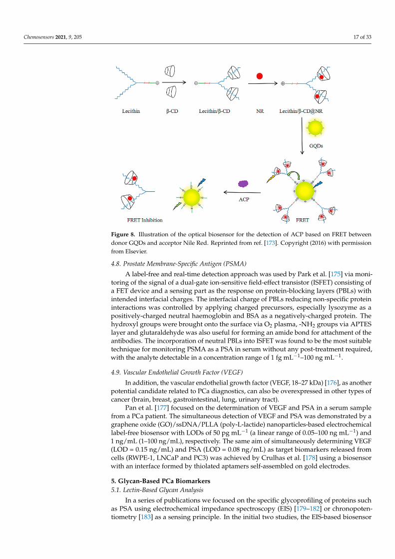
Chemosensors 2021, 9, 205 17 of 33
Chemosensors 2021, 9, x FOR PEER REVIEW 17 of 33
potentiometric biosensor affording better durability, decreased diffusion area, small sam-ple volume, and time-effectiveness. The early stage PCa diagnostics, human prostatic dis-eases, and hypophosphatasia can be performed through observation of the level of ACP activity in a blood sample.
Na et al. [173] took advantage of a lecithin/β-Cyclodextrin (lecithin/β-CD) complex as the linker for attachment of Nile red (NR, acceptor) to graphene quantum dots (GQDs, donor) through electrostatic and hydrophobic interaction in a Förster resonance energy transfer (FRET) biosensor detecting ACP with LOD of 28 μU mL−1 (Figure 8). The level of ACP can provide information about disorders related to kidneys, veins, and bones. The GQDs-based fluorescence biosensor facilitated in vitro imaging of human prostate cancer cells.
Figure 8. Illustration of the optical biosensor for the detection of ACP based on FRET between donor GQDs and acceptor Nile Red. Reprinted from ref. [173]. Copyright (2016) with permission from Elsevier.
4.7. Spondin-2 (SPON2) In an effort to achieve a more specific PCa diagnostics, Parra-Cabrera et al. [174], in
a real-time analysis, simultaneously examined two biomarkers such as PSA and spondin-2 (SPON2) within the clinically relevant range. Photolithography and a cast-moulding method were employed for fabrication of the lab-on-a-chip device with the integrated two sets of gold electrodes into the microfluidic channel. The blocking step was performed with 11-Mercaptoundecyl tetra(ethylene glycol) (PEG-Thiol) in ethanol. In the next step, biotinylated alkyl thiols with a polyethyleneglycol linker (biotin-thiol) were employed to form a layer. Subsequently, a biotin layer was covered with a streptavidin layer and in-situ immobilisation of biotinylated specific antibodies over this layer was performed to design the sensor. Both analytes could be detected in a clinically relevant concentration range of 1–10 ng mL−1 [174].
4.8. Prostate Membrane-Specific Antigen (PSMA) A label-free and real-time detection approach was used by Park et al. [175] via mon-
itoring of the signal of a dual-gate ion-sensitive field-effect transistor (ISFET) consisting of a FET device and a sensing part as the response on protein-blocking layers (PBLs) with intended interfacial charges. The interfacial charge of PBLs reducing non-specific protein
Figure 8. Illustration of the optical biosensor for the detection of ACP based on FRET betweendonor GQDs and acceptor Nile Red. Reprinted from ref. [173]. Copyright (2016) with permissionfrom Elsevier.
4.8. Prostate Membrane-Specific Antigen (PSMA)
A label-free and real-time detection approach was used by Park et al. [175] via moni-toring of the signal of a dual-gate ion-sensitive field-effect transistor (ISFET) consisting ofa FET device and a sensing part as the response on protein-blocking layers (PBLs) withintended interfacial charges. The interfacial charge of PBLs reducing non-specific proteininteractions was controlled by applying charged precursors, especially lysozyme as apositively-charged neutral haemoglobin and BSA as a negatively-charged protein. Thehydroxyl groups were brought onto the surface via O2 plasma, -NH2 groups via APTESlayer and glutaraldehyde was also useful for forming an amide bond for attachment of theantibodies. The incorporation of neutral PBLs into ISFET was found to be the most suitabletechnique for monitoring PSMA as a PSA in serum without any post-treatment required,with the analyte detectable in a concentration range of 1 fg mL−1–100 ng mL−1.
4.9. Vascular Endothelial Growth Factor (VEGF)
In addition, the vascular endothelial growth factor (VEGF, 18–27 kDa) [176], as anotherpotential candidate related to PCa diagnostics, can also be overexpressed in other types ofcancer (brain, breast, gastrointestinal, lung, urinary tract).
Pan et al. [177] focused on the determination of VEGF and PSA in a serum samplefrom a PCa patient. The simultaneous detection of VEGF and PSA was demonstrated by agraphene oxide (GO)/ssDNA/PLLA (poly-L-lactide) nanoparticles-based electrochemicallabel-free biosensor with LODs of 50 pg mL−1 (a linear range of 0.05–100 ng mL−1) and1 ng/mL (1–100 ng/mL), respectively. The same aim of simultaneously determining VEGF(LOD = 0.15 ng/mL) and PSA (LOD = 0.08 ng/mL) as target biomarkers released fromcells (RWPE-1, LNCaP and PC3) was achieved by Crulhas et al. [178] using a biosensorwith an interface formed by thiolated aptamers self-assembled on gold electrodes.
5. Glycan-Based PCa Biomarkers5.1. Lectin-Based Glycan Analysis
In a series of publications we focused on the specific glycoprofiling of proteins suchas PSA using electrochemical impedance spectroscopy (EIS) [179–182] or chronopoten-tiometry [183] as a sensing principle. In the initial two studies, the EIS-based biosensor
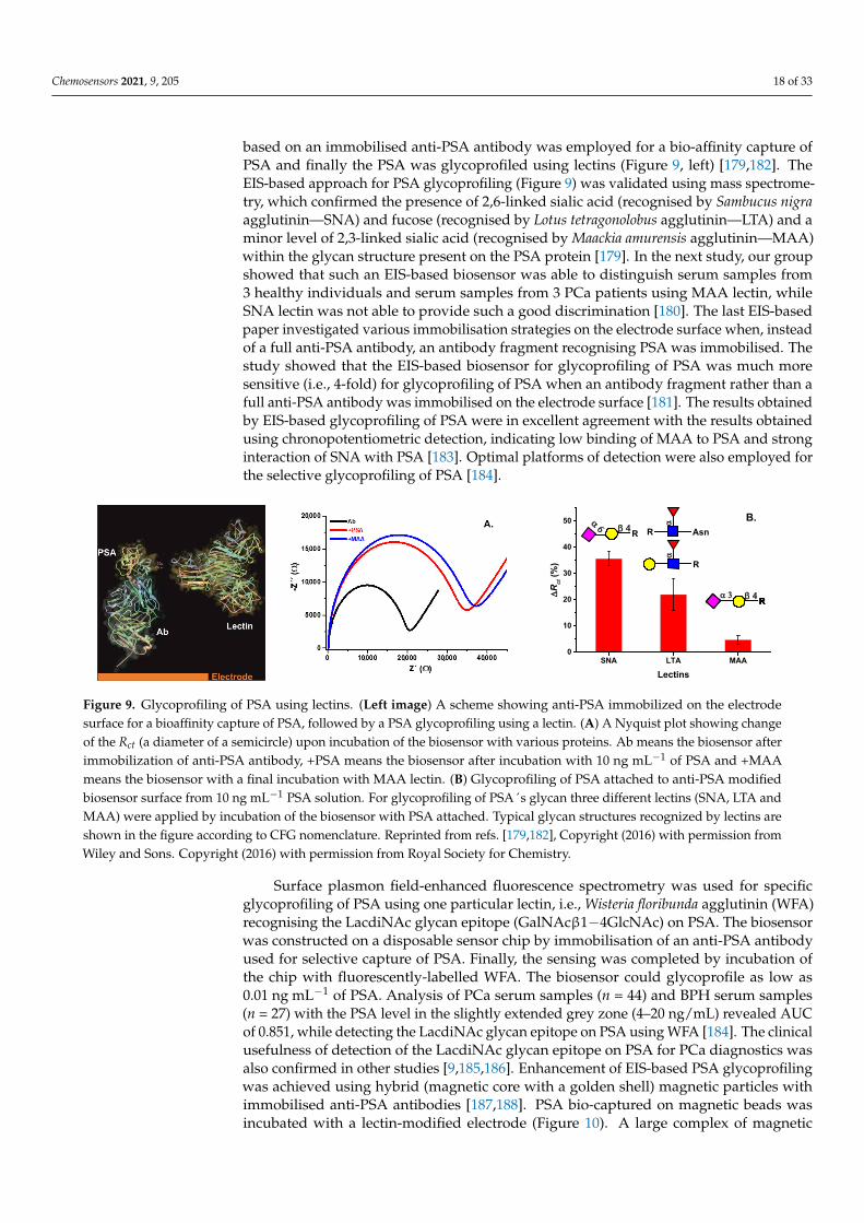
Chemosensors 2021, 9, 205 18 of 33
based on an immobilised anti-PSA antibody was employed for a bio-affinity capture ofPSA and finally the PSA was glycoprofiled using lectins (Figure 9, left) [179,182]. TheEIS-based approach for PSA glycoprofiling (Figure 9) was validated using mass spectrome-try, which confirmed the presence of 2,6-linked sialic acid (recognised by Sambucus nigraagglutinin—SNA) and fucose (recognised by Lotus tetragonolobus agglutinin—LTA) and aminor level of 2,3-linked sialic acid (recognised by Maackia amurensis agglutinin—MAA)within the glycan structure present on the PSA protein [179]. In the next study, our groupshowed that such an EIS-based biosensor was able to distinguish serum samples from3 healthy individuals and serum samples from 3 PCa patients using MAA lectin, whileSNA lectin was not able to provide such a good discrimination [180]. The last EIS-basedpaper investigated various immobilisation strategies on the electrode surface when, insteadof a full anti-PSA antibody, an antibody fragment recognising PSA was immobilised. Thestudy showed that the EIS-based biosensor for glycoprofiling of PSA was much moresensitive (i.e., 4-fold) for glycoprofiling of PSA when an antibody fragment rather than afull anti-PSA antibody was immobilised on the electrode surface [181]. The results obtainedby EIS-based glycoprofiling of PSA were in excellent agreement with the results obtainedusing chronopotentiometric detection, indicating low binding of MAA to PSA and stronginteraction of SNA with PSA [183]. Optimal platforms of detection were also employed forthe selective glycoprofiling of PSA [184].
Chemosensors 2021, 9, x FOR PEER REVIEW 19 of 33
while detecting the LacdiNAc glycan epitope on PSA using WFA [184]. The clinical use-fulness of detection of the LacdiNAc glycan epitope on PSA for PCa diagnostics was also confirmed in other studies [9,185,186]. Enhancement of EIS-based PSA glycoprofiling was achieved using hybrid (magnetic core with a golden shell) magnetic particles with immo-bilised anti-PSA antibodies [187,188]. PSA bio-captured on magnetic beads was incubated with a lectin-modified electrode (Figure 10). A large complex of magnetic beads accom-modated by anti-PSA antibodies and PSA molecules attached to the electrode interface significantly increased the charge-transfer resistance and thus also the EIS-based sensing with a sensitivity enhancement of 43-fold [187].
SNA LTA MAA
0
10
20
30
40
50R
α 6
∆R
ct (%
)
Lectins
Rβ 4
R
α
α 3RRRRβ 4
B.Asn
α
Figure 9. Glycoprofiling of PSA using lectins. (Left image) A scheme showing anti-PSA immobilized on the electrode surface for a bioaffinity capture of PSA, followed by a PSA glycoprofiling using a lectin. (A) A Nyquist plot showing change of the Rct (a diameter of a semicircle) upon incubation of the biosensor with various proteins. Ab means the bio-sensor after immobilization of anti-PSA antibody, +PSA means the biosensor after incubation with 10 ng mL−1 of PSA and +MAA means the biosensor with a final incubation with MAA lectin. (B) Glycoprofiling of PSA attached to anti-PSA modified biosensor surface from 10 ng mL−1 PSA solution. For glycoprofiling of PSA´s glycan three different lectins (SNA, LTA and MAA) were applied by incubation of the biosensor with PSA attached. Typical glycan structures recognized by lectins are shown in the figure according to CFG nomenclature. Reprinted from ref. [179,182], Copyright (2016) with per-mission from Wiley and Sons. Copyright (2016) with permission from Royal Society for Chemistry.
Figure 10. Sensitivity enhancement of electrochemical impedance spectroscopy-based PSA glycoprofiling using hybrid magnetic particles. (Left image) Modification of Au electrode (upper row) using carboxybetaine aryldiazonium derivative (CB) and lectin (SNA-I). In the first step, carboxybetaine-bearing SAM layer was formed using cyclic voltammetry, and subsequently served for the covalent immobilization of a lectin. The lower row shows hybrid magnetic nanoparticle spon-taneously modified with CB-derivative and subsequently with an antibody (Ab, anti-PSA). After enrichment of PSA from the human sample by magnetic particles using a permanent magnet, a sandwich was prepared, and the signal was evalu-ated electrochemically. (Right image) A response towards 0.1 ng mL−1 PSA in 10× diluted real human serum is shown (red curves). A calibration curve for PSA (blue) for a sandwich configuration is shown in the inset (error bars are too small to be visible). According to the slopes of calibration curves in the linear response range for both configurations, an amplifi-cation factor of 43 was calculated. Reprinted from ref. [187]. Copyright (2019) with permission from Elsevier.
Besides using antibodies against PSA, the PSA protein can be glycoprofiled in a sand-wich configuration using lectins and immobilised DNA aptamers [189]. The chemilumi-nescent biosensor operating in a microfluidic format can glycoprofile PSA down to 3 ng
Figure 9. Glycoprofiling of PSA using lectins. (Left image) A scheme showing anti-PSA immobilized on the electrodesurface for a bioaffinity capture of PSA, followed by a PSA glycoprofiling using a lectin. (A) A Nyquist plot showing changeof the Rct (a diameter of a semicircle) upon incubation of the biosensor with various proteins. Ab means the biosensor afterimmobilization of anti-PSA antibody, +PSA means the biosensor after incubation with 10 ng mL−1 of PSA and +MAAmeans the biosensor with a final incubation with MAA lectin. (B) Glycoprofiling of PSA attached to anti-PSA modifiedbiosensor surface from 10 ng mL−1 PSA solution. For glycoprofiling of PSA´s glycan three different lectins (SNA, LTA andMAA) were applied by incubation of the biosensor with PSA attached. Typical glycan structures recognized by lectins areshown in the figure according to CFG nomenclature. Reprinted from refs. [179,182], Copyright (2016) with permission fromWiley and Sons. Copyright (2016) with permission from Royal Society for Chemistry.
Surface plasmon field-enhanced fluorescence spectrometry was used for specificglycoprofiling of PSA using one particular lectin, i.e., Wisteria floribunda agglutinin (WFA)recognising the LacdiNAc glycan epitope (GalNAcβ1−4GlcNAc) on PSA. The biosensorwas constructed on a disposable sensor chip by immobilisation of an anti-PSA antibodyused for selective capture of PSA. Finally, the sensing was completed by incubation ofthe chip with fluorescently-labelled WFA. The biosensor could glycoprofile as low as0.01 ng mL−1 of PSA. Analysis of PCa serum samples (n = 44) and BPH serum samples(n = 27) with the PSA level in the slightly extended grey zone (4–20 ng/mL) revealed AUCof 0.851, while detecting the LacdiNAc glycan epitope on PSA using WFA [184]. The clinicalusefulness of detection of the LacdiNAc glycan epitope on PSA for PCa diagnostics wasalso confirmed in other studies [9,185,186]. Enhancement of EIS-based PSA glycoprofilingwas achieved using hybrid (magnetic core with a golden shell) magnetic particles withimmobilised anti-PSA antibodies [187,188]. PSA bio-captured on magnetic beads wasincubated with a lectin-modified electrode (Figure 10). A large complex of magnetic
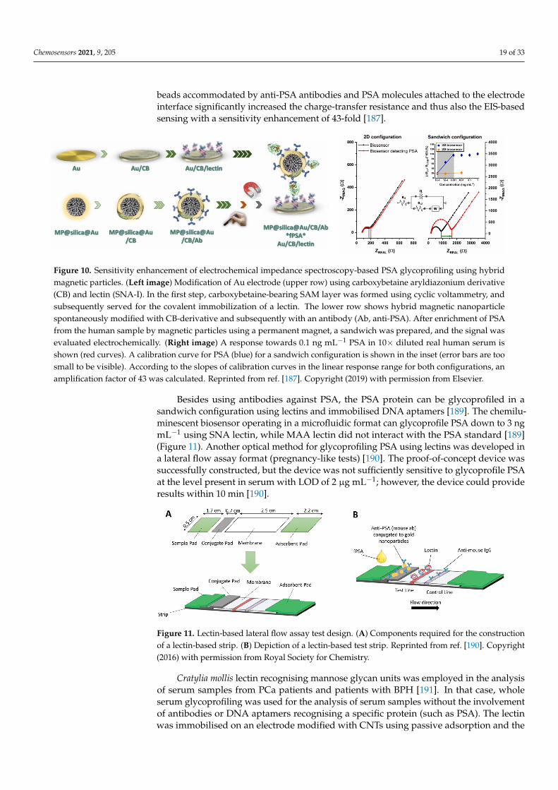
Chemosensors 2021, 9, 205 19 of 33
beads accommodated by anti-PSA antibodies and PSA molecules attached to the electrodeinterface significantly increased the charge-transfer resistance and thus also the EIS-basedsensing with a sensitivity enhancement of 43-fold [187].
Chemosensors 2021, 9, x FOR PEER REVIEW 19 of 33
while detecting the LacdiNAc glycan epitope on PSA using WFA [184]. The clinical use-fulness of detection of the LacdiNAc glycan epitope on PSA for PCa diagnostics was also confirmed in other studies [9,185,186]. Enhancement of EIS-based PSA glycoprofiling was achieved using hybrid (magnetic core with a golden shell) magnetic particles with immo-bilised anti-PSA antibodies [187,188]. PSA bio-captured on magnetic beads was incubated with a lectin-modified electrode (Figure 10). A large complex of magnetic beads accom-modated by anti-PSA antibodies and PSA molecules attached to the electrode interface significantly increased the charge-transfer resistance and thus also the EIS-based sensing with a sensitivity enhancement of 43-fold [187].
Figure 9. Glycoprofiling of PSA using lectins. (Left image) A scheme showing anti-PSA immobilized on the electrode surface for a bioaffinity capture of PSA, followed by a PSA glycoprofiling using a lectin. (A) A Nyquist plot showing change of the Rct (a diameter of a semicircle) upon incubation of the biosensor with various proteins. Ab means the bio-sensor after immobilization of anti-PSA antibody, +PSA means the biosensor after incubation with 10 ng mL−1 of PSA and +MAA means the biosensor with a final incubation with MAA lectin. (B) Glycoprofiling of PSA attached to anti-PSA modified biosensor surface from 10 ng mL−1 PSA solution. For glycoprofiling of PSA´s glycan three different lectins (SNA, LTA and MAA) were applied by incubation of the biosensor with PSA attached. Typical glycan structures recognized by lectins are shown in the figure according to CFG nomenclature. Reprinted from ref. [179,182], Copyright (2016) with per-mission from Wiley and Sons. Copyright (2016) with permission from Royal Society for Chemistry.
Figure 10. Sensitivity enhancement of electrochemical impedance spectroscopy-based PSA glycoprofiling using hybrid magnetic particles. (Left image) Modification of Au electrode (upper row) using carboxybetaine aryldiazonium derivative (CB) and lectin (SNA-I). In the first step, carboxybetaine-bearing SAM layer was formed using cyclic voltammetry, and subsequently served for the covalent immobilization of a lectin. The lower row shows hybrid magnetic nanoparticle spon-taneously modified with CB-derivative and subsequently with an antibody (Ab, anti-PSA). After enrichment of PSA from the human sample by magnetic particles using a permanent magnet, a sandwich was prepared, and the signal was evalu-ated electrochemically. (Right image) A response towards 0.1 ng mL−1 PSA in 10× diluted real human serum is shown (red curves). A calibration curve for PSA (blue) for a sandwich configuration is shown in the inset (error bars are too small to be visible). According to the slopes of calibration curves in the linear response range for both configurations, an amplifi-cation factor of 43 was calculated. Reprinted from ref. [187]. Copyright (2019) with permission from Elsevier.
Besides using antibodies against PSA, the PSA protein can be glycoprofiled in a sand-wich configuration using lectins and immobilised DNA aptamers [189]. The chemilumi-nescent biosensor operating in a microfluidic format can glycoprofile PSA down to 3 ng
Figure 10. Sensitivity enhancement of electrochemical impedance spectroscopy-based PSA glycoprofiling using hybridmagnetic particles. (Left image) Modification of Au electrode (upper row) using carboxybetaine aryldiazonium derivative(CB) and lectin (SNA-I). In the first step, carboxybetaine-bearing SAM layer was formed using cyclic voltammetry, andsubsequently served for the covalent immobilization of a lectin. The lower row shows hybrid magnetic nanoparticlespontaneously modified with CB-derivative and subsequently with an antibody (Ab, anti-PSA). After enrichment of PSAfrom the human sample by magnetic particles using a permanent magnet, a sandwich was prepared, and the signal wasevaluated electrochemically. (Right image) A response towards 0.1 ng mL−1 PSA in 10× diluted real human serum isshown (red curves). A calibration curve for PSA (blue) for a sandwich configuration is shown in the inset (error bars are toosmall to be visible). According to the slopes of calibration curves in the linear response range for both configurations, anamplification factor of 43 was calculated. Reprinted from ref. [187]. Copyright (2019) with permission from Elsevier.
Besides using antibodies against PSA, the PSA protein can be glycoprofiled in asandwich configuration using lectins and immobilised DNA aptamers [189]. The chemilu-minescent biosensor operating in a microfluidic format can glycoprofile PSA down to 3 ngmL−1 using SNA lectin, while MAA lectin did not interact with the PSA standard [189](Figure 11). Another optical method for glycoprofiling PSA using lectins was developed ina lateral flow assay format (pregnancy-like tests) [190]. The proof-of-concept device wassuccessfully constructed, but the device was not sufficiently sensitive to glycoprofile PSAat the level present in serum with LOD of 2 µg mL−1; however, the device could provideresults within 10 min [190].
Chemosensors 2021, 9, x FOR PEER REVIEW 20 of 33
mL−1 using SNA lectin, while MAA lectin did not interact with the PSA standard [189] (Figure 11). Another optical method for glycoprofiling PSA using lectins was developed in a lateral flow assay format (pregnancy-like tests) [190]. The proof-of-concept device was successfully constructed, but the device was not sufficiently sensitive to glycoprofile PSA at the level present in serum with LOD of 2 μg mL−1; however, the device could provide results within 10 min [190].
Figure 11. Lectin-based lateral flow assay test design. (A) Components required for the construction of a lectin-based strip. (B) Depiction of a lectin-based test strip. Reprinted from ref. [190]. Copyright (2016) with permission from Royal Society for Chemistry.
Cratylia mollis lectin recognising mannose glycan units was employed in the analysis of serum samples from PCa patients and patients with BPH [191]. In that case, whole se-rum glycoprofiling was used for the analysis of serum samples without the involvement of antibodies or DNA aptamers recognising a specific protein (such as PSA). The lectin was immobilised on an electrode modified with CNTs using passive adsorption and the SWV-based electrochemical biosensor was calibrated using fetuin as a model glycoprotein (Figure 12). The analyte was detected down to 0.017 μg mL−1 with a linear range of up to 25 μg mL−1 and the assay was highly reproducible and selective. The biosensor was finally employed in analysis of serum samples, exhibiting significant discrimination power for the analysis of 4 distinct groups of patients (BPH patients, PCa with Gleason score (GS) 6, PCa with GS 7 and PCa with GS 9) (Figure 13) [191].
Figure 12. Schematic illustration of the stepwise modification of the lectin-based nanobiosensor. Reprinted from ref. [191]. Copyright (2016) with permission from Elsevier.
Figure 11. Lectin-based lateral flow assay test design. (A) Components required for the constructionof a lectin-based strip. (B) Depiction of a lectin-based test strip. Reprinted from ref. [190]. Copyright(2016) with permission from Royal Society for Chemistry.
Cratylia mollis lectin recognising mannose glycan units was employed in the analysisof serum samples from PCa patients and patients with BPH [191]. In that case, wholeserum glycoprofiling was used for the analysis of serum samples without the involvementof antibodies or DNA aptamers recognising a specific protein (such as PSA). The lectinwas immobilised on an electrode modified with CNTs using passive adsorption and the
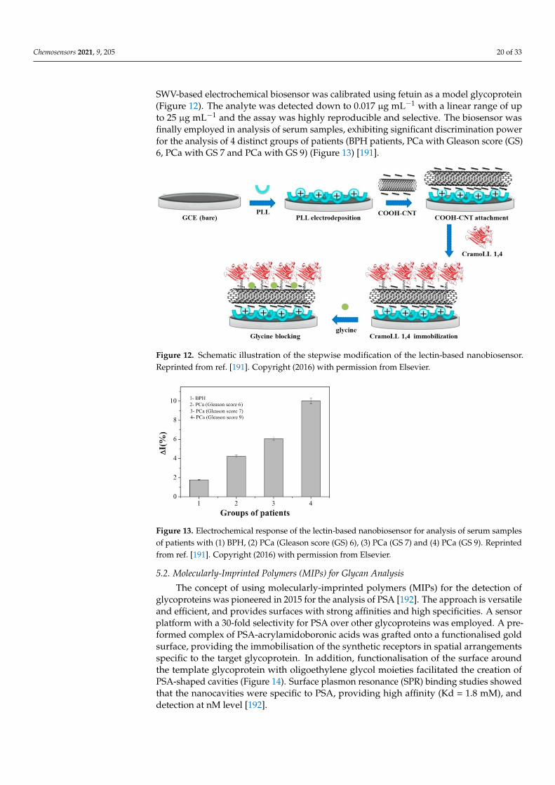
Chemosensors 2021, 9, 205 20 of 33
SWV-based electrochemical biosensor was calibrated using fetuin as a model glycoprotein(Figure 12). The analyte was detected down to 0.017 µg mL−1 with a linear range of upto 25 µg mL−1 and the assay was highly reproducible and selective. The biosensor wasfinally employed in analysis of serum samples, exhibiting significant discrimination powerfor the analysis of 4 distinct groups of patients (BPH patients, PCa with Gleason score (GS)6, PCa with GS 7 and PCa with GS 9) (Figure 13) [191].
Chemosensors 2021, 9, x FOR PEER REVIEW 20 of 33
mL−1 using SNA lectin, while MAA lectin did not interact with the PSA standard [189] (Figure 11). Another optical method for glycoprofiling PSA using lectins was developed in a lateral flow assay format (pregnancy-like tests) [190]. The proof-of-concept device was successfully constructed, but the device was not sufficiently sensitive to glycoprofile PSA at the level present in serum with LOD of 2 μg mL−1; however, the device could provide results within 10 min [190].
Figure 11. Lectin-based lateral flow assay test design. (A) Components required for the construction of a lectin-based strip. (B) Depiction of a lectin-based test strip. Reprinted from ref. [190]. Copyright (2016) with permission from Royal Society for Chemistry.
Cratylia mollis lectin recognising mannose glycan units was employed in the analysis of serum samples from PCa patients and patients with BPH [191]. In that case, whole se-rum glycoprofiling was used for the analysis of serum samples without the involvement of antibodies or DNA aptamers recognising a specific protein (such as PSA). The lectin was immobilised on an electrode modified with CNTs using passive adsorption and the SWV-based electrochemical biosensor was calibrated using fetuin as a model glycoprotein (Figure 12). The analyte was detected down to 0.017 μg mL−1 with a linear range of up to 25 μg mL−1 and the assay was highly reproducible and selective. The biosensor was finally employed in analysis of serum samples, exhibiting significant discrimination power for the analysis of 4 distinct groups of patients (BPH patients, PCa with Gleason score (GS) 6, PCa with GS 7 and PCa with GS 9) (Figure 13) [191].
Figure 12. Schematic illustration of the stepwise modification of the lectin-based nanobiosensor. Reprinted from ref. [191]. Copyright (2016) with permission from Elsevier. Figure 12. Schematic illustration of the stepwise modification of the lectin-based nanobiosensor.Reprinted from ref. [191]. Copyright (2016) with permission from Elsevier.
Chemosensors 2021, 9, x FOR PEER REVIEW 21 of 33
Figure 13. Electrochemical response of the lectin-based nanobiosensor for analysis of serum samples of patients with (1) BPH, (2) PCa (Gleason score (GS) 6), (3) PCa (GS 7) and (4) PCa (GS 9). Reprinted from ref. [191]. Copyright (2016) with permission from Elsevier.
5.2. Molecularly-Imprinted Polymers (MIPs) for Glycan Analysis The concept of using molecularly-imprinted polymers (MIPs) for the detection of gly-
coproteins was pioneered in 2015 for the analysis of PSA [192]. The approach is versatile and efficient, and provides surfaces with strong affinities and high specificities. A sensor platform with a 30-fold selectivity for PSA over other glycoproteins was employed. A pre-formed complex of PSA-acrylamidoboronic acids was grafted onto a functionalised gold surface, providing the immobilisation of the synthetic receptors in spatial arrangements specific to the target glycoprotein. In addition, functionalisation of the surface around the template glycoprotein with oligoethylene glycol moieties facilitated the creation of PSA-shaped cavities (Figure 14). Surface plasmon resonance (SPR) binding studies showed that the nanocavities were specific to PSA, providing high affinity (Kd = 1.8 mM), and detection at nM level [192].
Figure 14. Selective glycoprotein detection using boronate-based imprinting approach. Strategy for the fabrication of glycoprotein-imprinted surfaces using acrylamido-boronic acids (AM-BAs); (1) self-assembled monolayer (SAM) formation on a gold surface with DFC molecule; (2) incubation of the AM-BA receptors with the template target glycoprotein; (3) grafting of the preformed boronic acid–glycoprotein complex on the SAM via acrylamide co-polymerization; (4) azide-terminated ol-igoethylene glycol (Az-OEG) functionalization to provide glycoprotein-shaped cavities; (5) removal of the template protein, affording a nanocavity specific for the target glycoprotein. Reproduced from ref. [192] under a Creative Commons Attribution License.
Although a dual-recognition strategy for increasing the selectivity of glycoprotein detection using two types of MIPs was designed for the detection of a carcinoembryonic
Figure 13. Electrochemical response of the lectin-based nanobiosensor for analysis of serum samplesof patients with (1) BPH, (2) PCa (Gleason score (GS) 6), (3) PCa (GS 7) and (4) PCa (GS 9). Reprintedfrom ref. [191]. Copyright (2016) with permission from Elsevier.
5.2. Molecularly-Imprinted Polymers (MIPs) for Glycan Analysis
The concept of using molecularly-imprinted polymers (MIPs) for the detection ofglycoproteins was pioneered in 2015 for the analysis of PSA [192]. The approach is versatileand efficient, and provides surfaces with strong affinities and high specificities. A sensorplatform with a 30-fold selectivity for PSA over other glycoproteins was employed. A pre-formed complex of PSA-acrylamidoboronic acids was grafted onto a functionalised goldsurface, providing the immobilisation of the synthetic receptors in spatial arrangementsspecific to the target glycoprotein. In addition, functionalisation of the surface aroundthe template glycoprotein with oligoethylene glycol moieties facilitated the creation ofPSA-shaped cavities (Figure 14). Surface plasmon resonance (SPR) binding studies showedthat the nanocavities were specific to PSA, providing high affinity (Kd = 1.8 mM), anddetection at nM level [192].
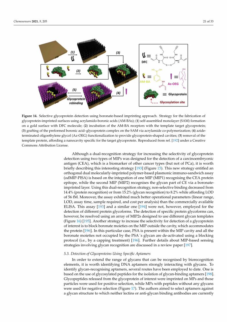
Chemosensors 2021, 9, 205 21 of 33
Chemosensors 2021, 9, x FOR PEER REVIEW 21 of 33
Figure 13. Electrochemical response of the lectin-based nanobiosensor for analysis of serum samples of patients with (1) BPH, (2) PCa (Gleason score (GS) 6), (3) PCa (GS 7) and (4) PCa (GS 9). Reprinted from ref. [191]. Copyright (2016) with permission from Elsevier.
5.2. Molecularly-Imprinted Polymers (MIPs) for Glycan Analysis The concept of using molecularly-imprinted polymers (MIPs) for the detection of gly-
coproteins was pioneered in 2015 for the analysis of PSA [192]. The approach is versatile and efficient, and provides surfaces with strong affinities and high specificities. A sensor platform with a 30-fold selectivity for PSA over other glycoproteins was employed. A pre-formed complex of PSA-acrylamidoboronic acids was grafted onto a functionalised gold surface, providing the immobilisation of the synthetic receptors in spatial arrangements specific to the target glycoprotein. In addition, functionalisation of the surface around the template glycoprotein with oligoethylene glycol moieties facilitated the creation of PSA-shaped cavities (Figure 14). Surface plasmon resonance (SPR) binding studies showed that the nanocavities were specific to PSA, providing high affinity (Kd = 1.8 mM), and detection at nM level [192].
Figure 14. Selective glycoprotein detection using boronate-based imprinting approach. Strategy for the fabrication of glycoprotein-imprinted surfaces using acrylamido-boronic acids (AM-BAs); (1) self-assembled monolayer (SAM) formation on a gold surface with DFC molecule; (2) incubation of the AM-BA receptors with the template target glycoprotein; (3) grafting of the preformed boronic acid–glycoprotein complex on the SAM via acrylamide co-polymerization; (4) azide-terminated ol-igoethylene glycol (Az-OEG) functionalization to provide glycoprotein-shaped cavities; (5) removal of the template protein, affording a nanocavity specific for the target glycoprotein. Reproduced from ref. [192] under a Creative Commons Attribution License.
Although a dual-recognition strategy for increasing the selectivity of glycoprotein detection using two types of MIPs was designed for the detection of a carcinoembryonic
Figure 14. Selective glycoprotein detection using boronate-based imprinting approach. Strategy for the fabrication ofglycoprotein-imprinted surfaces using acrylamido-boronic acids (AM-BAs); (1) self-assembled monolayer (SAM) formationon a gold surface with DFC molecule; (2) incubation of the AM-BA receptors with the template target glycoprotein;(3) grafting of the preformed boronic acid–glycoprotein complex on the SAM via acrylamide co-polymerization; (4) azide-terminated oligoethylene glycol (Az-OEG) functionalization to provide glycoprotein-shaped cavities; (5) removal of thetemplate protein, affording a nanocavity specific for the target glycoprotein. Reproduced from ref. [192] under a CreativeCommons Attribution License.
Although a dual-recognition strategy for increasing the selectivity of glycoproteindetection using two types of MIPs was designed for the detection of a carcinoembryonicantigen (CEA), which is a biomarker of other cancer types (but not of PCa), it is worthbriefly describing this interesting strategy [193] (Figure 15). This new strategy entitled anorthogonal dual molecularly-imprinted polymer-based plasmonic immuno-sandwich assay(odMIP-PISA) is based on the integration of one MIP (MIP1) recognising the CEA proteinepitope, while the second MIP (MIP2) recognises the glycan part of CE via a boronate-imprinted layer. Using this dual-recognition strategy, non-selective binding decreased from14.4% (protein recognition) or from 15.2% (glycan recognition) to 8.2% while affording LODof 56 fM. Moreover, the assay exhibited much better operational parameters (linear range,LOD, assay time, sample required, and cost per analysis) than the commercially availableELISA. This assay [193] and a similar one [194] were not, however, employed for thedetection of different protein glycoforms. The detection of specific protein glycoforms can,however, be resolved using an array of MIP2s designed to use different glycan templates(Figure 16) [195]. Another strategy to increase the selectivity for detection of a glycoproteinof interest is to block boronate moieties on the MIP outside the cavity, which accommodatesthe protein [196]. In this particular case, PSA is present within the MIP cavity and all theboronate moieties not occupied by the PSA´s glycan are de-activated using a blockingprotocol (i.e., by a capping treatment) [196]. Further details about MIP-based sensingstrategies involving glycan recognition are discussed in a review paper [197].
5.3. Detection of Glycoproteins Using Specific Aptamers
In order to extend the range of glycans that can be recognised by biorecognitionelements, it is worth identifying DNA aptamers strongly interacting with glycans. Toidentify glycan-recognising aptamers, several routes have been employed to date. One isbased on the use of glycosylated peptides for the isolation of glycan-binding aptamers [198].Glycopeptides released from the glycoprotein of interest were imprinted on MPs and thoseparticles were used for positive selection, while MPs with peptides without any glycanswere used for negative selection (Figure 17). The authors aimed to select aptamers againsta glycan structure to which neither lectins or anti-glycan binding antibodies are currently
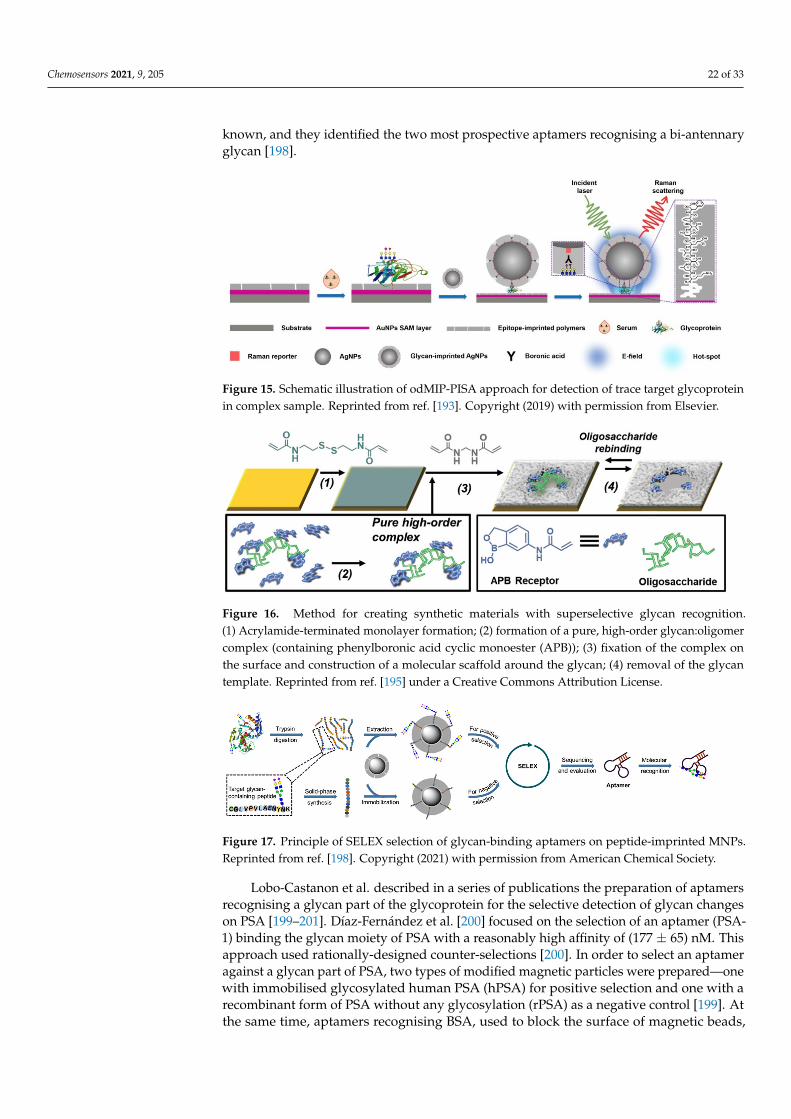
Chemosensors 2021, 9, 205 22 of 33
known, and they identified the two most prospective aptamers recognising a bi-antennaryglycan [198].
Chemosensors 2021, 9, x FOR PEER REVIEW 22 of 33
antigen (CEA), which is a biomarker of other cancer types (but not of PCa), it is worth briefly describing this interesting strategy [193] (Figure 15). This new strategy entitled an orthogonal dual molecularly-imprinted polymer-based plasmonic immuno-sandwich as-say (odMIP-PISA) is based on the integration of one MIP (MIP1) recognising the CEA protein epitope, while the second MIP (MIP2) recognises the glycan part of CE via a boro-nate-imprinted layer. Using this dual-recognition strategy, non-selective binding de-creased from 14.4% (protein recognition) or from 15.2% (glycan recognition) to 8.2% while affording LOD of 56 fM. Moreover, the assay exhibited much better operational parame-ters (linear range, LOD, assay time, sample required, and cost per analysis) than the com-mercially available ELISA. This assay [193] and a similar one [194] were not, however, employed for the detection of different protein glycoforms. The detection of specific pro-tein glycoforms can, however, be resolved using an array of MIP2s designed to use differ-ent glycan templates (Figure 16) [195]. Another strategy to increase the selectivity for de-tection of a glycoprotein of interest is to block boronate moieties on the MIP outside the cavity, which accommodates the protein [196]. In this particular case, PSA is present within the MIP cavity and all the boronate moieties not occupied by the PSA´s glycan are de-activated using a blocking protocol (i.e., by a capping treatment) [196]. Further details about MIP-based sensing strategies involving glycan recognition are discussed in a review paper [197].
Figure 15. Schematic illustration of odMIP-PISA approach for detection of trace target glycoprotein in complex sample. Reprinted from ref. [193]. Copyright (2019) with permission from Elsevier.
Figure 16. Method for creating synthetic materials with superselective glycan recognition. (1) Acrylamide-terminated monolayer formation; (2) formation of a pure, high-order glycan:oligomer complex (containing phenylboronic acid cyclic monoester (APB)); (3) fixation of the complex on the surface and construction of a molecular scaffold around the glycan; (4) removal of the glycan tem-plate. Reprinted from ref. [195] under a Creative Commons Attribution License.
5.3. Detection of Glycoproteins Using Specific Aptamers In order to extend the range of glycans that can be recognised by biorecognition ele-
ments, it is worth identifying DNA aptamers strongly interacting with glycans. To
Figure 15. Schematic illustration of odMIP-PISA approach for detection of trace target glycoproteinin complex sample. Reprinted from ref. [193]. Copyright (2019) with permission from Elsevier.
Chemosensors 2021, 9, x FOR PEER REVIEW 22 of 33
antigen (CEA), which is a biomarker of other cancer types (but not of PCa), it is worth briefly describing this interesting strategy [193] (Figure 15). This new strategy entitled an orthogonal dual molecularly-imprinted polymer-based plasmonic immuno-sandwich as-say (odMIP-PISA) is based on the integration of one MIP (MIP1) recognising the CEA protein epitope, while the second MIP (MIP2) recognises the glycan part of CE via a boro-nate-imprinted layer. Using this dual-recognition strategy, non-selective binding de-creased from 14.4% (protein recognition) or from 15.2% (glycan recognition) to 8.2% while affording LOD of 56 fM. Moreover, the assay exhibited much better operational parame-ters (linear range, LOD, assay time, sample required, and cost per analysis) than the com-mercially available ELISA. This assay [193] and a similar one [194] were not, however, employed for the detection of different protein glycoforms. The detection of specific pro-tein glycoforms can, however, be resolved using an array of MIP2s designed to use differ-ent glycan templates (Figure 16) [195]. Another strategy to increase the selectivity for de-tection of a glycoprotein of interest is to block boronate moieties on the MIP outside the cavity, which accommodates the protein [196]. In this particular case, PSA is present within the MIP cavity and all the boronate moieties not occupied by the PSA´s glycan are de-activated using a blocking protocol (i.e., by a capping treatment) [196]. Further details about MIP-based sensing strategies involving glycan recognition are discussed in a review paper [197].
Figure 15. Schematic illustration of odMIP-PISA approach for detection of trace target glycoprotein in complex sample. Reprinted from ref. [193]. Copyright (2019) with permission from Elsevier.
Figure 16. Method for creating synthetic materials with superselective glycan recognition. (1) Acrylamide-terminated monolayer formation; (2) formation of a pure, high-order glycan:oligomer complex (containing phenylboronic acid cyclic monoester (APB)); (3) fixation of the complex on the surface and construction of a molecular scaffold around the glycan; (4) removal of the glycan tem-plate. Reprinted from ref. [195] under a Creative Commons Attribution License.
5.3. Detection of Glycoproteins Using Specific Aptamers In order to extend the range of glycans that can be recognised by biorecognition ele-
ments, it is worth identifying DNA aptamers strongly interacting with glycans. To
Figure 16. Method for creating synthetic materials with superselective glycan recognition.(1) Acrylamide-terminated monolayer formation; (2) formation of a pure, high-order glycan:oligomercomplex (containing phenylboronic acid cyclic monoester (APB)); (3) fixation of the complex onthe surface and construction of a molecular scaffold around the glycan; (4) removal of the glycantemplate. Reprinted from ref. [195] under a Creative Commons Attribution License.
Chemosensors 2021, 9, x FOR PEER REVIEW 23 of 33
identify glycan-recognising aptamers, several routes have been employed to date. One is based on the use of glycosylated peptides for the isolation of glycan-binding aptamers [198]. Glycopeptides released from the glycoprotein of interest were imprinted on MPs and those particles were used for positive selection, while MPs with peptides without any glycans were used for negative selection (Figure 17). The authors aimed to select aptamers against a glycan structure to which neither lectins or anti-glycan binding antibodies are currently known, and they identified the two most prospective aptamers recognising a bi-antennary glycan [198].
Figure 17. Principle of SELEX selection of glycan-binding aptamers on peptide-imprinted MNPs. Reprinted from ref. [198]. Copyright (2021) with permission from American Chemical Society.
Lobo-Castanon et al. described in a series of publications the preparation of aptamers recognising a glycan part of the glycoprotein for the selective detection of glycan changes on PSA [199–201]. Díaz-Fernández et al. [200] focused on the selection of an aptamer (PSA-1) binding the glycan moiety of PSA with a reasonably high affinity of (177 ± 65) nM. This approach used rationally-designed counter-selections [200]. In order to select an aptamer against a glycan part of PSA, two types of modified magnetic particles were prepared—one with immobilised glycosylated human PSA (hPSA) for positive selection and one with a recombinant form of PSA without any glycosylation (rPSA) as a negative control [199]. At the same time, aptamers recognising BSA, used to block the surface of magnetic beads, were eliminated to obtain specific aptamers PSA-1, which selectively recognise the glycan part of PSA (Figure 18) [199].
Figure 18. Graphical representation of binding between DNA aptamer recognising glycan part on PSA glycoprotein. hPSA/T-PSAG-1 MD simulation (a) Ribbon and stick representations of the hPSA glycoprotein in complex with T-PSAG-1. The structure shown corresponds to a cluster representa-tive from the hPSA/T-PSAG-1 MD trajectory. The sugar residues and specific protein residues (num-bered as in the 3QUM crystal structure) are shown in stick models. The T-PSAG-1 aptamer is shown
Figure 17. Principle of SELEX selection of glycan-binding aptamers on peptide-imprinted MNPs.Reprinted from ref. [198]. Copyright (2021) with permission from American Chemical Society.
Lobo-Castanon et al. described in a series of publications the preparation of aptamersrecognising a glycan part of the glycoprotein for the selective detection of glycan changeson PSA [199–201]. Díaz-Fernández et al. [200] focused on the selection of an aptamer (PSA-1) binding the glycan moiety of PSA with a reasonably high affinity of (177 ± 65) nM. Thisapproach used rationally-designed counter-selections [200]. In order to select an aptameragainst a glycan part of PSA, two types of modified magnetic particles were prepared—onewith immobilised glycosylated human PSA (hPSA) for positive selection and one with arecombinant form of PSA without any glycosylation (rPSA) as a negative control [199]. Atthe same time, aptamers recognising BSA, used to block the surface of magnetic beads,
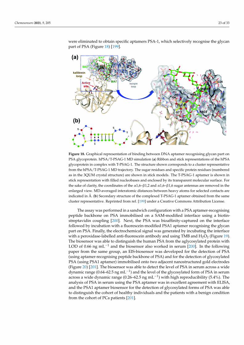
Chemosensors 2021, 9, 205 23 of 33
were eliminated to obtain specific aptamers PSA-1, which selectively recognise the glycanpart of PSA (Figure 18) [199].
Chemosensors 2021, 9, x FOR PEER REVIEW 23 of 33
identify glycan-recognising aptamers, several routes have been employed to date. One is based on the use of glycosylated peptides for the isolation of glycan-binding aptamers [198]. Glycopeptides released from the glycoprotein of interest were imprinted on MPs and those particles were used for positive selection, while MPs with peptides without any glycans were used for negative selection (Figure 17). The authors aimed to select aptamers against a glycan structure to which neither lectins or anti-glycan binding antibodies are currently known, and they identified the two most prospective aptamers recognising a bi-antennary glycan [198].
Figure 17. Principle of SELEX selection of glycan-binding aptamers on peptide-imprinted MNPs. Reprinted from ref. [198]. Copyright (2021) with permission from American Chemical Society.
Lobo-Castanon et al. described in a series of publications the preparation of aptamers recognising a glycan part of the glycoprotein for the selective detection of glycan changes on PSA [199–201]. Díaz-Fernández et al. [200] focused on the selection of an aptamer (PSA-1) binding the glycan moiety of PSA with a reasonably high affinity of (177 ± 65) nM. This approach used rationally-designed counter-selections [200]. In order to select an aptamer against a glycan part of PSA, two types of modified magnetic particles were prepared—one with immobilised glycosylated human PSA (hPSA) for positive selection and one with a recombinant form of PSA without any glycosylation (rPSA) as a negative control [199]. At the same time, aptamers recognising BSA, used to block the surface of magnetic beads, were eliminated to obtain specific aptamers PSA-1, which selectively recognise the glycan part of PSA (Figure 18) [199].
Figure 18. Graphical representation of binding between DNA aptamer recognising glycan part on PSA glycoprotein. hPSA/T-PSAG-1 MD simulation (a) Ribbon and stick representations of the hPSA glycoprotein in complex with T-PSAG-1. The structure shown corresponds to a cluster representa-tive from the hPSA/T-PSAG-1 MD trajectory. The sugar residues and specific protein residues (num-bered as in the 3QUM crystal structure) are shown in stick models. The T-PSAG-1 aptamer is shown
Figure 18. Graphical representation of binding between DNA aptamer recognising glycan part onPSA glycoprotein. hPSA/T-PSAG-1 MD simulation (a) Ribbon and stick representations of the hPSAglycoprotein in complex with T-PSAG-1. The structure shown corresponds to a cluster representativefrom the hPSA/T-PSAG-1 MD trajectory. The sugar residues and specific protein residues (numberedas in the 3QUM crystal structure) are shown in stick models. The T-PSAG-1 aptamer is shown instick representation with filled nucleobases and enclosed by its transparent molecular surface. Forthe sake of clarity, the coordinates of the α1,6–β1,2 and α1,6–β1,6 sugar antennas are removed in theenlarged view. MD-averaged interatomic distances between heavy atoms for selected contacts areindicated in Å. (b) Secondary structure of the complexed T-PSAG-1 aptamer obtained from the samecluster representative. Reprinted from ref. [199] under a Creative Commons Attribution License.
The assay was performed in a sandwich configuration with a PSA aptamer-recognisingpeptide backbone on PSA immobilised on a SAM-modified interface using a biotin-streptavidin coupling [200]. Next, the PSA was bioaffinity-captured on the interfacefollowed by incubation with a fluorescein-modified PSA1 aptamer recognising the glycanpart on PSA. Finally, the electrochemical signal was generated by incubating the interfacewith a peroxidase-labelled anti-fluorescein antibody and using TMB and H2O2 (Figure 19).The biosensor was able to distinguish the human PSA from the aglycosylated protein withLOD of 0.66 ng mL−1 and the biosensor also worked in serum [200]. In the followingpaper from the same group, an EIS-biosensor was developed for the detection of PSA(using aptamer-recognising peptide backbone of PSA) and for the detection of glycosylatedPSA (using PSA1 aptamer) immobilised onto two adjacent nanostructured gold electrodes(Figure 20) [201]. The biosensor was able to detect the level of PSA in serum across a widedynamic range (0.64–62.5 ng mL−1) and the level of the glycosylated form of PSA in serumacross a wide dynamic range (0.26–62.5 ng mL−1) with high reproducibility (5.4%). Theanalysis of PSA in serum using the PSA aptamer was in excellent agreement with ELISA,and the PSA1 aptamer biosensor for the detection of glycosylated forms of PSA was ableto distinguish the cohort of healthy individuals and the patients with a benign conditionfrom the cohort of PCa patients [201].
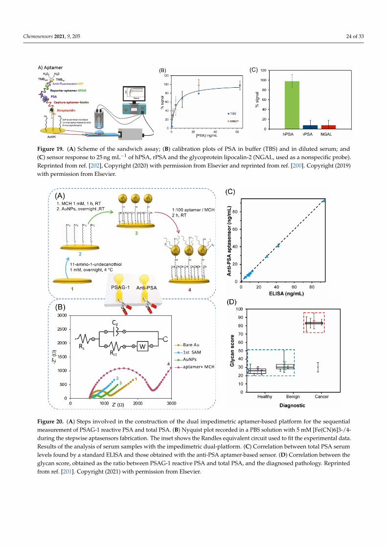
Chemosensors 2021, 9, 205 24 of 33
Chemosensors 2021, 9, x FOR PEER REVIEW 24 of 33
in stick representation with filled nucleobases and enclosed by its transparent molecular surface. For the sake of clarity, the coordinates of the α1,6–β1,2 and α1,6–β1,6 sugar antennas are removed in the enlarged view. MD-averaged interatomic distances between heavy atoms for selected contacts are indicated in Å. (b) Secondary structure of the complexed T-PSAG-1 aptamer obtained from the same cluster representative. Reprinted from ref. [199] under a Creative Commons Attribution Li-cense.
The assay was performed in a sandwich configuration with a PSA aptamer-recognis-ing peptide backbone on PSA immobilised on a SAM-modified interface using a biotin-streptavidin coupling [200]. Next, the PSA was bioaffinity-captured on the interface fol-lowed by incubation with a fluorescein-modified PSA1 aptamer recognising the glycan part on PSA. Finally, the electrochemical signal was generated by incubating the interface with a peroxidase-labelled anti-fluorescein antibody and using TMB and H2O2 (Figure 19). The biosensor was able to distinguish the human PSA from the aglycosylated protein with LOD of 0.66 ng mL−1 and the biosensor also worked in serum [200]. In the following paper from the same group, an EIS-biosensor was developed for the detection of PSA (using aptamer-recognising peptide backbone of PSA) and for the detection of glycosylated PSA (using PSA1 aptamer) immobilised onto two adjacent nanostructured gold electrodes (Figure 20) [201]. The biosensor was able to detect the level of PSA in serum across a wide dynamic range (0.64–62.5 ng mL−1) and the level of the glycosylated form of PSA in serum across a wide dynamic range (0.26–62.5 ng mL−1) with high reproducibility (5.4%). The analysis of PSA in serum using the PSA aptamer was in excellent agreement with ELISA, and the PSA1 aptamer biosensor for the detection of glycosylated forms of PSA was able to distinguish the cohort of healthy individuals and the patients with a benign condition from the cohort of PCa patients [201].
Figure 19. (A) Scheme of the sandwich assay; (B) calibration plots of PSA in buffer (TBS) and in diluted serum; and (C) sensor response to 25 ng mL−1 of hPSA, rPSA and the glycoprotein lipocalin-2 (NGAL, used as a nonspecific probe). Re-printed from ref. [202], Copyright (2020) with permission from Elsevier and reprinted from ref. [200]. Copyright (2019) with permission from Elsevier.
Figure 19. (A) Scheme of the sandwich assay; (B) calibration plots of PSA in buffer (TBS) and in diluted serum; and(C) sensor response to 25 ng mL−1 of hPSA, rPSA and the glycoprotein lipocalin-2 (NGAL, used as a nonspecific probe).Reprinted from ref. [202], Copyright (2020) with permission from Elsevier and reprinted from ref. [200]. Copyright (2019)with permission from Elsevier.
Chemosensors 2021, 9, x FOR PEER REVIEW 25 of 33
Figure 20. (A) Steps involved in the construction of the dual impedimetric aptamer-based platform for the sequential measurement of PSAG-1 reactive PSA and total PSA. (B) Nyquist plot recorded in a PBS solution with 5 mM [Fe(CN)6]3-/4-
during the stepwise aptasensors fabrication. The inset shows the Randles equivalent circuit used to fit the experimental data. Results of the analysis of serum samples with the impedimetric dual-platform. (C) Correlation between total PSA serum levels found by a standard ELISA and those obtained with the anti-PSA aptamer-based sensor. (D) Correlation between the glycan score, obtained as the ratio between PSAG-1 reactive PSA and total PSA, and the diagnosed pathology. Reprinted from ref. [201]. Copyright (2021) with permission from Elsevier.
6. Conclusions The first part of the review provides in-depth information about disease aetiology
describing molecular aspects of the disease and interplay between such biomolecules. A need for novel and more accurate PCa biomarkers was shortly discussed, as well. Several types of biomarkers have the potential to replace PSA for PCa diagnostics. The main rea-son for replacing PSA as a diagnostic biomarker is the high false negative rate (resulting in the development and advancement of PCa) or the high false positive rate (resulting in a substantial number of avoidable biopsies). This review proves the high potential of novel biomarkers for performing PCa diagnostics with a high accuracy. However, there is a need for such novel, alternative biomarkers to be fully validated by robust clinical validation studies by the analysis of some hundreds of samples. We found it especially interesting to focus on PCa diagnostics via glycan-based analysis of glycoproteins since this approach has a strong commercial potential and is currently implemented within our start-up Glycanostics.
Author Contributions: Conceptualization, T.B. and J.T.; resources, T.B., J.T. and P.K.; writing—orig-inal draft preparation, T.B., A.B., S.H., E.C., N.S., L.L., P.K. and J.T; writing—review and editing,
Figure 20. (A) Steps involved in the construction of the dual impedimetric aptamer-based platform for the sequentialmeasurement of PSAG-1 reactive PSA and total PSA. (B) Nyquist plot recorded in a PBS solution with 5 mM [Fe(CN)6]3-/4-during the stepwise aptasensors fabrication. The inset shows the Randles equivalent circuit used to fit the experimental data.Results of the analysis of serum samples with the impedimetric dual-platform. (C) Correlation between total PSA serumlevels found by a standard ELISA and those obtained with the anti-PSA aptamer-based sensor. (D) Correlation between theglycan score, obtained as the ratio between PSAG-1 reactive PSA and total PSA, and the diagnosed pathology. Reprintedfrom ref. [201]. Copyright (2021) with permission from Elsevier.
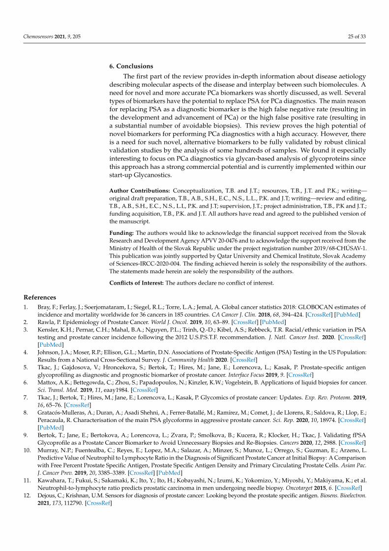
Chemosensors 2021, 9, 205 25 of 33
6. Conclusions
The first part of the review provides in-depth information about disease aetiologydescribing molecular aspects of the disease and interplay between such biomolecules. Aneed for novel and more accurate PCa biomarkers was shortly discussed, as well. Severaltypes of biomarkers have the potential to replace PSA for PCa diagnostics. The main reasonfor replacing PSA as a diagnostic biomarker is the high false negative rate (resulting inthe development and advancement of PCa) or the high false positive rate (resulting ina substantial number of avoidable biopsies). This review proves the high potential ofnovel biomarkers for performing PCa diagnostics with a high accuracy. However, thereis a need for such novel, alternative biomarkers to be fully validated by robust clinicalvalidation studies by the analysis of some hundreds of samples. We found it especiallyinteresting to focus on PCa diagnostics via glycan-based analysis of glycoproteins sincethis approach has a strong commercial potential and is currently implemented within ourstart-up Glycanostics.
Author Contributions: Conceptualization, T.B. and J.T.; resources, T.B., J.T. and P.K.; writing—original draft preparation, T.B., A.B., S.H., E.C., N.S., L.L., P.K. and J.T; writing—review and editing,T.B., A.B., S.H., E.C., N.S., L.L, P.K. and J.T; supervision, J.T.; project administration, T.B., P.K and J.T.;funding acquisition, T.B., P.K. and J.T. All authors have read and agreed to the published version ofthe manuscript.
Funding: The authors would like to acknowledge the financial support received from the SlovakResearch and Development Agency APVV 20-0476 and to acknowledge the support received from theMinistry of Health of the Slovak Republic under the project registration number 2019/68-CHÚSAV-1.This publication was jointly supported by Qatar University and Chemical Institute, Slovak Academyof Sciences-IRCC-2020-004. The finding achieved herein is solely the responsibility of the authors.The statements made herein are solely the responsibility of the authors.
Conflicts of Interest: The authors declare no conflict of interest.
References1. Bray, F.; Ferlay, J.; Soerjomataram, I.; Siegel, R.L.; Torre, L.A.; Jemal, A. Global cancer statistics 2018: GLOBOCAN estimates of
incidence and mortality worldwide for 36 cancers in 185 countries. CA Cancer J. Clin. 2018, 68, 394–424. [CrossRef] [PubMed]2. Rawla, P. Epidemiology of Prostate Cancer. World J. Oncol. 2019, 10, 63–89. [CrossRef] [PubMed]3. Kensler, K.H.; Pernar, C.H.; Mahal, B.A.; Nguyen, P.L.; Trinh, Q.-D.; Kibel, A.S.; Rebbeck, T.R. Racial/ethnic variation in PSA
testing and prostate cancer incidence following the 2012 U.S.P.S.T.F. recommendation. J. Natl. Cancer Inst. 2020. [CrossRef][PubMed]
4. Johnson, J.A.; Moser, R.P.; Ellison, G.L.; Martin, D.N. Associations of Prostate-Specific Antigen (PSA) Testing in the US Population:Results from a National Cross-Sectional Survey. J. Community Health 2020. [CrossRef]
5. Tkac, J.; Gajdosova, V.; Hroncekova, S.; Bertok, T.; Hires, M.; Jane, E.; Lorencova, L.; Kasak, P. Prostate-specific antigenglycoprofiling as diagnostic and prognostic biomarker of prostate cancer. Interface Focus 2019, 9. [CrossRef]
6. Mattox, A.K.; Bettegowda, C.; Zhou, S.; Papadopoulos, N.; Kinzler, K.W.; Vogelstein, B. Applications of liquid biopsies for cancer.Sci. Transl. Med. 2019, 11, eaay1984. [CrossRef]
7. Tkac, J.; Bertok, T.; Hires, M.; Jane, E.; Lorencova, L.; Kasak, P. Glycomics of prostate cancer: Updates. Exp. Rev. Proteom. 2019,16, 65–76. [CrossRef]
8. Gratacós-Mulleras, A.; Duran, A.; Asadi Shehni, A.; Ferrer-Batallé, M.; Ramírez, M.; Comet, J.; de Llorens, R.; Saldova, R.; Llop, E.;Peracaula, R. Characterisation of the main PSA glycoforms in aggressive prostate cancer. Sci. Rep. 2020, 10, 18974. [CrossRef][PubMed]
9. Bertok, T.; Jane, E.; Bertokova, A.; Lorencova, L.; Zvara, P.; Smolkova, B.; Kucera, R.; Klocker, H.; Tkac, J. Validating fPSAGlycoprofile as a Prostate Cancer Biomarker to Avoid Unnecessary Biopsies and Re-Biopsies. Cancers 2020, 12, 2988. [CrossRef]
10. Murray, N.P.; Fuentealba, C.; Reyes, E.; Lopez, M.A.; Salazar, A.; Minzer, S.; Munoz, L.; Orrego, S.; Guzman, E.; Arzeno, L.Predictive Value of Neutrophil to Lymphocyte Ratio in the Diagnosis of Significant Prostate Cancer at Initial Biopsy: A Comparisonwith Free Percent Prostate Specific Antigen, Prostate Specific Antigen Density and Primary Circulating Prostate Cells. Asian Pac.J. Cancer Prev. 2019, 20, 3385–3389. [CrossRef] [PubMed]
11. Kawahara, T.; Fukui, S.; Sakamaki, K.; Ito, Y.; Ito, H.; Kobayashi, N.; Izumi, K.; Yokomizo, Y.; Miyoshi, Y.; Makiyama, K.; et al.Neutrophil-to-lymphocyte ratio predicts prostatic carcinoma in men undergoing needle biopsy. Oncotarget 2015, 6. [CrossRef]
12. Dejous, C.; Krishnan, U.M. Sensors for diagnosis of prostate cancer: Looking beyond the prostate specific antigen. Biosens. Bioelectron.2021, 173, 112790. [CrossRef]
Chemosensors 2021, 9, 205 26 of 33
13. Sharma, S.; Zapatero-Rodríguez, J.; O’Kennedy, R. Prostate cancer diagnostics: Clinical challenges and the ongoing need fordisruptive and effective diagnostic tools. Biotechnol. Adv. 2017, 35, 135–149. [CrossRef]
14. Hodson, R. Small organ, big reach. Nature 2015, 528, S118–S119. [CrossRef]15. Siegel, R.L.; Miller, K.D.; Jemal, A. Cancer statistics, 2015. CA Cancer J. Clin. 2015, 65, 5–29. [CrossRef]16. McNally, C.J.; Ruddock, M.W.; Moore, T.; McKenna, D.J. Biomarkers That Differentiate Benign Prostatic Hyperplasia from
Prostate Cancer: A Literature Review. Cancer Manag. Res. 2020, 12, 5225–5241. [CrossRef]17. Martin, H. Laboratory measurement of urine albumin and urine total protein in screening for proteinuria in chronic kidney
disease. Clin. Biochem. Rev. 2011, 32, 97–102.18. Pereira, M.M.; Calixto, J.D.; Sousa, A.C.A.; Pereira, B.J.; Lima, Á.S.; Coutinho, J.A.P.; Freire, M.G. Towards the differential
diagnosis of prostate cancer by the pre-treatment of human urine using ionic liquids. Sci. Rep. 2020, 10, 14931. [CrossRef]19. Maywald, M.; Wessels, I.; Rink, L. Zinc Signals and Immunity. Int. J. Mol. Sci. 2017, 18, 2222. [CrossRef]20. Wessels, I.; Maywald, M.; Rink, L. Zinc as a Gatekeeper of Immune Function. Nutrients 2017, 9, 1286. [CrossRef]21. Kambe, T.; Matsunaga, M.; Takeda, T.-A. Understanding the Contribution of Zinc Transporters in the Function of the Early
Secretory Pathway. Int. J. Mol. Sci. 2017, 18, 2179. [CrossRef]22. Li, D.; Stovall, D.B.; Wang, W.; Sui, G. Advances of Zinc Signaling Studies in Prostate Cancer. Int. J. Mol. Sci. 2020, 21, 667.
[CrossRef]23. Costello, L.C.; Franklin, R.B. A comprehensive review of the role of zinc in normal prostate function and metabolism; and its
implications in prostate cancer. Arch. Biochem. Biophys. 2016, 611, 100–112. [CrossRef]24. Zhao, J.; Wu, Q.; Hu, X.; Dong, X.; Wang, L.; Liu, Q.; Long, Z.; Li, L. Comparative study of serum zinc concentrations in benign
and malignant prostate disease: A Systematic Review and Meta-Analysis. Sci. Rep. 2016, 6, 25778. [CrossRef]25. Hupe, M.C.; Philippi, C.; Roth, D.; Kümpers, C.; Ribbat-Idel, J.; Becker, F.; Joerg, V.; Duensing, S.; Lubczyk, V.H.; Kirfel, J.; et al.
Expression of Prostate-Specific Membrane Antigen (PSMA) on Biopsies Is an Independent Risk Stratifier of Prostate CancerPatients at Time of Initial Diagnosis. Front. Oncol. 2018, 8. [CrossRef]
26. Wüstemann, T.; Haberkorn, U.; Babich, J.; Mier, W. Targeting prostate cancer: Prostate-specific membrane antigen based diagnosisand therapy. Med. Res. Rev. 2019, 39, 40–69. [CrossRef]
27. Fujita, K.; Nonomura, N. Role of Androgen Receptor in Prostate Cancer: A Review. World J. Men Health 2019, 37, 288–295.[CrossRef]
28. Fu, S.; Wan, X.; Du, C.; Wang, H.; Zhou, J.; Wang, Z. A novel fluorescent probe for the early detection of prostate cancer based onendogenous zinc sensing. Prostate 2019, 79, 1378–1385. [CrossRef]
29. An, Y.; Chang, W.; Wang, W.; Wu, H.; Pu, K.; Wu, A.; Qin, Z.; Tao, Y.; Yue, Z.; Wang, P.; et al. A novel tetrapeptide fluorescencesensor for early diagnosis of prostate cancer based on imaging Zn(2+) in healthy versus cancerous cells. J. Adv. Res. 2020,24, 363–370. [CrossRef]
30. Goldberg, J.M.; Wang, F.; Sessler, C.D.; Vogler, N.W.; Zhang, D.Y.; Loucks, W.H.; Tzounopoulos, T.; Lippard, S.J. PhotoactivatableSensors for Detecting Mobile Zinc. J. Am. Chem. Soc. 2018, 140, 2020–2023. [CrossRef]
31. Teng, Y.; Singh, C.K.; Sadak, O.; Ahmad, N.; Gunasekaran, S. Electrochemical detection of mobile zinc ions for early diagnosis ofprostate cancer. J. Electroanal. Chem. 2019, 833, 269–274. [CrossRef]
32. Sarafanov, A.G.; Todorov, T.I.; Centeno, J.A.; Macias, V.; Gao, W.; Liang, W.M.; Beam, C.; Gray, M.A.; Kajdacsy-Balla, A.A. Prostatecancer outcome and tissue levels of metal ions. Prostate 2011, 71, 1231–1238. [CrossRef] [PubMed]
33. Lim, J.T.; Tan, Y.Q.; Valeri, L.; Lee, J.; Geok, P.P.; Chia, S.E.; Ong, C.N.; Seow, W.J. Association between serum heavy metals andprostate cancer risk—A multiple metal analysis. Environ. Int. 2019, 132, 105109. [CrossRef]
34. Vela, D. Iron Metabolism in Prostate Cancer; From Basic Science to New Therapeutic Strategies. Front. Oncol. 2018, 8, 547.[CrossRef]
35. Stelling, M.P.; Motta, J.M.; Mashid, M.; Johnson, W.E.; Pavão, M.S.; Farrell, N.P. Metal ions and the extracellular matrix in tumormigration. FEBS J. 2019, 286, 2950–2964. [CrossRef]
36. Serhan, M.; Jackemeyer, D.; Long, M.; Sprowls, M.; Diez Perez, I.; Maret, W.; Chen, F.; Tao, N.; Forzani, E. Total Iron Measurementin Human Serum with a Novel Smartphone-Based Assay. IEEE J. Trans. Eng. Health Med. 2020, 8, 2800309. [CrossRef]
37. Oshin, O.; Kireev, D.; Hlukhova, H.; Idachaba, F.; Akinwande, D.; Atayero, A. Graphene-Based Biosensor for Early Detection ofIron Deficiency. Sensors 2020, 20, 3688. [CrossRef] [PubMed]
38. Lim, S.D.; Sun, C.; Lambeth, J.D.; Marshall, F.; Amin, M.; Chung, L.; Petros, J.A.; Arnold, R.S. Increased Nox1 and hydrogenperoxide in prostate cancer. Prostate 2005, 62, 200–207. [CrossRef] [PubMed]
39. Han, C.; Wang, Z.; Xu, Y.; Chen, S.; Han, Y.; Li, L.; Wang, M.; Jin, X. Roles of Reactive Oxygen Species in Biological Behaviors ofProstate Cancer. Biomed. Res. Int. 2020, 2020, 1269624. [CrossRef]
40. Nimalasena, S.; Gothard, L.; Anbalagan, S.; Allen, S.; Sinnett, V.; Mohammed, K.; Kothari, G.; Musallam, A.; Lucy, C.; Yu, S.; et al.Intratumoral Hydrogen Peroxide with Radiation Therapy in Locally Advanced Breast Cancer: Results from a Phase 1 ClinicalTrial. Int. J. Rad. Oncol. Biol. Phys. 2020, 108, 1019–1029. [CrossRef] [PubMed]
41. Crulhas, B.P.; Ramos, N.P.; Castro, G.R.; Pedrosa, V.A. Detection of hydrogen peroxide releasing from prostate cancer cell using abiosensor. J. Solid State Electrochem. 2016, 20, 2427–2433. [CrossRef]
Chemosensors 2021, 9, 205 27 of 33
42. Chen, Q.; Cheng, K.; Wang, W.; Yang, L.; Xie, Y.; Feng, L.; Zhang, J.; Zhang, H.; Sun, H. A pyrene-based ratiometric fluorescentprobe with a large Stokes shift for selective detection of hydrogen peroxide in living cells. J. Pharm. Anal. 2020, 10, 490–497.[CrossRef]
43. Lorencova, L.; Bertok, T.; Dosekova, E.; Holazova, A.; Paprckova, D.; Vikartovska, A.; Sasinkova, V.; Filip, J.; Kasak, P.;Jerigova, M.; et al. Electrochemical performance of Ti(3)C(2)T(x) MXene in aqueous media: Towards ultrasensitive H2O2 sensing.Electrochim. Acta 2017, 235, 471–479. [CrossRef]
44. Hroncekova, S.; Bertok, T.; Hires, M.; Jane, E.; Lorencova, L.; Vikartovska, A.; Tanvir, A.; Kasak, P.; Tkac, J. Ultrasensitive Ti3C2TXMXene/Chitosan Nanocomposite-Based Amperometric Biosensor for Detection of Potential Prostate Cancer Marker in UrineSamples. Processes 2020, 8, 580. [CrossRef]
45. Strmiska, V.; Michalek, P.; Lackova, Z.; Guran, R.; Krizkova, S.; Vanickova, L.; Zitka, O.; Stiborova, M.; Eckschlager, T.;Klejdus, B.; et al. Sarcosine is a prostate epigenetic modifier that elicits aberrant methylation patterns through the SAMe-Dnmtsaxis. Mol. Oncol. 2019, 13, 1002–1017. [CrossRef]
46. Sreekumar, A.; Poisson, L.M.; Rajendiran, T.M.; Khan, A.P.; Cao, Q.; Yu, J.; Laxman, B.; Mehra, R.; Lonigro, R.J.; Li, Y.; et al.Metabolomic profiles delineate potential role for sarcosine in prostate cancer progression. Nature 2009, 457, 910–914. [CrossRef][PubMed]
47. Cernei, N.; Heger, Z.; Gumulec, J.; Zitka, O.; Masarik, M.; Babula, P.; Eckschlager, T.; Stiborova, M.; Kizek, R.; Adam, V. Sarcosineas a potential prostate cancer biomarker—A review. Int. J. Mol. Sci. 2013, 14, 13893–13908. [CrossRef] [PubMed]
48. Jornet-Martínez, N.; Henderson, C.J.; Campíns-Falcó, P.; Daly, R.; Hall, E.A.H. Towards sarcosine determination in urine forprostatic carcinoma detection. Sens. Actuators B Chem. 2019, 287, 380–389. [CrossRef]
49. Heger, Z.; Merlos Rodrigo, M.A.; Michalek, P.; Polanska, H.; Masarik, M.; Vit, V.; Plevova, M.; Pacik, D.; Eckschlager, T.;Stiborova, M.; et al. Sarcosine Up-Regulates Expression of Genes Involved in Cell Cycle Progression of Metastatic Models ofProstate Cancer. PLoS ONE 2016, 11, e0165830. [CrossRef]
50. Rebelo, T.S.; Pereira, C.M.; Sales, M.G.; Noronha, J.P.; Costa-Rodrigues, J.; Silva, F.; Fernandes, M.H. Sarcosine oxidase compositescreen-printed electrode for sarcosine determination in biological samples. Anal. Chim. Acta 2014, 850, 26–32. [CrossRef][PubMed]
51. Struys, E.A.; Heijboer, A.C.; van Moorselaar, J.; Jakobs, C.; Blankenstein, M.A. Serum sarcosine is not a marker for prostate cancer.Ann. Clin. Biochem. 2010, 47, 282. [CrossRef]
52. Ankerst, D.P.; Liss, M.; Zapata, D.; Hoefler, J.; Thompson, I.M.; Leach, R.J. A case control study of sarcosine as an early prostatecancer detection biomarker. BMC Urol. 2015, 15, 99. [CrossRef]
53. Wang, M.; Zou, L.; Liang, J.; Wang, X.; Zhang, D.; Fang, Y.; Zhang, J.; Xiao, F.; Liu, M. The Urinary Sarcosine/Creatinine Ratio is aPotential Diagnostic and Prognostic Marker in Prostate Cancer. Med. Sci. Monit. 2018, 24, 3034–3041. [CrossRef]
54. Gkotsos, G.; Virgiliou, C.; Lagoudaki, I.; Sardeli, C.; Raikos, N.; Theodoridis, G.; Dimitriadis, G. The Role of Sarcosine, Uracil,and Kynurenic Acid Metabolism in Urine for Diagnosis and Progression Monitoring of Prostate Cancer. Metabolites 2017, 7, 9.[CrossRef]
55. Kdadra, M.; Höckner, S.; Leung, H.; Kremer, W.; Schiffer, E. Metabolomics Biomarkers of Prostate Cancer: A Systematic Review.Diagnostics 2019, 9, 21. [CrossRef]
56. Spur, E.-M.; Decelle, E.A.; Cheng, L.L. Metabolomic imaging of prostate cancer with magnetic resonance spectroscopy and massspectrometry. Eur. J. Nucl. Med. Mol. Imaging 2013, 40, 60–71. [CrossRef]
57. Lima, A.R.; Bastos, M.d.L.; Carvalho, M.; Guedes de Pinho, P. Biomarker Discovery in Human Prostate Cancer: An Update inMetabolomics Studies. Transl. Oncol. 2016, 9, 357–370. [CrossRef]
58. Chatzakos, V.; Slätis, K.; Djureinovic, T.; Helleday, T.; Hunt, M.C. N-acyl taurines are anti-proliferative in prostate cancer cells.Lipids 2012, 47, 355–361. [CrossRef]
59. Fernández-Peralbo, M.A.; Gómez-Gómez, E.; Calderón-Santiago, M.; Carrasco-Valiente, J.; Ruiz-García, J.; Requena-Tapia, M.J.;Luque de Castro, M.D.; Priego-Capote, F. Prostate Cancer Patients–Negative Biopsy Controls Discrimination by UntargetedMetabolomics Analysis of Urine by LC-QTOF: Upstream Information on Other Omics. Sci. Rep. 2016, 6, 38243. [CrossRef]
60. Derezinski, P.; Klupczynska, A.; Sawicki, W.; Pałka, J.A.; Kokot, Z.J. Amino Acid Profiles of Serum and Urine in Search forProstate Cancer Biomarkers: A Pilot Study. Int. J. Med. Sci. 2017, 14, 1–12. [CrossRef]
61. Giskeødegård, G.F.; Hansen, A.F.; Bertilsson, H.; Gonzalez, S.V.; Kristiansen, K.A.; Bruheim, P.; Mjøs, S.A.; Angelsen, A.;Bathen, T.F.; Tessem, M.B. Metabolic markers in blood can separate prostate cancer from benign prostatic hyperplasia. Br. J. Cancer2015, 113, 1712–1719. [CrossRef] [PubMed]
62. Schmidt, J.A.; Fensom, G.K.; Rinaldi, S.; Scalbert, A.; Appleby, P.N.; Achaintre, D.; Gicquiau, A.; Gunter, M.J.; Ferrari, P.;Kaaks, R.; et al. Pre-diagnostic metabolite concentrations and prostate cancer risk in 1077 cases and 1077 matched controls in theEuropean Prospective Investigation into Cancer and Nutrition. BMC Med. 2017, 15, 122. [CrossRef] [PubMed]
63. Stabler, S.; Koyama, T.; Zhao, Z.; Martinez-Ferrer, M.; Allen, R.H.; Luka, Z.; Loukachevitch, L.V.; Clark, P.E.; Wagner, C.;Bhowmick, N.A. Serum methionine metabolites are risk factors for metastatic prostate cancer progression. PLoS ONE 2011,6, e22486. [CrossRef]
64. Schuster, D.M.; Nanni, C.; Fanti, S. Evaluation of Prostate Cancer with Radiolabeled Amino Acid Analogs. J. Nucl. Med. 2016,57, 61s–66s. [CrossRef]
Chemosensors 2021, 9, 205 28 of 33
65. Kobus, T.; Wright, A.J.; Weiland, E.; Heerschap, A.; Scheenen, T.W.J. Metabolite ratios in 1H MR spectroscopic imaging of theprostate. Magn. Res. Med. 2015, 73, 1–12. [CrossRef]
66. Lad, U.; Kale, G.M.; Bryaskova, R. Sarcosine Oxidase Encapsulated Polyvinyl Alcohol-Silica-AuNP Hybrid Films for SarcosineSensing Electrochemical Bioelectrode. J. Electrochem. Soc. 2014, 161, B98–B101. [CrossRef]
67. Narwal, V.; Kumar, P.; Joon, P.; Pundir, C.S. Fabrication of an amperometric sarcosine biosensor based on sarcosineoxidase/chitosan/CuNPs/c-MWCNT/Au electrode for detection of prostate cancer. Enzym. Microb. Technol. 2018, 113, 44–51.[CrossRef]
68. Kumar, P.; Narwal, V.; Jaiwal, R.; Pundir, C.S. Construction and application of amperometric sarcosine biosensor based onSOxNPs/AuE for determination of prostate cancer. Biosens. Bioelectron. 2018, 122, 140–146. [CrossRef]
69. Yang, H.; Wang, J.; Yang, C.; Zhao, X.; Xie, S.; Ge, Z. Nano Pt@ZIF8 Modified Electrode and Its Application to Detect Sarcosine.J. Electrochem. Soc. 2018, 165, H247–H250. [CrossRef]
70. Hu, J.; Wei, W.; Ke, S.; Zeng, X.; Lin, P. A novel and sensitive sarcosine biosensor based on organic electrochemical transistor.Electrochim. Acta 2019, 307, 100–106. [CrossRef]
71. Wang, Q.; Zhao, Y.; Yang, Q.; Du, D.; Yang, H.; Lin, Y. Amperometric sarcosine biosensor with strong anti-interference capabilitiesbased on mesoporous organic-inorganic hybrid materials. Biosens. Bioelectron. 2019, 141, 111431. [CrossRef]
72. Li, J.; Ma, J.; Zhang, Y.; Zhang, Z.; He, G. An amperometric biosensor for the assay of sarcosine based on the cross coupledchemical and electrochemical reactions with practical applications. J. Electroanal. Chem. 2019, 833, 568–572. [CrossRef]
73. Yang, Q.; Li, N.; Li, Q.; Chen, S.; Wang, H.-L.; Yang, H. Amperometric sarcosine biosensor based on hollow magnetic Pt–Fe3O4@Cnanospheres. Anal. Chim. Acta 2019, 1078, 161–167. [CrossRef]
74. Tang, P.; Wang, Y.; He, F. Electrochemical sensor based on super-magnetic metal–organic framework@molecularly imprintedpolymer for Sarcosine detection in urine. J. Saudi Chem. Soc. 2020, 24, 620–630. [CrossRef]
75. Özkütük, E.B.; Diltemiz, S.E.; Avcı, S.; Ugurag, D.; Aykanat, R.B.; Ersöz, A.; Say, R. Potentiometric sensor fabrication having 2Dsarcosine memories and analytical features. Mat. Sci. Eng. C 2016, 69, 231–235. [CrossRef] [PubMed]
76. Altunkök, N.; Biçen Ünlüer, Ö.; Birlik Özkütük, E.; Ersöz, A. Development of potentıometrıc bıosensor for dıagnosıs of prostatecancer. Mat. Sci. Eng. B 2021, 263, 114789. [CrossRef]
77. Nguy, T.P.; Van Phi, T.; Tram, D.T.N.; Eersels, K.; Wagner, P.; Lien, T.T.N. Development of an impedimetric sensor for the label-freedetection of the amino acid sarcosine with molecularly imprinted polymer receptors. Sens. Actuator B Chem. 2017, 246, 461–470.[CrossRef]
78. Lan, J.; Xu, W.; Wan, Q.; Zhang, X.; Lin, J.; Chen, J.; Chen, J. Colorimetric determination of sarcosine in urine samples of prostaticcarcinoma by mimic enzyme palladium nanoparticles. Anal. Chim. Acta 2014, 825, 63–68. [CrossRef]
79. Xue, Z.; Yin, B.; Wang, H.; Li, M.; Rao, H.; Liu, X.; Zhou, X.; Lu, X. An organic indicator functionalized graphene oxidenanocomposite-based colorimetric assay for the detection of sarcosine. Nanoscale 2016, 8, 5488–5496. [CrossRef]
80. Heger, Z.; Cernei, N.; Krizkova, S.; Masarik, M.; Kopel, P.; Hodek, P.; Zitka, O.; Adam, V.; Kizek, R. Paramagnetic Nanoparticlesas a Platform for FRET-Based Sarcosine Picomolar Detection. Sci. Rep. 2015, 5, 8868. [CrossRef]
81. Luo, Y.; Wang, J.; Yang, L.; Gao, T.; Pei, R. In vitro selection of DNA aptamers for the development of fluorescent aptasensor forsarcosine detection. Sens. Actuator B Chem. 2018, 276, 128–135. [CrossRef]
82. Cornu, J.-N.; Cancel-Tassin, G.; Ondet, V.; Girardet, C.; Cussenot, O. Olfactory Detection of Prostate Cancer by Dogs SniffingUrine: A Step Forward in Early Diagnosis. Eur. Urol. 2011, 59, 197–201. [CrossRef] [PubMed]
83. Pascual, L.; Campos, I.; Vivancos, J.-L.; Quintás, G.; Loras, A.; Martínez-Bisbal, M.C.; Martínez-Máñez, R.; Boronat, F.;Ruiz-Cerdà, J.L. Detection of prostate cancer using a voltammetric electronic tongue. Analyst 2016, 141, 4562–4567. [Cross-Ref] [PubMed]
84. Chen, Y.; Wang, K.; Liu, T.; Chen, J.; Lv, W.; Yang, W.; Xu, S.; Wang, X.; Li, L. Decreased glucose bioavailability and elevatedaspartate metabolism in prostate cancer cells undergoing epithelial-mesenchymal transition. J. Cell. Physiol. 2020, 235, 5602–5612.[CrossRef] [PubMed]
85. Zhao, Y.; Shen, Y.; Wen, Y.; Campbell, R.E. High-Performance Intensiometric Direct- and Inverse-Response Genetically EncodedBiosensors for Citrate. ACS Cent. Sci. 2020, 6, 1441–1450. [CrossRef]
86. Abarghoei, S.; Fakhri, N.; Borghei, Y.S.; Hosseini, M.; Ganjali, M.R. A colorimetric paper sensor for citrate as biomarker for earlystage detection of prostate cancer based on peroxidase-like activity of cysteine-capped gold nanoclusters. Spectrochim. Acta A Mol.Biomol. Spectrosc. 2019, 210, 251–259. [CrossRef] [PubMed]
87. Shen, H.; Liu, B.; Liu, D.; Zhu, X.; Wei, X.; Yu, L.; Shen, Q.; Qu, P.; Xu, M. Lanthanide coordination polymer-based biosensor forcitrate detection in urine. Anal. Methods 2019, 11, 1405–1409. [CrossRef]
88. Tsoi, T.H.; Chan, C.F.; Chan, W.L.; Chiu, K.F.; Wong, W.T.; Ng, C.F.; Wong, K.L. Urinary Polyamines: A Pilot Study on Their Rolesas Prostate Cancer Detection Biomarkers. PLoS ONE 2016, 11, e0162217. [CrossRef]
89. Fan, J.; Feng, Z.; Chen, N. Spermidine as a target for cancer therapy. Pharmacol. Res. 2020, 159, 104943. [CrossRef]90. Nair, R.R.; Debnath, S.; Das, S.; Wakchaure, P.; Ganguly, B.; Chatterjee, P.B. A Highly Selective Turn-On Biosensor for Measuring
Spermine/Spermidine in Human Urine and Blood. ACS Appl. Bio Mater. 2019, 2, 2374–2387. [CrossRef]91. Weinstein, S.J.; Mackrain, K.; Stolzenberg-Solomon, R.Z.; Selhub, J.; Virtamo, J.; Albanes, D. Serum creatinine and prostate cancer
risk in a prospective study. Cancer Epidemiol. Biomark. Prev. 2009, 18, 2643–2649. [CrossRef]
Chemosensors 2021, 9, 205 29 of 33
92. Pundir, C.S.; Kumar, P.; Jaiwal, R. Biosensing methods for determination of creatinine: A review. Biosens. Bioelectron. 2019,126, 707–724. [CrossRef]
93. Rule, A.D.; Jacobson, D.J.; Roberts, R.O.; Girman, C.J.; McGree, M.E.; Lieber, M.M.; Jacobsen, S.J. The association between benignprostatic hyperplasia and chronic kidney disease in community-dwelling men. Kidney Int. 2005, 67, 2376–2382. [CrossRef]
94. Moura-Melo, S.; Miranda-Castro, R.; de-Los-Santos-Álvarez, N.; Miranda-Ordieres, A.J.; Dos Santos Junior, J.R.; da Silva Fonseca, R.A.;Lobo-Castañón, M.J. A Quantitative PCR-Electrochemical Genosensor Test for the Screening of Biotech Crops. Sensors 2017,17, 881. [CrossRef] [PubMed]
95. Rosario, R.; Mutharasan, R. Nucleic acid electrochemical and electromechanical biosensors: A review of techniques anddevelopments. Rev. Anal. Chem 2014, 33, 213–230. [CrossRef]
96. Marks, L.S.; Bostwick, D.G. Prostate Cancer Specificity of PCA3 Gene Testing: Examples from Clinical Practice. Rev. Urol. 2008,10, 175–181.
97. Merola, R.; Tomao, L.; Antenucci, A.; Sperduti, I.; Sentinelli, S.; Masi, S.; Mandoj, C.; Orlandi, G.; Papalia, R.; Guaglianone, S.; et al.PCA3 in prostate cancer and tumor aggressiveness detection on 407 high-risk patients: A National Cancer Institute experience.J. Exp. Clin. Cancer Res. 2015, 34, 15. [CrossRef]
98. Barbera, M.; Pepe, P.; Paola, Q.; Aragona, F. PCA3 score accuracy in diagnosing prostate cancer at repeat biopsy: Our experiencein 177 patients. Arch. Ital. Urol. Androl. 2012, 84, 227–229.
99. Marks, L.S.; Fradet, Y.; Deras, I.L.; Blase, A.; Mathis, J.; Aubin, S.M.; Cancio, A.T.; Desaulniers, M.; Ellis, W.J.; Rittenhouse, H.; et al.PCA3 molecular urine assay for prostate cancer in men undergoing repeat biopsy. Urology 2007, 69, 532–535. [CrossRef]
100. Qin, Z.; Yao, J.; Xu, L.; Xu, Z.; Ge, Y.; Zhou, L.; Zhao, F.; Jia, R. Diagnosis accuracy of PCA3 level in patients with prostate cancer:A systematic review with meta-analysis. Int. Braz. J. Urol. 2020, 46, 691–704. [CrossRef] [PubMed]
101. Wang, T.; Qu, X.; Jiang, J.; Gao, P.; Zhao, D.; Lian, X.; Li, X. Diagnostic significance of urinary long non-coding PCA3 RNA inprostate cancer. Oncotarget 2017, 8, 58577–58586. [CrossRef]
102. Soares, J.C.; Soares, A.C.; Rodrigues, V.C.; Melendez, M.E.; Santos, A.C.; Faria, E.F.; Reis, R.M.; Carvalho, A.L.; Oliveira, O.N.Detection of the Prostate Cancer Biomarker PCA3 with Electrochemical and Impedance-Based Biosensors. ACS Appl. Mater. Interf.2019, 11, 46645–46650. [CrossRef]
103. Bertok, T.; Lorencova, L.; Chocholova, E.; Jane, E.; Vikartovska, A.; Kasak, P.; Tkac, J. Electrochemical Impedance SpectroscopyBased Biosensors: Mechanistic Principles, Analytical Examples and Challenges towards Commercialization for Assays of ProteinCancer Biomarkers. ChemElectroChem 2019, 6, 989–1003. [CrossRef]
104. Gajdosova, V.P.; Lorencova, L.; Blsakova, A.; Kasak, P.; Bertok, T.; Tkac, J. Challenges for impedimetric affinity sensors targetingproteins detection. Curr. Opin. Electrochem. 2021, 28, 100717. [CrossRef]
105. Rodrigues, V.C.; Soares, J.C.; Soares, A.C.; Braz, D.C.; Melendez, M.E.; Ribas, L.C.; Scabini, L.F.S.; Bruno, O.M.; Carvalho, A.L.;Reis, R.M.; et al. Electrochemical and optical detection and machine learning applied to images of genosensors for diagnosis ofprostate cancer with the biomarker PCA3. Talanta 2021, 222, 121444. [CrossRef]
106. Fu, X.; Wen, J.; Li, J.; Lin, H.; Liu, Y.; Zhuang, X.; Tian, C.; Chen, L. Highly sensitive detection of prostate cancer specific PCA3mimic DNA using SERS-based competitive lateral flow assay. Nanoscale 2019, 11, 15530–15536. [CrossRef]
107. Vilela, P.; El-Sagheer, A.; Millar, T.M.; Brown, T.; Muskens, O.L.; Kanaras, A.G. Graphene Oxide-Upconversion NanoparticleBased Optical Sensors for Targeted Detection of mRNA Biomarkers Present in Alzheimer’s Disease and Prostate Cancer. ACS Sens.2017, 2, 52–56. [CrossRef] [PubMed]
108. Yamkamon, V.; Htoo, K.P.P.; Yainoy, S.; Suksrichavalit, T.; Tangchaikeeree, T.; Eiamphungporn, W. Urinary PCA3 detectionin prostate cancer by magnetic nanoparticles coupled with colorimetric enzyme-linked oligonucleotide assay. EXCLI J. 2020,19, 501–513.
109. Ashrafizadeh, M.; Hushmandi, K.; Rahmani Moghadam, E.; Zarrin, V.; Hosseinzadeh Kashani, S.; Bokaie, S.; Najafi, M.;Tavakol, S.; Mohammadinejad, R.; Nabavi, N.; et al. Progress in Delivery of siRNA-Based Therapeutics Employing Nano-Vehiclesfor Treatment of Prostate Cancer. Bioengineering 2020, 7, 91. [CrossRef] [PubMed]
110. Tavallaie, R.; De Almeida, S.R.; Gooding, J.J. Toward biosensors for the detection of circulating microRNA as a cancer biomarker:An overview of the challenges and successes. Wiley Interdiscip. Rev. Nanomed. Nanobiotechnol. 2015, 7, 580–592. [CrossRef]
111. Kim, W.T.; Kim, W.-J. MicroRNAs in prostate cancer. Prostate Int. 2013, 1, 3–9. [CrossRef] [PubMed]112. D’Agata, R.; Spoto, G. Advanced methods for microRNA biosensing: A problem-solving perspective. Anal. Bioanal. Chem. 2019,
411, 4425–4444. [CrossRef]113. Kasomva, K.; Sen, A.; Paulraj, M.G.; Sailo, S.; Raphael, V.; Puro, K.-U.; Assumi, S.R.; Ignacimuthu, S. Roles of microRNA in
prostate cancer cell metabolism. Int. J. Biochem. Cell Biol. 2018, 102, 109–116. [CrossRef] [PubMed]114. Mompeón, A.; Ortega-Paz, L.; Vidal-Gómez, X.; Costa, T.J.; Pérez-Cremades, D.; Garcia-Blas, S.; Brugaletta, S.; Sanchis, J.;
Sabate, M.; Novella, S.; et al. Disparate miRNA expression in serum and plasma of patients with acute myocardial infarction: Asystematic and paired comparative analysis. Sci. Rep. 2020, 10, 5373. [CrossRef]
115. Sabahi, A.; Salahandish, R.; Ghaffarinejad, A.; Omidinia, E. Electrochemical nano-genosensor for highly sensitive detection ofmiR-21 biomarker based on SWCNT-grafted dendritic Au nanostructure for early detection of prostate cancer. Talanta 2020,209, 120595. [CrossRef]
116. Smith, D.A.; Newbury, L.J.; Drago, G.; Bowen, T.; Redman, J.E. Electrochemical detection of urinary microRNAs via sulfonamide-bound antisense hybridisation. Sens. Actuators B Chem. 2017, 253, 335–341. [CrossRef]

Chemosensors 2021, 9, 205 30 of 33
117. Ibrahim, N.H.; Abdellateif, M.S.; Thabet, G.; Kassem, S.H.; El-Salam, M.A.; El-Leithy, A.A.; Selim, M.M. Combining PHI andmiRNAs as Biomarkers in Prostate Cancer Diagnosis and Prognosis. Clin. Lab. 2019, 65. [CrossRef]
118. Bidarra, D.; Constâncio, V.; Barros-Silva, D.; Ramalho-Carvalho, J.; Moreira-Barbosa, C.; Antunes, L.; Maurício, J.; Oliveira, J.;Henrique, R.; Jerónimo, C. Circulating MicroRNAs as Biomarkers for Prostate Cancer Detection and Metastasis DevelopmentPrediction. Front. Oncol. 2019, 9. [CrossRef]
119. Nguyen, H.C.N.; Xie, W.; Yang, M.; Hsieh, C.-L.; Drouin, S.; Lee, G.-S.M.; Kantoff, P.W. Expression differences of circulatingmicroRNAs in metastatic castration resistant prostate cancer and low-risk, localized prostate cancer. Prostate 2013, 73, 346–354.[CrossRef]
120. Schaefer, A.; Jung, M.; Mollenkopf, H.-J.; Wagner, I.; Stephan, C.; Jentzmik, F.; Miller, K.; Lein, M.; Kristiansen, G.; Jung, K.Diagnostic and prognostic implications of microRNA profiling in prostate carcinoma. Int. J. Cancer 2010, 126, 1166–1176.[CrossRef]
121. Bryant, R.J.; Pawlowski, T.; Catto, J.W.F.; Marsden, G.; Vessella, R.L.; Rhees, B.; Kuslich, C.; Visakorpi, T.; Hamdy, F.C. Changes incirculating microRNA levels associated with prostate cancer. Br. J. Cancer 2012, 106, 768–774. [CrossRef]
122. Paiva, R.M.; Zauli, D.A.G.; Neto, B.S.; Brum, I.S. Urinary microRNAs expression in prostate cancer diagnosis: A systematicreview. Clin. Transl. Oncol. 2020, 22, 2061–2073. [CrossRef]
123. Fredsøe, J.; Rasmussen, A.K.I.; Thomsen, A.R.; Mouritzen, P.; Høyer, S.; Borre, M.; Ørntoft, T.F.; Sørensen, K.D. Diagnostic andPrognostic MicroRNA Biomarkers for Prostate Cancer in Cell-free Urine. Eur. Urol. Focus 2018, 4, 825–833. [CrossRef]
124. Yang, C.; Dou, B.; Shi, K.; Chai, Y.; Xiang, Y.; Yuan, R. Multiplexed and Amplified Electronic Sensor for the Detection ofMicroRNAs from Cancer Cells. Anal. Chem. 2014, 86, 11913–11918. [CrossRef]
125. El Aamri, M.; Yammouri, G.; Mohammadi, H.; Amine, A.; Korri-Youssoufi, H. Electrochemical Biosensors for Detection ofMicroRNA as a Cancer Biomarker: Pros and Cons. Biosensors 2020, 10, 186. [CrossRef] [PubMed]
126. Ivanov, Y.; Pleshakova, T.; Malsagova, K.; Kurbatov, L.; Popov, V.; Glukhov, A.; Smirnov, A.; Enikeev, D.; Potoldykova, N.;Alekseev, B.; et al. Detection of Marker miRNAs, Associated with Prostate Cancer, in Plasma Using SOI-NW Biosensor in Directand Inversion Modes. Sensors 2019, 19, 5248. [CrossRef]
127. Zheng, J.; Yang, R.; Shi, M.; Wu, C.; Fang, X.; Li, Y.; Li, J.; Tan, W. Rationally designed molecular beacons for bioanalytical andbiomedical applications. Chem. Soc. Rev. 2015, 44, 3036–3055. [CrossRef]
128. Jolly, P.; Batistuti, M.R.; Miodek, A.; Zhurauski, P.; Mulato, M.; Lindsay, M.A.; Estrela, P. Highly sensitive dual mode electrochemi-cal platform for microRNA detection. Sci. Rep. 2016, 6, 36719. [CrossRef] [PubMed]
129. Mucci, L.A.; Hjelmborg, J.B.; Harris, J.R.; Czene, K.; Havelick, D.J.; Scheike, T.; Graff, R.E.; Holst, K.; Möller, S.; Unger, R.H.; et al.Familial Risk and Heritability of Cancer Among Twins in Nordic Countries. JAMA 2016, 315, 68–76. [CrossRef]
130. Benafif, S.; Kote-Jarai, Z.; Eeles, R.A. A Review of Prostate Cancer Genome-Wide Association Studies (GWAS). Cancer Epidemiol.Biomark. Prev. 2018, 27, 845–857. [CrossRef]
131. Schumacher, F.R.; Al Olama, A.A.; Berndt, S.I.; Benlloch, S.; Ahmed, M.; Saunders, E.J.; Dadaev, T.; Leongamornlert, D.;Anokian, E.; Cieza-Borrella, C.; et al. Association analyses of more than 140,000 men identify 63 new prostate cancer susceptibilityloci. Nat. Genet. 2018, 50, 928–936. [CrossRef]
132. Wang, G.; Zhao, D.; Spring, D.J.; DePinho, R.A. Genetics and biology of prostate cancer. Genes Dev. 2018, 32, 1105–1140. [CrossRef][PubMed]
133. Ewing, C.M.; Ray, A.M.; Lange, E.M.; Zuhlke, K.A.; Robbins, C.M.; Tembe, W.D.; Wiley, K.E.; Isaacs, S.D.; Johng, D.; Wang, Y.; et al.Germline Mutations in HOXB13 and Prostate-Cancer Risk. N. Engl. J. Med. 2012, 366, 141–149. [CrossRef] [PubMed]
134. Eftekhari-Sis, B.; Karaminejad, S.; Karimi, F. A Nano-Biosensor for the Detection of 185delAG Mutation in BRCA1 Gene, Leadingto Breast Cancer. Cancer Investig. 2016, 34, 431–439. [CrossRef] [PubMed]
135. Filippidou, M.K.; Loukas, C.M.; Kaprou, G.; Tegou, E.; Petrou, P.; Kakabakos, S.; Tserepi, A.; Chatzandroulis, S. Detection ofBRCA1 gene on partially reduced graphene oxide biosensors. Microelectron. Eng. 2019, 216, 111093. [CrossRef]
136. Feng, D.; Su, J.; He, G.; Xu, Y.; Wang, C.; Zheng, M.; Qian, Q.; Mi, X. Electrochemical DNA Sensor for Sensitive BRCA1 DetectionBased on DNA Tetrahedral-Structured Probe and Poly-Adenine Mediated Gold Nanoparticles. Biosensors 2020, 10, 78. [CrossRef]
137. Wang, Z.; Wang, Y.; Zhang, J.; Hu, Q.; Zhi, F.; Zhang, S.; Mao, D.; Zhang, Y.; Liang, H. Significance of the TMPRSS2:ERG genefusion in prostate cancer. Mol. Med. Rep. 2017, 16, 5450–5458. [CrossRef]
138. Zhao, S.; Løvf, M.; Totland Carm, K.; Cathrine Bakken, A.; Hoff, A.M.; Skotheim, R.I. Novel transcription-induced fusion RNAsin prostate cancer. Oncotarget 2017, 8. [CrossRef]
139. Yang, Z.; Yu, L.; Wang, Z. PCA3 and TMPRSS2-ERG gene fusions as diagnostic biomarkers for prostate cancer. Chin. J. Cancer Res.2016, 28, 65–71.
140. Koo, K.M.; Wee, E.J.H.; Mainwaring, P.N.; Trau, M. A simple, rapid, low-cost technique for naked-eye detection of urine-isolatedTMPRSS2:ERG gene fusion RNA. Sci. Rep. 2016, 6, 30722. [CrossRef]
141. Bocedi, A.; Noce, A.; Marrone, G.; Noce, G.; Cattani, G.; Gambardella, G.; Di Lauro, M.; Di Daniele, N.; Ricci, G. GlutathioneTransferase P1-1 an Enzyme Useful in Biomedicine and as Biomarker in Clinical Practice and in Environmental Pollution.Nutrients 2019, 11, 1741. [CrossRef]
142. Pljesa-Ercegovac, M.; Savic-Radojevic, A.; Matic, M.; Coric, V.; Djukic, T.; Radic, T.; Simic, T. Glutathione Transferases: PotentialTargets to Overcome Chemoresistance in Solid Tumors. Int. J. Mol. Sci. 2018, 19, 3785. [CrossRef]
Chemosensors 2021, 9, 205 31 of 33
143. Kanwal, R.; Pandey, M.; Bhaskaran, N.; Maclennan, G.T.; Fu, P.; Ponsky, L.E.; Gupta, S. Protection against oxidative DNA damageand stress in human prostate by glutathione S-transferase P1. Mol. Carcinog. 2014, 53, 8–18. [CrossRef] [PubMed]
144. Gurioli, G.; Martignano, F.; Salvi, S.; Costantini, M.; Gunelli, R.; Casadio, V. GSTP1 methylation in cancer: A liquid biopsybiomarker? Clin. Chem. Lab. Med. 2018, 56, 702–717. [CrossRef]
145. Singh, S.; Shukla, G.C.; Gupta, S. MicroRNA Regulating Glutathione S-Transferase P1 in Prostate Cancer. Curr. Pharm. Rep. 2015,1, 79–88. [CrossRef] [PubMed]
146. Topkaya, S.N.; Ozkan-Ariksoysal, D.; Kosova, B.; Ozel, R.; Ozsoz, M. Electrochemical DNA biosensor for detecting cancerbiomarker related to glutathione S-transferase P1 (GSTP1) hypermethylation in real samples. Biosens. Bioelectron. 2012, 31, 516–522.[CrossRef] [PubMed]
147. Nur Topkaya, S.; Ozkan-Ariksoysal, D. Prostate Cancer Biomarker Detection with Carbon Nanotubes Modified Screen PrintedElectrodes. Electroanal 2016, 28, 1077–1084. [CrossRef]
148. Chiou, C.C.; Chang, P.Y.; Chan, E.C.; Wu, T.L.; Tsao, K.C.; Wu, J.T. Urinary 8-hydroxydeoxyguanosine and its analogs as DNAmarker of oxidative stress: Development of an ELISA and measurement in both bladder and prostate cancers. Clin. Chim. Acta2003, 334, 87–94. [CrossRef]
149. Sova, H.; Jukkola-Vuorinen, A.; Puistola, U.; Kauppila, S.; Karihtala, P. 8-Hydroxydeoxyguanosine: A new potential independentprognostic factor in breast cancer. Br. J. Cancer 2010, 102, 1018–1023. [CrossRef]
150. Ohtake, S.; Kawahara, T.; Ishiguro, Y.; Takeshima, T.; Kuroda, S.; Izumi, K.; Miyamoto, H.; Uemura, H. Oxidative stress marker8-hydroxyguanosine is more highly expressed in prostate cancer than in benign prostatic hyperplasia. Mol. Clin. Oncol. 2018,9, 302–304. [CrossRef]
151. Rosy Goyal, R.N. Determination of 8-Hydroxydeoxyguanosine: A potential biomarker of oxidative stress, using carbon-allotropicnanomaterials modified glassy carbon sensor. Talanta 2016, 161, 735–742. [CrossRef]
152. Gupta, P.; Oyama, M.; Goyal, R.N. Electrochemical investigations of 8-hydroxydeoxyguanosine and its determination at an edgeplane pyrolytic graphite electrode. RSC Adv. 2016, 6, 1722–1728. [CrossRef]
153. Kumar, N.; Rosy Goyal, R.N. A melamine based molecularly imprinted sensor for the determination of 8-hydroxydeoxyguanosinein human urine. Talanta 2017, 166, 215–222. [CrossRef] [PubMed]
154. Martins, G.V.; Tavares, A.P.M.; Fortunato, E.; Sales, M.G.F. Paper-Based Sensing Device for Electrochemical Detection of OxidativeStress Biomarker 8-Hydroxy-2′-deoxyguanosine (8-OHdG) in Point-of-Care. Sci. Rep. 2017, 7, 14558. [CrossRef]
155. Mohd Azmi, M.A.; Tehrani, Z.; Lewis, R.P.; Walker, K.A.D.; Jones, D.R.; Daniels, D.R.; Doak, S.H.; Guy, O.J. Highly sensitivecovalently functionalised integrated silicon nanowire biosensor devices for detection of cancer risk biomarker. Biosens. Bioelectron.2014, 52, 216–224. [CrossRef]
156. Song, E.; Hu, Y.; Hussein, A.; Yu, C.-Y.; Tang, H.; Mechref, Y. Characterization of the Glycosylation Site of Human PSA Promptedby Missense Mutation using LC–MS/MS. J. Proteome Res. 2015, 14, 2872–2883. [CrossRef] [PubMed]
157. Popovics, P.; Awadallah, W.N.; Kohrt, S.E.; Case, T.C.; Miller, N.L.; Ricke, E.A.; Huang, W.; Ramirez-Solano, M.; Liu, Q.;Vezina, C.M.; et al. Prostatic osteopontin expression is associated with symptomatic benign prostatic hyperplasia. Prostate 2020,80, 731–741. [CrossRef]
158. Gajdosova, V.; Lorencova, L.; Kasak, P.; Tkac, J. Electrochemical Nanobiosensors for Detection of Breast Cancer Biomarkers.Sensors 2020, 20, 4022. [CrossRef]
159. Khodavirdi, A.C.; Song, Z.; Yang, S.; Zhong, C.; Wang, S.; Wu, H.; Pritchard, C.; Nelson, P.S.; Roy-Burman, P. Increased Expressionof Osteopontin Contributes to the Progression of Prostate Cancer. Cancer Res. 2006, 66, 883–888. [CrossRef] [PubMed]
160. Moorman, H.R.; Poschel, D.; Klement, J.D.; Lu, C.; Redd, P.S.; Liu, K. Osteopontin: A Key Regulator of Tumor Progression andImmunomodulation. Cancers 2020, 12, 3379. [CrossRef] [PubMed]
161. Sharma, A.; Hong, S.; Singh, R.; Jang, J. Single-walled carbon nanotube based transparent immunosensor for detection of aprostate cancer biomarker osteopontin. Anal. Chim. Acta 2015, 869, 68–73. [CrossRef] [PubMed]
162. Li, Q.; Shi, Y.; Sa, R.; Hao, J.; Hu, J.; Xiao, M.; Wang, C.; Yan, L.; Qiao, B.; Chen, G. Altered staining patterns and expression levelof Engrailed-2 in benign prostatic hyperplasia and prostate Cancer predict prostatic disease progression. BMC Cancer 2020, 20.[CrossRef] [PubMed]
163. Killick, E.; Morgan, R.; Launchbury, F.; Bancroft, E.; Page, E.; Castro, E.; Kote-Jarai, Z.; Aprikian, A.; Blanco, I.; Clowes, V.; et al.Role of Engrailed-2 (EN2) as a prostate cancer detection biomarker in genetically high risk men. Sci. Rep. 2013, 3, 2059. [CrossRef][PubMed]
164. Marszałł, M.P.; Sroka, W.; Adamowski, M.; Słupski, P.; Jarzemski, P.; Siódmiak, J.; Odrowaz-Sypniewska, G. Engrailed-2 protein asa potential urinary prostate cancer biomarker: A comparison study before and after digital rectal examination. Eur. J. Cancer Prev.2015, 24, 51–56. [CrossRef]
165. Punia, N.; Primon, M.; Simpson, G.R.; Pandha, H.S.; Morgan, R. Membrane insertion and secretion of the Engrailed-2 (EN2)transcription factor by prostate cancer cells may induce antiviral activity in the stroma. Sci. Rep. 2019, 9. [CrossRef] [PubMed]
166. Lee, S.; Jo, H.; Her, J.; Lee, H.Y.; Ban, C. Ultrasensitive electrochemical detection of engrailed-2 based on homeodomain-specificDNA probe recognition for the diagnosis of prostate cancer. Biosens. Bioelectron. 2015, 66, 32–38. [CrossRef]
167. Hiragun, T.; Yanase, Y.; Kose, K.; Kawaguchi, T.; Uchida, K.; Tanaka, S.; Hide, M. Surface plasmon resonance-biosensor detectsthe diversity of responses against epidermal growth factor in various carcinoma cell lines. Biosens. Bioelectron. 2012, 32, 202–207.[CrossRef] [PubMed]
Chemosensors 2021, 9, 205 32 of 33
168. Ying, Z.; Feng, L.; Ji, D.; Zhang, Y.; Chen, W.; Dai, Y.; Janyasupab, M.; Li, X.; Wen, W.; Liu, C.-C. Phase-Regulated SensingMechanism of MoS2 Based Nanohybrids toward Point-of-Care Prostate Cancer Diagnosis. Small 2020, 16, 2000307. [CrossRef]
169. Lin, P.Y.; Cheng, K.L.; McGuffin-Cawley, J.D.; Shieu, F.S.; Samia, A.C.; Gupta, S.; Cooney, M.; Thompson, C.L.; Liu, C.C. Detectionof Alpha-Methylacyl-CoA Racemase (AMACR), a Biomarker of Prostate Cancer, in Patient Blood Samples Using a NanoparticleElectrochemical Biosensor. Biosensors 2012, 2, 377–387. [CrossRef]
170. Jolly, P.; Miodek, A.; Yang, D.-K.; Chen, L.-C.; Lloyd, M.D.; Estrela, P. Electro-Engineered Polymeric Films for the Development ofSensitive Aptasensors for Prostate Cancer Marker Detection. ACS Sens. 2016, 1, 1308–1314. [CrossRef]
171. Fernandes, F.C.B.; Bueno, P.R. Optimized electrochemical biosensor for human prostatic acid phosphatase. Sens. Actuator B Chem.2017, 253, 1106–1112. [CrossRef]
172. Kamel, A.H.; Galal, H.R.; Hanna, A.A. Novel planar chip biosensors for potentiometric immunoassay of acid phosphatase activitybased on the use of ion association complexes as novel electroactive materials. Int. J. Electrochem. Sci. 2014, 9, 5776–5787.
173. Na, W.; Liu, Q.; Sui, B.; Hu, T.; Su, X. Highly sensitive detection of acid phosphatase by using a graphene quantum dots-basedförster resonance energy transfer. Talanta 2016, 161, 469–475. [CrossRef] [PubMed]
174. Parra-Cabrera, C.; Samitier, J.; Homs-Corbera, A. Multiple biomarkers biosensor with just-in-time functionalization: Applicationto prostate cancer detection. Biosens. Bioelectron. 2016, 77, 1192–1200. [CrossRef] [PubMed]
175. Park, S.; Kim, M.; Kim, D.; Kang, S.H.; Lee, K.H.; Jeong, Y. Interfacial charge regulation of protein blocking layers in transistorbiosensor for direct measurement in serum. Biosens. Bioelectron. 2020, 147, 111737. [CrossRef] [PubMed]
176. Lin, C.-W.; Wei, K.-C.; Liao, S.-s.; Huang, C.-Y.; Sun, C.-L.; Wu, P.-J.; Lu, Y.-J.; Yang, H.-W.; Ma, C.-C.M. A reusable magneticgraphene oxide-modified biosensor for vascular endothelial growth factor detection in cancer diagnosis. Biosens. Bioelectron. 2015,67, 431–437. [CrossRef]
177. Pang, J.; Mendes, R.G.; Bachmatiuk, A.; Zhao, L.; Ta, H.Q.; Gemming, T.; Liu, H.; Liu, Z.; Rummeli, M.H. Applications of 2DMXenes in energy conversion and storage systems. Chem. Soc. Rev. 2019, 48, 72–133. [CrossRef] [PubMed]
178. Crulhas, B.P.; Karpik, A.E.; Delella, F.K.; Castro, G.R.; Pedrosa, V.A. Electrochemical aptamer-based biosensor developed tomonitor PSA and VEGF released by prostate cancer cells. Anal. Bioanal. Chem. 2017, 409, 6771–6780. [CrossRef]
179. Pihikova, D.; Pakanova, Z.; Nemcovic, M.; Barath, P.; Belicky, S.; Bertok, T.; Kasak, P.; Mucha, J.; Tkac, J. Sweet characterisation ofprostate specific antigen using electrochemical lectin-based immunosensor assay and MALDI TOF/TOF analysis: Focus on sialicacid. Proteomics 2016, 16, 3085–3095. [CrossRef]
180. Pihikova, D.; Kasak, P.; Kubanikova, P.; Sokol, R.; Tkac, J. Aberrant sialylation of a prostate-specific antigen: Electrochemicallabel-free glycoprofiling in prostate cancer serum samples. Anal. Chim. Acta 2016, 934, 72–79. [CrossRef]
181. Belicky, S.; Damborsky, P.; Zapatero-Rodriguez, J.; O’Kennedy, R.; Tkac, J. Full-length antibodies versus single-chain antibodyfragments for a selective impedimetric lectin-based glycoprofiling of prostate specific antigen. Electrochim. Acta 2017, 246, 399–405.[CrossRef] [PubMed]
182. Pihíková, D.; Belicky, Š.; Kasák, P.; Bertok, T.; Tkac, J. Sensitive detection and glycoprofiling of a prostate specific antigen usingimpedimetric assays. Analyst 2016, 141, 1044–1051. [CrossRef] [PubMed]
183. Belicky, S.; Cernocka, H.; Bertok, T.; Holazova, A.; Reblova, K.; Palecek, E.; Tkac, J.; Ostatna, V. Label-free chronopotentiometricglycoprofiling of prostate specific antigen using sialic acid recognizing lectins. Bioelectrochemistry 2017, 117, 89–94. [CrossRef]
184. Kaya, T.; Kaneko, T.; Kojima, S.; Nakamura, Y.; Ide, Y.; Ishida, K.; Suda, Y.; Yamashita, K. High-Sensitivity Immunoassay withSurface Plasmon Field-Enhanced Fluorescence Spectroscopy Using a Plastic Sensor Chip: Application to Quantitative Analysis ofTotal Prostate-Specific Antigen and GaINAc beta 1-4GIcNAc-Linked Prostate-Specific Antigen for Prostate Cancer Diagnosis.Anal. Chem. 2015, 87, 1797–1803. [PubMed]
185. Haga, Y.; Uemura, M.; Baba, S.; Inamura, K.; Takeuchi, K.; Nonomura, N.; Ueda, K. Identification of Multisialylated LacdiNAcStructures as Highly Prostate Cancer Specific Glycan Signatures on PSA. Anal. Chem. 2019, 91, 2247–2254. [CrossRef]
186. Bertokova, A.; Bertok, T.; Jane, E.; Hires, M.; Dubjaková, P.; Novotná, O.; Belan, V.; Fillo, J.; Tkac, J. Detection of N, N-diacetyllactosamine (LacdiNAc) containing free prostate-specific antigen for early stage prostate cancer diagnostics and foridentification of castration-resistant prostate cancer patients. Biorg. Med. Chem. 2021, 116156. [CrossRef]
187. Bertok, T.; Lorencova, L.; Hroncekova, S.; Gajdosova, V.; Jane, E.; Hires, M.; Kasak, P.; Kaman, O.; Sokol, R.; Bella, V.; et al.Advanced impedimetric biosensor configuration and assay protocol for glycoprofiling of a prostate oncomarker using Aunanoshells with a magnetic core. Biosens. Bioelectron. 2019, 131, 24–29. [CrossRef]
188. Bertok, T.; Lorencova, L.; Hroncekova, S.; Gajdosova, V.; Jane, E.; Hires, M.; Kasak, P.; Kaman, O.; Sokol, R.; Bella, V.; et al.Synthesis and characterization of Au nanoshells with a magnetic core and betaine derivatives. Methodsx 2019, 6, 1999–2012.[CrossRef] [PubMed]
189. Jolly, P.; Damborsky, P.; Madaboosi, N.; Soares, R.R.G.; Chu, V.; Conde, J.P.; Katrlik, J.; Estrela, P. DNA aptamer-based sandwichmicrofluidic assays for dual quantification and multi-glycan profiling of cancer biomarkers. Biosens. Bioelectron. 2016, 79, 313–319.[CrossRef]
190. Damborsky, P.; Koczula, K.M.; Gallotta, A.; Katrlik, J. Lectin-based lateral flow assay: Proof-of-concept. Analyst 2016,141, 6444–6448. [CrossRef]
191. Silva, P.M.S.; Lima, A.L.R.; Silva, B.V.M.; Coelho, L.; Dutra, R.F.; Correia, M.T.S. Cratylia mollis lectin nanoelectrode for differentialdiagnostic of prostate cancer and benign prostatic hyperplasia based on label-free detection. Biosens. Bioelectron. 2016, 85, 171–177.[CrossRef] [PubMed]
Chemosensors 2021, 9, 205 33 of 33
192. Stephenson-Brown, A.; Acton, A.L.; Preece, J.A.; Fossey, J.S.; Mendes, P.M. Selective glycoprotein detection through covalenttemplating and allosteric click-imprinting. Chem. Sci. 2015, 6, 5114–5119. [CrossRef]
193. Zhou, L.; Wang, Y.; Xing, R.; Chen, J.; Liu, J.; Li, W.; Liu, Z. Orthogonal dual molecularly imprinted polymer-based plasmonicimmunosandwich assay: A double characteristic recognition strategy for specific detection of glycoproteins. Biosens. Bioelectron.2019, 145, 111729. [CrossRef] [PubMed]
194. Duan, R.; Peng, C.; Sun, L.; Zhang, L.-X.; Bai, C.-C.; Dong, L.-Y.; Wang, X.-H. Integrating boronate affinity controllable-orientedsurface imprinting nylon wire and pH-triggered allochroic-graphene oxide for ultrasensitive detection of glycoprotein. Sens.Actuator B Chem. 2021, 330, 129310. [CrossRef]
195. Tommasone, S.; Tagger, Y.K.; Mendes, P.M. Targeting Oligosaccharides and Glycoconjugates Using Superselective BindingScaffolds. Adv. Funct. Mater. 2020, 30, 2002298. [CrossRef]
196. Matsumoto, H.; Sunayama, H.; Kitayama, Y.; Takano, E.; Takeuchi, T. Site-specific post-imprinting modification of molecularlyimprinted polymer nanocavities with a modifiable functional monomer for prostate cancer biomarker recognition. Sci. Technol.Adv. Mater. 2019, 20, 305–312. [CrossRef]
197. Tommasone, S.; Allabush, F.; Tagger, Y.K.; Norman, J.; Kopf, M.; Tucker, J.H.R.; Mendes, P.M. The challenges of glycan recognitionwith natural and artificial receptors. Chem. Soc. Rev. 2019, 48, 5488–5505. [CrossRef]
198. Li, W.; Ma, Y.Y.; Guo, Z.C.; Xing, R.R.; Liu, Z. Efficient Screening of Glycan-Specific Aptamers Using a Glycosylated Peptide as aScaffold. Anal. Chem. 2021, 93, 956–963. [CrossRef] [PubMed]
199. Diaz-Fernandez, A.; Miranda-Castro, R.; Diaz, N.; Suarez, D.; de-los-Santos-Alvarez, N.; Jesus Lobo-Castanon, M. Aptamerstargeting protein-specific glycosylation in tumor biomarkers: General selection, characterization and structural modeling.Chem. Sci. 2020, 11, 9402–9413. [CrossRef] [PubMed]
200. Diaz-Fernandez, A.; Miranda-Castro, R.; de-los-Santos-Alvarez, N.; Rodriguez, E.F.; Lobo-Castanon, M.J. Focusing aptamerselection on the glycan structure of prostate-specific antigen: Toward more specific detection of prostate cancer. Biosens. Bioelectron.2019, 128, 83–90. [CrossRef] [PubMed]
201. Diaz-Fernandez, A.; Miranda-Castro, R.; de-los-Santos-Alvarez, N.; Lobo-Castanon, M.J.; Estrela, P. Impedimetric aptamer-basedglycan PSA score for discrimination of prostate cancer from other prostate diseases. Biosens. Bioelectron. 2021, 175, 7. [CrossRef][PubMed]
202. Diaz-Fernandez, A.; Lorenzo-Gomez, R.; Miranda-Castro, R.; de-los-Santos-Alvarez, N.; Lobo-Castanon, M.J. Electrochemicalaptasensors for cancer diagnosis in biological fluids—A review. Anal. Chim. Acta 2020, 1124, 1–19. [CrossRef] [PubMed]


