Embed Size (px)
Citation preview

Novel Small-Molecule Inhibitors of Anthrax Lethal Factor Identified by High-ThroughputScreening
Igor A. Schepetkin,† Andrei I. Khlebnikov,‡ Liliya N. Kirpotina,† and Mark T. Quinn*,†
Department of Veterinary Molecular Biology, Montana State UniVersity, Bozeman, Montana 59717, and Department of Chemistry,Altai State Technical UniVersity, Barnaul 656038, Russia
ReceiVed May 1, 2006
Anthrax lethal factor (LF) is a key virulence factor of anthrax lethal toxin. We screened a chemolibrary of10 000 drug-like molecules for their ability to inhibit LF and identified 18 novel small molecules withpotent LF inhibitory activity. Three additional LF inhibitors were identified through further structure-activity relationship (SAR) analysis. All 21 compounds inhibited LF with an IC50 range of 0.8 to 11µM,utilizing mixed-mode competitive inhibition. An evaluation of inhibitory activity against a range of unrelatedproteases showed relatively high specificity for LF. Furthermore, pharmacophore modeling of thesecompounds showed a high degree of similarity to the model published by Panchal et al. (Nat. Struct. Mol.Biol. 2004, 11, 67-72), indicating that the conformational features of these inhibitors are structurallycompatible with the steric constraints of the substrate-binding pocket. These novel LF inhibitors and thestructural scaffolds identified as important for inhibitory activity represent promising leads to pursue forfurther LF inhibitor development.
Introduction
Anthrax is an acute infectious disease caused by toxigenicstrains of the spore-forming bacteriumBacillus anthracis. Thisdisease occurs most commonly in agricultural regions, whereit can be found in livestock and wild animals. Naturallyoccurring anthrax is extremely rare in humans and is primarilyassociated with exposure to infected animals or tissue frominfected animals.1 However, recent events have demonstratedthat B. anthracisnow poses a significant threat as an agent ofbiological warfare and terrorism, with a significant capacity tocause mortality.2
The major virulence factor ofB. anthracisis anthrax toxin,which is a binary A-B toxin comprised of protective antigen(PAa, 87.2 kDa)1 and two enzymatic moieties, lethal factor(LF, 90.2 kDa) and edema factor (EF, 88.8 kDa).3,4 PA bindsto cell-surface receptors and is cleaved by furin-like proteasesto form 63 kDa fragments (PA63) that oligomerize intohomoheptameric pores and bind LF and EF.5 Oligomerizationalso triggers endocytosis of the receptor-bound PA63-LF-EFcomplex by a clathrin-mediated process. Subsequently, LF andEF are packaged into endosomal carrier vesicles and deliveredto the cytoplasm by release from late endosomes.5
LF appears to be critical for pathogenesis, and bacterial strainslacking LF are not lethal in mice.6 LF is a Zn2+-dependentendopeptidase, which specifically cleaves mitogen-activatedprotein kinase kinases (MAPKK) near theirN-termini, therebyinterfering with MAPK-dependent signaling pathways thatrecruit other immune cells during the response to inflammatorystress.4 Although antibiotics are effective in clearingB. anthracisfrom the organism, high levels of the toxin may remain incirculation for several days. Thus, combination therapies ofantibiotics and toxin inhibitors have been proposed.7
There are many potential targets for therapeutic interventionagainst anthrax lethal toxin (i.e., complex of PA and LF), andnew strategies have been exploited on the basis of the recentunderstanding of the structure and function of the toxin proteins.These approaches include inhibitors of furin-related proteasesto block the proteolytic activation of PA,8 recombinant antibod-ies against PA,9 and polyvalent inhibitors of PA-LF interac-tions.10 Because of the key role played by LF in pathogenesis,a number of studies have also focused on the development ofLF inhibitors. For example, the lethal action of anthrax toxincan be blocked by synthetic or natural substances that inhibitLF protease activity.11-13 Peptide and small-molecule LFinhibitors have also been pursued as potential sources of newtherapeutics to treat anthrax,14-24 although relatively few potentcompetitive and noncompetitive LF inhibitors have been found.
Many LF inhibitors have been identified by high-throughputscreening (HTS) of libraries composed of a variety of syntheticand natural compounds.14,17-19,21,22,25Of note, Panchal et al.14
used HTS to screen a 1900-compound chemolibrary for LFinhibitors and identified 19 compounds with IC50 <20 µM.Using structures of six selected compounds that showed a rangeof LF inhibitory potency, the authors established a six-pointpharmacophore model of LF inhibitors.14 This model suggestedseveral common features essential for optimal LF inhibitorbinding and provides a rational approach for optimization ofcandidate small-molecule inhibitors.
In the present study, we utilized HTS to screen a chemicaldiversity library containing 10 000 drug-like molecules toidentify novel inhibitors of LF that have core structures distinctfrom currently known leads. We identified 21 small moleculesthat were potent inhibitors of LF protease activity (IC50 valuesof 0.5-11 µM), many highly selective for LF. In addition, weused substructure screening, fragment-focusing, and structure-activity relationship (SAR) analyses to further probe the parentchemical library and defined at least three groups of LFinhibitors: carboxylic acid derivatives of 2-phenylfurans,N-phenyldihydropyrazoles, andN-phenylpyrroles. A compound-based pharmacophore modeling of these inhibitors showed a
* To whom correspondence should be addressed. Phone: 406-994-5721.Fax: 406-994-4303. E-mail: [email protected].
† Montana State University.‡ Altai State Technical University.a Abbreviations: PA, protective antigen; LF, lethal factor; EF, edema
factor; HTS, high-throughput screening; SAR, structure-activity relation-ship; MMP, matrix metalloproteinase.
5232 J. Med. Chem.2006,49, 5232-5244
10.1021/jm0605132 CCC: $33.50 © 2006 American Chemical SocietyPublished on Web 07/28/2006

strong correlation to the current pharmacophore model of LFinhibitors, which was based on the X-ray crystal structure of asmall-molecule inhibitor bound to LF.14
Results and Discussion
Primary High-Throughput Screening. To identify novelcompounds that inhibit LF protease activity, we screened achemical diversity library of 10 000 drug-like compounds. Thislibrary was randomly assembled to maximize chemical diversitywith commonly accepted pharmaceutical hit structures, including810 nonfunctionalized carboxylic acid derivatives. However, itdid not contain compounds with hydroxamate groups or withaminoglycoside, tetracycline, and gallate scaffolds, all whichhave been reported previously as LF inhibitors.15,17-20,25
A compound was defined as a hit if it exhibited>75%inhibition of LF activity, with a final compound concentrationof 20µg/mL in fluorescence-based microplate assays. From theprimary enzymatic screening, 391 inhibitory compounds wereselected (3.9% hit rate). The size of the hit set was furtherreduced by applying a series of experimental filters to eliminatecrystallite- and aggregate-forming compounds, which couldnonspecifically inhibit enzymatic activity by absorption ofenzyme molecules to/into the aggregates.26 These filters includeddetermination of the solubility of selected compounds, evaluationof nonspecific LF inhibitory activity in the presence of 0.01%(w/v) bovine serum albumin, and measuring the dose-responserelationship for enzyme inhibition.26,27 The dose-responserelationship of 49 compounds resulted in typical sigmoidalsemilogarithmic curves associated with desirable inhibitors. Asan example, a representative curve for compound10 is shownin Figure 1. Eighteen compounds with the highest inhibitoryactivity for LF (IC50 <11 µM) were selected as a set ofprospective LF inhibitors, and the structures of these compoundsand their activities are presented in Figure 2 and Table 1,respectively.
To eliminate false positives resulting from fluorescencequenching by test compounds, high-performance liquid chro-matography (HPLC) was used to separate the cleavage productand inhibitor compounds.14 The separated cleavage product wasthen quantified in the absence of inhibitor, allowing us toevaluate fluorescence quenching by the compounds (exampleshown in Figure 3). For most of the compounds (13 of 18),
IC50 values obtained by the microplate and HPLC assays werequite close, falling<15% of each other and indicating that theobserved inhibition was not due to nonspecific quenching ofthe fluorescence signal (Table 1). In contrast, the apparent IC50
values of compounds1, 9, 13, 15, and 17 obtained by themicroplate assay were 15-35% lower than those from theHPLC-based assay, suggesting the possible quenching of partof the product fluorescence by these compounds.
Kinetic Features of Selected Inhibitors.For the selectedcompounds, LF inhibition occurred rapidly after the additionof inhibitor, with no lag period. As an example, reaction kineticsmeasured at different concentrations of compound10are shownin Figure 4A and indicate that the compound is a rapid inhibitorof LF protease activity. Dixon plots for hydrolysis of thefluorogenic substrate (2.5-20µM) by LF (5 nM) in the absenceand in the presence of the compounds (0.625-10 µg/mL)showed linear mixed-type inhibition for all 18 compounds (Table1), suggesting that these compounds can bind directly to LF aswell as to the LF-substrate complex. Double-reciprocal Lin-eweaver-Burk plots intersected at a single point above theabscissa (data for compound17 are shown as an example inFigure 4B), confirming mixed or partial competitive inhibitionand indicated that the inhibition constantKi for binding to thefree enzyme was greater thanKi′ for binding to the LF-substratecomplex (R > 1, whereKi′ ) RKi).28 For most compounds,affinity to LF was in the range of 0.8-3.5 µM. The mixed-type inhibition observed for all of the selected inhibitors is likelydue to the architecture of the LF substrate-binding region, whichappears to be an extended 40 Å groove.15,29This conformationmay allow inhibitors to bind to the LF adjacent to the cleavagesite and interfere at varying degrees with substrate binding. Thus,occlusion of a portion of the catalytic pocket by such an LFinhibitor could prevent substrate access to the catalytic residues,or the peptide substrate could bind in a nonproductive mode.Another possibility is that some of these inhibitors may bind toboth the peptide- and water-binding sites30 and could beclassified as multisubstrate inhibitors.31
Specificity of Selected Inhibitors. To evaluate inhibitorspecificity, we analyzed the effects of the selected compoundson six different proteases unrelated to the LF. These proteasesincluded two Zn2+-dependent proteases (porcine kidney ami-nopeptidase M (EC 3.4.11.2) and human matrix metallopro-teinase-9 (MMP-9) (EC 3.4.24.35)), three serine proteases(human pancreatic chymotrypsin (EC 3.4.21.1), human plasmakallikrein (EC 3.4.21.34), and human neutrophil elastase (EC3.4.21.37)), and a cysteine protease (human liver cathepsin B(EC 3.4.22.1)). As shown in Table 1, many of the selectedcompounds were quite specific for LF. None of the selectedcompounds inhibited aminopeptidase M, and most of thecompounds also did not inhibit chymotrypsin or MMP-9. Sevenof the compounds (2, 7, 9, 11-13, and16) were highly specific,either inhibiting none or only weakly inhibiting one of the otherproteases tested. These data further confirmed that LF inhibitionwas not due to nonspecific effects of the test compounds. Theactivity of kallikrein was inhibited by compounds1, 7, 8, 10,11, and15at relatively low concentrations (IC50 values of 5-36µM). Among these compounds, the nitrile15 was a relativelypotent kallikrein inhibitor, with IC50 ) 5.5 µM. It should benoted that several nitrile derivatives have been found previouslyto be inhibitors of serine proteases.32 Overall, this preliminarycharacterization of specificity indicates that a number of theselected LF inhibitors are relatively selective for LF and maybe the most promising leads for further characterization andoptimization.
Figure 1. Inhibition of LF protease activity by a representativecompound identified with high-throughput screening. Recombinant LFwas incubated with the indicated concentrations of compound10, andcleavage of fluorogenic LF protease substrate was monitored, asdescribed. The percent inhibition of LF activity is plotted againstlogarithm of inhibitor concentration. Data are presented as the mean( SD of four replicates. A representative experiment from threeindependent experiments is shown.
NoVel Inhibitors of the Anthrax Lethal Factor Journal of Medicinal Chemistry, 2006, Vol. 49, No. 175233

Substructure Analysis and Fragment Focusing.Severalmolecular substructures and scaffolds essential for the activityof synthetic nonhydroxamate LF inhibitors have been re-ported.14,16-22,25 Although some of these substructures wereincluded in the parent chemical diversity library, most com-pounds with the indicated fragments and scaffolds, with theexception of compound8, were not present in the highly activeprospective set because their IC50 values were>11 µM (Table2). One reason for the lower activities of these compounds maybe that they lacked the additional constituents necessary to
convey a high level of inhibitory activity. It should be notedthat the compound shown in row 2, column 5 of Table 2 (IC50
) 28.3 µM) was previously identified as a low-activity LFinhibitor with IC50 ) 140 µM.22 The difference in potencyobserved here may be due to the differences in the methodsused to monitor LF cleavage kinetics or the possible differencesin compound quality.
An examination of the 18 structures representing the mostpotent LF inhibitors (Table 1) resulted in the grouping of mostcompounds with two main classes of molecular fragments:
Figure 2. Chemical structures and molecular weights of the most potent LF inhibitors identified with high-throughput screening. Compounds withIC50 values<11 µM are shown. The compound numbers indicated in bold are used in this article to designate each compound.
5234 Journal of Medicinal Chemistry, 2006, Vol. 49, No. 17 Schepetkin et al.

2-phenylfurans (compounds1, 7, 9, 12, 15, and 17) andN-phenyldihydropyrazoles (compounds13and14). Compounds2 and18 possess both fragments. In addition, the set of activeLF inhibitors also contained phenylimidazole, phenylpyrrole,and phenylpyrazolidine substructures (compounds6, 10, and17, respectively), which were isosteric to phenylfuran andphenyldihydropyrazole moieties. In these fragments, the benzenering was linked directly to one of the heterocycles (designatedas substructure I), and 12 of the 18 active compounds containedsubstructure I (Table 3). Thus, this substructure seems tocontribute to the interaction of these inhibitors with LF. It shouldbe noted that substructures I, II, V, VI, VIII, IX, and X haveall been previously reported to be present in various LFinhibitors.21,22
Methylidene and imino moieties may be optimal linkersbecause 10 of the 18 high-activity LF inhibitors contained these
linkers in compounds with substructures III-VI and VIII-X(Table 3). Consistent with this finding, the methylidene linkerwas present in all LF inhibitors reported previously by Forinoand co-workers.21,22 The presence of a double bond in themethylidene moiety suggests the possibility ofcis-transisomerism in some inhibitor molecules, and we found that theactive compounds only had acis configuration. In theseinhibitors, an external substituent at the exocyclic double bondwas oriented in thecis position to a neighboring electron-withdrawing group in the cycle, that is, a nitrogen in inhibitors5 and6 and a carbonyl group in inhibitors1, 2, 11, 14, 17, and18 (Figure 2). It appears that an unsubstituted carboxyl groupmay be important for activity because∼78% of the activecompounds were nonfunctionalized carboxylic acids (Table 1).Indeed, the presence of a free carboxylic acid group increasedthe activity of a variety of compounds by∼4-9-fold (Table 3;
Table 1. Kinetic Characteristics of the Eighteen Most Potent LF Inhibitors Identified by High-Throughput Screening and Evaluation of Specificity
lethal factorother proteases
IC50 (µM)
compdno.
IC50
(µM)microplate
assay
IC50
(µM)HPLCassay
Ki
(µM)Ki′
(µM)mode of
inhibitionaminopep-tidase M
chymo-trypsin kallikrein elastase
cathepsinB MMP-9
highesttestedconcn(µM)
1 0.8 1.1 0.8 2.6 mixed N.I.a 72.6 13.5 19.6 59.4 N.I. 802 1.7 1.8 1.6 2.4 mixed N.I. N.I. N.I. N.I. 32.1 N.I. 753 3.0 3.2 2.7 4.9 mixed N.I. 90.3 67.4 6.7 26.9 N.I. 1354 3.1 2.9 2.4 4.9 mixed N.I. N.I. 67.9 N.I. 90.2 43.4 1755 3.4 3.7 1.1 15.2 mixed N.I. 143.6 120.1 84.8 52.3 105.9 2206 3.6 3.3 2.5 4.7 mixed N.I. N.I. N.I. 75.8 63.1 29.6 1007 3.6 4.1 2.9 4.1 mixed N.I. N.I. 35.4 N.I. N.I. N.I. 1258 3.9 4.8 0.9 11.5 mixed N.I. N.I. 28.1 78.3 5.2 N.I. 1159 4.2 5.2 2.4 5.6 mixed N.I. N.I. 46.2 N.I. N.I. N.I. 10010 4.3 3.8 1.5 6.7 mixed N.I. N.I. 30.1 51.5 55.0 43.0 22011 4.4 4.7 3.3 5.2 mixed N.I. N.I. 31.7 N.I. N.I. N.I. 10012 4.8 4.2 3.1 5.6 mixed N.I. N.I. N.I. N.I. 72.3 N.I. 12013 7.7 9.2 4.2 9.7 mixed N.I. N.I. N.I. N.I. N.I. 60.1 12014 7.9 8.2 0.9 13.1 mixed N.I. N.I. 54.2 N.I. 22.4 N.I. 10015 8.3 10.8 5.4 15.2 mixed N.I. 74.1 5.5 52.2 64.8 N.I. 10016 9.3 8.7 1.8 20.9 mixed N.I. N.I. N.I. N.I. N.I. N.I. 9017 10.5 13.9 7.2 15.1 mixed N.I. N.I. 96.2 N.I. 81.5 N.I. 12518 10.7 10.0 2.1 19.5 mixed N.I. N.I. 75.0 89.8 88.9 N.I. 90
a N.I., no inhibition seen at the highest concentration of compound tested (see the last column of the Table).
Figure 3. Analysis of LF inhibitors by HPLC. A representative HPLC profile is shown for determining the inhibitory activity of selected compounds.LF was incubated for 30 min with compound13 or a vehicle, and the incubation was terminated by the addition 9 volumes of 70% acetonitrile inwater (v/v) containing 0.1% TFA. Reaction samples were separated by HPLC, and fluorescent peaks representing the parent fluorogenic substrate(S) andC-terminal cleavage product (CS) were monitored, as described. For comparison, a control run using only the intact substrate (S) is alsoshown. Inset: Rate of change in peak fluorescence of CS vs the logarithm of concentration of LF (nM).
NoVel Inhibitors of the Anthrax Lethal Factor Journal of Medicinal Chemistry, 2006, Vol. 49, No. 175235

compare activities of compounds containing substructures I, III,V, and IX with those of compounds containing substructuresII, IV, VI, and X, respectively, which also contain a freecarboxyl group). Carboxyl groups possess potent Zn2+-bindingactivity, although less than that of hydroxamate and thiolgroups,33 and this may contribute to the observed activity.
Fragment-based focusing was used to probe the parentchemical diversity library for substructures enriched in theselected LF inhibitors (Table 4). In the set of active compounds(n ) 18), p-sulfonamido-N,N′-diphenylurea, carboxyphenol,2-(carboxyphenyl)furan,N-(carboxyphenyl)-4,5-dihydropyra-zole, andN-(carboxyphenyl)pyrrole substructures were enriched>100-fold compared to the parent set (n ) 10 000). Consistentwith this observation, phenylfuran, phenypyrazole, and phe-nylpyrrole substructures are among the most common pharma-cophoric moieties present in inhibitors of LF and otherproteases.21,22,34-37 Furthermore, previous molecular modelingstudies indicate that pyrrole is a submolecule that improves theactivity of LF inhibitors.14 Thus, three series of carboxylic acidderivatives containing 2-phenylfuran,N-phenyldihydropyrazole,and N-phenylpyrrole substructures were extracted from theparent chemical diversity set for further structure-activityrelationship (SAR) analyses, as described below.N-sulfonylated2-phenylfuran andN-phenyldihydropyrazole derivatives werealso considered because of their water solubility. Carboxyfunctionalized ester and/or lactone derivatives of these sub-structures were not considered, however, because of the apparentcontribution of a free carboxyl group to LF inhibitory activity.Indeed, ester and lactone derivatives of phenylfurans, phe-
nyldihydropyrazoles, and phenylpyrroles (23 compounds) wereinactive or only had low activity in the enzymatic assay (datanot shown). Note that the small subset size of thep-sulfonamido-N,N′-diphenylureas (6 water-soluble compounds) did not allowus to perform SAR analysis of these inhibitors in the presentstudies.
Carboxylic and Sulfonamide 2-Phenylfuran Derivatives.The parent chemical diversity library contained 26 water-solublecarboxylic 2-phenylfuran derivatives. Seven of these compoundswere active LF inhibitors (IC50 <11 µM; hit rate of 39%) withthe best compounds1 and2 having an IC50 of <2 µM, whereas12 other compounds had moderate to low activity (IC50 valuesof 14-50 µM) in the microplate-based enzymatic assay (Sup-porting Information, Table S1).
On the basis of a detailed analysis of substituents in the seriesof phenylfurans and consideration of LF inhibitory activity, weconclude that the highly active LF inhibitors (compounds1, 2,9, 12, 17, and18) possessed considerably more rigid R1 groupswith fused heterocyclic systems or two rings separated by onechemical bond. These data suggest that optimal distancesbetween key centers interacting with the enzyme site may bedetermined by this rigidity. The only exception to this paradigmwas compound7, which also has a low molecular weight and,thus, may differ from the other inhibitors in its coordinationwith the enzyme.
Among the carboxylic derivatives, low-activity phenylfurans21, 22, 25-27, and30 and nonactive compounds32, 34, and36 had only one ring (benzene or heterocycle) present assubstituent R1. Even if R1 contained two rings separated by onebond (compounds24 and 31) or fused rings (compound33),the cycle in R1 group was linked with the furan moiety by achain longer than two chemical bonds. Thus, the optimal linkerbetween the cycle in R1 and furan moiety seems to be amethylidene group, and our data suggest that this linker mayensure that the required distances and suitable degree of rigidityare maintained in the whole R1 substructure. Indeed, this linkeris present in all of the active phenylfuran compounds, exceptfor inhibitor 9, which possesses a very similar imino linker. Insome cases low-activity and nonactive carboxylic phenylfuranderivatives also contained an optimal methylidene linker;however, these compounds lacked other important features. Asmentioned above, the presence of a free carboxyl group wasnecessary for optimal inhibitory activity, and the preferredlocation of this substituent was in the phenylfuran moiety, ratherthan in the phenyl ring of the R1 group.
The inhibitory activity ofN-sulfonylated phenylfuran deriva-tives was also evaluated because the presence of this substituentstrongly enhances compound solubility. However, the sulfona-mide group has low nucleophilicity and does not form stablecomplexes with metal ions.38 The parent chemical diversitylibrary contained 9N-sulfonamide 2-phenylfuran derivatives(compounds15, 38-45). In contrast to the carboxylic acidderivatives, the sulfonamide derivatives were not efficient LFinhibitors. Even though six of these compounds (15, 38, 40,41, 43, and44) had the desirable substructures mentioned above,only compound15 had high activity. Thus, these data furthersupport the importance of a free carboxyl group in enzymeinhibition.
Carboxylic and Sulfonamide Derivatives of 1-Phenyl-4,5-dihydro-5-pyrazolone. The parent chemical diversity librarycontained 10 carboxylic and 5 sulfonamide water-solublederivatives of 1-phenyl-4,5-dihydro-5-pyrazolone (see Support-ing Information, Table S2). All high-activity compounds (IC50
<11 µM) had a carboxyl group in position R4 of the benzene
Figure 4. Kinetics of inhibition of LF protease activity by arepresentative compound identified with high-throughput screening.Panel A: Kinetic curves of fluorogenic substrate cleavage catalyzedby LF at different concentrations of compound10. Panel B: Double-reciprocal Lineweaver-Burk plot of LF inhibition by compound17.The concentrations of compound17 were 0 (9), 3 (O), 6 (b), and 12µM (4), and LF concentration was 5 nM. A representative experimentfrom three independent experiments is shown.
5236 Journal of Medicinal Chemistry, 2006, Vol. 49, No. 17 Schepetkin et al.

ring and the optimal methylidene linker between the pyrazolemoiety and furan cycle (inhibitors2 and18) or pyrazole moietyand benzene ring (inhibitor14). Nonactive and low-activitycompounds had no aromatic/heterocycle fragments in R2
(compounds46 and 51), or the linker between the pyrazolemoiety and benzene ring in R2 consisted of more than two bonds(compounds47and50). Although the low-activity and nonactivederivatives45 and48-49 also contained a methylidene linker,they contained only one cycle fragment in R2 (compounds45and48) and/or the carboxyl group was in the unfavorable R5
position (compounds49 and50).All sulfonamide derivatives had low activity (52 and53) or
were not active (38, 54, and55). It should be noted that inactivesulfonamide compounds38and52had a preferred phenylfuran-methylidene-phenylpyrazole substructure; however, they lackedthe necessary carboxyl group, which may form coordinationbonds with the catalytic Zn2+.
Carboxylic N-Phenylpyrrole Derivatives.The parent chemi-cal diversity library contained 10 water-soluble carboxylicN-phenylpyrrole derivatives. To increase the number of com-pounds in this set, seven analogues with various substituents inthe ortho, meta, or para positions of the phenyl ring wereobtained commercially so that 17 carboxylicN-phenylpyrrolederivatives were included in the SAR study (see SupportingInformation, Table S3). On the basis of kinetic assays, the mostpotent compounds (10, 56-58) were mixed-mode inhibitors(Tables 1 and 5). In addition, compounds56-58were relativelyselective for LF compared to the that of the six unrelatedproteases tested (Table 5). Thus, these compounds also representreasonable leads for further optimization and evaluation ofspecificity.
The small subset size of carboxylic phenylpyrrole derivatives(see Supporting Information, Table S3) did not allow us toperform a systematic SAR analysis of these inhibitors. However,some details of their chemical structures made it possible todefine differences between active and inactive pyrroles. Forexample, the substitution of the hydroxyl group in compound10 with a chlorine atom (compound71) significantly reducedLF inhibitory activity. Perhaps a hydroxyl group in this positionplays a role in the enzyme-inhibitor interaction of pyrrole10,forming a hydrogen bond with an appropriate counterpart inthe active site. This idea is supported by the fact that compound68, which lacks an analogous hydroxyl group, was inactive. Inaddition, switching the carboxyl and hydroxyl substituents incompound10 (see compound69) resulted in a loss of activity.Interestingly, relocation of the carboxyl group from the paraposition in pyrrole68 to the meta position (compound59)enhanced activity. However, moving the carboxyl group to theortho position (compound70) resulted in a complete loss ofactivity. Certain placements of chlorine atoms may alsocontribute to ligand recognition because chlorinated benzenederivatives possessed higher polarity and polarizability, and thismay promote binding with polar centers in the enzyme. Forexample, compound57was twice as active as its nonchlorinatedanalogue (compound59).
The exchange of hydrogen in the R2 position of compound59 with a bulky substituent (see compounds60 and64) led toa loss of LF inhibitory activity. Nonactive compounds61 and65-67 lacked a free carboxyl group in the benzene ring.Additionally, these compounds also had large substituentsinstead of a methyl group in the R1 and R3 positions. TheN-phenylpyrrole is a planar and rigid electron donor-acceptor
Table 2. Substructures Present in Previously Reported LF Inhibitors and Their Distributions in the Parent Compound Librarya
a The randomly selected 10 000-compound parent library did not contain several compounds/structures found previoiusly in LF inhibitors, includinghydroxamates, aminoglycosides, catechin-gallates, and gallate-like polyphenols.17-19,25 b For determining hit rate, compounds were considered active if IC50
<60 µM in enzymatic assays.
NoVel Inhibitors of the Anthrax Lethal Factor Journal of Medicinal Chemistry, 2006, Vol. 49, No. 175237

substructure with fast intramolecular charge transfer.39 However,the inclusion of more bulkyR-substituents in the pyrrole ringmay force the N-phenyl group out of the plane of theheterocycle40 or introduce steric interference, resulting in lessfavorable interactions with the enzyme. In support of this idea,inactive compound62contains a large side chain in the pyrrolering at position R2, whereas active compound59 lacks thissubstituent. Similarly, the incorporation of a CH2 between thebenzene ring and carboxyl group in compound68 converts thisnonactive compound into a compound with inhibitory activity(compound58). Furthermore, lengthening the chain by a sulfurbridge enhanced inhibitory activity even more (see compound56). However, this effect may also be due to the presence ofvacantd-orbitals and other specific properties of the sulfur atom,rather than the bulk of the substituent.
Conformational Analysis and Pharmacophore Modeling.Recently, Panchal et al.14 developed a six-point pharmacophoremodel of LF inhibitors that was based on overlapping confor-mations of LF inhibitors and a comparison with the X-ray crystalstructure of one of these inhibitors bound to LF. This model ischaracterized by axial symmetry of hydrophobic (aromatic)centersA andB separated by a polar centerD and a linkerFFigure 5A). Two other peripheral polar centers,C andE, arelocated in the vicinity of the hydrophobic groups. The hydro-phobic center in inhibitory molecules seems to be essential forbinding because the catalytic site of LF contains a hydrophobicsubstrate-binding groove.14,15In the present studies, we evaluatedthis model using an independent set of LF inhibitors.
From screening data, we identified 21 compounds with potentLF inhibitory activity (Tables 1 and 5). Although these inhibitorsrepresent a diverse set of compounds, a visual inspection oftheir structures allowed us to identify a phenyl-substituted five-membered ring comprising a polar group (carbonyl or furanoxygen) in many of the selected compounds. Moreover, thebenzene ring in this substructure also contained polar substit-uents, which correlated well with centersB, D and E of thepharmacophore model.14 Taking into account that most of thefive-membered rings are connected with other bulky groups ofaromatic and heteroaromatic nature associated with centerA,one can see the obvious similarity of these structures to that ofthe pharmacophore model.
To identify the pharmacophoric centers in our active LFinhibitors, we performed conformational analysis of 11 highlyactive compounds from Table 1 (1, 2, 5, 7-10, 12, 15, 17, and18). Considering that these are flexible molecules, we exploredtheir potential energy surfaces using a conformational searchwith an MM+ force field. The conformations within an energygap of 6 kcal/mol over the global minimum41 were stored andsubjected to cluster analysis42 to identify the representativeconformers, and the number of clusters obtained for the LFinhibitors are presented in Table 6. Optimization of the lowest-energy conformation from each cluster by the PM3 method ledto a set of geometric structures for further overlay and analysisof pharmacophoric features. The number of representativeconformations differed significantly, depending on the flexibilityof the compounds. As shown in Table 6, the most active
Table 3. Basic Substructures of the Selected LF Inhibitors and Their Distribution in the Parent Compound Library
a Dashed lines represent any bond type, and A represents any atom except hydrogen.b The number of compounds in the active set was 18 (see Table 1and Figure 2).
5238 Journal of Medicinal Chemistry, 2006, Vol. 49, No. 17 Schepetkin et al.

inhibitor (compound1) had only four clusters (SupportingInformation, Figure S1) allowing us to identify a conformationthat correlated quite well with the pharmacophore model.Although conformation 2 is stabilized by hydrogen bondsbetween the carboxyl group and furan (3.2 Å) and carbonyl(2.3 Å) oxygen atoms, it was 2.70 kcal/mol higher in energythan the global minimum conformation 1. We also found thatglobal minimum conformation of compound1 had the bestcorrespondence to the pharmacophore model of Panchal et al.14
(Figures 6 and 7). Consequently, conformer 1 can be regardedas a close approximation to the bioactive conformation ofcompound1, and it is reasonable to attribute the benzene ring
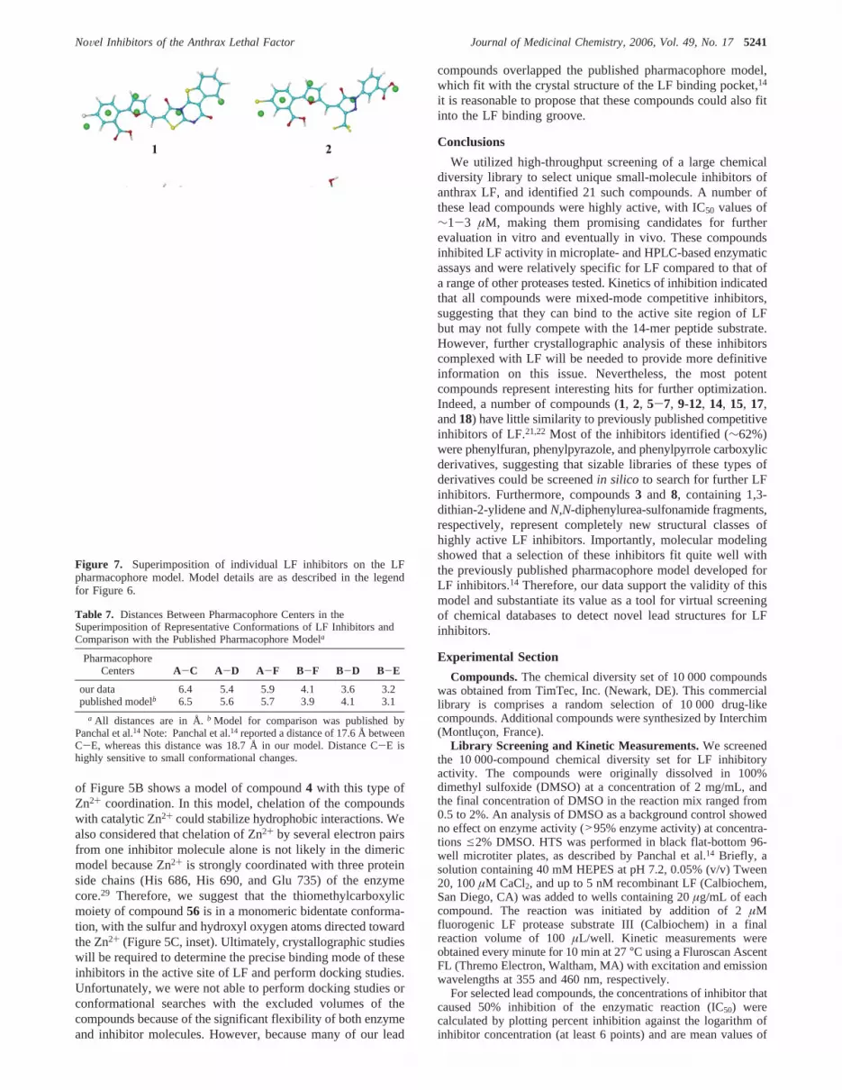
to the aromatic pharmacophore pointB and to assign the furanoxygen atom to the central polar regionD. In this case,substituents R1 in theR-position of the furan ring would overlapwith each other in suitable conformations. For compounds2,5, 7-10, 12, 15, 17, and18, it was possible, from the array ofrepresentative conformers, to choose a geometry that providedthe best overlap with the putative bioactive conformation ofinhibitor 1. Polar substituents (chlorine atoms, carbonyl andcarboxyl groups, and nitrogen atoms (heterocycle of compound8)) in this superimposition were close to a point that can beregarded as polar centerC (Figure 6). We refined the super-imposition by including distances from pharmacophore centerC to the polar groups in the rms fit, for compounds having suchgroups in the vicinity ofC. The overlaid geometries thusobtained are shown in Figure 6, and individual compounds inthe appropriate conformations are shown in Figure 7 togetherwith the pharmacophoric centers indicated for clarity. It can beseen that the hydrophobic center corresponding to regionA inthis overlay contains aromatic or heteroaromatic rings, and thethird polar centerE is clearly located in the vicinity of electron-acceptor substituents in para and meta positions of the phenylring (Figure 6). Finally, neutral linkerF is placed near theâ-carbon atoms of furan moieties in the overlay. Overall, thetotal arrangement of overlaid molecules corresponds extremelywell with the active site of LF, as modeled previously.14
Table 7 contains the distances between pharmacophoriccenters obtained by us in comparison with the distances reportedby Panchal et al.14 for their pharmacophore model. All distances,except forB-D, matched this model within a 0.2 Å tolerance.
Table 4. Functional Groups Present in the Selected LF Inhibitors andTheir Distribution in the Parent Compound Library
a Dashed lines represent any bond type, and A represents any atom excepthydrogen b The number of compounds in the active set was 18 (see Table1 and Figure 2).
Figure 5. Pharmacophore model of LF inhibitors with hypotheticalderivatives. Panel A: Spatial relationships in the published pharma-cophore model reported by Panchal et al.14 Inset: Pharmacophoremodeling of compound9. Panel B: Hypothetical dimeric pharmacoph-ore model with monodentate Zn2+ coordination. Inset: Modeling Zn2+
coordination for compound4. Panel C: Hypothetical monomericpharmacophore model with bidentate Zn2+ coordination. Inset: Model-ing Zn2+ coordination for compound58. The distances indicated inthe insets were calculated for the molecular conformation with thelowest energy (Table 7).
NoVel Inhibitors of the Anthrax Lethal Factor Journal of Medicinal Chemistry, 2006, Vol. 49, No. 175239

Good agreement among these values indicates that the geom-etries used for the superimposition were close to bioactiveconformations of the compounds under investigation. Althougha 6 kcal/mol energy gap was used during the conformationalsearch, most of these conformations were within 3 kcal/molabove the corresponding global minima (Table 6), a valuegenerally considered as acceptable in pharmacophore mappingstudies.43 It should be noted that polar centerB coincides withbenzene rings in the superimposed molecules, whereas molecularmoieties corresponding to centerA have a more diversecharacter, comprising aromatic and heteroaromatic cycles as wellas acyclic fragments of hydrophobic nature. Although theposition of Zn2+ with respect to the centers also is not definedin the published pharmacophore model,14 it is reasonable toassociate it with the position of polar regionD in the middle ofthe model (Figure 5A). Indeed, the crystal structure of LF incomplex with a small-molecule inhibitor shows that its ureamoiety, containing polar centerD and a linkerF, is very closeto the catalytic Zn2+, and the distance between the urea groupand Zn2+ was determined to be within 4 Å, at a resolution of2.9 Å.14 At these distances (<2.05 Å), donor-acceptor coor-dination interactions between Zn2+ and polar groups of a givenprotease inhibitor would be possible.44,45
It should be noted that the superimposed compounds did notalways contain groups that could be attributed to all of thepharmacophoric centers shown in Figures 6 and 7. For example,inhibitors 5 and 10 only partially occupied space within theoverlaid structures. However, partial binding within the activesite could also lead to inhibition of the enzyme. For theremaining 10 compounds of the 21 active inhibitors, it is difficultto suggest a defined pharmacophore model, at least for theirconformations found within the 6 kcal/mol energy gap. Nev-ertheless, it is possible to propose mechanisms in which theseand other LF inhibitors could interact with the active site. Forexample, carboxylic acid groups in compounds4, 6, 11, 13,14, 16, and56-58as well as sulfamate and sulfonamide groupsin compounds6, 13, and16could act as donors for the catalytic
Zn2+.36,46Likewise, inhibitors3, 6, 11, and14 could be ligatedto Zn2+ through one of the carbonyl oxygen atoms in the linkerchain or heterocycle. Analogous to the pharmacophore model,the spatial arrangement of two aromatic and two polar centersin inhibitors 4, 11, and56-58 could result in a symmetrical,dimeric monodentate model (Figure 5B). For example, the inset
Table 5. Characteristics of the Most Potent CarboxylicN-Phenylpyrrole LF Inhibitors Found Among the Analogues from Interchim
lethal factorother proteases
IC50 (µM)
compd
IC50
(µM)microplate
assayKi
(µM)Ki′
(µM)mode of
inhibitionaminopep-tidase M
chymo-trypsin kallikrein elastase
cathepsinB MMP-9
highesttestedconcn(µM)
56 (m.w. 261.3) 2.9 1.6 4.6 mixed N.I.a N.I. N.I. 15.5 58.3 N.I. 16057 (m.w. 249.7) 6.8 5.6 7.6 mixed N.I. N.I. N.I. 160.2 7.3 N.I. 17558 (m.w. 299.3) 7.6 0.9 10.9 mixed N.I. N.I. 60.5 124.9 10.1 N.I. 155
a N.I., no inhibition seen at the highest concentrations of compound tested (see the last column of the Table).
Table 6. Number of Representative Conformations Found for LFInhibitors and Relative Energy Levels (kcal/mol) of the Molecules in theFitted Conformationa
compdno. 1 2 5 7 8 9 10 12 15 17 18
Nconf 4 16 2 81 50 16 2 10 3 16 8∆Eb 0 0.06 2.33 1.03 2.07 2.64 0.03 3.41 0.56 3.66 2.72
a The number of representative conformations within 6 kcal/mol ofthe global energy minimum are indicated.b Energy difference with respectto the global minimum for the lowest energy conformation of eachcompound.
Figure 6. Comparison of LF inhibitor conformation with the that ofpublished LF inhibitor pharmacophore model. A spatial overlay of 11representative compounds on the published pharmacophore coordinatesis shown. Compound conformations shown represent the best rms fitfrom all clusters within 6 kcal/mol of the global energy minimum. Thesuperimposition is shown in two projections, and the green ellipsesare located at the pharmacophore centers of the superimposition in bothprojections. The centers are also indicated at the top of the Figure forclarity, with letter notation corresponding to the hydrophobic (A andB), polar (C, D, andE), and linker (F) centers, as previously reported.14
For all structures, carbon is sky-blue, nitrogen is blue, oxygen is red,sulfur and bromine are yellow, and chlorine is white.
5240 Journal of Medicinal Chemistry, 2006, Vol. 49, No. 17 Schepetkin et al.
of Figure 5B shows a model of compound4 with this type ofZn2+ coordination. In this model, chelation of the compoundswith catalytic Zn2+ could stabilize hydrophobic interactions. Wealso considered that chelation of Zn2+ by several electron pairsfrom one inhibitor molecule alone is not likely in the dimericmodel because Zn2+ is strongly coordinated with three proteinside chains (His 686, His 690, and Glu 735) of the enzymecore.29 Therefore, we suggest that the thiomethylcarboxylicmoiety of compound56 is in a monomeric bidentate conforma-tion, with the sulfur and hydroxyl oxygen atoms directed towardthe Zn2+ (Figure 5C, inset). Ultimately, crystallographic studieswill be required to determine the precise binding mode of theseinhibitors in the active site of LF and perform docking studies.Unfortunately, we were not able to perform docking studies orconformational searches with the excluded volumes of thecompounds because of the significant flexibility of both enzymeand inhibitor molecules. However, because many of our lead
compounds overlapped the published pharmacophore model,which fit with the crystal structure of the LF binding pocket,14
it is reasonable to propose that these compounds could also fitinto the LF binding groove.
Conclusions
We utilized high-throughput screening of a large chemicaldiversity library to select unique small-molecule inhibitors ofanthrax LF, and identified 21 such compounds. A number ofthese lead compounds were highly active, with IC50 values of∼1-3 µM, making them promising candidates for furtherevaluation in vitro and eventually in vivo. These compoundsinhibited LF activity in microplate- and HPLC-based enzymaticassays and were relatively specific for LF compared to that ofa range of other proteases tested. Kinetics of inhibition indicatedthat all compounds were mixed-mode competitive inhibitors,suggesting that they can bind to the active site region of LFbut may not fully compete with the 14-mer peptide substrate.However, further crystallographic analysis of these inhibitorscomplexed with LF will be needed to provide more definitiveinformation on this issue. Nevertheless, the most potentcompounds represent interesting hits for further optimization.Indeed, a number of compounds (1, 2, 5-7, 9-12, 14, 15, 17,and18) have little similarity to previously published competitiveinhibitors of LF.21,22 Most of the inhibitors identified (∼62%)were phenylfuran, phenylpyrazole, and phenylpyrrole carboxylicderivatives, suggesting that sizable libraries of these types ofderivatives could be screenedin silico to search for further LFinhibitors. Furthermore, compounds3 and 8, containing 1,3-dithian-2-ylidene andN,N-diphenylurea-sulfonamide fragments,respectively, represent completely new structural classes ofhighly active LF inhibitors. Importantly, molecular modelingshowed that a selection of these inhibitors fit quite well withthe previously published pharmacophore model developed forLF inhibitors.14 Therefore, our data support the validity of thismodel and substantiate its value as a tool for virtual screeningof chemical databases to detect novel lead structures for LFinhibitors.
Experimental Section
Compounds.The chemical diversity set of 10 000 compoundswas obtained from TimTec, Inc. (Newark, DE). This commerciallibrary is comprises a random selection of 10 000 drug-likecompounds. Additional compounds were synthesized by Interchim(Montlucon, France).
Library Screening and Kinetic Measurements.We screenedthe 10 000-compound chemical diversity set for LF inhibitoryactivity. The compounds were originally dissolved in 100%dimethyl sulfoxide (DMSO) at a concentration of 2 mg/mL, andthe final concentration of DMSO in the reaction mix ranged from0.5 to 2%. An analysis of DMSO as a background control showedno effect on enzyme activity (>95% enzyme activity) at concentra-tions e2% DMSO. HTS was performed in black flat-bottom 96-well microtiter plates, as described by Panchal et al.14 Briefly, asolution containing 40 mM HEPES at pH 7.2, 0.05% (v/v) Tween20, 100µM CaCl2, and up to 5 nM recombinant LF (Calbiochem,San Diego, CA) was added to wells containing 20µg/mL of eachcompound. The reaction was initiated by addition of 2µMfluorogenic LF protease substrate III (Calbiochem) in a finalreaction volume of 100µL/well. Kinetic measurements wereobtained every minute for 10 min at 27°C using a Fluroscan AscentFL (Thremo Electron, Waltham, MA) with excitation and emissionwavelengths at 355 and 460 nm, respectively.
For selected lead compounds, the concentrations of inhibitor thatcaused 50% inhibition of the enzymatic reaction (IC50) werecalculated by plotting percent inhibition against the logarithm ofinhibitor concentration (at least 6 points) and are mean values of
Figure 7. Superimposition of individual LF inhibitors on the LFpharmacophore model. Model details are as described in the legendfor Figure 6.
Table 7. Distances Between Pharmacophore Centers in theSuperimposition of Representative Conformations of LF Inhibitors andComparison with the Published Pharmacophore Modela
PharmacophoreCenters A-C A-D A-F B-F B-D B-E
our data 6.4 5.4 5.9 4.1 3.6 3.2published modelb 6.5 5.6 5.7 3.9 4.1 3.1
a All distances are in Å.b Model for comparison was published byPanchal et al.14 Note: Panchal et al.14 reported a distance of 17.6 Å betweenC-E, whereas this distance was 18.7 Å in our model. Distance C-E ishighly sensitive to small conformational changes.
NoVel Inhibitors of the Anthrax Lethal Factor Journal of Medicinal Chemistry, 2006, Vol. 49, No. 175241
at least three experiments with relative standard deviations<15%.Km and Vmax were calculated using the double-reciprocal Line-weaver-Burk plot of rate versus substrate concentration, and thevalues of Ki and Ki′ were found using replots of inhibitorconcentration againstKm/Vmax and I/Vmax values, respectively.47
Lineweaver-Burk plots were linear (R2 ) 0.998) over the relevantconcentration range of fluorogenic substrate (0.5-3 µM), indicatingthat the inner filter effect was very small at substrate concentrationse3 µM and our typical conditions of fluorescence measurementusing black flat-bottom 96 well microtiter plates.48 Kinetic studiesconsisted of three independent experiments with four replicates ineach experiment.
HPLC Assay.Reaction mixtures (20µL total volume) containing40 mM HEPES at pH 7.2, 0.05% (v/v) Tween 20, 100µM CaCl2,and 2µM fluorogenic LF protease substrate III, with or withoutinhibitor, were incubated with 0.5µg/mL of LF for 30 min at 27°C. The reactions were stopped by adding 180µL of 70% (v/v)acetonitrile in 0.1% (v/v) trifluoracetic acid (TFA), and the productswere separated by reverse-phase HPLC on an automated HPLCsystem (Shimadzu, Torrance, CA) with a Phenomenex Jupiter C18300A column (5µm, 25× 0.46 cm) eluted with acetonitrile/water(35/65, v/v) containing 0.1% (v/v) TFA at a flow rate of 0.8 mL/min at 30 °C over 15 min. The elution of LF inhibitors wasmonitored using a diode array detector (Shimadzu SPD-M10A VP)at specific wavelength regions where the inhibitor showed greatestabsorbance. The fluorescence peak of theC-terminal cleavageproduct was detected using a fluorescence detector (Shimadzu RF-10A XL) at excitation and emission wavelengths of 340 and 430nm, respectively. For the selected inhibitors, the IC50 values wereevaluated using six different concentrations of the inhibitor rangingfrom 0.3 to 40µg/mL and measuring the fluorescence peak of theC-terminal cleavage product.
The incubation of the fluorogenic substrate with LF resulted inone new peak in the chromatogram (Rt ) 5.84), with a concomitantdecrease in peak height of the substrate peak (Rt ) 4.08) (Figure3), and the rate of increase in the peak fluorescence was linearlyrelated to the amount of LF (Figure 3, inset). A comparative analysisof microplate- and HPLC-based assays resulted in a linear correla-tion between the values of fluorescence signals (R2 >0.999),measured at same time point of incubation of the substrate withLF. In addition, the peaks of selected inhibitors and the fluorescenceC-terminal cleavage product were completely separated at theindicated elution conditions.
Analysis of Inhibitor Specificity. Selected compounds wereevaluated for their ability to inhibit a range of proteases in 100µLreaction volumes at 30°C. Aminopeptidase M inhibitory activitywas determined by a modification of the method of Ishida et al.49
Reaction mixtures contained 0.1 M Tris-HCl buffer at pH 7.0,0.05% (v/v) Tween 20, 2 mU porcine kidney aminopeptidase M(Calbiochem), test compounds, and 0.4 mML-alanyl-p-nitroanilide(Calbiochem). Pro-MMP-9 was activated at 37°C for 2 h inbufferA (50 mM Tris-HCl at pH 7.6, 150 mM NaCl, 5 mM CaCl2, 0.01%Brij 35) with 0.2 mMp-aminophenylmercuric acetate. The MMP-9inhibition assay was performed in reaction mixtures containingbuffer A, 3 nM MMP-9 (Calbiochem), test compounds, and 20µMMMP substrate III (DABCYL-GABA-Pro-Gln-Gly-Leu-Glu-(EDANS)-Ala-Lys-NH2, Calbiochem). The kallikrein inhibitionassay was performed in reaction mixtures containing 50 mM Tris-HCl at pH 8.0, 100 mM NaCl, 0.05% (v/v) Tween-20, 2 nM humanplasma kallikrein (Calbiochem), test compounds, and 50µMsubstrate (benzyloxycarbonyl-Phe-Arg-7-amino-4-methylcoumarin,Calbiochem). The elastase inhibition assay was performed inreaction mixtures containing 200 mM Tris-HCl at pH 7.5, 0.001%HSA, 20 mU/mL of human neutrophil elastase (Calbiochem), testcompounds, and 750µM elastase substrate (MeOSuc-Ala-Ala-Pro-Val-7-amino-4-methylcoumarin, Calbiochem). The chymotrypsininhibition assay was performed in reaction mixtures containing 50mM Tris-HCl at pH 8.0, 30 nM human pancreas chymotrypsin(Calbiochem), test compounds, and 250µM chymotrypsin substrate(Suc-Ala-Ala-Pro-Phe-7-amino-4-methylcoumarin, Calbiochem).The cathepsin B inhibition assay was performed in reaction mixtures
containing 250 mM sodium potassium phosphate buffer at pH 6.0,150 nM human liver cathepsin B (Calbiochem), test compounds,and 400µM cathepsin B substrate III (benzyloxycarbonyl-Arg-Arg-7-amino-4-methylcoumarin, Calbiochem). Reactions for ami-nopeptidase M were monitored at 405 nm using a SpectraMax Plusmicrotiter plate reader (Molecular Devices, Sunnyvale, CA). Forall other proteases, fluorescence substrate cleavage was monitoredwith a Fluoroskan Ascent FL microtiter plate reader with excitationand emission wavelengths of 355 and 460 nm, respectively.
Conformational Analysis. For 11 inhibitors, sets of conforma-tions were generated using the Conformational Search Module, asimplemented in HyperChem Version 7.0 (Hypercube, Inc., Canada).The systematic search of conformations for each compound wasperformed by energy minimization, starting with 1000 initialgeometries at random values of torsion angles about exocyclic singlebonds and chemical bonds within nonaromatic cycles. Energy wasminimized by the Polak-Ribiere conjugate gradient method withthe MM+ force field (HyperChem). Attainment of an rms gradient<0.02 kcal/mol‚Å was used as the termination condition forminimization. Conformations were compared and considered asequal if their rms difference in atomic coordinates was less than0.25 Å, and unique conformations were clustered as described.42
Independent conformations obtained after cluster analysis were thenoptimized by semiempirical parametric method 3 (PM3) and usedfor alignment and generation of the pharmacophore.
To determine a geometry favorable for the interaction of aninhibitor with the enzyme site, we fitted conformations of compound1 to the pharmacophore model developed previously by Panchalet al.14 for a different set of LF inhibitors. The conformation chosen(see Results) was then used as a template for alignment ofrepresentative conformations of the other selected compounds.Using the resulting overlay of structures, the coordinates ofpharmacophore centers were determined from the positions ofbenzene rings (B), polar substituents in para and ortho positions(E), electron-withdrawing atoms in the five-membered ring linkedto benzene (D), and aromatic or heteroaromatic substructures nearthis ring (A). CenterC was located in the vicinity of polar groupspresent in some compounds far from the five-membered ring. Theoverlay of structures was refined by a rms fitting of the conforma-tions to the pharmacophore points.
Acknowledgment. We thank Suzanne Wilson for technicalhelp in processing the chemical diversity library. This work wassupported in part by Department of Defense Grant W9113M-04-1-0001, National Institutes of Health Grant RR020185, andthe Montana State University Agricultural Experimental Station.The U.S. Army Space and Missile Defense Command, 64Thomas Drive, Frederick, MD 21702 is the awarding andadministering acquisition office. The content of this report doesnot necessarily reflect the position or policy of the U.S.Government.
Supporting Information Available: Structures and activitiesof the carboxylic andN-sulfonylated phenylfuran derivatives,carboxylic and sulfonamide derivatives of 1-phenyl-4,5-dihydro-5-pyrazolone, and carboxylicN-penylpyrrole derivatives as wellas an example of the optimization of LF inhibitor conformation.This material is available free of charge via the Internet at http://pubs.acs.org.
References(1) Keim, P.; Smith, K. L. Bacillus anthracis evolution and epidemiology.
Curr. Top. Microbiol. Immunol.2002, 271, 21-32.(2) Baillie, L. W. Bacillus anthracis, a story of nature subverted by man.
Lett. Appl. Microbiol.2005, 41, 227-229.(3) Collier, R. J.; Young, J. A. Anthrax toxin.Annu. ReV. Cell DeV. Biol.
2003, 19, 45-70.(4) Abrami, L.; Reig, N.; van der Goot, F. G. Anthrax toxin: the long
and winding road that leads to the kill.Trends Microbiol.2005, 13,72-78.
5242 Journal of Medicinal Chemistry, 2006, Vol. 49, No. 17 Schepetkin et al.

(5) Krantz, B. A.; Melnyk, R. A.; Zhang, S.; Juris, S. J.; Lacy, D. B.;Wu, Z.; Finkelstein, A.; Collier, R. J. A phenylalanine clamp catalyzesprotein translocation through the anthrax toxin pore.Science2005,309, 777-781.
(6) Mock, M.; Mignot, T. Anthrax toxins and the host: a story ofintimacy.Cell. Microbiol. 2003, 5, 15-23.
(7) Dixon, T. C.; Meselson, M.; Guillemin, J.; Hanna, P. C. Anthrax.N.Engl. J. Med.1999, 341, 815-826.
(8) Komiyama, T.; Swanson, J. A.; Fuller, R. S. Protection from anthraxtoxin-mediated killing of macrophages by the combined effects offurin inhibitors and chloroquine.Antimicrob. Agents Chemother.2005, 49, 3875-3882.
(9) Huber, M.; Vor Dem, E. U.; Grunow, R.; Bessler, W. G. Generationof mouse polyclonal and human monoclonal antibodies againstBacillus anthracistoxin. Drugs Exp. Clin. Res.2005, 31, 35-43.
(10) Mourez, M.; Kane, R. S.; Mogridge, J.; Metallo, S.; Deschatelets,P.; Sellman, B. R.; Whitesides, G. M.; Collier, R. J. Designing apolyvalent inhibitor of anthrax toxin.Nat. Biotechnol.2001, 19, 958-961.
(11) Tonello, F.; Seveso, M.; Marin, O.; Mock, M.; Montecucco, C.Screening inhibitors of anthrax lethal factor.Nature2002, 418, 386.
(12) Montecucco, C.; Tonello, F.; Zanotti, G. Stop the killer: how toinhibit the anthrax lethal factor metalloprotease.Trends Biochem.Sci.2004, 29, 282-285.
(13) Kim, C.; Gajendran, N.; Mittrucker, H. W.; Weiwad, M.; Song, Y.H.; Hurwitz, R.; Wilmanns, M.; Fischer, G.; Kaufmann, S. H. HumanR-defensins neutralize anthrax lethal toxin and protect against its fatalconsequences.Proc. Natl. Acad. Sci. U.S.A.2005, 102, 4830-4835.
(14) Panchal, R. G.; Hermone, A. R.; Nguyen, T. L.; Wong, T. Y.;Schwarzenbacher, R.; Schmidt, J.; Lane, D.; McGrath, C.; Turk, B.E.; Burnett, J.; Aman, M. J.; Little, S.; Sausville, E. A.; Zaharevitz,D. W.; Cantley, L. C.; Liddington, R. C.; Gussio, R.; Bavari, S.Identification of small molecule inhibitors of anthrax lethal factor.Nat. Struct. Mol. Biol.2004, 11, 67-72.
(15) Turk, B. E.; Wong, T. Y.; Schwarzenbacher, R.; Jarrell, E. T.; Leppla,S. H.; Collier, R. J.; Liddington, R. C.; Cantley, L. C. The structuralbasis for substrate and inhibitor selectivity of the anthrax lethal factor.Nat. Struct. Mol. Biol.2004, 11, 60-66.
(16) Min, D. H.; Tang, W. J.; Mrksich, M. Chemical screening by massspectrometry to identify inhibitors of anthrax lethal factor.Nat.Biotechnol.2004, 22, 717-723.
(17) Lee, L. V.; Bower, K. E.; Liang, F. S.; Shi, J.; Wu, D.; Sucheck, S.J.; Vogt, P. K.; Wong, C. H. Inhibition of the proteolytic activity ofanthrax lethal factor by aminoglycosides.J. Am. Chem. Soc.2004,126, 4774-4775.
(18) Numa, M. M.; Lee, L. V.; Hsu, C. C.; Bower, K. E.; Wong, C. H.Identification of novel anthrax lethal factor inhibitors generated bycombinatorial Pictet-Spengler reaction followed by screening in situ.ChemBioChem2005, 6, 1002-1006.
(19) Fridman, M.; Belakhov, V.; Lee, L. V.; Liang, F. S.; Wong, C. H.;Baasov, T. Dual effect of synthetic aminoglycosides: antibacterialactivity againstBacillus anthracisand inhibition of anthrax lethalfactor.Angew. Chem., Int. Ed.2005, 44, 447-452.
(20) Kocer, S. S.; Walker, S. G.; Zerler, B.; Golub, L. M.; Simon, S. R.Metalloproteinase inhibitors, nonantimicrobial chemically modi-fied tetracyclines, and ilomastat blockBacillus anthracis lethalfactor activity in viable cells.Infect. Immun.2005, 73, 7548-7557.
(21) Forino, M.; Johnson, S.; Wong, T. Y.; Rozanov, D. V.; Savinov, A.Y.; Li, W.; Fattorusso, R.; Becattini, B.; Orry, A. J.; Jung, D.;Abagyan, R. A.; Smith, J. W.; Alibek, K.; Liddington, R. C.; Stron-gin, A. Y.; Pellecchia, M. Efficient synthetic inhibitors of anthraxlethal factor. Proc. Natl. Acad. Sci. U.S.A.2005, 102, 9499-9504.
(22) Johnson, S. L.; Jung, D.; Forino, M.; Chen, Y.; Satterthwait, A.;Rozanov, D. V.; Strongin, A. Y.; Pellecchia, M. Anthrax lethal factorprotease inhibitors: synthesis, SAR, and structure-based 3D QSARstudies.J. Med. Chem.2006, 49, 27-30.
(23) Xiong, Y.; Wiltsie, J.; Woods, A.; Guo, J.; Pivnichny, J. V.; Tang,W.; Bansal, A.; Cummings, R. T.; Cunningham, B. R.; Friedlander,A. M.; Douglas, C. M.; Salowe, S. P.; Zaller, D. M.; Scolnick, E.M.; Schmatz, D. M.; Bartizal, K.; Hermes, J. D.; MacCoss, M.;Chapman, K. T. The discovery of a potent and selective lethal factorinhibitor for adjunct therapy of anthrax infection.Bioorg. Med. Chem.Lett. 2006, 16, 964-968.
(24) Jiao, G. S.; Cregar, L.; Goldman, M. E.; Millis, S. Z.; Tang, C.Guanidinylated 2,5-dideoxystreptamine derivatives as anthrax lethalfactor inhibitors. Bioorg. Med. Chem. Lett.2006, 16, 1527-1531.
(25) Dell’Aica, I.; Dona, M.; Tonello, F.; Piris, A.; Mock, M.; Montecucco,C.; Garbisa, S. Potent inhibitors of anthrax lethal factor from greentea.EMBO Rep.2004, 5, 418-422.
(26) McGovern, S. L.; Caselli, E.; Grigorieff, N.; Shoichet, B. K. Acommon mechanism underlying promiscuous inhibitors from virtualand high-throughput screening.J. Med. Chem.2002, 45, 1712-1722.
(27) Blanchard, J. E.; Elowe, N. H.; Huitema, C.; Fortin, P. D.; Cechetto,J. D.; Eltis, L. D.; Brown, E. D. High-throughput screening identifiesinhibitors of the SARS coronavirus main proteinase.Chem. Biol.2004, 11, 1445-1453.
(28) Cornish-Bowden, A.Fundamentals of Enzyme Kinetics; Butterworth& Company, Ltd.: Woburn, MA, 1979.
(29) Pannifer, A. D.; Wong, T. Y.; Schwarzenbacher, R.; Renatus, M.;Petosa, C.; Bienkowska, J.; Lacy, D. B.; Collier, R. J.; Park, S.;Leppla, S. H.; Hanna, P.; Liddington, R. C. Crystal structure of theanthrax lethal factor.Nature2001, 414, 229-233.
(30) Tonello, F.; Naletto, L.; Romanello, V.; Dal, M. F.; Montecucco, C.Tyrosine-728 and glutamic acid-735 are essential for the metallo-proteolytic activity of the lethal factor ofBacillus anthracis. Biochem.Biophys. Res. Commun.2004, 313, 496-502.
(31) Krantz, A. A classification of enzyme-inhibitors.Bioorg. Med. Chem.Lett. 1992, 2, 1327-1334.
(32) Thompson, S. A.; Andrews, P. R.; Hanzlik, R. P. Carboxyl-modifiedamino acids and peptides as protease inhibitors.J. Med. Chem.1986,29, 104-111.
(33) Babine, R. E.; Bender, S. L. Molecular recognition of protein-ligandcomplexes: Applications to drug design.Chem. ReV. 1997, 97,1359-1472.
(34) Pruitt, J. R.; Pinto, D. J.; Galemmo, R. A., Jr.; Alexander, R. S.;Rossi, K. A.; Wells, B. L.; Drummond, S.; Bostrom, L. L.; Burdick,D.; Bruckner, R.; Chen, H.; Smallwood, A.; Wong, P. C.; Wright,M. R.; Bai, S.; Luettgen, J. M.; Knabb, R. M.; Lam, P. Y.; Wexler,R. R. Discovery of 1-(2-aminomethylphenyl)-3-trifluoromethyl-N-[3-fluoro-2′-(aminosulfonyl)[1,1′-biphenyl)]-4-yl]-1H-pyrazole-5-carboxyamide (DPC602), a potent, selective, and orally bio-available factor Xa inhibitor (1).J. Med. Chem.2003, 46, 5298-5315.
(35) Kevin, N. J.; Duffy, J. L.; Kirk, B. A.; Chapman, K. T.; Schleif, W.A.; Olsen, D. B.; Stahlhut, M.; Rutkowski, C. A.; Kuo, L. C.; Jin,L.; Lin, J. H.; Emini, E. A.; Tata, J. R. Novel HIV-1 proteaseinhibitors active against multiple PI-resistant viral strains: coadmin-istration with indinavir.Bioorg. Med. Chem. Lett.2003, 13, 4027-4030.
(36) Huang, Q. Q.; Huang, M.; Nan, F. J.; Ye, Q. Z. Metalloform-selectiveinhibition: synthesis and structure-activity analysis of Mn(II)-form-selective inhibitors ofEscherichia colimethionine aminopeptidase.Bioorg. Med. Chem. Lett.2005, 15, 5386-5391.
(37) Lu, Z.; Bohn, J.; Rano, T.; Rutkowski, C. A.; Simcoe, A. L.; Olsen,D. B.; Schleif, W. A.; Carella, A.; Gabryelski, L.; Jin, L.; Lin, J. H.;Emini, E.; Chapman, K.; Tata, J. R. Orally bioavailable highly potentHIV protease inhibitors against PI-resistant virus.Bioorg. Med. Chem.Lett. 2005, 15, 5311-5314.
(38) Bellu, S.; Hure, E.; Trape, M.; Rizzotto, M.; Sutich, E.; Sigrist, M.;Moreno, V. The interaction between mercury(II) and sulfathiazole.Quim. NoVa 2003, 26, 188-192.
(39) Yoshihara, T.; Druzhinin, S. I.; Zachariasse, K. A. Fast intramolecularcharge transfer with a planar rigidized electron donor/acceptormolecule.J. Am. Chem. Soc.2004, 126, 8535-8539.
(40) Lepailleur, A.; Bureau, R.; Paillet-Loilier, M.; Fabis, F.; Saettel, N.;Lemaitre, S.; Dauphin, F.; Lesnard, A.; Lancelot, J. C.; Rault, S.Molecular modeling studies focused on 5-HT7 versus 5-HT1Aselectivity. Discovery of novel phenylpyrrole derivatives with highaffinity for 5-HT7 receptors.J. Chem. Inf. Model.2005, 45, 1075-1081.
(41) Nicklaus, M. C.; Wang, S. M.; Driscoll, J. S.; Milne, G. W. A.Conformational-changes of small molecules binding to proteins.Bioorg. Med. Chem.1995, 3, 411-428.
(42) Shenkin, P. S.; Mcdonald, D. Q. Cluster-analysis of molecular-conformations.J. Comput. Chem.1994, 15, 899-916.
(43) Wermuth, C. G.; Langer, T. Pharmacophore Identification. In3D QSAR in Drug Design. Theory Methods and Applications;Kubinyi, H., Ed.; ESCOM: Leiden, The Netherlands, 1993; pp 117-136.
(44) Brandstetter, H.; Grams, F.; Glitz, D.; Lang, A.; Huber, R.; Bode,W.; Krell, H. W.; Engh, R. A. The 1.8-A crystal structure of a matrixmetalloproteinase 8-barbiturate inhibitor complex reveals a previouslyunobserved mechanism for collagenase substrate recognition.J. Biol.Chem.2001, 276, 17405-17412.
NoVel Inhibitors of the Anthrax Lethal Factor Journal of Medicinal Chemistry, 2006, Vol. 49, No. 175243

(45) Voisin, S.; Rognan, D.; Gros, C.; Ouimet, T. A three-dimensionalmodel of the neprilysin 2 active site based on the X-ray structure ofneprilysin. Identification of residues involved in substrate hydrolysisand inhibitor binding of neprilysin 2.J. Biol. Chem.2004, 279,46172-46181.
(46) Nishino, N.; Powers, J. C. Design of potent reversible inhibitorsfor thermolysin. Peptides containing zinc coordinating ligands andtheir use in affinity chromatography.Biochemistry1979, 18, 4340-4347.
(47) O’Donohue, M. J.; Beaumont, A. The roles of the prosequence ofthermolysin in enzyme inhibition and folding in vitro.J. Biol. Chem.1996, 271, 26477-26481.
(48) Liu, Y.; Kati, W.; Chen, C. M.; Tripathi, R.; Molla, A.; Kohlbrenner,W. Use of a fluorescence plate reader for measuring kineticparameters with inner filter effect correction.Anal. Biochem.1999,267, 331-335.
(49) Ishida, K.; Kato, T.; Murakami, M.; Watanabe, M.; Watanabe, M.F. Microginins, zinc metalloproteases inhibitors from the cyano-bacteriumMicrocystis aeruginosa. Tetrahedron2000, 56, 8643-8656.
JM0605132
5244 Journal of Medicinal Chemistry, 2006, Vol. 49, No. 17 Schepetkin et al.


