Embed Size (px)
Citation preview
GOVERNMENT OF KERALA
STANDARD TREATMENT GUIDELINES
DEPARTMENT OF HEALTH AND FAMILY WELFARE
Obstetrics

Committee for Development of Standard Treatment Guidelines GI diseases
Conveners for STG in obstetrics
1. Dr Nandini.V.R,Professor ,O&G
2. Dr.Sreekumari.R,Professor,O&G
Members
1. Dr.Nirmala.C,HOD,Govt Medical College,Trivandrum
2. Dr.Lalithambica Karunakaran,HOD,TDMC Alappuzha
3. Dr.Ambujam ,HOD,Govt Medical College,Thrissur
4. Dr.Mini.C.H ,HOD,Govt Medical College Calicut
5. Dr.Jacob,HOD,Govt Medical College,Manjeri
6. Dr.P.K.ShyamalaDevi,Rtd Professor &HOD
7. Dr.Sheela Shenoy.Rtd Professor&HOD
8. Dr.Presannakumari.B,Rtd Professor, Past president, KFOG
Additional Chief Secretary, Department of Health and Family Welfare, Government
of Kerala, the process of preparation of Standard Treatment Guidelines (STG) was
initiated by the Director of Medical Education Dr. Remla Beevi A. The process of
developing and finalizing the STG’s were coordinated by Dr. Sreekumari K. Joint
Director Medical education and Dr. Suma T K, Professor of Medicine and ably
supported by a dedicated team of experts, including external faculty”.
“Driven by the inspiration drawn from Shri. Rajeev Sadanandan IAS,

TABLE OF CONTENTS
Section 1:COMPREHENSIVE ANTENATAL CARE
Section2:HYPEREMESIS GRAVIDARUM
Section 3:ANTIBIOTIC POLICY IN OBSTETRICS
Section 4.ANEMIA IN PREGNANCY
1.1 Goal
1.2 Antenatal visits
1.3 Immunisation in pregnancy
1.4 First trimester
1.5 Second trimester
1.6 Third trimester
1.7 Annexure
1.7.1. Annexure 1
1.7.2. Annexure 2
2.1. Definition
2.2. Pathogenesis
2.3. Differential Diagnosis
2.4. Complications
2.4.1. Maternal Complications
2.4.2. Fetal Complications
2.5. PUQE SCORE
2.6. Aims Of Management
2.7. Investigations
2.8. Management
4.1. Introduction
4.2. Definition
4.3. Diagnosis
4.4. Investigations
4.5. Management
4.5.1. MOHFW RECOMMENDTION
4.5.2. Parenteral iron
4.5.2.A.Indication
4.5.2.B.Contraindication
4.5.2.C.Preparation
4.5.2.D.Recombinant Human Erythropoietin
4.5.3. Packed Red Cell
4.5.3.A. Indication in Antepartum period
4.5.3.B.Indication in Intrapartum period
4.5.3.C.Indication in Postpartum period
4.6. Intrapartum Severe Anemia
4.7. Anemia In Puerperium
4.8. Deworming
4.9. Treatment of malaria
4.10 .Treatment outcomes
6.1. Incidence
6.2. Definition
6.3. Complication
6.3.1. Maternal complication
6.3.2. Fetal complication
Section 5. DIABETES IN PREGNANCY
Section 6.PRETERM PRELABOUR RUPTURE OF MEMBRANES
5.1. Introduction
5.2. Gestational Diabetes Mellitus
5.3. Risk to Mother and Fetus
5.4. Screening and Diagnosis
5.5. Methodology
5.6. Pre gestational Diabetes.
5.7. Timing of Delivery
5.8. Management
5.9. Energy requirement during Pregnancy
5.9.1. Energy requirement according to BMI in Diabetics
5.10. Medical treatment\
5.11. Labour And Delivery
5.12. Neonatal management
5.13. Follow up of GDM
5.14. Prepregnancy counselling
5.15. Management of DKA during pregnancy
5.16. Action profile of Insulin
6.4. Specific conditions and tests
6.4. Management
6.4.1. Initial assessment
6.4.2. Treatment
6.4.2.A.Expectant management
6.4.2.B.Antibiotics
6.4.2.C.Cervical encirclage
6.4.2.D.Tocolytic
6.5. Mgso4 Neuroprotection
6.6. Modeof delivery
7.1. Introduction
7.2. Obstetric haemorrhage
7.3. Clinical assessment
7.4. Examination
7.4.1 Abdominal palpation
7.4.2. Local examination/per speculum
7.5. Management
7.5.1. If patient is haemo dynamically unstable
7.5.2. Placenta previa
7.5.3. Abruption placentae(revealed/concealed/mixed)
7.5.4. Vasa praevia
7.6. Post natal issues
8.1. Introduction
8.2. Risk factors
8.3. Prevention
8.3.1. Antenatal period
8.3.2. Intra partum
8.3.3. In Labour
8.3.4. Postpartum
8.3.5. Preventive Measures If The Patient Had
A Previous Caesarean Section
8.4. Pitfalls In The Diagnosis Of PPH
8.5. Early detection of PPH
8.6. Resuscitation and initial assessment -team work
Section
Section 8.POSTPARTUM HAEMORRHAGE
7.ANTEPARTUM HEMORRHAGE
8.7. Directed therapy
8.7 .1. Atonic PPH
8.7.2. Tissue
8.7.3. Trauma.
8.7.4. Thrombin
8.7.5. Traction
8.8. Hidden bleeding
8.9. When to transfuse
8.10. Uterine artery embolisation
8.11. Referral –whom, when and how?
9.1. Introduction
9.2. Risk Factors
9.3. Diagnosis
9.3.1. Biochemistry
9.3.2. Ultrasound
9.3.3. MRI
9.5. Management
9.5.1 Antepartum considerations
9.5.2. Intra-operative considerations
9.5.3. Postoperative care after hysterectomy
9.6. Checklist
10.1. HYPERTENsion in pregnancy
10.2. Preeclampsia
10.3. Screening methods
10.4. Preventive strategies
10.5. Risk factors and aspirin use
Section
Section 10.Hypertensive disorders of Pregnancy
9.PLACENTA ACCRETA SPECTRUM
9.4. Relevant Considerations for Case Optimization
in Planned PlacentaAccreta Spectrum
9.7. “Unexpected” and Unplanned Intraoperative
Recognition of Placenta Accreta Spectrum
9.8. Uterine Preservation and Expectant Management
9.9. Triple-P PROCEDURE
10.5.1.High risk
10.5.2.Moderate risk
10.5.3.Low risk
10.6. Recommendations for measuring proteinuria
10.7. Management
10.7.1.Preeclampsia aand GHTN
10.7.2.Severe preeclampsia
10.8 MgSO4
10.9. HELLP
10.10. Post partum
10.11. Chronic hypertension
10.12. Chronic hypertension with superimposed
preeclampsia
10.13. Emergent treatment for acute onset, severe
hypertension during pregnancy and postpartum period
11.1. Definition
11.2. Diagnosis
11.3. Differential Diagnosis
11.4. Initial Patient assessment and stabilization
11.4.1.General assessment
11.4.2.Initial investigations
11.4.3 Stabilising patient
11.4.4.General supportive measures
11.5. Anticonvulsant therapy
11.5.1 Dosage schedule
11.5.2 Monitoring toxicity
11.5.3 Management of toxicity of Mgso4
11.5.4.Absolute contraindication for Mgso4
11.6. Antihypertensive management
11.7. Obstetric Management
11.8. Postpartum Management
11.9. Indication for LSCS
11.10. Anaesthesia
11.11. Indication for Imaging in Eclampsia
Section 11 Eclampsia
11.12. Puerperal Care
12.1. Introduction
12.2. Defining fetal growth restriction
12.3. Etiology of fetal growth restriction
12.4. Screening for fetal growth restriction
12.4.1.Abdominal examination
12.4.2.First trimester biomarker
12.4.3.Ultrasound with Doppler
12.4.3.A.Uterine artery Doppler
12.4.3.B.Umbilical artery Doppler
12.4.3.C.MCA Doppler
12.4.3.D.Aortic isthmus Doppler
12.4.3.E.Venous Doppler Assessment
12.5. Role of corticosteroids and Magnesium sulphate
12.6. Monitoring
12.6.1.Early onset FGR
12.6.2.Late onset FGR
12.7. Quick reference
12.8. Complications of Growth Restricted babies
13.1. Definition
13.2. Introduction
13.3. Perinatal outcomes
13.4. Adverse Long term Infant outcomes
13.5. General principles
13.6. Indications
13.6.1.Medical conditions
13.6.2.Placental conditions
13.6.3.Fetal conditions
13.6.4.Maternal conditions
13.6.5.Obstetric conditions
13.7. Contraindications for induction
13.8. Pre induction assessment
Section
Section 613.Induction of Labour
12. Intra Uterine Growth restriction
13.9. Overview of cervical ripening methods
13.10. Monitoring during induction
13.11. Complcations
13.12. Failed induction
13.12.1.Management options in failed induction
13.13. Special situation for induction of labour in previous LSCS
13.14. Oxytocin in Labour
13.15. Vaginal examination in labour
14.1 Categories of LSCS
14.2 Pre-requisites for LSCS
14.3. Pre-operative testing and preparation
14.4 Anaesthesia
14.5 Surgical technique
14.5.1 skin incision
14.5.2.uterine incision
14.5.3.Uterine closure
14.5.4.Closure of abdomen
14.6. Antibiotic usage
14.7 Thromboprophylaxis
14.8. Post operative care
14.9 LSCS wound care
14.10 Complications of LSCS
Section 14.CAESAREAN SECTION


Message
The Government is taking many initiatives to ensure providing quality
health care to all. Out of the five missions launched by the Government, the
Aardram mission is primarily focussed to improve Primary Health Care to
provide standard health care facilities to people at grassroots. This initiative is
complemented by strategic investment for the improvement of infrastructure in
secondary and tertiary health care institutions to provide quality health care
services.
I am happy to note that the Department of Health is also taking
initiatives to bring standardization in treatment for various disciplines like
Cardiology, Critical care, Diabetes Mellitus, Cancer Care, etc. It is a noteworthy
initiative to improve the qualitative aspects of the health service delivery. I
appreciate the efforts taken by the experts from Government sector and private
sector from Kerala and also the subject experts from outside the state. I am
hopeful that the introduction of standard guidelines for diagnosis and
treatment will ensure better quality and consistency in health care.
I wish all the success to this endeavour.
13
Pinarayi VijayanChief Minister
SecretariatThiruvananthapuram
Pinarayi VijayanChief Minister

Message
15

Foreword
Patient care has moved away from management by an
individual based on personal knowledge and skill to an evidence
based, team managed operation. Decisions are reviewed more
rigorously post facto and their alignment verified with standard
practice. With the mode of payment for care moving from out of
pocket payments to third party payers there will be a demand for
rigorous documentation and evidence of having conformed to
standard practice. When analysis of big data and machine learning
becomes the norm it will require a standard set of procedures to act
as the baseline from which to measure deviations and differences in
impact.
To meet the requirement of these developments in the field
of medicine, it is necessary to have explicit, objectively verifiable set
of standard operating procedures. They have to be prepared based
on international guidelines with the highest acceptance, but have to
be modified to suit local knowledge and practice, so that there is
local ownership. Government of Kerala has been trying to get the
guidelines prepared for some time now. I would like to thank and
congratulate Dr. Sreekumari, Joint Director of Medical Education
and Dr. T.K.Suma, Professor of Medicine, T.D. Medical College,
Alappuzha who took on the task of preparing standard treatment
guidelines and completed it through a long, consultative process. I
also thank the conveners of the different thematic groups who
coordinated the work in their field as well as the innumerable
number of participants, in government and private sector, who
contributed their effort and knowledge to improve the guidelines.
Professional associations have also contributed in their fields. Their
efforts have resulted in a product they and Kerala can be proud of.
Treatment guidelines cannot be static if they are to remain
relevant. They must be updated based on new knowledge and the
17
experience of treatment based on these guidelines. To do this the
group which prepared the guidelines has to remain active and have
a system for collecting data on the results of practice based on
these guidelines. I hope such an activity is institutionalised and
periodic revisions of the guidelines are prepared and published.
I wish that these guidelines contribute to raising the quality of
patient care in Kerala.
Rajeev Sadanandan IAS
Addl Chief Secretary
Health & Family Welfare
Department
18
COMPREHENSIVE ANTENATAL CARE
Section I

SECTION 1
COMPREHENSIVE ANTENATAL CARE
1.1GOAL:
1.2.ANTENATAL VISITS ( low risk)
1.3.IMMUNISATION IN PREGNANCY:
2. INFLUENZA VACCINE:
1.4.FIRST TRIMESTER
l For identification and surveillance of pregnant women and her
expected child
l Recognition and management of pregnancy related complications
l Treatment of underlying or concurrent illnesses
l Preventive measures including immunisation, deworming and
anemia prophylaxis
l Promote post natal Family planning /Birth spacing
l Health education on nutrition, danger signs of pregnancy
l Prediction ,detection and initial management of perinatal mental
health
l Monthly till 28 weeks
l Fortnightly till 36 weeks
l Weekly till 40 weeks
1.Td:
First dose of Inj.Td to be administered at the first visit and second
dose 4 weeks later.
If the previous pregnancy is less than 3 years only one booster
dose is needed provided she is immunized previously.
Third dose can be given 6 months after the second dose to
provide protection for 5 years (WHO)
Safe during pregnancy
Dose: 0.5 ml IM single dose Deltoid
INDICATIONS FOR ECOSPIRIN: (FIGO)
l History taking and risk stratification
ANNEXURE 1
Se
cti
on
I
STANDARD TREATMENT GUIDELINES - OBSTETRICS
21
High Risk
Medical comorbidities
ART pregnancy
Bad obstetric history
Elderly and teenage pregnancies
Obesity
Short stature
Previous caesarean
Malpresentations
Antepartum hemorrrhage
Preeclampsia, eclampsia
Gestational diabetes
Multiple pregnancy
l General examination including CVS, BMI, Breast examination
l Folic acid 5 mg to be started ( ideally to be started
preconceptionally)
l Blood Investigations :
Hemoglobin,
CBC,
ESR (>60 ) is significant,
RFT
LFT
Urine Routine (to be done monthly)
GTT if patient tolerates or FBS /PPBS
Blood grouping and Rh typing
ICT in all trimesters
Viral markers(HIV ,HCV,HBsAg)
VDRL
Se
cti
on
ISTANDARD TREATMENT GUIDELINES - OBSTETRICS
22
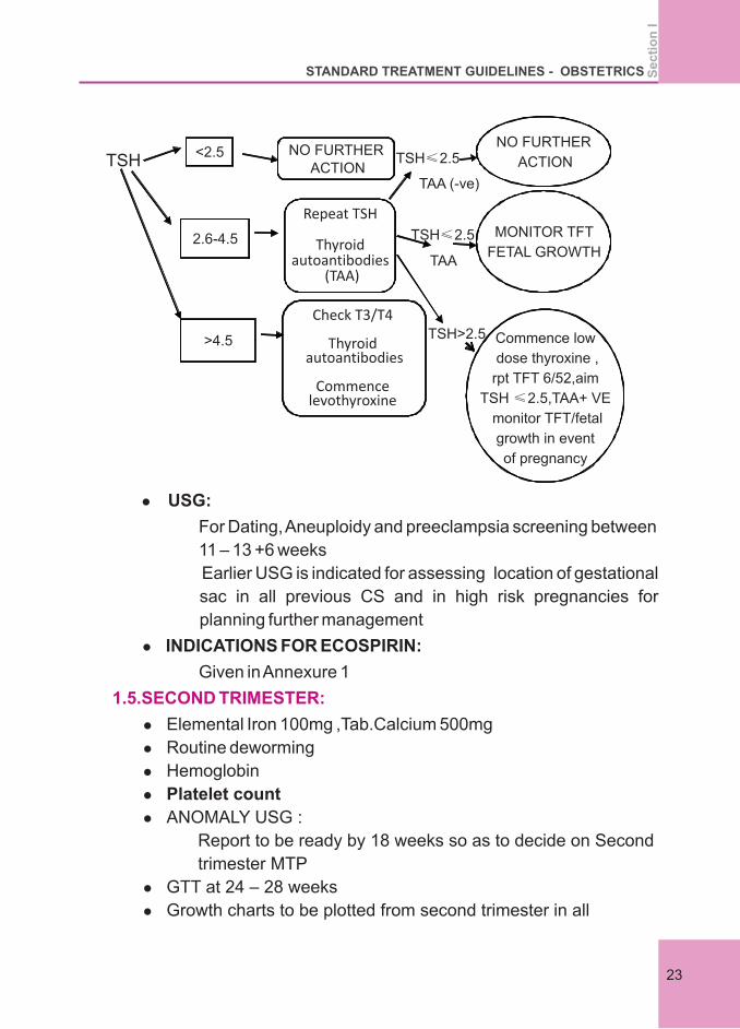
Commence low
dose thyroxine ,
rpt TFT 6/52,aim
TSH ≤2.5,TAA+ VE
monitor TFT/fetal
growth in event
of pregnancy
MONITOR TFT
FETAL GROWTH
NO FURTHER
ACTION
Check T3/T4
Thyroid autoantibodies
Commence levothyroxine
Repeat TSH
Thyroid autoantibodies
(TAA)
NO FURTHER ACTION
<2.5
2.6-4.5
>4.5
TSH TSH≤2.5
TAA (-ve)
TSH≤2.5
TAA
TSH>2.5
l USG:
For Dating, Aneuploidy and preeclampsia screening between
11 – 13 +6 weeks
Earlier USG is indicated for assessing location of gestational
sac in all previous CS and in high risk pregnancies for
planning further management
l INDICATIONS FOR ECOSPIRIN:
Given in Annexure 1
l Elemental Iron 100mg ,Tab.Calcium 500mg
l Routine deworming
l Hemoglobin
l Platelet count
l ANOMALY USG :
Report to be ready by 18 weeks so as to decide on Second
trimester MTP
l GTT at 24 – 28 weeks
l Growth charts to be plotted from second trimester in all
1.5.SECOND TRIMESTER:
Se
cti
on
I
STANDARD TREATMENT GUIDELINES - OBSTETRICS
23
pregnancies
l Fetal ECHO (indication see ANNEXUE 2)
l If ICT negative , Antenatal ANTI-D 300micrograms at 28 weeks
in Rh negative at deltoid
Haemoglobin
Platelet count
TSH
GTT
Growth scans based on clinical indication at 32 and
36 +WEEKS
Breast examination
1.6.THIRD TRIMESTER:
1.7ANNEXURE
1.7.1.ANNEXURE 1
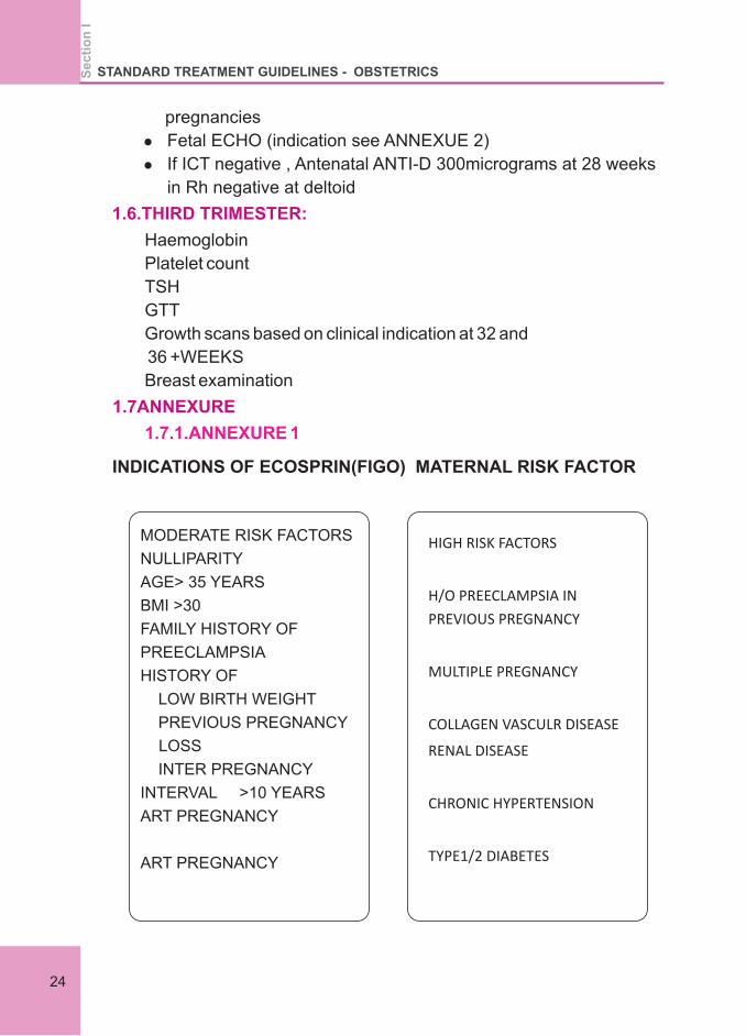
INDICATIONS OF ECOSPRIN(FIGO) MATERNAL RISK FACTOR
MODERATE RISK FACTORS
NULLIPARITY
AGE> 35 YEARS
BMI >30
FAMILY HISTORY OF
PREECLAMPSIA
HISTORY OF
LOW BIRTH WEIGHT
PREVIOUS PREGNANCY
LOSS
INTER PREGNANCY
INTERVAL >10 YEARS
ART PREGNANCY
ART PREGNANCY
HIGH RISK FACTORS
H/O PREECLAMPSIA IN
PREVIOUS PREGNANCY
MULTIPLE PREGNANCY
COLLAGEN VASCULR DISEASE
RENAL DISEASE
CHRONIC HYPERTENSION
TYPE1/2 DIABETES
Se
cti
on
ISTANDARD TREATMENT GUIDELINES - OBSTETRICS
24
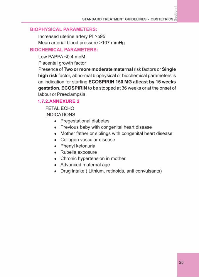
BIOPHYSICAL PARAMETERS:
BIOCHEMICAL PARAMETERS:
Increased uterine artery PI >p95
Mean arterial blood pressure >107 mmHg
Low PAPPA <0.4 moM
Placental growth factor
Presence of Two or more moderate maternal risk factors or Single
high risk factor, abnormal biophysical or biochemical parameters is
an indication for starting ECOSPIRIN 150 MG atleast by 16 weeks
gestation. ECOSPIRIN to be stopped at 36 weeks or at the onset of
labour or Preeclampsia.
FETAL ECHO
INDICATIONS
l Pregestational diabetes
l Previous baby with congenital heart disease
l Mother father or siblings with congenital heart disease
l Collagen vascular disease
l Phenyl ketonuria
l Rubella exposure
l Chronic hypertension in mother
l Advanced maternal age
l Drug intake ( Lithium, retinoids, anti convulsants)
1.7.2.ANNEXURE 2
Se
cti
on
I
STANDARD TREATMENT GUIDELINES - OBSTETRICS
25

SECTION II
HYPEREMESIS GRAVIDARUM

SECTION 2
HYPEREMESIS GRAVIDARUM
2.1Definition:
2.2.Pathogenesis:
2.3.Differential diagnosis:
2.4.Complications:
It is a severe form of nausea and vomiting seen in 1-3% of pregnant
women. It is characterised by severe, protracted nausea and
vomiting associated with weight loss of more than 5% of pre-
pregnancy weight, fluid loss or dehydration.
For most women, these symptoms improve or disappear by around
week 14, although for some women it can last longer.
Despite extensive research in the field, the pathogenesis of
predisposition in addition to previously studied factors such as
infections, psychiatric disturbances and hormonal causes.
Infections, metabolic, gastrointestinal, neurologic, and iatrogenic
causes can cause similar symptoms.
.
Maternal morbidity is due to clinical complications from nutritional
deficiencies, gastrointestinal trauma and in rare cases, neurological
damage. In addition, psychological effects & financial burden also
add to the problem.
2.4.1.Maternal Complications
l Dehydration increases the risk of diabetic ketoacidosis in
those with type 1 diabetes, immobilisation increases the risk
of thromboembolism
l Electrolyte disturbances as seen in any patient with persistent
vomiting – hypochloraemic alkalosis, hypokalaemia and
hyponatraemia
l Protein-calorie malnutrition
l Vitamin/mineral deficiencies and accompanying problems –
e.g. thiamine deficiency can cause Wernicke's
encephalopathy, a serious neurological disorder associated
with acute mental confusion, short term memory loss, ataxia,
ocular abnormalities such as nystagmus and peripheral
Se
cti
on
II
STANDARD TREATMENT GUIDELINES - OBSTETRICS
29
neuropathy. Wernicke's encephalopathy can lead to
irreversible neurological impairment.
l Folate & iron deficiency
l Thyroid dysfunction – e.g. “pseudo-thyrotoxicosis” –
suppressed TSH with high free thyroxine resulting from
thyroid stimulation by HCG
l Renal dysfunction – (reversible) elevated urea and creatinine
l Hepatic dysfunction – elevated ALT, AST, low albumin,
elevated bilirubin, due to malnutrition and catabolic changes
l Ulcerative oesophagitis
l Psychological morbidity e.g. post-traumatic stress disorder
l Mallory –Weiss tears.
l Fetal loss as a result of maternal Wernicke's encephalopathy
l Intrauterine growth restriction (IUGR) or small for gestational
age infants associated with prolonged hyperemesis and loss
of >5% body weight.
l Undernutrition in early intrauterine period increases risk of
chronic illness in adult life.
2.4.2.Fetal Complications
2.5.Classification:
Recently, a classification system was created to categorize
hyperemesis gravidarum called the PUQE (pregnancy-unique
quantification of emesis and nausea) scoring index. This index
accounts for the daily number of vomiting episodes, the length of
nausea per day in hours, and the number of retching episodes per
day
Se
cti
on
II
STANDARD TREATMENT GUIDELINES - OBSTETRICS
30
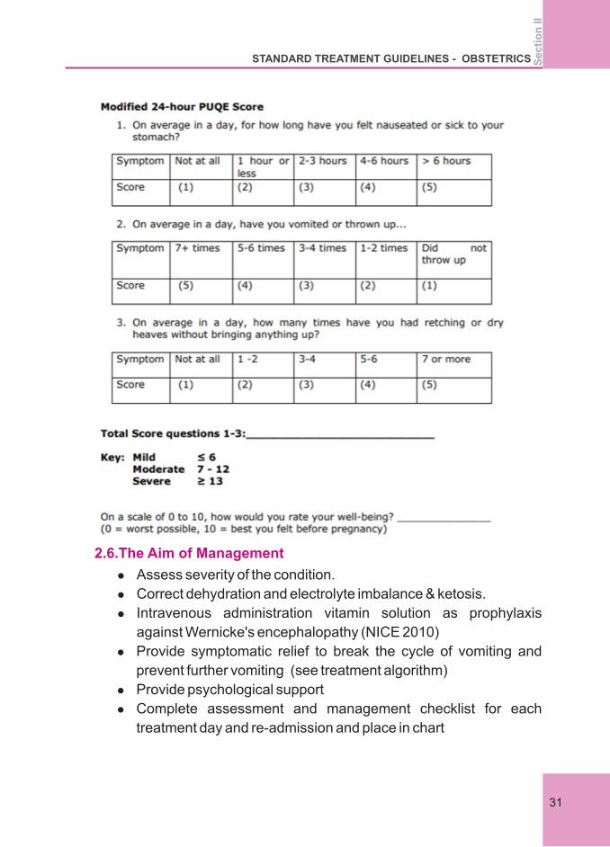
l Assess severity of the condition.
l Correct dehydration and electrolyte imbalance & ketosis.
l Intravenous administration vitamin solution as prophylaxis
against Wernicke's encephalopathy (NICE 2010)
l Provide symptomatic relief to break the cycle of vomiting and
prevent further vomiting (see treatment algorithm)
l Provide psychological support
l Complete assessment and management checklist for each
treatment day and re-admission and place in chart
2.6.The Aim of Management
Se
cti
on
II
STANDARD TREATMENT GUIDELINES - OBSTETRICS
31
Inpatient management should be considered if there is at least one of
the following:
1. Continued nausea and vomiting and inability to keep down
oral antiemetics
2. Continued nausea and vomiting associated with ketonuria
and/or weight loss (greater than 5% of body weight) despite
oral antiemetics
3. Confirmed or suspected comorbidity (such as urinary tract
infection and inability to tolerate oral antibiotics).
l BLOOD –CBC, Blood urea c Creatinine,S. Electrolytes,LFT,
Blood Glucose
l TFT
l Urine acetone every 12 hrs until negative
l USG to rule out multiple pregnancy & vesicular mole.
l SEVERE CASES- Serum Amylase , lipase, ABG
l IV fluid therapy for ketone positive women, with electrolyte
monitoring
l Keep nil orally &give iv fluids depending on severity of
dehydration.
l Normal saline or RL preferred
l N saline 2pints (each unit over 2hrs – 3 hrs depending on ketosis)
l RL or hartmans solution – 2 pints
l 5% Dextrose must be given with 100 mg of thiamine ( to prevent
Wernicke's encephalopathy)
l First line – promethazine (12.5 – 25 mg iv / im 4hrly)
l Second line - Metclopromide 10 mg / odansetron 4mg Q8H
l Third line- Serotonin Receptor Antagonist(4-6 mg Q6-8H
PO,8mg 15 minutes 12 HRLY)
l If one group fails a combination can be used
l If evidence of gastro esophageal reflux or gastritis -proton
pump inhibitors or Methyl prednisolone may be used as last
2.7. Investigations
2.8. Management:
Anti emetics
Se
cti
on
II
STANDARD TREATMENT GUIDELINES - OBSTETRICS
32
resort.
l Thromboprophylaxis with heparin till patient is recovered
l Consultation with Endocrinologist , Gastro enterologist &
Psychiatrist may be needed.
l Termination of pregnancy in exceptional situations where all
medical measures fail.
1. Nice guidelines 2016, Green-top Guideline No. 69 June 2016
2. Hyperemesis Education and Research (HER) Foundation
[http://www.helpher.org]
3. National Institute for Health and Care Excellence. Intravenous
fluid therapy in adults in hospital. NICE clinical guideline 174.
[Manchester]: NICE; 2013
4. Gill SK, O'Brien L, Einarson TR, Koren G. The safety of proton
pump inhibitors (PPIs) in pregnancy: a metaanalysis. Am J
Gastroenterol 2009;104:1541–5.
5. Treatments for Hyperemesis Gravidarum and Nausea and
Vomiting in Pregnancy: A Systematic Review. JAMA. 2016 Oct
4;316(13):1392-1401. doi: 10.1001/jama.2016.14337
6. Hyperemesis Gravidarum: A Review of Recent Literature London
V. · Grube S. · Sherer D.M. · Abulafia O.
7. Kjeldgaard HK, Eberhard-Gran M, Benth JŠ, Nordeng H,
Vikanes ÅV: History of depression and risk of hyperemesis
gravidarum: a population-based cohort study. Arch Womens
Ment Health 2017;20:397-404
8. Novak 2002; Godfrey 2000; Barker 1998
9. Briggs G, Freeman RK. (2015) Drugs in pregnancy and lactation:
a reference guide to fetal and neonatal risk. 10th ed: Lippincott
Williams & Wilkins.
10. Pasternak B, Svanström H, Hviid A. (2013) 'Ondansetron in
Pregnancy and Risk of Adverse Fetal Outcomes'. New England
Journal of Medicine 368(9), 814-23.
Resistant cases-
Reference:
Se
cti
on
II
STANDARD TREATMENT GUIDELINES - OBSTETRICS
33

Section III
ANTIBIOTIC POLICY IN OBSTETRICS

SECTION 3
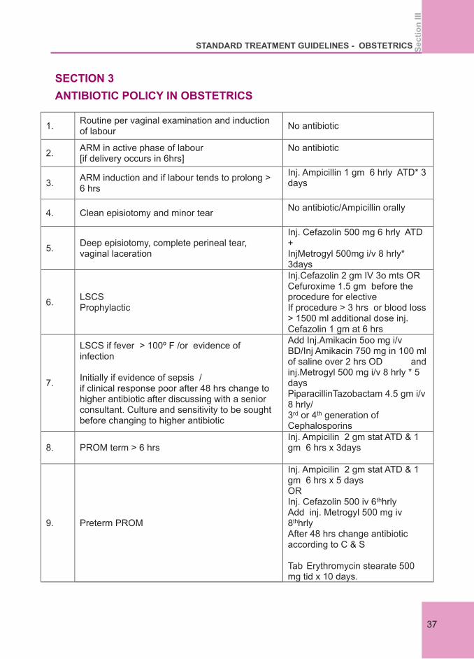
ANTIBIOTIC POLICY IN OBSTETRICS
1. Routine per vaginal examination and induction of labour
No antibiotic
2. ARM in active phase of labour [if delivery occurs in 6hrs]
No antibiotic
3. ARM induction and if labour tends to prolong > 6 hrs
Inj. Ampicillin 1 gm 6 hrly ATD* 3 days
4. Clean episiotomy and minor tear No antibiotic/Ampicillin orally
5. Deep episiotomy, complete perineal tear, vaginal laceration
Inj. Cefazolin 500 mg 6 hrly ATD + InjMetrogyl 500mg i/v 8 hrly* 3days
6. LSCS Prophylactic
Inj.Cefazolin 2 gm IV 3o mts OR Cefuroxime 1.5 gm before the procedure for elective If procedure > 3 hrs or blood loss > 1500 ml additional dose inj. Cefazolin 1 gm at 6 hrs
7.
LSCS if fever > 100º F /or evidence of infection Initially if evidence of sepsis / if clinical response poor after 48 hrs change to higher antibiotic after discussing with a senior consultant. Culture and sensitivity to be sought before changing to higher antibiotic
Add Inj.Amikacin 5oo mg i/v BD/Inj Amikacin 750 mg in 100 ml of saline over 2 hrs OD and inj.Metrogyl 500 mg i/v 8 hrly * 5 days PiparacillinTazobactam 4.5 gm i/v 8 hrly/ 3rd or 4th generation of Cephalosporins
8. PROM term > 6 hrs Inj. Ampicilin 2 gm stat ATD & 1 gm 6 hrs x 3days
9. Preterm PROM
Inj. Ampicilin 2 gm stat ATD & 1 gm 6 hrs x 5 days OR Inj. Cefazolin 500 iv 6thhrly Add inj. Metrogyl 500 mg iv 8thhrly After 48 hrs change antibiotic according to C & S Tab Erythromycin stearate 500 mg tid x 10 days.
Se
cti
on
III
STANDARD TREATMENT GUIDELINES - OBSTETRICS
37
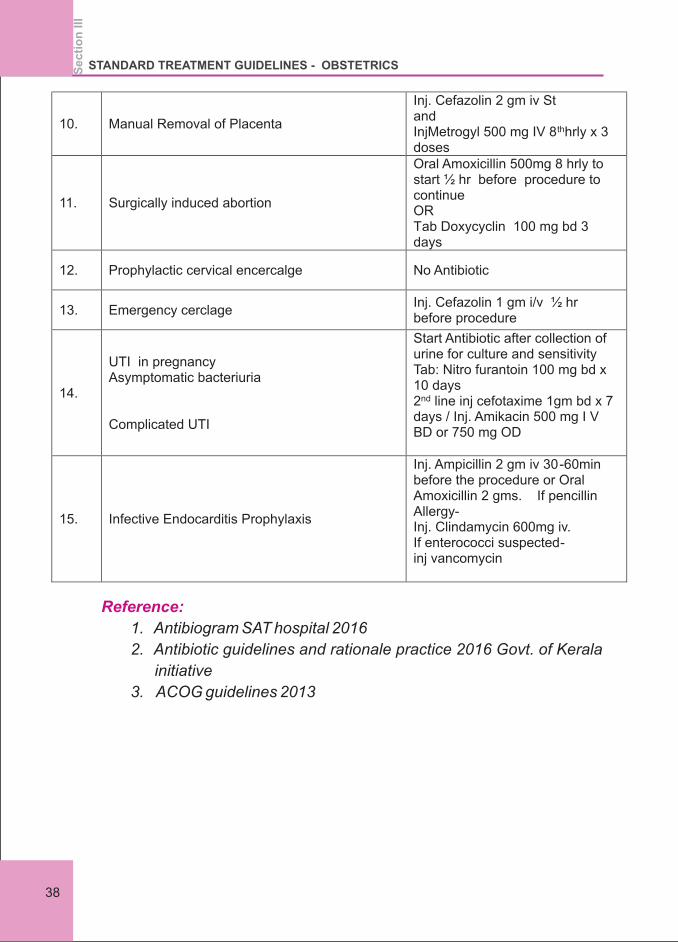
10. Manual Removal of Placenta
Inj. Cefazolin 2 gm iv St and InjMetrogyl 500 mg IV 8thhrly x 3 doses
11. Surgically induced abortion
Oral Amoxicillin 500mg 8 hrly to start ½ hr before procedure to continue OR Tab Doxycyclin 100 mg bd 3 days
12. Prophylactic cervical encercalge No Antibiotic
13. Emergency cerclage Inj. Cefazolin 1 gm i/v ½ hr before procedure
14.
UTI in pregnancy Asymptomatic bacteriuria Complicated UTI
Start Antibiotic after collection of urine for culture and sensitivity Tab: Nitro furantoin 100 mg bd x 10 days 2nd line inj cefotaxime 1gm bd x 7 days / Inj. Amikacin 500 mg I V BD or 750 mg OD
15. Infective Endocarditis Prophylaxis
Inj. Ampicillin 2 gm iv 30-60min before the procedure or Oral Amoxicillin 2 gms. If pencillin Allergy- Inj. Clindamycin 600mg iv. If enterococci suspected- inj vancomycin
Reference:
1. Antibiogram SAT hospital 2016
2. Antibiotic guidelines and rationale practice 2016 Govt. of Kerala
initiative
3. ACOG guidelines 2013
Se
cti
on
III
STANDARD TREATMENT GUIDELINES - OBSTETRICS
38
Section IV
ANEMIA IN PREGNANCY

SECTION 4
ANEMIA IN PREGNANCY
4.1.introduction:
4.2.definition:
u According to WHO, world wide 32.4 million pregnant women
suffer from anemia.
u Defined as Hb < 2SD below the median value for a healthy
age matched population
u Hb< 11 g% or Hct < 33% (WHO in all trimesters)
u 50% cases – Iron deficiency anemia (Most Common cause)
u Iron deficiency anemia during pregnancy increases the risk of
Low Birth Weight, preterm birth, maternal and perinatal mortality
and poor APGAR score.
u India has highest prevalence of anemia( 57-96.2%) among
South Asian countries.
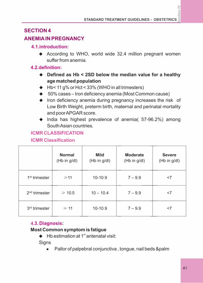
ICMR CLASSIFICATION
ICMR Classification
Normal
(Hb in g/dl)
Mild
(Hb in g/dl)
Moderate
(Hb in g/dl)
Severe
(Hb in g/dl)
1st trimester ≥11 10-10.9 7 – 9.9 <7
2nd trimester ≥ 10.5 10 – 10.4 7 – 9.9 <7
3rd trimester ≥ 11 10-10.9 7 – 9.9 <7
4.3. Diagnosis:
Most Common symptom is fatiguest
u Hb estimation at 1 antenatal visit:
Signs
l Pallor of palpebral conjunctiva , tongue, nail beds &palm
Se
cti
on
IV
STANDARD TREATMENT GUIDELINES - OBSTETRICS
41
l Alopecia
l Atrophy of lingual papillae
l Stomatitis
l Koilonychia
l Restless leg syndrome
l Pagophagia & pica
l Hepatomegaly
l Splenomegaly
l Edema
l Look for CCF – tachycardia, raised JVP and fine crepitations
in lung base
Family history – anemia, thalassemia, and sickle cell anemia
Suspect sickle cell anemia in tribal and certain geographical
areas
u Hb, Complete Blood Count, Mean Corpuscular Volume, Mean
Corpuscular Hemoglobin, Mean Corpuscular Hemoglobin
Concentratiom, RDW (red cell with)
u Peripheral smear-microcytic hypochromic
u Serum ferritin-<15mcg/dl –iron depletion
<30mcg/dl –early iron depletion – initiate treatment
More specific: S. Transferrin Receptor estimation for early iron
deficiency anemia. Its level rises even before S. ferritin starts to fall.
u Urine routine
u TFT – Thyroid function test
u RFT – Renal function test, urine culture and sensitivity
u Hemoglobin electrophoresis
u Stool routine and microscopy
u Ultrasonography of abdomen
u Bone Marrow study in indicated cases
u Nutritional supplementation/cooking in iron utensils
u Iron rich foods
l Jaggery, Beetroot, Green Leafy vegetable, Pulses, Cereals,
nuts, meat, liver, poultry, egg, legumes, dry beans and dry
4.4. Investigations
4.5.management:
Se
cti
on
VI
STANDARD TREATMENT GUIDELINES - OBSTETRICS
42
fruits.
l Iron and calcium should not be taken together
l Along with iron ensure adequate protein, B and folic acid 12
and haemoglobin synthesis.
l Avoid milk and milk products along with iron intake
u Trial of iron therapy- if doesn't respond within 2 weeks – detailed
investigation
u ORAL IRON THERAPY
Iron preparations available: Ferrous sulphate , ferrous fumarate,
ferrous ascorbate, ferric ammonium citrate,.
200mg Feso4 tablet contains 60mg elemental Fe.
Response to oral Iron:
1. Reticulocyte count increases by 0.2% per day in 5-7 days.
2. Hb increases by 0.8-1g/ wk in 2-3 wks
3. By 6-8 weeks- Hb comes back to normal.
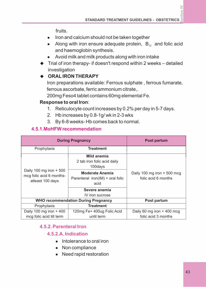
4.5.1.MoHFW recommendation
During Pregnancy
Post partum
Prophylaxis
Treatment
Daily 100 mg iron + 500
mcg folic acid 6 months- atleast 100 days
Mild anemia 2 tab iron folic acid daily
100days
Daily 100 mg iron + 500 mcg
folic acid 6 months
Moderate Anemia
Parenteral iron(IM) + oral folic
acid
Severe anemia IV iron sucrose
WHO recommendation During Pregnancy
Post partum
Prophylaxis
Treatment
Daily 100 mg iron + 400
mcg folic acid till term
120mg Fe+ 400ug Folic Acid
until term
Daily 60 mg iron + 400 mcg
folic acid 3 months
4.5.2. Parenteral Iron
4.5.2.A. Indication
l Intolerance to oral iron
l Non compliance
l Need rapid restoration
Se
cti
on
IV
STANDARD TREATMENT GUIDELINES - OBSTETRICS
43
l Suspected malabsorption
l History of anaphylactic reaction to parenteral iron therapy
l 1st trimester of pregnancy, chronic liver disease &active
infection
l Oral iron should be stopped at least 24 hrs prior to therapy
4.5.2.B.Contraindication
4.5.2.C.Preparations Of Parenteral Iron:
4.5.2.D.Recombinant human erythropoietin
4.5.3.Indications for packed cell transfusion in pregnancy
(RCOG)
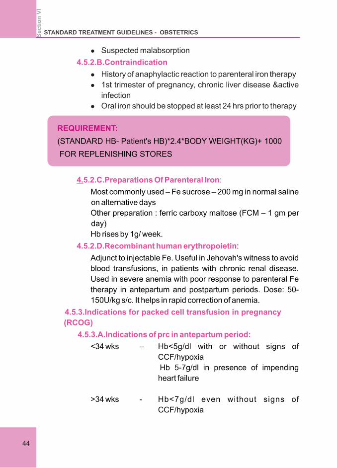
4.5.3.A.Indications of prc in antepartum period:
Most commonly used – Fe sucrose – 200 mg in normal saline
on alternative days
Other preparation : ferric carboxy maltose (FCM – 1 gm per
day)
Hb rises by 1g/ week.
:
Adjunct to injectable Fe. Useful in Jehovah's witness to avoid
blood transfusions, in patients with chronic renal disease.
Used in severe anemia with poor response to parenteral Fe
therapy in antepartum and postpartum periods. Dose: 50-
150U/kg s/c. It helps in rapid correction of anemia.
<34 wks – Hb<5g/dl with or without signs of
CCF/hypoxia
Hb 5-7g/dl in presence of impending
heart failure
>34 wks - Hb<7g/dl even without signs of
CCF/hypoxia
REQUIREMENT:
(STANDARD HB- Patient's HB)*2.4*BODY WEIGHT(KG)+ 1000
FOR REPLENISHING STORES
Se
cti
on
VI
STANDARD TREATMENT GUIDELINES - OBSTETRICS
44
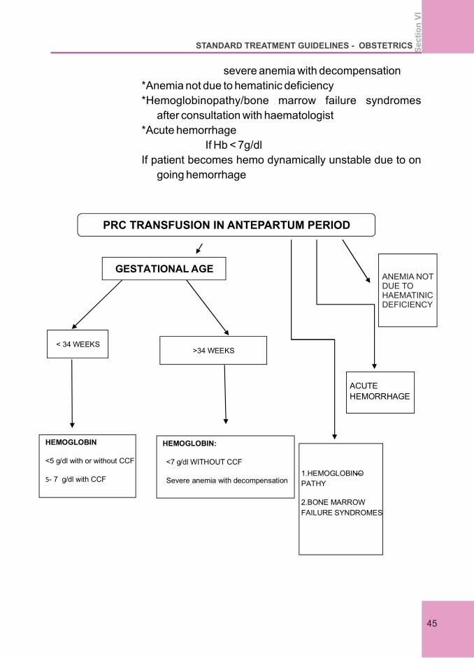
severe anemia with decompensation
*Anemia not due to hematinic deficiency
*Hemoglobinopathy/bone marrow failure syndromes
after consultation with haematologist
*Acute hemorrhage
If Hb < 7g/dl
If patient becomes hemo dynamically unstable due to on
going hemorrhage
< 34 WEEKS
>34 WEEKS
HEMOGLOBIN-
<5 g/dl with or without CCF
5- 7 g/dl with CCF
HEMOGLOBIN:
<7 g/dl WITHOUT CCF
Severe anemia with decompensation
ACUTE
HEMORRHAGE
1.HEMOGLOBINO---
PATHY
2.BONE MARROW
FAILURE SYNDROMES
PRC TRANSFUSION IN ANTEPARTUM PERIOD
GESTATIONAL AGEANEMIA NOTDUE TO HAEMATINIC DEFICIENCY
Se
cti
on
VI
STANDARD TREATMENT GUIDELINES - OBSTETRICS
45
4.5.3.B.Indications of prc in intrapartum period
4.5.3.C.Indications of prc in postpartum period
Hb<7g/dl (in labour)
Decision for blood transfusion depends on medical history
or symptoms
Anaemia with signs of shock/acute hemorrhage with signs of
hemodynamic instability
Hb <7 g/dl (postpartum) –decision of blood transfusion
depends on medical history or symptoms
1. Adequate cross matched blood
2. Administration of oxygen to decrease fetal hypoxia
3. Asepsis to be maintained strictly to avoid puerperal sepsis
4. Cut short second stage using forceps or vacuum
5. Active management of third stage of labour
6. Prompt management of PPH
l Continue oral Fe for atleast 6 Months
l Advice spacing and contraception
l Close monitoring for sepsis, DVT, cardiac failure
l Thrombo prophylaxis for moderate and severe anemia
l As an additional intervention to reduce anemiast
l Single dose 400mg Albendazole after 1 trimester preferably ndin 2 trimester
l Chloroquine for Pl.vivaxst nd rd
l Quinine for Pl.falciparum during 1 trimester &ACT in 2 &3
trimester.
An existing Cochrane systematic review assessing the benefits and
harms of iron compared the daily provision of iron supplements alone
or in combination with folic acid or other micronutrients with no
intervention, placebo or versus the use of the same supplements but
4.6. Intrapartum Management Of Severe Anemia
4.7.Puerperium
4.8. Deworming
4.9.Treatment of malaria
4.10.Treatment outcomes- WHO (2)
Se
cti
on
IV
STANDARD TREATMENT GUIDELINES - OBSTETRICS
46
without iron (e.g. only folic acid) among pregnant women living in a
variety of settings, including malaria-endemic areas.
Overall, women taking daily iron supplements were less likely to
have low birth weight babies compared with controls (average
relative risk (RR) 0.81) and the mean birth weight was 30.81g greater
for those infants whose mothers received iron during pregnancy.
There was no significant effect on preterm birth or neonatal death.
Daily iron supplementation reduced the risk of maternal anaemia at
term by 70% and iron deficiency at term by 57%, it had no significant
effect on the risk of infections during pregnancy.
Women receiving iron had 8.88 g more haemoglobin per litre at or
near term than those who did not receive iron. At the same time,
women who received iron supplements tended to report more
frequently side-effects and were at increased risk of high
haemoglobin concentrations (i.e. greater than 13.0 mg/L) during the
second and third trimesters of pregnancy.
1. FOGSI General Clinical Practice Recommendations
Management of Iron Deficiency Anemia in Pregnancy May 2016.
2. WHO Guideline: Daily iron and folic acid supplementation in
pregnant women, Geneva, World Health Organization, 2012.
3. WHO, The Global Prevalence of Anemia in 2011. Geneva: World
Health Organisation; 2015.
4. Treatment of Anemia in pregnancy - Indian Guidelines/speciality
medical dialogues.
5. Blood transfusion in Obstetrics, Green top guideline No. 47.
RCOG 2015.th
6. James, Steer, High Risk Pregnancy, Volume 1, 5 edition, South
Asian 2018.
7. ACOG Practice Bulletin No. 95, Anemia in Pregnancy, July 2008.
1. MoHFW- Ministry of health and Family welfare
2. WHO- World Health Organization
References
Abbreviatons
Se
cti
on
IV
STANDARD TREATMENT GUIDELINES - OBSTETRICS
47

DIABETES IN PREGNANCY
Section V

SECTION 5
DIABETES IN PREGNANCY
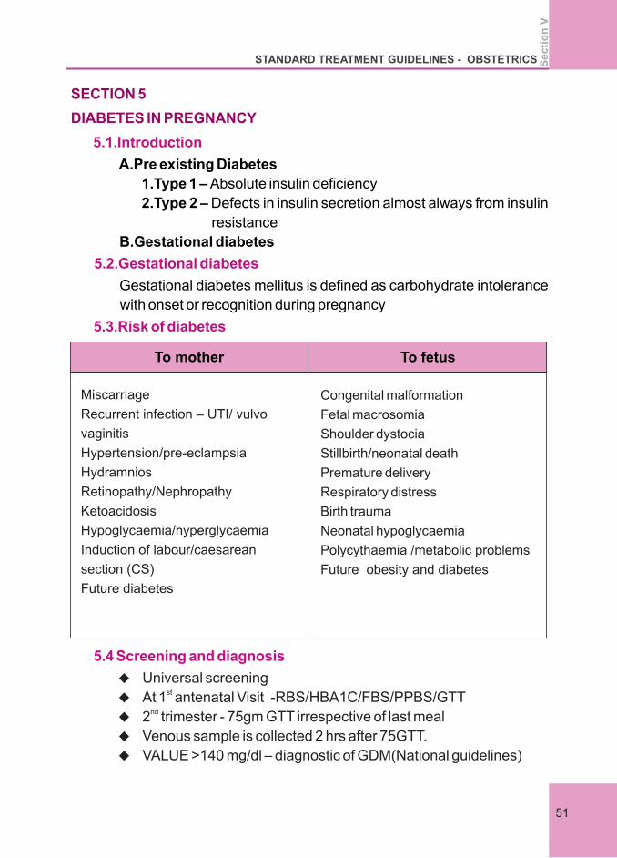
5.1.Introduction
5.2.Gestational diabetes
5.3.Risk of diabetes
A.Pre existing Diabetes
1.Type 1 – Absolute insulin deficiency
2.Type 2 – Defects in insulin secretion almost always from insulin
resistance
B.Gestational diabetes
Gestational diabetes mellitus is defined as carbohydrate intolerance
with onset or recognition during pregnancy
To mother To fetus
Miscarriage
Recurrent infection – UTI/ vulvo
vaginitis
Hypertension/pre-eclampsia
Hydramnios
Retinopathy/Nephropathy
Ketoacidosis
Hypoglycaemia/hyperglycaemia
Induction of labour/caesarean
section (CS)
Future diabetes
Congenital malformation
Fetal macrosomia
Shoulder dystocia
Stillbirth/neonatal death
Premature delivery
Respiratory distress
Birth trauma
Neonatal hypoglycaemia
Polycythaemia /metabolic problems
Future obesity and diabetes
5.4 Screening and diagnosis
u Universal screeningst
u At 1 antenatal Visit -RBS/HBA1C/FBS/PPBS/GTTnd
u 2 trimester - 75gm GTT irrespective of last meal
u Venous sample is collected 2 hrs after 75GTT.
u VALUE >140 mg/dl – diagnostic of GDM(National guidelines)
Se
cti
on
V
STANDARD TREATMENT GUIDELINES - OBSTETRICS
51
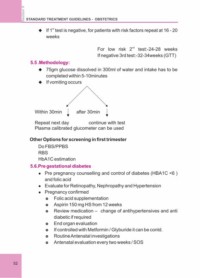
stu If 1 test is negative, for patients with risk factors repeat at 16 - 20
weeks
nd
For low risk 2 test:-24-28 weeks
If negative 3rd test:-32-34weeks (GTT)
u 75gm glucose dissolved in 300ml of water and intake has to be
completed within 5-10minutes
u If vomiting occurs
Other Options for screening in first trimester
Do FBS/PPBS
RBS
HbA1C estimation
l Pre pregnancy counselling and control of diabetes (HBA1C <6 )
and folic acid
l Evaluate for Retinopathy, Nephropathy and Hypertension
l Pregnancy confirmed
£ Folic acid supplementation
£ Aspirin 150 mg HS from 12 weeks
£ Review medication – change of antihypertensives and anti
diabetic if required
£ End organ evaluation
£ If controlled with Metformin / Glyburide it can be contd.
£ Routine Antenatal investigations
£ Antenatal evaluation every two weeks / SOS
5.5 .Methodology:
5.6.Pre gestational diabetes
Within 30min after 30min
Repeat next day continue with test Plasma calibrated glucometer can be used
Se
cti
on
VSTANDARD TREATMENT GUIDELINES - OBSTETRICS
52
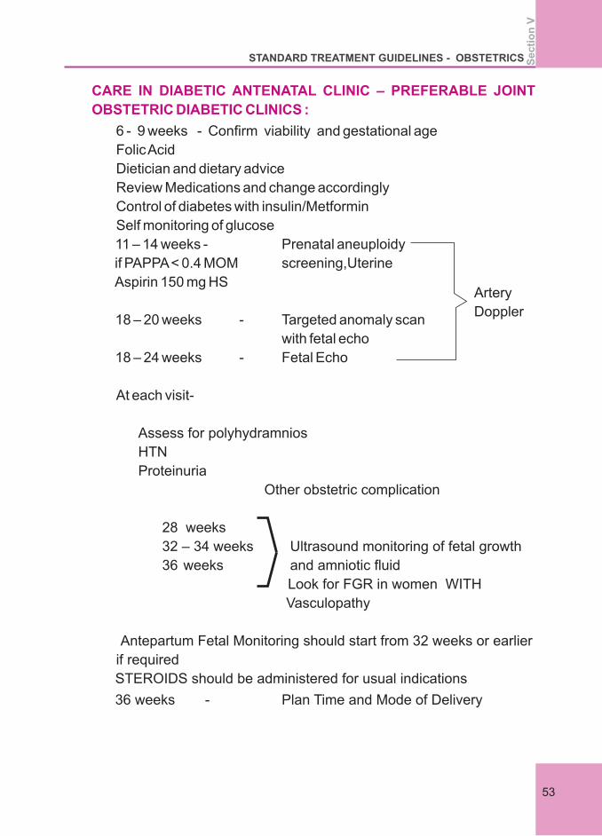
CARE IN DIABETIC ANTENATAL CLINIC – PREFERABLE JOINT
OBSTETRIC DIABETIC CLINICS :
6 - 9 weeks - Confirm viability and gestational age
Folic Acid
Dietician and dietary advice
Review Medications and change accordingly
Control of diabetes with insulin/Metformin
Self monitoring of glucose
11 – 14 weeks - Prenatal aneuploidy
if PAPPA < 0.4 MOM screening,Uterine
Aspirin 150 mg HS
18 – 20 weeks - Targeted anomaly scan
with fetal echo
18 – 24 weeks - Fetal Echo
At each visit-
Assess for polyhydramnios
HTN
Proteinuria
Other obstetric complication
28 weeks
32 – 34 weeks Ultrasound monitoring of fetal growth
36 weeks and amniotic fluid
Look for FGR in women WITH
Vasculopathy
Antepartum Fetal Monitoring should start from 32 weeks or earlier
if required
STEROIDS should be administered for usual indications
36 weeks - Plan Time and Mode of Delivery
Artery
Doppler
Se
cti
on
V
STANDARD TREATMENT GUIDELINES - OBSTETRICS
53

5.7 Timing of delivery
5.8 management:
l Pre gestational diabetes well controlled no complications by 38
weeks
l Pre gestational diabetes with complication time accordingly
l Gestational diabetes on insulin well controlled without
complication by 38 weeks.
l Gestational on MNT 39 – 40 weeks
Pregnant women with GDM
Medical nutritional therapy and physical exercise
Target blood glucose level in mg/dL
FBS 95
1 hour PPBS 140
2 hour PPBS 120
Sedentary work 2250kcal/day
Moderate work 2580kcal/day
Heavy work 3200kcal/day
Underweight <18.5 40kcal/kg
Normal weight 18.5-22.9 30kcal/kg
Overweight 23-24.9 25kcal/kg
Obese >25 12.5kcal/kg
MNT:Carbohydrate controlled balanced meal planIndividualization is importantAdjustment should be made based on the BMI and weight gain pattern
5.9. Energy requirement during pregnancy
5.9.1. Energy requirement according to BMI in pregnancy with diabetes
Se
cti
on
VSTANDARD TREATMENT GUIDELINES - OBSTETRICS
54
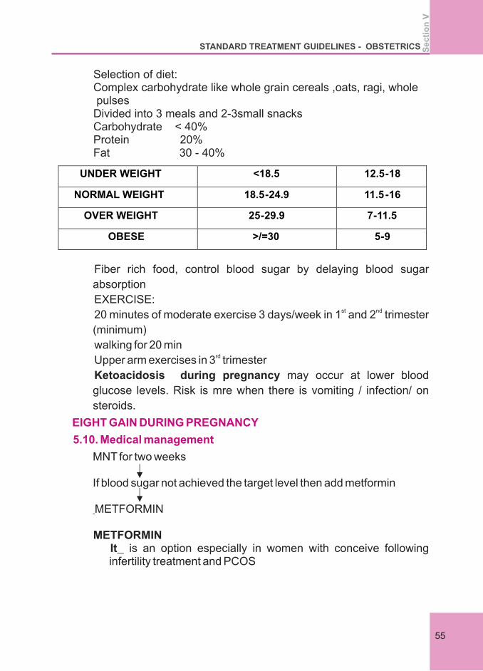
Selection of diet:Complex carbohydrate like whole grain cereals ,oats, ragi, whole pulsesDivided into 3 meals and 2-3small snacksCarbohydrate < 40%Protein 20%Fat 30 - 40%
UNDER WEIGHT <18.5 12.5-18
NORMAL WEIGHT 18.5-24.9 11.5-16
OVER WEIGHT 25-29.9 7-11.5
OBESE >/=30 5-9
Fiber rich food, control blood sugar by delaying blood sugar
absorption
EXERCISE:st nd
20 minutes of moderate exercise 3 days/week in 1 and 2 trimester
(minimum)
walking for 20 minrd
Upper arm exercises in 3 trimester
Ketoacidosis during pregnancy may occur at lower blood
glucose levels. Risk is mre when there is vomiting / infection/ on
steroids.
MNT for two weeks
If blood sugar not achieved the target level then add metformin
METFORMIN
METFORMINIt is an option especially in women with conceive following infertility treatment and PCOS
EIGHT GAIN DURING PREGNANCY
5.10. Medical management
Se
cti
on
V
STANDARD TREATMENT GUIDELINES - OBSTETRICS
55
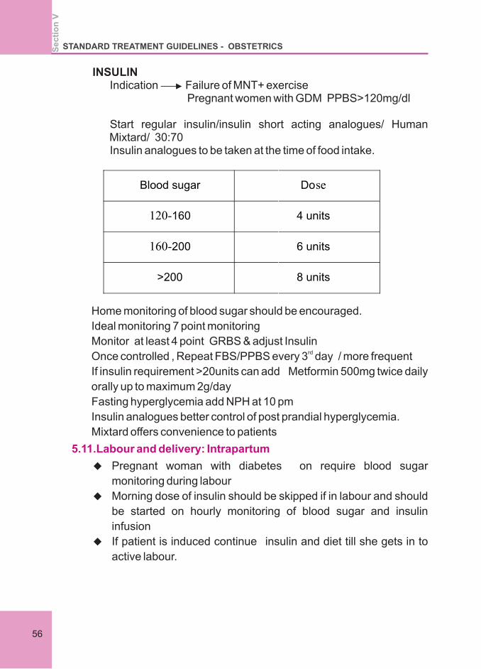
INSULINIndication Failure of MNT+ exercise Pregnant women with GDM PPBS>120mg/dl
Start regular insulin/insulin short acting analogues/ Human Mixtard/ 30:70 Insulin analogues to be taken at the time of food intake.
Home monitoring of blood sugar should be encouraged.
Ideal monitoring 7 point monitoring
Monitor at least 4 point GRBS & adjust Insulin rd
Once controlled , Repeat FBS/PPBS every 3 day / more frequent
If insulin requirement >20units can add Metformin 500mg twice daily
orally up to maximum 2g/day
Fasting hyperglycemia add NPH at 10 pm
Insulin analogues better control of post prandial hyperglycemia.
Mixtard offers convenience to patients
u Pregnant woman with diabetes on require blood sugar
monitoring during labour
u Morning dose of insulin should be skipped if in labour and should
be started on hourly monitoring of blood sugar and insulin
infusion
u If patient is induced continue insulin and diet till she gets in to
active labour.
5.11.Labour and delivery: Intrapartum
Blood sugar Dose
120-160 4 units
160-200 6 units
>200 8 units
Se
cti
on
VSTANDARD TREATMENT GUIDELINES - OBSTETRICS
56
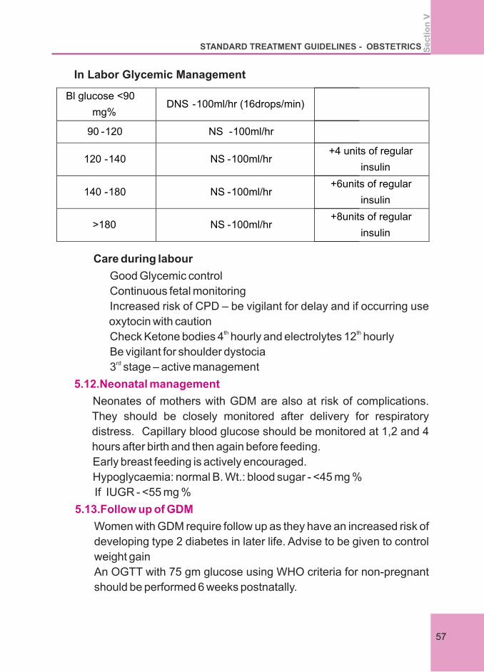
In Labor Glycemic Management
Bl glucose <90
mg% DNS -100ml/hr (16drops/min)
90 -120 NS -100ml/hr
120 -140 NS -100ml/hr +4 units of regular
insulin
140 -180 NS -100ml/hr +6units of regular
insulin
>180 NS -100ml/hr +8units of regular
insulin
Care during labour
Good Glycemic control
Continuous fetal monitoring
Increased risk of CPD – be vigilant for delay and if occurring use
oxytocin with cautionth thCheck Ketone bodies 4 hourly and electrolytes 12 hourly
Be vigilant for shoulder dystocia rd
3 stage – active management
Neonates of mothers with GDM are also at risk of complications.
They should be closely monitored after delivery for respiratory
distress. Capillary blood glucose should be monitored at 1,2 and 4
hours after birth and then again before feeding.
Early breast feeding is actively encouraged.
Hypoglycaemia: normal B. Wt.: blood sugar - <45 mg %
If IUGR - <55 mg %
Women with GDM require follow up as they have an increased risk of
developing type 2 diabetes in later life. Advise to be given to control
weight gain
An OGTT with 75 gm glucose using WHO criteria for non-pregnant
should be performed 6 weeks postnatally.
5.12.Neonatal management
5.13.Follow up of GDM
Se
cti
on
V
STANDARD TREATMENT GUIDELINES - OBSTETRICS
57
If normal, GTT is repeated every year to determine if glucose
intolerance has returned to normal or progressed
They should be made aware of the symptoms of hyperglycaemia and
advice given regarding the importance of healthy eating and exercise
patterns
Contraception to be advised
Low dose COC –to be avoided in hypertension, coronary artery
disease
Progesterone only pill may avoided in case of women with h/o GDM.
1.Tight glycemic control should be advised maintaining HBA1C - <6.5
2.In Type 1Diabetes – must conceive after attaining Target HBA1C
and After completion of CARDIO,NEPHRO,OPHTHAL Evaluation.
5.14.Prepregnancy counselling.
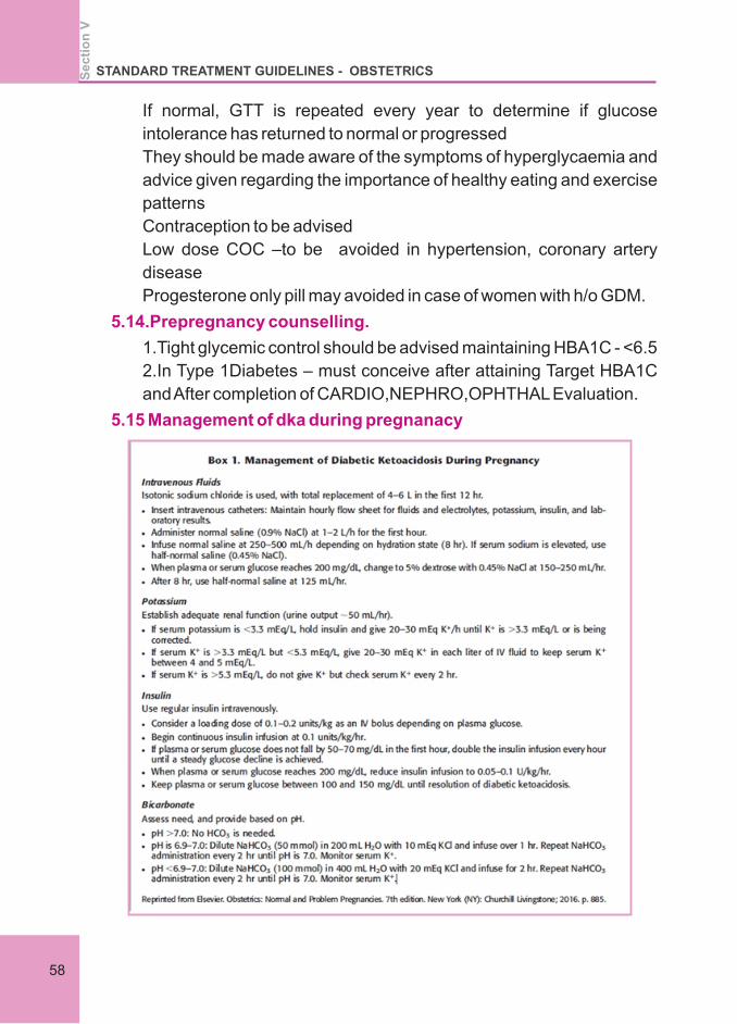
5.15 Management of dka during pregnanacy
Se
cti
on
VSTANDARD TREATMENT GUIDELINES - OBSTETRICS
58
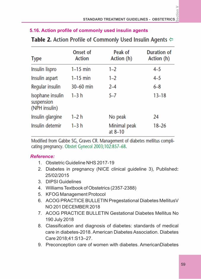
5.16. Action profile of commonly used insulin agents
1. Obstetric Guideline NHS 2017-19
2. Diabetes in pregnancy (NICE clinical guideline 3), Published:
25/02/2015
3. DIPSI Guidelines
4. Williams Textbook of Obstetrics (2357-2388)
5. KFOG Management Protocol
6. ACOG PRACTICE BULLETIN Pregestational Diabetes MellitusV
NO 201 DECEMBER 2018
7. ACOG PRACTICE BULLETIN Gestational Diabetes Mellitus No
190 July 2018
8. Classification and diagnosis of diabetes: standards of medical
care in diabetes-2018. American Diabetes Association. Diabetes
Care 2018;41:S13–27.
9. Preconception care of women with diabetes. AmericanDiabetes
Reference:
Se
cti
on
V
STANDARD TREATMENT GUIDELINES - OBSTETRICS
59

Association. Diabetes Care 2004;27(suppl 1): S76–8. (2004A)
10. Gabbe SG, Graves CR. Management of diabetes mellitus
complicating pregnancy. Obstet Gynecol 2003;102:857–68.
11. WHO guideline 2016
Se
cti
on
VSTANDARD TREATMENT GUIDELINES - OBSTETRICS
60
PRETERM PRELABOUR
RUPTURE OF MEMBRANES
Section VI

SECTION 6
PRETERM PRELABOUR RUPTURE OF MEMBRANES (PPROM)
6.1.Incidence
6.2.Definition
6.3.Complications
6.4.Management – PPROM
l Rupture of membranes before the onset of uterine contractions –
10%
l PPROM – PROM before 37 weeks – 2-3%
l PRE VIABLE PPROM – Before 26 weeks – 0.3% - 0.7%
l Chorioamnionitis
l Placental abruption
l Retained placenta
l Puerperal sepsis
l Increased chances of operative vaginal delivery
l Fetal infection
l Cord prolapse
l Prematurity
l RDS
l NEC
l IVH
l Pulmonary hypoplasia
l Long term sequelae, PVL, CP, Hearing and visual defects
l Initial Assessment – Detailed history and examination
l Confirmation of Diagnosis
l Management – decisions based on Maternal and fetal
parameters and gestational age
l ADMISSION
l Detailed history
– Gestational Age
6.3.1.Maternal
6.3.2. Fetal
6.4.1.Initial Assessment
Se
cti
on
VI
STANDARD TREATMENT GUIDELINES - OBSTETRICS
63
– Gush of fluid/Watery discharge from vagina
– Duration
– Associated bleeding, pain, decreased fetal movement
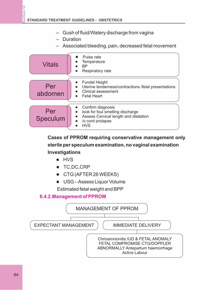
Cases of PPROM requiring conservative management only
sterile per speculum examination, no vaginal examination
Investigations
l HVS
l TC,DC,CRP
l CTG (AFTER 26 WEEKS)
l USG – Assess Liquor Volume
Estimated fetal weight and BPP
6.4.2.Management of PPROM
Vitals
Per abdomen
Per Speculum
Pulse rate Temperature BP Respiratory rate
Fundal Height Uterine tenderness/contractions /fetal presentations Clinical assessment Fetal Heart
Confirm diagnosis look for foul smelling discharge Assess Cervical length and dilatation /o cord prolapse HVS
MANAGEMENT OF PPROM
EXPECTANT MANAGEMENT IMMEDIATE DELIVERY
Chrioamnionitis IUD & FETAL ANOMALYFETAL COMPROMISE CTG/DOPPLER
ABNORMALLY Antepartum haemorrhageActive Labour
Se
cti
on
VI
STANDARD TREATMENT GUIDELINES - OBSTETRICS
64
6.4.2.a.Expectant Management
6.4.2.b.Antibiotics
22-24 Weeks – Individualize the case, termination may be
preferred in view of high risk of infection
– Steroids (from 24 weeks)
– Antibiotics, Magnesium sulphate for neuro protection
– Tocolysis – Not Recommended
Counsel the couple , explain the long term complication
and take appropriate decision.
24-28 Weeks
– Counselling – Obstetrician and neonatologist
– Steroids
– Antibiotics
– Tocolysis for latency for steroid to act up till 48 hours
– Magnesium sulphate for neuro protection
28-34 Weeks
Antibiotics
l Steroids
l Tocolysis- for obtaining latent period for steroids to act
l Magnesium sulphate for neuro protection
34-36 weeks
l give time for steroids to act and then delivery
l Antibiotics
l No magnesium sulphate
Steroids
l Advantages
l Decreases the incidence of RDS
l Reduces mortality by 50%
l Decreases IVH, NEC in first 48 hours of life
Options
l Inj. BETAMETHASONE IM 12 mg, inj. 2 Doses 24 hour
apart
l Inj Dexamethasone 6 mg I.M. 4 doses 12 hour apart
l Caution : - blood sugar may shoot up
l Erythromyin 250 mg qid orally for 10 days for a maximum
Se
cti
on
VI
STANDARD TREATMENT GUIDELINES - OBSTETRICS
65
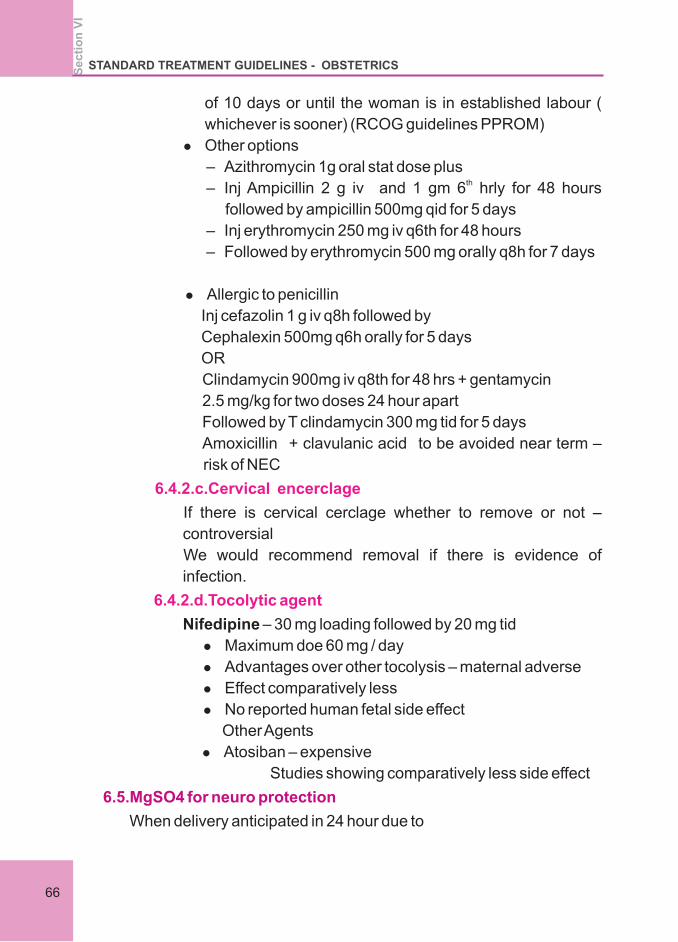
of 10 days or until the woman is in established labour (
whichever is sooner) (RCOG guidelines PPROM)
l Other options
– Azithromycin 1g oral stat dose plusth
– Inj Ampicillin 2 g iv and 1 gm 6 hrly for 48 hours
followed by ampicillin 500mg qid for 5 days
– Inj erythromycin 250 mg iv q6th for 48 hours
– Followed by erythromycin 500 mg orally q8h for 7 days
l Allergic to penicillin
Inj cefazolin 1 g iv q8h followed by
Cephalexin 500mg q6h orally for 5 days
OR
Clindamycin 900mg iv q8th for 48 hrs + gentamycin
2.5 mg/kg for two doses 24 hour apart
Followed by T clindamycin 300 mg tid for 5 days
Amoxicillin + clavulanic acid to be avoided near term –
risk of NEC
If there is cervical cerclage whether to remove or not –
controversial
We would recommend removal if there is evidence of
infection.
Nifedipine – 30 mg loading followed by 20 mg tid
l Maximum doe 60 mg / day
l Advantages over other tocolysis – maternal adverse
l Effect comparatively less
l No reported human fetal side effect
Other Agents
l Atosiban – expensive
Studies showing comparatively less side effect
When delivery anticipated in 24 hour due to
6.4.2.c.Cervical encerclage
6.4.2.d.Tocolytic agent
6.5.MgSO4 for neuro protection
Se
cti
on
VI
STANDARD TREATMENT GUIDELINES - OBSTETRICS
66
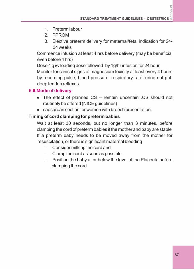
1. Preterm labour
2. PPROM
3. Elective preterm delivery for maternal/fetal indication for 24-
34 weeks
Commence infusion at least 4 hrs before delivery (may be beneficial
even before 4 hrs)
Dose 4 g i/v loading dose followed by 1g/hr infusion for 24 hour.
Monitor for clinical signs of magnesium toxicity at least every 4 hours
by recording pulse, blood pressure, respiratory rate, urine out put,
deep tendon reflexes.
l The effect of planned CS – remain uncertain .CS should not
routinely be offered (NICE guidelines)
l caesarean section for women with breech presentation.
Timing of cord clamping for preterm babies
Wait at least 30 seconds, but no longer than 3 minutes, before
clamping the cord of preterm babies if the mother and baby are stable
If a preterm baby needs to be moved away from the mother for
resuscitation, or there is significant maternal bleeding
– Consider milking the cord and
– Clamp the cord as soon as possible
– Position the baby at or below the level of the Placenta before
clamping the cord
6.6.Mode of delivery
Se
cti
on
VI
STANDARD TREATMENT GUIDELINES - OBSTETRICS
67
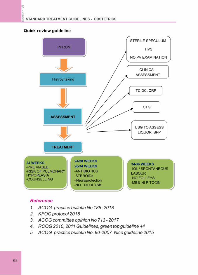
Quick review guideline
PPROM
Histroy taking
ASSESSMENT
STERILE SPECULUM
HVS
NO PV EXAMINATION
CLINICAL
ASSESSMENT
TC,DC, CRP
CTG
USG TO ASSESS
LIQUOR ,BPP
TREATMENT
24 WEEKS
-PRE VIABLE
-RISK OF PULMONARY HYPOPLASIA
-COUNSELLING
24-28 WEEKS
28-34 WEEKS
-ANTIBIOTICS
-STEROIDs
-
Neuroprotection
-NO TOCOLYSIS
34-36 WEEKS
-IOL / SPONTANEOUS
LABOUR
-NO FOLLEYS
-MBS >6 PITOCIN
Reference
1. ACOG practice bulletin No 188 -2018
2. KFOG protocol 2018
3. ACOG committee opinion No 713 - 2017
4. RCOG 2010, 2011 Guidelines, green top guideline 44
5 ACOG practice bulletin No. 80-2007 Nice guideline 2015
Se
cti
on
VI
STANDARD TREATMENT GUIDELINES - OBSTETRICS
68
Section VII
ANTEPARTUM HEMORRHAGE

Section 7
ANTEPARTUM HEMORRHAGE
7.1.Introduction
7.2.Obstetric Haemorrhage
7.3.Clinical assessment
7.4.Examination
Definition – Bleeding from or in to the genital tract from 24 + 0 weeks of pregnancy and prior to the birth baby.
Complicates – 3-5 % of pregnancies
Obstetric haemorrhage is the number one cause of maternal death
About 1/3 of all haemorrhage is APH
APH may be placenta previa or abruption or unclassified
USG will help in the differential diagnosis
In 10% of placenta previa bleeding abruption is also present
Rarely Vasa Previa is the cause of APH, but usually bleeding occurs only in labour
To establish the need for urgent intervention and triage
Consider the following
History taking
Assess haemodynamic stability
Coexisting symptoms – pain
Extent of vaginal bleeding
Fetal well being
If hemodynamically unstable first resuscitate
Remember initial Hb may not reflect blood loss
Abdominal palpation
Tenderness/woody feel/fundal height-utrine contraction
Local Examination/per speculum
Visualise lower genital tract / cervical dilatation/ altered or fresh bleed
CTG
No Vaginal Examination before placental location
Se
cti
on
VII
STANDARD TREATMENT GUIDELINES - OBSTETRICS
71
7.5.Management
7.5.1.If patient is haemo dynamically unstable
Call for help – team / ALERT O T / blood bank
Evaluate – A/B/C – KEEP patient WARM- acidosis – coagulopathy
Facial o2 – 10 – 15 L/min
2 IV cannula – 14/16 G
Take blood for full blood count/RFT/LFT, Bed side clotting test, Clotting screen, Cross match 6 units Blood and products
Commence Fluids – up to 2L of 0.9% NS / RL fluid warmed,
O – ve blood (if condition critical) Cross matched blood as soon as
possible,
Indwelling catheter
Vital Recording – Pulse/BP/CVP/SpO 2/Urine output/ continuous
FHR/ fundal height / blood loss drugs administered (time / type/
dose) have to be documented.
4 -6 hrly checking – Hb, platelet count and coagulation profile
Treatment goals after resuscitation
Rapid restoration of circulating blood volume and O 2 carryin
capacity
HB > 8 gm / dl
Restoration / maintenance of normal acoagulation platelet >
50000, PT and aPTT < 1.5 x mean ontrol and fibrinogen > 200 mg
%
Vitals
Pulse < 100 / min
SBP > 100 mm of HG
Confirmation of diagnosis
USG to diagnose placenta previa and to locate any retro placental
clots
Abruption is a clinical diagnosis no reliable diagnostic tests
available
USG – limited Sensitivity 24% speciality 96%.
Se
cti
on
VII
STANDARD TREATMENT GUIDELINES - OBSTETRICS
72
7.5.2.If it is Placenta previa ?
7.5.3.If it is abruption placentae – revealed/concealed/mixed
Placenta is inserted wholly or in part into the lower segment
Grade 1 : placental edge in lower segment but does not reach the
os (Low lying placenta) MINOR PP.
Grade 2 : Placental edge reaches but does not cover the internal
os (marginal PP) – minor PP
Grade 3 : placenta covers the internal os and is asymmetrically
situated ( partial PP) MAJOR PP
Grade 4 : Placenta overs the internal os and is centrally situated
(Total / central PP) MAJOR PP.
Plan of delivery
l Elective LSCS – type 2 posterior, type 3-4
l Informed consent – should include possibility of PPH and
blood transfusion, hysterectomy if needed.
l Don't allow to go beyond 37 weeks
l CS can be lower segment transverse
l But if lower segment very vascular may consider vertical
incision or underpinning vessles.
l Don't cut though the placenta
l During CS in case of placental sinus bleeding use purse string
stich.
l Amount of blood loss – often underestimated
l Blood from the introitus – may not represent the total blood
loss – concealed abruption
l Management strategy will depend on
l GRADE
l Effect on fetus
l Duration of gestation
GRADES – BLEEDING IS MATERNAL
GRADE 0 : Asymptomatic retro placental clot seen after
placental delivery
Grade 1 : bleeding and tenderness : visible retro placental
clot after delivery – incidence 40%
STANDARD TREATMENT GUIDELINES - OBSTETRICS
73
Se
cti
on
VII
GRADE 2: Revealed bleeding +/- significant maternal signs
foetal compromise / death, incident – 15%
A – without coagulopathy
B – with coagulopathy ( 30% will have DIC)
Grade 1, preterm, self limited bleeding
Confirm with Ultra sound scan may consider conservative
management, start steroids for lung maturation.
Grade 1 term, no effect on foetus, Try for vaginal delivery with
continuous foetal monitoring.
Grade 2 and Grade 3
If in labour and immediate delivery anticipated may consider
vaginal delivery.
All other as sess, even with dead foetus, if immediate delivery
not possible, consider CS.
Tube coagulation test –test for haemostasis
Bed side test
Easy to do and not time consuming
Take 2 test tubes – 1 ml blood in each test tube and after 4
min, start tilting the first tube and let the second stand, after
blood clots in the first tube start titling. Second tube every
minute, note the time blood clots in the second tube – this is
considered the clotting time.
Be vigilant if it crosses six minutes
If it is prolonged signifies – severe deficiency of cogulation
factors
Vaginal Delivery
Favourable cervical status
Amniotomy
Oxytocin augmentation
Continuous CTG
Immediate CS if suspicious CTG – partial abruption can
proceed rapidly to comlete abruption.
AMTSL
Anticipate PPH – ergometrine in absence of hypertension
STANDARD TREATMENT GUIDELINES - OBSTETRICSSe
cti
on
VII
74
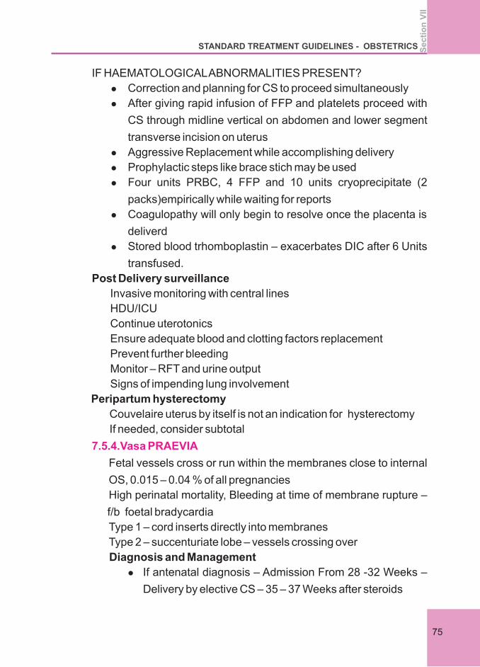
IF HAEMATOLOGICAL ABNORMALITIES PRESENT?
l Correction and planning for CS to proceed simultaneously
l After giving rapid infusion of FFP and platelets proceed with
CS through midline vertical on abdomen and lower segment
transverse incision on uterus
l Aggressive Replacement while accomplishing delivery
l Prophylactic steps like brace stich may be used
l Four units PRBC, 4 FFP and 10 units cryoprecipitate (2
packs)empirically while waiting for reports
l Coagulopathy will only begin to resolve once the placenta is
deliverd
l Stored blood trhomboplastin – exacerbates DIC after 6 Units
transfused.
Post Delivery surveillance
Invasive monitoring with central lines
HDU/ICU
Continue uterotonics
Ensure adequate blood and clotting factors replacement
Prevent further bleeding
Monitor – RFT and urine output
Signs of impending lung involvement
Peripartum hysterectomy
Couvelaire uterus by itself is not an indication for hysterectomy
If needed, consider subtotal
Fetal vessels cross or run within the membranes close to internal
OS, 0.015 – 0.04 % of all pregnancies
High perinatal mortality, Bleeding at time of membrane rupture –
f/b foetal bradycardia
Type 1 – cord inserts directly into membranes
Type 2 – succenturiate lobe – vessels crossing over
Diagnosis and Management
l If antenatal diagnosis – Admission From 28 -32 Weeks –
Delivery by elective CS – 35 – 37 Weeks after steroids
7.5.4.Vasa PRAEVIA
STANDARD TREATMENT GUIDELINES - OBSTETRICS
75
Se
cti
on
VII
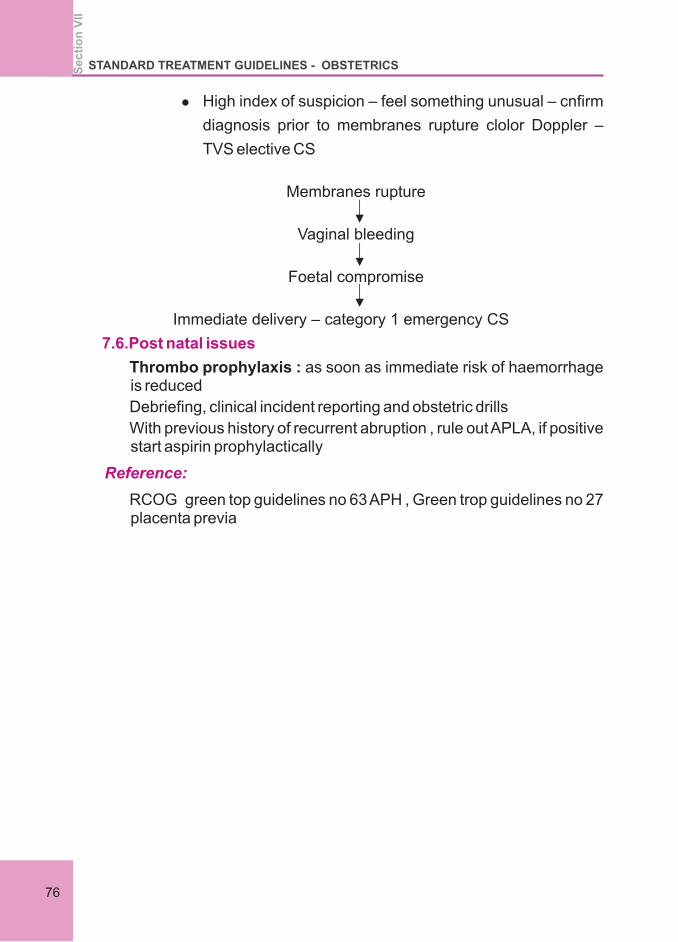
l High index of suspicion – feel something unusual – cnfirm
diagnosis prior to membranes rupture clolor Doppler –
TVS elective CS
Membranes rupture
Vaginal bleeding
Foetal compromise
Immediate delivery – category 1 emergency CS
Thrombo prophylaxis : as soon as immediate risk of haemorrhage is reduced
Debriefing, clinical incident reporting and obstetric drills
With previous history of recurrent abruption , rule out APLA, if positive start aspirin prophylactically
RCOG green top guidelines no 63 APH , Green trop guidelines no 27 placenta previa
7.6.Post natal issues
Reference:
STANDARD TREATMENT GUIDELINES - OBSTETRICSSe
cti
on
VII
76
Section VII
POST PARTUM HAEMORRHAGE

Section 8
POST PARTUM HAEMORRHAGE
8.1.Introduction
l PPH is the most preventable but still the leading cause of
maternal mortality !!PPH is an adverse event, not a diagnosis.
l There is no universally accepted definition for PPH. WHO
defines PPH as blood loss more than 500ml after a vaginal
delivery or more than 1000ml after a CS.
l ACOG defines postpartum hemorrhage as cumulative blood loss
equal to 1000 mL or more along with signs or symptoms of
hypovolemia within 24 hours after delivery (including intrapartum
loss), regardless of route of delivery. (ACOG practice bulletin 183
2017)
l Fallacies of this definition are that visual estimates of blood loss
are notoriously low (often only one third of actual blood loss).
l Even smaller amounts of blood loss is critical in patients with
anemia, dehydration and pre eclampsia. Also in postpartum
patients abdominal or pelvic bleeding can be hidden.
l So any amount of blood loss that produces hemodynamic
instability in the patient is to be considered as PPH.
l Although PPH is prone to happen in high risk patients, it can occur
in any delivering patient irrespective of the risk.
l So anticipation of PPH in every patient and preparation to handle
the situation if a need arises is the first step to prevent maternal
morbidity and mortality due to PPH.
l Major PPH is defined as blood loss more than 1000ml . (UK)
l Massive haemorrhage- no universally accepted definition. , but
blood loss >150ml/mt or >50% blood volume loss in 3 hours.
To reduce deaths due to PPH
1. Prevention
2. Risk factor assessment
3. Early detection
4. Timely intervention
Se
cti
on
VII
I
STANDARD TREATMENT GUIDELINES - OBSTETRICS
79
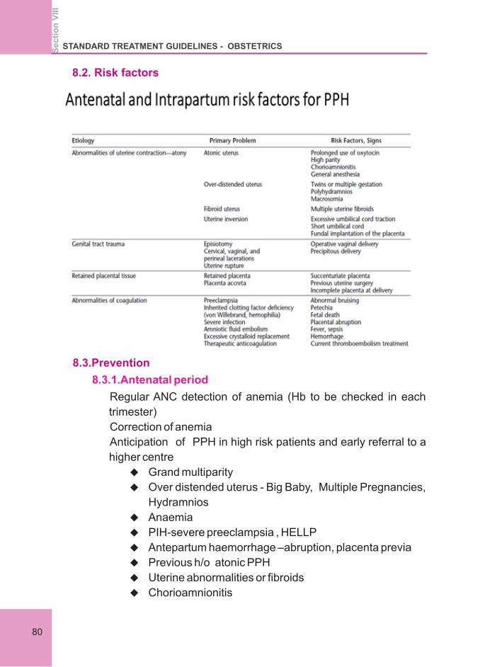
8.2. Risk factors
8.3.Prevention
8.3.1.Antenatal period
Regular ANC detection of anemia (Hb to be checked in each
trimester)
Correction of anemia
Anticipation of PPH in high risk patients and early referral to a
higher centre
u Grand multiparity
u Over distended uterus - Big Baby, Multiple Pregnancies,
Hydramnios
u Anaemia
u PIH-severe preeclampsia , HELLP
u Antepartum haemorrhage –abruption, placenta previa
u Previous h/o atonic PPH
u Uterine abnormalities or fibroids
u Chorioamnionitis
Se
cti
on
VII
ISTANDARD TREATMENT GUIDELINES - OBSTETRICS
80
u Maternal fever
u Induction & augmentation of labour
u Rapid and prolonged labour.
u Intra partum fever
l Avoid dehydration & prolonged labour
l All delivering patients should have 16/ 18G cannula
l Monitoring labour with partogram
l AMTSL (Active Management of Third Stage Of Labour) -3
steps
1. Administration of an uterotonic drug (oxytocin 5 units diluted
up to 5 ml with saline ) within 1 minute of delivery of baby for
immediate action followed by 10units IM or 20 units IV
infusion for sustained action the infusion should last for 2hrs.
(KFOG protocol)
OR
WHO / ACOG Protocol 10 units of oxytocin IV/IM
2. Controlled cord traction
3. Gentle uterine massage
u Pulse rate , volume , Blood pressure monitoring
u Monitor for vaginal bleeding & uterine hardness
l every 30 mts x first 2 hrs
l every 60 mts x next 2 hrs
u Encourage the woman to keep her bladder empty & early
breast feeding
u Teach the woman to massage her uterus & keep it firm
8.3.2.Intra partum risk factors
8.3.3.In Labour
8.3.4.Postpartum
STANDARD TREATMENT GUIDELINES - OBSTETRICS
81
Se
cti
on
VII
I
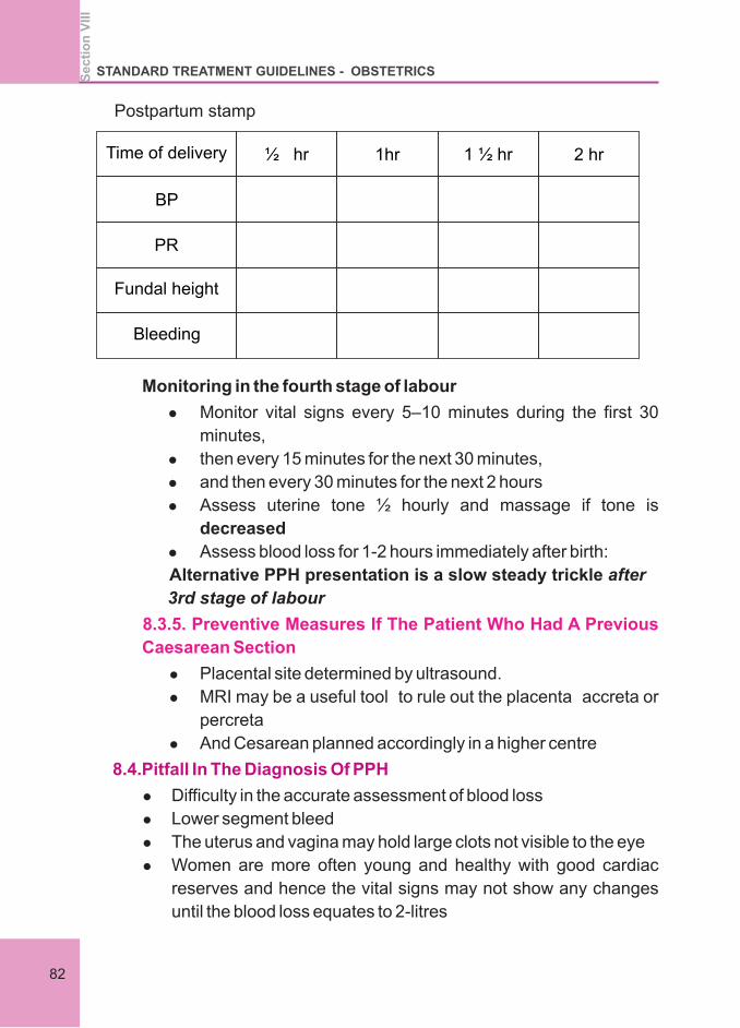
Postpartum stamp
Time of delivery ½ hr 1hr 1 ½ hr 2 hr
BP
PR
Fundal height
Bleeding
Monitoring in the fourth stage of labour
l Monitor vital signs every 5–10 minutes during the first 30
minutes,
l then every 15 minutes for the next 30 minutes,
l and then every 30 minutes for the next 2 hours
l Assess uterine tone ½ hourly and massage if tone is
decreased
l Assess blood loss for 1-2 hours immediately after birth:
Alternative PPH presentation is a slow steady trickle after
3rd stage of labour
l Placental site determined by ultrasound.
l MRI may be a useful tool to rule out the placenta accreta or
percreta
l And Cesarean planned accordingly in a higher centre
l Difficulty in the accurate assessment of blood loss
l Lower segment bleed
l The uterus and vagina may hold large clots not visible to the eye
l Women are more often young and healthy with good cardiac
reserves and hence the vital signs may not show any changes
until the blood loss equates to 2-litres
8.3.5. Preventive Measures If The Patient Who Had A Previous
Caesarean Section
8.4.Pitfall In The Diagnosis Of PPH
STANDARD TREATMENT GUIDELINES - OBSTETRICSS
ec
tio
n V
III
82
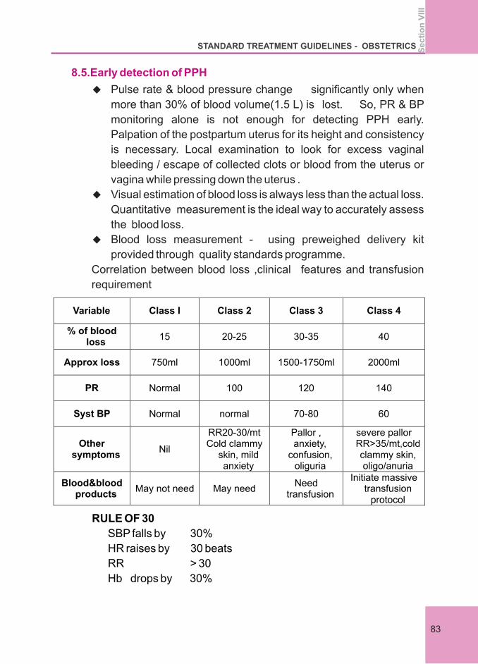
8.5.Early detection of PPH
u Pulse rate & blood pressure change significantly only when
more than 30% of blood volume(1.5 L) is lost. So, PR & BP
monitoring alone is not enough for detecting PPH early.
Palpation of the postpartum uterus for its height and consistency
is necessary. Local examination to look for excess vaginal
bleeding / escape of collected clots or blood from the uterus or
vagina while pressing down the uterus .
u Visual estimation of blood loss is always less than the actual loss.
Quantitative measurement is the ideal way to accurately assess
the blood loss.
u Blood loss measurement - using preweighed delivery kit
provided through quality standards programme.
Correlation between blood loss ,clinical features and transfusion
requirement
Variable Class I Class 2 Class 3 Class 4
% of blood loss
15 20-25 30-35 40
Approx loss
750ml
1000ml 1500-1750ml
2000ml
PR
Normal
100
120
140
Syst BP
Normal
normal
70-80
60
Other symptoms
Nil
RR20-30/mt
Cold clammy skin, mild anxiety
Pallor , anxiety,
confusion, oliguria
severe pallor RR>35/mt,cold clammy skin, oligo/anuria
Blood&blood products
May not need
May need
Need transfusion
Initiate massive transfusion
protocol
RULE OF 30
SBP falls by 30%
HR raises by 30 beats
RR > 30
Hb drops by 30%
STANDARD TREATMENT GUIDELINES - OBSTETRICS
83
Se
cti
on
VII
I
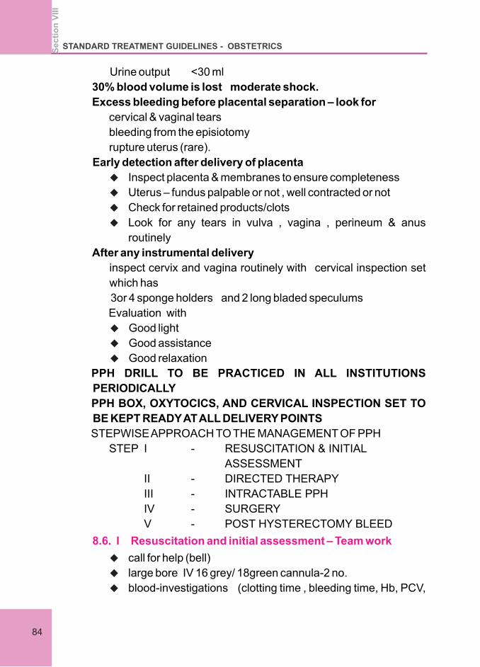
Urine output <30 ml
30% blood volume is lost moderate shock.
Excess bleeding before placental separation – look for
cervical & vaginal tears
bleeding from the episiotomy
rupture uterus (rare).
Early detection after delivery of placenta
Inspect placenta & membranes to ensure completeness
Uterus – fundus palpable or not , well contracted or not
Check for retained products/clots
Look for any tears in vulva , vagina , perineum & anus
routinely
After any instrumental delivery
inspect cervix and vagina routinely with cervical inspection set
which has
3or 4 sponge holders and 2 long bladed speculums
Evaluation with
Good light
Good assistance
Good relaxation
PPH DRILL TO BE PRACTICED IN ALL INSTITUTIONS
PERIODICALLY
PPH BOX, OXYTOCICS, AND CERVICAL INSPECTION SET TO
BE KEPT READY AT ALL DELIVERY POINTS
STEPWISE APPROACH TO THE MANAGEMENT OF PPH
STEP I - RESUSCITATION & INITIAL
ASSESSMENT
II - DIRECTED THERAPY
III - INTRACTABLE PPH
IV - SURGERY
V - POST HYSTERECTOMY BLEED
call for help (bell)
large bore IV 16 grey/ 18green cannula-2 no.
blood-investigations (clotting time , bleeding time, Hb, PCV,
u
u
u
u
u
u
u
u
u
u
8.6. I Resuscitation and initial assessment – Team work
STANDARD TREATMENT GUIDELINES - OBSTETRICSS
ec
tio
n V
III
84
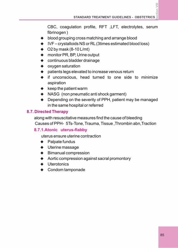
CBC, coagulation profile, RFT ,LFT, electrolytes, serum
fibrinogen )
blood grouping cross matching and arrange blood
IVF – crystalloids NS or RL (3times estimated blood loss)
O2 by mask (8-10 L/mt)
monitor PR, BP, Urine output
continuous bladder drainage
oxygen saturation
patients legs elevated to increase venous return
if unconscious, head turned to one side to minimize
aspiration
keep the patient warm
NASG (non pneumatic anti shock garment)
Depending on the severity of PPH, patient may be managed
in the same hospital or referred
along with resuscitative measures find the cause of bleeding
Causes of PPH- 5Ts-Tone, Trauma, Tissue ,Thrombin abn,Traction
uterus ensure uterine contraction
Palpate fundus
Uterine massage
Bimanual compression
Aortic compression against sacral promontory
Uterotonics
Condom tamponade
u
u
u
u
u
u
u
u
u
u
u
u
u
u
u
u
u
8.7. Directed Therapy
8.7.1.Atonic uterus-flabby
STANDARD TREATMENT GUIDELINES - OBSTETRICS
85
Se
cti
on
VII
I
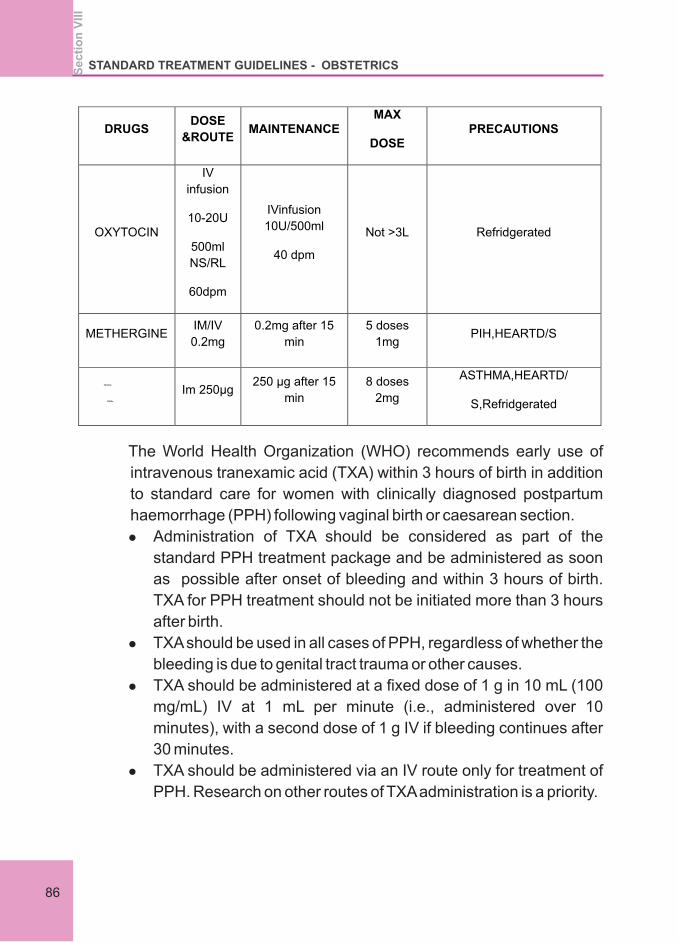
DRUGS DOSE
&ROUTE MAINTENANCE
MAX
DOSE
PRECAUTIONS
OXYTOCIN
IV
infusion
10-20U
500ml
NS/RL
60dpm
IVinfusion
10U/500ml
40 dpm
Not >3L
Refridgerated
METHERGINE
IM/IV
0.2mg
0.2mg after 15
min
5 doses
1mg
PIH,HEARTD/S
15mehyl
PGF2α
Im 250µg
250 µg after 15
min
8 doses
2mg
ASTHMA,HEARTD/
S,Refridgerated
The World Health Organization (WHO) recommends early use of
intravenous tranexamic acid (TXA) within 3 hours of birth in addition
to standard care for women with clinically diagnosed postpartum
haemorrhage (PPH) following vaginal birth or caesarean section.
l Administration of TXA should be considered as part of the
standard PPH treatment package and be administered as soon
as possible after onset of bleeding and within 3 hours of birth.
TXA for PPH treatment should not be initiated more than 3 hours
after birth.
l TXA should be used in all cases of PPH, regardless of whether the
bleeding is due to genital tract trauma or other causes.
l TXA should be administered at a fixed dose of 1 g in 10 mL (100
mg/mL) IV at 1 mL per minute (i.e., administered over 10
minutes), with a second dose of 1 g IV if bleeding continues after
30 minutes.
l TXA should be administered via an IV route only for treatment of
PPH. Research on other routes of TXA administration is a priority.
STANDARD TREATMENT GUIDELINES - OBSTETRICSS
ec
tio
n V
III
86
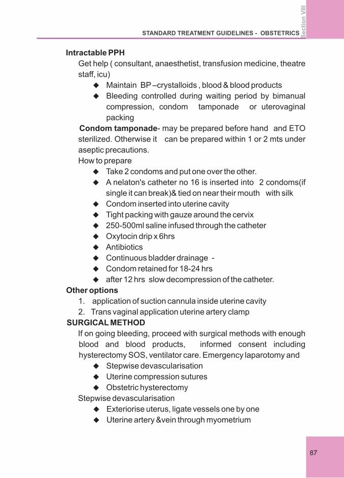
Intractable PPH
Get help ( consultant, anaesthetist, transfusion medicine, theatre
staff, icu)
u Maintain BP –crystalloids , blood & blood products
u Bleeding controlled during waiting period by bimanual
compression, condom tamponade or uterovaginal
packing
Condom tamponade- may be prepared before hand and ETO
sterilized. Otherwise it can be prepared within 1 or 2 mts under
aseptic precautions.
How to prepare
u Take 2 condoms and put one over the other.
u A nelaton's catheter no 16 is inserted into 2 condoms(if
single it can break)& tied on near their mouth with silk
u Condom inserted into uterine cavity
u Tight packing with gauze around the cervix
u 250-500ml saline infused through the catheter
u Oxytocin drip x 6hrs
u Antibiotics
u Continuous bladder drainage -
u Condom retained for 18-24 hrs
u after 12 hrs slow decompression of the catheter.
Other options
1. application of suction cannula inside uterine cavity
2. Trans vaginal application uterine artery clamp
SURGICAL METHOD
If on going bleeding, proceed with surgical methods with enough
blood and blood products, informed consent including
hysterectomy SOS, ventilator care. Emergency laparotomy and
u Stepwise devascularisation
u Uterine compression sutures
u Obstetric hysterectomy
Stepwise devascularisation
u Exteriorise uterus, ligate vessels one by one
u Uterine artery &vein through myometrium
STANDARD TREATMENT GUIDELINES - OBSTETRICS
87
Se
cti
on
VII
I
u Utero-ovarian anastomoses
u Descending cervical
Internal iliac artery ligation
Internal iliac ligation converts pelvic circulation to venous
system-
l Artery ligated 2.5 cm below its origin
u Tied with absorbable suture
u Femoral pulse must be checked
u Risk of injury to vein
u Requires surgical expertise
u Don't overdo –septicaemia
Burchell, ascertained that ligating the hypogastric artery turned
the pelvic circulation like a venous system, thereby aiding clotting
and controlling PPH.
It is effective in Uterine atony,
Midline perforation,
Large broad ligament or lateral pelvic wall haematoma,
Multiple cervical tears and
Lower segment bleeding
Uterine compression sutures
u B- Lynch suture(75%success)
u modified B– Lynch
u Cho square sutures
u Haymann suture -preferred
Obstetric hysterectomy
Decision for hysterectomy should not be delayed especially
in multipara and patients with fibroid.
coagulation failure must be simultaneously corrected
u Last resort in severe PPH
u Vertical incision
u Exteriorise uterus
u Retain ovaries
u Clamp, cut & drop technique
u Only subtotal hysterectomy. Unless placenta previa
,cervical , forniceal tears
STANDARD TREATMENT GUIDELINES - OBSTETRICSS
ec
tio
n V
III
88
u Edges of vagina/ cervix over sewed
u Leave drain or keep vault open
u retained blood clots,placental bits,membranes- evacuate
u retained placenta-empty the bladder, 20 units IV oxytocin
drip, manual removal under GA
u Examination using cervical inspection set-minimum 2 long
bladed speculum and 3sponge holding forceps.
u Proper visualisation
u Good lighting & assistance
u Careful repair-lacerations (cx,vag.,perineum)-
u If apex of cervical tear not seen ,lower uterine extension
cannot be ruled , so laparotomy may be necessary.
u Replace clotting factors
u FFP (pt inr >1.5- 15 ml/ kg)
u Cryo precipitate
u Platelet transfusion
u Recombinant factor VII a
u Complete or partial due to mismanaged 3rd stage of labour
causing shock and haemorrhage
u Prevention by controlled cord traction
u Per abdomen- fundus of uterus not palpable
u Vaginal examination-soft globular swelling in the vagina
Management
Resuscitation and immediate replacement of the inverted
uterus under GA
The part that came out first –fundus should be the last part to
be reverted
Placental removal after repositioning only
Oxytocics after repositioning to ensure uterine contraction
If manual repositioning fails- hydrostatic method using warm
8.7.2. Tissue (Retained products)
8.7.3. Trauma
8.7.4. Thrombin-(abnormalities of coagulation)
8.7.5. Traction (Inversion uterus )
STANDARD TREATMENT GUIDELINES - OBSTETRICS
89
Se
cti
on
VII
I
saline
If that also fail ,surgical methods- laparotomy and
Huntington's method or Haultain's method
u Pallor ,tachycardia and tachypnoea is out of proportion to the
external bleeding
u Episiotomy pain, urinary retention, sense of defecation after
delivery
haematoma( vulval, vaginal) -<3cm close observation
>3cm - evacuate , catch bleeder, occlude cavity , tightly pack the
vagina, catheterise and antibiotics.
Broad ligament haematoma- uterus not in the midline, tender adnexal
mass laparotomy & haematoma evacuation
Rupture uterus–signs of intra peritoneal bleeding and patient going
for hemodynamic stability
Laparotomy - rent repair if possible or hysterectomy
Post LSCS bleeding
Causes inferior epigastric artery injury during CS
Uterine angle bleeding following CS
Sterilization stump bleeding
apid fluid replacement with NS or Ringer lactate
8.8. Hidden bleeding –be vigilant when
l The cornerstones of resuscitation during PPH are
restoration of both blood volume and oxygen-carrying
capacity. (Glover, 2003; Toledo et al, 2007; Patel et al, 2006).
l Oxygen by mask at 10–15 litres/minute
l Intravenous access (large gauge cannula x 2)
l Position flat
l Keep the woman warm
l Until blood is available, infuse up to 3.5 litres of warmed fluid
solution, crystalloid (2 litres) and/or colloid (1–2 litres), rapidly as
required
l Apply clinical judgement in each situation
l Take blood for cross matching, and obtain a CBC count,
coagulation screen, urea level, creatinine value, and
electrolyte status.
l Begin immediate r
STANDARD TREATMENT GUIDELINES - OBSTETRICSS
ec
tio
n V
III
90
solution.
l ransfuse with PRBCs as available and appropriate.
l Order coagulation screen (International Normalized Ratio,
activated partial thrombo plastin time) if fibrinogen, thrombin time,
blood film, and D-dimer results are abnormal
l Mortality is increased when hypothermia and acidosis occur
with coagulopathy– the
l Avoid hypothermia
Warmed saline
Use fluid warmers and forced air warmers
Minimise exposure
Remove wet linen
Provide warm blankets
Temporizing measures recommended for intractable atonic and
non-atonic PPH include:
1. External aortic compression
2. Bimanual uterine compression
3. Non-pneumatic anti-shock garment (NASG)
u Any bleeding more than 1000ml
u Any hemodynamic instability
u Ongoing bleeding
u Previously anemic without even the stipulated loss
WHEN TO INITIATE MASSIVE TRANSFUSION PROTOCOL?
u Rapid blood loss >150ml/mt
u More than 50% blood loss in 3 hours
u Risk is that of DIC
u Packed cell:FFP :platelet – 1:1:1 ratio
u 95% success
u Done in stable patient
u Angiographic facility needed
u Takes 1-2hrs
u Temporary – gel foam –better
u Permanent- microspheres
'Lethal Triad'.
8.9.When to transfuse?
8.10.Uterine artery embolisation
STANDARD TREATMENT GUIDELINES - OBSTETRICS
91
Se
cti
on
VII
I
u Internal iliac artery lign should not be done if uae needed
Post hysterectomy bleeding
u Abdominal packing which is removed 24 hrs later after
correction of DIC
u Angiographic embolization
After managing PPH
u Closely monitor in ICU/ LR x 24hrs
u Correction of anemia & coagln. Abnlty-blood& blood products
u Assessment of complications.
u Antenatal reference
u High risk women for PPH
u Previous cesarean patients with placenta previa referred early
at least by 32 weeks of pregnancy
u Intrapartum reference
u In properly equipped ambulance
u Accompanied by paramedical or other competent staff
u Two iv cannulas with crystalloids
u Oxygen
NASG
u Condom tamponade and urinary catheter in situ
u Properly labeled sample of patient's blood
u Need for blood and blood products explained and extra
people for blood donation
u Inform the referring hospital staff
PPH BOX
1. Green and gray cannula
2. Vacutainers
3. Blood bank request form
4. Lab investigation form
5. Spirit swab
6. Adhesive
7. Scissors for cutting
8. I V set
9. Blood set
8.11.Referral –whom, when and how?
STANDARD TREATMENT GUIDELINES - OBSTETRICSS
ec
tio
n V
III
92
10. Normal Saline
11. Ringer Lactate
12. Haemacel
13. Foleys Catheter No.14
14. Uro bag
15. O2 mask
16. PPH drugs
a. Oxytocin
b. Methergin
c. PGF2α
d. PGE1
17. Syringes
18. Gloves
Drugs to be refrigerated
1. WHO guidelines for management of Post-partum haemorrhage
2. Improving maternity care in Kerala-Quality standards on
postpartum haemorrhage and hypertensive disorders of
pregnancy-First edition 2013
3. KFOG protocols
4. Why mothers die Kerala 2006-2009
5. Updated WHO Recommendation on Tranexamic Acid for the
Treatment of Postpartum Haemorrhage the World Health
Organization's 2017 Global Recommendation
l Potential sequelae of PPH (life after PPH)
l Orthostatic hypotension,
l Anemia and fatigue which can make breastfeeding and
maternal care of the newborn more difficult.
l Postpartum hemorrhage may increase the risk of postpartum
depression and acute stress reactions.
l Transfusion may be necessary and carries associated risks
including infection and transfusion reaction.
l Hemorrhagic shock may lead to Sheehan's Syndrome
(posterior pituitary ischemia with delay or failure of lactation),
occult myocardial ischemia, or death.
References
STANDARD TREATMENT GUIDELINES - OBSTETRICS
93
Se
cti
on
VII
I
6. Committee on Practice Bulletins-Obstetrics. Practice Bulletin No.
183: Postpartum Hemorrhage. Obstet Gynecol. 2017
Oct;130(4):e168-e186.
7. A Textbook of POSTPARTUM HEMORRHAGE A comprehensive
guide to evaluation, management and surgical intervention
Edited by Christopher B-Lynch FRCS, FRCOG, D.Univ Milton
Keynes General Hospital NHS Foundation Trust, Oxford
Regional Health Authority, UK
8. Dahlke JD, Mendez-Figueroa H, Maggio L, Hauspurg AK,
Sperling JD, Chauhan SP, Rouse DJ. Prevention and
management of postpartum hemorrhage: a comparison of 4
national guidelines. Am. J. Obstet. Gynecol. 2015
Jul;213(1):76.e1-76.e10.
9. Oyelese Y, Ananth CV. Postpartum hemorrhage: epidemiology,
risk factors, and causes. Clin Obstet Gynecol. 2010
Mar;53(1):147-56.
STANDARD TREATMENT GUIDELINES - OBSTETRICSS
ec
tio
n V
III
95

Section IX
PLACENA ACCRETA SPECTRUM

Section 9
PLACENA ACCRETA SPECTRUM
9.1. Introduction
9.2. Risk factors:
Placenta accreta spectrum (PAS) is one of the most morbid
conditions encountered during pregnancy. It is associated with
hemorrhage, often massive, and associated problems such as
multiorgan failure and even death. In addition, most cases require a
difficult hysterectomy with high rates of complications including
bladder and ureteral injury and rectovaginal fistula as well as loss of
fertility. The rate of the condition has dramatically increased over the
past 30 years owing to a concomitant increase in the rate of
cesarean delivery
Three grades of abnormal placental attachment are defined
according to the depth of invasion:
Accreta: Placenta implants totally/partially/focally through the
decidua basalis. Chorionic villi attach to the myometrium, rather than
being restricted within the decidua basalis.
Increta: Chorionic villi invade into the myometrium.
Percreta: Chorionic villi invade fully through the myometrium. It may
breach the serosa and invade surrounding structures.
An important risk factor for placenta accreta is placenta praevia in the
presence of a uterine scar.
Placenta praevia is an independent risk factor for placenta accreta.
The frequency of placenta accreta relates directly to number of
cesarean deliveries and presence or absence of placenta previa .
Additional reported risk factors for placenta accreta include
l Advanced maternal age
l Multiparity
l Prior uterine surgery
l Prior uterine curettage
l Uterine irradiation/Endometrial ablation
l Asherman syndrome
l Uterine leiomyomata
Se
cti
on
IX
STANDARD TREATMENT GUIDELINES - OBSTETRICS
97
l Uterine anomalies
l IVF pregnancies
l Smoking.
Morbidly adherent placentation may be suspected when there is a
placenta praevia in a woman with a history of caesarean section or
other uterine surgery.
The diagnosis is usually established by ultrasonography and
occasionally supplemented by magnetic resonance imaging
(MRI)
Prenatal diagnosis is a key factor in optimizing the counselling,
treatment, and outcome of patients with placenta accrete
Features contributing to the diagnosis of placenta accreta include:
Increased AFP and hCG in second trimester
l First trimester
Gestational sac in LUS
Irregular vascular spaces in placental bed
CS scar implantation
l Second trimester
The presence of placental vascular lacunae is the most
sensitive of all diagnostic markers (80-90% in second
trimester).
l Third trimester
Sonographic findings that have been associated with
placenta accreta
l Loss of normal hypoechoic retroplacental zone
l Multiple vascular lacunae (irregular vascular spaces)
within placenta, giving “Swisscheese” appearance
l Blood vessels or placental tissue bridging uterine-
placental margin, myometrial-bladder interface, or
crossing uterine serosa
l Retroplacental myometrial thickness of<1mm
9.3. Diagnosis
9.3.1.Biochemistry
9.3.2.Ultrasound
STANDARD TREATMENT GUIDELINES - OBSTETRICSSe
cti
on
IX
98
l Numerous coherent vessels visualized with 3-
dimensional power Doppler in basal view
l Multiple, complex placental vascular lacunae
l Serosa-bladder interface: disruption, irregularity,
vascularity
DOPPLER:
l Turbulent lacunar blood flow
l Peak systolic flow > 15 cm/s
l Disruption of continuous blood flow in myometrium
l Distorted badder wall with increased vascularity
l Dilated vascular channels with pulsatile venous flow
over cervix
l Uterine bulging
l Heterogeneous signal intensity within placenta
l Dark intra-placental bands
l Abnormal placental vascularity
l Focal interruptions in myometrium
l Tenting of bladder
l Direction visualisation of invasion of adjacent organs
9.3.3.MRI
Magnetic resonance imaging may be useful for diagnosis of
difficult cases, such as posterior placenta praevia, and to assess
depth of invasionin suspected percreta
FINDINGS:
Preoperative
l Maximization of preoperative hemoglobin values
l Verification of specific timing of planned delivery
l Identification of exact location of delivery (surgical suite and
its associated capabilities)
l Verification that necessary preoperative consultations have
occurred
l Consideration of patient and family needs given temporary
9.4. Relevant considerations for case optimization in planned
placentaaccreta spectrum needs multidisciplinary approach
STANDARD TREATMENT GUIDELINES - OBSTETRICS
99
Se
cti
on
IX
relocation
Intraoperative
l Verification of appropriate complement of surgical expertise
involved or available, or both
l Verification of availability of related services as necessary
(eg, interventional radiology)
l Coordination of blood bank with scheduling or timing of case
Postoperative
l Assurance that critical care services are engaged and
available for postoperative care
9.5. Management
9.5.1 Antepartum considerations
As with all women at risk of major obstetric hemorrhage, those
with suspected placenta accreta should be encouraged to remain
close to hospital of confinement for the duration of the third
trimester of pregnancy.
A proper well documented plan should be there for all diagnosed
case of placenta accrete.
Timing of elective caesarean delivery
This should be individualized and the decision should be
made jointly with the woman, the Obstetrician and
Neonatologist Although the evidence does not support any
particular gestation for elective delivery, combined maternal
and neonatal outcome is optimized in stable patients with a
planned delivery from 34 weeks GESTATION to 36 WEEKS
GESTATION
The gestation for elective delivery should balance the
neonatal risks of prematurity against the maternal risks of
emergent delivery. In the absence of clear evidence to guide
this decision, a plan should be made tailored to the individual
patient, taking into account the ultrasound findings, history of
bleeding, medical comorbidities, surgeon availability and
patient preferences.
In general, features of placenta percreta would warrant
consideration of earlier delivery (i.e.32-34 weeks)
STANDARD TREATMENT GUIDELINES - OBSTETRICSSe
cti
on
IX
100
Antenatal corticosteroids as well as magnesium sulphate for
neuro-protection should be administered prior to delivery at
all gestations less than 34 weeks
Among women with suspected placenta accreta, emergency
caesarean section is mostly performed in:
l The
l
Preoperative Patient Counseling:
This is to be carried out by the Consultant Obstetrician and
should include
The following risk factors
A consent form should be completed including all possible
treatments including hysterectomy, interventional
radiological procedures and conservative management,
elective ventilation, injury to viscera, torrential hemorrhage
and regarding mortality.
l Anaesthesia: General anaesthesia preferred in most
centres
l Skin incision: Midline vertical
l Uterine incision: Classical Caesarean. /Fundal transverse
l Uterine incision be located to avoid the placenta during
entry into the uterine cavity.
l After delivery of baby, Wait for spontaneous separation, if
presence of vaginal bleeding
PPROM and/or uterine contractions
l Potential need for hysterectomy,in case of fertility
sparing the option of retaining placenta in situ and
chemotherapy and its consequences.
l Risks of profuse hemorrhage
l Prolonged surgery ,injury to bowel, bladder, ureter
l Need of prolonged hospitalization, need of continuous
bladder drainage, post operative ventilation and ICU
care
l Real risk of mortality
l Involve the family in all discussions for medico-legal
reasons
9.5.2. Intra-operative considerations:
STANDARD TREATMENT GUIDELINES - OBSTETRICS
101
Se
cti
on
IX
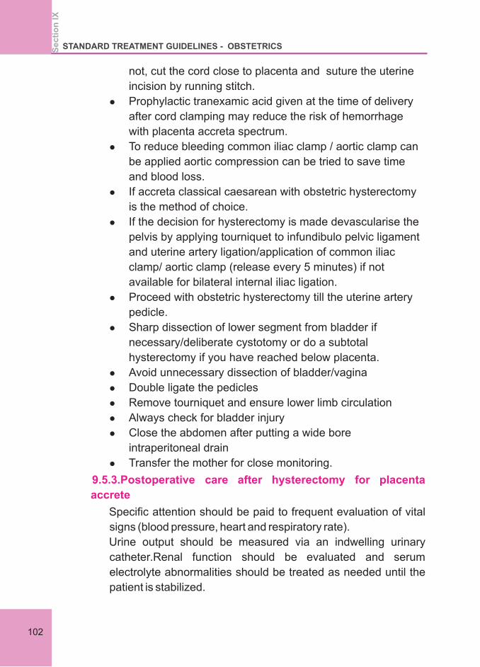
not, cut the cord close to placenta and suture the uterine
incision by running stitch.
To common iliac clamp / aortic clamp can
be applied aortic compression can be tried to save time
and blood loss.
If accreta
Remove
Specific attention should be paid to frequent evaluation of vital
signs (blood pressure, heart and respiratory rate).
Urine output should be measured via an indwelling urinary
catheter.Renal function should be evaluated and serum
electrolyte abnormalities should be treated as needed until the
patient is stabilized.
l Prophylactic tranexamic acid given at the time of delivery
after cord clamping may reduce the risk of hemorrhage
with placenta accreta spectrum.
l reduce bleeding
l classical caesarean with obstetric hysterectomy
is the method of choice.
l If the decision for hysterectomy is made devascularise the
pelvis by applying tourniquet to infundibulo pelvic ligament
and uterine artery ligation/application of common iliac
clamp/ aortic clamp (release every 5 minutes) if not
available for bilateral internal iliac ligation.
l Proceed with obstetric hysterectomy till the uterine artery
pedicle.
l Sharp dissection of lower segment from bladder if
necessary/deliberate cystotomy or do a subtotal
hysterectomy if you have reached below placenta.
l Avoid unnecessary dissection of bladder/vagina
l Double ligate the pedicles
l tourniquet and ensure lower limb circulation
l Always check for bladder injury
l Close the abdomen after putting a wide bore
intraperitoneal drain
l Transfer the mother for close monitoring.
9.5.3.Postoperative care after hysterectomy for placenta
accrete
STANDARD TREATMENT GUIDELINES - OBSTETRICSSe
cti
on
IX
102
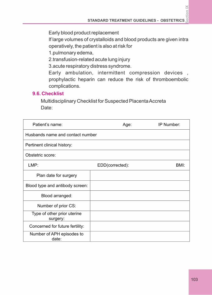
Early blood product replacement
If large volumes of crystalloids and blood products are given intra
operatively, the patient is also at risk for
1.pulmonary edema,
2.transfusion-related acute lung injury
3.acute respiratory distress syndrome.
Early ambulation, intermittent compression devices ,
prophylactic heparin can reduce the risk of thromboembolic
complications.
Multidisciplinary Checklist for Suspected Placenta Accreta
Date:
9.6. Checklist
Patient’s name: Age: IP Number:
Husbands name and contact number
Pertinent clinical history:
Obstetric score:
LMP:
EDD(corrected): BMI:
Plan date for surgery
Blood type and antibody screen:
Blood arranged:
Number of prior CS:
Type of other prior uterine surgery:
Concerned for future fertility:
Number of APH episodes to
date:
STANDARD TREATMENT GUIDELINES - OBSTETRICS
103
Se
cti
on
IX
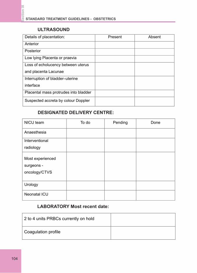
ULTRASOUND
Details of placentation: Present Absent
Anterior Posterior
Low lying Placenta or praevia
Loss of echolucency between uterus
and placenta Lacunae
Interruption of bladder–uterine
interface
Placental mass protrudes into bladder
Suspected accreta by colour Doppler
DESIGNATED DELIVERY CENTRE:
NICU team To do Pending Done
Anaesthesia
Interventional
radiology
Most experienced
surgeons -
oncology/CTVS
Urology
Neonatal ICU
LABORATORY Most recent date:
2 to 4 units PRBCs currently on hold
Coagulation profile
STANDARD TREATMENT GUIDELINES - OBSTETRICSSe
cti
on
IX
104
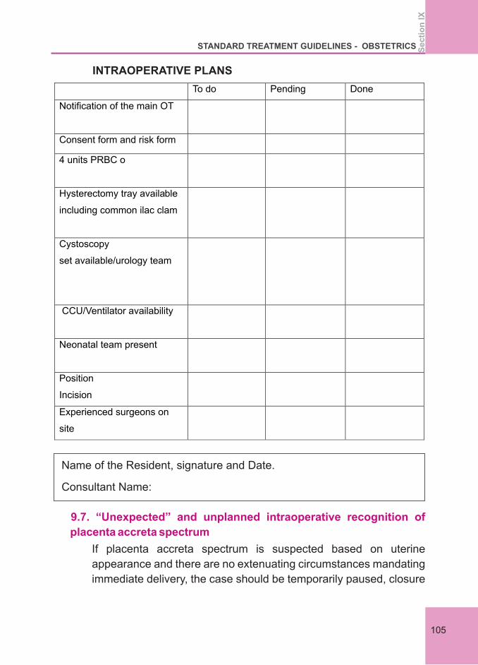
INTRAOPERATIVE PLANS
To do Pending Done
Notification of the main OT
Consent form and risk form
4 units PRBC o
Hysterectomy tray available
including common ilac clam
Cystoscopy
set available/urology team
CCU/Ventilator availability
Neonatal team present
Position
Incision
Experienced surgeons on
site
Name of the Resident, signature and Date.
Consultant Name:
9.7. “Unexpected” and unplanned intraoperative recognition of
placenta accreta spectrum
If placenta accreta spectrum is suspected based on uterine
appearance and there are no extenuating circumstances mandating
immediate delivery, the case should be temporarily paused, closure
STANDARD TREATMENT GUIDELINES - OBSTETRICS
105
Se
cti
on
IX
of abdomen and consider transfer to a higher centreif facilities not
available.
Sometimes placenta accreta spectrum is unexpectedly recognized
at the time of cesarean delivery or after the uterus is opened, the
fetus is delivered and attempts to removethe placenta have failed.
Uterine closure and proceed with hysterectomy and mobilize the
multidisciplinary team.
In addition, the anesthesia team should be alerted and consideration
given to general anesthesia, additional intravenous access should
be obtained, blood products should be ordered, and critical care
personnel should be alerted.
Expectant management is defined as leaving the placenta either
partially or totally in situ in exceptional situations. Life threatening
complications like hemorrahage and sepsis may follow.
l Perioperative placental loclisation of superior edge of placenta
l Pelvic devascularisation including intarterial balloon catheter
l no attempt to remove the entire placentawith large myometrial
excision and uterine repair
9.8. Uterine preservation and expectant management
9.9.Triple –p procedure
References
1. Shamshirsaz A, Fox K, Erfani H, et al. Outcomes of planned
compared with urgent deliveries using a multidisciplinary team
approach for morbidly adherent placenta. Obstet Gynecol.
2018;131: 234–241
2. Jauniaux E, Bhide A, Burton GJ. Pathophysiology of accreta. In:
Silver R, ed. Placenta accretasyndrome. Portland: CRC Press;
2017:13–28.
3. Jauniaux E, Chantraine F, Silver RM, et al. for the FIGO placenta
accreta diagnosis and management expert consensus panel.
FIGO consensus guidelines on placenta accreta spectrum
disorders: Epidemiology. Int J Gynecol Obstet .
2018;140:265–273
4. Jauniaux E, Jurkovic D. Placenta accreta: pathogenesis of a 20th
STANDARD TREATMENT GUIDELINES - OBSTETRICSSe
cti
on
IX
106
century iatrogenic uterine disease. Placenta. 2012;33:244–251
5. Collins SL, Ashcroft A, Braun T, et al. Proposal for standardized
ultrasound descriptions of abnormallyinvasive placenta (AIP).
UltrasoundObstet Gynecol. 2016;47:271–275.
6. D'Antonio F, Iacovella C, Bhide A. Prenatal identification of
invasive placentation using ultrasound: systematicreview and
meta-analysis. Ultrasound Obstet Gynecol2013;42:509–17.
7. Simonazzi G, Bisulli M, Saccone G, Moro E, Marshall A,
Berghella V. Tranexamic acid for preventing postpartum blood
loss after cesarean delivery: a systematic review and meta-
analysis of randomized controlled trials. Acta Obstet Gynecol
Scand 2016;95:28–37.
STANDARD TREATMENT GUIDELINES - OBSTETRICS
107
Se
cti
on
IX

Section X
.MANAGEMENT OF HYPERTENSIVE
DISORDERS IN PREGNANCY

Section 10
MANAGEMENT OF HYPERTENSIVE DISORDERS IN PREGNANCY
The hypertensive disorders of pregnancy remain leading causes of
maternal and perinatal morbidity and mortality. This protocol
summarizes the quality of the relevant existing evidence and provides a
reasonable approach to the diagnosis, evaluation, and treatment of
hypertensive disorders of pregnancy.
Definition
GHTN:New onset hypertension systolic≥ 140mmHg and or
diastolic ≥ 90mmHg at least 4 hrs apart developing after 20
weeks of gestation without proteinuria and resolving within 12
weeks postpartum.
l Hypertensive disorders of pregnancy still classified as :
m Chronic Hypertension
m Gestational Hypertension
m Preeclampsia
m Eclampsia
m Chronic hypertension with superimposed preeclampsia
l The terms mild and severe preeclampsia have been
eliminated.
l New categories are preeclampsia with and without severe
features .
Pre eclampsia Diagnosis
l BP ≥ 140/90 on two occasions four hours apart, (or single
recording of BP ≥ 160/110 ) after 20 weeks
10.1Hypertension in pregnancy
10.2.Preeclampsia
l Most changes have to do with diagnosis and
management of pre-eclampsia
The change is to emphasize those severe features may develop
at any time in women with preeclampsia and to emphasize
vigilance in looking for these features.
AND
Se
cti
on
X
STANDARD TREATMENT GUIDELINES - OBSTETRICS
111
l ≥ 300 mg on 24 hours protein OR . 0.3 protein/ creatinine
ratio OR dipstick of +1 protein ( if other two methods are not
available)
l Any of the following feature in the absence of proteinuria (
non proteinuric hypertension with any of the below mentioned
features.
¦ Platelets < 100,000
¦ Creatinine > 1.1 OR doubling of creatinine in absence of
other renal disease
¦ Doubling of AST or ALT
OR
10.3.Screening methods that are useful to identify women at risk of
developing hypertensive disorders of pregnancy
10.4.Prevention strategies for reducing the risk of hypertensive
disorders of pregnancy
First trimester of pregnancy, it has been reported that a combination of
1. low maternal serum concentrations of PlGF,
2. high uterine artery pulsatility index,
3. Low PAPPA, identified patients who would develop preeclampsia
requiring delivery before 34 weeks of gestation
The calculated positive predictive value was only21.2%, indicating
that approximately 79% of the women in the screen-positive group
would not develop hypertensive disorders during pregnancy
Strategies to prevent preeclampsia have been studied extensively
over the past 30 years. To date, no intervention has been proved
unequivocally effective at eliminating the risk of preeclampsia
1. A meta-analysis of 13 trials (15,730 women) reported a significant
reduction in preeclampsia with calcium supplementation, with the
greatest effect among women with low-baseline calcium intake
2. In a recent meta-analysis of aggregate data from 45 randomized
trials, only a modest reduction in preeclampsia was noted when
low-dose aspirin was started after 16 weeks of gestation (relative
risk [RR], 0.81; 95% CI, 0.66–0.99) but a more significant
reduction in severe preeclampsia (RR, 0.47; 95% CI, 0.26–0.83)
and fetal growth restriction (RR, 0.56; 95% CI, 0.44–0.70) was
STANDARD TREATMENT GUIDELINES - OBSTETRICS Se
cti
on
X
Se
cti
on
X
112
demonstrated when low-dose aspirin was started before 16
weeks of gestation
In a recent multicenter, double blind, placebo controlled trial,
pregnant women at increased risk of preterm preeclampsia (less
than 37 weeks of gestation) were randomly assigned to receive
aspirin, at dose (150 mg/day), or placebo from 11 weeks to 14
weeks of gestation until 36 weeks of gestation low-dose aspirin in
women at high risk of preeclampsia was associated with a lower
incidence for preterm preeclampsia. However, there were no
differences in the rates of term preeclampsia between study groups.
l History of preeclampsia, especially when accompanied by an
adverse outcome
l Multifetal gestation
l Chronic hypertension
l Type 1 or 2 diabetes
l Renal disease
l Autoimmune disease (ie, systemic lupus erythematosus, the
antiphospholipid syndrome
Recommend low-dose aspirin 150 mg /day if the patient has
one or more of these high-risk factors
l Nulliparity
l Obesity (body mass index greater than 30)
l Family history of preeclampsia (mother or sister)
l Sociodemographic characteristics (African American
l race, low socioeconomic status)
l Age 35 years or older
l Personal history factors (eg, low birth weight or small for
gestational age, previous adverse pregnancy outcome, more
than 10-year pregnancy interval
Consider low-dose aspirin if the patient has more than one of
these moderate-risk
10.5.Clinical Risk Factors and Aspirin Use*
10.5.1.High risk
10.5.2.moderate risk
STANDARD TREATMENT GUIDELINES - OBSTETRICS
113
Se
cti
on
X
10.5.3.Low risk
10.7.1 Preeclampsia and gestational hypertension
Previous uncomplicated full term delivery:
Do not recommend low dose aspirin
l All pregnant women should be assessed for proteinuria and at
each visit
l Significant proteinuria should be defined as 0.3 g/dl in a plete
24-h urine collection or 30 mg/mmol urinary creatinine in a spot
(random) urine sample
l More definitive testing for proteinuria (by urinary protein :
creatinine ratio or 24-h urine collection) is encouraged when there
is a suspicion of preeclampsia, including: 1+ dipstick
proteinuria, in the setting of hypertension with rising BP, or when
BP is normal but women have symptoms or signs suggestive of
preeclampsia
l Proteinuria testing need not be repeated once the significant
proteinuria of preeclampsia has been confirmed
m BP≥ 160/110
m Platelets < 100,000/micro litre
m Doubling AST or ALT AND /Or severe right upper quadrant pain
not explained by other pathology
m Creatinine > 1.1 OR doubling of creatinine in absence of renal
disease
m Pulmonary edema
m New cerebral or visual disturbances
Intrauterine growth restriction and >5g on 24 hour proteinuria is
no longer part of severe preeclampsia
Preterm management non-severe preeclampsia and
gestational hypertension
l Daily self – assessment preeclampsia symptoms and fetal
kick count
10.6.Recommendations for measuring protenuria
10.6.Definition of Preeclampsia with severe features:
10.7 Management
≥≥
≥
STANDARD TREATMENT GUIDELINES - OBSTETRICSSe
cti
on
X
114
l BP measurement twice weekly, at least one in the office
l Platelets, liver enzymes, weekly
l Protein assessment weekly in women with
gestational hypertension but no need to repeat once
diagnostic proteinuria is present
l Antenatal testing · Serial growth ultrasounds with
Doppler studies 2 weekly,more frequently in presence of
FGR
l Do not recommend bed rest
l
Severe preeclampsia at ≥ 34 weeks or if unstable woman or
fetus
Severe preeclampsia at ≤ 34 weeks and stable woman
and fetus
l Antihypertensives according to BP
l Give steroids and try to delay delivery for 48 hours
If severe preeclampsia, viable fetus at < 34 weeks
immediate termination indicated if
m Preterm premature rupture of membranes\
m Platelets < 100000
m Persistent elevated AST or ALT (over twice upper limit
normal
m Growth restriction ( < 3 % EFW for EGA)
twice
Delivery recommended at 37 weeks
Stabilization and delivery
10.7.2.Severe pre-eclampsia
The expectant management of preeclampsia with severe
features before 34 0/7 weeks of gestation is based on strict
selection criteria of those appropriate candidates and is best
accomplished in a setting with resources appropriate for
maternal and neonatal care. Because expectant
management is intended to provide neonatal benefit at the
expense of maternal risk, expectant management is not
advised when neonatal survival is not anticipated. During
expectant management, delivery is recommended at any
time in the case of deterioration of maternal or fetal condition
STANDARD TREATMENT GUIDELINES - OBSTETRICS
115
Se
cti
on
X
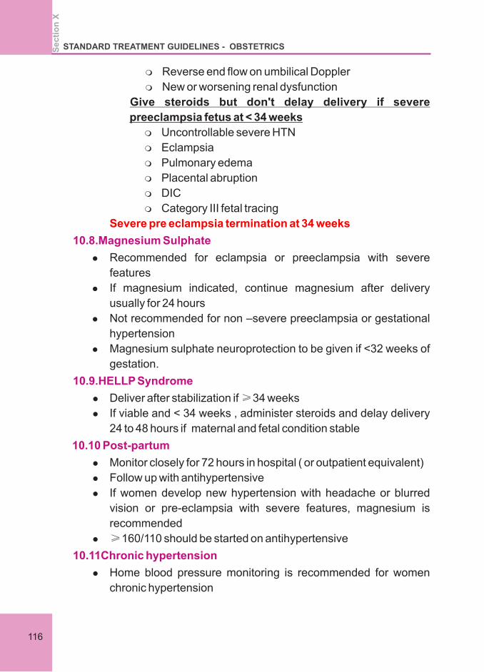
m Reverse end flow on umbilical Doppler
m New or worsening renal dysfunction
Give steroids but don't delay delivery if severe
preeclampsia fetus at < 34 weeks
m Uncontrollable severe HTN
m Eclampsia
m Pulmonary edema
m Placental abruption
m DIC
m Category III fetal tracing
l Recommended for eclampsia or preeclampsia with severe
features
l If magnesium indicated, continue magnesium after delivery
usually for 24 hours
l Not recommended for non –severe preeclampsia or gestational
hypertension
l Magnesium sulphate neuroprotection to be given if <32 weeks of
gestation.
l Deliver after stabilization if ≥ 34 weeks
l If viable and < 34 weeks , administer steroids and delay delivery
24 to 48 hours if maternal and fetal condition stable
l Monitor closely for 72 hours in hospital ( or outpatient equivalent)
l Follow up with antihypertensive
l If women develop new hypertension with headache or blurred
vision or pre-eclampsia with severe features, magnesium is
recommended
l ≥ 160/110 should be started on antihypertensive
l Home blood pressure monitoring is recommended for women
chronic hypertension
Severe pre eclampsia termination at 34 weeks
10.8.Magnesium Sulphate
10.9.HELLP Syndrome
10.10 Post-partum
10.11Chronic hypertension
STANDARD TREATMENT GUIDELINES - OBSTETRICSSe
cti
on
X
116
l Recommended antihypertensive : labetalol, nifedipine,
methyldopa
l Not recommended: ACE, ARB, renin inhibitor, mineral corticoid
receptor antagonist
l Target blood pressure for those on antihypertensive is 110 to
140/80 to 85mmHg
l Assess fetal growth from 26 weeks and with serial growth
ultrasounds 2-4 weekly when
m On antihypertensive
m Superimposed preeclampsia
m IUGR
l If chronic hypertension and no other maternal or fetal
complications delivery is not recommended until ≥ 38 weeks
l Watch carefully for signs and symptoms of superimposed
preeclampsia which occurs in about 20% of chronic hypertensive
l If need to increase antihypertensive medication in any trimester
consider admission to monitor blood pressure and rule out
superimposed preeclampsia
l
l Introducing standardized, evidence-based clinical guidelines for
the management of patients with preeclampsia and eclampsia
has been demonstrated to reduce the incidence of adverse
maternal outcomes.
l Pregnant women or women in the postpartum period with acute-
onset, severe systolic hypertension; severe diastolic
hypertension; or both require urgent antihypertensive therapy.
l Close maternal and fetal monitoring by a physician and nursing
staff are advised during the treatment of acute-onset, severe
hypertension.
l After initial stabilization, the team should monitor blood pressure
10.12.Chronic hypertension with superimposed preeclampsia
10.13.Emergent Therapy for Acute-Onset, Severe Hypertension
During Pregnancy and the Postpartum Period
Decision to deliver is similar to other preeclamptic
women based on gestational age and presence of
severe feature
STANDARD TREATMENT GUIDELINES - OBSTETRICS
117
Se
cti
on
X
closely and institute maintenance therapy as needed.
l Intravenous (IV) labetalol and hydralazine have long been
considered first-line medications for the management of acute-
onset, severe hypertension in pregnant women and women in the
postpartum period.
l Immediate release oral Nifedipine also may be considered as
a first-line therapy, particularly when IV access is not
available.
l The use of IV labetalol, IV hydralazine, or immediate release oral
nifedipine for the treatment of acute-onset, severe hypertension
for pregnant or postpartum patients does not require cardiac
monitoring.
l In the rare circumstance that IV bolus labetalol, hydralazine, or
immediate release oral nifedipine fails to relieve acute-onset,
severe hypertension and is given in successive appropriate
doses, emergent consultation with an anesthesiologist,
maternal–fetal medicine subspecialist, or critical care
subspecialist to discuss second-line intervention is
recommended.
l Acute-onset, severe hypertension that is accurately measured
using standard techniques and is persistent for 15 minutes or
more is considered a hypertensive emergency. It is well known
that severe hypertension can cause central nervous system
injury
l Accurate measurement of blood pressure is necessary to
optimally manage hypertension in pregnancy. Standardized
protocols to measure BP in pregnant patients facilitate accuracy
and ensure that appropriate steps are followed across all units
regardless of patient arm size or shape.
Mercury sphygmomanometer is the gold standard; however,
validated equivalent automated equipment also can be used. It is
necessary to obtain the correct cuff size (a range of cuff sizes with
directions to determine appropriate cuff size based on arm shape
should be available) and patients should be positioned in a sitting
or semireclining position with the back supported.
10.13.Mesurement of blood Pressure:
l
STANDARD TREATMENT GUIDELINES - OBSTETRICSSe
cti
on
X
118
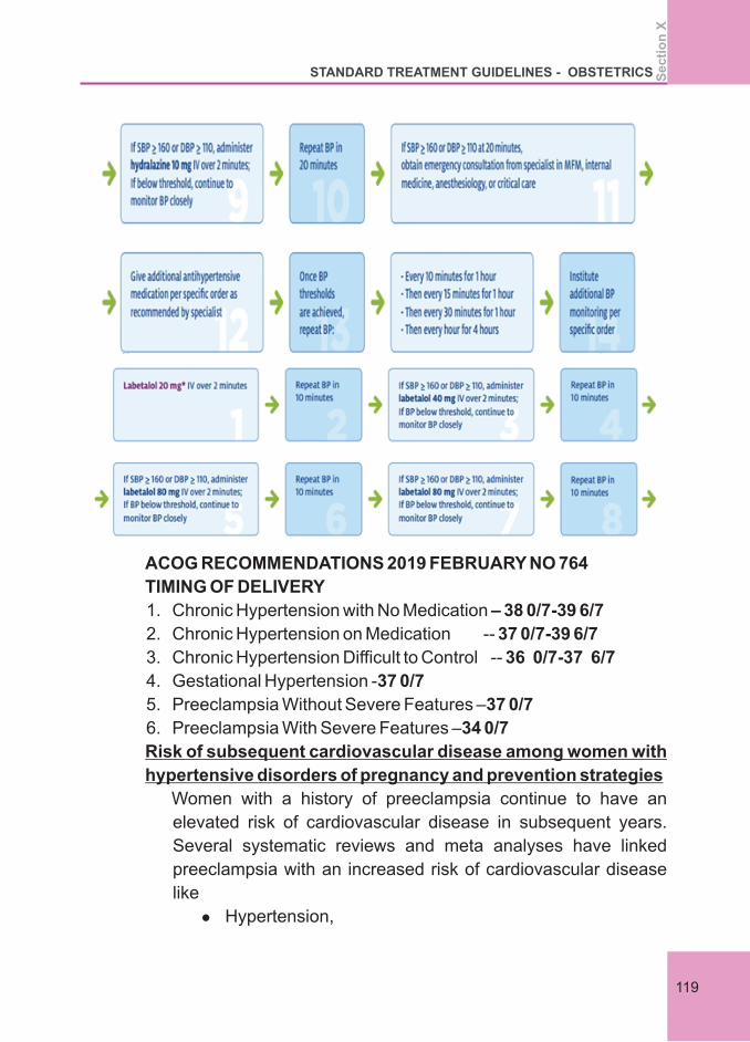
ACOG RECOMMENDATIONS 2019 FEBRUARY NO 764
TIMING OF DELIVERY
1. Chronic Hypertension with No Medication – 38 0/7-39 6/7
2. Chronic Hypertension on Medication -- 37 0/7-39 6/7
3. Chronic Hypertension Difficult to Control -- 36 0/7-37 6/7
4. Gestational Hypertension -37 0/7
5. Preeclampsia Without Severe Features –37 0/7
6. Preeclampsia With Severe Features –34 0/7
Risk of subsequent cardiovascular disease among women with
hypertensive disorders of pregnancy and prevention strategies
Women with a history of preeclampsia continue to have an
elevated risk of cardiovascular disease in subsequent years.
Several systematic reviews and meta analyses have linked
preeclampsia with an increased risk of cardiovascular disease
like
l Hypertension,
STANDARD TREATMENT GUIDELINES - OBSTETRICS
119
Se
cti
on
X
l Myocardial Infarction,
l Congestive Heart Failure),
l Cerebrovascular Events (Stroke),
l Peripheral Arterial Disease,
and cardiovascular mortality later in life, with an estimated
doubling of odds compared with women unaffected by
preeclampsia
Preventive strategies to be considered by patients and health care
providers may warrant closer long-term follow-up and lifestyle
modifications to better manage risk factors for cardiovascular
disease (eg, achieving healthful weight, exercise, diet, smoking
cessation), for which women and their primary care providers may
maintain ongoing care and vigilance.
5. ACog practice bulletin chronic hypertension in pregnancy
number 203 January 2019 Obstetrics
6. ACOG PRACTICE BULLETIN Gestational Hypertension and
Preeclampsia NUMBER 202 JANUARY 2019.
7. ACOG COMMITTEE OPINION Emergent Therapy for Acute-
Onset, Severe Hypertension During Pregnancy and the
Postpartum Period Number 767 FEBRUARY 2019.
10. Sibai BM. Diagnosis, prevention, and management of eclampsia.
Obstet Gynecol 2005;105:402–10.
Referrences
1. Acog recommendations 2019 february no 764
2. US task force recommendations 2012-13
3. Hypertension in pregnancy nice 107 ,2011th4. Williams Obstetrics 25 edition
8. ASPRE Trial: Incidence of Preterm Preeclampsia in Patients
Fulfilling ACOG and NICE Criteria According to Risk by FMF
Algorithm Obstetrical and Gynecological Survey 2018.
9. Controversies Regarding Diagnosis and Treatment of Severe
Hypertension in Pregnancy CLINICAL OBSTETRICS AND
GYNECOLOGY Volume 60, Number 1, 198–205 MARCH 2017.
STANDARD TREATMENT GUIDELINES - OBSTETRICSSe
cti
on
X
120
Section XI
Eclampsia

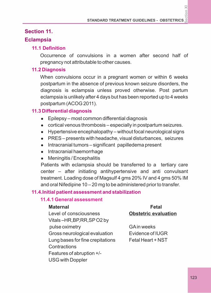
Section 11.
Eclampsia
11.1 Definition
11.2 Diagnosis
11.3 Differential diagnosis
11.4.Initial patient assessment and stabilization
Occurrence of convulsions in a women after second half of
pregnancy not attributable to other causes.
When convulsions occur in a pregnant women or within 6 weeks
postpartum in the absence of previous known seizure disorders, the
diagnosis is eclampsia unless proved otherwise. Post partum
eclampsia is unlikely after 4 days but has been reported up to 4 weeks
postpartum (ACOG 2011).
l Epilepsy – most common differential diagnosis
l cortical venous thrombosis – especially in postpartum seizures.
l Hypertensive encephalopathy – without focal neurological signs
l PRES – presents with headache, visual disturbances, seizures
l Intracranial tumors – significant papilledema present
l Intracranial haemorrhage
l Meningitis / Encephalitis
Patients with eclampsia should be transferred to a tertiary care
center – after initiating antihypertensive and anti convulsant
treatment. Loading dose of Magsulf 4 gms 20% IV and 4 gms 50% IM
and oral Nifedipine 10 – 20 mg to be administered prior to transfer.
Maternal Fetal
Level of consciousness Obstetric evaluation
Vitals –HR,BP,RR,SP O2 by
pulse oximetry GA in weeks
Gross neurological evaluation Evidence of IUGR
Lung bases for fine crepitations Fetal Heart + NST
Contractions
Features of abruption +/-
USG with Doppler
11.4.1 General assessment
Se
cti
on
XI
STANDARD TREATMENT GUIDELINES - OBSTETRICS
123
11.4.2 Initial investigations
11.4.3 Stabilizing patient
11.4.4 General Supportive measures
11.5 Anticonvulsant Therapy
11.5.1 Dosage schedule
11.5.2 Monitoring of toxicity
l CBC with platelet count / peripheral smear
l Urine RE
l LFT / LDH
l RFT
l Coagulation profile
l General supportive measures
l Stabilizing convulsions (Anti convulsant therapy)
l Stabilizing hypertension (Anti Hypertensive Therapy)
l Obstetric management
1 Patient in left lateral position
2 Padded mouth gag – to prevent injury to tongue
3 Railed cot
4 Mouth suction – to remove secretions and vomitus
5 Oxygen by mask
6 Start IV line – Restrict IVF to 75 – 80 ml per hr
7 CBD – after loading dose of Magsulf is administered
l Drug of choice : is Magnesium sulfate
Loading dose – 4 gm IV + 4 gm IM
1. 4 gm I V as 20 ml of 20% solution over 10 minutes
2. 4 gm Deep IM as 50 % solution 2 gm in each buttocks
Loading dose MgSo4 is given regardless of urine output
and renal function test
Maintenance dose
4 gm of 50% Magsulf in 100 ml normal saline run at rate of 25
ml/hr - that is 1 gm / hr
If convulsions recur after loading dose, additional 2 gm of
20% solutions IV is given over 3 to 5 minutes
1. Patellar reflex
STANDARD TREATMENT GUIDELINES - OBSTETRICSSe
cti
on
XI
124
2. Respiratory rate < 14/mt
3. SPO > 96%
4. Urine output > 30 ml
MgSo4 to be continued till 24 hrs after delivery or 24 hrs after the
last seizure, which even occurs later
Serum Magnesium to be checked if
l Signs of toxicity +
l Renal impairment – to adjust dosage of MgSo4
1. Absent patellar reflex - skip MgSo4
l Can restart MgSo4 when reflex returns
2. Respiratory depression or fall in SPO2
l Skip MgSO4
l IV Calcium Gluconate 10 ml of 10% solution over 10 mts
l Oxygen by mask
l Still hypoxic – endotracheal intubation
3. Decreased urine output < 20 ml/hr
l Stop MgSO4
l Immediate termination by delivery / CS
l Myasthenia gravis
l Recent myocardial infarction/pulmonary edema /CCF
l Renal failure
BP must be controlled immediately – systolic BP should be
maintained at 140-150 and diastolic at 90-100 mmHg.
Antihypertensive drugs used are listed below
1. LabetalolI V bolus –– 20 mgover 2min(after 10 mts) 40 mg
over2min (after 10 mts) 80 mg (after 2 mts)
Maximum 220 mg / 24hrs
2. Labetalol infusion
1 amp - 4 ml – 20 mgm
1 amp in 100 ml NS - Run at 26 drops/min in 1 hour dose is 20
mgm/hr
11.5.3 Management of Toxicity of MgSo4
11.5.4Absolute Contra indication for MgSO4
11.6 Anti hypertensive Management
STANDARD TREATMENT GUIDELINES - OBSTETRICS
125
Se
cti
on
XI
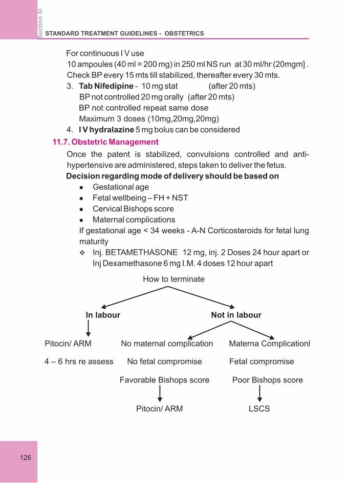
For continuous I V use
10 ampoules (40 ml = 200 mg) in 250 ml NS run at 30 ml/hr (20mgm] .
Check BP every 15 mts till stabilized, thereafter every 30 mts.
3. Tab Nifedipine - 10 mg stat (after 20 mts)
BP not controlled 20 mg orally (after 20 mts)
BP not controlled repeat same dose
Maximum 3 doses (10mg,20mg,20mg)
4. I V hydralazine 5 mg bolus can be considered
Once the patent is stabilized, convulsions controlled and anti-
hypertensive are administered, steps taken to deliver the fetus.
Decision regarding mode of delivery should be based on
l Gestational age
l Fetal wellbeing – FH + NST
l Cervical Bishops score
l Maternal complications
If gestational age < 34 weeks - A-N Corticosteroids for fetal lung
maturity
v Inj. BETAMETHASONE 12 mg, inj. 2 Doses 24 hour apart or
Inj Dexamethasone 6 mg I.M. 4 doses 12 hour apart
11.7. Obstetric Management
How to terminate
In labour Not in labour
Pitocin/ ARM No maternal complication Materna l Complication
4 – 6 hrs re assess No fetal compromise Fetal compromise
Favorable Bishops score Poor Bishops score
Pitocin/ ARM LSCS
STANDARD TREATMENT GUIDELINES - OBSTETRICSSe
cti
on
XI
126
Intrapartum Management
l Restrict IV fluids to 80 ml/hr
l Continue Magsulf
l Continuous CTG monitoring
l Partogram for progress of labour
l No prolonged labour
l Cut short second stage of labor by vacuum or forceps
l AMTSL – Methergine should not be used
l Oxytocin 5 units IV bolus diluted in 5ml saline followed by 20
units I V infusion
l Continue MgSo4 for 24 hrs after delivery or 24 hrs after last
seizure whichever is later
l Continue antihypertensives – Tab Nifidepine or Tab labetalol /
atenolol and taper and withdraw gradually.
l Thromboprohylaxis is – with Inj. LMWH 0.4 or 0.6 ml s/c ODth
l Monitor BP 4 hourly
l Retain in labour room for 24 to 48 hours till BP settles
1. Unfavorable cervix
2. Severe fetal growth restriction
3. Fetal compromise – abnormal NST/ oligamnios / abnormal
doppler
4. Maternal complications
a. Pulmonery edema
b. Renal failure
c. Placental abruption
d. Cerebral haemorrhage
5. Obstetric Indications
a. Malpresentation
b. CPD
c. Intrapartum fetal distress
d. Failed induction
11.8. Postpartum Management
11.9. Indication of LSCS
STANDARD TREATMENT GUIDELINES - OBSTETRICS
127
Se
cti
on
XI
11.10. Anaesthesia
11.11. Indication for imaging in eclampsia
11.12. Puerperal care
Reference:
Epidural analgesia/anesthesia is recommended for labour or LSCS
Combined spinal epidural may also be used
General anesthesia is associated with risk of difficult intubation and
acute rise of BP during intubation
1 patient with focal neurological deficit.
2 Patient in coma.
3 Suspected i/c heamorrhage.
4 Atypical presentation <20wks, >48hrsafter delivery.
5 Eclampsia refractory to Mgsulphate therapy.
l Continue anti hypertensives – till BP becomes normal
l If BP elevated at two weeks post partum – evaluate for chronic
hypertension, thrombophilia, chronic kidney disease
l Contraceptive advice - IUCD, progesterone only contraception,
injectable progesterone's are safe
l Combined oral contraceptive may be started after BP returns to
normal.
1. KFOG Protocol 2018
2. American College of Obstetricians and Gynecologists, Society
for Maternal-Fetal Medicine: Magnesium sulfate use in
obstetrics. Committee Opinion No. 652, January 2016c
3. NICE Guidelines – Clinical guideline 3 -2015
4. WHO recommendations for Prevention and treatment of pre-
eclampsia and eclampsia 2010th
5. Williams text book of obstetrics 25 edition
STANDARD TREATMENT GUIDELINES - OBSTETRICSSe
cti
on
XI
128
Section XII
FETAL GROWTH RESTRICTION (FGR)

Section12.
FETAL GROWTH RESTRICTION (FGR)
12.1 Introduction
12.2.Defining FGR
After prematurity, FGR is the second leading cause of prenatal
morbidity and mortality.It is widely accepted that there are 2 types of
FGR: early onset and late onset. This classification not only
describes timing of the problem, but also points to the etiology of the
disease.
Most national societies agree on the tenth percentile for the EFW as
a diagnostic cutoff for small for gestational age (SGA). The
disadvantage of this cutoff is the inclusion of a variable number of
normal constitutionally small fetuses that do not require surveillance.
Using an EFW less than the third percentile or a decreased AC
growth rate is more likely to identify “true FGR but has the
disadvantage that less severe forms of FGR at risk for deterioration
are missed and therefore their risk for stillbirth remains high.
Combining an EFW less than the third percentile with either an
abnormal UA, MCA,UtA or cerebro placental ratio (CPR, defined
as UA/MCA index), increases the identification of the small
fetus at risk for adverse outcome. Although UA Doppler
velocimetry is sufficient for the diagnosis of FGR before 32
weeks gestation, thereafter MCA Doppler is also required to
represent the whole clinical spectrum found in early-onset and
late-onset placental disease
More recently, the definition of FGR has been readdressed by an
international consensus
l Early onset (gestational age <32wk):
EFW/AC<3rd centile and/or UA-AEDF;
or EFW/AC<10th centile combined with UtA-PI>95th centile thand/or UA-PI>95 centile;
l Late onset (gestational age >32wk):
EFW/AC<3rd centile
or at least2 of 3 of the following
Se
cti
on
XII
STANDARD TREATMENT GUIDELINES - OBSTETRICS
131
(1) EFW/AC<10thcentile;
(2) EFW/AC crossing centiles>2 quartiles on growth centiles; th(3) A cerebroplacental ratio <5th or UA-PI>95 centile.
l The predictive value of abdominal palpation and symphysial-
fundal height (SFH) measurements as the primary
surveillance method for estimating fetal weight in the third
trimester is limited
l SFH measurement must be taken from the top of the fundus
to the fixed point at the upper edge of the pubic symphysis.
Measure along the fetal axis, using a non-elastic tape
measure (Figueras & Gardosi 2011)
12.3 Etiology of Fetal Growth Restriction
12.4.Screening for Fetal Growth Restriction
Maternal medical conditions
· Pregestational diabetes mellitus
· Renal insufficiency
l Autoimmune disease (eg, systemic lupus erythematosus)
l Cyanotic cardiac disease
l Pregnancy-related hypertensive diseases of pregnancy (eg,
chronic hypertension, gestational hypertension, or
preeclampsia)
l Antiphospholipid antibody syndrome
l Substance use and abuse (eg, tobacco, alcohol, cocaine, or
narcotics)
l Multiple gestation
l Teratogen exposure (eg, cyclophosphamide, valproic acid, or
antithrombotic drugs)
l Infectious diseases (eg, malaria, cytomegalovirus, rubella,
toxoplasmosis, or syphilis)
l Genetic and structural disorders (eg, trisomy 13, trisomy
congenital heart disease, or gastroschisis)
l Placental disorders and umbilical cord abnormalities
Physical Examination or History
12.4.1Abdominal examination
STANDARD TREATMENT GUIDELINES - OBSTETRICSSe
cti
on
XII
132
l Serial measurement of fundal height and plotting on a growth
chart is a useful screening tool and is recommended
Low PAPP-A may be considered as a biomarker in predicting
IUGR
Arterial and venous Doppler techniques provide detailed
information on the development of the maternal-fetal-placental
unit. The characteristics of the Doppler waveform depend on the
vascular bed that is being examined.
In particular, arterial Doppler waveforms mirror vascular
resistance, therefore providing information about the
downstream distribution of cardiac output.
Venous Doppler waveforms provide completely different
information, reflecting forward cardiac function by assessing
compliance, contractility, and afterload of the heart.
Doppler indices in clinical practice are semi quantitative
measurements, such as pulsatility index (PI), resistance index,
and systolic/diastolic ratio. Among these indices, PI has a smaller
measurement error and narrower reference limit and is therefore
the preferred index.
UtA Doppler assessment provides information regarding the
maternal side of the placenta, and is an effective evaluation
method for assessing the degree of trophoblastic invasion
Placental infarcts and inadequate trophoblast invasion may
be depicted in the UtA flow by increased resistance and/or the
prolonged persistence of a diastolic notch.Can be done as
early as 12 weeks with NT scan
UA Doppler is a reflection of villous branching in the fetal side
of the placenta. Similar to UtA development, high resistance
is seen in the UA due to incomplete invasion of the villous
branching . UA Doppler assessment should be taken in the
12.4.2 First trimester biomarkers
12.4.3 Ultrasound with doppler
12.4.3. A.UtA Doppler
12.4.3.B.UA Doppler
STANDARD TREATMENT GUIDELINES - OBSTETRICS
133
Se
cti
on
XII
mid cord, color box, and magnification should target the area
of interest only, and the Doppler gate should be positioned
between the walls of the vessel.
If the umbilical artery Doppler demonstrates increased
resistance (Pulsatility
Index >95th centile), the sonographic surveillance should be
increased to weekly intervals or more frequently if deemed
necessary by the managing clinician
As gestational age advances, normal development leads to
an increase in the peak systolic velocity (PSV) and average
diastolic velocity in the MCA. As both values increase
contingently, this allows indices of cerebral vascular
resistance to remain high throughout fetal life
It is therefore not surprising to measure high-resistance flow
during normal development throughout pregnancy, and low
resistance during FGR due to the brain sparing effect in the
fetus. The resistance of this vessel can be measured with the
PI, which is used in the management of FGR, and the
absolute velocity in the MCA can be used in assessing fetal
anemia.
Measurement should be taken at the transverse plane of the
fetal head, and the angle of insonation between the
ultrasound beam and the direction of blood flow should be
kept to 0 degrees
12.4.3.C.MCA Doppler
12.4.3.D.Aortic Isthmus Doppler
The aortic isthmus (AoI) Doppler is associated with
increased fetal mortality and neurological morbidity in early-
onset FGR . This vessel reflects the balance between the
impedance of the brain and systemic vascular systems.
Reverse AoI flow is a sign of advanced deterioration and a
further step in the sequence starting with the UA and MCA
Dopplers AoI has a strong association with both adverse
perinatal and neurological outcome
STANDARD TREATMENT GUIDELINES - OBSTETRICSSe
cti
on
XII
134
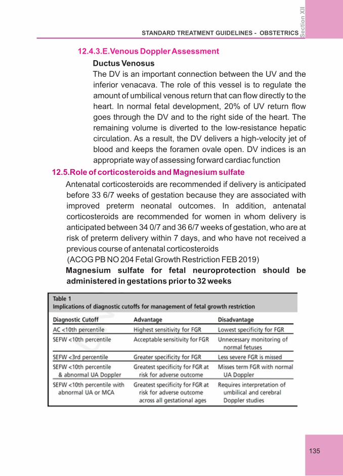
12.4.3.E.Venous Doppler Assessment
Ductus Venosus
The DV is an important connection between the UV and the
inferior venacava. The role of this vessel is to regulate the
amount of umbilical venous return that can flow directly to the
heart. In normal fetal development, 20% of UV return flow
goes through the DV and to the right side of the heart. The
remaining volume is diverted to the low-resistance hepatic
circulation. As a result, the DV delivers a high-velocity jet of
blood and keeps the foramen ovale open. DV indices is an
appropriate way of assessing forward cardiac function
Magnesium sulfate for fetal neuroprotection should be
administered in gestations prior to 32 weeks
12.5.Role of corticosteroids and Magnesium sulfate
Antenatal corticosteroids are recommended if delivery is anticipated
before 33 6/7 weeks of gestation because they are associated with
improved preterm neonatal outcomes. In addition, antenatal
corticosteroids are recommended for women in whom delivery is
anticipated between 34 0/7 and 36 6/7 weeks of gestation, who are at
risk of preterm delivery within 7 days, and who have not received a
previous course of antenatal corticosteroids
(ACOG PB NO 204 Fetal Growth Restriction FEB 2019)
STANDARD TREATMENT GUIDELINES - OBSTETRICS
135
Se
cti
on
XII
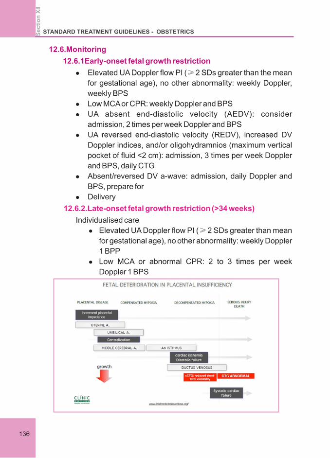
12.6.Monitoring
12.6.1Early-onset fetal growth restriction
l Elevated UA Doppler flow PI (≥ 2 SDs greater than the mean
for gestational age), no other abnormality: weekly Doppler,
weekly BPS
l Low MCA or CPR: weekly Doppler and BPS
l UA absent end-diastolic velocity (AEDV): consider
admission, 2 times per week Doppler and BPS
l UA reversed end-diastolic velocity (REDV), increased DV
Doppler indices, and/or oligohydramnios (maximum vertical
pocket of fluid <2 cm): admission, 3 times per week Doppler
and BPS, daily CTG
l Absent/reversed DV a-wave: admission, daily Doppler and
BPS, prepare for
l Delivery
Individualised care
l Elevated UA Doppler flow PI (≥ 2 SDs greater than mean
for gestational age), no other abnormality: weekly Doppler
1 BPP
l Low MCA or abnormal CPR: 2 to 3 times per week
Doppler 1 BPS
12.6.2.Late-onset fetal growth restriction (>34 weeks)
STANDARD TREATMENT GUIDELINES - OBSTETRICSSe
cti
on
XII
136
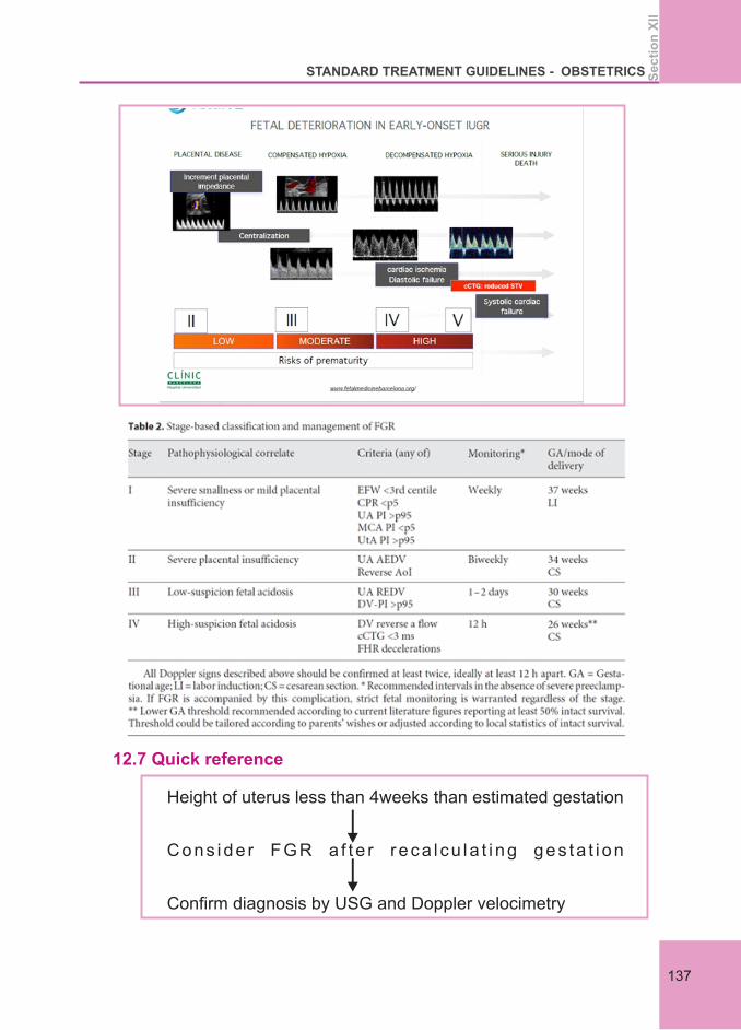
12.7 Quick reference
Height of uterus less than 4weeks than estimated gestation
Cons ide r FGR a f t e r r eca l cu la t i ng ges ta t i on
Confirm diagnosis by USG and Doppler velocimetry
STANDARD TREATMENT GUIDELINES - OBSTETRICS
137
Se
cti
on
XII
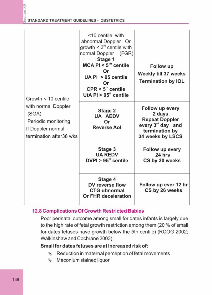
Growth < 10 centile
with normal Doppler
(SGA)
Periodic monitoring
If Doppler normal
termination after38 wks
<10 centile with abnormal Doppler Or
rdgrowth < 3 centile with
normal Doppler (FGR) Stage 1
THMCA PI < 5 centileOr
UA PI > 95 centileOr
thCPR < 5 centile
thUtA PI > 95 centile
Stage 2UA AEDV
OrReverse AoI
Stage 3UA REDV
thDVPI > 95 centile
Stage 4DV reverse flowCTG ubnormal
Or FHR deceleration
Follow up ever 12 hr CS by 26 weeks
Follow up every 24 hrs
CS by 30 weeks
Follow up every 2 days
Repeat Doppler rdevery 3 day and
termination by 34 weeks by LSCS
Follow up
Weekly till 37 weeks
Termination by IOL
12.8 Complications Of Growth Restricted Babies
Poor perinatal outcome among small for dates infants is largely due
to the high rate of fetal growth restriction among them (20 % of small
for dates fetuses have growth below the 5th centile) (RCOG 2002;
Walkinshaw and Cochrane 2003)
Small for dates fetuses are at increased risk of:
Ä Reduction in maternal perception of fetal movements
Ä Meconium stained liquor
STANDARD TREATMENT GUIDELINES - OBSTETRICSSe
cti
on
XII
138
Ä Abnormal heart rate patterns intrapartum
Ä Intrauterine fetal death
Ä Hypoxic ischaemic encephalopathy
Ä Poor neurological development
Ä Delay in cognitive development
Ä Sudden infant death syndrome
In adult life
Type 2 diabetes and hypertension (RCOG 2002)
NB: Good catch up growth in the first few months of life may predict a
healthy outcome
Patients with previous history of IUGR should be started on Ecospirin
150mg not later than16 weeks of gestation.
2. CLINICAL OBSTETRICS AND GYNECOLOGY Volume 60,
Number 3, 668–678 2017
3. Unterscheider J, Daly S, Geary MP, et al. Optimizing the definition
of intrauterine growth restriction: the multicenter prospective
PORTO Study. Am JObstet Gynecol. 2013;208:290e1–290.e6
4. Gordijn SJ, Beune IM, Thilaganathan B, et al.Consensus
definition of fetal growth restriction: a Delphi procedure.
Ultrasound Obstet Gynecol.2016;48:333–339.
5. Figueras F, Gardosi J. Intrauterine growth restriction: new
concepts in antenatal surveillance, diagnosis and management.
AJOG 2011; 204: 288-300.
References
1. Fetal growth restriction. ACOG Practice Bulletin No. 204.
American College of Obstetricians and Gynecologists Obstet
Gynecol 2019;133:e97–109. Huisman TW. Doppler assessment
of the fetal venous system. Semin Perinatol. 2001;25:21–31
6. Del Rio M, et al: Doppler assessment of the aortic isthmus and
perinatal outcome in preterm fetuses with severe intrauterine
growth restriction. Ultrasound Obstet Gynecol 2008;31: 41–47.
7. Update on the Diagnosis and Classification of Fetal Growth
Restriction and Proposalof a Stage-Based Management
Protocol Francesc Figueras Eduard GratacósBarcelona Center
STANDARD TREATMENT GUIDELINES - OBSTETRICS
139
Se
cti
on
XII

of Maternal-Fetal Medicine and Neonatology (2014)
8. KEY CURRENT ISSUES In IUGR Eduard Gratacós Maternal-
Fetal Medicine Department Hospital Clínic, Universidad de
Barcelona
STANDARD TREATMENT GUIDELINES - OBSTETRICSSe
cti
on
XII
140

13.INDUCTION OF LABOUR
Section XIIIINDUCTION OF LABOUR

Section 13.
INDUCTION OF LABOUR
13.1 Definition:
13.2 Introduction
13.3 Perinatal Outcomes
13.4 Adverse Long-Term Infant Outcomes
Induction implies stimulation of contractions before the spontaneous
onset of labour, with or without ruptured membranes.
In 2009, several large cohort studies, including a study from the
Eunice Kennedy Shriver National Institute of Child Health and
Human Development (NICHD) Maternal-Fetal Medicine Units
Network,reported that early-term delivery (37 0/7–38 6/7 weeks of
gestation) was associated with increased neonatal and infant
morbidity and mortality compared with deliveries at 39 weeks of
gestation or more. The NICHD study also found that more than one
third of non medically indicated cesarean deliveries occurred at less
than 39 weeks of gestation
The risk of adverse outcomes is greater for neonates delivered in the
early-term period (37 0/7–38 6/7 weeks of gestation) compared with
neonates delivered at 39 weeks of gestation . Because pulmonary
development continues well into early childhood, respiratory
morbidity is relatively common in neonates delivered in the early-
term period. A retrospective cohort study by the Consortium on Safe
Labor, which included 233,844 births, found that among all infants
delivered at 37 weeks of gestation, regardless of indication, there
were higher rates of respiratory failure (adjusted odds ratio [OR], 2.8;
95% CI, 2.0–3.9) and ventilator use (adjusted OR, 2.8; 95% CI,
2.3–3.4) compared with infants delivered at 39 weeks of gestation
In addition to immediate adverse perinatal outcomes, multiple
studies have shown increased rates of adverse long-term infant
outcomes associated with late-preterm and early-term delivery
compared with full-term delivery. Studies have reported that
compared with children born full term, children born late preterm and
early term experience additional long-term adverse consequences
Se
cti
on
XII
I
STANDARD TREATMENT GUIDELINES - OBSTETRICS
143
including increased hospitalizations up to age 18 slower neurologic
development , worse cognitive performance , more school-related
problems, and poorer academic achievement .
l Induction of labour should be performed only when there is a
clear indication for it – maternal or fetal.
l Informed written consent should be taken before induction.
l Induction of labour should be performed with caution since the
procedure carries the risk of uterine hyper stimulation, rupture
and fetal distress.
l Wherever induction of labour is carried out, facilities should be
available for assessing maternal and fetal well-being and for
emergency caesarean section.
l Women receiving oxytocin, misoprostol or other prostaglandins
should never be left unattended.
l Preeclampsia, eclampsia, gestational hypertension, or
complicated chronic hypertension
l Pregestational diabetes with vascular disease
l Pregestational or gestational diabetes—poorly controlled
l Cholestasis of pregnancy
l Alloimmunization of pregnancy with known or suspected fetal
effects
l Placenta previa -----Late preterm/early term 36 –38 weeks of
gestation
l Suspected accreta, increta, or percretay -----Late preterm 34
–36 weeks of gestation
l Vasa previa Late preterm/early term ------34 –37 weeks of
gestation
l Prior classical cesarean Late preterm/early term 36 –37
13.5 GENERAL PRINCIPLES related to the practice of induction of
labour
13.6 Indications for termination(IOL/LSCS)
:
13.6.1 Medical Indications for Late-Preterm or Early-Term
Deliveries*
13.6.2 Placental/Uterine Conditions(timing of delivery)
STANDARD TREATMENT GUIDELINES - OBSTETRICSSe
cti
on
XII
I
144
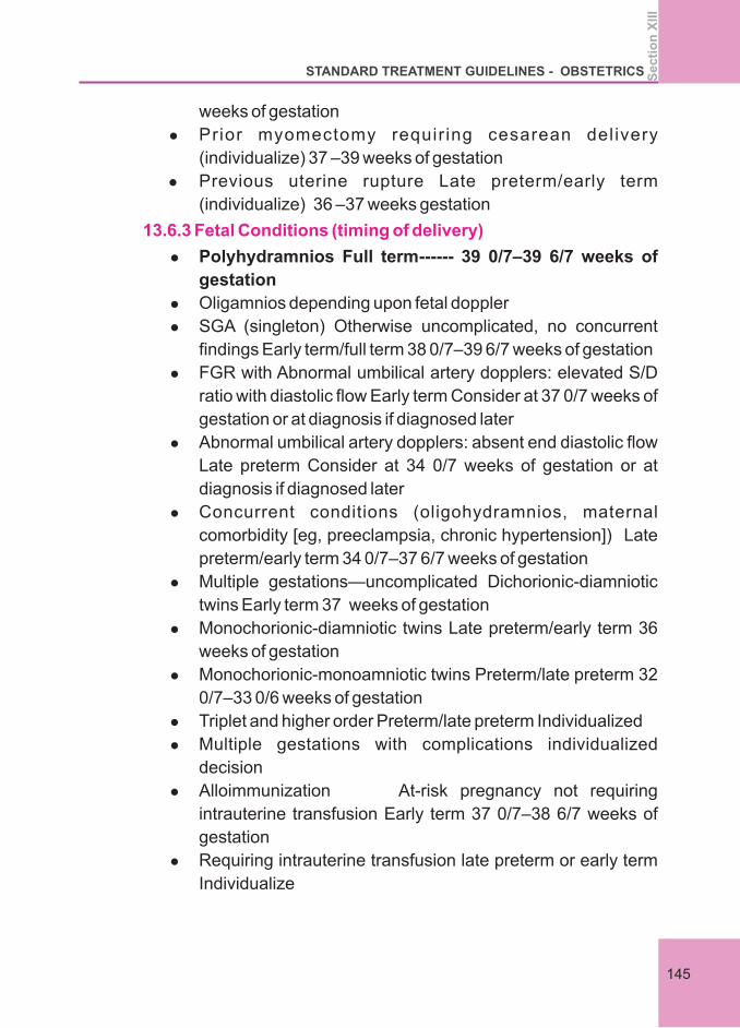
weeks of gestation
l Prior myomectomy requir ing cesarean del ivery
(individualize) 37 –39 weeks of gestation
l Previous uterine rupture Late preterm/early term
(individualize) 36 –37 weeks gestation
l Polyhydramnios Full term------ 39 0/7–39 6/7 weeks of
gestation
l Oligamnios depending upon fetal doppler
l SGA (singleton) Otherwise uncomplicated, no concurrent
findings Early term/full term 38 0/7–39 6/7 weeks of gestation
l FGR with Abnormal umbilical artery dopplers: elevated S/D
ratio with diastolic flow Early term Consider at 37 0/7 weeks of
gestation or at diagnosis if diagnosed later
l Abnormal umbilical artery dopplers: absent end diastolic flow
Late preterm Consider at 34 0/7 weeks of gestation or at
diagnosis if diagnosed later
l Concurrent conditions (oligohydramnios, maternal
comorbidity [eg, preeclampsia, chronic hypertension]) Late
preterm/early term 34 0/7–37 6/7 weeks of gestation
l Multiple gestations—uncomplicated Dichorionic-diamniotic
twins Early term 37 weeks of gestation
l Monochorionic-diamniotic twins Late preterm/early term 36
weeks of gestation
l Monochorionic-monoamniotic twins Preterm/late preterm 32
0/7–33 0/6 weeks of gestation
l Triplet and higher order Preterm/late preterm Individualized
l Multiple gestations with complications individualized
decision
l Alloimmunization At-risk pregnancy not requiring
intrauterine transfusion Early term 37 0/7–38 6/7 weeks of
gestation
l Requiring intrauterine transfusion late preterm or early term
Individualize
13.6.3 Fetal Conditions (timing of delivery)
STANDARD TREATMENT GUIDELINES - OBSTETRICS
145
Se
cti
on
XII
I
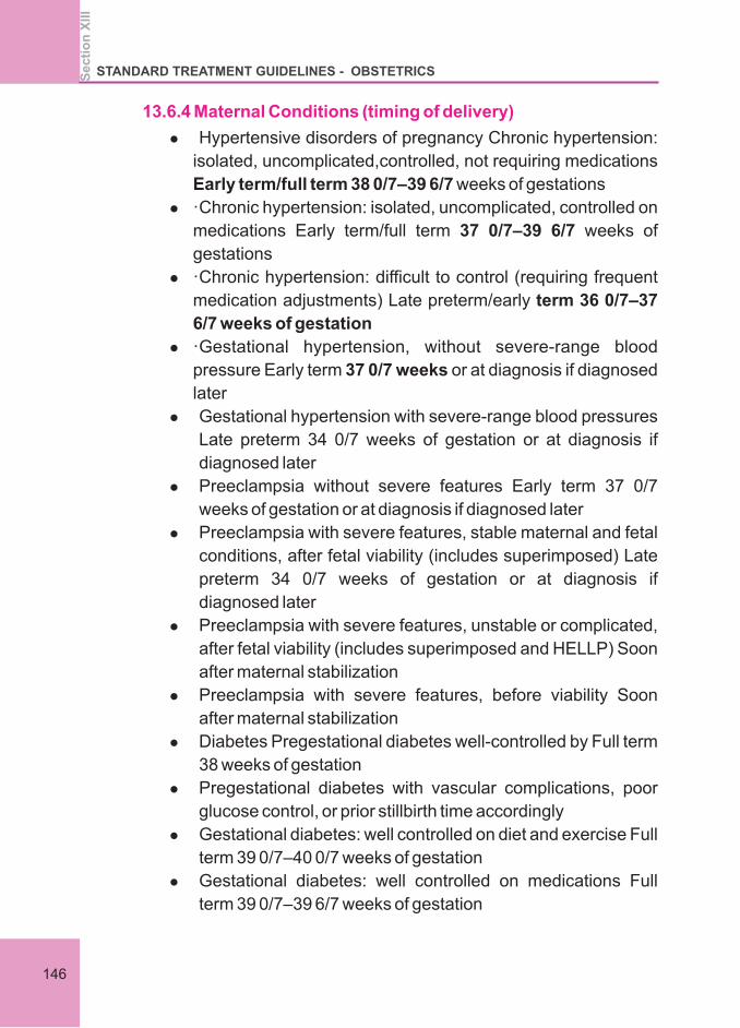
13.6.4 Maternal Conditions (timing of delivery)
l Hypertensive disorders of pregnancy Chronic hypertension:
isolated, uncomplicated,controlled, not requiring medications
Early term/full term 38 0/7–39 6/7 weeks of gestations
l ·Chronic hypertension: isolated, uncomplicated, controlled on
medications Early term/full term 37 0/7–39 6/7 weeks of
gestations
l ·Chronic hypertension: difficult to control (requiring frequent
medication adjustments) Late preterm/early term 36 0/7–37
6/7 weeks of gestation
l ·Gestational hypertension, without severe-range blood
pressure Early term 37 0/7 weeks or at diagnosis if diagnosed
later
l Gestational hypertension with severe-range blood pressures
Late preterm 34 0/7 weeks of gestation or at diagnosis if
diagnosed later
l Preeclampsia without severe features Early term 37 0/7
weeks of gestation or at diagnosis if diagnosed later
l Preeclampsia with severe features, stable maternal and fetal
conditions, after fetal viability (includes superimposed) Late
preterm 34 0/7 weeks of gestation or at diagnosis if
diagnosed later
l Preeclampsia with severe features, unstable or complicated,
after fetal viability (includes superimposed and HELLP) Soon
after maternal stabilization
l Preeclampsia with severe features, before viability Soon
after maternal stabilization
l Diabetes Pregestational diabetes well-controlled by Full term
38 weeks of gestation
l Pregestational diabetes with vascular complications, poor
glucose control, or prior stillbirth time accordingly
l Gestational diabetes: well controlled on diet and exercise Full
term 39 0/7–40 0/7 weeks of gestation
l Gestational diabetes: well controlled on medications Full
term 39 0/7–39 6/7 weeks of gestation
STANDARD TREATMENT GUIDELINES - OBSTETRICSSe
cti
on
XII
I
146
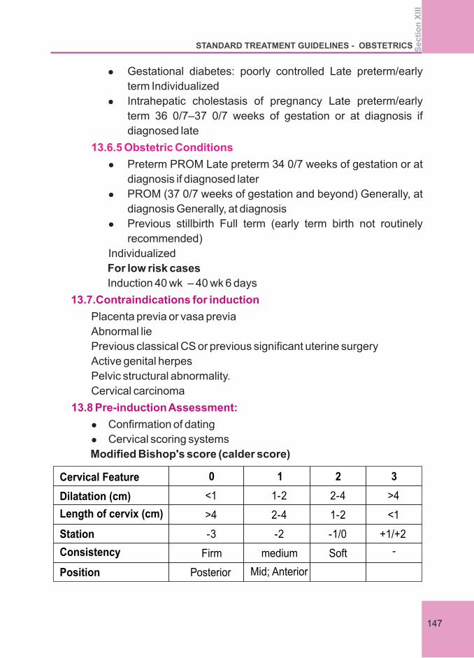
l Gestational diabetes: poorly controlled Late preterm/early
term Individualized
l Intrahepatic cholestasis of pregnancy Late preterm/early
term 36 0/7–37 0/7 weeks of gestation or at diagnosis if
diagnosed late
l Preterm PROM Late preterm 34 0/7 weeks of gestation or at
diagnosis if diagnosed later
l PROM (37 0/7 weeks of gestation and beyond) Generally, at
diagnosis Generally, at diagnosis
l Previous stillbirth Full term (early term birth not routinely
recommended)
Individualized
13.6.5 Obstetric Conditions
For low risk cases
Induction 40 wk – 40 wk 6 days
Placenta previa or vasa previa
Abnormal lie
Previous classical CS or previous significant uterine surgery
Active genital herpes
Pelvic structural abnormality.
Cervical carcinoma
l Confirmation of dating
l Cervical scoring systems
Modified Bishop's score (calder score)
13.7.Contraindications for induction
13.8 Pre-induction Assessment:
Cervical Feature 0 1 2 3
Dilatation (cm) <1 1-2 2-4 >4
Length of cervix (cm) >4
2-4
1-2
<1
Station -3
-2
-1/0
+1/+2
Consistency Firm medium Soft -
Position Posterior
Mid; Anterior
STANDARD TREATMENT GUIDELINES - OBSTETRICS
147
Se
cti
on
XII
I
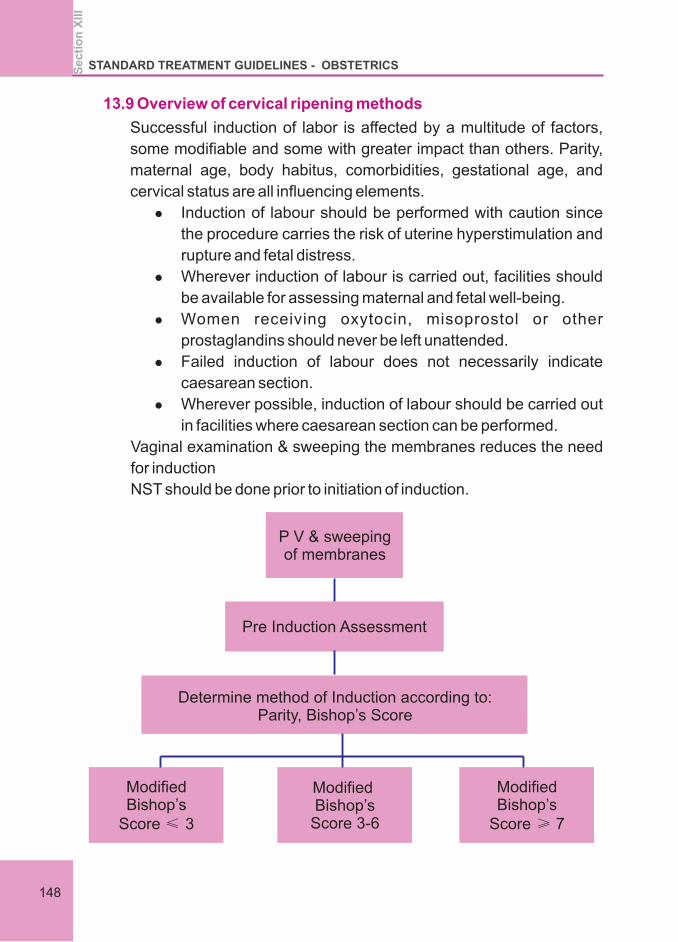
13.9 Overview of cervical ripening methods
Successful induction of labor is affected by a multitude of factors,
some modifiable and some with greater impact than others. Parity,
maternal age, body habitus, comorbidities, gestational age, and
cervical status are all influencing elements.
l Induction of labour should be performed with caution since
the procedure carries the risk of uterine hyperstimulation and
rupture and fetal distress.
l Wherever induction of labour is carried out, facilities should
be available for assessing maternal and fetal well-being.
l Women receiving oxytocin, misoprostol or other
prostaglandins should never be left unattended.
l Failed induction of labour does not necessarily indicate
caesarean section.
l Wherever possible, induction of labour should be carried out
in facilities where caesarean section can be performed.
Vaginal examination & sweeping the membranes reduces the need
for induction
NST should be done prior to initiation of induction.
P V & sweeping of membranes
Pre Induction Assessment
Determine method of Induction according to: Parity, Bishop’s Score
Modified Bishop’s
Score ≤ 3
Modified Bishop’s
Score 3-6
Modified Bishop’s
Score ≥ 7
STANDARD TREATMENT GUIDELINES - OBSTETRICSSe
cti
on
XII
I
148
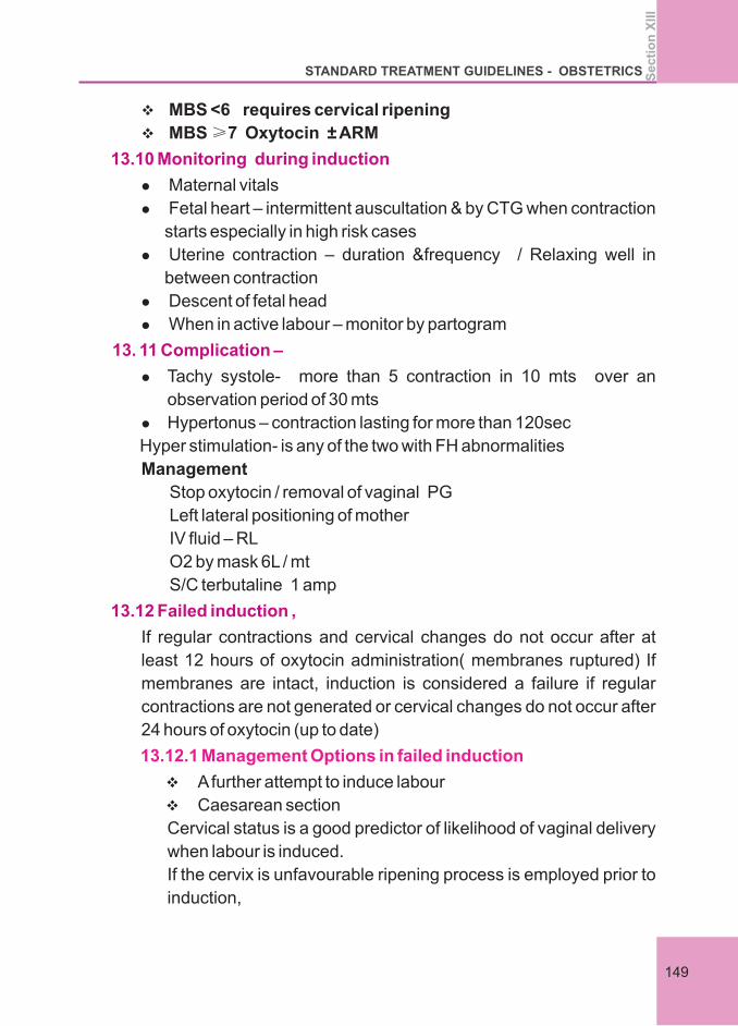
v MBS <6 requires cervical ripening
v MBS ≥7 Oxytocin ± ARM
l Maternal vitals
l Fetal heart – intermittent auscultation & by CTG when contraction
starts especially in high risk cases
l Uterine contraction – duration &frequency / Relaxing well in
between contraction
l Descent of fetal head
l When in active labour – monitor by partogram
l Tachy systole- more than 5 contraction in 10 mts over an
observation period of 30 mts
l Hypertonus – contraction lasting for more than 120sec
Hyper stimulation- is any of the two with FH abnormalities
Management
Stop oxytocin / removal of vaginal PG
Left lateral positioning of mother
IV fluid – RL
O2 by mask 6L / mt
S/C terbutaline 1 amp
If regular contractions and cervical changes do not occur after at
least 12 hours of oxytocin administration( membranes ruptured) If
membranes are intact, induction is considered a failure if regular
contractions are not generated or cervical changes do not occur after
24 hours of oxytocin (up to date)
v A further attempt to induce labour
v Caesarean section
Cervical status is a good predictor of likelihood of vaginal delivery
when labour is induced.
If the cervix is unfavourable ripening process is employed prior to
induction,
13.10 Monitoring during induction
13. 11 Complication –
13.12 Failed induction ,
13.12.1 Management Options in failed induction
STANDARD TREATMENT GUIDELINES - OBSTETRICS
149
Se
cti
on
XII
I
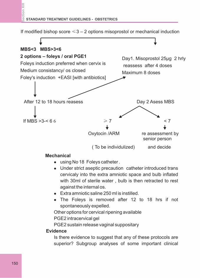
If modified bishop score ≤3 – 2 options misoprostol or mechanical induction
MBS<3 MBS>3<6
2 options – foleys / oral PGE1
Foleys induction preferred when cervix is
Medium consistancy/ os closed
Foley's induction +EASI [with antibiotics]
Day1. Misoprostol 25µg 2 hrly
reassess after 4 doses
Maximum 8 doses
After 12 to 18 hours reasess Day 2 Asess MBS
If MBS >3-< 6 6 ≥ 7 < 7
Oxytocin /ARM re assessment by senior person
( To be individulized) and decide
Mechanical
l using No 18 Foleys catheter .
l Under strict aseptic precaution catheter introduced trans
cervicaly into the extra amniotic space and bulb inflated
with 30ml of sterile water , bulb is then retracted to rest
against the internal os.
l Extra amniotic saline 250 ml is instilled.
l The Foleys is removed after 12 to 18 hrs if not
spontaneously expelled.
Other options for cervical ripening available
PGE2 intracervical gel
PGE2 sustain release vaginal suppositary
Evidence
Is there evidence to suggest that any of these protocols are
superior? Subgroup analyses of some important clinical
STANDARD TREATMENT GUIDELINES - OBSTETRICSSe
cti
on
XII
I
150
outcomes show a clear dose effect. For example, when
comparing oral misoprostol with dinoprostone, the rate of
hyperstimulation increases as the initial dose rises from 25 12to 200 µg . ,It would therefore appear that there are safety
benefits of using doses of 20–25 µg, even if they may result
in a slower induction process.
This is supported by a systematic review of the studies that 16
used 20–25 µg of oral misoprostol, which found lower
caesarean section and lower hyperstimulation rates 8
compared with standard induction methods. And whereas in
previous studies researchers have been forced to either use
cut 200-mcg tablets or solution, high-quality 25- µg tablets
are now available.
Findings from a non-inferiority randomised controlled trial
(RCT) of oral misoprostol 50mcg versus Foley catheter for
induction of labour showed equivalent safety and 13
effectiveness, whereas misoprostol tablets (25 µg) has
recently been found to be more an effective than Foley
catheter when given orally in a large Medical Research 15 Council (MRC) labour induction study. The use of regimens
in which misoprostol is given every 2 hours is supported by
pharmacokinetic studies that show that oral misoprostol
reaches its peak serum level within 30 minutes, but that its
half-life is only 90 minutes as misoprostol is rapidly 11
metabolised by the liver and excreted by the kidneys. With
oral misoprostol sustained uterine activity is achieved in 90
minutes and the duration of action is approximately 2 hours.
The 4–6 hourly dosage regimens have stemmed from an
incorrect assumption that the oral pharmacokinetic data is 11
the same as that for vaginal doses.
Misoprostol has advantages in being cheap and stable at
room temperature and widely available also in most
resource-poor settings. Misoprostol is included in the World
Health Organization (WHO) essential medicine list on
several indications including labor induction . Orally
STANDARD TREATMENT GUIDELINES - OBSTETRICS
151
Se
cti
on
XII
I
administrated misoprostol has in several publications been
shown to be an effective method for induction of labor,
comparable with PGE2 and oxytocin.
To prepare the misoprostol oral solution, one tablet of 200 μg
misoprostol was dissolved in 20 ml of water. The solution thus
held 10 μg of misoprostol/ml. This method of administration
has been tested by the Swedish Institute of Pharmacology
and approved to be accurate in terms of correct dosage. A
dose of 2.5 ml/25 μg of misoprostol was administered orally
to the women every two hours until frequent painful
contractions were obtained. The dose could be repeated up
to eight times if necessary. When ripening of the cervix had
been achieved (MBS ≥ 6), amniotomy and oxytocin were
used to support uterine contractions.
ARRIVE TRIAL(Randomised trial of induction v/s expectant
management. The study suggests induction of labour in the
39 th week, resulted in fewer caesarean section. ACOG
does not currently recommend routine induction of labour for
low risk pregnancy at 39 weeks.
Induction not preferred wait for spontaneous onset of labour
Can use mechanical methods and later controlled oxytocin
PGE2 can be used with caution (NICE Guidelines)
AUGMENTATION OF LABOUR will be done when there is delay
of progress of labour in partogram
Augmentation is given with 5units oxytocin in 500ml of RL
Start with 4 drops/ mt
Increasing dosage every 30mts titrating against uterine contraction
& FH
Uterine contraction may last for 45 sec /.ensure relaxation in
between
FH should be reassuring – ideal by continuous CTG
Max dosage is 60drops / mt
13.13 Special situations for induction of labour in previous
caesarean
13.14 Oxytocin in labour
STANDARD TREATMENT GUIDELINES - OBSTETRICSSe
cti
on
XII
I
152
If required a second infusion with 5 units in 500ml of RL is given
starting with half the infusion rate reached in the first infusion
When parturient reaches second stage maintain oxytocin infusion in
the same concentration & rate
AMTSL should be strictly adhered to.
Preparation of infusion
5 units in 500ml of RL
= 10 milliunits in 1 ml
1 ml = 16 drops
4drops/mt = 2.5 mu /mt
Max - 60 drops / mt
l First vaginal examination of a woman with symptoms suggestive
of labour will be done when she has contractions lasting for more
than 20 sec occurring at least once in 4 minutes
l Active labour is diagnosed when cervical dilatation reaches 6cm
- Partogram to be initiated.
l Repeat vaginal examination is done after 4 hours in active labour.
l Repeat vaginal examination if woman is in latent phase or false
labour is not warranted.
l Vaginal examination may be done earlier if there is FH
abnormality.
l If vaginal examination was not done on admission and there are
no contractions after an observation period of 6 hours woman
may be shifted from labour ward.
13.15.Vaginal examination in labour
STANDARD TREATMENT GUIDELINES - OBSTETRICS
153
Se
cti
on
XII
I
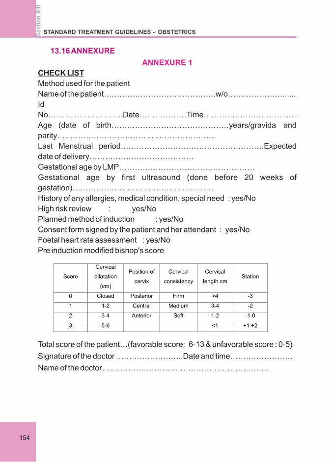
13.16 ANNEXURE
ANNEXURE 1
CHECK LIST
Method used for the patient
Name of the patient…………………………………….w/o…………………......
Id
No………………………..Date………………Time…………………………...…
Age (date of birth………………………………………years/gravida and
parity…………………………………………………….
Last Menstrual period……………………………………………….Expected
date of delivery………………………………….
Gestational age by LMP…………………………………………….
Gestational age by first ultrasound (done before 20 weeks of
gestation)………………………………………………
History of any allergies, medical condition, special need : yes/No
High risk review : yes/No
Planned method of induction : yes/No
Consent form signed by the patient and her attendant : yes/No
Foetal heart rate assessment : yes/No
Pre induction modified bishop's score
Total score of the patient…(favorable score: 6-13 & unfavorable score : 0-5)
Signature of the doctor ……………………..Date and time……………………
Name of the doctor……………………………………………………….
Score
Cervical
dilatation
(cm)
Position of
cervix
Cervical
consistency
Cervical
length cmStation
0
Closed
Posterior
Firm
>4 -3
1
1-2
Central
Medium
3-4 -2
2
3-4
Anterior
Soft
1-2 -1-0
3
5-6
<1 +1 +2
STANDARD TREATMENT GUIDELINES - OBSTETRICSSe
cti
on
XII
I
154
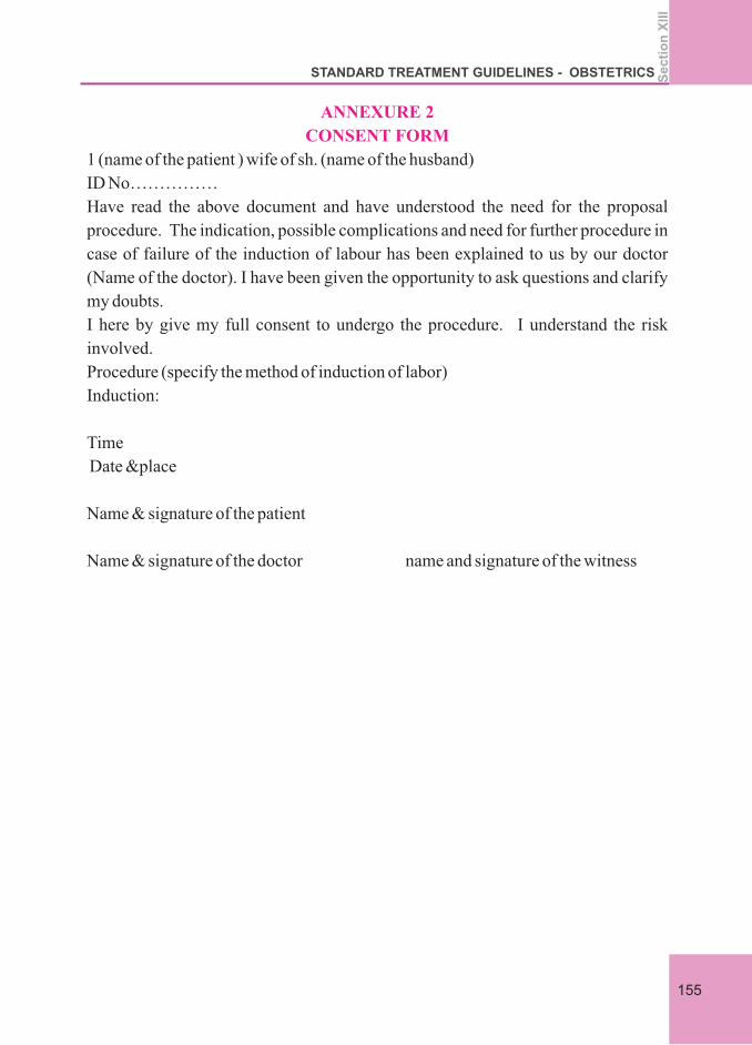
ANNEXURE 2
CONSENT FORM
1 (name of the patient ) wife of sh. (name of the husband)
ID No……………
Have read the above document and have understood the need for the proposal
procedure. The indication, possible complications and need for further procedure in
case of failure of the induction of labour has been explained to us by our doctor
(Name of the doctor). I have been given the opportunity to ask questions and clarify
my doubts.
I here by give my full consent to undergo the procedure. I understand the risk
involved.
Procedure (specify the method of induction of labor)
Induction:
Time
Date &place
Name & signature of the patient
Name & signature of the doctor name and signature of the witness
STANDARD TREATMENT GUIDELINES - OBSTETRICS
155
Se
cti
on
XII
I
Reference
1. SOGC guideline 2013
www.uptodate.com
2. April2017
3. Medically indicated late-preterm and early-term deliveries.
ACOG Committee Opinion No. 764. American College of
Obstetr ic ians and Gynecologists. Obstet Gynecol
2019;133:e151–55.
4. Clark SL, Miller DD, Belfort MA, Dildy GA, Frye DK, Meyers JA.
Neonatal and maternal outcomes associated with elective term
delivery. Am J Obstet Gynecol 2009; 200:156.e1–4
5. Oshiro BT, Henry E, Wilson J, Branch DW, Varner MW.
Decreasing elective deliveries before 39 weeks of gestation in an
integrated health care system. Women and Newborn Clinical
Integration Program. Obstet Gynecol 2009;113: 804–11
6. Tita AT, Landon MB, Spong CY, Lai Y, Leveno KJ, Varner MW, et
al. Timing of elective repeat cesarean delivery at term and
neonatal outcomes. Eunice Kennedy Shriver NICHD Maternal-
Fetal Medicine Units Network. N EnglJ Med 2009;360:111–20.
7. Zhang X, Kramer MS. Variations in mortality and morbidity by
gestational age among infants born at term. J Pediat
2009;154;358–62, 362.e1.
8. Induction of labor. ACOG Practice Bulletin No. 107. American
College of Obstetricians and Gynecologists. Obstet Gynecol
2009;114:386–97.
9. Osterman MJ, Martin JA. Recent declines in induction of labor by
gestational age. NCHS Data Brief 2014;155:1–8. Schoen CN,
Tabbah S, Iams JD, Caughey AB, Berghella VWhy the United
States preterm birth rate is declining. Am J Obstet Gynecol
2015;213:175–80.
10. Nonmedically indicated early-term deliveries and associated
neonatal morbidities. ACOG Committee Opinion No.765.
STANDARD TREATMENT GUIDELINES - OBSTETRICSSe
cti
on
XII
I
156
American College of Obstetricians and Gynecologists. Obstet
Gynecol 2019;133:e156–63.
11.FIGO.[http://www.figo.org/sites/default/files/uploads/projectpubli
cations/Miso/Misoprostol_Recommended%20Dosages%2020
12.pdf] Accessed 7 July 2016
12. Alfirevic Z, Aflaifel N, Weeks A. Oral misoprostol for induction
oflabour. The Cochrane database of systematic reviews.
2014;(6):
13. M. L. G. Ten Eikelder, K. Oude Rengerink, M. Jozwiak et al.,
“Induction of labour at term with oral misoprostol versus a Foley
catheter (PROBAAT-II): a multicentre randomised controlled
non-inferiority trial,” The Lancet, vol. 387, no. 10028, pp.
1619–1628, 2016.
14. M. L. Stephenson and D. A. Wing, “Misoprostol for induction of
labor,” Seminars in Perinatology, vol. 39, no. 6, pp. 459–462,
2015
15. G. J. Hofmeyr, “Oral misoprostol is as safe as Foley catheter for
labour induction...or is it?” The Lancet, vol. 387, no. 10028, pp.
1593-1594, 2016
16. Z. Alfirevic, E. Keeney, T. Dowswell et al., “Methods to induce
labour: a systematic review, network meta-analysis and cost-
effectiveness analysis,” BJOG: An International Journal of
Obstetrics and Gynaecology, vol. 123, no. 9, pp. 1462–1470,
2016
STANDARD TREATMENT GUIDELINES - OBSTETRICS
157
Se
cti
on
XII
I


Section XIV
CAESAREAN SECTION

Section14
CAESAREAN SECTION
Caesarean section is one of most commonly performed major
operations in women throughout the world. There are many possible
approaches to performing a caesarean section, the aim being to achieve
safe delivery of the infant with a minimum maternal morbidity.
Category 1:EMERGENCY:immediate threat to the life of the woman
or foetus(within 30 mts)
Category 2:URGENT:maternal or foetal compromise which is not
immediately life-threatening( within 75 mts)
Category3:SCHEDULED:no maternal or foetal compromise but
needs early delivery
Category 4:ELECTIVE:delivery timed to suit woman or
staff(elective caesarean section to be planned at 39 weeks.If
planned before 39 weeks steroids recommended.
l Offer pregnant women information and support.
l This information should include indications for CS, what the
procedure involves, associated risks and benefits, and
implications for future pregnancies and birth after CS.
l Communication and information should be provided in a form
that is accessible.
l Consent for CS should be requested after providing pregnant
women with evidence-based information.
l Blood routine which includes haemoglobin, packed cell volume,
total count, Platelet count.
l Blood grouping & Rh typing
l Screening - HbsAg, HIV, VDRL, Anti HCV, Platelet, BT,CT,
Electrolytes, Na and K
14.1 Categories of cesarean section
14.2.Pre requisites for caesarean section
14.3 Preoperative testing & preparation of CS
Se
cti
on
XIV
STANDARD TREATMENT GUIDELINES - OBSTETRICS
161
aspiration pneumonia
l Women who are having induction of regional anaesthesia for CS
should be cared for in theatre because this does not increase the
woman's anxiety.
l Cardiac patients – cardiac anesthesia according to their lesion
and pathology
l Women having a CS should be offered antiemetics to reduce
nausea and vomiting during CS.
l The operating table for CS should have a lateral tilt of 15°,
because this reduces maternal hypotension.
General recommendations for safe surgical practice to ensure strict
aspects should be followed at CS to reduce the risk of HIV infection of
staff.
l Vaginal cleansing with povidone iodine solution immediately
prior to caesarean birth may reduce post op endometritis
especially when membranes ruptured or early in
labour.(WHO)
Abdomen should be cleaned using chlorhexidine.CS should be
performed using a transverse abdominal incision because this is
associated with less postoperative pain and an improved
cosmetic effect compared with a midline incision.
Low transverse incision(Kerr incision)When there is a well
formed lower uterine segment, blunt rather than sharp extension
of the uterine incision should be used because it reduces blood
loss incidence of postpartum haemorrhage and the need for
transfusion at CS.
Vertical incision:
Placenta accreta
Anterior leiomyoma obstructing the lower uterine segment
14.5.Surgical technique
14.5.1.Skin Incision
14.5.2 Uterine incision:
:
Vertical
incision in situations like complete placenta praevia ,accreta and
lower segment fibroids
15
STANDARD TREATMENT GUIDELINES - OBSTETRICSSe
cti
on
XIV
162
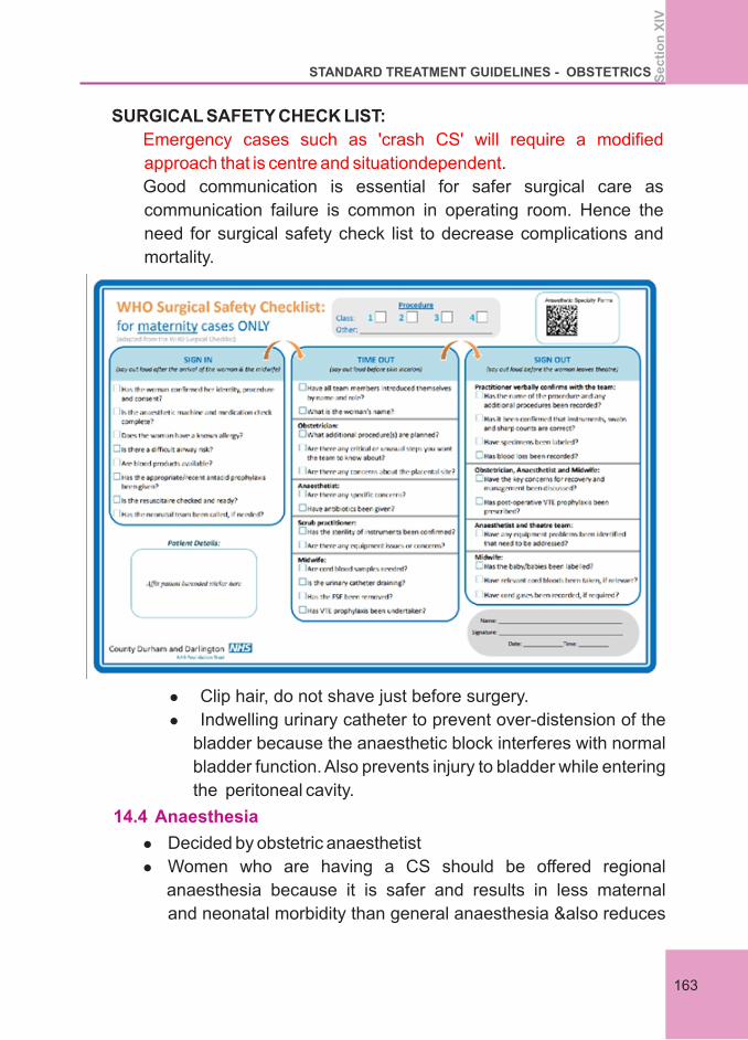
SURGICAL SAFETY CHECK LIST:
.
Good communication is essential for safer surgical care as
communication failure is common in operating room. Hence the
need for surgical safety check list to decrease complications and
mortality.
Emergency cases such as 'crash CS' will require a modified
approach that is centre and situationdependent
l Clip hair, do not shave just before surgery.
l Indwelling urinary catheter to prevent over-distension of the
bladder because the anaesthetic block interferes with normal
bladder function. Also prevents injury to bladder while entering
the peritoneal cavity.
l Decided by obstetric anaesthetist
l Women who are having a CS should be offered regional
anaesthesia because it is safer and results in less maternal
and neonatal morbidity than general anaesthesia &also reduces
14.4 Anaesthesia
163
Se
cti
on
XIV
STANDARD TREATMENT GUIDELINES - OBSTETRICS
Fetus in a backdown transverse lie
Complete placenta previa
l Women who are having a CS should be informed that
the risk of fetal lacerations is about 2%.
l Forceps should only be used at CS if there is difficulty
delivering the baby's head. The effect on neonatal
morbidity of the routine use of forceps at CS remains
uncertain.
l Delayed cord clamping (by 30-120 sec)
l Oxytocin 5 IU by slow intravenous injection should be
used at CS to encourage contraction of the uterus and
to decrease blood loss
l At CS, the placenta should be removed using
controlled cord traction and not manual removal as this
reduces the risk of endometritis.
l Intraperitoneal repair of the uterus at CS should be
undertaken. Routine exteriorisation of the uterus is not
recommended because it is associated with more pain
and does not improve operative outcomes such as
haemorrhage and infection.
The effectiveness and safety of single layer closure of the uterine
incision is uncertain. Except within a research context, the uterine
incision should be closed in two layers with no.1vicryl.Separate
sutures for uterine angles 1 cm from the edge before continuous
suturing is recommended.
The visceral and parietal peritoneum need not be approximated
at CS.
Rectus sheath with continuous sutures with prolene,placement
of sutures minimum 1 cm from margin and at about 1 cm intervals
l In the rare circumstances that a midline abdominal
incision is used at CS, mass closure with slowly
absorbable continuous sutures should be used because
this results in fewer incisional hernias and less
14.5.3 Uterine closure:
14.5.4: Closure of abdomen:
STANDARD TREATMENT GUIDELINES - OBSTETRICSSe
cti
on
XIV
164
dehiscence than layered closure.
l Routine closure of the subcutaneous tissue space should
not be used, unless the woman has more than 2 cm
subcutaneous fat, because it does not reduce the
incidence of wound infection.
l C- section wounds are better closed with subcuticular
sutures than staples to avoid postoperative wound
complications which are higher with staples.
l Offer women prophyactic antibiotics at CS before skin incision.
Inform them that this reduces the risk of maternal infection more
than prophylactic antibiotics given after skin incision, and that no
effect on the baby has been demonstrated.
l Prophylactic antibiotics at CS reduces the risk of postoperative
infections.
l InjCefazolin 1gm IV/ Cefazolin + Azithromycin (for non elective
CS)is the preferred choice .
l If the length of surgery exceeds 4 hours or blood loss is > 1500 ml,
an additional intra op dose of same antibiotic given for pre incision
prophylaxis is administered.
l Low molecular weight heparin to be given prophylactically after
risk stratification.
l For the women who are low risk for thrombosis early ambulation is
recommended.
After CS, women should be observed on a one-to-one basis by a
properly trained member of staff until they have regained airway
control and cardiorespiratory stability and are able to communicate.
Early maternal skin to skin contact with baby & breast feeding to be
initiated (within 1 hour)
l After recovery from anaesthesia, observations (respiratory
rate, heart rate, blood pressure, pain and sedation) should be
continued every half hour for 2 hours, and hourly thereafter
14.6 Antibiotic use
14.7 Thromboprophylaxis for CS
14.8 Postoperative care
STANDARD TREATMENT GUIDELINES - OBSTETRICS
165
Se
cti
on
XIV
provided that the observations are stable or satisfactory. If
these observations are not stable, more frequent
observations and medical review are recommended.
l Postoperative analgesia given.
l Providing there is no contraindication, non-steroidal anti-
inflammatory drugs should be offered post-CS as an adjunct
to other analgesics, because they reduce the need for
opoids.
l Women who are recovering well after CS and who do not
have complications can eat and drink when they feel hungry
or thirsty.
l Removal of the urinary bladder catheter should be carried out
once a woman is mobile after a regional anaesthetic and not
sooner than 12 hours after the last epidural “top up” dose.
l removing the dressing 24 hours after the CS
l specific monitoring for fever
l assessing the wound for signs of infection (such as increasing
pain, redness or discharge), separation or dehiscence
l encouraging the woman to wear loose, comfortable clothes and
cotton underwear , gently cleaning and drying the wound daily ,if
needed, planning the removal of sutures.
l Increased likelihood of blood transfusions
l Risk of anaesthesia
l Organ injury
l Infection
l Thromboembolic diseases
l Neonatal respiratory distress
Long term:
l Placenta accreta
l Uterine rupture
l Infertility
l Intraabdominal adhesions
14.9.CS wound care should include:
14.10 Complications of CS:
STANDARD TREATMENT GUIDELINES - OBSTETRICSSe
cti
on
XIV
166
Non clinical interventions to reduce unnecessary primary CS:
l Educational interventions for woman
l Evidence based clinical guidelines and second opinion
l Clinical guidelines, audit and feedback to health care
professional
l Decision to delivery interval to be used as audit standards.
Primary section rate to be kept around 20%
1. NICE guideline2019.
2. WHO recommendations 2018.
3. ACOG practice bulletin September 2018.
Reference :
STANDARD TREATMENT GUIDELINES - OBSTETRICS
167
Se
cti
on
XIV


Annexe II, Secretariat Thiruvananthapuram
Kerala-695001
Department Of Health And Fa ily WelfaremGovernment Of Kerala
Ke HEALTHrala
Feb
rua
ry 2
021


