Embed Size (px)
Citation preview
Molecular Immunology, Vol. 25, No. 4, pp. 329-335, 1988 Printed in Great Britain.
0161-5890/88 $3.00 + 0.00 Pergamon Press plc
OLIVE (OLEA EUROPEA) POLLEN ALLERGENS-I. IMMUNO~HEM~~AL CHARACTERIZATION BY
IMMUNOBLOTTING, CRIE AND IMMUNODETECTION BY A MONOCLONAL ANTIBODY*
P. LAUZURICA, C. GURBINDO, N. MARURI, B. GALOCHA, R. DIAZ,
J. GONZALEZ, R. GARCIA and C. LAHOZ~ Department of Immunology, Fundacibn Jimenez Diaz Avda, Reyes Catolicos 2, 28040 Madrid, Spain
(First received 19 December 1986; accepted in revised form 24 April 1987)
Abstract-The pattern of reactivity of the Olea europea crude extract antigens was analysed after electroblotting to nitrocellulose from SDS-PAGE. The antigens contained in the 17; 19 and 42 K bands were most reactive with specific IgE from individual sera. Following immunization with a crude extract, one monoclonal antibody (O&l) was raised against components which exhibited IgE binding capacity in el~troblotting and crossed radioimmunoel~trophoresis (CRIE). Monoclonal antibody OL-I reacted with the 17 and 19 K antigens and with three arcs of crossed immunoeIectrophoresis (CIE), one of which is considered to contain a major allergen by CRIE
INTRODUCHON
The characterization of IgE binding antigens is im- portant not only because of their possible use for diagnostic purposes but also because their chemical manipulation may induce the dampening of the IgE response at least in animal models (Santilli et al., 1980; Goldstein et al., 1980; Kanellopoulos-Langevin et al., 1984; King ef al., 1979; Malley and Deppe, 1980; Metzger et al., 1980).
dition, we describe a monoclonal antibody, OL-1, which identifies IgE reactive components by two different methods. These components react with almost all patients’ sera tested.
MATERIALS AND METHODS
Studies carried out by different investigators in the last decade have not been able to demonstrate the existence of any specific structural feature which allows a given molecule to produce and react prefer- entially with antibodies belonging to the IgE class (Viander et al., 1979; Lapkoff and Goodfriend, 1974; King, 1979; Elsayed and Bennich, 1975; Klapper et al., 1980). Nevertheless, structural studies leading to the identification of the determinants responsible for the IgE response are of great importance (Olson and Klapper, 1986; Elsayed and Apold, 1983; Ekramoddoullah et al., 1986; Baur et al., 1986).
Patients’ sera. Sera were selected from patients with d~umented positive clinical histories and cu- taneous test for otive pollen. All patients’ sera were screened with Phadebast RAST and showed specific IgE binding (RAST class 3-4). Some sera were used as individual sera while others were pooled prior to use.
The pollen of Olea europea is responsible for an increasing number of allergic problems particularly in the Mediterranean area, where this species is widely distributed (Bousquet et al., 1985). In this report, the IgE reactive components of 0. europea pollen have been analysed. Aqueous extracts from this pollen were fractionated by SDS-polyacrylamide gel electro- phoresis and transferred to nitrocellulose mem- branes. A panel of 22 sera from 0. europea pollen allergic patients were used to identify the IgE reactive components. The same extract was also analysed by crossed radioimmunoel~trophoresis (CRIE). In ad-
Preparation of pollen extract. Five grams of 0. europea pollen (Allergon, Sweden) were deffated with diethyl-ether. After drying, 200 ml of 0.01 M am- monium bicarbonate were added and the suspension was kept overnight at 4°C with continuous stirring. After ~nt~fugation at 12,OOOg for 20 min at 4°C the supematant was filtered through a 0.22pm membrane (Millipore, France) and freeze-dried. The Iyophilized extract was exhaustively dialysed (Visking tubing 18/30, Union Carbide, Canada) at 4°C against 0.01 M ammonium bicarbonate and non-dialysable components were designated as crude extract (CE).
*This work was partially supported by FISS, project I3/81. +Author to whom correspondence should be addressed.
Chemical analysis of crude extract. SDS-PAGE was performed according to the method of Laemmli (1970). A continuous gradient of 7-15% acrylamide was used. The mobility of the bands was measured as described by Weber and Osborn (1969) by com- parison with high and low mol. wt markers (Bio-Rad laboratories, CA). Isoelectrof~using was performed on a 3-10pH gradient, with a 5% poIyacrylamide gel containing 10% glycerol and 2% ampholytes (Pharmacia Fine Chemicals) for 2 hr at 1500 V and
329
330 P. LAU~~JRICA et al.
30 W. Protein determinations were carried out using the method described by Bradford (1976).
Probing of nitrocellulose membrane strips. Electro- phoretic blotting of the crude extract from the SDS-PAGE was carried out onto nitrocellulose mem- branes, according to the method of Towbin et al. (1979) as modified by Sutton et al. (1982). Unbound sites on the membrane (Bio-Rad Laboratories, CA) were blocked by overnight incubation with 0.2% Tween-20 (Difco, Surrey, U.K.) in PBS.
Detection of ZgE binding protein bands. Strips of nitrocellulose were incubated overnight with atopic serum, diluted l/4 with 0.1% Tween-20 in PBS and then washed extensively. The IgE binding bands were developed with radiolabelled “‘I-rabbit anti-human IgE (Pharmacia Fine Chemicals) for 6 hr. After ex- tensive washing with 0.05% Tweet-r-20 in PBS the nitrocellulose strips were air dried at room temp and autoradiographed using photographic film (X-Omat- GRS Kodak) and stored at - 70°C for 72 hr. Follow- ing autoradiography, the strips were scanned using a Quick Scan Densitometer (Helena Laboratories Fex).
Con-A binding antigens. Glycoprotein detection was carried out using the method of Wood and Sarinana (1975) modified for nitrocellulose mem- brane. Briefly, blotted nitrocellulose strips were incu- bated with 0.1 mg/ml Con-A in 0.1% Tween PBS, for 3 hr. After exhaustive washing, the strips were incubated with 0.1 mg/ml peroxidase in 0.1% Tween- 20 PBS. The coloured bands were developed using 4-chloro-1-naphthol as substrate.
Preparation of monoclonal antibody OL- 1. BALB/c mice (Iffa Credo, Lyon, France) were immunized
intraperitoneally with 50 pg/mouse of crude extract in 200 ~1 of PBS in complete Freund’s adjuvant (1: 1). Boosting injections with the same amount of antigen and incomplete Freund’s adjuvant were given on days 8 and 15. On the 32nd day, spleen cells from immune mice were fused with P3-NS/l -Ag 4- 1 (NP3) myeloma cells according to the method described by Kohler and Milstein (1976). Cultures secreting anti- body specific for the immunizing antigen were identified by an enzyme immunoassay. Purification of the monoclonal antibody (MAb) from ascites fluid was achieved by affinity chromatography on Protein- A Sepharose (Pharmacia Fine Chemicals AB, Uppsala, Sweden) as described by Ey et al. (1978).
Characterization of the speciJicity of MAb OL-1. Blotted nitrocellulose strips were incubated with 100,000 cpm of ‘2SI-labelled OL-I. After extensive washing with PBS-0.2% Tween-20, the strips were air dried and used for autoradiography. The same pro- cedure was carried out with CIE plates for detection of the precipitation arcs recognized by the MAb. Affinity purified MAb from secreting P3 X-63/Ag 8 (P3) myeloma cell line was used as a negative control
RZA inhibition with MAb OL-1. Inhibition experi- ments were performed by incubating sensitizing EIA plates (Costar, Cambridge, MA) with 0. europea crude extract at lOpgg/ml in PBS. After 2 hr of incubation with OL-1 at two different concns the plates was incubated overnight with a pool of sera from sensitive patients. P3 supernatants were used as control. Bound IgE was developed with ‘25I anti- human IgE (Pharmacia, Uppsala, Sweden).
Crossed immunoelectrophoresis (CZE) and crossed
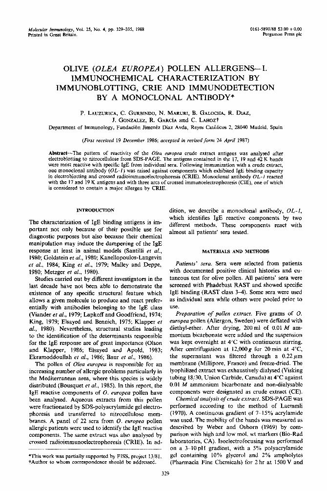
Fig. 1, (a) Seven to fifteen per cent SDS-PAGE and its corresponding densitometer scan. Standard markers were run on the lanes marked MK and their values are given at the side. (b) Analytical isoelectric focusing of 0. europea crude extracts (CE). The corresponding densitometric analysis of the polyacrylamide gel
is represented.
Characterization of OIeu europea pollen allergens 331
radioimmunoelectrophoresis (CRIE). These were performed according to the methods described by Weeke and Lowenstein (1973) and Weeke (1973), respectively.
RESULTS
Analysis of0. europea crude extract. Figure l(a) shows the SDS-PAGE results. Multiple bands of mol. wts ranging from 10 to 100 K were observed. The densitometric profile on the right of the figure confirms that the main protein peaks correspond to the 17 and 19 K bands. In Fig. l(b) results of isoelectrofocusing of the same crude extract and of its corresponding densitometric profile are shown. Although there were protein bands distributed along the pH gradient from 3 to 10, most of the bands were
located in the acidic pH range. When the proteins from the SDS-PAGE were
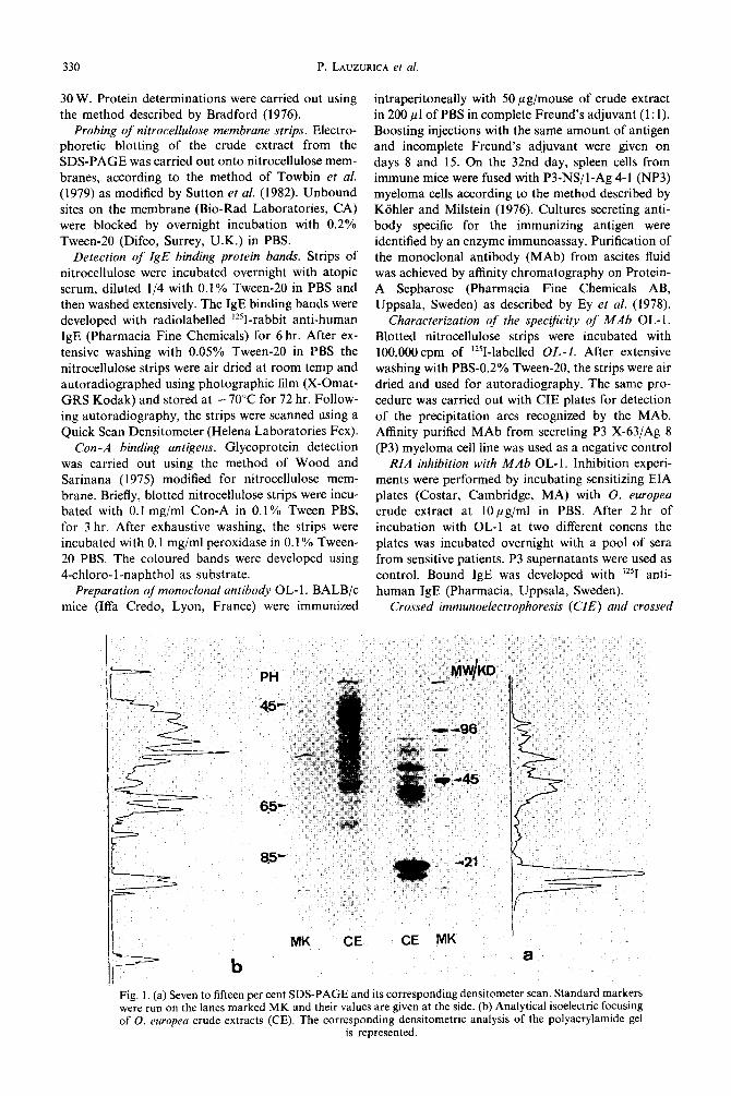
transferred to nitrocellulose strips and treated with Con-A and peroxidase, several glycoprotein bands appeared. As shown in Fig. 2 the most relevant bands were those of 42 and 19 K. In this way, the two major proteins of the crude extract, the 17 and 19 K bands, can be distinguished.
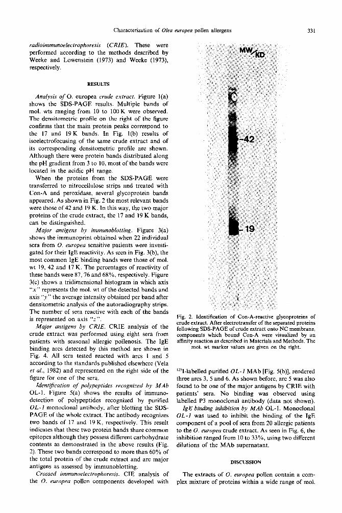
Major antigens by immunoblotting. Figure 3(a) shows the immunoprint obtained when 22 individual sera from 0. europea sensitive patients were investi- gated for their IgE reactivity. As seen in Fig. 3(b), the most common IgE binding bands were those of mol. wt 19, 42 and 17 K. The percentages of reactivity of these bands were 87,76 and 68%, respectively. Figure 3(c) shows a tridimensional histogram in which axis “x” represents the mol. wt of the detected bands and axis “y” the average intensity obtained per band after densitometric analysis of the autoradiography strips. The number of sera reactive with each of the bands is represented on axis “z”.
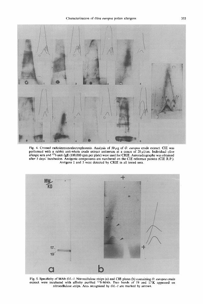
Major antigens by CRIE. CRIE analysis of the crude extract was performed using eight sera from patients with seasonal allergic pollenosis. The IgE binding arcs detected by this method are shown in Fig. 4. All sera tested reacted with arcs 1 and 5 according to the standards published elsewhere (Vela et al., 1982) and represented on the right side of the figure for one of the sera.
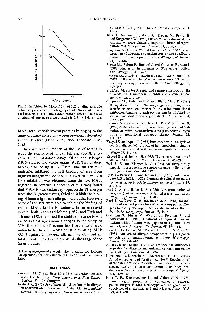
IdentiJication of polypeptides recognized by MAb OL-I. Figure 5(a) shows the results of immuno- detection of polypeptides recognized by purified OL-1 monoclonal antibody, after blotting the SDS- PAGE of the whole extract. The antibody recognizes two bands of 17 and 19 K, respectively. This result indicates that these two protein bands share common epitopes although they possess different carbohydrate contents as demonstrated in the above results (Fig. 2). These two bands correspond to more than 60% of the total protein of the crude extract and are major antigens as assessed by immunoblotting.
Crossed immunoelectrophoresis. CIE analysis of the 0. europea pollen components developed with
Fig. 2. Identification of Con-A-reactive glycoproteins of crude extract. After electrotransfer of the separated proteins following SDS-PAGE of crude extract onto NC membrane, components which bound Con-A were visualized by an affinity reaction as described in Materials and Methods. The
mol. wt marker values are given on the right.
‘251-labelled purified OL-1 MAb [Fig. 5(b)], rendered three arcs 3, 5 and 6. As shown before, arc 5 was also found to be one of the major antigens by CRIE with patients’ sera. No binding was observed using labelled P3 monoclonal antibody (data not shown).
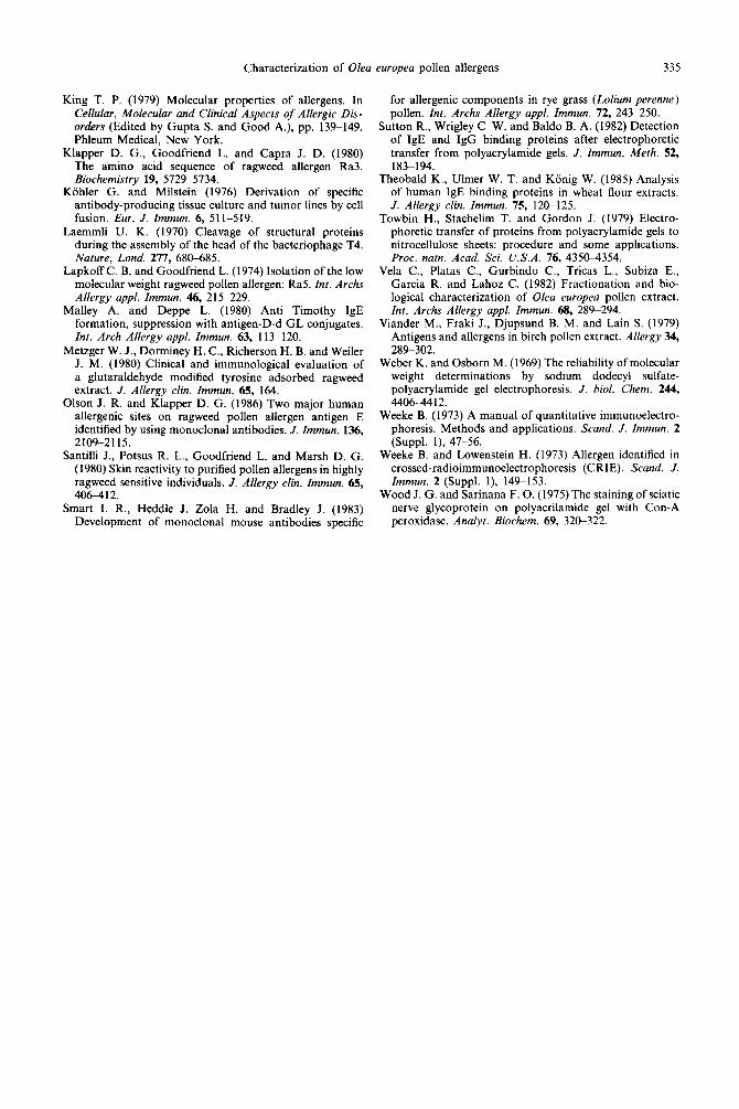
IgE binding inhibition by MAb OL-1. Monoclonal OL-1 was used to inhibit the binding of the IgE component of a pool of sera from 20 allergic patients to the 0. europea crude extract. As seen in Fig. 6, the inhibition ranged from 10 to 33%, using two different dilutions of the MAb supernatant.
DISCUSSION
The extracts of 0. europea pollen contain a com- plex mixture of proteins within a wide range of mol.
332 P. LAUZURICA et al.
ABCDEFQHIJ K L P Q R S T V V
1.
.
.
.
.
.
.
.
.
.
.
.
I I 17 19 28 32 39 42 48 55 60 66 70 85 95 I IO 130 Band 1 mw/ kd 1
sew
Fig. 3. IgE reactivity of 22 individual sera from 0. europea allergic patients. (a) Immunoprint of the 22 sera. (b) List of patients’ sera showing their reaction with the identified allergenic bands. (c) Tri- dimensional histogram of frequency distribution of patient’ sera. Axis “x” represents the mol. wt of the detected bands; axis “y” the average intensity obtained per band after densitometric analysis of the autoradiography strips. The number of sera reactive with each of the bands is represented on axis “z”.
wts. At least 16 proteins bands were demonstrated, of which the 17 and 19 K bands were the most apparent. When the extract was analysed by IEF, the majority of proteins were detected in the acidic range, as has been described for other pollen antigens (Anderson and Baer, 1984).
17, 42 and 19 K bands show the strongest radio- staining, with no apparent 15 K band detected.
It was demonstrated by Con-A binding that the 17 and 19 K bands were structurally different, in terms of their giycoprotein content. Nevertheless, both these bands react with the same MAb (OL-1) indi- cating that they share at least one antigenic epitope.
The detection of IgE binding components by immunoblotting has been of great value for defining the spectrum of allergens recognized by individual allergic patients (Sutton et al., 1982; Ford et al., 1985; Ford and Baldo, 1986). This technique is increasingly replacing the use of crossed immunoelectrophoreses to detect major and minor antigens since it is not dependent on the quality and potency of precipitating antisera (Baldo, 1985; Bengtsson et al., 1985).
The profiles of IgE reactivity of some patients’ sera We have obtained a MAb, OL-I, using 0. europea
were followed. The immunoprint from the different whole extract as the immunizing antigen. This anti- sera developed by immunodetection of IgE binding body reacts with two protein bands of mol. wt 17 and bands showed a heterogeneous pattern. Some of the 19 K by immunoblotting techniques. Therefore OL-I
sera reacted with only one or two bands, whereas is an extremely useful tool for the study of this the majority reacted with a large number of protein particular allergen since, as we have demonstrated, bands. The major bands were those of 19 and 17 K, the 17 and 19 K bands are two of the most important which reacted with 87 and 68% of the sera, respect- IgE binding bands of 0. europea pollen extract. This ively. In addition, a band of mol. wt 42 K reacted MAb was also used to detect the corresponding with 76% of the patients’ sera. We can conclude that components of the crude extract revealed by CRIE. these three bands of 17, 19 and 42 K represent major Three arcs, 3, 5 and 6 were detected, of which at least allergens. In a previous report, Blanca et al. (1983) arc 5 corresponds to a major antigen. This MAb assigned the category of major allergen to a poly- probably recognizes an antigenic determinant peptide of mol. wt 15 and 17 K. In our experience the common to two different proteins. Allergen-specific
Characterization of Olea europea pollen allergens 333
Fig. 4. Crossed radioimmunoelectrophoresis. Analysis of 5Opg of 0. europea crude extract. CIE was performed with a rabbit anti-whole crude extract antiserum at a concn of 20~1/cm. Individual olive allergic sera and ‘2SI-anti-IgE (100,000 cpm per plate) were used for CRIE. Autoradiography was obtained after 5 days’ incubation. Antigenic components are numbered on the CIE reference pattern (CIE R.P.).
Antigens 1 and 5 were detected by CRIE in all tested sera.
Fig. 5. Specificity of MAb OL-1. Nitrocellulose strips (a) and CIE plates (b) containing 0. europea crude extract were incubated with affinity purified ‘251-MAb. Two bands of 19 and 17 K appeared on
nitrocellulose strips. Arcs recognized by OL-I are marked by arrows.
334 P. LAUZUR~CA et al.
w MAb dihtions
Fig. 6. Inhibition by MAb OL-I of IgE binding to crude extract of pool sera from allergic patients. Supernatant was used undiluted (x 1), and concentrated 4 times ( x 4). Serial dilutions of pooled sera were used (0 l/2, 0 l/4, + l/S).
MAbs reactive with several proteins belonging to the same antigenic extract have been previously described in the literature (Haas et al., 1986; Theobald et al., 1985).
There are several reports of the use of MAbs to study the reactivity of human IgE and specific aller- gens. In an inhibition assay, Olson and Klapper (1986) studied five MAbs against AgE. Two of these MAbs, directed against different sites on the AgE molecule, inhibited the IgE binding of sera from ragweed-allergic individuals to a level of 50%. An 80% inhibition was obtained using the two MAbs together. In contrast, Chapman et al. (1984) found that MAbs to two distinct epitopes on the Pl allergen from the D. pteronyssinus, failed to inhibit the bind- ing of human IgE from allergic individuals. However, some of the sera were able to inhibit the binding of certain MAbs to the Pl antigen. In an unrelated system, both Kahn and Marsh (1982) and Esch and Klapper (1985) reported the ability of murine MAbs raised against Rye Group 1 antigen to inhibit up to 25% the binding of human IgE from grass-allergic individuals. In our inhibition studies using MAb OL-1 against 0. europea allergen, we obtained in- hibitions of up to 33%, more within the range of the
latter studies.
Acknowledgement-We would like to thank Dr Dolores Jaraquemada for her valuable discussions and continuous help.
REFERENCES
Anderson M. C. and Baer H. (1984) Rast inhibition and isoelectric focusing. Third International Paul-Ehrlich - Seminar, Vol. 79. Stuttgart.
Baldo B. A. (1985) Use of monoclonal antibodies in allergen standardization. Proceedings of the XII International Congress of Allergology and Clinical Immunology (Edited
by Reed C. E.), p. 411. The C.V. Mosby Company, St. Louis.
Baur X., Aschauer H., Mazur G., Dewair M., Prelicz H. and Steigemann W. (1986) Structure and antigenic deter- minants of some clinically important insect allergens: chironomid hemoglobins. Science 233, 351-354.
Bengtsson A., Rolfsen W. and Einarsson R. (1985) Charac- terization of allergens and patient sera by a nitrocellulose immunoprint technique. Int. Archs Allergy appl. Immun. 78, 139-144.
Blanca M., Bulton P., Brostoff J. and Gonzalez-Reguera I. (1983) Studies of the allergens of Olea europea pollen. Clin. Allergy 13, 473478.
Bousquet J., Guerin B., Hewitt B., Lim S. and Michel F. B. (1985) Allergy in the Mediterranean area III: cross- reactivity among Oleaceae pollens. Clin. Allergy 15, 43948.
Bradford M. (1976) A rapid and sensitive method for the quantitation of microgram quantities of protein. Analyf. Biochem. 72, 248-254.
Chapman M., Sutherland W. and Platts Mills T. (1984) Recognition of two Dermatophagoides pteronyssinus specific epitopes on antigen Pl by using monoclonal antibodies: binding to each epitope can be inhibited by serum from dust mite-allergic patients. J. Immun. 133, 2488-2495.
Ekramoddoullah A. K. M., Kisil F. T. and Sehon A. H. (1986) Partial characterization of an antigenic site of high molecular weight basic antigen, a ryegrass pollen allergen using a monoclonal antibody. Molec. Immun. 23, 111~117.
Elsayed S. and Apold J. (1983) Immunochemical analysis of cod fish allergen M: location of immunoglobulin binding sites as demonstrated by the native and synthetic peptides. Allergy 38, 449453.
Elsayed S. and Bennich H. (1975) The primary structure of allergen M from cod. Sand. J. Immun. 4, 203S210.
Esch R. E. and Klapper D. G. (1985) An allergenically important, cross-sensitive epitope on grass pollen group I antigens. Fedn Proc. 44, 1879.
Ey P. L.: Prowse S. J. and Jenkin C. R. (1978) Isolation of pure IeGl, IaG2a. IeG2b. Immunoelobulins from mouse serum-using-pro&IA Sepharose. immunochemistry 15, 429436.
Ford S. A. and Baldo B. A. (1986) A re-examination of ryegrass (Lo&m perenne) pollen allergens. Inr. Archs Allergy appl. Immun. 81, 193-203.
Ford S. A., Tovey E. R. and Baldo B. A. (1985) Identifi- cation of orchard grass (Dactylis glomerata) pollen aller- gens following electrophoretic transfer to nitrocellulose. Int. Archs Allergy appl. Immun. 78, 15-21.
Goldstein S., Muller V., Wypych J., Reisman R. and Arbesman C. (1980) Treatment of ragweed sensitive patients with a fraction-A conjugated to D-ghtamk acid and D-lysine. J. Allergy clin. Immun. 65, 164-165.
Haas H., Becker W.-M., Maasch H. J. and Schlaak M. (1986) Analysis of allergen components in grass pollen extracts using immunoblotting. Int. Archs Allergy appl. Immun. 79, 434440.
Kahn C. R. and Marsh D. G. (1982) Monoclonal antibodies as probes for allergenic and antigenic determinants on the rye I allergen. Fedn Proc. 41, 826.
Kanellopoulos-Langevin C., Matheeson B. J., Perkins A., Maynard A. and Asofsky R. (1984) Regulation of anti-hapten antibody response in uivo: memory, carrier- specific Lyt-2+ T cells can terminate antibody pro- duction without altering the peak of response. J. Immun. 132, 1639-1646.
King T. P., Kochoumiang L. and Chiorazzi N. (1979) Immunological properties of conjugates of ragweed pollen antigen E with methoxypolyetilene glycol or a copolymer of D-glutamic acid and D-lysine. J. exp. Med. 149, 424430.
Characterization of OIea europea pollen allergens 335
King T. P. (1979) Molecular properties of allergens. In Cellular. Molecular and Clinical Asoects of Allewic Dis-
for allergenic components in rye grass (L&urn perenne) nollen. Int. Archs Allergy aDpI. Immun. 72, 243-250.
orders (Edited by Gupta S. and Good A.j, pp. l!39-149. S&on R., Wrigley C W. &dBaldo B. A. (1982) Detection Phleum Medical, New York. of IgE and IgG binding proteins after electrophoretic
Klapper D. G., Goodfriend L. and Capra J. D. (1980) transfer from polyacrylamide gels. J. Immun. Meth. 52, The amino acid sequence of ragweed allergen Ra3. 183-194. Biochemistry 19, 5724-5734. -
Kijhler G. and Milstein (1976) Derivation of specific antibody-producing tissue culture and tumor lines by cell fusion. Eur. J. Immun. 6, 511-519.
Laemmli U. K. (1970) Cleavage of structural proteins during the assembly of the head of the bacteriophage T4. Nature, Lond. 277, 68G685.
Lapkoff C. B. and Goodfriend L. (1974) Isolation of the low molecular weight ragweed pollen allergen: Ra5. Int. Archs Allergy appl. Immun. 46, 215-229.
Malley A. and Deppe L. (1980) Anti Timothy IgE formation, suppression with antigen-D-d GL conjugates. Int. Arch Allergy appl. Immun. 63, 113-120.
Metzger W. J., Dorminey H. C., Richerson H. B. and Weiler J. M. (1980) Clinical and immunoloeical evaluation of a glutaralddhyde modified tyrosine idsorbed ragweed extract. J. Allergy clin. Immun. 65, 164.
Olson J. R. and Klapper D. G. (1986) Two major human allergenic sites on ragweed pollen allergen antigen E identified by using monoclonal antibodies. J. Immun. 136, 2109-2115.
Santilli J., Potsus R. L., Goodfriend L. and Marsh D. G. (1980) Skin reactivity to purified pollen allergens in highly ragweed sensitive individuals. J. Allergy clin. Immun. 65, 40&412.
Smart I. R., Heddle J. Zola H. and Bradley J. (1983) Development of monoclonal mouse antibodies specific
Theobald K., Ulmer W. T. and Konig W. (1985) Analysis of human IgE binding proteins in wheat flour extracts. J. Allergy clin. Immun. 75, 1213125.
Towbin H., Staehehm T. and Gordon J. (1979) Electro- phoretic transfer of proteins from polyacrylamide gels to nitrocellulose sheets: procedure and some applications. Proc. natn. Acad. Sci. U.S.A. 76, 435&4354.
Vela C., Platas C., Gurbindo C., Tricas L., Subiza E., Garcia R. and Lahoz C. (1982) Fractionation and bio- logical characterization of Olea europea pollen extract. Int. Archs Allergy appl. Immun. 68, 289-294.
Viander M., Fraki J., Djupsund B. M. and Lain S. (1979) Antigens and allergens in birch pollen extract. Allergy 34, 289-302.
Weber K. and Osborn M. (1969) The reliability ofmolecular weight determinations by sodium dodecyl sulfate- polyacrylamide gel electrophoresis. J. biol. Chem. 244, 4406-4412.
Weeke B. (1973) A manual of quantitative immunoelectro- phoresis. Methods and applications. Stand. J. Immun. 2 (Suppl. I), 47-56.
Weeke B. and Lowenstein H. (1973) Allergen identified in crossed-radioimmunoelectrophoresis (CRIE). Stand. J. Immun. 2 (Suppl. I), 149-153.
Wood J. G. and Sarinana F. 0. (1975) The staining of sciatic nerve glycoprotein on polyacrilamide gel with Con-A peroxidase. Analyt. Biochem. 69, 320-322.


