Embed Size (px)
Citation preview

One Pretty Amazing T.rex: A presentation for “100 years of tyrannosaurus rex”
Hosted by the Black Hills Institute
Mary Higby Schweitzer1,2,3, Jennifer L. Wittmeyer1, John R. Horner3
1Department of Marine, Earth and Atmospheric Sciences, North Carolina State University, Raleigh NC 27695 2North Carolina Museum of Natural Science, Raleigh, NC 27695 3Museum of the Rockies, Montana State University, Bozeman, MT 59717
Determining gender in extinct animals is difficult, because most features commonly
used to assign gender are lost in the process of fossilization. Despite this difficulty, many
bony features of dinosaurs have been interpreted to be evidence of sexual dimorphism,
including degree of ‘robustness’ in sauropods and their close relatives (Weishampel and
Chapman 1990; Galton 1997; Benton et al. 2000), theropods (Larson 1994; Smith 1998)
and protoceratopsids (Tereschenko and Alifanov 2003); horn core size in ceratopsids
(Godfrey and Holmes, 2003), or presence or absence of the first caudal chevron (Larson
and Frey, 1992; Larson 1994) to name a few. However, even if such features could
definitively be shown to be products of sexual differentiation, it remains impossible to
assign a particular feature unambiguously to a specific gender (e.g. the robust morph
being female; Carpenter 1990, Larson 1994). At best, assigning gender to a specific
morphotype of dinosaurs has fallen within the realm of speculation. What is needed is
an unambiguous way to assign a particular gender to male and female morphs. One
possibility is the identification of medullary bone in dinosaurs.
Medullary bone is an ephemeral reproductive tissue, among living taxa found
exclusively in female, actively reproducing birds. This bony tissue lines the medullary
cavities of the long bones of extant birds, and is chemically and morphologically distinct
from other bone types. Special characteristics of composition and structure contribute to
the high metabolic rates of medullary bone. In fact, it is capable of being metabolized 10-
15 times faster than cortical bone (Simkiss 1967; Dacke et al. 1993), and serves as an
easily mobilized calcium storage tissue for the production of calcareous eggshell
(Sugiyama and Kusuhara 2001). Its presence in dinosaurs would indicate gender,

support phylogenetic proximity, suggest shared reproductive physiological strategies with
extant birds, and indicate reproductive phase at the time of death.
Comparison of medullary and cortical bone characteristics:
In addition to protection and support of vital internal organs, bone plays an important
role in calcium metabolism in vertebrates, including all avian taxa (Miller and Bowman
1981). Long bone formation in extant birds proceeds much the same as in other
vertebrate taxa, via endochondral ossification of pre-existing cartilage models
(Whitehead, 2004; Taylor et al. 1971). Bone elongation involves periosteal deposition,
and concurrent endosteal osteoclastic resorption at the metaphyseal region, resulting in
overall maintenance of bone morphology and thickness during longitudinal growth
(Taylor et al. 1971).
In both formation and elongation, bone production involves two phases, reflecting the
composite nature of bone material. In the first, the bone forming cells (osteoblasts)
secrete organic matrix, called osteoid (Taylor et al. 1971; McKee et al. 1993). This
matrix consists primarily of the fibrous helical protein collagen I and the accessory
collagen V; non-collagenous proteins osteocalcin, osteopontin, osteonectin (Bonucci
and Gherardi 1975; McKee et al. 1993; Gerstenfield et al. 1994; Sugiyama and Kasuhura
2001; Wang et al. 2005), and bone sialoprotein (Gerstenfield et al. 1994; Robey 1996 and
references therein); serum proteins, including hemoglobin and albumin (McKee et al.
1993), and various glycosaminoglycans (Bonucci and Gherardi 1975; Dacke et al. 1993;
Arias and Fernandez 2001; Wang et al. 2005) . Therefore, cortical and trabecular bone
have a specific, characteristic and defineable chemical/molecular profile.
However, in female birds, a unique bone type is formed as the result of a surge in
blood estrogen levels at the onset of sexual maturity (Bonucci and Gherardi 1975; Knott
and Bailey 1999; Dacke et al. 1993, 2004; Whitehead, 2004 ). Medullary bone does not
occur naturally in any other taxon (Elsey and Wink 1986; Dacke 2004), and is present
only during the reproductive period in all living female birds, filling the marrow cavities
of many skeletal elements (Wilson and Thorpe 1998; Van Neer et al. 2002). It is
produced by specialized osteoblasts that lie within the endosteum, a thin connective
tissue layer that lines the marrow surfaces of the bones (Van Neer et al.2002). Medullary
bone exists only to offset the effects of bone resorption during shelling by serving as an

easily mobilized source for calcium, and has no direct biomechanical function (Bonucci
and Gherardi 1975; Wilson and Thorp 1998). It is chemically and morphologically
distinct from other bone types. Although medullary bone has been assumed to be present
in extant paleognaths, it has not been previously imaged or studied, and no data exists
regarding the morphology or chemistry of this bone type in ratites.
The mineral phase of both medullary and cortical bone is primarily hydroxyapatite
(Ca10(PO4)6(OH)2 ), but the ratio of mineral to organics is measurably higher in
medullary bone (Ascenzi et al. 1963; Taylor et al.1971; Dacke et al. 1993; Dacke 2004),
and medullary bone incorporates a higher proportion of calcium carbonate (Pelligrino and
Blitz 1970) than other bone types. Medullary bone is not only more highly mineralized
than cortical bone, the distribution of minerals is different between the two bone types.
In cortical bone, the mineral crystals are regularly distributed at the head of the A-bands
of collagen molecules (Taylor et al. 1971), but in medullary bone, mineral distribution
and orientation is much more random, with mineral crystals additionally deposited in
intrafibrillar spaces (Ascenzi et al. 1963; Taylor et al. 1971). In addition, medullary bone
does not exhibit birefringence because of the random arrangement of both collagen fibrils
and mineral, whereas other bone types are anisotropic in polarized light (Miller and
Bowman, 1981; Wilson and Thorp, 1998). Finally, the mineral crystals incorporated into
medullary bone are somewhat larger than the microcrystalline apatite of other bone types
(Ascenzi et al. 1963).
The organic phase of medullary bone differs significantly from that of cortical
and/or trabecular bone. Collagen makes up a greater proportion of the organic matrix of
cortical bone, while the percentage of non–collagenous proteins to collagen is far greater
in medullary bone, comprising approximately 40% of the total organics (Knott and
Bailey; 1999). The concentration of various glyclosaminoglycans is greater in medullary
than cortical bone, and it incorporates different amino sugars (Bonucci and Gherardi
1975). Hexosamine and keratan sulfate are much more prevalent in medullary than
cortical bone (Taylor et al. 1971; Wang et al. 2005), which incorporates chondroitin
sulfate instead. In addition, relatively high concentrations of tartrate-resistant acid
phosphatase (TRAP), an enzyme involved in digestion of bone (Sugiyama and Kusuhara
2001), are found in medullary bone. These chemical differences are reflected in the

differential response of the two bone types to various histochemical stains (Figure 1, also
Taylor et al. 1971; Sugiyama and Kusuhara 2001; Wang et al 2005).
Function of medullary bone.
Unlike other bone types, medullary bone has no biomechanical or other supportive
function, and exists solely as a calcium storage tissue that aids in mineral mobilization to
the shell gland during lay (Dacke et al. 1993; Wilson and Thorp 1998; Whitehead 2004).
As mentioned previously, medullary bone formation in birds is triggered by increased
levels of both estrogen and androgens that accompany ovulation, activating osteoblasts to
begin secretion of osteoid, while inhibiting osteoclast activity (Dacke et al. 1993;
Whitehead 2004). The formation of medullary bone begins ~1-2 weeks before lay. It is
maintained during the full laying cycle, and may persist up to one week post-lay before
resorption is complete (Reynolds 2003). Medullary bone osteoclasts in female birds are
specialized to contain estrogen receptors in their cell membranes, which, when triggered
by rising reproductive hormones, increases the efficiency of mobilizing stored calcium
(Miller 1981). While evidence of medullary bone may be found in virtually all skeletal
elements of extant birds, it is most abundant in the femur and tibiotarsus of most birds
studied (Reynolds 2003), and, consistent with its function as a source of rapid calcium
mobilization, it is infused with abundant vessels and blood sinuses. In fact, it has been
shown that up to 40% of the calcium used in eggshell formation comes directly from the
resorption of medullary bone (Mueller et al. 1969; Dacke et al. 1993). Although it is not
known to serve a direct mechanical function, in reducing the resorption of cortical and
trabecular bone, it may aid in maintaining integrity and strength of structurally important
bone (Whitehead 2004), and indeed, the presence of medullary bone in long bones of
laying birds has been shown to increase fracture resistance of these elements (Fleming et
al. 1998).
Like birds, most reptiles, including crocodiles and alligators, also produce calcareous
eggshell, but apparently do not produce medullary bone (Elsey and Wink 1986; Dacke
2004). This may be because of different mechanisms of shelling (Jackson et al. 2002)
and overall greater bone density that can offset the calcium draw without requiring
additional bone storage sources. Thus, extant non-avian archosaurs undergo bone

resorption during lay, but the structural integrity and biomechanical function of these
organisms is not apparently compromised during shelling.
Although medullary bone has not been previously observed or noted in dinosaurs, it
was proposed that reproducing dinosaurs, at least in the theropod lineage most closely
related to avian dinosaurs, would possess this ephemeral tissue (Martill et al. 1996;
Chinsamy and Barrett 1997). The failure to observe or identify these fragile reproductive
tissues in dinosaurs previously may be due to a number of taphonomic and biological
factors. First, we do not have any way of estimating the length of reproductive cycle in
theropods. There is a wide range of reproductive strategies amongst living birds, and the
extent and distribution of medullary bone in these taxa differ correspondingly (Schraer
and Hunter 1985). If theropods reproduce seasonally, they may only possess the tissue
for a maximum of a month or less. Second, in extant birds, the tissue is quite fragile, and
separates easily from the overlying cortex (Fig 1b, c). It may be that the tissues are lost,
either during fossilization, or subsequent recovery and preparation. Third, it may be that
medullary bone differs sufficiently from that of extant derived birds that it is not
recognized.
At the end of field season in 2002, a well preserved specimen of Tyrannosaurus rex
(Museum of the Rockies (MOR) specimen 1125) was found as an association of
disarticulated elements. The site was located at the base of the Hell Creek Formation,
about 8 m above the Fox Hills Sandstone. Soft, well-sorted sandstones derived from an
estuarine or fluvial setting surrounded the skeletal elements. Some of the elements
evidenced slight crushing, but overall preservation was excellent. MOR 1125, nick-
named “B-rex” after its discoverer, Bob Harmon, is a relatively small, but fully adult T.
rex. In comparison with the Chicago Field Museum Tyrannosaurus rex (FMNH
PR2081), with a femur length of about 131 cm, the femur of MOR 1125 is only 107 cm
in length. Using lines of arrested growth (LAG), MOR 1125 was calculated to be about
18 years old at the time of death (Horner and Padian 2004).
The remote region where elements of MOR 1125 were recovered had no roads into
the site, requiring a helicopter to transport jackets to the MOR labs. However, the jacket
containing the femur and other elements was too heavy to be airlifted out, and the bone
and jacket were broken and re-jacketed for removal. In the process, many internal

fragments that were visually free of preservative or consolidants were collected for
analyses.
When these fragments were examined in hand sample, a bony tissue lining the
endosteal surface of the bone could be seen that was distinct in texture, appearance and
distribution from other described dinosaur bone types. The morphological similarity of
the new tissues to avian medullary bone was immediately apparent (Schweitzer et al.
2005b). Figure two shows fresh-fracture images of Tyrannosaurus rex endosteal tissues
(A, B), compared with medullary bone tissues in reproducing ostrich (C) and emu (D).
The hallmark traits of medullary bone, dense vascularity and random, woven bone
pattern, are clearly visible in all samples. Large erosion rooms are visible in all
medullary tissues (*), indicating that calcium mobilization has begun.
Demineralization of extant bony tissues is commonly employed to more clearly
observe microstructural characteristics, such as fibril orientation; and, when mineral is
removed, the primarily collagenous protein matrix is exposed. It has been assumed that
when fossilized dinosaur bone is subjected to the same treatment, the bone would
dissolve completely as no proteinaceous material would persist over the course of
geological time.
In order to determine characteristics of presumed medullary tissues, we prepared a
partial demineralization, designed to etch mineral enough to expose underlying patterns.
At this point, we discovered an unexpected and totally novel characteristic to this bony
tissue. As minerals were dissolved from the medullary bone, the sample did not
distintegrate, but, similar to extant bone, tissues remained (Schweitzer et al. 2005a).
Furthermore, these dinosaur tissues exhibited apparent original flexibility, comparable to
that seen in extant ratites. However, these characteristics are not germane to this paper,
and will be discussed elsewhere (in preparation), but the retention of a pliable and fibrous
matrix after demineralization speaks to unusual preservation in this dinosaur material and
suggests that perhaps theorized modes of fossilization may need to be re-evaluated.
Figure 3 demonstrates the persistence of fibrous tissues after demineralization. Small
fragments of emu (A) and dinosaur (B) demineralized medullary bone tissues show
random fiber orientation, and large open spaces for vessels and vascular sinuses permeate

the tissues. The morphological similarity between extant and fossil samples is clearly
visible and supports the hypothesis of a common origin to the tissues.
Summary:
The endosteally derived bone tissues observed in MOR 1125 have all of the
characteristics of medullary bone, a distinctive avian reproductive tissue. While not
identical in morphology to published accounts of extant neognaths, the dinosaur tissues
fall within the range of variation observed in ratites. This bone tissue is derived from the
endosteum, it is highly vascular, it exhibits the random, woven-bone arrangement
consistent with very rapidly deposited bone. In addition, it has been identified on the
endosteal surfaces of both femora and one tibia, the only bones examined for the
presence of this tissue. The distribution is consistent with that seen in extant birds, and
suggests an organismal, rather than pathological response. Pathologies of the endosteum
are relatively rare and localized, and are usually accompanied by cortical bone anomalies
in the affected regions, which was not observed either grossly or microscopically in MOR
1125. In light of the fact that the relationship between theropod dinosaurs and birds is
robustly supported (e.g. Gauthier 1986; Sereno 1997; Holtz 2004) it is most parsimonious
to conclude that this novel tissue seen in MOR 1125 is medullary bone, and its presence
in theropods not only adds independent support of the robustly relationship between
theropods and birds, but also suggests that similar reproductive physiological strategies
were employed. In addition, its presence provides a means for unambiguous assignment
of gender in dinosaurs.
References
Arias JL, Fernandez MS. 2001. Role of extracellular matrix molecules in shell formation and structure. World’s Poultry Sci. J. 57:349-355. Ascenzi A, Francois C, Bocciarelli DS. 1963. On the bone induced by estrogens in birds. J. Ultrastructure Research 8:491-505. Benton MJ, Juul L, Storrs GW, Galton PM. 2000. Anatomy and systematics of the prosauropod dinosaur Thecodontosaurus antiquus from the Upper Triassic of southwest England. J. Vert. Paleont. 20(1):77–108. Bonucci E, Gherardi G. 1975. Histochemical and electron microscope investigations on medullary bone. Cell Tiss. Res. 163:81-97. Carpenter, K. (1990) - Variations in Tyrannosaurus rex, in Carpenter K. & Currie, P.J. (Eds), Dinosaur Systematics, Cambridge Univ. Press, Cambridge, pp. 141-146. Chinsamy A, Barrett PM. 1997. Sex and old bones? J. Vert. Paleontol. 17(2): 450-450. Dacke CG, Arkle S, Cook DJ, Wormstone IM, Jones S, Zaidi M, Bascal ZA. 1993. Medullary bone and avian calcium regulation. J. Exp Biol. 184: 63-88. Dacke CG. 2004. Elsey RM, Wink CS 1986. The effects of estradiol on plasma calciumand femoral bone structure in alligators (Alligator mississippiensis). Comp. Biochem. Physiol. 84A(1):107-110. Fleming RH, McCormack HA, McTeir L, Whitehead CC. 1998. Medullary bone an dhumeral breaking strength in laying hens. Res. Vet. Sci 64:63-67. Galton, PM. 1997. Comments on sexual dimorphism in the prosauropod dinosaur Plateosaurus engelhardti (Upper Triassic, Trossingen). Neues Jahrbuch fu¨r Geologie und Pala¨ontologie, Monatshefte, 1997:674–682. Gauthier J. 1986. Saurischian monophyly and the origin of birds. In: The origin of birds and the evolution of flight (Padian K, ed). Calif. Acad. Sci. Mem. 8:1-55. Godfrey SJ, Holmes R. 1995. Cranial morphology and systematics of Chasmosaurus (Dinosauria:Ceratopsidae) from the Upper Cretaceous of western Canada. J. Vert. Paleont. 15(4):726-742. Horner JR, Padian K. 2004. Age and growth dynamics of Tyrannosaurus rex. Proc. R. Soc. London Ser. B 271, 1875-1880.
Holtz, T.R., Jr. 2004. Tyrannosauroidea. In The Dinosauria. Second Edition. D.B. Weishampel, P. Dodson and H. Osmólska (eds.), University of California Press, pp 111-136. Jackson FD, Schweitzer MH, Schmitt JG. 2002. Dinosaur eggshell study using scaning electron microscopy. Scanning 24:217-223. Knott L, Bailey AJ. 1999. Collagen biochemistry of avian bone: comparison of bone type and skeletal site. Br. Poultry Sci. 40:371:379. Larson PL & Frey E. (1992) - Sexual Dimorphism in the Abundant Upper Cretaceous Theropod, Tyrannosaurus rex, Jour. Vert. Paleontol., 12 (Supp. to No. 3): 38a.
Larson PL. (1994) - Tyrannosaurus Sex, in Rosenberg, G.D. & Wolberg, D.L. (Eds), Dinofest, Paleontol. Soc. Spec. Pub. 7: 139-155.
Martill DM, Barker MJ, Dacke CG. 1996. Dinosaur nesting or preying? Nature 379: 778-778.
McKee MD, Farachcarson MC, Butler WT, Hauschka PV, Nanci A. 1993. Ultrastructural immunolocalization of noncollagenous (osteopontin and osteocalcin) and plasma (alabumin and alpha-2HS glycoprotein) proteins in rat bone. J. Bone Min. Res. 8 (4): 485-496 Miller SC, Bowman BM. 1981. Medullary bone oseogenesis following estrogen administration to mature male Japanese Quail. Developmental Biology 87:52-63. Miller SC. 1981. Osteclast cell-surface specializations and nuclear kinetics during egg-laying in Japanese quai. Am.J. Anat. 162:35-43. Mueller WJ, Brubaker RL, Caplan MD. 1969. Eggshell formation and bone resportion in egg-laying hens. Fedn Proc. Fedn. Am. Soc. Exp. Biol. 28:1851-1855. Pelligrino ED, Blitz RM 1970. Calcium carbonate in medullary bone. Calcif. Tiss. Res. 6:168-171. Reynolds SJ. 2003. Mineral retention, medullary bone formation and reproduction in the white-tailed ptarmigan (Lagopus Leucurus): a critique of Larson Et al. (2001). Auk 120(1):224-228. Robey PG. 1996. Vertebrate mineralized matrix proteins: Structure and function. Conn. Tiss. Res. 35 (1-4): 185-190. Schraer H, Hunter SJ. 1985. The development of medullary bone: a model for osteogenesis. Comp. Biochem. Biophys. 82A(1):13-17

Schweitzer MH, Wittmeyer JL, Horner JR, Toporski J. 2005a. Soft tissue vessels and cellular preservation in Tyrannosaurus rex. Science 307:1952-1955. Schweitzer MH, Wittmeyer JL, Horner JR. 2005b. Gender specific reproductive tissue in ratites and Tyrannosaurus rex. Science 308: 1456-1460 Sereno PC. 1997. The origin and evolution of dinosaurs. Ann. Rev. Earth Planet. Sci. 25: 435-489 1997 Simkiss K. 1961. Calcium metabolism and avian reproduction. Biol. Rev 36:321-367. Smith DK. 1998. A morphometric analysis of Allosaurus. J. Vert. Paleont. 18 (1): 126-142. Sugiyama T, Kusuhara S. 2001. Avian calcium metabolis and bone function. Asian-Aust. J. Anim. Sci. 2001. 14:82-90. Taylor TG, Simkiss K, Stringer DA. 1971. The skeleton: its structure and metabolism. In: Physiology and Biochemistry of the Domestic Fowl, Freeman BM, ed. Acad. Press 2:621-640. Tereschenko VS, Alifanov VR. 2003. Bainoceratops efremovi, a new Protoceratopid dinosaur (Protoceratopidae, Neoceratopsia) from the Bain-Dzak locality (south Mongolia) PALEONT. J. 37 (3): 293-302. Van Neer W, Noyen K, DeCupere B. 2002. On the use of endosteal layers and medullary bone from domestic fowl in archaeozoological studies. J. Arch. Sci. 29:123-134.
Wang X, Ford BC, Praul CA, Leach RM. 2005. Characterization of the non-collagenous proteins in avian cortical and medullary bone. Comp. Biochem. Phys B. 140:665-572. Weishampel, D. B., and R. E. Chapman. 1990. Morphometric study of Plateosaurus from Trossingen (Baden-Wu¨rttemberg, Federal Republic of Germany); pp. 43–51 in K. Carpenter and P. J. Currie (eds.), Dinosaur Systematics: Approaches and Perspectives. Cambridge University Press, Cambridge. Whitehead CC. 2004. Overview of bone biology in the egg-laying hen. Poult. Sci. 83:193-199. Wilson S, Thorp BH. 1998. Estrogen and cancellous bone loss in the fowl. Calcif. Tissue Int. 62:506-511.

Figure Legends: Figure 1. Medullary bone in extant laying hen. A) gross cross section of femur of actively laying hen shows extensive medullary bone formation. New bone is randomly oriented and much more porous than overlying cortical bone. B) low magnification and C) high magnification of histological section of demineralized bone from laying hen. Chemical differences between cortical and medullary bone are indicated by differential response of each bone type to hematoxylin and eosin staining. In C, separation of the medullary bone from cortical bone is seen as sectioning artifact. Large, multinucleated osteoclasts are visible around bone spicules, and small osteoblasts align along preexisting bone spicules, active in deposition of new bone. CB = cortical bone, MB = medullary bone, ELB = endosteal laminar bone, OCL = osteocyte lacunae, OC = osteoclast, OB = osteoblast. Scales as indicated. Figure 2. Fresh fracture of tibiae of MOR 1125 (A, B), ostrich (C), and emu (D). The morphology and microstructure of medullary bone is observed in all cases as distinct from overlying cortical bone. Medullary bone is less organized and more vascular. Large vascular sinuses can be seen in the medullary bone, and in some cases, large erosion rooms (*) are visible at the interface between medullary and cortical bone and in the medullary bone itself, indicating some resorption of bone has occurred. In emu (D) a large elongate erosion room is infilled with new medullary bone with characteristic “crumbly” texture. Abbreviations as in Figure 1, T = trabecular spicule. Scales as indicated. Figure 3. Demineralized fragments of medullary bone from emu (A) and MOR 1125 (B). The fibrous, woven pattern of bone matrix is visible in both cases, and the relatively “lacy” appearance results from penetration of the bone by blood vessels. Scales as indicated. For methods on demineralization, see Schweitzer et al. 2005a, supplemental online information.

Figure 1.
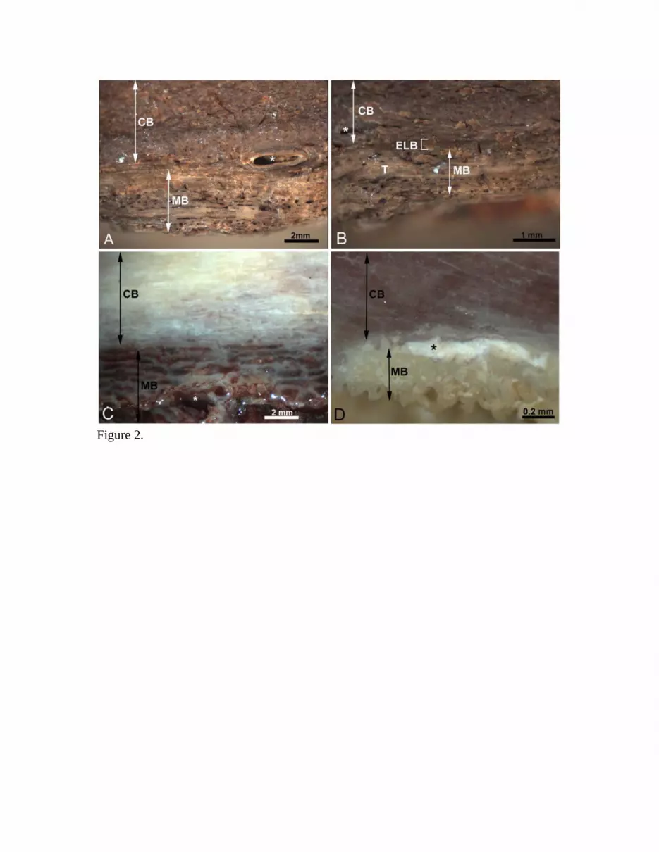
Figure 2.

Figure 3