Embed Size (px)
Citation preview

Biol. Chem., Vol. 393, pp. 343–353, May 2012 • Copyright © by Walter de Gruyter • Berlin • Boston. DOI 10.1515/hsz-2011-0253
One round of SELEX for the generation of DNA aptamers directed against KLK6
Steve Arnold 1 , Georgios Pampalakis 2 , Kalliopi Kantiotou 2 , Dilson Silva 3 , Celia Cortez 3 , Sotiris Missailidis 1, * and Georgia Sotiropoulou 2, *
1 Department of Chemistry , The Open University, Milton, Keynes MK7 6AA , UK 2 Department of Pharmacy , University of Patras, Rion-Patras 26500 , Greece 3 Universidade do Estado do Rio de Janeiro , Rio de Janeiro 20550-170 , Brazil
* Corresponding authors e-mail: [email protected]; [email protected]
Abstract
Kallikrein-related peptidase 6 (KLK6) is an active serine protease that has been implicated in common pathologies, including neurodegenerative disorders such as Parkinson and Alzheimer disease and certain types of cancer. Antibodies, either polyclonal or monoclonal, that exhibit specifi city for distinct members of the extended kallikrein family, including KLK6, were developed. With the exception of KLK3/PSA, the identifi cation and generation of aptamers, as potential new tools with improved characteristics demanded for therapeutic and diagnostic applications, has not been explored for KLKs. Here, we report for the fi rst time the identifi cation of novel DNA aptamers against KLK6 that were isolated using a modi-fi ed systemic evolution of ligands by exponential enrichment technique. The identifi ed aptamers were characterized using fl uorescence spectroscopy, competition ELISA, and quartz crystal microbalance, and two aptamers (008 and 022) were found to exhibit high affi nity ( K d in the low nanomolar range) for KLK6. Aptamers were tested for their ability to bind to serum albumin, to demonstrate their specifi city for their target, and the possible involvement of such proteins in the transport of aptamers into the bloodstream. The developed aptamers are expected to assist the development of novel diagnostic, biosensing, and therapeutic strategies.
Keywords: DNA aptamers; kallikrein-related peptidase 6; SELEX.
Introduction
The development of high-quality and high-affi nity reagents to target human proteins represents a major challenge in the current medical and diagnostic fi eld. Aptamers, sometimes referred as ‘ chemical antibodies ’ , are single-stranded DNA or RNA oligonucleotides that specifi cally recognize and bind
with high affi nity to a molecular target. The discovery of aptamers is based on the fact that nucleic acids not only can hybridize to one another based on a simple code as it widely known but can also form complex structures that act as mole-cular scaffolds to support complex formation with proteins or small molecules. Aptamers are generated by in vitro selection according to the systematic evolution of ligands by exponen-tial enrichment (SELEX) process (Bunka and Stockley , 2006 ; Keefe et al. , 2010 ).
There are certain major advantages of aptamers against antibodies for usage as therapeutic, diagnostic, imaging, and sensor agents. Aptamers can easily be produced by automated chemical synthesis, and there is an extended modifi cation chemistry that can be applied to improve clearance (Borbas et al. , 2007 ; Keefe et al. , 2010 ) or to deliver therapeutic or radiotherapeutic agents (Borbas et al. , 2007 ; Missailidis and Perkins , 2007 ; Da Pieve et al. , 2009 ), they can be stored for long periods at ambient temperatures without signifi cant degradation or denaturation (Velasco -Garcia and Missailidis, 2009 ), and they are not immunogenic (Keefe et al. , 2010 ).
As their generation through in vitro methods is easy, their recognition and binding properties can be tailored to virtu-ally any target including proteins, drugs, peptides, even ions (Bunka and Stockley , 2006 ; Keefe et al. , 2010 ). Examples of successful aptamers include the aptamer that targets throm-bin, which was the fi rst DNA aptamer to be generated (Bock et al. , 1992 ), for VEGF approved by the FDA for treatment of the age-related macular degeneration (pegaptanib, Pfi zer) (Ng et al. , 2006 ) and for the small-molecule cocaine with potential sensing and analytical applications (Stojanovic and Landry , 2002 ).
In the present study, we undertook a new approach to identify specifi c aptamers that target the kallikrein-related peptidase 6 (KLK6). KLK6 is a member of the extended family of KLKs that was originally cloned based on its differential expression in metastatic and primary breast cancer cells (Anisowicz et al. , 1996 ). Recently, we showed that KLK6 plays important roles in breast cancer growth and progression in a concentration-dependent manner (Pampalakis et al. , 2009 ). KLK6 is consid-ered a potential biomarker in blood for ovarian cancer (Yousef et al. , 2003 ). KLK6 is involved in physiological processes, and it has also been implicated in neurodegenerative diseases such as the progression of multiple sclerosis because of its ability to control the myelination process (Bando et al. , 2006 ), and as administration of anti-KLK6 antibodies to mouse mod-els of multiple sclerosis resulted in delayed onset and attenu-ation of disease symptoms (Blaber et al. , 2004 ). It has been considered a new potential biomarker for secondary progres-sive multiple sclerosis (Scarisbrick et al. , 2008 ). It may also be involved in Parkinson disease as a regulator of α -synuclein
Brought to you by | University of Patras (University of Patras)Authenticated | 172.16.1.226
Download Date | 4/24/12 3:30 PM

344 S. Arnold et al.
turnover (Iwata et al. , 2003 ) and is involved in neuronal inju-ries, especially in spinal cord injury (Terayama et al. , 2004 ; Scarisbrick et al. , 2006 ). Further, KLK6 represents a potential biomarker in plasma for Alzheimer disease (Diamandis et al. , 2000 ; Menendez -Gonzalez et al., 2008 ). Recently, KLK6 was shown to regulate the lymphocyte survival, thus participat-ing in a range of immunological responses (Scarisbrick et al. , 2011 ). Cumulatively, KLK6 represents a potential therapeutic and diagnostic target (Sotiropoulou et al., 2009; Bayani and Diamandis , 2011 ; Lawrence et al. , 2010 ).
The postulated roles of KLK6 in most common patholo-gies indicate the need to develop novel agents that specifi cally recognize KLK6 for use as new diagnostic and/or therapeutic agents. The developed aptamers were tested for their spec-ifi city and stability in serum, as required for future in vivo studies.
Results
Aptamer selection
The classical SELEX process involves multiple rounds of selection and amplifi cation (Bock et al. , 1992 ). Here, a modi-fi ed SELEX selection was performed using a single round of selection and elution of aptamer binding species using a step gradient salt (NaCl) elution, as described in Materials and methods. Briefl y, the immobilized KLK6 protein was incubated with the aptamer library. Subsequently, unbound or nonspecifi cally bound aptamer species were removed using salt solutions in a step elution gradient, whereas the highest salt concentration eluted the tightly bound, specifi c aptamers. These aptamers were cloned and sequenced; 17 sequences were obtained (Figure 1 A).
As shown in Figure 1A, considerable motif similarity was observed, with oligonucleotide sequences, ATAGGGAC, ATAGGGAG, and, in particular, ATAGG or ATAGA (high-lighted in yellow), occurring eight times, including twice
in 001. Sequences 013, 014, and 021 also show similarity (TTTG) (highlighted in red). Several sequences are either identical, such as 008 and 009, 022 and 023, or highly homo-logous, such as 022, 023, 026, and 027. The 002 sequence was truncated; therefore, it was not further analyzed, and it is not shown.
The RNA Shapes program ( http://bibiserv.techfak.uni-bielefeld.de/rnashapes/submission.html ) predicts low-energy foldings of RNA or DNA (Steffen et al. , 2006 ). An abstract shape is produced by the program predicting probable base pairing that defi nes stem regions and loops or tails where base pairing is unlikely. The priming sequences were included in structure prediction analysis, as they may be intimately involved in forming the overall structure. Eleven aptamers for the KLK6 protein were selected (marked with a star in Figure 1A). These were chosen for further study to evaluate binding, as they showed high probabilities of stem loop struc-ture formation. The common motifs ATAGG and ATAGA are marked with a blue arc in Figure 1B (shown for aptamers 008 and 022). All predictions show a stem-loop structure with the ATAGG sequence occurring consistently at the end of a stem region. An exposed loop containing the motif ATAG occurs in aptamers 008, 022, and 027, with ATA occurring as a single-stranded section in 008, 013, 022, and 027.
Investigation of KLK6-aptamer interaction
with fl uorescence spectroscopy
Fluorescence spectroscopy is a highly sensitive technique that allows measurements of molecular interactions between species at very low concentrations (Missailidis et al. , 2003 ). KLK6 contains the aromatic residues Trp, Tyr, and Phe, which fl uoresce upon UV excitation. The dominant fl uore-scent mole cule in proteins is Trp, and KLK6 contains four Trp residues, which are depicted in Figure 2 A (Gomis -R ü th et al., 2002 ). An additional Trp residue is located in the presignal sequence, which is removed after synthesis.
022008
ΔG=-20.04 kcal/mol ΔG=-10.52 kcal/mol
BA
014 021 013 022 023 027 026 001 003 004 007 008009 012005 006
CCCTAGTCTGGGTTGTATTTTGCGT------------ 25 ------TGTGGATTGTTTTTTGACTGATCGG------ 25 ----TCAACCATTTGTCTATGATAGGATGCAAT---- 29 ----------CAAGGCATAGGGAGCATGAAAGGAC-- 25 ----------CAAGGCATAGGGAGCATGAAAGGAC-- 25 ----------CAAGGCATAGGGACGATGAAACCA--- 24 ----------CAAGGCATAGGGACGTTTTGGAC---- 23 ----------ATAAACATAGGGACCAATAGGATAT-- 25 -------------GGCATCGGAGTGATTCGGCTGATA 24 ---------GATGGGTGTTAAAAAGGCGGTATT---- 24 -------CTCACTGGTACTCCGTGTTTAAAAT----- 25 -------TTGCTCCTTATAGAGCCGTCCGAGC----- 25
-------TTGCTCCTTATAGAGCCGTCCGAGC----- 25 ----------CTGGGTGTACGAGAATTAGGTGCC--- 24 -------TGTTCGAGTGCTGACTCCATCTGGG----- 25 ----------AATCATATTGTAAGCTGCTGTCTAG-- 25
Figure 1 (A) Multiple sequence alignment ( http://www.ebi.ac.uk/Tools/clustalw2 ) of the 16/17 aptamers found in the current study. A star indicates the aptamers that were selected based on their predicted secondary structure for KLK6 binding testing in this study. (B) Example of predicted structures where the stem-loop structure is easily recognized.
Brought to you by | University of Patras (University of Patras)Authenticated | 172.16.1.226
Download Date | 4/24/12 3:30 PM

KLK6 aptamers 345
Using 0.5 µ m of KLK6, aptamer was titrated directly into the cuvette at concentrations ranging from 0.01 to 0.1 µ m . Fluorescence measurements were performed at 37 ° C, after testing for optimal instrument settings including the excita-tion wavelength and bandwidth, as well as KLK6 and aptamer concentrations, to obtain a maximal signal for KLK6 emis-sion and minimal signal for aptamer emission. Each individ-ual titration point was scanned fi ve times and averaged, and each titration was performed in triplicate. KLK6 and aptamer controls were titrated with PBS only. A 20 % drop in fl uores-cence occurred after 10 titrations of PBS at λ max , most likely the result of photobleaching (data not shown). This reduction in fl uorescence intensity is linear, and the standard deviation (SD) of average fl uorescence between 344 and 350 nm was < 6 % . On examining the normalized data (relative change in fl uorescence) when aptamers are titrated into KLK6, the reduction in fl uorescence showed a nonlinear relationship in all cases (Figure 2B). After the fi nal titration point, it varied between 20 % , for aptamer 021 (no more than that observed for photobleaching of KLK6), and 35 % , for aptamer 022. The collated normalized data (Figure 2B) for KLK6 alone and
KLK6 plus aptamer titrations reveal that most of the aptam-ers do not show a quenching effect > 4 % . However, aptamers 008, 001, and 022 produce quenching effects of 9 % , 13 % , and 15 % , respectively, on top of photobleaching. Furthermore, it should be noted that this reduction in fl uorescence is observed with titrations of only a small amount of aptamer (maximum protein aptamer ratio of 5:1 after the last titration point) to avoid large contributions from the aptamer ’ s intrinsic fl uo-rescence. Furthermore, it was found that λ max shifted to lon-ger wavelengths, up to 10 nm for aptamers 008 (Figure 2C), 006, and 014 (not shown), whereas no shift was observed for aptamers 001, 005, 013, and 022 (not shown). The remain-ing aptamers demonstrated intermediate shifts. In interpreting fl uorescence data, it should be taken into consideration that shifts in wavelength occur if binding molecules cause confor-mational changes affecting the environment around any of the four Trp residues (Missailidis et al. , 2003 ). On the other hand, quenching arises from collisional effects and/or complex for-mation involving Trp residues at the surface. Aptamers that did not show fl uorescence quenching much above the KLK6 pho-tobleaching included 006, 007, 013, 014, 021, 026, and 027,
Aptamer 008 (μM)
0
20 000
40 000
60 000
80 000
100 000
120 000
300 320 340 360 380 400 420 440Wavelength (nm)
Fluo
resc
ence
(a.u
.) 0.010.020.030.040.050.060.070.080.090.10
KLK6
0.60
0.64
0.68
0.72
0.76
0.80
0.84
0.88
0.92
0.96
1.00
0.00 0.01 0.02 0.03 0.04 0.05 0.06 0.07 0.08 0.09 0.10Aptamer (μM)
Rel
ativ
e flu
ores
cenc
e F
corr
/Fi
001005006007008013014021022026027
KLK6
BA
C
Figure 2 (A) Three-dimensional structure of proKLK6 (1GVL) (Gomiz-R ü th et al., 2002) showing the four Trp residues with blue side chains generated with Chimera ( http://www.cgl.ucsf.edu/chimera ). The buried residue is Trp141. (B) Relative fl uorescence change for all aptamers titrated into 0.5 µ m KLK6. (C) The spectra of 0.5 µ m KLK6 titrated by aptamer 008 presenting a 10-nm red shift in arbitrary units (a.u.).
Brought to you by | University of Patras (University of Patras)Authenticated | 172.16.1.226
Download Date | 4/24/12 3:30 PM
346 S. Arnold et al.
which are likely false positives of SELEX that either do not actually bind or their binding constants are much lower and, consequently, a larger amount of aptamer would need to be titrated before a quenching effect can be observed. However, four of the aptamers (001, 005, 008, and 022) showed a small but signifi cant amount of quenching even at such low con-centration and despite the fact that the molar ratio of KLK6/aptamer does not exceed 5:1. The fact that fi ve aptamers (006, 008, 014, 021, and 027) showed a red shift up to 10 nm sug-gests changes of conformation caused by interaction of these aptamers with KLK6 (Silva et al. , 2010 ).
Further, by fi tting the fl uorescence titration data as described by Missailidis et al. (2003) and detailed here in Materials and methods, we have calculated the dissociation constants for the aptamers 008 and 022 and found K d008 = 15 × 10 -9 ( ± 1.6 × 10 -9 ) m and K d022 = 21 × 10 -9 ( ± 8 × 10 -9 ) m , respectively. These values are in the nanomolar range, thus verifying the high affi nity of the aptamers for their target protein KLK6.
Characterization of the HSA-aptamer interaction
with fl uorescence spectroscopy
The same principle described for KLK6-aptamer interaction was applied. Human serum albumin (HAS) is a 582-amino acid residue protein that contains one Trp (Trp 214 ) that is partially buried (Hein et al. , 2010 ). The conditions detailed in Materials and Methods were selected to minimize aptamer and maximize
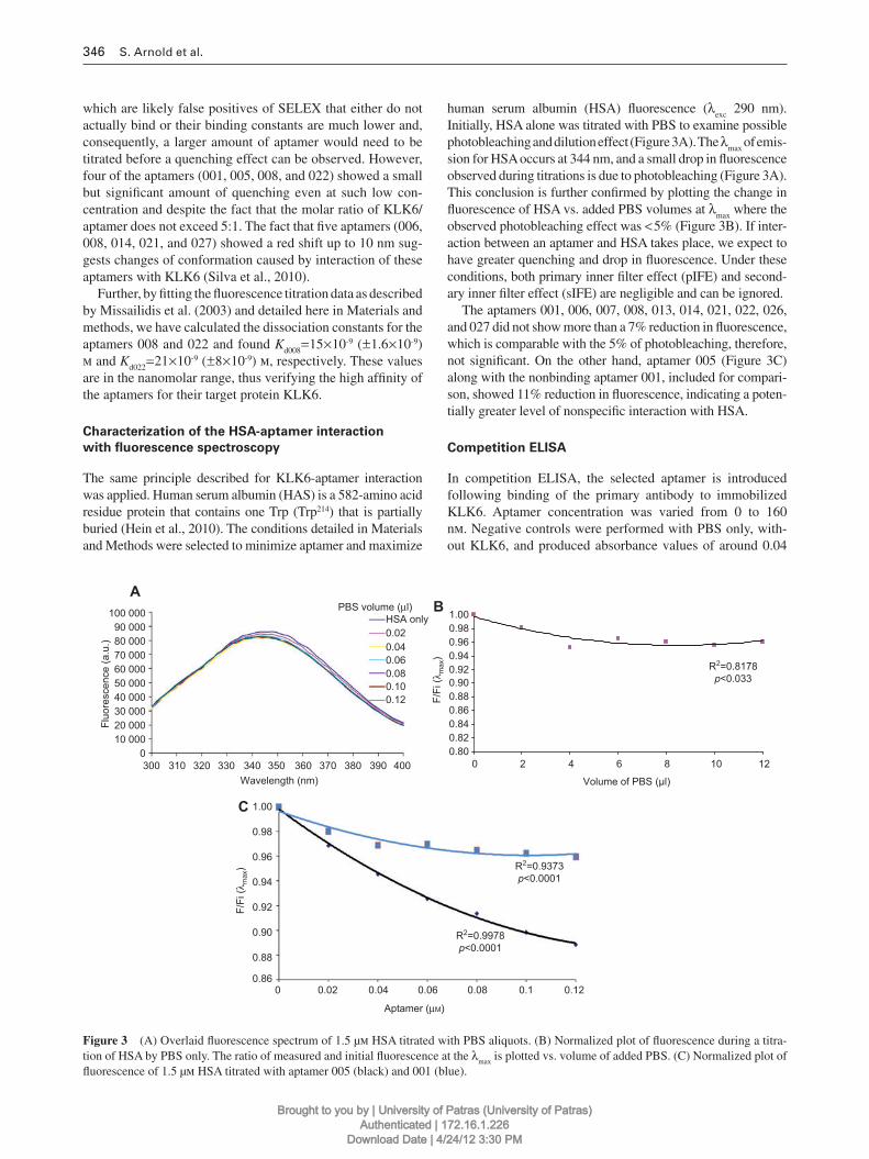
human serum albumin (HSA) fl uorescence ( λ exc 290 nm). Initially, HSA alone was titrated with PBS to examine possible photobleaching and dilution effect (Figure 3A). The λ max of emis-sion for HSA occurs at 344 nm, and a small drop in fl uorescence observed during titrations is due to photobleaching (Figure 3 A). This conclusion is further confi rmed by plotting the change in fl uorescence of HSA vs. added PBS volumes at λ max where the observed photobleaching effect was < 5 % (Figure 3B). If inter-action between an aptamer and HSA takes place, we expect to have greater quenching and drop in fl uorescence. Under these conditions, both primary inner fi lter effect (pIFE) and second-ary inner fi lter effect (sIFE) are negligible and can be ignored.
The aptamers 001, 006, 007, 008, 013, 014, 021, 022, 026, and 027 did not show more than a 7 % reduction in fl uorescence, which is comparable with the 5 % of photobleaching, therefore, not signifi cant. On the other hand, aptamer 005 (Figure 3C) along with the nonbinding aptamer 001, included for compari-son, showed 11 % reduction in fl uorescence, indicating a poten-tially greater level of nonspecifi c interaction with HSA.
Competition ELISA
In competition ELISA, the selected aptamer is introduced following binding of the primary antibody to immobilized KLK6. Aptamer concentration was varied from 0 to 160 n m . Negative controls were performed with PBS only, with-out KLK6, and produced absorbance values of around 0.04
R2=0.8178p<0.033
0.800.820.840.860.880.900.920.940.960.98
HSA onlyPBS volume (μl)
0.020.040.060.080.100.12
1.00
0300 310 320 330 340 350 360
Wavelength (nm)370 380 400390
10 00020 00030 00040 00050 000
Fluo
resc
ence
(a.u
.)
60 00070 00080 00090 000
100 000
0 2 4 6 8 10 12
Volume of PBS (μl)
F/Fi
( λm
ax)
BA
C 1.00
0.98
0.96
0.94R2=0.9373p<0.0001
R2=0.9978p<0.0001
0.92F/Fi
( λm
ax)
0.90
0.88
0.860 0.02 0.04 0.06
Aptamer (μM)
0.08 0.1 0.12
Figure 3 (A) Overlaid fl uorescence spectrum of 1.5 µ m HSA titrated with PBS aliquots. (B) Normalized plot of fl uorescence during a titra-tion of HSA by PBS only. The ratio of measured and initial fl uorescence at the λ max is plotted vs. volume of added PBS. (C) Normalized plot of fl uorescence of 1.5 µ m HSA titrated with aptamer 005 (black) and 001 (blue).
Brought to you by | University of Patras (University of Patras)Authenticated | 172.16.1.226
Download Date | 4/24/12 3:30 PM

KLK6 aptamers 347
(shown with black boxes in Figure 4 ). We have tested all 11 aptamers identifi ed against KLK6 following the above con-ditions to verify their ability to compete with an anti-KLK6 primary monoclonal antibody. The concentration-dependent competition between the already bound primary antibody and aptamers 008 and 022 are shown in Figure 4, demonstrating the ability of these aptamers to compete with the anti-KLK6.
Investigation of a KLK6 aptamer interaction
with quartz crystal microbalance biosensor
Initially, the gold (Au) surface that covers the crystal was modifi ed with thiol chemistry as described in Materials and methods to activate the surface with 3,3 ′ -dithiodipropionic acid di( N -succinimidyl ester) (DSP). Avidin, which contains Lys, can covalently bind to DSP via its side-chain amino group. The biotinylated aptamers will bind to avidin, forming a coating that spaces the aptamer away from the surface. This coating method roughly follows that of Liss et al. (2002) and is depicted in Figure 5 A. Specifi c interactions between the KLK6 aptamers on the crystal and KLK6 in solution pass-ing through the cell can be recorded as a change in Δ F. The Au area on the crystal was 1.13 cm 2 . The resonant frequency used was 5 MHz, the quartz density is 2648 kg/m 3 , and the velocity of sound in crystal is 3340 m/s (Liss et al. , 2002 ). From this, the sensitivity factor was calculated to be 0.044 Hz/ng, allowing calculation of the mass change ( Δ m ) on the crystal.
Before loading the analyte, the aptamer-coated crystals were placed into the cell chamber, a constant fl ow of PBS was initiated, and the crystal was allowed to equilibrate until a steady resonance reading was obtained. The capacitance of the crystal was locked onto the resonant frequency to limit signal drift. A total of 100 µ l of 2 µ g/ml KLK6 was introduced into the cell at 25 µ l/min using a peristaltic pump. All record-ings were made at room temperature. Representative results
0 20 40 60 80 100 120Aptamer (nM)
140 1600.00
0.05
0.10
0.15
Abs
orba
nce
(450
nm
)
0.20
022: y=0.13762*e-x/15.08245 + 0.0764, R2=0.96182
008: y=0.11923*e-x/13.3762 + 0.066, R2=0.98814
0.25
Background
Aptamer 022
Aptamer 008
Figure 4 Competition ELISA for aptamer 008 (open triangles) and 022 (open boxes), demonstrating their ability to compete out the KLK6 MAb at a concentration of approximately 20 n m . Error bars are shown as ± SD of three replicates. Black boxes indi-cate background signals.
for aptamer 006 (nonbinding) and 022 (binding) are shown in Figure 5B. For aptamer 022, the maximum decrease in fre-quency was 8.3 Hz compared with 6.5 Hz for 006. Aptamer 022 showed a 5.7-Hz decrease after equilibration, equivalent to 122 ng of KLK6. In addition, however, 006 was washed away and the original value of the resonant frequency of the crystal was fully recovered, indicating a nonspecifi c binding event.
Digestion of KLK6 aptamers in human and mouse
serum
DNA aptamers are rapidly degraded by digestive enzymes present in human serum (Shaw et al. , 1991 ). Those enzymes include endonucleases or exonucleases, and human serum predominantly contains 3 ′ exonucleases (Schmidt et al. , 2004 ). To ascertain the resistance to degradation by serum nucleases, all aptamers were subjected to a ‘ digestion ’ assay in both human and mouse sera.
Figure 6 A shows, as an example, the stability of aptamers 022 and 026 to human serum. Visible small fragments of DNA between 25 and 50 nucleotide (nt) long can be identifi ed mainly for 026, which intensifi es as the exposure time increases, espe-cially after incubation for 120 min. However, the DNA aptamer remains largely intact, even after incubation for 360 min, indicated by an intense aptamer band at 75 nt. Thus, the gen-eral trend for the aptamers was that some, but incomplete, digestion occurs when incubated with human serum. To fur-ther evaluate the degradation of aptamers in human serum, the incubation period for the aptamers was increased to 24 h. Even after 24 h, the aptamers are partially digested, as can be seen, for example, in aptamer 022 in Figure 6B.
Figure 6C shows the products formed during the incu-bation of aptamer 026 and 022 in mouse serum. In mouse serum, there is evidence of small fragments of DNA < 25 nt already after 30 min of incubation. The original aptamer is considerably degraded as exposure time increases, and this was generally indicated for all of the aptamers (not shown). The band of the aptamer almost disappears after 360 min. In general, the aptamers showed degradation at a much faster rate in mouse compared with human serum.
As degradation is mainly caused by the serum exonu-cleases (in theory), the same 11 KLK6 aptamers biotinylated at the 3 ′ end were incubated in mouse serum to examine the possibility that the biotin may provide protection (a representative example for 022 is given in Figure 6D). Biotinylated aptamers are often used in diagnostic applica-tions in sensors, which makes their potential serum stabil-ity more relevant. It was observed that aptamers 001 and 013 showed little degradation even after 5 h of incubation. However, aptamers 005, 007, 008, 021, and 027 were mode-rately degraded with short fragments of DNA < 50 nt visible after only 2 h of incubation, which progressively increase in intensity as time progresses (not shown). Finally, aptamers 006, 014, 022, and 026 were degraded to a higher degree in a short period. In these aptamers, after 60 min, the bands at 50 nt have almost completely disappeared. Compared with nonbiotinylated aptamers in mouse serum, where there is
Brought to you by | University of Patras (University of Patras)Authenticated | 172.16.1.226
Download Date | 4/24/12 3:30 PM

348 S. Arnold et al.
BA
5006734.0
5006736.0
5006738.0
5006740.0
5006742.0
5006744.0
5006746.0
5006748.0
5006750.0
0.0 1.0 2.0 3.0 4.0 5.0 6.0 7.0 8.0 9.0 10.0 11.0 12.0 13.0 14.0 15.0 16.0
Time (min)
Freq
uenc
y (H
z)
4983996.0
4984000.0
4984004.0
4984008.0
4984012.0
4984016.0
0.0 1.0 2.0 3.0 4.0 5.0 6.0 7.0 8.0 9.0 10.0Time (min)
Freq
uenc
y (H
z)
Au
S
OO
NO
O
Au
S
OO
NO
O
S
OO
NO O
S
O
O
NO
O
Avidin biotin
Au
S
OO
NO
O
Au
S
OHN
Au
S
OHN
NO O
OH
Aptamer
006
022
Figure 5 (A) Aptamer immobilization procedure. Briefl y, the Au surface was functionalized with DSP, washed, and allowed to react with avidin. Then, biotinylated aptamer was incubated with the Au-surface coated avidin for immobilization. For details, see Materials and methods. (B) Sensogram for a 5-MHz crystal coated with aptamer 006 showing a decrease in resonant frequency when the KLK6 runs over the crystal surface, which recovers completely after 16 min of constant fl ow (upper). Sensogram for a 5-MHz crystal coated with aptamer 022 showing a decrease in resonant frequency when the KLK6 runs over the crystal surface, which only partially recovers after 10 min of constant fl ow (lower).
A
026100
755035
252015
10
022
B
026
C
D
022
1 2 3 4 5 6 7 8 9 10 1 2 3 4 5 6 7 8 9 10
Biot-022
1 2 3 4 5 6 7 8 9 10
022
1 2 3
100755035
2520
15
10
Figure 6 (A) Aptamer 026 (upper) and 022 (lower) exposed to human serum. Lanes: 1, step ladder; 2, serum only; 3, aptamer only; 4 – 9, digestion products after 30, 60, 120, 180, 240, and 360 min of incubation; 10, DNA step ladder (nucleotide). (B) Digestion after 24 h, lanes: 1, DNA step ladder; 2, aptamer KLK6 022; 3, aptamer 022 digestion. (C) Aptamer 026 (upper) and 022 (lower) exposed to mouse serum. Lanes as in (A). (D) Biotinylated aptamer (indicated with biot-), 022 exposed to mouse serum. Lanes as in (A).
Brought to you by | University of Patras (University of Patras)Authenticated | 172.16.1.226
Download Date | 4/24/12 3:30 PM
KLK6 aptamers 349
generally fast degradation, the results demonstrate that, at least in some of the aptamers, protection from degradation is offered by biotinylation. However, it is obvious that this is not an effi cient protection methodology, and further modi-fi cations may be necessary in animal models but probably not in humans.
Discussion
Here, we describe the development and use of a single-round SELEX method applied to the generation of DNA aptamers specifi c for KLK6. The method is based on the sequential elution of the bound aptamers with increasing concentration of NaCl solution. The selection method resulted in apta mers showing sequence similarity in more than 50 % of cases. Subsequent testing using competition ELISA indicated that for aptamers 008 and 022, binding to KLK6 occurs at or near to the primary antibody epitope. Also, we can report that the ELISA result for aptamer 006 (not shown) gave an indication of binding of either lower affi nity or at a site that partially restricts the primary antibody from gaining full access to its epitope, although in quartz crystal microbal-ance (QCM), this aptamer did not show binding. In com-petition ELISA, the remaining aptamers showed no signal above that of negative control, indicative of either binding that does not sterically hinders the primary antibody or no binding (not shown). Aptamers 008 and 022 demonstrated higher quenching effects than the remainder, which is con-sistent with a reduction signal in competition ELISAs by virtue of binding at or near the epitope recognized by the monoclonal antibody. Comparing the remaining aptamers, there is little indication of binding, apart from 006, whereas the fl uorescence data indicated no quenching above the KLK6 photobleaching.
In QCM experiments, both 008 and 022 found to bind to KLK6 in contrast to 006. We have used QCM experiments because it provides a potential biosensing platform with multiple applications to provide a proof of principle for the incorporation of such aptamers to the area of biosensing at nanoscale levels (Samanta and Sarkar , 2011 ).
The fl uorescent spectroscopy studies showed that aptamer 005 induced a quenching effect on the emission of HSA. This indicated that a protein-aptamer interaction takes place in close to the Trp residue in HSA. The quenching effect of 10 % was reached at a concentration of 0.1 µ m (molar ratio aptamer/protein = 1:15). The fact that aptamer 005 may form a complex with HSA may subsequently limit its potential use in vivo or in diagnostic assays performed in serum. However, no other aptamer against KLK6 showed complex formation with HSA, demonstrating specifi city and potential applicabil-ity in biological media or in vivo.
The serum stability assay showed that all the apta-mers are relatively stable when exposed to the nucleases in human serum for up to 5 h. In contrast, degradation products start to be seen when aptamers were incubated in mouse serum, in some cases after only 30 min. The results here suggest that aptamers are suitable for use in humans
without modifi cations that protect from nuclease attack, whereas animal testing using mice would not be possible without modifi cations. 3 ′ -Biotinylated aptamers showed varied behavior in mouse serum. Two aptamers show little degradation, fi ve showed moderate degradation, and the rest were fully degraded. This suggests that a 3 ′ modifi -cation, such as the biotin, does not guarantee protection. Furthermore, endonucleases or 5 ′ exonucleases may also be acting to degrade these aptamers. For future in vivo experi-ments, it will be important to introduce new modifi cations such as the ones that are produced by the incorporation of 2 ′ -fl uoro-nucleotide derivatives (Rhie et al. , 2003 ) or others (e.g., boranophosphates, locked nucleic acids) (Wilson and Keefe , 2006 ).
Moreover, as predicted for the aptamers tested here, it was found that the most protected, 001 and 013, have a high struc-tural percentage of loops, whereas the others, which are all degraded to a greater or lesser extent, show generally lower loop to stem ratios, with the exception of 022. Therefore, their structure and sequence composition may infl uence their resis-tance to nucleases in a limited fashion.
In conclusion, aptamers 008 and 022 have demonstrated binding to their KLK6 target. The specifi city of the selected aptamers (008 and 022) has been verifi ed through compari-sons with commonly used generic proteins, such as HSA and bovine serum albumin (BSA) (data not shown) and aptamers have been found to be specifi c for KLK6. Importantly, aptamer 022 showed less degradation in mouse serum, therefore, it may be the most useful for murine assays, while both 008 and 022 have potential for human use. Although several antibodies that exhibit specifi city for distinct KLK proteases have been raised, the identifi cation and generation of aptamers as poten-tial new tools with improved characteristics for KLK-based therapeutics and diagnostics has not been explored. Only very recently, two studies reported on aptamers that target KLK3/PSA. Specifi cally, an RNA aptamer for active KLK3/PSA was generated by screening of an RNA library with a central vari-able 40-mer region (Jeong et al. , 2010 ) and a DNA aptamer for KLK3/PSA was generated and incorporated into a biosensing platform (Savory et al. , 2010 ). Given the fact that the KLK6 full-length protein shares amino acid identity with other KLKs ranging from 32 % to 46 % (Harvey et al. , 2000 ), which may reach about 50 % for mature enzymes, whereas the three-di-mensional structures of KLKs display common structural fea-tures, cross-reactivity of KLK6-directed aptamers cannot be completely excluded. Given the important pathophysiological roles of KLK6, we expect that our generated aptamers, mainly 008 and 022, will fi nd applications in the development of novel therapeutic and/or diagnostic agents.
Materials and methods
Chemicals and oligonucleotides
All chemicals used were obtained from either Sigma (St. Louis, MO, USA) or Merck (Darmstadt, Germany). Ultrapure water was used in all experiments. Oligonucleotide DNA libraries with priming
Brought to you by | University of Patras (University of Patras)Authenticated | 172.16.1.226
Download Date | 4/24/12 3:30 PM

350 S. Arnold et al.
sequences of 23 and 24 nt at each end and a central variable sequence of 25 nt were purchased from Eurofi ns MWG operon (Ebersberg, Germany) or Integrated DNA Technologies (Glasgow, UK). The li-brary and primer details are as follows: library: 5 ′ -GGG AGA CAA GAA TAA ACG CTC AA N 25 TTC GAC AGG AGG CTC ACA ACA GGC-3 ′ ; forward: 5 ′ -GGG AGA CAA GAA TAA ACG CTC AA-3 ′ ; reverse: 5 ′ -GCC TGT TGT GAG CCT CCT GTC GAA-3 ′ . Libraries were amplifi ed by PCR before selection.
Active recombinant KLK6
Recombinant pro-KLK6 (R80Q) was produced in Pichia pastoris KM71, as described (Bay é s et al., 2004 ). Briefl y, a small culture was set in 10 ml BMGY for 24 h at 30 ° C, then 1 ml was trans-ferred to 1 l BMGY and grown for 2 days. Cells were recovered by centrifugation (3000 × g for 10 min) and resuspended in 200 ml of BMMY (1 % CH 3 OH) growth medium for induction of KLK6 expression in the supernatant for two additional days. Supernatant was clarifi ed by centrifugation, and KLK6 was purifi ed following a number of steps, including an initial purifi cation step by hydro-phobic interaction chromatography on a Butyl-Toyopearl 650M column matrix, elution of the protein with a decreasing gradient of (NH 4 ) 2 SO 4 , dialysis in large volumes of 10 m m Tris · HCl, pH 8.0, and a fi nal stage of anion exchange FPLC purifi cation step. The yield was approximately 3 – 4 mg/l. Purifi ed rpro-KLK6 (R80Q) was left to fully autoactivate to mature rKLK6 (R80Q), and its ac-tivity was confi rmed using the BAEE and Z-Phe-Arg-MCA sub-strates (Bay é s et al., 2004 ).
Selection by modifi ed SELEX
A total of 100 µ l of 2 µ g/ml KLK6 in 10 m m Tris · HCl, pH 8.4, was added to a 96-well ELISA plate, covered, and incubated overnight at 4 ° C for immobilization. The well was dry blotted, and 50 µ l of amplifi ed single-stranded aptamer library was added and incubated for 1 h. This was repeated with another 50 µ l of aptamer library. The plate was dry blotted, and 100 µ l of PBS (150 m m NaCl + 5 m m MgCl 2 ) was added, incubated for 5 min, and the solution collected, labeled, and stored. This was repeated using 0.5, 1.0, 1.2, 1.4, and 1.5 m NaCl, incubating for 5 min each time, solutions collected, labeled, and stored. As the concentration of NaCl is increased, it progres-sively removes low to high affi nity binding species of aptamer. The molecular basis for this effect is mainly through the ability of NaCl to break electrostatic interactions (Perez -Jimenez et al., 2004 ), which are the main interactions between the aptamer and the protein (James , 2001 ). The collected 1.4 and 1.5 m NaCl solutions were desalted using microcon fi lters of 10 kDa MW cutoff, amplifi ed by PCR, resolved on 2 % agarose gel, and visualized with ethidium bromide. Samples showing 75-bp bands on the gel were purifi ed, cloned, and sequenced (Macrogen, Seoul, Korea).
Fluorescence quenching
This method is described by Silva et al. (2010, 2003) and involves the quenching of Trp residues contained in the enzyme as a means to show interactions with the aptamers. The quenching or decrease in fl uorescence can include dynamic quenching due to collision, static
quenching due to binding between the fl uorophore and quenching molecules, and molecular rearrangements and energy transfer. By normalizing the recorded observations and plotting titration curves, the differences between the amounts of fl uorescence quenching when KLK6 was titrated by the selected aptamers can be compared and in-formation about the binding interactions between the two molecules obtained.
An inherent phenomenon to be considered when measuring fl uo-rescence is the absorption of exciting (pIFE) and/or emitted radia-tion (sIFE) by the molecules in solution, including the fl uorophore, resulting in decrease in fl uorescence intensity. For KLK6 and DNA aptamers, the sIFE was negligible as the absorbance at 350 nm of these molecules was found to be minimal. The equation that cor-rects for pIFE is given by the Parker equation below (Puchalski et al. , 1991 ):
F corr = 2.303 × F o × D ( d 2 -d 1 )/(10 -Dd1 -10 -Dd2 )
By measuring the absorption at the excitation wavelength ( D ) of the dissolved species, corrected values for fl uorescence can be deter-mined. F corr and F o refer to the corrected and observed fl uorescence, respectively, whereas d 2 and d 1 refer to distances (cm) specifi c to cuvette geometry (Puchalski et al. , 1991 ).
Fluorescence was measured in the range between 300 and 450 nm, using a 1-ml cuvette (Hellma, Ref CXA-145-298S 104F QG). Instrument settings on the Horiba, Jobin Yvon Fluoromax-P Fluorimeter, including bandwidth, excitation wavelength and integra-tion time, were established to maximize KLK6 emission and minimize aptamer emission. This is because any fl uorescent measurement is the combined sum of all dissolved fl uorophores, and thus, the compound-ing effects of any aptamer fl uorescence would add to the detected sig-nal (Missailidis et al. , 2003 ). Fluorimeter parameters were determined as follows: 280 nm excitation, bandwidth settings of 5 nm excitation and 1 nm detection of emission, iterations of 2 nm, and integration time of 0.05 s, fi ve scans were averaged for each titration point, each titration series was performed in triplicate, allowing warming up time of 5 min when placing the cuvette in the heated stage before any re-cordings were taken, measurements were performed at 37 ° C, 0.5 µ m of KLK6 were used, and titrations of aptamer between 0.01 and 0.1 µ m from stock of 8 µ m in PBS were performed.
The same method was also used to assess the interaction of KLK6 aptamers with HSA. Settings were 290-nm excitation wavelength with bandwidth of 1.05 nm excitation and 3 nm detection of emis-sion, iterations of 2 nm, and integration time of 0.1 s. A total of 1.5 µ m of HSA and aliquots of aptamer from 0.02 to 0.12 µ m from a stock solution of 8 µ m were used as follows: 800 µ l of PBS was added to the cuvette, and the background spectrum was recorded; 800 µ l of 1.5 µ m HSA in PBS was added to the cuvette, and its spectrum recorded; aptamer titrations of 2 µ l were added to the cuvette, and the spectrum recorded each time. Measurements were strictly timed in 150-s intervals. This allowed time for the mixing of the dissolved molecules.
Calculation of dissociation constants ( K d )
Dissociation constants for the aptamer-KLK6 complex were derived from fl uorescence titration data as described by Missailidis et al. , (2003) . Briefl y, titration curves were fi tted with the equation
21 2 0 0 0
calc 0 2
( - )[(1 )- (1 ) -4 ] 1( )
2
E E K D K P K D K P K K D PA P E D
K D
⎡ ⎤⎡ ⎤+ × + × + × + × × × × ×⎢ ⎥⎢ ⎥= + × ××⎢ ⎥⎢ ⎥⎣ ⎦⎣ ⎦
Brought to you by | University of Patras (University of Patras)Authenticated | 172.16.1.226
Download Date | 4/24/12 3:30 PM

KLK6 aptamers 351
where K is the equilibrium association constant, D is the concen-tration of the protein, E 1 is the maximum value of A calc ( P 0 ), E 2 is the ratio F / F i at F i = F (no aptamer present), A calc ( P 0 ) is the calcu-lated parameter representing F / F i (the maximum fl uorescence of the free protein divided by the fl uorescence signals in the presence of quenching aptamer). Values of K and E 1 are iteratively manipulated by Microcal Origin Software until the best fi t is obtained between the experimental data and the curve. The dissociation constant is the inverse association constant.
Competition ELISA
The method described by Ferreira et al. (2008) was used to test for aptamer/enzyme binding, where both aptamer and control antibody bind at or near the same site and thus compete for this location. For immobilization, 100 µ l of 40 n m KLK6 in PBS was added on wells of ELISA plates and incubated for 16 h at 4 ° C. Then, the well surface was blocked with 200 µ l of 10 mg/ml BSA in PBS for 20 min at room temperature. Negative controls that lack KLK6 were also included. The wells were washed three times with 200 µ l PBST (PSB with 0.05 % Tween-20). Then 100 µ l of a primary anti-KLK6 rabbit antibody (sc-20624; Santa Cruz) (1:1000 in PBST containing 0.1 % Tween-20 and 0.5 % BSA) was added to each well and left to incubate for 1 h at room temperature. The aptamer was serially diluted in PBS to give a range of 0-, 10-, 20-, 40-, 80-, and 160-n m solutions; subsequently, 100 µ l of aptamer solutions was added. The plate was incubated for 1 h at room temperature to allow aptamers to bind. A total of 100 µ l of secondary antibody anti-rabbit peroxidase-labeled solution (1:700) was then added to each well and further incubated for 1 h. A total of 50 µ l of chromogen solution (5 ml H 2 O, 1.21 ml 0.5 m sodium acetate, 37.5 µ l 0.5 m citric acid, 62.5 µ l of 10 mg/ml 3,3 ′ ,5,5 ′ -tetramethyl benzidine in DMSO, and 7.5 µ l of 30 % H 2 O 2 ) was added and incubated for 30 min. The reaction was terminated with 25 µ l of 10 % H 2 SO 4 , and absorbance was measured at 450 nm.
Quartz crystal microbalance
A QMC is a weighing device capable of ultrasensitive measurements of mass (nanogram range) based on the piezoelectric properties of a sensor crystal, where the resonant frequency ( Δ F ) is proportional to the mass on the surface. The relationship is defi ned by the Sauerbrey equation:
Δ F = S Δ m
where constant S is equal to -2 F 2 / Z p , Δ m is the change in mass per unit area (ng/cm 2 ), S is the mass sensitivity (Hz/ng/cm 2 ), F is the res-onant frequency (Hz), and Z p is the acoustic impedance (Hz ng/cm 2 ) (the density of quartz × the velocity of sound in quartz) (Liss et al. , 2002 ).
The gold surface of a Maxtek crystal were cleaned using a glass pipette by two injections with 500 µ l of ice-cold piranha solution (30 % H 2 O 2 /H 2 SO 4 , 1:4) in a square polypropylene tank with a sup-port rack, followed by an immediate wash with 500 µ l of cold H 2 O after each injection. Surfaces were soaked submerged in H 2 O for 5 min to rinse and dried for 1 h at 100 ° C in an air-circulating oven. Gold surfaces were activated by covering the gold disc with 40 mg/ml of DSP in water-free N , N -dimethylacetamide. All solutions were degassed using argon, and the test cell was kept clean and dry be-fore insertion of the crystal, especially the noncoated surface. After the crystal had been immersed in running fl uid (PBS + , PBS contain-ing 1 m m MgCl 2 ), suffi cient time was allowed for the system to
equilibrate before use (until a steady frequency for the quartz crystal was obtained). The sample loading tubes were changed and fl ushed through with fresh PBS after use. The equipment used was 5-MHz Maxtek crystals, with a Stanford Instruments RQCM200 oscilla-tor, crystal cell, and Stanford Instruments RQCM200 (Sunnyvale, CA, USA) analysis software. An Instech Model P720 peristaltic pump was used for sample loading and wash steps. A total of 50 µ l biotin-labeled aptamer solutions of a concentration of 100 µ m in PBS were heated to 95 ° C for 3 min and chilled on ice to ensure correct intramolecular folding. This solution was pipetted onto the middle of the gold surface of the crystal, spread with a pipette tip, and incubated at room temperature for at least 1 h. Aptamer-coated crystals were either used immediately or covered with a 0.02 % NaN 3 in PBS and stored at 4 ° C.
Stability assay of KLK6 aptamers in human
and mouse serum
This assay was conducted at 37 ° C by incubating 1 µ l of aptamer (stock 100 µ m ) in 18 µ l of either mouse or human serum for dif-ferent time periods. The reaction was arrested by adding 2 µ l of 1 m EDTA after 30, 60, 120, 180, 240, and 300 min and stored at -20 ° C. The digestion products were resolved onto 12 % native polyacrylamide gels in TBE buffer and visualized with ethidium bromide. Low-range or ultra-low-range DNA nt markers were used to compare sizes, and serum only or aptamer in buffer were used as negative controls.
Acknowledgments
We acknowledge funding provided by the European Association for Cancer Research (EACR) and ECCO-European Cancer Organization through a Mike Price Fellowship (2008) awarded to Dr. Sotiris Missailidis. Drs. Sotiris Missailidis and Steve Arnold received fi nan-cial support from the Open University. Dr. Dilson Silva and Prof. Celia Cortez received fi nancial support from CNPq.
References
Anisowicz, A., Sotiropoulou, G., Stenman, G., Mok, S.C., and Sager, R. (1996). A novel protease homolog differentially expressed in breast and ovarian cancer. Mol. Med. 2 , 624 – 636.
Bando, Y., Ito, S., Nagai, Y., Terayama, R., Kishibe, M., Jiang, Y.P., Mitrovic, B., Takahashi, T., and Yoshida, S. (2006). Implications of protease M/neurosin in myelination during experimen-tal demyelination and remyelination. Neurosci. Lett. 405 , 175 – 180.
Bayani, J. and Diamandis, E.P. (2012). The physiology and pathophys-iology of human kallikrein-related peptidase 6 (KLK6). Clin. Chem. Lab. Med., in press.
Bay é s, A., Tsetsenis, T., Ventura, S., Vendrell, J., Aviles, F.X., and Sotiropoulou, G. (2004). Human kallikrein 6 activity is regulated via an autoproteolytic mechanism of activation/inactivation. Biol. Chem. 385 , 517 – 524.
Blaber, S.I., Ciric, B., Christophi, G.P., Bernett, M.J., Blaber, M., Rodriguez, M., and Scarisbrick, I.A. (2004). Targeting kallikrein 6 proteolysis attenuates CNS infl ammatory disease. FASEB J. 18 , 920 – 922.
Bock, L.C., Griffi n, L.C., Latham, J.A., Vermaas, E.H., and Toole, J.J. (1992). Selection of single-stranded DNA molecules that bind and inhibit human thrombin. Nature 355 , 564 – 566.
Brought to you by | University of Patras (University of Patras)Authenticated | 172.16.1.226
Download Date | 4/24/12 3:30 PM

352 S. Arnold et al.
Borbas, K.E., Ferreira, C.S., Perkins, A., Bruce, J.I., and Missailidis, S. (2007). Design and synthesis of mono- and multimeric targeted radiopharmaceuticals based on novel cyclen ligands coupled to anti-MUC1 aptamers for the diagnostic imaging and targeted radiotherapy of cancer. Bioconjug. Chem. 18 , 1205 – 1212.
Bunka, D.H. and Stockley, P.G. (2006). Aptamers come of age – at last. Nat. Rev. Microbiol. 4 , 588 – 596.
Da Pieve, C., Perkins, A.C., and Missailidis, S. (2009). Anti-MUC1 aptamers: radiolabelling with 99m Tc and biodistribution in MCF-7 tumour-bearing mice. Nuclear Med. Biol. 36 , 703 – 710.
Diamandis, E.P., Yousef, G.M., Petraki, C., and Soosaipillai, A.R. (2000). Human kallikrein 6 as a biomarker of Alzheimer ’ s dis-ease. Clin. Biochem. 33 , 663 – 667.
Ferreira, C.S., Papamichael, K., Guilbault, G., Schwarzacher, T., Gariepy, J., and Missailidis, S. (2008). DNA aptamers against the MUC1 tumour marker: design of aptamer-antibody sand-wich ELISA for the early diagnosis of epithelial tumours. Anal. Bioanal. Chem. 390 , 1039 – 1050.
Gomis-R ü th, F.X., Bay é s, A., Sotiropoulou, G., Pampalakis, G., Tsetsenis, T., Villegas, V., Avil é s, F.X., and Coll, M. (2002). The structure of human prokallikrein 6 reveals a novel activa-tion mechanism for the kallikrein family. J. Biol. Chem. 277 , 27273 – 27281.
Harvey, T.J., Hooper, J.D., Myers, S.A., Stephenson, S.A., Ashworth, L.K., and Clements, J.A. (2000). Tissue-specifi c expression pat-terns and fi ne mapping of the human kallikrein (KLK) locus on proximal, 19q13.4. J. Biol. Chem. 275 , 37397 – 37406.
Hein, K.L., Kragh-Hansen, U., Morth, J.P., Jeppesen, M.D., Otzen, D., Moller, J.V., and Nissen, P. (2010). Crystallographic analysis reveals a unique lidocaine binding site on human serum albumin. J. Struct. Biol. 171 , 353 – 360.
Iwata, A., Maruyama, M., Akagi, T., Hashikawa, T., Kanazawa, I., Tsuji, S., and Nukina, N. (2003). Alpha-synuclein degradation by serine protease neurosin: implication for pathogenesis of synu-cleinopathies. Hum. Mol. Genet. 15 , 2625 – 2635.
James, W. (2001). Nucleic acid and polypeptide aptamers: a pow-erful approach to ligand discovery. Curr. Opin. Pharmacol. 1 , 540 – 546.
Jeong, S., Han, S.R., Lee, Y.J., and Lee, S.W. (2010). Selection of RNA aptamers specifi c to active prostate-specifi c antigen. Biotechnol. Lett. 32 , 379 – 385.
Keefe, A.D., Pai, S., and Ellington, A. (2010). Aptamers as therapeu-tics. Nat. Rev. Drug Discov. 9 , 537 – 550.
Lawrence, M.G., Lai, J., and Clements, J.A. (2010). Kallikreins on steroids: structure, function, and hormonal regulation of prostate-specifi c antigen and the extended kallikrein locus. Endocr. Rev. 31 , 407 – 446.
Liss, M., Petersen, B., Wolf, H., and Prohaska, E. (2002). An aptamer-based quartz crystal protein biosensor. Anal. Chem. 74 , 4488 – 4495.
Menendez-Gonzalez, M., Castro-Santos, P., Suarez, A., Calatayud, M.T., Perez-Pinera, P., Martinez, M., Ribacoba, R., and Gutierrez, C. (2008). Value of measuring plasmatic levels of neurosin in the diagnosis of Alzheimer ’ s disease. J. Alzheimers Dis. 14 , 59 – 67.
Missailidis, S. and Perkins, A. (2007). Aptamers as novel radiophar-maceuticals: their applications and future prospects in diagnosis and therapy. Cancer Biother. Radiopharm. 4 , 453 – 468.
Missailidis, S., Brady, K., Lo, B., and Chen, Y. (2003). Antibody engineering: Characterisation of antibody-antigen interactions by fl uorescence spectroscopy. (Totowa, NJ: Humana Press), pp. 431 – 441.
Ng, E.W., Shima, D.T., Calias, P., Cunningham, E.T. Jr., Guyer, D.R., and Adamis, A.P. (2006). Pegaptanib, a targeted anti-VEGF
aptamer for ocular vascular disease. Nat. Rev. Drug Discov. 5 , 123 – 132.
Pampalakis, G., Prosnikli, E., Agalioti, T., Vlahou, A., Zoumpourlis, V., and Sotiropoulou, G. (2009). A tumor-protective role for human kallikrein-related peptidase 6 in breast cancer mediated by inhibition of epithelial-to-mesenchymal transition. Cancer Res. 69 , 3779 – 3787.
Perez-Jimenez, R., Godoy-Ruiz, R., Ibarra-Molero, B., and Sanchez-Ruiz, J.M. (2004). The effi ciency of different salts to screen charge interactions in proteins: a Hofmeister effect ? Biophys. J. 86 , 2414 – 2429.
Puchalski, M.M., Morra, M.J., and Wandruszka, R. (1991). Assessment of inner fi lter effect corrections in fl uorimetry. Fresnius J. Anal. Chem. 340 , 341 – 344.
Rhie, A., Kirby, L., Sayer, N., Wellesley, R., Disterer, P., Sylvester, I., Gill, A., Hope, J., James, W., and Tahiri-Alaoui, A. (2003). Characterization of 2 ′ -fl uoro-RNA aptamers that bind preferen-tially to disease-associated conformations of prion protein and inhibit converstion. J. Biol. Chem. 278 , 39697 – 39705.
Samanta, D. and Sarkar, A. (2011). Immobilization of bio-macromol-ecules on self-assembled monolayers: methods and sensor appli-cations. Chem. Soc. Rev. 40 , 2567 – 2592.
Savory, N., Abe, K., Sode, K., and Ikebukuro, K. (2010). Selection of DNA aptamer against prostate specifi c antigen using a genetic algorithm and application to sensing. Biosens. Bioelectron. 15 , 1386 – 1391.
Scarisbrick, I.A., Epstein, B., Cloud, B.A., Yoon, H., Wu, J., Renner, D.N., Blaber, S.I., Blaber, M., Vandell, A.G., and Bryson, A.L. (2011). Functional role of kallikrein 6 in regulating immune cell survival. PLoS One 6 , e18376.
Scarisbrick, I.A., Linbo, R., Vandell, A.G., Keegan, M., Blaber, S.I., Blaber, M., Sneve, D., Lucchinetti, C.F., Rodriguez, M., and Diamandis, E.P. (2008). Kallikrein are associated with secondary progressive multiple sclerosis and promote neurodegeneration. Biol. Chem. 389 , 739 – 745.
Scarisbrick, I.A., Sabharwal, P., Cruz, H., Larsen, N., Vandell, A.G., Blaber, S.I., Ameenuddin, S., Papke, L.M., Fehlings, M.G., Reeves, R.K., et al. (2006). Dynamic role of kallikrein 6 in trau-matic spinal cord injury. Eur. J. Neurosci. 24 , 1457 – 1469.
Schmidt, K.S., Borkowski, S., Kurreck, J., Stephens, A.W., Bald, R., Hecht, M., Friebe, M., Dinkelborg, L., and Erdmann, V.A. (2004). Application of locked nucleic acids to improve aptamer in vivo stability and targeting function. Nucleic Acids Res. 32 , 5757 – 5765.
Shaw, J.P., Kent, K., Bird, J., Fishback, J., and Froehler, B. (1991). Modifi ed deoxyoligonucleotides stable to exonuclease degrada-tion in serum. Nucleic Acids Res. 19 , 747 – 750.
Silva, D., Cortez, C.M., and Louro, S.R.W. (2003). Chlorpromazine interaction to sera albumins a study by quenching of fl uores-cence. Spect. Acta Part A 60 , 1215 – 1223.
Silva, D., Cortez-Moreira, M., Cunha Bastos, V.L., Cunha Bastos, J., and Martins Cortez, C. (2010). The interaction of methyl-para-thion with serum and albumin of the neo-tropical fi sh Piaractus mesopotamicus . Ecotoxicol. Environ. Saf. 73 , 32 – 37.
Sotiropoulou, G., Pampalakis, G., and Diamandis, E.P. (2009). Functional roles of human kallikrein-related peptidases. J. Biol. Chem. 284 , 32989 – 32994.
Steffen, P., Voss, B., Rehmsmeier, J., Reeder, J., and Giegerich, R. (2006). RNA shapes: an integrated RNA analysis package based on abstract shapes. Bioinformatics 22 , 500 – 503.
Stojanovic, M.N. and Landry, D.W. (2002). Aptamer-based colori-metric probe for cocaine. J. Am. Chem. Soc. 124 , 9678 – 9679.
Brought to you by | University of Patras (University of Patras)Authenticated | 172.16.1.226
Download Date | 4/24/12 3:30 PM

KLK6 aptamers 353
Terayama, R., Bando, Y., Takahashi, T., and Yoshida, S. (2004). Differential expression of neurosin and protease M/neurosin in oligodendrocytes after injury to the spinal cord. Glia 48 , 91 – 101.
Velasco-Garcia, M.N. and Missailidis, S. (2009). New trends in aptamer-based electrochemical biosensors. Gene Ther. Mol. Biol. 13 , 1 – 10.
Wilson, C. and Keefe, A.D. (2006). Building oligonucleotide thera-peutics using non-natural chemistries. Curr. Opin. Chem. Biol. 10 , 607 – 614.
Yousef, G.M., Polymeris, M.E., Yacoub, G.M., Scorilas, A., Soosaipillai, A., Popalis, C., Fracchioli, S., Katsaros, D., and Diamandis, E.P. (2003). Parallel overexpression of seven kallikrein genes in ovarian cancer. Cancer Res. 63 , 2223 – 2227.
Received November 8, 2011; accepted January 31, 2012
Brought to you by | University of Patras (University of Patras)Authenticated | 172.16.1.226
Download Date | 4/24/12 3:30 PM


