Embed Size (px)
Citation preview
Optogenetically evoked gamma oscillations are disturbed by cocaine
administration
Running Title: Acute cocaine disrupts gamma oscillations
Jonathan E. Dilgen PhD1^, Tamas Tompa PhD
1,2^, Shalini Saggu PhD
1,3 Thomas P. Naselaris,
PhD1 and Antonieta Lavin.PhD
1*
1 Dept. of Neuroscience, Medical University of South Carolina, Charleston, SC, USA
2 Department of Preventive Medicine, Faculty of Healthcare, University of Miskolc, Miskolc,
Hungary
3Department of Biology, Faculty of Sciences, University of Tabuk, Tabuk, Saudi Arabia
^ Both authors contributed equally.
*Corresponding Author:
Antonieta Lavin, PhD
Dept. of Neuroscience
Medical University of South Carolina
Charleston, SC, 29425
e-mail: [email protected]
Abstract
Drugs of abuse have enormous societal impact by degrading the cognitive abilities, emotional
state and social behavior of addicted individuals. Among other events involved in the addiction
cycle, the study of a single exposure to cocaine, and the contribution of the effects of that event
to the continuous and further use of drugs of abuse are fundamental.
Gamma oscillations are thought to be important neural correlates of cognitive processing in
the prefrontal cortex (PFC) which include decision making, set shifting and working memory. It
follows that cocaine exposure might modulate gamma oscillations, which could result in reduced
cognitive ability. Parvalbumin-positive fast-spiking interneurons play an orchestrating role in
gamma oscillation induction and it has been shown recently that gamma oscillations can be
induced in an anesthetized animal using optogenetic techniques. We use a knock-in mouse model
together with optogenetics and in vivo electrophysiology to study the effects of acute cocaine on
PFC gamma oscillation as a step toward understanding the cortical changes that may underlie
continuous use of stimulants. Our results show that acute cocaine administration increases
entrainment of the gamma oscillation to the optogentically induced driving frequency. Our
results also suggest that this modulation of gamma oscillations is driven trough activation of D1
receptors.
The acute cocaine-mediated changes in mPFC may underlie the enhancement of attention and
awareness commonly reported by cocaine users and may contribute to the further use and abuse
of psychostimulants.

Key Words: cocaine, gamma oscillations, prefrontal cortex, dopamine, optogenetics, in vivo.
Introduction
Use of psychostimulants such as cocaine is a serious health problem and opens the door to
neurobiological changes in limbic and cortical circuits that engage cognitive and emotive
processing. Recently, we have just began to understand the cellular adaptations that occur in
cortex following a single exposure to cocaine and their contribution to the continuous and further
use of drugs of abuse.
The behavioral consequences of first time cocaine use are varied and appear to be somewhat
contradictory. First time cocaine users often report feeling a sharpening of the senses (Ashley C
and Hitzeman, 1990), and anecdotal information suggest that acute cocaine increases attention.
Indeed, individuals with ADHD will sometimes self-medicate with cocaine (Weiss and Mirin,
1986). Contrastingly, Jentsch and colleagues (Jentsch et al., 2002) have shown that acute cocaine
administration impairs performance on a reversal learning task, and several studies have reported
compromised performance during repeated acquisition tasks in monkeys (Thompson et al.,
1979;Evans and Wenger, 1992). Additionally, imaging studies in humans have shown that acute
cocaine administration induces prominent activation of the prefrontal cortex, primarily in the
dorsolateral regions (Howell et al., 2010). Furthermore, acute cocaine administration has been
linked to poor impulse control (Jentsch et al., 2002;Fillmore et al., 2002;Garavan et al., 2008).
Therefore, it seems that first time cocaine use may give users a sense of enhanced awareness,
while cognitive performance is diminished.
Neuronal oscillations are thought to be general and fundamental mechanisms for enabling coordinated
activity during normal brain functions (Engel et al., 2001; Buzsaki and Draguhn, 2004;Jutras et al., 2009)
and it is within this framework that the function of gamma oscillations have recently acquired importance.
Gamma oscillations in the cortex involve the reciprocal interaction between interneurons, mainly PV+
fast spiking interneurons (FS PV+) and principal cells (Traub et al., 1997The predominant mechanism
underlying this type of rhythm is the phasic excitation (via fast, AMPA receptor mediated PSPs) of
interneurons following orthodromic spike generation in principal cells (Parra et al., 1998). At spike rates
in the tens of Hz, the massive convergence of local excitatory inputs onto interneurons is sufficient to
overcome frequency-dependent and metabotropic receptor-mediated (Parra et al., 1998; Vignes et al
1998) attenuation of postsynaptic responses, and is therefore able to produce large (2–10 mV at rmp)
compound EPSPs. The resulting divergence of outputs from individual interneurons back to the principal
cell leads to temporally modulated principal cell output, provided, of course, that different interneurons
fire synchronously.
Gamma oscillations appear to be a critical mechanism underlying the cognitive and
behavioral function of mPFC. It is therefore highly likely that gamma oscillations in mPFC
would be altered by cocaine administration. In this study we investigated changes in gamma
oscillations following an acute administration of cocaine. Our studies show that acute cocaine
administration narrows the bandwidth of gamma oscillation responses evoked by optogenetic
stimulation, resulting in precise entrainment at 40 Hz. Furthermore, pharmacological studies
suggest that the cocaine effect is mediated by DAergic receptors.
Since PV+ interneurons are an integral part of the generation of gamma oscillations (Traub et
al., 1997), the data presented here also support the hypothesis that acute cocaine administration
affects gamma oscillations via DAergic modulation of fast spiking, PV+ interneurons.
Materials and Methods
All procedures were done in accordance to the National Institute of Health guidelines as
approved by the Medical University of South Carolina Institutional Animal Care and Use
Committee.
AAV injection: Male PV-Cre mice (B6;129P2-Pvalb tm1(Cre)Arbr/J
Jackson Laboratory (Bar
Harbor, ME) were anesthetized with ketamine/xylazine (120 mg/kg ketamine, 15 mg/kg
xylazine) and secured in a mouse/neonate rat adapter (51625, Stoelting, Wood Dale, IL) fit to a
stereotaxic apparatus (Narashige, Japan). Using aseptic technique, a small burr hole was drilled
over the medial PFC, (2.0 mm anterior to Bregma, 0.5 mm lateral from the midline) of each
hemisphere with a dental drill. Animal core temperature was maintained at 37.5 ± 0.5 °C using a
heating pad. The viral vector (AAV2/5.EF1a.DIO.hChR2(H134R)-EYFP.WPRE.hGH, Penn
Vector Core, Univ. of Pennsylvania; 5-50x106 particles per µl in 10% sucrose 0.1M PBS) was
delivered via a glass micropipette driven by a microinjector (Nanoject II, Drummond Scientific
Company, Broomall, Pa). After being loaded with virus suspension, the injection pipette was
lowered into the medial PFC (-1.0 mm ventral from the pial surface) and five injections of 41.4
nl each (207 nl total) were given at 1 minute intervals (rate = 23 nl/sec). The pipette was left in
place for 3-5 min before being withdrawn from the brain. The injection procedure was then
repeated on the contralateral side. The burr holes were then covered with bonewax, the skin was
repositioned and closed with sutures. Animals were returned to their home cages after regaining
movement and a post-injection analgesic was given.
Immunohistochemistry: To confirm the selective expression of YFP in PV-containing neurons,
mice were deeply anaesthetized with ketamine/xylazine and transcardially perfused with
paraformaldehyde (4%, Sigma). After 24-48 hrs in paraformaldehyde, brains were transferred to
a 30% sucrose solution overnight. Coronal (40 µm) sections were cut using a cryostat and
alternating sections were collected for subsequent immunohistochemistry.
Dual-labeling to visualize the ChR2-YFP conjugate and PV was achieved by first incubating
sections in a blocking solution consisting of 0.3% Triton, 1% BSA (OmniPur,
EMD4biosciences), 5% normal goat serum (Abcam), in PBS for 1hr. Sections were then
incubated for 24 hrs at 4◦C in rabbit-anti-PV(ABCAM) diluted at 1:1,000 in the same blocking
solution. After 24 hrs, sections were thoroughly rinsed several times in PBS followed by
incubation in a goat antirabbit IgG secondary antibody conjugated to the Alexa 594 fluorophore
(Invitrogen) used at 10µg/ml in 1% BSA in PBS for 1-2 hours in the dark on a shaker, washed
and mounted with Prolonged Gold antifade reagent (Invitrogen).
Sections were examined with a confocal laser-scanning microscope (Leica TCS Sp5) using a
Plan Apochromatic 63X objective. Images were acquired using sequential acquisition of the
different channels to avoid cross-talk between fluorophores.
Electrophysiological recordings: Three to 6 weeks following viral infection, animals were
anesthetized as described above (initiation by Ketamine/xylazine, 0.5-1% isoflurane - O2/CO2
anesthesia was administered through a gas anesthesia platform for mice (50264, Stoelting, Wood
Dale, IL), and adjusted to maintain adequate anesthesia levels, (monitored by toe pinch reflex
and breathing rate), positioned in the stereotaxic apparatus with mouse adapter. Animal core
temperature was maintained at 37.5 ± 0.5 °C using a heating pad and DC Temperature Controller
coupled to a thermal probe (40-90-8, FHC, Bowdoin, Maine, USA). A midline incision was
made over the recording site, the scalp retracted, and a small craniotomy was drilled around the
injection site. Remaining bone was removed and dura mater was resected. A custom made
optrode consisting of a 0.5 mm borosilcate glass pipette (7-10 MOhms) glued to a 50 µm
diameter optic fiber (Thorlabs, Inc, Newton, NJ, USA) was constructed. The recording pipette
was filled with 2M NaCl and 2% Chicago Sky Blue was added to prevent light-induced artifact.
As an extra precaution, care was taken to ensure the tip of the recording pipette was not directly
in the laser beam emitting from the optic fiber: approximately 200 µm of space was left from the
tip of the optic fiber to the tip of the recording pipette. The optrode was lowered into the cortex
and 30 minutes was allowed to elapse to allow the brain tissue to “settle” around the optrode.
The optrode was lowered through the brain using a Narishige (Japan) hydraulic microdrive.
Extracellular signals were amplified by a Grass amplifier (Grass Technologies, West Warwick,
RI, USA), digitized at 10 kHz by a 1401plus data acquisition system, visualized using Spike2
software (Cambridge Electronic Design, LTD., Cambridge, UK) and stored on a PC for offline
analysis. Line noise was eliminated by using a HumBug 50/60 Hz Noise Eliminator (Quest
Scientific Inc., Canada). The signal was band-pass filtered online between 0.1-10 kHz for single-
or multi-unit activity, or between 0.1-130 Hz for LFP recordings.
Light stimulation was generated by a 473nm laser (DPSS Laser System, OEM Laser Systems
Inc, East Lansing, MI, USA), controlled via a 1401plus digitizer and Spike2 software
(Cambridge Electronic Design,LTD., Cambridge, UK). Light pulses were delivered via the 50-
µm diameter optical fiber glued to the recording electrode (Thorlabs, Inc, Newton, NJ, USA).
After tissue stabilization, single unit recordings were obtained and the minimal amount of laser
power needed to elicit a response was found. After establishing this parameter, laser power was
subsequently fixed at minimum amount of laser power+ 25% more.
Extracellular recordings were made from single neurons and multiunits in the prelimbic and
infralimbic part of the mPFC with low impedance glass pipette electrodes, using the following
stereotaxic coordinates: 2 mm anterior to bregma; 0.5 mm lateral to midline; 1mm vertical
Recorded signals were amplified, band-pass filtered according to the needs (lfp: 1- 200 Hz, units:
300 Hz – 3 kHz), displayed on analog oscilloscope, digitized (1401 plus interface, Cambridge
Electronic Design), and recorded on a Pentium 4 personal computer (PC) using Spike2 software
(version 4; Cambridge Electronic Design).
Individual neurons were identified by the configuration, shape, and amplitude of the action
potentials (spikes). Pyramidal neurons can be distinguished from interneurons based on their
broader action potential waveform (peak-to-valley >500 μs) and lower baseline discharge rate
(<10 Hz). The broader waveform means wider half-width, which is measured at 50% of height of
the AP. Spikes in the spike2 system are detected and recorded based on the waveform signal that
crosses a trigger level and matches a pre-set shape or template, which is created for the
individual neuron at the beginning of the recording period.
Experimental Protocol: At the top of the recording track the efficacy of optical stimulation was
assessed by monitoring single-unit or multi-unit responses to various light pulses (duration 10-
250 ms). High firing rate action potentials, low half-width amplitude (presumably from PV-
positive interneurons) during the light stimulation, and/or the inhibition of regular spiking units
was considered confirmation of optical stimulation of ChR2 expressing PV+ interneurons. The
optrode was repositioned along the dorsal ventral axis if no response was found. Upon finding a
stable response, filters were changed to record field potentials (0.1 – 100 Hz). Optical
stimulation was delivered for three seconds at 40 Hz (120 pulses, 1 ms duration). Field potential
activity was monitored for a minimum of 10 minutes while occasionally stimulating at 40 Hz to
ensure the stability of the electrode placement and the ability to induce the oscillation.
Additionally LFP activity was monitored as a tertiary method of assessing anesthesia levels.
Several animals were excluded from analysis due to fluctuating levels of LFP activity that
resulted from titration of anesthesia levels during the experiment. Optical stimulation was
repeated immediately before and after the administration of cocaine (15 mg/kg, ip) and then
repeated every five minutes for a total of 30 minutes. This was immediately followed by
injection of a selective dopamine receptor antagonist (D1 antagonist SCH23390, 1.0 mg/kg ip or
the D2 antagonist Sulpiride, 15 mg/kg, ip) and optical stimulation. Again, light stimulation was
repeated every five minutes for 30 minutes. Additionally, a several points throughout the
recording a 3 second long 8 Hz train stimulation (1 ms pulses, ISI = 124 ms) was administered as
a control for specificity. It has been demonstrated that 8 Hz stimulation does not enhance 8 Hz
oscillations in animals which have ChR2 expressed in PV+ interneurons, but 8 Hz stimulation
will enhance 8 Hz oscillations in animals where ChR2 is expressed in pyramidal neurons (16).
In several other animals, a shorter 40 Hz stimulation was used which consisted of 10 light pulses,
each 10 ms in duration, with 15 ms between pulses. These recordings were included in the
assessment of changes in peak power.
Drugs: Drugs were delivered through a secure ip line implanted in aseptic conditions before
placing the animal in the stereotaxic frame. Drugs were dissolved in physiological saline or
DMSO (sulpiride) and administered intraperitoneally (sulpiride, 15mg/kg1(17-19); SCH23390
[R(_)-7-chloro-8-hydroxy-3-methyl-1-phenyl-2,3,4,5-tetrahydro-1H-3-benzazepine
hydrochloride],1.0mg/kg (20-22). Cocaine HCl, SCH 23390, sulpiride, and other reagents were
purchased from Sigma (St. Louis, MO, USA). Infusions of physiological saline were used as a
control but not DMSO.
Analysis and Statistics: LFP signals were analyzed using a custom Matlab (MathWorks)
script. The power of the induced oscillation was assessed by comparing the power spectral
densities in the three seconds prior to stimulation, to that in the three seconds during the
stimulation. Relative power and the power ratio were assessed using methods similar to Cardin,
et al., (2009). Briefly, relative power was calculated by dividing the power in a band (varying
from 0-20 Hz;see Fig. 5) centered on the stimulation frequency (i.e. 40 Hz), by the total power
present in the power spectrum from 0 to 100 Hz. The power ratio is defined as the relative power
while the stimulation is on divided by the relative power while the stimulation is off.
The effectiveness of inducing oscillations with optical stimulation was assessed by comparing
the relative power in an 8 Hz band before and during the stimulation (see figure 3C and D). Drug
effects were assessed by comparing relative power as well as peak power (power at 40 Hz)
among drug conditions. Relative power assessments consisted of only those recordings in which
3 second long optical stimulation was utilized, while peak power assessments also included
recordings with a different stimulation pattern (i.e.25, 10 ms duration light pulses at 40 Hz).
Differences in power were assessed using the Student’s t-test or one-way ANOVA, unless
otherwise noted.
To measure the effects of cocaine administration on gamma oscillations, power ratios were
calculated for each recording under naïve and cocaine conditions. For each recording, power-
ratio decay curves were calculated by varying between 0 and 20 Hz the size of the frequency
band used to calculate the power ratio (in all cases the band was centered at 40 Hz). Power-ratios
appeared to decay as a hyperbolic function of the size of the frequency band:
r∆= r0/(1+α∆)
where r∆ is the power-ratio for a given frequency band of size ∆ (centered at 40 Hz), the scale
factor r0 is the power-ratio ratio for a band of size 0, and the decay factor is the rate at which
the power-ratio decays with increasing frequency band size. For large values of α the power-ratio
will decay rapidly as the frequency band size increases. Therefore, large alpha values indicate a
gamma oscillation that is tightly entrained to the stimulation frequency (40 Hz). The parameters
r0 and α were estimated by fitting a hyperbolic function to the empirically measured power-ratio
decay curves using the lsqnonlin utility in MATLAB. Fits were made for each recording
independently for a total of n = 12 fits (6 subjects x 1 observation x 2 conditions).
We used a permutation test to determine if cocaine administration induced significant changes
in r0 and/or α. The r0 and α parameters were re-estimated for all possible permutations of the
naive and cocaine conditions. This resulted in a null distribution of median (across subjects)
differences in the r0 and α values between the naive and cocaine conditions. The statistical
significance of the median observed differences in r0 and α were determined with respect to this
null distribution.
Results
Cell type specific expression of ChR2
Examination of coronal PFC sections using confocal microscopy confirmed successful
infection of the medial PFC via expression of the reporter protein YFP (Fig 1a). This staining
was confirmed to be in PV+ interneurons through the double-labeling of PV and YFP (n = 55
cells, 49 cells labeled for YFP, 53 cells labeled for PV, 47 cells double labeled, 10 sections, Fig
1b). The infectiosn encompassed the cingulate cortex and prelimbic cortex, in some cases, also
the dorsal infralimbic cortex was infected. To confirm light activation of the ChR2 protein,
single, multi-unit (MU) and local field potentials recordings (LFP) were performed in
anesthetized mice 4-6 weeks after injection of the virus into the medial PFC. Single-unit
recordings confirmed that putative FS PV+ neurons were excited by laser stimulation (Fig. 2A).
MU recordings provide evidence that laser activation at various durations elicited an inhibition
of MU activity during the stimulation (Fig. 2B). This reflects a pause in spontaneous firing
among principal neurons as a consequence of the inhibition presumably caused by light-evoked
excitation of PV+ GABAergic interneurons.
Activation of FS PV+ interneurons generates Gamma oscillations: effects of acute cocaine
administration.
It has been demonstrated that synchronous activation of FS interneurons from 8 to 200 Hz
generates gamma oscillations (Cardin et al., 2009). Therefore, activating FS interneurons in the
mPFC of Cre-PV mice infected with floxed ChR2-YFP at gamma frequency is expected to elicit
a gamma-elevation in the local field potential (LFP). FS interneurons were activated in 14
infected mice at 8 and 40 Hz with light pulses (1 ms duration) while recording LFP’s. Laser
stimulation at 40 Hz elicited a robust increase in gamma power near 40 Hz (Figs. 3B,D and
4A,C). However, light stimulation at 8 Hz did not produce an increase in power near 8 Hz (Fig.
3A and C). These findings confirm previous work by Cardin et al., (2009), demonstrating that a
cortical network containing ChR2 expressing PV+ interneurons will produce light-induced
oscillations in the gamma range, but not at lower frequencies. This result also suggests that the
evoked 40 Hz oscillation observed in the LFP is not a product of light-induced artifacts.
Furthermore, stimulation at 40 Hz produces an increase in relative power in the gamma range
(Fig. 3D). Taken together, these findings suggest that light-induced gamma oscillations in this
preparation are indeed the product of a physiological network effect.
Following successful optical induction of gamma oscillations, we investigated the effect of
acute systemic cocaine administration (15 mg/kg i.p.) upon this phenomenon. Due to fluctuating
anesthesia levels, resulting in unstable background activity, several animals were excluded from
the analysis (see methods). Cocaine administration appeared to have maximal effect at 15
minutes post-injection. Following cocaine administration, the induced oscillation appeared to
have increased peak power, with a simultaneous decrease in bandwidth (Fig C,D). However,
further investigation revealed that the peak power is not significantly altered (median difference
in r0 between cocaine and naive = 8.0022 p = 0.11, permutation test). Contrastingly, the
bandwidth of the induced oscillation was significantly decreased after cocaine as evidenced by a
significant increase in the decay of the power-ratio with increasing band size (median difference
in alpha between cocaine and naive = 2.3370 p < 0.01, permutation test). Therefore the main
effect of cocaine is that it increases the entrainment of the laser-induced oscillation to the driving
frequency, resulting in a very narrow-bandwidth gamma oscillation centered at 40 Hz.
Dopamine modulates cocaine effects on gamma oscillations
Following cocaine injections, either a D1 or D2 selective antagonist was systemically
administered. The D1 antagonist SCH23390 significantly blocked the effect of cocaine on
bandwidth (Fig. 5). The permutation test revealed that both the scale factor and the decay factor
of the hyperbolic function fitted to the data were significantly different in SCH23390 vs. cocaine
conditions (p < 0.01, n = 2, permutation test). Conversely, the D2 antagonist sulpiride appeared
to enhance the effect of cocaine (Fig. 6). However, this effect was not significantly different in
sulpiride vs cocaine conditions (p > 0.05, n = 4, permutation test). These results suggest that
perhaps the effects of cocaine are being mediated through activation of D1 receptors; however
the “n” of these experiments is small.
In a different set of experiments we investigate the contributions of D1and D2 receptors on
laser-evoked gamma oscillations. We systemically administered either SCH23390 or sulpiride
and compared the laser-evoked oscillations before and after administration of the drug. The D1
antagonist SCH23390 had a trend toward decreasing the peak of the evoked response (p = 0.066,
MATLAB permutation test, n= 5 animals)), while the bandwidth had a trend towards being more
narrow (p = 0.138, MATLAB permutation test). On the other hand, the DAD2 antagonist
sulpiride did not have an effect on either the peak or the bandwidth of the evoked response (p =
0.237 and p = 0.254 respectively, MATLAB permutation test, n=3 animlas). Thus, it appears
that D1 receptors might contribute to some characteristics of laser-evoked gamma oscillations,
suggesting a role for dopaminergic regulation of these oscillations, and by extension of cognitive
processes which are believed to rely upon such oscillations. Moreover, these data support the
suggestion that changes in laser-evoked gamma oscillations are mediated through activation of
D1 receptors.
In summary, we have successfully demonstrated the expression of ChR2 in PV+
interneurons as exhibited via immunohistochemistry and electrophysiology. Additionally, we
have confirmed that laser activation of PV+ interneurons is sufficient to induce gamma
oscillations in vivo, as originally shown by Cardin and colleagues, (2009) and Carlen et al.,
(2010). Furthermore, we have demonstrated that acute cocaine administration can reduce the
bandwidth of the optically-induced oscillations and activation of D1 receptors affects gamma
oscillations. These findings suggest that cocaine may not affect the power of induced oscillation,
but rather changes its character as evidenced by the sharpening of the bandwidth. This result may
reflect an increase in synchronicity during the induced oscillations.
Discussion
Our results show that acute cocaine administration alters the nature of laser-evoked
gamma oscillations.
Gamma oscillations in cortex, including the PFC, involve the reciprocal interaction
between fast spiking, PV+ interneurons and principal cells (Traub et al., 1997). Given our
previous results demonstrating that cocaine administration affects the activity of FS PV+
inteneurons (Kroener and Lavin, 2010), we hypothesized that cocaine administration would alter
laser-evoked gamma oscillations in the medial PFC. Our results show that indeed, acute cocaine
administration increases entrainment of optically induced gamma oscillations.
Following acute cocaine administration we observed a significant decrease in the spread
of the induced oscillation. This suggests that the firing of principal neurons reflected in the
oscillation becomes more synchronous following cocaine administration (i.e. increased
entrainment). An increase in synchronicity suggests increased entrainment of the cortical
network leading to alterations in cortical processing. We suggest that this could be the
mechanism by which cocaine increases attention and awareness in first time users. For example,
first time cocaine users often report feeling a sharpening of the senses (Ashley and Hitzeman
1990). The misapprehended introspective impression could reflect the tightening of prefrontal
processing systems. This short–live feeling of enhanced awareness and attention mediate by
increases in gamma oscillations may motive the user to keep using and increasing the doses of
stimulants in an effort to keep the feelings of euphoria constant.
Moreover, our results show that activation of D1R seems to mediate the increase in
gamma synchronicity elicited by acute cocaine.
While dopamine plays a well documented role in attention and working memory (for
review see: Robins and Arnsten, 2009) until very recently there was a dearth of information
regarding the role of DA in mediating oscillatory activity related with these executive functions.
Several authors reported that systemic administration of amphetamine, apomorphine or
metamphetamine do not affect gamma oscillations in rat cortex or hippocampus (Ehrlichman et
al., 2009; Ma and Leung, 2000; Pinault, 2008), but recently in a very elegant review, Furth and
colleagues (2013) suggest that D4R in PFC and hippocampus may play a role in regulating
gamma oscillations, and Dzirasa and colleagues (2006,2009) have shown that DAT1knockout
mice have increased hippocampal gamma power and enhanced hippocampus-PFC synchronicity,
whereas Demiralp and colleagues (2007) using EEG data, have shown that DA increases the
power of cortical oscillations.
Studies in vitro have shown contradictory results regarding the role of DA in gamma
oscillations: Whereas pharmacological induction of gamma oscillations in PFC was reduced by
bath application of haloperidol (Jones et al., 2012), in CA1 DA increased the power and duration
of electrical–induced gamma oscillations (Wojtowicz et al., 2009). On the other hand, when
oscillations in CA1 were chemically evoked, haloperidol decreased the power and duration
(Weiss et al., 2003), and application of D1R agonists decreased chemically induced gamma
power (Weiss et al., 2003). These data show that the effects of DA may depend on the different
means used to elicit gamma oscillations, as chemically-induced gamma and electrically- induced
gamma are underlined by different mechanisms.
Whereas the effects of DA in gamma oscillations are not yet clear, we have previously
demonstrated that DA has a biphasic effect on sIPSCs. At lower doses, DA increased sIPSCs
amplitude via activation of D1R, whereas at higher doses DA decreases sIPSCs amplitude via
activation of D2R (Trantham-Davison et al.,2004; Kroener and Lavin, 2010).
Furthermore, we have demonstrated that activation of DAD1 receptors increases intrinsic
excitability of pyramidal cells (Lavin and Grace 2001). Thus, we propose that acute cocaine
administration is increasing endogenous DA to levels that activate both D1 and D2 receptors
eliciting a decrease in amplitude of sIPSCs (noise), and an increase in intrinsic excitability
(signal), thereby, augmenting the temporal precision and increasing the entrainment of FSPV+
interneurons.
Conclusion
In conclusion, we propose that acute cocaine administration may disrupt normal cognitive
functioning via increase of synchronicity in gamma oscillations, thus altering cortical networks
and disrupting cognitive processes.

Conflict of Interest Statement
This research was conducted in the absence of any commercial or financial relationships that
could be construed as a potential conflict of interest.
Author Contributions:
Dr. Jonathan Dilgen and Tama Tompa contributed equally. They performed the experiments,
analyzed the data and contributed to writing the manuscript.
Dr. Shalini Saggu performed the YFP-Chr double labeling and generated the pictures.
Dr. Thomas D. Naselaris performed the analysis.
Dr Lavin supervised the experiments and wrote the manuscript.
References
Ashley C and Hitzemann RA (1990) Pharmacology of cocaine, in Cocaine in the Brain (Volkow
ND and Swann AC eds) pp 117–134, Rudgers UP, London.
Buzsáki G, Draguhn A. (2004) Neuronal oscillations in cortical networks. Science.
304(5679):1926-9.
Cardin JA, Carlén M, Meletis K, Knoblich U, Zhang F, Deisseroth K, Tsai LH, Moore CI.(2009)
Driving fast-spiking cells induces gamma rhythm and controls sensory responses. Nature.
459(7247):663-7.
Demiralp T, Herrmann CS, Erdal ME, Ergenoglu T, Keskin YH, Ergen M, Beydagi H.(2007).
DRD4 and DAT1 polymorphisms modulate human gamma band responses. Cereb Cortex.
17(5):1007-19
Dzirasa K, Ribeiro S, Costa R, Santos LM, Lin SC, Grosmark A, Sotnikova TD, Gainetdinov
RR, Caron MG, Nicolelis MA.(2006) Dopaminergic control of sleep-wake states. J Neurosci.
11;26(41):10577-89.
Dzirasa K, Ramsey AJ, Takahashi DY, Stapleton J, Potes JM, Williams JK, Gainetdinov RR,
Sameshima K, Caron MG, Nicolelis MA. (2009) Hyperdopaminergia and NMDA receptor
hypofunction disrupt neural phase signaling J Neurosci. 24;29(25):8215-24.
Ehrlichman RS, Gandal MJ, Maxwell CR, Lazarewicz MT, Finkel LH, Contreras D, Turetsky
BI, Siegel SJ. (2009) N-methyl-d-aspartic acid receptor antagonist-induced frequency
oscillations in mice recreate pattern of electrophysiological deficits in schizophrenia.
Neuroscience. 23;158(2):705-12.
Evans EB, Wenger GR. (1992) Effects of drugs of abuse on acquisition of behavioral chains in
squirrel monkeys. Psychopharmacology (Berl). 107(1):55-60.
Fillmore, M. T., Rush, C. R., Hays, L. (2002) Acute effects of oral cocaine on inhibitory control
of behavior in humans. Drug Alcohol Depend. 67(2):157-67.
Furth KE, Mastwal S, Wang KH, Buonanno A, Vullhorst D. (2013) Dopamine, cognitive
function, and gamma oscillations: role of D4 receptors. Front Cell Neurosci. 2;7:102
Garavan, H., Kaufman, J. N., Hester, R. (2008) Acute effects of cocaine on the neurobiology of
cognitive control. Philos Trans R Soc Lond B Biol Sci. 363(1507):3267-76.
Howell LL, Votaw JR, Goodman MM, Lindsey KP.(2010) Cortical activation during cocaine
use and extinction in rhesus monkeys. Psychopharmacology (Berl). 208(2):191-9.
Jentsch JD, Olausson P, De La Garza R 2nd, Taylor JR (2002) Impairments of reversal learning
and response perseveration after repeated, intermittent cocaine administrations to monkeys.
Neuropsychopharmacology. 26(2):183-90.
Jones NC, Reddy M, Anderson P, Salzberg MR, O'Brien TJ, Pinault D. (2012) Acute
administration of typical and atypical antipsychotics reduces EEG γ power, but only the
preclinical compound LY379268 reduces the ketamine-induced rise in γ power.Int J
Neuropsychopharmacol. 15(5):657-68
Jutras MJ, Fries P, Buffalo EA. (2009) Gamma-band synchronization in the macaque
hippocampus and memory formation. J Neurosci. 29(40):12521-31.
Kroener S, Lavin (2010) AAltered dopamine modulation of inhibition in the prefrontal cortex of
cocaine-sensitized rats Neuropsychopharmacology. 35(11):2292-304
Lavin A, Grace AA. (2001) Stimulation of D1-type dopamine receptors enhances excitability in
prefrontal cortical pyramidal neurons in a state-dependent manner. Neuroscience.;104(2):335-46.
Ma J, Leung LS. (2000) Relation between hippocampal gamma waves and behavioral
disturbances induced by phencyclidine and methamphetamine Behav Brain Res. 15;111(1-2):1-
11.
Parra P, Gulyás AI, Miles R. (1998) How many subtypes of inhibitory cells in the hippocampus?
Neuron. 20(5):983-93.
Pinault D (2008) N-methyl-d-aspartate receptor antagonists ketamine and MK-801 induce wake-
related aberrant gamma oscillations in rat neocortex. Biol. Psychiatry 63: 730-735.
Robbins TW, Arnsten AF. (2009) The neuropsychopharmacology of fronto-executive function:
monoaminergic modulation Annu Rev Neurosci.;32:267-87.
Roopun AK, Cunningham MO, Racca C, Alter K, Traub RD, Whittington MA (2008) Region-
specific changes in gamma and beta2 rhythms in NMDA receptor dysfunction models of
schizophrenia. Schizophr Bull. 34(5):962-73.
Trantham-Davidson H, Neely LC, Lavin A, Seamans JK (2004) Mechanisms underlying
differential D1 versus D2 dopamine receptor regulation of inhibition in prefrontal cortex. J
Neurosci. 24;24(47):10652-9.
Thompson DM, Moerschbaecher JM. (1979 )An experimental analysis of the effects of d-
amphetamine and cocaine on the acquisition and performance of response chains in monkeys J
Exp Anal Behav. 32(3):433-44.
Traub RD, Jefferys JG, Whittington MA. (1997) Simulation of gamma rhythms in networks of
interneurons and pyramidal cells. J Comput Neurosci.;4(2):141-50.
Weiss RD, Mirin SM.(1986) Subtypes of cocaine abusers. Psychiatr Clin North Am.; 9(3):491-
501.
. Vignes M, Clarke VR, Parry MJ, Bleakman D, Lodge D, Ornstein PL, Collingridge GL(1998) .
The GluR5 subtype of kainate receptor regulates excitatory synaptic transmission in areas CA1
and CA3 of the rat hippocampus. Neuropharmacology. 37(10-11):1269-77
Weiss T, Veh RW, Heinemann U. (2003) Dopamine depresses cholinergic oscillatory network
activity in rat hippocampus.Eur J Neurosci. 18(9):2573-80.
Wójtowicz AM, van den Boom L, Chakrabarty A, Maggio N, Haq RU, Behrens CJ, Heinemann
U.(2009) Monoamines block kainate- and carbachol-induced gamma-oscillations but augment
stimulus-induced gamma-oscillations in rat hippocampus in vitro.Hippocampus. 19(3):273-88
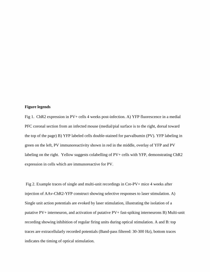
Figure legends
Fig 1. ChR2 expression in PV+ cells 4 weeks post-infection. A) YFP fluorescence in a medial
PFC coronal section from an infected mouse (medial/pial surface is to the right, dorsal toward
the top of the page) B) YFP labeled cells double-stained for parvalbumin (PV). YFP labeling in
green on the left, PV immunoreactivity shown in red in the middle, overlay of YFP and PV
labeling on the right. Yellow suggests colabelling of PV+ cells with YFP, demonstrating ChR2
expression in cells which are immunoreactive for PV.
Fig 2. Example traces of single and multi-unit recordings in Cre-PV+ mice 4 weeks after
injection of AAv-ChR2-YFP construct showing selective responses to laser stimulation. A)
Single unit action potentials are evoked by laser stimulation, illustrating the isolation of a
putative PV+ interneuron, and activation of putative PV+ fast-spiking interneurons B) Multi-unit
recording showing inhibition of regular firing units during optical stimulation. A and B: top
traces are extracellularly recorded potentials (Band-pass filtered: 30-300 Hz), bottom traces
indicates the timing of optical stimulation.
Fig 3. Activation of PV+ interneurons in the medial prefrontal cortex generates gamma
oscillations. Power spectral densities from 3 second epochs occurring just prior to (grey) or
during (black) optical stimulation delivered at 8 Hz (A) or 40 Hz (B). A) 8 Hz stimulation was
ineffective at inducing oscillations. B) Stimulation at 40 Hz induces a clear peak in the gamma
range (37-43 Hz). C and D represent the relative power induced by laser stimulation (see
methods). C) Optical stimulation at 8 Hz did not significantly increase oscillations. D) In
contrast, optical stimulation at 40 Hz significantly increased oscillations, demonstrating
successful induction of gamma oscillations (paired, one-tailed, Student’s t-test: p < 0.05, n = 6).
Fig 4. Effect of Cocaine on the induced oscillation. A through D are power spectra (0 - 100 Hz)
for six subjects which received 40 Hz optical stimulation for 3 second duration. Thin grey lines
represent individual data, while the thicker black line represents the mean of the individual data.
In a few cases significant line noise was present at precisely 60 Hz, this was excluded from the
graphs by setting the power at 60 Hz to the mean of the power at 59 and 61 Hz. A and C are prior
to i.p. cocaine injection while B and D are after. A and B represent immediately preceding the
optical stimulation, while C and D are during stimulation. In C and D, there is a clearly
distinguishable peak at the stimulation frequency, with a harmonic echo at 80 Hz. The shape of
the peak of the induced oscillation in the cocaine treated animals (D) appears to be sharper, with
increased peak power relative to the naïve condition (C). E) Comparison of power ratios across
bandwidth frequencies from 0 – 20 Hz. Power ratio values plotted against the bandwidth
‘window’ surrounding the 40 Hz stimulation frequency. Cocaine appears to have a sharpening
effect on the induced oscillation reflected here as an increase in peak power, while decreasing the
bandwidth of the induced oscillation. However, only the change in bandwidth is significant (p =
0.008), see text). Power ratio values are shown for cocaine (red) and naïve (black) conditions.
Thicker solid lines represent means, dashed lines are individual animals. The boxed inset depicts
an expanded view of the induced oscillation at 40 Hz under naïve and cocaine conditions.
Fig 5. Effect of the D1 antagonist SCH23390 on the cocaine induced change to the induced
oscillation. A) Power spectra around the stimulated frequency in naïve (A1), cocaine (A2), and
SCH23390 (A3) conditions. Sharpening of the peak is seen here again and appears to be reversed
by SCH23390. B) Power ratio vs. Bandwidth plot reflecting the changes in peak power and
bandwidth seen above. SCH23390 appears to block the sharpening effect of cocaine treatment.
C) Comparison of power ratio at the peak, across treatment conditions. Above the line represents
a larger power ratio in cocaine treatment vs either naïve (red) or SCH23390 (blue). Data points
are nearly overlapping demonstrating that SCH23390 appears similar to the naïve condition.
Fig 6. Effect of the D2 antagonist Sulpiride on cocaine induced change to the induced oscillation.
A) Power spectra around the stimulated frequency in naïve (A1), cocaine (A2), and Sulpiride
(A3) conditions. Sharpening of the peak is seen here again and appears to be enhanced by
Sulpiride. B) Power ratio vs. Bandwidth plot reflecting the changes in peak power and bandwidth
seen above. Sulpiride appears to increase the sharpening effect of cocaine treatment. C)
Comparison of power ratio at the peak, across treatment conditions. Above the line represents a
larger power ratio in cocaine treatment vs either naïve (red) or Sulpiride (blue). Sulpiride data
points are shifted to the right demonstrating that Sulpiride may increase the peak power further
than cocaine.


