Embed Size (px)
Citation preview
DOI: 10.14260/jemds/2015/1943
ORIGINAL ARTICLE
J of Evolution of Med and Dent Sci/ eISSN- 2278-4802, pISSN- 2278-4748/ Vol. 4/ Issue 78/ Sept 28, 2015 Page 13582
STUDY OF CONGENITAL MALFORMATIONS IN NEWBORN K. Koteswara Rao1, A. Krishna Prasad2, D. Manikyamba3, K. Adi Reddy4, Solomon Saawan P5, S. Anusha6
HOW TO CITE THIS ARTICLE: K. Koteswara Rao, A. Krishna Prasad, D. Manikyamba, K. Adi Reddy, Solomon Saawan P, S. Anusha. “Study of Congenital Malformations in Newborn”. Journal of Evolution of Medical and Dental Sciences 2015; Vol. 4, Issue 78, September 28; Page: 13582-13595, DOI: 10.14260/jemds/2015/1943
ABSTRACT: The etiology of congenital malformations has not been clearly defined. It is interesting to
note that certain congenital malformations are more prevalent in some areas1. Neural tubal defects
(NTD) are common in Punjab and cleft lip (CL) is common in south India. Gastrointestinal tract
defects are detected from south i.e. Mysore, Trivandrum, Hyderabad and Pondicherry. Polydactyly
was prevalent in south India and Chandigarh. Incidence of Talipes was highest in Delhi and next in
order were Chennai, Pondicherry and Patna. Malformations are controlled by genetic and
environmental factors and a thorough analysis would indicate the factors responsible for their
genesis and thereby, their means of prevention2. Thus it is imperative that in every region the
prevalence and peculiarities of malformations should be studied. Therefore, the present study was
carried out in Government General Hospital, Kakinada, where consanguineous marriages are very
much prevalent. The aim is to study the spectrum, incidence and maternal risk factors associated
with congenital malformations.
KEYWORDS: Congenital, Malformations, Birth Defects, Consanguinity, Anomalies.
INTRODUCTION: STUDY METHOD: The study was undertaken in the NICU, Department of
Pediatrics, Govt. General Hospital, Kakinada in collaboration with Department of Gynecology and
Obstetrics, Govt. General Hospital, Kakinada. This is a prevalence study. The present study was
carried out from September 2014 to August 2015 and 10, 720 consecutive births were studied in
Government General Hospital, Kakinada.
RESULTS:
Out of 146 cases, CNS defects were seen in 29 cases and accounted for19.8%, GIT defects in 25
cases (17.1%), Genitourinary malformations were 15 cases (10.3%), CVS were 14 cases (9.6%),
musculoskeletal anomalies were 12 cases (8.2%), Oral clefts were 12 cases (8.2%), syndromes
were 10 cases (6.8%), skin defects were 8 cases (5.5%), multiple anomalies were 9 cases
(6.2%), others were 12 cases (8.2%).
Incidence among male babies was 14.8/1000 total births, among females was 12.3/1000 total
births. Incidence of malformations was 1.17% among live born babies and 8.6% among still
born babies3.
Out of 24 cases of stillborn babies, 8 cases (36%) were having CNS malformations, 7 cases
(32%) were GI anomalies and 3 cases (14%) had multiple defects.
History of Polyhydramnios was recorded among mothers of 10.3% malformed babies,4
prematurity in 8.2% and oligohydramnios in 6.8%. Maternal history of PIH was obtained in
6.2%. There was history of fever in 1st trimester in 4.1%, history of repeated abortions in
1.37% and maternal history of Diabetes and hypothyroidism was observed in 2.7% and 0.68%
respectively.
DOI: 10.14260/jemds/2015/1943
ORIGINAL ARTICLE
J of Evolution of Med and Dent Sci/ eISSN- 2278-4802, pISSN- 2278-4748/ Vol. 4/ Issue 78/ Sept 28, 2015 Page 13583
The parents of 8.3% of malformed babies were married to their first cousins.513.6% were
married to a relative further than first cousin. In the present study, parental consanguinity had
been noted in 21.9% of all malformations and 33.4% of Musculoskeletal defects, 30% of
syndromes, 20% of G.I defects & G.U defects, 13.8% of C.N.S defects, 33.3% of miscellaneous
defects.
CONCLUSION: No definite environmental insult could be established as a cause in any of the cases. It
was presumed that the abnormalities were mainly caused by genetic factors.6 A direct relation
between the maternal age and occurrence of Down syndrome was noted.7 Maternal disorders
complicating pregnancy like Polyhydramnios, oligohydramnios, maternal febrile illness, PIH were
found to be one of the associated factors in the development of congenital malformations in
newborns.
Good and thorough antenatal care regarding identification and treatment of maternal
complications can prevent malformations.8 Whenever possible, a thorough screening of the mother
and fetus is to be done to rule out intra uterine infections. People should be counseled about
consanguinous marriages and about the relation between higher maternal age and Down syndrome.
A good and sound genetic counseling will prevent the recurrence of another congenitally malformed
baby.9 Antenatal diagnosis of congenital malformations by means of ultrasonography, Amniocentesis
and chorionic villous biopsy is very essential as mother could to terminate her pregnancy so that a lot
of morbidity can be avoided.
MATERIALS AND METHODS:
Source of Data: The present study was carried out from September 2014 to August 2015and 10, 720
consecutive births were studied in Government General Hospital, Kakinada. All of the children are
born at this hospital. Out of these, 146 cases were having congenital malformations. The cases
represent random mixture of all categories of people as the hospital caters to the needs of people
from all strata coming mainly from two Godavari districts. The population consists of people from all
walks of life, though majority of them are poor and illiterate.
Inclusion Criteria: All of the children who were born at this hospital during the study period were
included in this study.
Exclusion Criteria: Children born outside and brought to this institute were excluded from the
study.
Method of Collection of Data: A detailed history of mother regarding risk factors, maternal
investigations like ante natal scans along with relevant laboratory investigations and detailed family
history was taken and the babies were thoroughly examined for congenital malformations and data
was recorded as per proforma. Relevant available investigations were done whenever necessary.
RESULTS: In the present study, the incidence of congenital malformations in newborn was
13.6/1000 total births.10
DOI: 10.14260/jemds/2015/1943
ORIGINAL ARTICLE
J of Evolution of Med and Dent Sci/ eISSN- 2278-4802, pISSN- 2278-4748/ Vol. 4/ Issue 78/ Sept 28, 2015 Page 13584
No. of
Births
No. of
Malformations
Incidence/
1000 Births
Total 10, 720 146 13.6
Male 5, 585 83 14.8
Female 5, 135 63 12.3
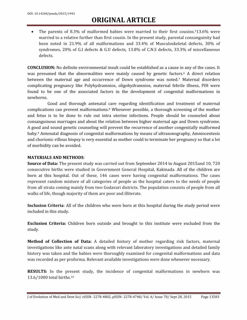
Sex Distribution: Out of 10, 720 babies born in this period 5, 585 were male and 5, 135 were female
and the incidence was 14.8/1000 births among males and 12.3/1000 among females.
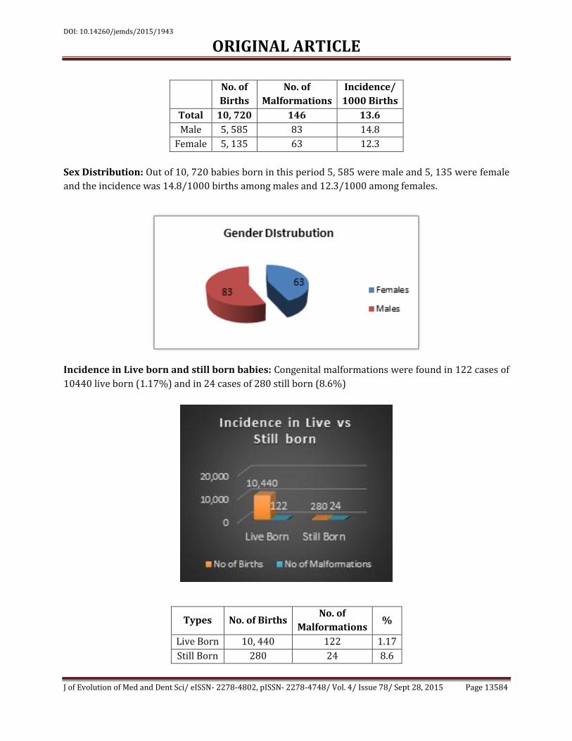
Incidence in Live born and still born babies: Congenital malformations were found in 122 cases of
10440 live born (1.17%) and in 24 cases of 280 still born (8.6%)
Types No. of Births No. of
Malformations %
Live Born 10, 440 122 1.17
Still Born 280 24 8.6
DOI: 10.14260/jemds/2015/1943
ORIGINAL ARTICLE
J of Evolution of Med and Dent Sci/ eISSN- 2278-4802, pISSN- 2278-4748/ Vol. 4/ Issue 78/ Sept 28, 2015 Page 13585
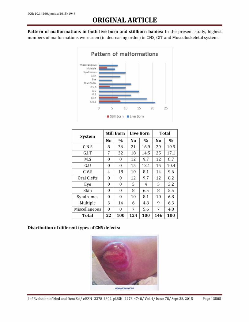
Pattern of malformations in both live born and stillborn babies: In the present study, highest
numbers of malformations were seen (in decreasing order) in CNS, GIT and Musculoskeletal system.
System Still Born Live Born Total
No % No % No %
C.N.S 8 36 21 16.9 29 19.9
G.I.T 7 32 18 14.5 25 17.1
M.S 0 0 12 9.7 12 8.7
G.U 0 0 15 12.1 15 10.4
C.V.S 4 18 10 8.1 14 9.6
Oral Clefts 0 0 12 9.7 12 8.2
Eye 0 0 5 4 5 3.2
Skin 0 0 8 6.5 8 5.5
Syndromes 0 0 10 8.1 10 6.8
Multiple 3 14 6 4.8 9 6.3
Miscellaneous 0 0 7 5.6 7 4.8
Total 22 100 124 100 146 100
Distribution of different types of CNS defects:
DOI: 10.14260/jemds/2015/1943
ORIGINAL ARTICLE
J of Evolution of Med and Dent Sci/ eISSN- 2278-4802, pISSN- 2278-4748/ Vol. 4/ Issue 78/ Sept 28, 2015 Page 13586
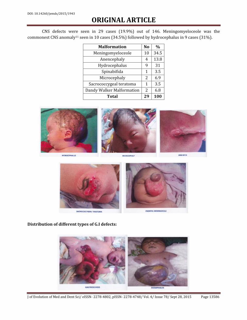
CNS defects were seen in 29 cases (19.9%) out of 146. Meningomyeloceole was the
commonest CNS anomaly11 seen in 10 cases (34.5%) followed by hydrocephalus in 9 cases (31%).
Malformation No %
Meningomyeloceole 10 34.5
Anencephaly 4 13.8
Hydrocephalus 9 31
Spinabifida 1 3.5
Microcephaly 2 6.9
Sacrococcygeal teratoma 1 3.5
Dandy Walker Malformation 2 6.8
Total 29 100
Distribution of different types of G.I defects:
DOI: 10.14260/jemds/2015/1943
ORIGINAL ARTICLE
J of Evolution of Med and Dent Sci/ eISSN- 2278-4802, pISSN- 2278-4748/ Vol. 4/ Issue 78/ Sept 28, 2015 Page 13587
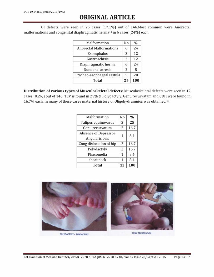
GI defects were seen in 25 cases (17.1%) out of 146.Most common were Anorectal
malformations and congenital diaphragmatic hernia12 in 6 cases (24%) each.
Malformation No %
Anorectal Malformations 6 24
Exomphalos 3 12
Gastroschisis 3 12
Diaphragmatic hernia 6 24
Duodenal atresia 2 8
Tracheo-esophageal Fistula 5 20
Total 25 100
Distribution of various types of Musculoskeletal defects: Musculoskeletal defects were seen in 12
cases (8.2%) out of 146. TEV is found in 25% & Polydactyly, Genu recurvatam and CDH were found in
16.7% each. In many of these cases maternal history of Oligohydramnios was obtained.13
Malformation No %
Talipes equinovarus 3 25
Genu recurvatum 2 16.7
Absence of Depressor
Angularis oris 1 8.4
Cong dislocation of hip 2 16.7
Polydactyly 2 16.7
Phacomelia 1 8.4
short neck 1 8.4
Total 12 100

DOI: 10.14260/jemds/2015/1943
ORIGINAL ARTICLE
J of Evolution of Med and Dent Sci/ eISSN- 2278-4802, pISSN- 2278-4748/ Vol. 4/ Issue 78/ Sept 28, 2015 Page 13588
Distrubution of various types of Genito Urinary defects: Genito urinary defects were seen in 15
cases (10.3%). Among the G.U defects one peculiar case of absence of penis was observed. There
were 5 cases of hypospadias14, 2 cases of hydro ureteronephrosis, one case of epispadias, ambiguous
genitalia and dystrophic kidney each.
Malformation No %
Hypospadias 5 33.2
Exstropy Of Bladder +Epispadias 1 6.7
Congenital Hydrocele 3 20
Hydro uretronephrosis 2 13.3
Dystrophic Kidney 1 6.7
Absence of Penis 1 6.7
Ambiguous genitalia 1 6.7
Ovarian cyst 1 6.7
Total 15 100
Distribution of Cardiovascular Anomalies: Cardiovascular defects were seen in 14 cases (9.6%),
consisting of 35.6% cases of Acyanotic Heart Disease and 21.4% cases of Complex Heart Disease and
Cyanotic Heart Diseases each. One each case of atrial myxoma, Hypo plastic Heart, and Intra cardiac
tumor were observed. The diagnosis was by antenatal ultrasonography and X-Ray, ECG and 2D ECHO
of baby.
Malformation No %
Acyanotic Heart Disease 5 35.6
Cyanotic Heart Disease 3 21.4
Intra Cardiac Tumor 1 7.2
Hypo plastic Heart 1 7.2
Atrial Myxoma 1 7.2
Complex Heart Disease 3 21.4
Total 14 100
DOI: 10.14260/jemds/2015/1943
ORIGINAL ARTICLE
J of Evolution of Med and Dent Sci/ eISSN- 2278-4802, pISSN- 2278-4748/ Vol. 4/ Issue 78/ Sept 28, 2015 Page 13589
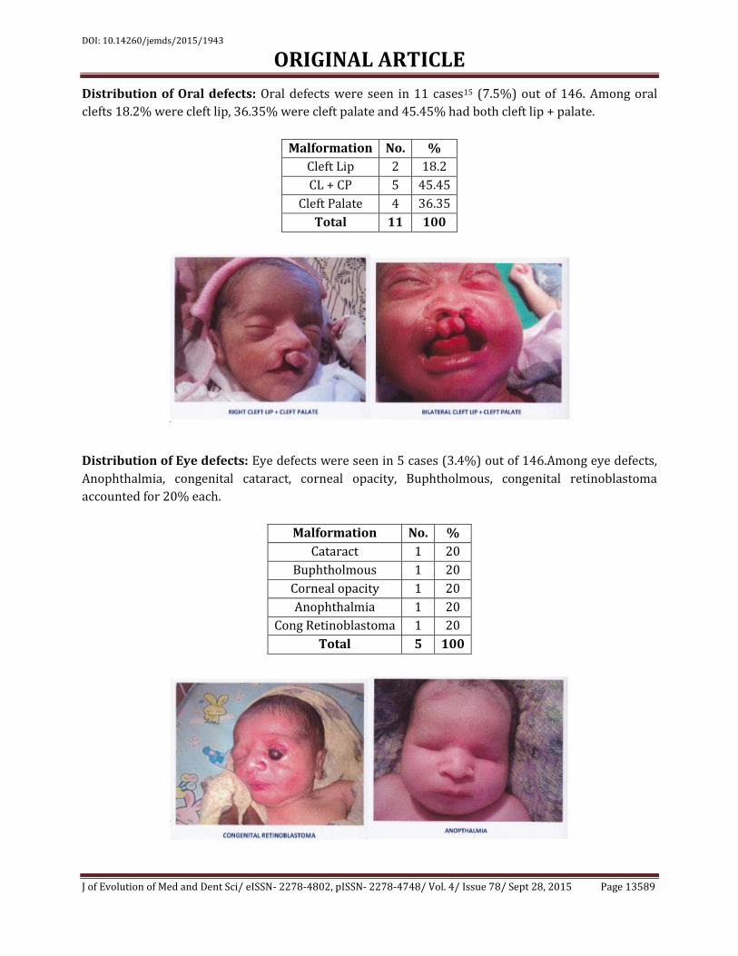
Distribution of Oral defects: Oral defects were seen in 11 cases15 (7.5%) out of 146. Among oral
clefts 18.2% were cleft lip, 36.35% were cleft palate and 45.45% had both cleft lip + palate.
Malformation No. %
Cleft Lip 2 18.2
CL + CP 5 45.45
Cleft Palate 4 36.35
Total 11 100
Distribution of Eye defects: Eye defects were seen in 5 cases (3.4%) out of 146.Among eye defects,
Anophthalmia, congenital cataract, corneal opacity, Buphtholmous, congenital retinoblastoma
accounted for 20% each.
Malformation No. %
Cataract 1 20
Buphtholmous 1 20
Corneal opacity 1 20
Anophthalmia 1 20
Cong Retinoblastoma 1 20
Total 5 100
DOI: 10.14260/jemds/2015/1943
ORIGINAL ARTICLE
J of Evolution of Med and Dent Sci/ eISSN- 2278-4802, pISSN- 2278-4748/ Vol. 4/ Issue 78/ Sept 28, 2015 Page 13590
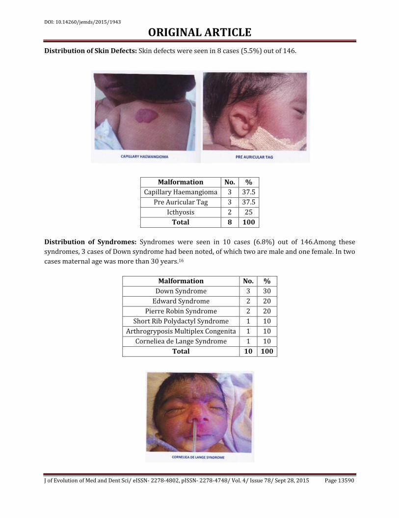
Distribution of Skin Defects: Skin defects were seen in 8 cases (5.5%) out of 146.
Malformation No. %
Capillary Haemangioma 3 37.5
Pre Auricular Tag 3 37.5
Icthyosis 2 25
Total 8 100
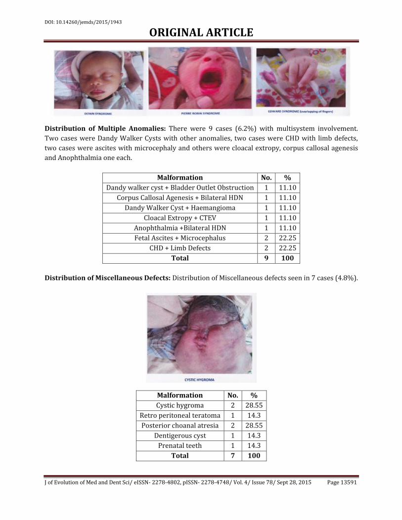
Distribution of Syndromes: Syndromes were seen in 10 cases (6.8%) out of 146.Among these
syndromes, 3 cases of Down syndrome had been noted, of which two are male and one female. In two
cases maternal age was more than 30 years.16
Malformation No. %
Down Syndrome 3 30
Edward Syndrome 2 20
Pierre Robin Syndrome 2 20
Short Rib Polydactyl Syndrome 1 10
Arthrogryposis Multiplex Congenita 1 10
Corneliea de Lange Syndrome 1 10
Total 10 100
DOI: 10.14260/jemds/2015/1943
ORIGINAL ARTICLE
J of Evolution of Med and Dent Sci/ eISSN- 2278-4802, pISSN- 2278-4748/ Vol. 4/ Issue 78/ Sept 28, 2015 Page 13591
Distribution of Multiple Anomalies: There were 9 cases (6.2%) with multisystem involvement.
Two cases were Dandy Walker Cysts with other anomalies, two cases were CHD with limb defects,
two cases were ascites with microcephaly and others were cloacal extropy, corpus callosal agenesis
and Anophthalmia one each.
Malformation No. %
Dandy walker cyst + Bladder Outlet Obstruction 1 11.10
Corpus Callosal Agenesis + Bilateral HDN 1 11.10
Dandy Walker Cyst + Haemangioma 1 11.10
Cloacal Extropy + CTEV 1 11.10
Anophthalmia +Bilateral HDN 1 11.10
Fetal Ascites + Microcephalus 2 22.25
CHD + Limb Defects 2 22.25
Total 9 100
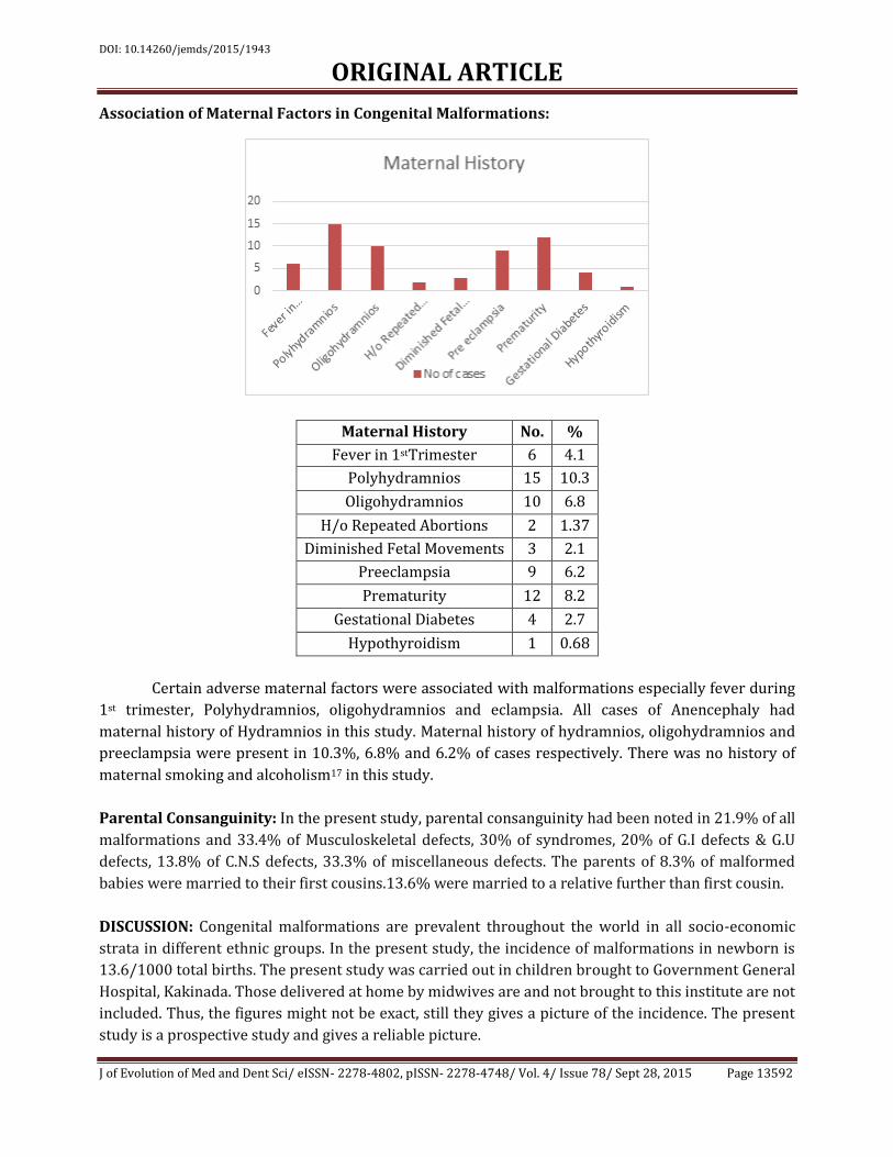
Distribution of Miscellaneous Defects: Distribution of Miscellaneous defects seen in 7 cases (4.8%).
Malformation No. %
Cystic hygroma 2 28.55
Retro peritoneal teratoma 1 14.3
Posterior choanal atresia 2 28.55
Dentigerous cyst 1 14.3
Prenatal teeth 1 14.3
Total 7 100
DOI: 10.14260/jemds/2015/1943
ORIGINAL ARTICLE
J of Evolution of Med and Dent Sci/ eISSN- 2278-4802, pISSN- 2278-4748/ Vol. 4/ Issue 78/ Sept 28, 2015 Page 13592
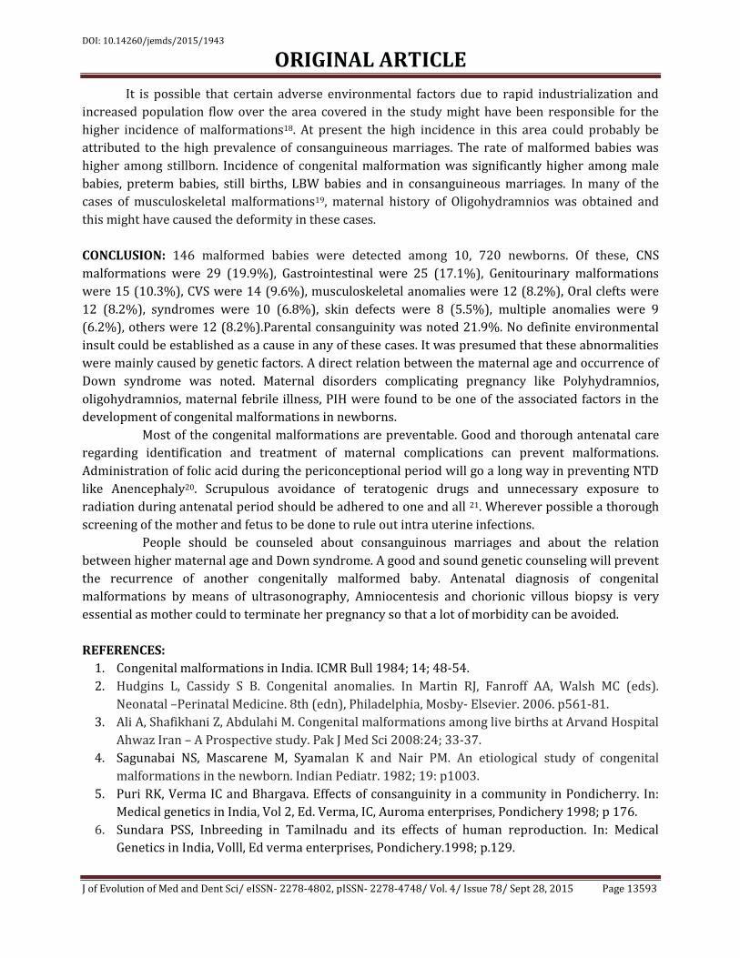
Association of Maternal Factors in Congenital Malformations:
Maternal History No. %
Fever in 1stTrimester 6 4.1
Polyhydramnios 15 10.3
Oligohydramnios 10 6.8
H/o Repeated Abortions 2 1.37
Diminished Fetal Movements 3 2.1
Preeclampsia 9 6.2
Prematurity 12 8.2
Gestational Diabetes 4 2.7
Hypothyroidism 1 0.68
Certain adverse maternal factors were associated with malformations especially fever during
1st trimester, Polyhydramnios, oligohydramnios and eclampsia. All cases of Anencephaly had
maternal history of Hydramnios in this study. Maternal history of hydramnios, oligohydramnios and
preeclampsia were present in 10.3%, 6.8% and 6.2% of cases respectively. There was no history of
maternal smoking and alcoholism17 in this study.
Parental Consanguinity: In the present study, parental consanguinity had been noted in 21.9% of all
malformations and 33.4% of Musculoskeletal defects, 30% of syndromes, 20% of G.I defects & G.U
defects, 13.8% of C.N.S defects, 33.3% of miscellaneous defects. The parents of 8.3% of malformed
babies were married to their first cousins.13.6% were married to a relative further than first cousin.
DISCUSSION: Congenital malformations are prevalent throughout the world in all socio-economic
strata in different ethnic groups. In the present study, the incidence of malformations in newborn is
13.6/1000 total births. The present study was carried out in children brought to Government General
Hospital, Kakinada. Those delivered at home by midwives are and not brought to this institute are not
included. Thus, the figures might not be exact, still they gives a picture of the incidence. The present
study is a prospective study and gives a reliable picture.
DOI: 10.14260/jemds/2015/1943
ORIGINAL ARTICLE
J of Evolution of Med and Dent Sci/ eISSN- 2278-4802, pISSN- 2278-4748/ Vol. 4/ Issue 78/ Sept 28, 2015 Page 13593
It is possible that certain adverse environmental factors due to rapid industrialization and
increased population flow over the area covered in the study might have been responsible for the
higher incidence of malformations18. At present the high incidence in this area could probably be
attributed to the high prevalence of consanguineous marriages. The rate of malformed babies was
higher among stillborn. Incidence of congenital malformation was significantly higher among male
babies, preterm babies, still births, LBW babies and in consanguineous marriages. In many of the
cases of musculoskeletal malformations19, maternal history of Oligohydramnios was obtained and
this might have caused the deformity in these cases.
CONCLUSION: 146 malformed babies were detected among 10, 720 newborns. Of these, CNS
malformations were 29 (19.9%), Gastrointestinal were 25 (17.1%), Genitourinary malformations
were 15 (10.3%), CVS were 14 (9.6%), musculoskeletal anomalies were 12 (8.2%), Oral clefts were
12 (8.2%), syndromes were 10 (6.8%), skin defects were 8 (5.5%), multiple anomalies were 9
(6.2%), others were 12 (8.2%).Parental consanguinity was noted 21.9%. No definite environmental
insult could be established as a cause in any of these cases. It was presumed that these abnormalities
were mainly caused by genetic factors. A direct relation between the maternal age and occurrence of
Down syndrome was noted. Maternal disorders complicating pregnancy like Polyhydramnios,
oligohydramnios, maternal febrile illness, PIH were found to be one of the associated factors in the
development of congenital malformations in newborns.
Most of the congenital malformations are preventable. Good and thorough antenatal care
regarding identification and treatment of maternal complications can prevent malformations.
Administration of folic acid during the periconceptional period will go a long way in preventing NTD
like Anencephaly20. Scrupulous avoidance of teratogenic drugs and unnecessary exposure to
radiation during antenatal period should be adhered to one and all 21. Wherever possible a thorough
screening of the mother and fetus to be done to rule out intra uterine infections.
People should be counseled about consanguinous marriages and about the relation
between higher maternal age and Down syndrome. A good and sound genetic counseling will prevent
the recurrence of another congenitally malformed baby. Antenatal diagnosis of congenital
malformations by means of ultrasonography, Amniocentesis and chorionic villous biopsy is very
essential as mother could to terminate her pregnancy so that a lot of morbidity can be avoided.
REFERENCES:
1. Congenital malformations in India. ICMR Bull 1984; 14; 48-54.
2. Hudgins L, Cassidy S B. Congenital anomalies. In Martin RJ, Fanroff AA, Walsh MC (eds).
Neonatal –Perinatal Medicine. 8th (edn), Philadelphia, Mosby- Elsevier. 2006. p561-81.
3. Ali A, Shafikhani Z, Abdulahi M. Congenital malformations among live births at Arvand Hospital
Ahwaz Iran – A Prospective study. Pak J Med Sci 2008:24; 33-37.
4. Sagunabai NS, Mascarene M, Syamalan K and Nair PM. An etiological study of congenital
malformations in the newborn. Indian Pediatr. 1982; 19: p1003.
5. Puri RK, Verma IC and Bhargava. Effects of consanguinity in a community in Pondicherry. In:
Medical genetics in India, Vol 2, Ed. Verma, IC, Auroma enterprises, Pondichery 1998; p 176.
6. Sundara PSS, Inbreeding in Tamilnadu and its effects of human reproduction. In: Medical
Genetics in India, Volll, Ed verma enterprises, Pondichery.1998; p.129.
DOI: 10.14260/jemds/2015/1943
ORIGINAL ARTICLE
J of Evolution of Med and Dent Sci/ eISSN- 2278-4802, pISSN- 2278-4748/ Vol. 4/ Issue 78/ Sept 28, 2015 Page 13594
7. Stoll B J. Congenital anomalies. In Kliegman R M, Jenson H B, Behrman R E, Stanton B F(eds)
Nelson Textbook of paediatrics 18th (edn) Philadelphia. WB Saunders, 2008.pp.711-13.
8. Kalter H, Warkany J. 1983. (Medical Progress) Congenital malformations etiologic factors and
their role in prevention. New England Journal of Medicine 308(8):424–431(Part I);491–
497(Part II).
9. World Health Organization (WHO). 1999. Human Genetics: Services for the Prevention and
Management of Genetic Disorders and Birth Defects in Developing Countries: Report of a Joint
WHO/WOAPBD Meeting. Geneva: WHO.
10. Swain S. Agarwal A, Bhatia BD. Congenital malformations at birth. Ind Pediatr 1994;
31(10):1187-91.
11. Cherian A, Seena S, Bullock RK, Antony AC. Incidence of neural tube defects in the least
developed area of India: A population-based study. Lancet 2005; 366:930-931.
12. Mishra PC, Baweja R. Congenital malformations in the newborns. A prospective study. Ind
Pediatr1989; 26(1): 32-35.
13. Verma M. Chhatwal J. Singh D. Congenital malformations. A prospective study of 10, 000 cases.
Ind J Pediat.1991; 58(2): 245-52.
14. Gupta R K, Singh A, Gupta R. Pattern of Congenital Anomalies In Newborn at Birth: A Hospital
Based
a. Prospective Study. Scientific paper presented during the Proceedings at 42nd national
conference of Indian academy of Pediatrics (Pedicon) at KolKatta, 2005.
15. Smith HW, Keen M, Edwards E. 1991. Cleft lip and palate surgery in La Ceiba, Honduras.
Archives of Otolaryngology—Head and Neck Surgery 117(12):1356–1359.
16. Hook EB. 1982. The epidemiology of Down syndrome. In Pueschel SM (ed.). Down syndrome:
Advances in Biomedicine and the Behavioral Sciences. Cambridge, MA: Ware Press. Pp. 11–18.
17. Gladstone J, Nulman I, Koren G. 1996. Reproductive risks of binge drinking during pregnancy.
Reproductive Toxicology 10(1):3–13.
18. Kuliev AM, Modell B. 1990. Problems in the control of genetic disorders. Biomedical Science
1(1):3–17.
19. Gupta RK, Gupta CR, Singh D. Incidence of Congenital malformations of the musculoskeletal
system in live borns in Jammu. JK Science 2003; 4: 157-60.
20. Berry RJ, Li Z, Erickson JD, Li S, Moore CA, Wang H, Mulinare J, Zhao P, Wong LC, Gindler J, Hong
S, Correa A. 1999. Prevention of neural-tube defects with folic acid inChina. New England
Journal of Medicine 341(20):1485–1490.
21. Schull WJ, Otake M. 1999. Cognitive function and peri natal exposure to ionizing radiation.
Teratology 59(4):222–226.
DOI: 10.14260/jemds/2015/1943
ORIGINAL ARTICLE
J of Evolution of Med and Dent Sci/ eISSN- 2278-4802, pISSN- 2278-4748/ Vol. 4/ Issue 78/ Sept 28, 2015 Page 13595
AUTHORS:
1. K. Koteswara Rao
2. A. Krishna Prasad
3. D. Manikyamba
4. K. Adi Reddy
5. Solomon Saawan P.
6. S. Anusha
PARTICULARS OF CONTRIBUTORS:
1. In charge Professor, Department of
Pediatrics, Rangaraya Medical College,
Kakinada.
2. Associate Professor, Department of
Pediatrics, Rangaraya Medical College,
Kakinada.
3. Professor & HOD, Department of
Pediatrics, Rangaraya Medical College,
Kakinada.
4. Junior Resident, Department of Pediatrics,
Rangaraya Medical College, Kakinada.
FINANCIAL OR OTHER
COMPETING INTERESTS: None
5. Junior Resident, Department of ENT,
Rangaraya Medical College, Kakinada.
6. Junior Resident, Department of Pediatrics,
Rangaraya Medical College, Kakinada.
NAME ADDRESS EMAIL ID OF THE
CORRESPONDING AUTHOR:
Dr. K. Adi Reddy,
Junior Resident,
Department of Pediatrics,
Rangaraya Medical College,
Kakinada.
E-mail: [email protected]
Date of Submission: 18/09/2015.
Date of Peer Review: 19/09/2015.
Date of Acceptance: 21/09/2015.
Date of Publishing: 28/09/2015.


![Gabriel Figueroa and Juan Rulfo [ENGLISH Version of Original Spanish Article]](https://img.pdfslide.net/doc/110x75/635d0d1ffd007d475b02f8d2/gabriel-figueroa-and-juan-rulfo-english-version-of-original-spanish-article.jpg)