Embed Size (px)
Citation preview

Overcoming the restriction barrier to plasmidtransformation and targeted mutagenesis inBifidobacterium breve UCC2003
Mary O’Connell Motherway,1,2 Jonathan O’Driscoll,1†
Gerald F. Fitzgerald1,2,3 and Douwe van Sinderen1,2*1Alimentary Pharmabiotic Centre, 2Department ofMicrobiology and 3Department of Food and NutritionalSciences , National University of Ireland, Cork, WesternRoad, Cork, Ireland.
Summary
In silico analysis of the Bifidobacterium breveUCC2003 genome predicted two distinct loci, whichencode three different restriction/modification sys-tems, each comprising a modification methylase and arestriction endonuclease. Based on sequence homol-ogy and observed protection against restriction weconclude that the first restriction endonuclease, des-ignated BbrI, is an isoschizomer of BbeI, the second,BbrII, is a neoschizomer of SalI, while the third, BbrIII,is an isoschizomer of PstI. Expression of each of the B.breve UCC2003 methylase-encoding genes in B. breveJCM 7017 established that BbrII and BbrIII are activeand restrict incoming DNA. By exploiting knowledgeon restriction/modification in B. breve UCC2003 wesuccessfully increased the transformation efficiencyto a level that allows the reliable generation of mutantsby homologous recombination using a non-replicativeplasmid.
Introduction
The commensal gut microbiota has long been appreci-ated for its influence on gut health (reviewed by O’Haraand Shanahan, 2006; Turroni et al., 2008). Bifidobacteriaconstitute a specific group of mostly commensal bacteria,which inhabit the gastrointestinal tract (GIT) of mammals,including the human GIT, where they are estimated torepresent 3–6% of the adult faecal flora (Ventura et al.,2004; Saxelin et al., 2005; Zoetendal and Vaughan,2006). The presence of bifidobacteria in the human GIT
has been associated with many beneficial health effects,such as the prevention of diarrhoea, amelioration oflactose intolerance and immunomodulation (reviewed byLeahy et al., 2005). Indeed, the health benefits of probi-otic bacteria such as bifidobacteria have been shown toextend beyond the GIT (Lenoir-Wijnkoop et al., 2007).These many positive attributes have led to the wide-spread incorporation of bifidobacteria as live componentsof commercial health-promoting probiotic foods. Despitethese commercial and scientific interests, fundamentalknowledge is still scarce regarding the exact molecularmechanisms by which bifidobacteria contribute to hosthealth and well-being. Such scientific knowledge isessential to scientifically explain the purported health ben-efits, and consequently support the inclusion of such bac-teria as probiotics in functional foods.
The genome sequences on Bifidobacterium longumsubsp. longum NCC2705 (Schell et al., 2002), B. longumsubsp. longum DJ010A (Lee et al., 2008), B. adolescentisATCC15703 (Suzuki et al., 2006), B. adolescentis L2-32(Fulton et al., 2007), B. dentium ATCC27678 (Sudar-sanam et al., 2008) and B. animalis subsp. lactis HN019(Collett et al., 2008) have recently become available andhave contributed very significantly to advancing ourknowledge on bifidobacterial genetics and metabolism.However, the availability of a genome sequence is merelya first step towards a better understanding of a specificprobiotic property, and unravelling the molecular mecha-nisms by which bifidobacteria bring about positive hostresponses demands the availability of suitable moleculartools. To date, relatively few molecular tools for bifidobac-teria have been developed, which explains why the genet-ics of these microbes is rather poorly understood,certainly when compared with other bacteria of industrialimportance.
Available genetic tools for bifidobacteria include bifido-bacterial plasmids, which were first reported by Sgorbatiand colleagues (1982). In recent years significant efforthas focused on identifying and sequencing plasmids frombifidobacteria, and exploiting some of these native bifido-bacterial replicons for the creation of Escherichiacoli–Bifidobacterium shuttle vectors (Lee and O’Sullivan2006; Alvarez-Martín et al., 2007; Cronin et al., 2007;Sangrador-Vegas and colleagues, 2007). A limitation of
Received 18 September, 2008; revised 24 October, 2008; accepted27 October, 2008. *For correspondence. E-mail [email protected]; Tel. (+353) 21 490 1365; Fax (+353) 21 490 3101. †Presentaddress: New England Biolabs, Inc., 240 County Road, Ipswich, MA01938-2723, USA.
Microbial Biotechnology (2009) 2(3), 321–332 doi:10.1111/j.1751-7915.2008.00071.x
No claim to original Irish government worksJournal compilation © 2008 Society for Applied Microbiology and Blackwell Publishing Ltd

many of these shuttle vectors is the low transformationefficiency of many of the bifidobacteria tested, coupledin some cases with segregational instability (Lee andO’Sullivan, 2006).
The observed differences in transformation efficiencyamong different strains of bifidobacteria may be attrib-uted, at least in part, to restriction/modification (R–M)systems, which are ubiquitous among prokaryotes andgenerally comprise of a restriction endonuclease (REase)and cognate methyltransferase (MTase) (Murray, 2002;Tock and Dryden, 2005). R–M systems are believed toserve primarily as defensive instruments that protectprokaryotic cells against invading DNA such as promiscu-ous plasmids or infecting bacteriophage. R–M systemsare classified into four groups (designated type I, II, III andIV) on the basis of their subunit composition, co-factorrequirement, recognition sequence structure and thecleavage site relative to the recognition sequence(Roberts et al., 2003). Type I R–M systems consist ofthree different subunits, HsdM, HsdR and HsdS, that areresponsible for modification, restriction and sequence rec-ognition respectively. Type I REases require ATP, MG2+
and AdoMet for activity. In general they interact with twoasymmetrical bi-partite recognition sites, translocate theDNA in an ATP hydrolysis-dependent manner and cut theDNA distal to the recognition sites, approximately half-way between two sites (Murray, 2002). Typically, in a typeII R–M system the REase recognizes and cleaves withina short (4–8 bp) palindromic DNA sequence. Protection of‘self’ DNA from restriction occurs by methylation using anMTase, which modifies specific adenosyl or cytosyl resi-dues within the sequence recognized by the correspond-ing REase (Kobayashi, 2001; Pingoud et al., 2005). TypeIII R–M systems consist of two subunits, Mod, responsiblefor DNA recognition and modification, and Res, respon-sible for DNA cleavage. Active nucleases require ATP andMG2+ for activity and are stimulated by AdoMet. Theholoenzyme, composed of two Res and two Mod sub-units, interacts with two unmodified asymmetric targetsites positioned in inverse orientations with respect toeach other and cuts the DNA close to one recognition site(Janscak et al., 2001). Type IV R–M systems are specifiedby either one of two structural genes encoding proteinswith specificities for methylated, hydroxymethylated orglucosyl-hydroxymethylated bases in the target DNA mol-ecule (Roberts et al., 2003).
REase activity in Bifidobacterium was first described byKhosaka and colleagues (1982) and to date a total of 23bifidobacterial proven or putative R–M systems have beenidentified, as listed on the REBASE website (http://rebase.neb.com/rebase). BbeI, the first bifidobacterialREase to be described, was isolated from Bifidobacteriumbreve YIT4006, recognizing and cleaving the sequence5′-GGCGC↓C-3′. However, two copies of the BbeI recog-
nition sequence are required for full endonuclease activity(Khosaka et al., 1982). Subsequently Khosaka and col-leagues (1983) reported on the identification of the REasesBinSI and BinSII from B. longum subsp. infantis S76e.BinSI is an isoschizomer of EcoRII (recognizing and cleav-ing the sequence 5′↓CCWGG-3′), while BinSII exhibits thesame restriction specificity as BbeI (5′-GGCGC↓C-3′).BinI was isolated from B. longum subsp. infantis 659, andrecognizes the asymmetric pentanucleotide sequence5′-GGATCNNNN↓N-3′ (Khosaka and Kiwaki, 1984).Skrypina and colleagues (1988) showed that four out of 12bifidobacterial strains exhibited REase activity, of whichtwo, BadI from B. adolescentis LVA1 and BbfI from B.bifidum LVA3, are isoschizomers of XhoI (5′-C↓TCGAG-3′),while the REases Bbf7411I from B. bifidum 7411 andBla7920I from B. lactentis 7920 are neoschizomers ofBspMII (5′-T↓CCGGA-3′). Hartke and colleagues (1996)identified two REases from B. longum subsp. longum BL2:BloI is an isoschizomer of XhoII (5′-R↓GATCY-3′), whileBloII is an isoschizomer of PstI (5′-CTGCA↓G-3′).
In the current study we report on the identificationand preliminary characterization of three R–M systemsencoded on the genome of B. breve UCC2003. Circum-venting these R–M systems allowed the development of areliable method for the creation of gene disruptions in B.breve UCC2003.
Results
Sequence, genetic organization and amino acid analysisof the BbrI, BbrII and BbrIII R–M systems from B. breveUCC2003
Two loci, predicted to encode three different R–Msystems, were identified from the annotation of thegenome sequence of B. breve UCC2003 (S. Leahy. M.O’Connell Motherway, J. Moreno Munoz, G.F. Fitzgerald,D. Higgins and D. van Sinderen, unpubl. results) anddesignated BbrI, BbrII and BbrIII (Fig. 1A). The G+Ccontent for each system is 58% which is in agreementwith the approximately 60% G+C content for bifidobacte-ria (Ventura et al., 2007). The first gene of the BbrI R–Msystem, bbrIM, codes for a protein (M.BbrI; 43.2 kDa) with60% and 53% identity to cytosine-specific MTases fromClavibacter michiganesis and Photorhabdus luminescensrespectively; M.BbrI also contains the six highly con-served motifs characteristic of known 5′-methylcytosineMTases (Kumar et al., 1984) (Fig. 1B). The cytosine-specific MTases from C. michiganesis and P. luminescensare known to methylate of the sequence 5′-GGC(m5)GCC-3′, which is also the recognition sequence of the BbeIREase identified by Khosaka and colleagues (1982) fromB. breve YIT4006. The protein product of the second ORF,bbr0215, exhibits 94% identity to a hypothetical proteinencoded by B. longum subsp. longum NCC2705 (Schell
322 M. O’Connell Motherway et al.
No claim to original Irish government worksJournal compilation © 2008 Society for Applied Microbiology and Blackwell Publishing Ltd, Microbial Biotechnology, 2, 321–332
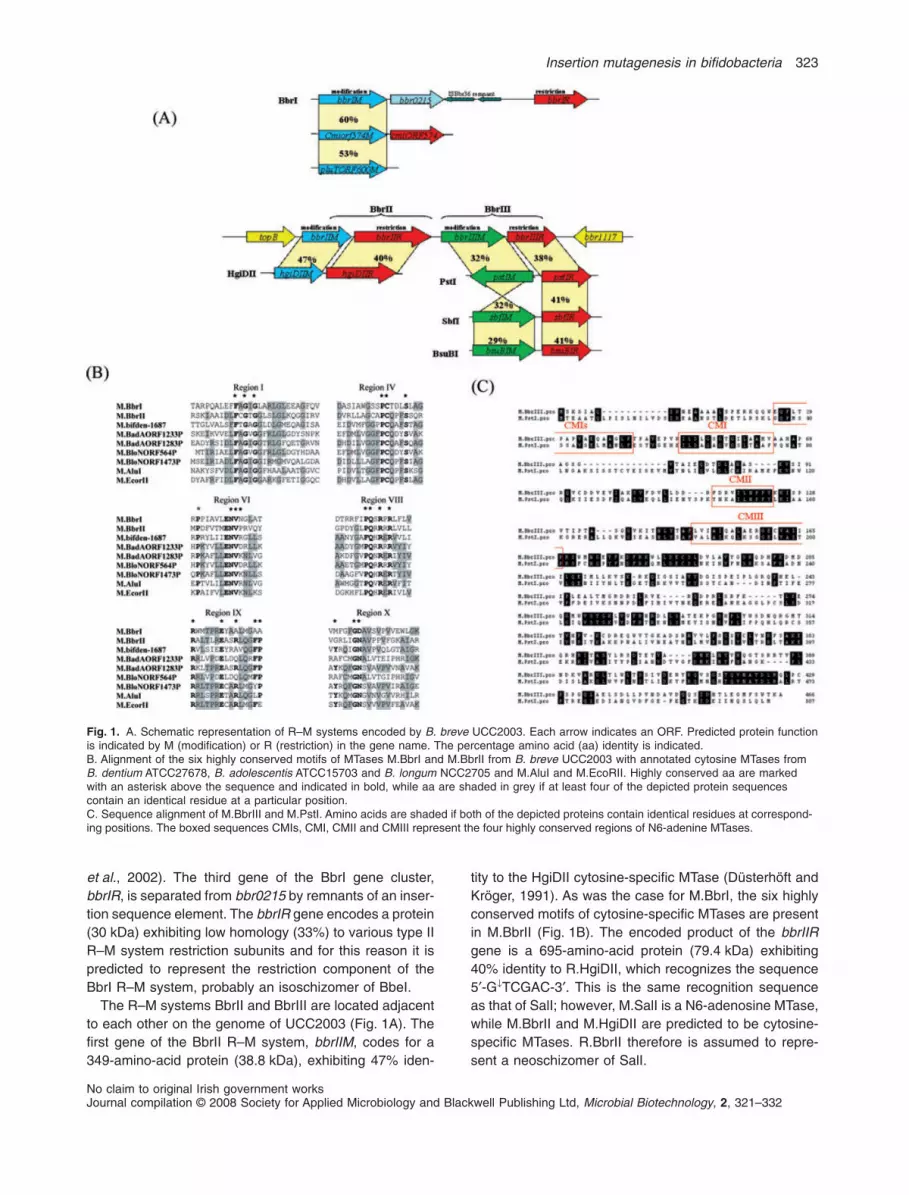
et al., 2002). The third gene of the BbrI gene cluster,bbrIR, is separated from bbr0215 by remnants of an inser-tion sequence element. The bbrIR gene encodes a protein(30 kDa) exhibiting low homology (33%) to various type IIR–M system restriction subunits and for this reason it ispredicted to represent the restriction component of theBbrI R–M system, probably an isoschizomer of BbeI.
The R–M systems BbrII and BbrIII are located adjacentto each other on the genome of UCC2003 (Fig. 1A). Thefirst gene of the BbrII R–M system, bbrIIM, codes for a349-amino-acid protein (38.8 kDa), exhibiting 47% iden-
tity to the HgiDII cytosine-specific MTase (Düsterhöft andKröger, 1991). As was the case for M.BbrI, the six highlyconserved motifs of cytosine-specific MTases are presentin M.BbrII (Fig. 1B). The encoded product of the bbrIIRgene is a 695-amino-acid protein (79.4 kDa) exhibiting40% identity to R.HgiDII, which recognizes the sequence5′-G↓TCGAC-3′. This is the same recognition sequenceas that of SalI; however, M.SalI is a N6-adenosine MTase,while M.BbrII and M.HgiDII are predicted to be cytosine-specific MTases. R.BbrII therefore is assumed to repre-sent a neoschizomer of SalI.
Fig. 1. A. Schematic representation of R–M systems encoded by B. breve UCC2003. Each arrow indicates an ORF. Predicted protein functionis indicated by M (modification) or R (restriction) in the gene name. The percentage amino acid (aa) identity is indicated.B. Alignment of the six highly conserved motifs of MTases M.BbrI and M.BbrII from B. breve UCC2003 with annotated cytosine MTases fromB. dentium ATCC27678, B. adolescentis ATCC15703 and B. longum NCC2705 and M.AluI and M.EcoRII. Highly conserved aa are markedwith an asterisk above the sequence and indicated in bold, while aa are shaded in grey if at least four of the depicted protein sequencescontain an identical residue at a particular position.C. Sequence alignment of M.BbrIII and M.PstI. Amino acids are shaded if both of the depicted proteins contain identical residues at correspond-ing positions. The boxed sequences CMIs, CMI, CMII and CMIII represent the four highly conserved regions of N6-adenine MTases.
Insertion mutagenesis in bifidobacteria 323
No claim to original Irish government worksJournal compilation © 2008 Society for Applied Microbiology and Blackwell Publishing Ltd, Microbial Biotechnology, 2, 321–332

The third identified R–M system on the genome of B.breve UCC2003, BbrIII, is predicted to encode an isoschi-zomer of PstI and BloII, the latter representing a REaseidentified from B. longum subsp. longum BL2 (Hartkeet al., 1996). The first gene, bbrIIIM, encodes a 315-amino-acid protein (36.3 kDa), which shares 32% identitywith M.PstI, an N6-adenosine MTase (Walder, Walderet al., 1984). The four conserved motifs characteristic ofN6-adenosine-methyltransferase, CMIs, CMI, CMII andCMIII (Timinskas et al., 1995), can be identified in M.BbrIII(Fig. 1C). The second gene bbrIIIR encodes a 355-amino-acid protein (36.6 kDa), exhibiting 38% identity to theREase Pst1 (5′-CTGCA↓G-3′).
Assessment of R–M activity in B. breve UCC2003
To establish if the identified R–M systems are functionalin B. breve UCC2003 and whether they affect trans-
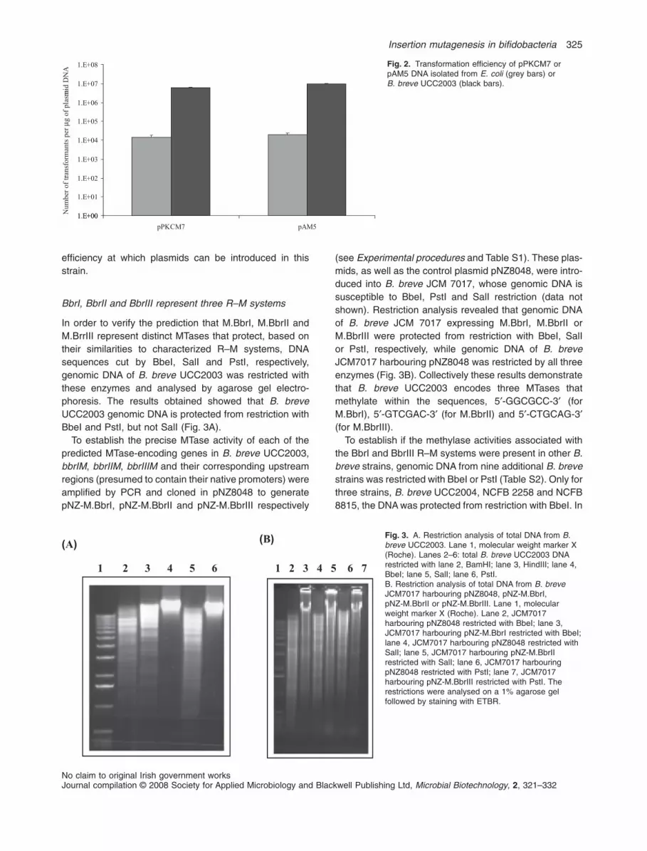
formation efficiency of this strain, the transformationfrequency of two E. coli–bifidobacterial shuttle vectors,pPKCM7 and pAM5 (Table 1), was determined whenthese plasmids had been isolated either from B. breveUCC2003 (DNA protected from R–M) or from E. coliJM101 (DNA sensitive to R–M). 200 ng quantities ofeach of these plasmid DNAs isolated from these twodifferent hosts was used to transform B. breve UCC2003by electroporation. Transformants were selected on RCAsupplemented with chloramphenicol (Cm) in case ofplasmid pPKCM7, or tetracycline (Tet) in case of plasmidpAM5, and enumerated following anaerobic incubationat 37°C for 48 h (Fig. 2). For each plasmid there was a500-fold higher transformation efficiency of the plasmidDNA isolated from B. breve UCC2003 as compared withthe DNA isolated from E. coli, thus indicating that oneor more of the identified R–M systems encoded byB. breve UCC2003 is functional and contributes to the
Table 1. Bacterial strains and plasmids used in this study.
Strain or plasmid Relevant characteristics Reference or source
StrainsE. coli strainsEC101 Cloning host, repA+ kmr Law et al. (1995)JM109 F’ traD36 proAB lacIqZ M15 recA1 relA1 endA1 thi hsdR17JM101 supE, thi (lacproAB) (F’ traD36 proAB lacIqZ M15 Yanisch-Perron et al. (1985)BM101 E. coli JM101with bbrIIM and bbrIIIM integrated in the chromosome
and transcribed by an IPTG-inducible lac promoterThis study
B. breve strainsNCFB 2257 Isolate from infant intestine NCFBNCFB 2258 Isolate from infant intestine NCFBNCFB 8815 Isolate from nursling stool NCFBNCFB 11815 Isolate from infant intestine NCFBYakult Isolate from infant intestine Oishi et al. (2008)LMG 13208 Isolate from infant intestine UCCJCM 7017 Isolate from infant intestine JCMJCM 7019 Isolate from infant intestine JCMUCC2004 Isolate from nursling stool UCCUCC2003 Isolate from nursling stool Mazé et al. (2007)UCC2003-galA-744 pORI19-tet-G744 insertion mutant of UCC2003 This studyUCC2003-gaIA-476 pORI19-tet-G476 insertion mutant of UCC2003 This studyUCC2003-apuB-939 pORI19-tet-apuB insertion mutant of UCC2003 This study
PlasmidspNZ8048 Cmr, nisin-inducible translational fusion vector de Ruyter et al. (1996)pNZ-M.BbrI pNZ8048 derivative containing bbrIM (bbr0216) This studypNZ-M.BbrII pNZ8048 derivative containing bbrIIM (bbr1121) This studypNZ-M.BbrIII pNZ8048 derivative containing bbrIIIM (bbr1119) This studypNZ-M.BbrII + M.BbrIII pNZ8048 derivative containing bbrIIM and bbrIIIM This studypAM5 pBC1-puC19-Tcr Alvarez-Martín et al. (2007)pPKCM7 pblueCm harbouring rep pCIBA089 Cronin et al. (2007)pREP4 Low-copy-number LacI expressing pQE60 companion plasmid QiagenpQE60 AmpR overexpression vector QiagenpQE60 M.BbrII + M.BbrIII pQE60 derivative containing bbrIIM and bbrIIIM transcriptionally fused
to IPTG-inducible promoterThis study
pKVB2 Tcr, Kmr containing internally deleted E. coli glgB gene 11.7 kb Kiel et al. (1987)pKVB2-M.BbrII-M.BbrIII pKVB2 derivative containing bbrIIM and bbrIIIM transcriptionally fused
to IPTG-inducible promoterThis study
pORI19 Emr, repA-, ori+, cloning vector Law et al. (1995)pORI19-tet-G744 Internal 744 bp fragment of galA and tetW cloned in pORI19 This studypORI19-tet-G476 Internal 476 bp fragment of galA and tetW cloned in pORI19 This studypORI19-tet-apuB Internal 939 bp fragment of apuB and tetW cloned in pORI19 This study
JCM, Japan Collection of Microorganisms; NCFB, National Collection of Food Bacteria, Reading, UK; UCC, University College Cork, Cork, Ireland.
324 M. O’Connell Motherway et al.
No claim to original Irish government worksJournal compilation © 2008 Society for Applied Microbiology and Blackwell Publishing Ltd, Microbial Biotechnology, 2, 321–332
efficiency at which plasmids can be introduced in thisstrain.
BbrI, BbrII and BbrIII represent three R–M systems
In order to verify the prediction that M.BbrI, M.BbrII andM.BrrIII represent distinct MTases that protect, based ontheir similarities to characterized R–M systems, DNAsequences cut by BbeI, SalI and PstI, respectively,genomic DNA of B. breve UCC2003 was restricted withthese enzymes and analysed by agarose gel electro-phoresis. The results obtained showed that B. breveUCC2003 genomic DNA is protected from restriction withBbeI and PstI, but not SalI (Fig. 3A).
To establish the precise MTase activity of each of thepredicted MTase-encoding genes in B. breve UCC2003,bbrIM, bbrIIM, bbrIIIM and their corresponding upstreamregions (presumed to contain their native promoters) wereamplified by PCR and cloned in pNZ8048 to generatepNZ-M.BbrI, pNZ-M.BbrII and pNZ-M.BbrIII respectively
(see Experimental procedures and Table S1). These plas-mids, as well as the control plasmid pNZ8048, were intro-duced into B. breve JCM 7017, whose genomic DNA issusceptible to BbeI, PstI and SalI restriction (data notshown). Restriction analysis revealed that genomic DNAof B. breve JCM 7017 expressing M.BbrI, M.BbrII orM.BbrIII were protected from restriction with BbeI, SalIor PstI, respectively, while genomic DNA of B. breveJCM7017 harbouring pNZ8048 was restricted by all threeenzymes (Fig. 3B). Collectively these results demonstratethat B. breve UCC2003 encodes three MTases thatmethylate within the sequences, 5′-GGCGCC-3′ (forM.BbrI), 5′-GTCGAC-3′ (for M.BbrII) and 5′-CTGCAG-3′(for M.BbrIII).
To establish if the methylase activities associated withthe BbrI and BbrIII R–M systems were present in other B.breve strains, genomic DNA from nine additional B. brevestrains was restricted with BbeI or PstI (Table S2). Only forthree strains, B. breve UCC2004, NCFB 2258 and NCFB8815, the DNA was protected from restriction with BbeI. In
Fig. 2. Transformation efficiency of pPKCM7 orpAM5 DNA isolated from E. coli (grey bars) orB. breve UCC2003 (black bars).
mid
DN
A
1.E+07
1.E+08er
μg o
f pl
asm
1.E+05
1.E+06
rans
form
ants
p
1.E+03
1.E+04
Num
ber
of tr
1 E+00
1.E+01
1.E+02
1.E+00
5MAp7MCKPp
Fig. 3. A. Restriction analysis of total DNA from B.breve UCC2003. Lane 1, molecular weight marker X(Roche). Lanes 2–6: total B. breve UCC2003 DNArestricted with lane 2, BamHI; lane 3, HindIII; lane 4,BbeI; lane 5, SalI; lane 6, PstI.B. Restriction analysis of total DNA from B. breveJCM7017 harbouring pNZ8048, pNZ-M.BbrI,pNZ-M.BbrII or pNZ-M.BbrIII. Lane 1, molecularweight marker X (Roche). Lane 2, JCM7017harbouring pNZ8048 restricted with BbeI; lane 3,JCM7017 harbouring pNZ-M.BbrI restricted with BbeI;lane 4, JCM7017 harbouring pNZ8048 restricted withSalI; lane 5, JCM7017 harbouring pNZ-M.BbrIIrestricted with SalI; lane 6, JCM7017 harbouringpNZ8048 restricted with PstI; lane 7, JCM7017harbouring pNZ-M.BbrIII restricted with PstI. Therestrictions were analysed on a 1% agarose gelfollowed by staining with ETBR.
(A) (B)
1 2 3 4 5 6 1 2 3 4 5 6 7
Insertion mutagenesis in bifidobacteria 325
No claim to original Irish government worksJournal compilation © 2008 Society for Applied Microbiology and Blackwell Publishing Ltd, Microbial Biotechnology, 2, 321–332

addition, DNA from B. breve NCFB 8815 was alsoprotected from restriction with PstI. Genomic DNA fromthe remaining six strains was restricted by these twoenzymes. This would indicate that different strains of B.breve exhibit quite a variety of different R–M activities.
To determine the individual effect of each R–M systemon the transformation frequency of B. breve UCC2003, wefirst introduced plasmid pAM5, which harbours one PstI,two SalI and three BbeI sites, into B. breve JCM7017strains harbouring either pNZ8048, pNZ-M.BbrI, pNZ-M.BbrII or pNZ-M.BbrIII. The methylation of the pAM5DNA at the appropriate sequence in each of the methy-lase expressing strains was confirmed by restrictionanalysis (results not shown) prior to introducing 200 ng ofeach plasmid preparation into B. breve UCC2003 by elec-troporation. The number of transformants was determinedafter 48h of anaerobic incubation at 37°C on RCA withtetracycline selection (Fig. 4). pAM5 DNA isolated fromJCM7017 expressing M.BbrIII allowed an almost 1000-fold higher transformation frequency as compared withpAM5 isolated from E. coli or JCM 7017 harbouringpNZ8048. A 10- and 5-fold higher transformation effi-ciency was observed for pAM5 isolated from JCM7017expressing M.BbrII and M.BbrI respectively. The transfor-mation frequency obtained with pAM5 DNA isolated fromJCM 7017 expressing M.BbrIII was comparable to thetransformation frequency obtained with pAM5 plasmidDNA isolated from B. breve UCC2003. However, in thelatter case DNA preparations contain just the pAM5plasmid, while in the former case the DNA preparationwould have contained a mixture of pAM5 and pNZ-M.BbrIII. These results demonstrate that the BbrIII restric-tion endonuclease (isoschizomer of PstI) is highly activein B. breve UCC2003 and that the activity of this restric-tion endonuclease appears to represent the main limita-tion to the genetic accessibility of B. breve UCC2003, atleast for plasmid pAM5.
Expression of M.BbrII and M.BbrIII in E. coli andmethylation of plasmid DNA
From the data presented above it was clear that all threeREases BbrI, BbrII and BbrIII are active in B. breveUCC2003. In order to enhance transformation efficienciesof B. breve UCC2003 by prior methylation of plasmidDNA, two E. coli strains expressing both M.BbrII andM.BbrIII were constructed. In the first, E. coli pNZ-M.BbrII-M.BbrIII, two of the bifidobacterial methylases wereexpressed on plasmid pNZ8048 (see Experimental pro-cedures and Table S1). As expected, chromosomal (andplasmid) DNA from E. coli strain EC101 harbouring pNZ-M.BbrII-M.BbrIII is protected from restriction with PstI. Thesecond E. coli strain, BM1, harbours bbrIIM and bbrIIIMunder the control of an IPTG-inducible promoter inte-grated into the glgB gene on the E. coli JM101 chromo-some (see Experimental procedures). Upon inductionwith 10 mM IPTG total DNA from E. coli BM1 is protectedfrom restriction with PstI (Fig. S1A). However, completeprotection from SalI restriction was not observed (resultsnot shown) and this may be due to the lower level expres-sion of bbrIIM from the E. coli chromosome as comparedwith expression from plasmid pNZ-M.BbrII-M.BbrIII. Inaddition, SalI can restrict hemi-methylated DNA, thereforethe observed restriction by SalI may be a reflection ofincomplete methylation.
To evaluate the effect of methylation of plasmid DNA ontransformation efficiency, pAM5 was introduced into E.coli pNZ-M.BbrII-M.BbrIII and E. coli BM1 by electropora-tion. Expression of M.BbrII and M.BbrIII in BM1 harbour-ing pAM5 was effected by the addition of 10 mM IPTGprior to the isolation of plasmid DNA. Plasmid prepara-tions of E. coli harbouring pNZ-M.BbrII-M.BbrIII or E. coliBM1 were then used for B. breve UCC2003 transforma-tion. pAM5 DNA isolated from E. coli harbouring pNZ-M.BbrII-M.BbrIII gave a 1000-fold higher transformation
Fig. 4. Transformation efficiency of B. breveUCC2003 using pAM5 plasmid DNA isolatedfrom UCC2003, B. breve JCM7017harbouring pNZ8048, pNZ-M.BbrI,pNZ-M.BbrII, pNZ-M.BbrIII or E. coli.
1.E+07
1.E+08D
NA
1 E+05
1.E+06
g of
pla
smid
D
1.E+04
1.E+05
orm
ants
per
μg
1.E+02
1.E+03
mbe
r of
tran
sfo
1.E+00
1.E+01
UCC2003 JCM 7017 pNZ8048 JCM 7017 pNZ- JCM 7017 pNZ- JCM 7017 pNZ- E. coli
Num
M.BbrI M.BbrII M.BbrIII
326 M. O’Connell Motherway et al.
No claim to original Irish government worksJournal compilation © 2008 Society for Applied Microbiology and Blackwell Publishing Ltd, Microbial Biotechnology, 2, 321–332

frequency as compared with pAM5 from E. coli pNZ8048while plasmid DNA isolated from E. coli BM1 gave a50-fold higher transformation frequency. pAM5 DNA iso-lated from EC101 pNZ-M.BbrII-M.BbrIII gave transforma-tion efficiencies comparable to those obtained withplasmid DNA isolated from B. breve (Fig. S1B).
Disruption of the galA and apuB genes of B. breveUCC2003
In order to establish if methylation of a non-replicatingplasmid by the B. breve UCC2003 MTases wouldincrease transformation efficiency to a sufficiently highlevel that would allow site-specific homologous recombi-nation, two genes, galA and apuB, were selected asmutational targets. The galA and apuB genes encode anendogalactanase and an amylopullulanase, respectively,which are involved in extracellular polysaccharidemetabolism by B. breve UCC2003 (Hinz et al., 2005;Ryan et al., 2006; O’Connell Motherway et al., 2008). Toestablish if gene disruption could be achieved usinghomologous recombination, DNA fragments of 476 and744 bp, representing internal fragments of the galA gene,and a 939 bp internal fragment of the apuB gene werecloned in pORI19 and provided with a tetracycline resis-tance marker, generating plasmids pORI19-tet-G744,pORI19-tet-G476 and pORI19-tet-apuB respectively (seeExperimental procedures). These plasmids, being deriva-tives of pORI19, cannot replicate in B. breve UCC2003 asthey lack a functional replication protein (Law et al.,1995). These pORI19 derivatives were introduced into E.coli EC101 harbouring pNZ-M.BbrII-M.BbrIII to facilitatemethylation, and preparations of the resulting methylatedpORI19-derived plasmids were then introduced into B.breve UCC2003 by electroporation. Tetracycline-resistanttransformants were isolated at a frequency of 50 per mg oftransformed DNA when greater than 700 bp of homolo-gous DNA was used. The number of potential integrantswas slightly reduced when the smaller region of homolo-gous DNA was used. All transformants obtained wereexpected to carry galA or apuB gene disruptions, while nosuch transformants were obtained when unmethylatedpORI19 constructs were introduced into B. breveUCC2003. The suspected chromosomal integration of thepORI constructs was verified by colony PCR on a selec-tion of Tetr transformants using a forward primer upstreamof the region of integration and a reverse primer based onpORI19 (galAp1 and pORI19A, or apuBp1 and pORI19B)(results not shown). Southern hybridizations confirmedthe assumed integration of the individual pORI-derivedplasmids by homologous recombination. For the pre-sumed galA disruptions of B. breve UCC2003, Southernhybridizations were performed using SphI-digestedgenomic DNA and employing a 2.6 kb PCR fragment
encompassing galA as a probe. SphI was selected for thegenomic digests since there are no corresponding restric-tion sites within the galA sequence. The galA fragmenthybridized to a 6.1 kb fragment of UCC2003 genomicDNA, while in the UCC2003 derivatives with a presumedpORI-tet-G476 or pORI-tet-G744 integration this bandwas absent, and expected hybridization signals of 10.5 kband 557 bp, or 10.8 kb and 848 bp, respectively, wereobserved (Fig. 5). For two of each of the UCC2003 mutantstrains examined the galA probe also hybridized to a5.3 kb or 5.5 kb Sph1 fragment for the pORI19-tet-G476and pORI19-tet-G744 integrants respectively [Fig. 5B(i),lanes 4 and 5; Fig. 5B(ii), lanes 5 and 6]. These hybrid-ization signals indicate that duplication of pORI19-tet-galAplasmids had occurred after integration of the plasmid intothe bacterial chromosome in these mutant strains. For thesuspected apuB integrants of strain UCC2003, Southernhybridizations were performed using BamHI-digestedgenomic DNA and a 1 kb probe encompassing an internalfragment of apuB. The apuB fragment hybridized to a3.6 kb fragment of UCC2003 genomic DNA. For the apuBmutant strains the anticipated hybridization signals of 2.1and 7.2 kb were obtained (Fig. S2).
Collectively these results demonstrate that methylationof plasmid DNA by the B. breve UCC2003 MTases M.BbrIIand M.BbrIII in E. coli circumvents the BbrII and BbrIIIREase activities in B. breve UCC2003 and allows a suf-ficiently high transformation efficiency so as to allow reli-able homologous recombination in B. breve UCC2003. Inaddition, these data illustrate that chromosomal integra-tion in B. breve UCC2003 can be achieved with less than500 bp of homologous DNA.
Phenotypic analysis of the B. breve UCC2003plasmid integrants
In order to verify the expected phenotypic consequencesof the created gene disruptions in galA and apuB, strainsB. breve UCC2003, and individual representatives ofB. breve UCC2003 mutants generated by insertion ofpORI19-tet-G744 or pORI19-tet-G476, designated hereas UCC2003-galA-476 and UCC2003-galA-744, respec-tively, were analysed for their ability to grow on galactanas the sole carbohydrate source (Fig. 6A). Similarly, B.breve UCC2003 and a derivative with an integratedpORI19-tet-apuB (designated UCC2003-apuB-939) wereanalysed for the ability to grow on starch, amylopectin,glycogen or pullulan as the sole carbohydrate source(Fig. 6B). In contrast to the wild-type B. breve UCC2003,the B. breve UCC2003-galA-476 or UCC2003-galA-744mutant strains failed to grow on potato galactan, whilecomparable growth of the parent and galA mutant strainswas observed when glucose was the sole carbohydratesource. In a similar manner it was shown that B. breve
Insertion mutagenesis in bifidobacteria 327
No claim to original Irish government worksJournal compilation © 2008 Society for Applied Microbiology and Blackwell Publishing Ltd, Microbial Biotechnology, 2, 321–332

UCC2003-apuB-939 failed to grow on starch, amylopec-tin, glycogen or pullulan, which contrasted with observedgood growth on these substrates by the parent strain.Comparable growth for parent and mutant strains wasobserved when glucose was used as the sole carbohy-drate source. These results confirm that the chromosomalplasmid integrations in UCC2003 cause a demonstrablephenotype and clearly illustrate the importance of theextracellular enzymes specified by galA and apuB in themetabolism of specific high-molecular-weight polysaccha-rides by B. breve UCC2003.
Discussion
Bifidobacterial strains demonstrate substantial variabilityin the efficiency of transformation by plasmids from E. coli,while many strains exhibit complete resistance to trans-
formation (Lee and O’Sullivan, 2006). Progress in theevaluation of probiotic factors in bifidobacteria has beenslow due to the lack of efficient and versatile systems forgenetic manipulation (Ventura et al., 2004). While quite anumber of E. coli–bifidobacterial shuttle vectors havebeen developed, it has been noted that widespread appli-cation of these plasmids among bifidobacterial species islimited (Lee and O’Sullivan, 2006).
As shown here, R–M systems are one of the majorobstacles hindering progress in the genetic accessibilityand analysis of B. breve UCC2003, and are likely to do thisin other (bifido)bacteria as well. Convincing evidence tosupport this notion can be obtained from the availablebifidobacterial genome sequences. Genes specifying R–Msystems can be identified in all sequenced bifidobacterialgenomes. The genomes of B. longum subsp. longumNCC2705 (Schell et al., 2002) and B. longum subsp.
Fig. 5. A. Schematic representation of therelevant regions of the B. breve UCC2003and UCC2003-galA-476 andUCC2003-galA-744 (in brackets)chromosomes. Chromosomal DNA isrepresented by a thin line, the galA gene isrepresented by a black arrow, the internalgalA fragment is indicated by a solid grey lineand pORI19 is indicated by a boxed line. SphIsites relevant to the Southern hybridizationanalysis are indicated.B. Southern hybridization analysis of SphIchromosomal DNAs of (i) B. breve UCC2003(lane 2) and four representative B. breveUCC2003-galA-476 mutants (lanes 3–6); (ii)B. breve UCC2003 (lane 2) and fourrepresentative B. breve UCC2003-galA-744mutants (lanes 3–6). The hybridization signalsfor molecular weight marker X (Roche) are inlane 1 and the molecular weight of therelevant hybridization signals are indicated tothe left of the panel. A PCR product of 2.6 kbencompassing galA was used as a probe forthe hybridization.
SphI SphI6.1 kb
B. breve UCC2003(A)
galA bbr0423 bbr0425bbr0424
SphISphI
pORI19-tetW
10.5 kb(10.8 kb)
SphI
560 bp(848 bp)
B. breve UCC2003::galA-476 bp (UCC2003::galA-744 bp) insertion mutant
bbr0423 bbr0425bbr0424
SphI SphI SphI10.5 kb
(10.8 kb)SphI
*5.2 kb x n
(* 5.5 kb x n)
pORI19-tet-galA integration followed by plasmid duplication
bbr0423 bbr0425bbr0424
557 bp(848 bp)
(B)
1 2 3 4 5 61 2 3 4 5 6
)ii()i(
10.8 kb
*5.5 kb6.1 kb
10.5 kb
6.1 kb
*5.3 kb
848 b557 b 848 bp557 bp
328 M. O’Connell Motherway et al.
No claim to original Irish government worksJournal compilation © 2008 Society for Applied Microbiology and Blackwell Publishing Ltd, Microbial Biotechnology, 2, 321–332

longum DJ010A (Lee et al., 2008) both harbour a singletype I R–M system, two type II R–M systems and one typeIV R–M system. The type II REases specified by blo_1473and bld_0356 are predicted to be isoschizomers of EcoRII,which restricts within the sequence ↓CCWGG, while theREases specified by blo_564 and bln_1359 are predictedto be isoschizomers of Sau3A1, which recognizes thesequence ↓GATC. The recognition sequence of the type Iand type IV R/M systems in the sequenced B. longumgenomes are unknown. The genome of B. adolescentisATCC15703 (Suzuki et al., 2006) specifies two MTasesubunits and six REase subunits. The restriction subunitsspecified by bad_1283 and bad_1232 are predicted to beisoschizomers of KpnII and Sau3AI, respectively, while theremaining four are as yet unknown. The sequencedgenomes of B. dentium ATCC27678 (Sudarsanam et al.,2008) and B. animalis HN019 (Collett et al., 2008) bothharbour a single type II R–M system, where the REase ispredicted to be an isoschizomer of AvaII, which recognizesthe sequence G↓GWCC (Sutcliffe and Church, 1978).Based on the results obtained for B. breve UCC2003, it istempting to speculate that exploiting the MTases encodedby the aforementioned sequenced bifidobacterial strainswould allow the transformation efficiencies of these strainsto be improved. For bifidobacterial strains that are particu-larly recalcitrant to transformation or where the complete
genome sequence is not known it may be possible tomethylate plasmid DNA isolated from E. coli by incubatingthe DNA with crude cell extracts of the Bifidobacteria inthe presence of S-adenosylmethionine thereby possiblyimproving the transformation efficiency.
An alternative method that would circumvent bifidobac-terial R–M systems would be to introduce plasmid DNA byconjugation. To date conjugation has not been conclu-sively demonstrated for the genus Bifidobacterium. Untilrecently the only evidence supporting the possibility ofconjugation in bifidobacteria was the identification ofgenes encoding proteins potentially involved in theconjugation process on various bifidobacterial plasmids.Putative relaxase-encoding genes have been identifiedon plasmids pJK36 and pJK50 from B. longum subsp.longum (Park et al., 1999; 2000), while homologues ofseptal DNA translocator (Tra) proteins have been identi-fied on the B. breve plasmid pCIBb1 (O’Riordan andFitzgerald, 1999) and the B. pseudocatenulatum plasmidp4M (Gibbs et al., 2006). Recently, Shkoporov and col-leagues (2008) sequenced three plasmids of bifidobacte-rial origin: pB44 from B. longum, pB90 from B. bifidum andpB21a from B. breve. Both pB44 and pB90 harbour genesencoding potential mobilization functions while pB21Aencodes a putative Tra protein. These proteins wereexploited in efforts to achieve conjugation in bifidobacte-
33
)B()A(
2
2.5
3
600
nm
2
2.5
600
nm
1
1.5
al d
ensi
ty O
D
1
1.5
al d
ensi
ty O
D 6
0
0.5
galactan glucose
Opt
ica
0
0.5
starc
h
opec
tin
ycog
en
ullula
n
glucos
e
Opt
ica
Carbohydrate source
s
amyl gl p glu
Carbohydrate source
Fig. 6. A. Growth profile analysis of B. breve UCC2003 (white), B. breve UCC2003-galA-476 (grey) and B. breve UCC2003-galA-744 (black)in modified Rogosa broth supplemented with potato galactan or glucose.B. Growth profile analysis of B. breve UCC2003 (white) and B. breve UCC2003-apuB (black) in modified Rogosa broth supplemented withstarch, amylopectin, glycogen, pullulan or glucose as indicated.
Insertion mutagenesis in bifidobacteria 329
No claim to original Irish government worksJournal compilation © 2008 Society for Applied Microbiology and Blackwell Publishing Ltd, Microbial Biotechnology, 2, 321–332

ria, and although antibiotic-resistant, PCR-positive andthus putative transconjugants were obtained, plasmidtransfer has as yet not been demonstrated.
The difficulties associated with obtaining sufficientlyhigh transformation efficiencies so as to allow insertionalmutagenesis in B. breve UCC2003 through homologousrecombination led us to believe that R–M systems werethe barrier that needed to be overcome in order to achievethis. In the present study we describe three different R–Msystems specified by the genome of UCC2003: BbrI, anisoschizomer of BbeI; BbrII, a neoschizomer of SalI; andBbrIII, an isoschizomer of PstI. Restriction analysis ofchromosomal DNA from UCC2003 showed that the DNAis protected from restriction with BbeI and PstI, but notSalI. The observed restriction of DNA by SalI can beexplained by M.SalI being a N6-adenosine-methylase,while M.BbrII is predicted to be cytosine-specific MTase,which may therefore not confer (full) protection againstSalI restriction. However, the finding that M.BbrII doesprovide full protection against SalI restriction when it isexpressed from a multicopy plasmid in B. breve JCM7017 would indicate that M.BbrII in such circumstances ismore abundant, thereby eliciting complete methylationand concomitant protection of the DNA. The three R–Msystems identified in B. breve UCC2003 do not appear tobe highly conserved among B. breve strains, just onestrain examined in this study, B. breve NCIMB 8815, wasshown to exhibit protection of BbrI and BbrIII recognitionsites indicating that this species and indeed the genusBifidobacterium is likely to harbour a very diverse range ofR–M activities.
The contribution of each R–M system in impedingplasmid transformation of B. breve UCC2003 was deter-mined and established that all three systems impact ontransformation efficiency, with BbrIII, at least under thecircumstances used here, providing the biggest hurdle toincoming DNA. To facilitate methylation of plasmid DNAby M.BbrII and M.BbrIII, thereby enhancing the transfor-mation frequency of B. breve UCC2003, two E. colistrains were constructed, where bbrIIM and bbrIIIM wereexpressed in different ways, either from their own pro-moter on plasmid pNZ8048 or from an IPTG-induciblepromoter on the E. coli chromosome. The observedhigher transformation efficiency for pAM5 DNA isolatedfrom E. coli pNZ-M.BbrII-M.BbrIII may be attributed to thehigh copy number of pNZ8048 plasmids in E. coli andresulting higher expression levels of the MTases as com-pared with expression from single copy on the E. colichromosome in E. coli BM1.
Having established that the use of M.BbrII- andM.BbrIII-methylated plasmid DNA results in a significantlyincreased transformation efficiency of B. breve UCC2003,we conclusively showed that gene disruptions in B. breveUCC2003 can be created using a non-replicating and
M.BbrII- and M.BbrIII-methylated plasmid. We have pre-viously produced a gene disruption in the apuB gene of B.breve UCC2003 by adaptation of the lactococcal twoplasmid homologous recombination system (O’ConnellMotherway et al., 2008). However, in our hands thissystem was very tedious, time-consuming and not reliable(O’Connell Motherway et al., 2008; our unpublishedresults). Therefore, insertional mutagenesis of the apuBgene was deemed an appropriate control to evaluate thevalidity and reliability of the plasmid methylation strategy.By M.BbrII-M.BbrIII-mediated methylation of plasmid DNAin E. coli prior to transformation into B. breve UCC2003,gene disruptions not only in apuB, but also in galA weresuccessfully and reliably created, as verified by geneticand phenotypic analyses.
This, to the best of our knowledge, therefore representsthe first reliable system for creating insertional mutation ina member of the genus Bifidobacterium. The ability toachieve chromosomal integration of a non-replicativeplasmid with less than 500 bp of homologous DNA alsoopens the opportunity for the creation of a bank of B.breve UCC2003-derived mutants carrying random chro-mosomal integrations, which in turn will provide a range ofpossibilities to further advance fundamental knowledgeon the physiology, biochemistry and genetics of this strain.Such information will obviously be relevant to other bifi-dobacteria and will be crucial to understand the health-promoting properties that have been attributed to variousmembers of this genus.
Experimental procedures
The description of the experimental procedures resides inAppendix S1 in Supporting information.
Acknowledgements
This research was financially supported by the Science Foun-dation Ireland Alimentary Pharmabiotic Centre located at Uni-versity College Cork.
References
Alvarez-Martín, P., O’Connell-Motherway, M., van Sinderen,D., and Mayo, B. (2007) Functional analysis of the pBC1replicon from Bifidobacterium catenulatum L48. ApplMicrobiol Biotechnol 76: 1395–1402.
Collett, M.A., Depree, K.M., Rand, C.J., Mason, C., andStanton, J.-A.L. (2008) Bifidobacterium animalis subsp.lactis HN019, whole genome shotgun sequence. NCBIDatabase.
Cronin, M., Knobel, M., O’Connell-Motherway, M., Fitzgerald,G.F., and van Sinderen, D. (2007) Molecular dissection ofa bifidobacterial replicon. Appl Environ Microbiol 73: 7858–7866.
Düsterhöft, A., and Kröger, M. (1991) Cloning, sequenceand characterization of m5C-methyltransferase-encoding
330 M. O’Connell Motherway et al.
No claim to original Irish government worksJournal compilation © 2008 Society for Applied Microbiology and Blackwell Publishing Ltd, Microbial Biotechnology, 2, 321–332

gene, hgiDIIM (GTCGAC), from Herpetosiphon giganteusstrain Hpa2. Gene 106: 87–92.
Fulton, L., Clifton, S., Fulton, B., Xu, J., Minx, P., Pepin, K.H.,et al. (2007) Bifidobacterium adolescentis (L2-32) genomesequence. NCBI Database.
Gibbs, M.J., Smeianov, V.V., Steele, J.L., Upcroft, P., andEfimov, B.A. (2006) Two families of rep-like genes thatprobably originated by interspecies recombination are rep-resented in viral, plasmid, bacterial, and parasitic proto-zoan genomes. Mol Biol Evol 6: 1097–1100.
Hartke, A., Benachour, A., Bouibonnes, P., and Auffray, Y.(1996) Characterisation of a complex restriction/modification system detected in a Bifidobacterium longumstrain. Appl Microbiol Biotechnol 45: 132–136.
Hinz, S.W., Pastink, M.I., van den Broek, L.A., Vincken, J.P.,and Voragen, A.G. (2005) Bifidobacterium longum endoga-lactanase liberates galactotriose from type I galactans.Appl Environ Microbiol 71: 5501–5510.
Janscak, P., Sandmeier, U., Szczelkun, M.D., and Bickle, T.A.(2001) Subunit assembly and mode of DNA cleavage of thetype III restriction endonucleases EcoP1I and EcoP15I. JMol Biol 306: 417–431.
Khosaka, T., and Kiwaki, M. (1984) BinI: a new site-specificendonuclease from Bifidobacterium infantis. Gene 31:251–255.
Khosaka, T., Sakurai, T., Takahashi, H., and Saito, H. (1982)A new site-specific endonuclease BbeI from Bifidobacte-rium breve. Gene 17: 117–122.
Khosaka, T., Kiwaki, M., and Rak, B. (1983) Two site-specificendonucleases BinSI and BinSII from Bifidobacteriuminfantis. FEBS Lett 163: 170–174.
Kiel, J.A., Vossen, J.P., and Venema, G. (1987) A generalmethod for the construction of Escherichia coli mutants byhomologous recombination and plasmid segregation. MolGen Genet 207: 294–301.
Kobayashi, I. (2001) Behaviour of restriction-modificationsystems as selfish mobile elements and their impact ongenome evolution. Nucleic Acids Res 29: 3742–3756.
Kumar, S., Cheng, X., Klimasauskas, S., Mi, S., Posfai, J.,Roberts, R.J., and Wilson, G.G. (1984) The DNA (cytosine-5) methyltransferases. Nucleic Acids Res 22: 1–10.
Law, J., Buist, G., Haandrikman, A., Kok, J., Venema, G., andLeenhouts, K. (1995) A system to generate chromosomalmutations in Lactococcus lactis which allows fast analysisof targeted genes. J Bacteriol 177: 7011–7018.
Leahy, S.C., Higgins, D.G., Fitzgerald, G.F., and van Sin-deren, D. (2005) Getting better with bifidobacteria. J ApplMicrobiol 98: 1303–1315.
Lee, J.H., and O’Sullivan, D.J. (2006) Sequence analysis oftwo cryptic plasmids from Bifidobacterium longum DJO10Aand construction of a shuttle cloning vector. Appl EnvironMicrobiol 72: 527–535.
Lee, J.H., Karamychev, V.N., Kozyavkin, S.A., Mills, D.,Pavlov, A.R., Pavlova, N.V., et al. (2008) Comparativegenomic analysis of the gut bacterium Bifidobacteriumlongum reveals loci susceptible to deletion during pureculture growth. BMC Genomics 9: 247.
Lenoir-Wijnkoop, I., Sanders, M.E., Cabana, M.D., Caglar,E., Corthier, G., Rayes, N., et al. (2007) Probiotic andprebiotic influence beyond the intestinal tract. Nutr Rev 65:469–489.
Mazé, A., O’Connell-Motherway, M., Fitzgerald, G.F., Deut-scher, J., and van Sinderen, D. (2007) Identification andcharacterization of a fructose phosphotransferase systemin Bifidobacterium breve UCC2003. Appl Environ Microbiol73: 545–553.
Murray, N.E. (2002) Immigration control of DNA in bacteria:self versus non-self. Microbiology 148: 3–20.
O’Connell Motherway, M., Fitzgerald, G.F., Neirynck, S.,Ryan, S., Steidler, L., and van Sinderen, D. (2008) Char-acterisation of ApuB, an extracellular type II amylopullula-nase from Bifidobacterium breve UCC2003. Appl EnvironMicrobiol 74: 6271–6279.
O’Hara, A.M., and Shanahan, F. (2006) The gut flora as aforgotten organ. EMBO Rep 7: 688–693.
Oishi, K., Sato, T., Yokoi, W., Yoshida, Y., Ito, M., andSawada, H. (2008) Effect of probiotics, Bifidobacteriumbreve and Lactobacillus casei, on bisphenol A exposure inrats. Biosci Biotechnol Biochem 72: 1409–1415.
O’Riordan, K., and Fitzgerald, G.F. (1999) Molecular charac-terization of a 5.75 kb cryptic plasmid from Bifidobacteriumbreve NCFB 2258 and determination of the mode of repli-cation. FEMS Microbiol Lett 174: 285–294.
Park, M.S., Shin, D.W., Lee, K.H., and Ji, G.E. (1999)Sequence analysis of plasmid pKJ50 from Bifidobacteriumlongum. Microbiology 145: 585–592.
Park, M.S., Shin, D.W., Lee, K.H., and Ji, G.E. (2000) Char-acterisation of plasmid pKJ36 from Bifidobacterium longumand construction of an E. coli–Bifidobacterium shuttlevector. J Microbiol Biotechnol 10: 310–320.
Pingoud, A., Fuxreiter, M., Pingoud, V., and Wende, W.(2005) Type II restriction endonucleases: structure andmechanism. Cell Mol Life Sci 62: 685–707.
Roberts, R.J., Belfort, M., Bestor, T., Bhagwat, A.S., Bickle,T.A., Bitinaite, J., et al. (2003) A nomenclature for restrictionenzymes, DNAmethyltransferases, homing endonucleasesand their genes. Nucleic Acids Res 31: 1805–1812.
de Ruyter, P.G., Kuipers, O.P., and de Vos, W.M. (1996)Controlled gene expression systems for Lactococcus lactiswith the food-grade inducer nisin. Appl Environ Microbiol62: 3662–3667.
Ryan, S.M., Fitzgerald, G.F., and van Sinderen, D. (2006)Screening for and identification of starch-, amylopectin-,and pullulan-degrading activities in bifidobacterial strains.Appl Environ Microbiol 72: 5289–5296.
Sangrador-Vegas, A., Stanton, C., Van Sinderen, D., Fitzger-ald, G.F., and Ross, R.P. (2007) Characterization ofplasmid pAS479 from Bifidobacterium pseudolongumsubsp. globosum and its use for expression vector con-struction. Plasmid 58: 140–147.
Saxelin, M., Tynkkynen, S., Sandholm, T.M., and de Vos,W.M. (2005) Probiotic and other functional microbes: frommarkets to mechanisms. Curr Opin Biotechnol 16: 204–211.
Schell, M.A., Karmirantzou, M., Snel, B., Vilanova, D.,Berger, B., Pessi, G., et al. (2002) The genome sequenceof Bifidobacterium longum reflects its adaptation to thehuman gastrointestinal tract. Proc Natl Acad Sci USA 99:14422–14427.
Sgorbati, B., Scardovi, V., and Leblanc, D.J. (1982) Plasmidsin the genus Bifidobacterium. J Gen Microbiol 128: 2121–2131.
Insertion mutagenesis in bifidobacteria 331
No claim to original Irish government worksJournal compilation © 2008 Society for Applied Microbiology and Blackwell Publishing Ltd, Microbial Biotechnology, 2, 321–332
Shkoporov, A.N., Efimov, B.A., Khokhlova, E.V., Steele, J.L.,Kafarskaia, L.I., and Smeianov, V.V. (2008) Characteriza-tion of plasmids from human infant Bifidobacteriumstrains: sequence analysis and construction of E. coli–Bifidobacterium shuttle vectors. Plasmid 60: 136–148.
Skrypina, N.A., Kramarov, V.M., Liannaia, A.M., andSmolianinov, V.V. (1988) Restriction endonucleases fromBifidobacteria. Mol Gen Mikrobiol Virusol 5: 15–16.
Sudarsanam, P., Ley, R., Guruge, J., Turnbaugh, P.J.,Mahowald, M., Liep, D., and Gordon, J. (2008) Draftgenome sequence of Bifidobacterium dentium (ATCC27678). NCBI Database.
Sutcliffe, J.G., and Church, G.M. (1978) The cleavage site ofthe restriction endonuclease Ava II. Nucleic Acids Res 5:2313–2319.
Suzuki, T., Tsuda, Y., Kanou, N., Inoue, T., Kumazaki, K.,Nagano, S., et al. (2006) Bifidobacterium adolescentisATCC 15703 complete genome sequence. NCBI Database.
Timinskas, A., Butkus, V., and Janulaitis, A. (1995) Sequencemotifs characteristic for DNA (cytosine-N4) and DNA(adenine0N6) methyltransferases. Classification of all DNAmethyltransferases. Gene 157: 3–11.
Tock, M.R., and Dryden, D.T.F. (2005) The biology of restric-tion and anti-restriction. Curr Opin Microbiol 8: 466–472.
Turroni, F., Ribbera, A., Foroni, E., van Sinderen, D., andVentura, M. (2008) Human gut microbiota and bifidobacte-ria: from composition to functionality. Antonie Van Leeu-wenhoek 94: 35–50.
Ventura, M., van Sinderen, D., Fitzgerald, G.F., and Zink, R.(2004) Insights into the taxonomy, genetics and physiologyof bifidobacteria. Antonie Van Leeuwenhoek 86: 205–223.
Ventura, M., Canchaya, C., Tauch, A., Chandra, G., Fitzgerald,G.F., Chater, K.F., and van Sinderen, D. (2007) Genomics ofActinobacteria: tracing the evolutionary history of an ancientphylum. Microbiol Mol Biol Rev 71: 495–548.
Walder, R.Y., Walder, J.A., and Donelson, J.E. (1984) Theorganization and complete nucleotide sequence of the PstIRestriction-Modification System. J Biol Chem 259: 8015–8026.
Yanisch-Perron, C., Vieira, J., and Messing, J. (1985)Improved M13 phage cloning vectors and host strains:nucleotide sequences of the M13mp18 and pUC19vectors. Gene 33: 103–109.
Zoetendal, E.G., and Vaughan, E. E., and de Vos, W.M.(2006) A microbial world within us. Mol Microbiol 59: 1639–1650.
Supporting information
Additional Supporting Information may be found in the onlineversion of this article:
Fig. S1. A. Restriction analysis of E. coli JM101 and tworepresentative JM101 bbrIIM and bbrIIIM methylase integra-tion strains. Lane 1, molecular weight marker X (Roche).Lanes 2–4, Pst1 digest of total DNA isolated from JM101following induction with 0, 1 or 10 mM IPTG. Lanes 5–7 andlane 8–10, PstI digests of total DNA isolated from two repre-sentative JM101 bbrIIM and bbrIIIM methylase integrationstrains after induction with 0, 1 or 10 mM IPTG.B. Transformation efficiency of B. breve UCC2003 with pAM5plasmid DNA isolated from B. breve UCC2003, E. coli pNZ-M.BbrII-M.BbrIII, E. coli BM1 or E. coli pNZ8048.Fig. S2. A. Schematic representation of the relevant regionsof the B. breve UCC2003 and UCC2003-apuB-939 chromo-some. Chromosomal DNA is represented by a thin line, theapuB gene is represented by a black arrow, the internal apuBfragment is indicated by a solid grey line and pORI19 isindicated by a boxed line. BamHI sites relevant to the South-ern hybridization analysis are indicated.B. Southern hybridization analysis of BamHI-digested chro-mosomal DNAs of B. breve UCC2003 (lane 1) and fourrepresentative B. breve UCC2003-apuB-939 mutants (lanes2–5). The molecular weight of the relevant hybridizationsignals are indicated to the left of the panel. The internal1 kb PCR amplicon of apuB was used as a probe for thehybridization.Table S1. Oligonucleotide primers used in this study.Table S2. Restriction analysis of genomic DNA from B.breve strains with BbeI and PstI.Appendix S1. Experimental procedures.
Please note: Wiley-Blackwell are not responsible for thecontent or functionality of any supporting materials suppliedby the authors. Any queries (other than missing material)should be directed to the corresponding author for the article.
332 M. O’Connell Motherway et al.
No claim to original Irish government worksJournal compilation © 2008 Society for Applied Microbiology and Blackwell Publishing Ltd, Microbial Biotechnology, 2, 321–332


