Embed Size (px)
Citation preview

The Plant Cell, Vol. 3, 1275-1288, December 1991 O 1991 American Society of Plant Physiologists
Overexpression of Phytochrome B lnduces a Short Hypocotyl Phenotype in Transgenic Arabídopsís
Doris Wagner, James M. Tepperman, and Peter H. Quail‘
Department of Plant Biology, University of California, Berkeley, California 94720, and University of California, Berkeley/United States Department of Agriculture Plant Gene Expression Center, Albany, California, 9471 O
The photoreceptor phytochrome is encoded by a small multigene family in higher plants. phyA encodes the well- characterized etiolated-tissue phytochrome. The product of the phyB gene, which has properties resembling those of “green tissue” phytochrome, is as yet poorly characterized. We have developed a phytochrome B overexpression system for analysis of the structure and function of this protein. Using newly generated polyclonal and monoclonal antibodies that are selective for phytochrome B, we have demonstrated high levels of expression of full-length rice and Arabidopsis phytochrome B under the control of the cauliflower mosaic virus 35s promoter in transgenic Arabidopsis. The overexpressed phytochrome is spectrally active, undergoes red/far-red-light-dependent confor- mational changes, is synthesized in its inactive red light-absorbing form, and is stable in the light. Overexpression of phytochrome 6 is tightly correlated with a short hypocotyl phenotype in transgenic seedlings. This phenotype is strictly light dependent, thus providing direct evidence that phytochrome B is a biologically functional photoreceptor. Based on similarities to phenotypes obtained by overexpression of phytochrome A, it appears that phytochromes A and B can control similar responses in the plant.
INTRODUCTION
Plants perceive the intensity, direction, and quality of light, as well as the length of the photoperiod, and utilize this information to optimize photosynthesis, to sense the den- sity of neighboring plants, and to regulate the timing of flowering and germination. Phytochrome is the best char- acterized of the photoreceptors involved in these light- dependent responses (Kendrick and Kronenberg, 1986). From physiological, biochemical, and mutant analyses, evidence has mounted that multiple pools of phytochromes might control such diverse responses. Initially, physiologi- cal data led to speculation on the heterogeneity of the phytochrome pool (Hillman, 1967). In vivo spectral analysis of oat (Jabben and Holmes, 1983) pointed toward two pools of phytochrome: a light-labile (or type 1) pool pre- dominantly present in etiolated tissue and a light-stable (or type 2) pool present in green tissue. Biochemical analysis of light-grown oat identified, in addition to the light-labile species, at least one spectrally and immunologically dis- tinct type of phytochrome that predominated in extracts from green plants (Shimazaki et al., 1983; Tokuhisa and Quail, 1983, 1987,1989; Shimazaki and Pratt, 1985,1986;
’ To whom correspondence should be addressed at UC-Berkeleyl USDA Plant Gene Expression Center, 800 Buchanan St., Albany, CA 9471 O.
Tokuhisa et al., 1985; Cordonnier et al., 1986; Pratt et al., 1991; Wang et al., 1991). Light-grown dicot (pea) plants (Abe et al., 1985, 1989; Konomi et al., 1987), on the other hand, contained an immunologically but not spectrally dis- tinct type of phytochrome in green plant extracts. Data from analysis of the responses of photomorphogenic long hypocotyl mutants were also consistent with lesions in either the light-labile (aurea mutant of tomato) or the light- stable (Ih mutant of cucumber, hy3 mutant of Arabidopsis) types of physiologically active phytochrome (Adamse et al., 1988a; Furuya, 1989; Smith and Whitelam, 1990; Tomizawa et al., 1990). The light-labile type of phyto- chrome has been implicated in the “high-irradiance re- sponse” of etiolated seedlings, whereas the light-stable type is necessary for the “end-of-day far-red response,” where increases in hypocotyl length are observed when a far-red light pulse is given prior to the dark period (Smith and Whitelam, 1990).
The discovery of a multigene family encoding phyto- chrome in Arabidopsis (phyA, phyB, phyC, phyD, and phyE, Sharrock and Quail 1989; R. Sharrock, personal communication) and rice (phyA, phyB, and phyC, Dehesh et al., 1991; K. Dehesh and P. Quail, unpublished data) and the subsequent isolation of the individual phytochrome genes provide the opportunity for the molecular char- acterization of the distinct classes of the photoreceptor

1276 The Plant Cell
described above. Comparison of the phyA-demed aminoacid sequences with protein sequences from proteolyticfragments of phytochrome purified from etiolated tissueindicates that phyA encodes the well-characterized light-labile or type 1 phytochrome (Lagarias and Rapoport,1980; Hershey et al., 1985; Yamamoto, 1987; Grimm etal., 1988; Sato, 1988; Jones and Quail, 1989). This phy-tochrome species shows an apparent molecular mass of116 to 127 kD depending on the plant species (Vierstra etal., 1984) and occurs as a dimer (Jones and Quail, 1986).The holoprotein has a covalently attached linear tetrapyr-role chromophore (Rudiger and Scheer, 1983; Vierstra andQuail, 1986). Irradiation with red (660 nm) light convertsthe molecule from the inactive, red light-absorbing (Pr)form to the active, far-red-light-absorbing (Pfr) form. Thisprocess can be reversed by irradiation with far-red (730nm) light.
The amino acid sequence derived from phyB is moreconserved between the monocot rice and the dicot Arabi-dopsis (73%) than that of phyA between the same twospecies (63%). Yet phyA compared with phyB in riceshows only 50% identity at the amino acid level, as doesphyA compared with phyB in Arabidopsis (Sharrock andQuail, 1989; Dehesh et al., 1991). In addition, the mRNAfor phytochrome B is constitutively expressed in white lightin contrast to that of phyA, which is down regulated(Sharrock and Quail 1989; Dehesh et al., 1991). LimitedN-terminal sequences of proteolytic fragments from type2 phytochrome of pea show higher homology to the phyB-derived amino acid sequences of rice and Arabidopsis thanto those of the other reported phy genes. Thus, it hasbeen an attractive hypothesis that phyB encodes a light-stable phytochrome. Because of the potentially central roleof light-stable phytochrome in fully green plants, we areinterested in understanding the structural properties, bio-logical function, and mechanism of action of phytochromeB. One approach to these questions is to overexpressphyB sequences in transgenic plants and monitor for analtered phenotype. This approach has been used success-fully for phytochrome A (Boylan and Quail, 1989, 1991;Kay et al., 1989; Keller et al., 1989; Cherry et al., 1991;Nagatani et al., 1991).
Overexpression is useful for a number of purposes. First,it provides the opportunity to examine whether the phyBsequences that have been cloned do indeed encode func-tionally active photoreceptors. Second, overexpressionallows determination of whether phytochrome A and phy-tochrome B elicit similar, distinct, or overlapping pheno-types as an initial indication of whether the individualphytochromes are capable of regulating similar or differentcellular processes. Third, the overexpression system canbe used for dissection of functionally important domains inthe polypeptide by in vitro mutagenesis and subsequentanalysis for activity in transgenic plants. To this end, wereport here the overexpression of rice and Arabidopsis Bphytochromes in transgenic Arabidopsis and examine thephenotypic consequences of this overexpression.
RESULTS
Arabidopsis and Rice phyB Constructs Induce ShortHypocotyls in Transgenic Arabidopsis
To study phytochrome B by overexpression in Arabidop-sis, clones encoding full-length copies of either rice orArabidopsis phytochrome B polypeptides were generated.For introduction into Arabidopsis and expression underthe control of the cauliflower mosaic virus (CaMV) 35Spromoter, the cointegrate vector pMON316 was chosen.This vector also carries the neomycin phosphotransferaseII gene for selection of transformants on kanamycin(Rogers etal., 1987).
Based on transformation to kanamycin resistance, 29transgenic Arabidopsis lines carrying the rice phyB
B
Figure 1. Segregation of Transgenic Ft, Arabidopsis Seedlingsfor a Short Hypocotyl Phenotype.
Ri seedlings were derived from selfed Arabidopsis lines trans-formed with the rice phyB construct (RBO line) or the ArabidopsisphyB construct (ABO line).(A) Seven-day-old RBO seedlings segregate for a short hypocotylphenotype.(B) Seven-day-old ABO seedlings segregate for a short hypocotylphenotype.

Overexpression of Phytochrome B in Arabidopsis 1277
g 2 0 - m Q - L
z n = 10- 5
o.
A
. ,-.
I EV (emptv vector controij
I long : 94 KanS:l O
20 “-1 ‘ 7n 1
O 1 2 3 4 5 6 7 B hypocotyl length (mm)
30
I RBO (rice B overexpression)
O 1 2 3 4 5 6 7 C hypocotyl length (mm)
40 i ABO (Ara. B overexpression)
30 - 2 p 2 0 -
m Q - o, n E C
10 -
I
O 1 2 3 4 5 6 7 hypocotyl length (mm)
Figure 2. Hypocotyl Length Distribution of 7-Day-Old Seedlings of Three Transgenic Arabidopsis Lines.
construct and 10 lines carrying the Arabidopsis phyB construct were selected for further characterization. For each construct, all lines segregating for kanamycin resist- ance in the R1 generation also segregated for a pro- nounced short hypocotyl phenotype when grown without kanamycin selection. When the short and tal1 plants of the R1 generation of each line were transferred to plates containing kanamycin, the short plants showed resistance and the tal1 plants were sensitive. Figure 1 A shows the R, progeny of a selfed transformant carrying the rice phyB construct (referred to as RBO, for rice phytochrome B overexpression). The seedlings segregated for short and long hypocotyls after 7 days of growth in the light. Figure 1 B shows that plants of the same age carrying the Arabi- dopsis phyB construct (ABO, for Arabidopsis phytochrome B overexpression) also segregated for the short hypocotyl phenotype. The ABO line exhibited even shorter hypoco- tyls than the RBO line. This more pronounced phenotype was found in all A 6 0 lines tested so far (data not shown). Hypocotyl lengths of 7-day-old seedlings of both lines depicted in Figure 1 were compared with a line transformed with the “empty” vector (pMON316 without insert). The hypocotyl length of the empty vector control averaged 5.5 mm, as shown in Figure 2A. However, both the RBO and ABO lines showed a bimodal distribution of hypocotyl length with about 75% of the plants clustered around 2 and 1 mm, respectively, and about 25% of the plants averaging 5.5 mm similar to the control (Figures 2B and 2C). The values obtained for the short (<4.0 mm) to long segregation as well as for kanamycin resistance to sensi- tivity are shown in the insets. For all three lines, the ratios found are close to 3: l .
(A) Hypocotyl length distribution of the transformation control line. An Arabidopsis lhe transformed with the empty pMON316 vector (no insert, [EV]) segregates 3:l (x2 = 0.008) for kanamycin resistance to sensitivity (see inset), respectively. This line has hypocotyl lengths of >4.0 mm when grown without kanamycin selection. Ninety-four seedlings were scored. (B) Hypocotyl length distribution of the RBO lhe. The RBO line shown in (A) segregates 3:l for kanamycin resistance (x’ = 0.266) and shows bimodal distribution of hypocotyl length with approxi- mately 75% (48) of the seedling heights significantly <4.0 mm and 25% (13) r4 .0 mm when grown without kanamycin selection. The x2 for 3:l hypocotyl length segregation is 0.44. (C) Hypocotyl length distribution of the ABO lhe. The ABO line shown in (B) segregates 3:l for kanamycin resistance (x’ = 0.070) and shows bimodal distribution of hypocotyl length with 75% (82) of the seedling heights significantly <4.0 mm and 25% (27) >4.0 mm when grown without kanamycin selection. The x2 for 3:l hypocotyl length segregation is 0.003. Absolute numbers of short and long hypocotyls and of kanamycin- resistant (KanR) and kanamycin-sensitive (KanS) seedlings were determined in separate experiments and are boxed.

1278 The Plant Cell
Newly Generated Antibodies Are Selective forPhytochrome B
The ncephyB construct was overexpressed in Escherichiacoli using the T7 expression system (Studier and Moffat,1986). The pftyB-encoded product was purified by bandisolation after acrylamide gel electrophoresis and used asan antigen to immunize mice. Polyclonal serum as well asseveral monoclonal cell line supernatants that recognizephytochrome B selectively were obtained. The two mono-clonal antibodies (MAbs) used in this study did not detectE. co//'-produced, purified Arabidopsis A and C phyto-chromes in immunoblot analysis, as shown in lanes 1 and2 of Figure 3 in the top (MAb 1) and center (MAb 2) panels.In contrast, both MAbs reacted well with £. co//-produced,purified rice and Arabidopsis B phytochromes (lanes 3 to8). MAb 1 reacted less strongly (twofold, data not shown)with the Arabidopsis than with the rice phytochrome B(compare lanes 3 and 4) and was used for most of the
rice B
phy
plantspecies
A C B
P̂ s [ Ar Ar Ar Ri Ar Ri Ar Ri
122 kD —
122 kD —
118 kD —
1 2 3 4 5 6 7 8Figure 3. Selectivity of MAbs toward Phytochrome B.
The top panel shows an immunoblot probed with MAb 1. Threehundred nanograms each of E. co//-produced, gel-purified Arabi-dopsis (Ar) phytochrome A and phytochrome C were loaded ontolane 1 and lane 2, respectively. Lanes 3 through 8 show decreas-ing amounts of E. co//-produced, gel-purified Arabidopsis (Ar, odd-numbered lanes) and rice (Ri, even-numbered lanes) B phyto-chromes. Amounts of phytochrome B loaded were 30 ng (lanes 3and 4), 20 ng (lanes 5 and 6), and 10 ng (lanes 7 and 8) for bothproteins. The apparent molecular mass of Arabidopsis phyto-chrome B (122 kD) is indicated. The center panel shows animmunoblot probed with MAb 2. The loading was identical to thetop panel. The bottom panel shows a Coomassie blue-stainedgel. The loading was identical to the top panel. The apparentmolecular mass of Arabidopsis phytochrome A (118 kD) isindicated.
122KD
______ Ara. BI i I—————iER RBO RBO EV ABO ABO EA
tall short short tall
•JUM>|- ^^^^^ •SWfl!
B122KD —
1 2 3 4 5 6 7
Figure 4. Immunoblot Analysis of Phytochrome B PolypeptidesProduced by Homozygous RBO, ABO, and Control Lines.
(A) Immunoblot probed with MAb 1. Crude protein extracts (50/ug per lane) were prepared from homozygous, 7-day-old, light-grown seedlings (lanes 2 to 6). Homozygous short RBO and ABOseedlings were assayed in lanes 3 and 5, respectively. Lane 4contains extracts of the homozygous control plants (EV). Homo-zygous siblings of the RBO and ABO lines that segregate for wild-type seedling height (tall) were tested in lanes 2 and 6, respec-tively. Lanes 1 and 7 contain 30 ng of E. co//-produced,gel-purified rice (ER) and Arabidopsis (EA) B phytochromes,respectively.(B) Immunoblot probed with polyclonal phytochrome B-selectiveascites depleted of Arabidopsis phytochrome B-reactive antibod-ies. The lanes are identical to those in (A).
analysis. MAb 2 showed similar apparent affinity for bothB phytochromes. On the Coomassie Brilliant Blue R 250-stained gel, only phytochromes A and C, which wereloaded at 10-fold higher concentrations than phytochromeB, could be visualized (Figure 3, bottom panel, lanes 1 and2). Both antibodies were mapped to the first 266 aminoacids of phytochrome B by expressing deleted derivativesof rice phyB in E. coli (data not shown). However, MAb 2recognizes a different epitope than MAb 1 (based onreactivity with additional phytochrome fragments upon invitro proteolysis; data not shown) and was used for theproteolysis analysis discussed below.
Short Hypocotyl Phenotype Is Correlated withOverexpression of Phytochrome B
Phytochrome B levels were quantitated by immunoblotanalysis of crude extracts from light-grown homozygousindividuals of the control, RBO, and ABO lines discussedabove, as well as from siblings of the RBO and ABO linesthat segregate for wild-type height (tall). Figure 4 showsthat the tall siblings and the control line had barely

Overexpression of Phytochrome B in Arabidopsis 1279
detectable levels of phytochrome B. In contrast, bothoverexpressing lines showed high levels of phytochromeB (Figure 4, lanes 3 and 5). Overexpression of either riceor Arabidopsis phyB yielded a full-length polypeptide, asdetermined by comparison with the corresponding E. coli-produced, gel-purified protein (Figure 4A). The overex-pressed Arabidopsis phytochrome B also comigrated withendogenous Arabidopsis phytochrome B (barely visiblehere; data not shown). We estimate that in 50 ^9 of crudeArabidopsis protein loaded, 14 ng of overexpressed pro-tein was present in the extract of RBO seedlings (Figure4, lane 3), and 82 ng of overproduced protein was presentin the ABO-derived extract. These estimates are based ondensitometric determination of concentration by compari-son with 30 ng each of E. co//-produced, gel-purified ricephytochrome B (lane 1) and Arabidopsis phytochrome B(lane 7) in Figure 4, as well as by serial dilution comparisonsof both the E, co//-produced proteins and the extractsderived from RBO, ABO, and control lines (data notshown). The levels of Overexpression are comparable withthose seen in tomatoes overexpressing oat phytochromeA (Boylan and Quail, 1989). Compared with the proteinlevels reportedly expected for type 2 phytochrome(Furuya, 1989) and to the endogenous levels of Arabidop-sis phytochrome B (data not shown), these levels repre-sent increases of from threefold to fivefold (RBO lines) to18-to 30-fold (ABO lines).
Because the MAb 1 (Figure 4A) reacted with both riceand Arabidopsis B phytochromes, it was possible that, inthe RBO line tested here, we simply detected increasedlevels of endogenous Arabidopsis phytochrome B causedby the introduction of the rice phyB construct. To excludethis possibility, polyclonal antiserum reactive to both Bphytochromes was depleted of the Arabidopsis reactiveantibodies by incubation with E. co//-produced Arabidopsisphytochrome B and subsequent centrifugation. The de-pleted antiserum reacted only very weakly with the Arabi-dopsis phytochrome B, as shown in Figure 4B (lanes 5and 7). However, it reacted strongly with rice phytochromeB (Figure 4B, lanes 1 and 3). This result indicates that theprotein recognized by MAb 1 in lane 3 is rice phytochromeB. Thus, elevated levels of the phyB gene product arepresent in the RBO and ABO plants. These levels corre-lated strictly with the short hypocotyl phenotype, showingthat both rice and Arabidopsis phyB encode functionalphytochromes.
To compare the levels of overexpressed phytochromeB protein in several different RBO and ABO lines, crudeextracts were prepared from light-grown seedlings of fiveindependent lines for each construct. The data for threerepresentative lines of each are shown in Figure 5. Allthree RBO lines (lanes 1 to 3) segregated for a single locusof T-DNA insertion, as did two of the ABO lines (lanes 5and 6). The third ABO line segregated for two independentT-DNA loci (lane 7). The control extract is shown in lane4. The levels of overproduced protein were similar among
RBO EV ABO
122 kD —
1 2 3 4 5 6 7Figure 5. Similar Levels of Overexpressed Phytochrome B inThree Independent RBO and ABO Lines.
Crude extracts were prepared from light-grown R, seedlings ofthree independent RBO and ABO lines. Forty-eight microgramsof crude protein were loaded in each lane. Lanes 1 to 3 show theRBO lines and lanes 5 and 6 are the ABO lines segregating for asingle locus of T-DNA insertion; lane 7 is an ABO line thatsegregates for two independent T-DNA insertion loci. Lane 4represents the empty vector (EV) control, which also segregatesfor a single locus of T-DNA insertion. Lanes 1 and 5 show theRBO and ABO lines used throughout this study.
all RBO lines tested and among all ABO lines tested, yetthe levels of the RBO lines were consistently lower thanthose of the ABO lines. The higher protein levels correlatedin all cases tested with larger decreases in hypocotyllength.
Overexpressed Phytochrome B Is Stable in Light
Because endogenous green tissue phytochrome is stablein both red and white light, we tested the behavior of theoverexpressed protein under those conditions. Figure 6displays the immunologically detectable levels of theoverexpressed phytochromes in homozygous seedlingsgrown for 7 days in darkness, in darkness followed by 6hr of red light exposure, and with 16-hr light/8-hr darkcycles. In the overexpressing lines (RBO, lanes 4, 5, and6; ABO, lanes 7, 8, and 9), the level of phytochrome Bpresent did not change with either light treatment (Figure6A). Thus, the behavior of the overexpressed protein issimilar to that of green tissue phytochrome in that it isstable in the light. In contrast, the endogenous phyto-chrome A levels probed with a phytochrome A-specificMAb 073D (Shanklin, 1988; Somers et al., 1991) droppedbelow the level of detection during both the red light andwhite light treatments (Figure 6B). Phytochrome A waspresent at equal levels in the overexpressing (RBO andABO) and the control lines in the dark (Figure 6B, lanes 1,4, and 7), indicating that phytochrome B Overexpressiondoes not affect the synthesis of photochemically functionalphytochrome A.
When the dark- and red light-grown plants were assayedfor spectral activity (Figure 6C), crude extracts from dark-grown tissue of the ABO line showed a substantial in-crease in AA4 levels when compared with extracts of thecontrol line. The spectral activity in the control line

1280 The Plant Cell
EV RBO ABO
D R WL D R WL D R WL
122 kD —
B121 kD —
2Q 3 - 20 2 - 39 13 -
1 2 3 4 5 6 7 8 9Figure 6. Light Stability of Overexpressed Rice and ArabidopsisB Phytochromes in Transgenic Arabidopsis.
(A) Immunoblot of crude extracts of the control (EV) as well as ofRBO and of ABO seedlings grown under different light conditions.The plants were grown for 7 days either in the dark (D), in darknessfollowed by a 6-hr treatment with red light (R), or under white lightconditions (WL, 16-hr day /8-hr night). Forty-five microliters ofcrude extract (= 30 mg fresh weight of tissue) for each treatmentwas loaded per lane. Lanes 1 to 3 show the control seedlings;lanes 4 to 6, RBO seedlings; lanes 7 to 9, ABO seedlings. Theblot was probed with MAb 1 .(B) Immunoblot of ABO, RBO, and control seedlings probed witha phytochrome A-selective antibody (073D). The lanes are asgiven in (A).(C) Spectral activity of crude extracts from etiolated and red light-treated ABO, RBO, and control lines. AA/4 values are arbitraryunits. The dash in lanes 3, 6, and 9 indicates "not determined."
decreased to background levels after 6 hr of continuousred light irradiation (Figure 6C, lane 2), as did the immu-nologically detectable light-labile protein (Figure 6B, lane2). However, increased spectral activity was maintainedafter 6 hr of red light illumination in the ABO line (Figure6C, lane 8). The remaining measurable spectral activity islikely exclusively due to the overexpressed protein be-cause the difference in activity observed between darkand red light treatments (Figure 6C, lanes 7 and 8) corre-sponds approximately to the loss of endogenous spectralsignal (Figure 6C, lanes 1 and 2), which is presumablyprimarily attributable to phytochrome A. These data indi-cate that the overexpressed Arabidopsis phyB-encodedprotein is photochemically active and light stable in thecell. Crude extracts from the RBO line, on the other hand,showed no increase in spectral activity relative to thecontrol line in either darkness or after 6 hr of red light
exposure. Because the RBO line showed a sixfold lowerlevel of immunochemically detectable phytochrome B thanthe ABO line (see Figures 4 and 5), the expected increasein spectral activity associated with this amount of additionalprtyB-encoded protein would be below the level of detect-ability of the spectrophotometric assay used in these ex-periments. This finding together with the phenotype ob-served (Figure 1A) suggests that only relatively minorchanges in the level of phytochrome B can lead to pro-nounced phenotypic consequences.
Overexpressed Arabidopsis Phytochrome B IsSpectrally Similar to Phytochrome A
Figure 7 shows difference spectrum analysis of concen-trated green tissue extracts derived from the RBO andABO overexpressing lines as well as from the controlline. Both overexpressing lines showed reproducibly
8CO_aoCO_a
ABO 0.001 A
RBO 0.001 A
EV 0.001 A
*Hfift500 600 700
Wavelength (nm)800
Figure 7. Spectral Activity of Overexpressed Rice and Arabidop-sis B Phytochromes in Transgenic Arabidopsis.
Difference spectra (red minus far-red) of extracts derived fromgreen tissue of the ABO (top), RBO (center), and control (EV,bottom) seedlings were recorded from 500 to 800 nm. Barsindicate absorbance differences. All samples were concentrated10-fold from the crude extracts and depleted of chlorophyll (seeMethods).

Overexpression of Phytochrome B in Arabidopsis 1281
Pfr PfrPr Pfr Pr Pfr Pr Pfr Pr Pr
t 0 5 10 20 30 0min
BPfr Pfr Pr Pfr Pr Pfr Pr Pfr Pr Pr
measurable spectral activity with this procedure. At thesame concentration, however, green tissue extracts of thecontrol line showed no detectable activity. Thus, the ob-served difference spectra appear to represent exclusivelythe overexpressed B phytochromes. The spectral activityin the ABO line was about sixfold higher (0.006 A/4) thanthat of the RBO line (0.001 A/4), paralleling the differenceobserved in levels of overexpressed protein immunochem-ically detected in both lines (Figures 5 and 6 and discussion
— above).— •) 06 We estimate the absorbance maxima and minima for
overexpressed Arabidopsis phytochrome B from the dif-— 80 ference spectrum to be at about 664 and 729 nm, respec-
tively. The peak positions for Arabidopsis phytochrome Ahave been reported at 665 and 730 nm, respectively (Parkset al., 1989). Thus, phytochrome A and phytochrome Bmay have very similar spectral properties in Arabidopsis.
~50 No estimate of peak positions was attempted for ricephytochrome B because of the low signal in this analysis.
It is well established that phytochrome A is synthesizedin the Pr form in the cell (Vierstra and Quail, 1986). Todetermine in which form phytochrome B is synthesized intransgenic Arabidopsis, we performed spectral analysis ofcrude extracts from dark-grown seedlings of the ABO line.In three independent measurements, we could not detectsignificant changes in absorbance after initial far-red irra-diation of the extract, whereas red irradiation yielded thesame absorbance changes as obtained after successivefar-red/red irradiations (data not shown). These resultsestablish that all of the phytochrome in dark-grown plants,including the additional overexpressed phytochrome B,was present in the Pr form.
-106
-80
-50
t 0min
10 20 30 0
Figure 8. Photoinduced Conformational Changes in Overex-pressed Rice and Arabidopsis B Phytochromes.
(A) Immunoblot showing differential in vitro proteolysis of over-expressed rice phytochrome B. Separate aliquots of a crudeextract from the RBO line were irradiated with far-red or red lightto convert the phytochrome pool to the Pr and Pfr forms, respec-tively. Chymotrypsin was added to 1.2 ng/mL and digestion wasallowed to proceed for 0 to 30 min at 23°C. Fifty micrograms of
Overexpressed Phytochrome B UndergoesPhotoconversion-lnduced Conformational Changes
The biological activity of phytochrome A is dependent onthe attachment of the linear tetrapyrrole chromophore.Only the holoprotein undergoes reversible Conformationalchanges in a red/far-red-light-dependent manner. These
crude protein was loaded per lane. The Pfr and Pr samples wererun in alternating lanes for each time point. The blot was probedwith MAb 2. Proteolytic fragments retained or appearing prefer-entially in the Pfr form are marked by arrows. Sizes of markerstandards (in kilodaltons) are indicated.(B) Immunoblot showing differential in vitro proteolysis of over-expressed Arabidopsis phytochrome B. Aliquots of a crude ex-tract of the ABO line were irradiated as given in (A). Chymotrypsinwas added to 1.6 ng/mL and digestion was allowed to proceedas given in (A). Twenty-two micrograms of crude protein wasloaded per lane; the order of loading was as given in (A). The blotwas probed with MAb 2, and fragments retained or appearingpreferentially in the Pfr form are marked with arrows. Sizes ofmarker standards (in kilodaltons) are indicated.
1282 The Plant Cell
conformational changes can be demonstrated by differ-ential susceptibility to cleavage at several sites by exoge-nous proteases (Vierstra et al., 1984; Lagarias andMercuric, 1985). After testing several different proteases,chymotrypsin was found most suitable for the analysis ofphytochrome B. Figure 8 shows that chymotrypsin treat-ment of crude extracts derived from homozygous RBO(Figure 8A) and ABO (Figure 8B) lines yielded differentproteolytic fragments when phytochrome B was in the Pfror Pr form. These data provide direct evidence that thephytochrome B polypeptide undergoes light-induced con-formational changes in the holoprotein.
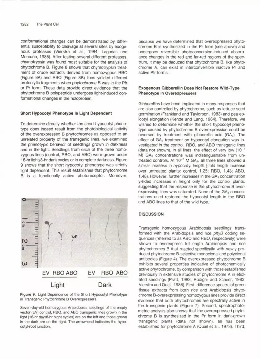
Short Hypocotyl Phenotype Is Light Dependent
To determine directly whether the short hypocotyl pheno-type does indeed result from the photobiological activityof the overexpressed B phytochromes as opposed to anunrelated property of the transgenic lines, we examinedthe phenotypic behavior of seedlings grown in darknessand in the light. Seedlings from each of the three homo-zygous lines (control, RBO, and ABO) were grown under16-hr light/8-hr dark cycles or in complete darkness. Figure9 shows that the short hypocotyl phenotype was strictlylight dependent. This result establishes that phytochromeB is a functionally active photoreceptor. Moreover,
o3
ro -o3
T Y
EV RBO ABO EV RBO ABO
Light DarkFigure 9. Light Dependence of the Short Hypocotyl Phenotypein Transgenic Phytochrome B Overexpressers.
Seven-day-old homozygous Arabidopsis seedlings of the emptyvector (EV) control, RBO, and ABO transgenic lines grown in thelight (16-hr day/8-hr night cycles) are on the left and those grownin the dark are on the right. The arrowhead indicates the hypo-cotyl-root junction.
because we have determined that overexpressed phyto-chrome B is synthesized in the Pr form (see above) andundergoes reversible photoconversion-induced absorb-ance changes in the red and far-red regions of the spec-trum, it may be deduced that phytochrome B, like phyto-chrome A, can exist in interconvertible inactive Pr andactive Pfr forms.
Exogenous Gibberellin Does Not Restore Wild-TypePhenotype in Overexpressers
Gibberellins have been implicated in many responses thatare also controlled by phytochrome, such as lettuce seedgermination (Frankland and Taylorson, 1983) and pea ep-icotyl elongation (Kende and Lang, 1964). Therefore, wewished to determine whether the short hypocotyl pheno-type caused by phytochrome B overexpression could bereversed by treatment with gibberellic acid (GA3). Theeffect of GA3 treatment on hypocotyl elongation was in-vestigated in the control, RBO, and ABO transgenic lines(data not shown). In all lines, the effect of very low (10~7
M) GA3 concentrations was indistinguishable from un-treated controls. At 1CT5 M GA3, all three lines showed asimilar increase in hypocotyl length (-fold length increaseover untreated plants: control, 1.25; RBO, 1.43; ABO,1.48). However, further increases in the GA3 concentrationyielded increases in height only for the control plants,suggesting that the response in the phytochrome B over-expressing lines was saturated. None of the GA3 concen-trations used restored the hypocotyl length in the RBOand ABO lines to that of the wild type.
DISCUSSION
Transgenic homozygous Arabidopsis seedlings trans-formed with the Arabidopsis and rice phyB coding se-quences (referred to as ABO and RBO, respectively) wereshown to overexpress full-length Arabidopsis and ricephytochromes B that reacted specifically with newly pro-duced phytochrome B-selective monoclonal and polyclonalantibodies (Figure 4). The overexpressed phytochrome Bexhibits several properties indicative of photochemicallyactive phytochrome, by comparison with those establishedpreviously in extensive studies of phytochrome A in etiol-ated seedlings (Pratt, 1983; Rudiger and Scheer, 1983;Vierstra and Quail, 1986). First, difference spectra of greentissue extracts from both rice and Arabidopsis phyto-chrome B-overexpressing homozygous lines provide directevidence that both phytochromes are spectrally active inthe transgenic plants (Figure 7). Second, spectrophoto-metric analysis also shows that the overexpressed phyto-chrome B is synthesized in the Pr form in dark-growntransgenic plants (data not shown), as has beenestablished for phytochrome A (Quail et al., 1973). Third,

Overexpression of Phytochrome B in Arabidopsis 1283
both B phytochromes were cleaved differentially by exog- enous protease after exposure to red or to far-red light. These results indicate that the overexpressed phyto- chrome B has formed a holoprotein in the plant and that it undergoes conformational changes in a red/far-red-light- dependent manner. Photoinduced conformational changes detectable by differential proteolytic sensitivity of phyto- chrome A have been shown to be dependent on holopro- tein formation (chromophore attachment) in vitro and in vivo (Lagarias and Lagarias, 1989; Parks and Quail, 1991) and result from the transition from the inactive Pr to the active Pfr form. Thus, we conclude that phy8 encodes a photochemically active photoreceptor that under- goes reversible, photoconversion-induced conformational changes analogous to those of phytochrome A.
The observation that, in all lines tested, overexpressed phytochrome 6 was stable in the light (Figure 6) is con- sistent with the properties of type 2 phytochrome. More- over, because Arabidopsis phytochrome B is stable in transgenic Arabidopsis, the data demonstrate that the stability of this protein is not simply the result of expression of a foreign (i.e., monocot) polypeptide that is less suscep- tible to specific degradation in a heterologous host. On the other hand, the data presented here leave open the pos- sibility that stability is the result of CaMV 35s promoter- driven expression of the protein in cell types that do not contain the degradative machinery responsible for selec- tive Pfr turnover. The extent of such “ectopic” expression is currently unknown but can be assessed when the expression pattern and localization of the endogenous phytochrome B are better characterized. However, be- cause recent independent experiments have shown that endogenous Arabidopsis phytochrome B is stable in the light (Somers et al., 1991), the observed stability of the overexpressed molecule is most likely primarily due to determinants residing within the polypeptide.
Light-stable phytochrome from oat showed spectral properties different from those of light-labile phytochrome in the same species in that the Pr absorption maximum was shifted 15 nm to a shorter wavelength (Tokuhisa and Quail, 1987, 1989; Pratt et al., 1991). In contrast, light- stable phytochrome showed similar spectral properties to light-labile phytochrome in pea (Konomi et al., 1987; Abe et al., 1989). Using transgenic Arabidopsis plants overex- pressing Arabidopsis phytochrome B, we estimated ab- sorption maxima and minima in difference spectra similar to those of Arabidopsis phytochrome A. Because phyto- chrome B appears to be a major component of the light- stable phytochromes thus far examined in Arabidopsis (SOmerS et al., 1991), it may follow that in dicots the light- labile and the predominant light-stable phytochrome have similar spectral properties. We propose to determine the spectral properties of overexpressed rice (monocot) phy- tochrome B upon further purification to asses whether it shows a similar shift toward shorter wavelength, as has been demonstrated for light-stable phytochrome from oat.
Severa1 lines of evidence indicate that overexpressed rice or Arabidopsis phytochrome 6 regulates seedling pho- tomorphogenesis in transgenic Arabidopsis. First, trans- genic lines show a tight correlation between transformation with the phy8 constructs and a short hypocotyl phenotype (Figures 3 and 5), inasmuch as all positive (kanamycin- resistant) transformants exhibit the short hypocotyl phe- notype. Second, all short hypocotyl lines tested show increased levels of phytochrome B. Conversely, neither siblings that segregate for wild-type height nor the trans- formation control shows elevated levels of phytochrome 6. Third, the short hypocotyl phenotype is strictly light dependent because elevated levels of phytochrome 6 have no effect on plants grown in the dark (data not shown). These data show that the phenotype is the result of altered photomorphogenesis, providing compelling evidence that it results directly from the overexpression of a biologically active photoreceptor. In conjunction with the finding that phytochrome B is synthesized in the Pr form (data not shown), we conclude that the Pfr form of phytochrome B is the biologically active form of the photoreceptor. In addition, because the phenotype observed in response to overexpression of Arabidopsis and rice B phytochromes is qualitatively similar, we conclude that the observed phenotype is due to elevated levels of the phytochrome B photoreceptor, rather than overexpression of a foreign (i.e., monocot) molecule in the plant.
The relatively small (threefold to fivefold) increase in immunochemically detectable phytochrome B over endog- enous levels observed in the rice phyB-expressing lines (Figures 4 and 5 and data not shown) as well as the small increase in spectral activity (Figure 7) result, nonetheless, in a 70% reduction in hypocotyl length when compared with the transformation control (data not shown). Phyto- chrome B may be, therefore, highly active or, alternatively, the plant response system may be very sensitive to small changes in the photoequilibrium of phytochrome B. In contrast, the larger (1 8- to 30-fold) increases in phyto- chrome B in the Arabidopsis phy8-overexpressing lines lead to disproportionately small additional effects on hy- pocotyl elongation, i.e., a decrease in hypocotyl length to 15% of that of the transformation control. These results may indicate a saturation of the photomorphogenic re- sponse system with respect to levels of phytochrome B photoreceptor.
The pronounced inhibition of hypocotyl elongation in light-grown seedlings overexpressing rice or Arabidopsis phytochrome B is similar to the short hypocotyl phenotype observed in Arabidopsis plants overexpressing oat phyto- chrome A (Boylan and Quail, 1991). Hence, both phyto- chromes A and B can regulate hypocotyl elongation in transgenic Arabidopsis. These overexpression phenotypes complement the long hypocotyl phenotypes observed in photomorphogenic mutants deficient in either phyto- chrome A or phytochrome B. The long hypocotyl aurea mutant of tomato is deficient in light-labile phytochrome A

1284 The Plant Cell
(Parks et al., 1987) but retains normal levels of light-stable phytochrome (Adamse et al., 1988~). Conversely, based on photobiological studies, it has been proposed that the Ih long hypocotyl mutant of cucumber (Adamse et al., 1987, 1988b) and the hy3 long hypocotyl mutant of Arabidopsis (Koornneef et al., 1980) are deficient in light- stable phytochrome but have normal levels of light-labile phytochrome (Adamse et al., 1988a). The hy3 mutant has now been shown to be deficient in phytochrome B, the predominant light-stable phytochrome thus far examined in Arabidopsis (Somers et al., 1991). The observation that overexpression of either phytochrome A or phytochrome B leads to exaggerated shortening of hypocotyl length, in conjunction with the evidence from the mutant analyses that both phytochromes are necessary for correct hypo- cotyl length, suggests that the combined total amounts of active phytochrome A and B in the plant can determine the rate of hypocotyl elongation. This notion is supported by the finding that only slight increases in the leve1 of phytochrome B in the overexpressing seedling (RBO, see above) cause a pronounced reduction of hypocotyl elon- gation. Thus mutant, wild type, and overexpressers may represent a continuum of phenotypic expression in re- sponse to increasing levels of phytochrome (Boylan and Quail, 1989). However, because the pattern of expression of phytochromes A and B may be altered spatially and temporally under the regulation of the CaMV 35s promoter in overexpressers, it is also possible that the effect in transgenic Arabidopsis is the result of “ectopic” expression per se rather than a simple reflection of the amount of phytochrome present.
Phytochrome function through regulation of phytohor- mone levels has been suggested by severa1 authors (Frankland and Taylorson, 1983; Cosgrove, 1986). Gibberellin-deficient and gibberellin-insensitive mutants in Arabidopsis (Koornneef and van der Veen, 1980; Koornneef et al., 1985) show phenotypes potentially similar to phytochrome 6-overexpressing transgenic Arabidopsis lines. Therefore, we tested whether the short hypocotyl phenotype caused by phytochrome B overexpression could be reversed by treatment with exogenous GAs. None of the GA3 concentrations used could restore the hypocotyl length in the overexpressing lines to that of the control line. Hence, our data provide no evidence in support of the hypothesis that GA3 mediates the phenotype caused by overexpression.
In conclusion, transgenic Arabidopsis plants overex- pressing phytochrome B have provided (1) a direct dem- onstration that this gene family member encodes a pho- tochemically and biologically functional photoreceptor, (2) information on the spectral and physiological properties of the chromoprotein in vivo, and (3) support for the notion that phytochromes A and B may be capable of regulating similar cellular processes based on the similar phenotypes obtained when either is overexpressed in Arabidopsis. However, different photosensory roles have been
proposed for light-labile and light-stable phytochromes: the high-irradiance response has been hypothesized to be mediated by light-labile phytochrome, whereas the shade avoidance and end-of-day far-red responses are attributed to the light-stable phytochrome pool (Smith and Whitelam, 1990). Arabidopsis seedlings overexpressing phytochrome B may prove useful in determining the properties of a light- stable phytochrome through analysis of the response of the transgenic lines under various light regimes. Compari- son with the behavior of phytochrome A overexpressers may help to determine whether the different phytochromes can have discrete physiological effects in the plant. In addition, the overexpression system described here pro- vides a powerful tool for further analysis of the biochemical and molecular properties of the phytochrome 8 photore- ceptor. As we have demonstrated, transgenic Arabidopsis can be used to determine the spectral properties of low- abundance phytochromes. This approach may be helpful in the spectral characterization of other members of the gene family, alleviating the need for extensive purification, which in the past has led to partia1 degradation and re- sulted in changes of the spectral properties of the protein (Pratt et al., 1991). Finally, this Arabidopsis overexpression system can be used for rapid analysis of the functional domains of phytochrome B. In contrast to tobacco and tomato, Arabidopsis has a very short generation time, and the transformation procedure is both rapid and simple. In vitro mutagenesis of the coding sequence of phytochrome B and subsequent analysis in transgenic Arabidopsis are thus greatly facilitated.
METHODS
Construction and Cloning of Full-Length Rice and Arabidopsis phyB Coding Sequences
Full-length rice phy8 was generated from a central 2586-bp Bglll-EcoRI fragment of a cDNA clone and an upstream genomic clone as well as a downstream polymerase chain reaction (PCR)- generated clone (Dehesh et al., 1991). An SnaBI site was intro- duced 51 bp upstream of the ATG start codon by site-directed mutagenesis (Kunkel, 1985). Similarly, a Kpnl site was introduced 52 bp downstream of the TGA stop codon. The plant transfor- mation vector pMON316 (Rogers et al., 1987) was linearized at the Clal site (blunted) and then digested with Kpnl. In one ligation reaction, the linearized vector, the 5‘ SnaBl to Bglll fragment, the central Bglll to EcoRl region, and the 3‘ EcoRl to Kpnl fragment were included to yield the final transformation vector pMRB316. For overexpression in Escherichia coli, the full-length rice phyB was reconstructed in plasmid pPO9 (Rottman et al., 1991). An Xbal site was introduced 4 bp 3’ to the ATG start codon. The Xbal-Bglll (genomic), Bglll-EcoRI (cDNA), and EcoRI-Kpnl (PCR from RNA) fragments were ligated into pPO9 digested with Nhel and Kpnl. The full-length construct, pRB6, is a perfect translational fusion.

Overexpression of Phytochrome B in Arabidopsis 1285
The full-length Arabidopsis phy6 clone was generated by PCR from a cDNA clone of phyB in X EMBL3 (Somers et al., 1991). Both primers contained Kpnl sites. This clone was subcloned into pBluescript KS-. The fidelity of approximately 75% of the PCR- generated clone was verified by sequence analysis (Tabor and Richardson, 1987), and the full-length clone was transferred into pMON316 to yield the final transformation vector pMAB316. This clone contains none of the original 5’ and 3’ untranslated leader sequences. The correct orientation in pMON316 was ascertained by sequence analysis and restriction mapping. For overexpression in E. coli, the Arabidopsis phyB clone was introduced into the plasmid pet3c (Somers et al., 1991).
Arabidopsis Transformation
Arabidopsis thaliana (ecotype NO-O) seedlings were grown in sterile culture for 3 weeks on growth medium (GM, Valvekens et al., 1988). The cointegrate vector was transferred into Agrobac- terium tumefaciens by triparental mating as described by Rogers et al. (1986). Roots were infected with A. tumefaciens carrying pMON316 alone or pMRB316 or pMAB316. The root transfor- mation procedure was according to Valvekens et al. (1988).
Plant Material and Extraction
Seeds were sterilized for 1 O min in 30% bleach and 0.02% Triton X-100, plated onto GM, and incubated at 4OC for 3 days. Kana- mycin sensitivity was assayed on GM plates using 50 pg/mL kanamycin. Otherwise, plants were grown for 7 days without kanamycin selection in the light (1 6-hr day/8-hr night cycles). Alternatively, plants were grown on identical medium but in dark- ness for 7 days or in darkness followed by 6 hr of red light irradiation (0.2 W/m’). Crude extracts were prepared essentially as described in Parks et al. (1987). The 2 x extraction buffer (1 O0 mM 3-[N-morpholino]propanesulfonic acid, pH 7.6, 50% ethylene glycol, 5 mM EDTA, 56 mM mercaptoethanol, 2 mM phenylmeth- ylsulfonyl fluoride) was adjusted to 1 pg/mL aprotenin, pepstatin, and leupeptin just before use. Tissue (400 mg) was ground in 300 pL of 2 x extraction buffer, and after 10-min centrifugation at 48,00Og, the supernatant was mixed 1:l with 2 x sample buffer (Laemmli, 1970) and frozen in liquid nitrogen. Crude protein con- centrations were determined by Bradford analysis (Bradford, 1976). All extractions were performed in dim green safelight. Green plant extracts for difference spectrum analysis were pre- pared essentially as described in Pratt et al. (1991). To the extraction buffer aprotenin, pepstatin, and leupeptin were added to 1 pg/mL. By scaling down the extraction procedure, the tissue could be handled faster and spectral activity could be measured within 1 hr of the start of extraction. The ammonium sulfate pellet was routinely resuspended in resuspension buffer at one-tenth of the volume of the crude extract. For the gibberellin analysis, plants were germinated on GM plates containing lO-’, 10-5, 10-4, and 10-3 M GA3 (Sigma). Stock solutions of GA3 were prepared at 100 mM in DMSO.
Production and Purification of Rice and Arabidopsis B Phytochromes in E. coli
Overexpression of pRB6 and Arabidopsis phyB in pet-3c in BL21 (DE3) was as described in Studier and Moffat (1986). lnclusion
bodies were isolated as described in Nagai and Thragersen (1987) with the following modifications. For the DNase treatment, MnCh was omitted and the DNase concentration was raised to 0.5 mg/ mL. Resuspension of the final pellet was in 1:lOO to 150 of the original volume in 50 mM Tris, pH 7.8, 0.5 mM DTT, 1 mM EDTA, 6 M urea, and 1% SDS. The proteins were further purified by electrophoresis on a 3.0-mm preparative 6% acrylamide gel. Bands were visualized with CuClp staining (Lee et al., 1987), excised, and electroeluted into 0.01% SDS, 190 mM glycine, 25 mM Tris, pH 7.8, at 100 V for 24 hr.
Antibody Preparation
Four mice (Swiss Webster and BALB/c) were used for antibody production. Polyclonal ascites were produced essentially as de- scribed in Hunter et al. (1990). The antigen was €. coli-prcduced, gel-purified rice phytochrome B. Ten micrograms of antigen emul- sified in 2 x reconstituted adjuvant (RIBI Adjuvant System, RlBl lmmunochemical Research Incorporated, Hamilton, MT) was used for four consecutive immunizations. Polyclonal ascites were ob- tained after 6 weeks.
The monoclonal cell line supernatants were obtained essentially as described in Karu (1991). After hyperimmunization, the spleen of a high-titer mouse was removed, and electrofusion between myeloma and spleenocytes was performed. Hybridoma superna- tants were transferred to immunological assay plates using a robotic sampling system (Karu et al., 1985). In ELISA-based screens, 300 positive cell lines were identified that reacted with the rice phytochrome B antigen. These were further tested for phytochrome B selectivity on ELISA microtiter plates and immu- noblots. Crude epitope mapping was performed by testing E. coli- produced deletion clones of rice phytochrome B. Cell lines that recognized the same deletion-defined region of the polypeptide were tested further for differential recognition of proteolytic pat- terns of rice phytochrome B obtained with V8 protease (Cleveland, 1983) and other proteases. Six phytochrome B selective hybrid- oma lines were subcloned to obtain monoclonal cell lines.
lmmunoblotting
Proteins from crude extracts were separated by SDS-PAGE (Laemmli, 1970). The acrylamide gels consisted of 1.5-mm thick, 6% resolving, and 4.5% stacking gels except after proteolysis, where 8% to 5% resolving gradient gels with 4% stacking gels were used. After blotting onto nitrocellulose membranes (600 mA for 2 hr), the remaining binding sites were saturated with 4% milk buffer (powdered milk dissolved in 20 mM Tris, pH 7.5, 0.15 mM NaCI, 0.05% Nonidet P-40). Both monoclonal cell supernatants were diluted 1 :1 O0 in this buffer. The polyclonal antiserum was depleted of Arabidopsis phytochrome B-reactive antibodies by incubation with one-half volume of E. coli-overproduced Arabidop- sis phy6-encoded protein at 4OC overnight with stirring. The antibody-antigen complexes were removed by centrifugation in a microcentrifuge for 20 min. The supernatant was diluted 1 :200 in the milk buffer for use on immunoblots. After 3-hr incubation with the primary antibodies, the blots were washed 3 x 10 min with the milk buffer and incubated for 1 hr with anti-mouse lg alkaline phosphatase conjugate (Promega) at 15000 dilution in milk buffer. After washing as above, the blots were developed in 100 mM Tris, pH 8.9, 100 mM NaCI, 5 mM MgCI2 solution, to which 75

1286 The Plant Cell
mg/mL 5-bromo-4-chloro-3-indolyl phosphate and 1 50 mg/mL nitroblue tetrazolium were added. Video imaging was used for densitometric analysis of immunoblots to determine the relative concentration of phytochrome in different samples.
In Vitro Proteolysis
Crude extracts were prepared as above except aprotenin was omitted from the extraction buffer. After extraction, the protease inhibitors were removed by 1 -min centrifugation through a packed Sephadex G-50 spin column, which had been equilibrated with 1 x extraction buffer without proteases. Crude extract (600 pL) was loaded on a column with a 3-mL bed volume and centrifuged at 5000g for 1 min. Samples were pooled, divided in half, and irradiated for 5 min with red or far-red light (on ice). Time O time points were taken immediately after the irradiation. Chymotrypsin (Sigma) aliquots (prepared at 0.5 mg/mL in 1 x extraction buffer without protease inhibitors and stored at -80°C) were thawed on ice, diluted 1:lO with 1 x extraction buffer, and added to a final concentration of 1.6 pg/mL for the ABO line (0.56 pg/mL crude protein) or 1.2 pg/mL for the RBO line (1.1 pg/mL crude protein). Digestion was stopped after 5, 10, 20, and 30 min by removal of aliquots and addition to boiling sample buffer, followed by freezing in liquid nitrogen.
Spectral Analysis
Crude extract supernatants (as described above) were prepared from 7-day-old dark-grown seedlings or after irradiation of such seedlings for 6 hr with red light. Spectrally active phytochrome (AAA) was measured with 730- and 800-nm measuring beams in a dual-wavelength spectrophotometer (Pratt et al., 1985) using Caco3 as a scattering agent, and the resulting values were presented in arbitrary units per gram fresh weight of tissue. Difference spectra were measured in clear, chlorophyll-depleted, concentrated green extracts (prepared as described above to yield a resuspended ammonium sulfate precipitate 10-fold con- centrated relative to the crude extract) in a Shimadzu dual- wavelength, double-beam recording spectrophotometer (model UV-3000, Shimadzu Corporation, Kyoto, Japan).
ACKNOWLEDGMENTS
We thank David E. Somers for the PCR product of Arabidopsis phy6, the Arabidopsis phy8 clone in the pet-3c expression vector, and the E. coli-purified Arabidopsis phytochromes A and C; Dr. Katayoon Dehesh for the rice phy6 DNA; Paul Oeller for the E. coli expression vector pPO9; Drs. John Shanklin and Richard Vierstra for the generous gift of the phytochrome A reactive antibody; and Dr. Stephen Rogers (Monsanto, St. Louis, MO) for providing Agrobacterium vectors and strains. We are grateful to John D. Wagner and Dr. Timothy Caspar for useful discussion throughout this project and thank Dr. Xing-Wang Deng for intro- duction to the Arabidopsis transformation and Dr. Brian M. Parks for assistance with the spectrophotometric analysis. We thank Dr. Judith M. Healy for critically reading this manuscript. We produced the polyclonal ascites and the monoclonal hybridoma
lines at University of California, Berkeley, College of Natural Re- sources Hybridoma facility, which is in part supported by National Science Foundation Grant DIR 90-43099. We especially thank Dr. Alex Karu and his colleagues for instruction, assistance, and advice with the antibody production. Our research is supported by Grant No. DE-FG03-87ER 13742 from the U.S. Department of Energy, Washington, DC, Division of Energy Biosciences and by U.S. Department of Agriculture, Agricultura1 Research Service, CRlS NO. 5335-23000-004-00D.
Received August 27, 1991; accepted October 14, 1991.
REFERENCES
Abe, H., Yamamoto, K.T., Nagatani, A., and Furuya, M. (1985). Characterization of green tissue-specific phytochrome isolated immunochemically from pea seedlings. Plant Cell Physiol. 326,
Abe, H., Takio, K., Titani, K., and Furuya, M. (1989). Amino- terminal amino acid sequences of pea phytochrome II fragments obtained by limited proteolysis. Plant Cell Physiol. 30,
Adamse, P., Jaspers, P.A.P.M., Kendrick, R.E., and Koornneef, M. (1 987). Photomorphogenetic responses of a long hypocotyl mutant of Cucumis sarivus L. J. Plant Physiol. 127, 481 -491.
Adamse, P., Kendrick, R.E., and Koornneef, M. (1988a). Pho- tomorphogenetic mutants of higher plants. Photochem. Photo- biol. 48, 833-841.
Adamse, P., Jaspers, P.A.P.M., Bakker, J.A., Kendrick, R.E., and Koornneef, M. (1988b). Photophysiology and phytochrome Content of long-hypocotyl mutant and wild-type cucumber seed- lings. Plant Physiol. 87, 264-268.
Adamse, P., Jaspers, P.A.P.M., Bakker, J.A., Wesselius, J.C., Heeringa, G.H., Kendrick, R.E., and Koornneef, M. (1988~). Photophysiology of a tomato mutant deficient in light-labile phytochrome. J. Plant Physiol. 133, 436-440.
Boylan, M.T., and Quail, P.H. (1989). Oat phytochrome is biolog- ically active in transgenic tomatoes. Plant Cell 1, 765-773.
Boylan, M.T., and Quail, P.H. (1 991). Phytochrome A overexpres- sion inhibits hypocotyl elongation in transgenic Arabidopsis. Proc. Natl. Acad. Sci. USA, in press.
Bradford, M.M. (1976). A rapid and sensitive method for the quantitation of microgram quantities of protein utilizing the principle of protein-dye binding. Anal. Biochem. 72, 248-254.
Cherry, J.R., Hershey, H.P., and Vierstra, R.D. (1991). Charac- terization of tobacco expressing functional oat phytochrome. Plant Physiol. 96, 775-785.
Cleveland, D.W. (1983). Peptide mapping in one dimension by limited proteolysis of sodium dodecyl sulfate-solubilized pro- teins. Methods Enzymol. 96, 222-229.
Cordonnier, M.-M., Greppin, H., and Pratt, L.H. (1986). Phyto- chrome from green Avena shoots characterized with a mono- clonal antibody to phytochrome from etiolated Pisum shoots. Biochemistry 25, 7657-7666.
1387-1 399.
1089-1 097.

Overexpression of Phytochrome 6 in Arabidopsis 1287
Cosgrove, D.J. (1 986). Photomodulation of growth. In Photomor- phogenesis in Plants, R. E. Kendrick and G. H. M. Kronenberg, eds (Dordrecht, The Netherlands: Martinus Nijhoff), pp.
Dehesh, K., Tepperman, J., Christensen, A.H., and Quail, P.H. (1 991). phyB is evolutionarily conserved and constitutively ex- pressed in rice seedling shoots. MOI. Gen. Genet. 225,
Frankland, B., and Taylorson, R. (1983). Light control of seed germination. In Photomorphogenesis, W. Shropshire, Jr., and H. Mohr, eds (Berlin: Springer-Verlag), pp. 428-456.
Furuya, M. (1989). Molecular properties and biogenesis of phy- tochrome I and II. Adv. Biophys. 25, 133-167.
Grimm, R., Eckerskorn, C., Lottspeich, F., Zenger, C., and Riidiger, W. (1 988). Sequence analysis of proteolytic fragments of 124-kilodalton phytochrome from etiolated Avena sativa L.: Conclusions on the conformation of the native protein. Planta
Hershey, H.P., Barker, R.F., Idler, K.B., Lissemore, J.L., and Quail, P.H. (1985). Analysis of cloned cDNA and genomic sequences for phytochrome: Complete amino acid sequences for two gene products expressed in etiolated Avena. Nucl. Acids Res. 13,8543-8559.
Hillman, W.S. (1 967). The physiology of phytochrome. Annu. Rev. Plant Physiol. 18, 301-324.
Hunter, B.G., Richardson, J., Dietzgen, R.G., Karu, A.E., Sylvester, E.S., Jackson, A.O., and Morris, J.T. (1990). Puri- fication and characterization of strawberry crinkle virus. Phyto- pathology 80,282-287.
Jabben, M., and Holmes, M.G. (1983). Phytochrome in light grown plants. In Photomorphogenesis, W. Shropshire, Jr. and H. Mohr, eds (Berlin: Springer-Verlag), pp. 704-722.
Jones, A.M., and Quail, P.H. (1986). Quarternary structure of 124-kDa phytochrome from Avena sativa. Biochemistry 25,
Jones, A.M., and Quail, P.H. (1 989). Phytochrome structure: Peptide fragments from the amino-terminal domain involved in protein-chromophore interactions. Planta 178, 147-1 56.
Karu, A.E. (1991). Monoclonal antibodies and their use in meas- urements of environmental contaminants. In Hazard Assess- ment of Chemicals-Current Developments, Vol. 8, J. Saxena, ed (Washington, DC: Hemisphere Publishing Corporation), in press.
Karu, A.E., Miller, P.L., Chase, C., and Cornutt, W. (1985). A point-addressable transfer system for automated sampling, feeding, and expansion of hybridoma cultures. J. Immunol. Methods 76, 145-1 56.
Kay, S.A., Nagatani, A., Keith, B., Deak, M., Furuya, M., and Chua, N.-H. (1989). Rice phytochrome is biologically active in transgenic tobacco. Plant Cell 1,775-782.
Keller, J.M., Shanklin, J., Vierstra, R.D., and Hershey, P.H. (1 989). Expression of functional monocotyledonous phyto- chrome in transgenic tobacco. EMBO J. 8, 1005-1012.
Kende, H., and Lang, A. (1964). Gibberellins and light inhibition of stem growth in pea. Plant Physiol. 39, 435-440.
Kendrick, R.E., and Kronenberg, G.H.M., eds (1 986). Photo- morphogenesis in Plants. (Dordrecht, The Netherlands: Martinus Nijhoff).
341 -365.
305-31 3.
174,396-401.
2987-2995.
Konomi, K., Abe, H., and Furuya, M. (1987). Changes in the content of phytochrome I and II apoproteins in embryonic axes of pea seedlings during imbibition. Plant Cell Physiol. 28,
Koornneef, M., and van der Veen, J.H. (1980). lnduction and analysis of gibberellin sensitive mutants in Arabidopsis thaliana (L.) Heynh. Theor. Appl. Genet. 58, 257-263.
Koornneef, M., Rolff, E., and Spruit, C. (1980). Genetic control of light-inhibited hypocotyl elongation in Arabidopsis thaliana (L.) Heynh. Z. Pflanzenphysiol. 100, 147-160.
Koornneef, M., Elgersma, A., Hanhart, C.J., van Loenen- Martinet, E.P., van Rijn, L., and Zeevaart, J.A.D. (1985). A gibberelin insensitive mutant of Arabidopsis thaliana. Physiol. Plant. 65, 33-39.
Kunkel, T.A. (1 985). Rapid and efficient site-specific mutagenesis without phenotypic selection. Proc. Natl. Acad. Sci. USA 82,
Laemmli, U.K. (1 970). Cleavage of structural proteins during the assembly of the head of bacteriophage T4. Nature 227,
Lagarias, C.J., and Lagarias, D.M. (1 989). Self-assembly of synthetic phytochrome holoprotein in vitro. Proc. Natl. Acad. Sci. USA 86,5778-5780.
Lagarias, C.J., and Mercurio, F.M. (1 985). Structure function studies on phytochrome. J. Biol. Chem. 260, 2415-2433.
Lagarias, C.J., and Rapoport, H. (1980). Chromopeptides from phytochrome. The structure and linkage of the Pr form of the phytochrome chromophore. J. Am. Chem. SOC. 102,
Lee, C., Levin, A., and Branton, D. (1987). Copper staining: A five minute protein stain for sodium dodecyl sulfate-polyacryl- amide gels. Anal. Biochem. 166, 308-312.
Nagai, K., and Thogersen, C. (1987). Synthesis and sequence- specific proteolysis of hybrid proteins produced in Escherichia coli. Methods Enzymol. 153, 461-481.
Nagatani, A., Kay, S.A., Deak, M., Chua, N.-H., and Furuya, M. (1 991). Rice type I phytochrome regulates hypocotyl elongation in transgenic tobacco seedlings. Proc. Natl. Acad. Sci. USA 88,
Parks, B.M., and Quail, P.H. (1 991). Phytochrome-deficient hy l and hy2 long hypocotyl mutants of Arabidopsis are defective in phytochrome chromophore biosynthesis. Plant Cell. 3,
Parks, B.M., Jones, A.M., Adamse, P., Koornneef, M., Kendrick, R.E., and Quail, P.H. (1987). The aurea mutant of tomato is deficient in spectrophotometrically and immunochemically de- tectable phytochrome. Plant MOI. Biol. 9, 97-107.
Parks, B.M., Shanklin, J., Koornneef, M., Kendrick, R.E., and Quail, P.H. (1 989). lmmunochemically detectable phytochrome is present at normal levels but is photochemically nonfunctional in the hy l and hy2 long hypocotyl mutants of Arabidopsis. Plant MOI. Biol. 12, 425-437.
Pratt, L.H. (1 983). Assay of photomorphogenic photoreceptors. In Photomorphogenesis, W. Shropshire, Jr. and H. Mohr, eds (Berlin: Springer-Verlag), pp. 152-177.
Pratt, L.H., Wampler, J.E., and Rich, E.S.J. (1985). An automated dual-wavelength spectrophotometer optimized for phyto- chrome assay. Anal. Instrument. 13, 269-287.
1443-1451.
488-492.
680-685.
4821 -4828.
5207-5211.
11 77-1 186.

1288 The Plant Cell
Pratt, L.H., Shimazaki, Y., Stewart, S.J., and Cordonnier, M.-M. (1 991). Large-scale purification of phytochrome from green leaves of Avena sativa L. Planta 184, 81 -86.
Quail, P.H-, Schafer, E., and Marmé, D- (lg73). Tw'nover of phytochrome in pumpkin hooks. Plant Physiol. 52, 124-1 27.
Rogers, S.G., Horsch, R.B., and Fraley, R.T. (1986). Gene trans-
Studier, F.W., and Moffat, B.A. (1986). Use of bacteriophage T7 RNA polymerase to direct selective high-leve1 expression of cloned genes. J. MOI. Biol. 189, 113-130.
Tabor, S., and Richardson, C.C. (1987). DNA sequence analysis with a modified bacteriophage T7 DNA polymerase. Proc. Natl. Acad. Sci. USA 14,4767-4771.
fer in plants: Production of transformed plants using Ti plasmid vectors. Methods Enzymol. 118, 627-640.
Rogers, S.G., Klee, H.J., Horsch, R.B., and Fraley, R.T. (1987). lmproved vectors for plant transformation: Expression cassette vectors and new selectable markers. Methods Enzymol. 153,
Rottmann, W.H., Peter, G.F., Oeller, P.W., Keller, J.A., Shen, N.F., Nagy, B.P., Taylor, L.P., Campbell, A.D., and Theologis, A. (1 991). 1 -Aminopropane-1-carboxylate synthase in tomato is encoded by a multigene family whose transcription is induced during fruit and floral senescence. J. MOI. Biol. 222, in press.
Rüdiger, W., and Scheer, H. (1983). Chromophores in photo- morphogenesis. In Photomorphogenesis, W. Shropshire, Jr. and H. Mohr, eds (Berlin: Springer-Verlag), pp. 1 1 9-1 51.
Sato, N. (1988). Nucleotide sequence and expression of the phytochrome gene in Pisum sativum: Differential regulation by light of multiple transcripts. Plant MOI. Biol. 11, 697-710.
Shanklin, J. (1988). Phytochrome degradation in oat. PhD Thesis (Madison, WI: University of Wisconsin).
Sharrock, R.A., and Quail, P.H. (1 989). Nove1 phytochrome se- quences in Arabidopsis thaliana: Structure, evolution, and dif- ferential expression of a plant regulatory photoreceptor family. Genes Dev. 3,1745-1 757.
Shimaraki, Y., and Pratt, L.H. (1 985). lmmunochemical detection with rabbit polyclonal and mouse monoclonal antibodies of different pools of phytochrome from etiolated and green Avena shoots. Planta 164, 333-344.
Shimazaki, Y., and Pratt, L.H. (1 986). lmmunoprecipitation of phytochrome from green Avena by rabbit antisera to phyto- chrome from etiolated Avena. Planta 168, 512-51 5.
Shimaraki, Y., Cordonnier, M.-M., and Pratt, L.H. (1983). Phy- tochrome quantitation in crude extracts of Avena by enzyme- linked immunoabsorbent assay with monoclonal antibodies. Planta 159, 534-544.
Smith, H., and Whitelam, G.C. (1990). Phytochrome, a family of photoreceptors with multiple physiological roles. Plant Cell En- viron. 13, 695-707.
Somers, D.E., Sharrock, R.A., Tepperman, J.M., and Quail, P.H. (1991). The hy3 long hypocotyl mutant of Arabidopsis is defi- cient in phytochrome B. Plant Cell 3, 1263-1274.
253-277.
Tokuhisa, J.G., and Quail, P.H. (1 983). Spectral and immuno- chemical characterization of phytochrome isolated from light grown Avena sativa. Plant Physiol. 72 (suppl.), 85 (abstr.).
Tokuhisa, J.G., and Quail, P.H. (1987). The levels of two distinct species of phytochrome are regulated differently during germi- nation in Avena. Planta 172. 371 -377.
Tokuhisa, J.G., and Quail, P.H. (1989). Phytochrome in green tissue: Partia1 purification and characterization of the 1 1 8- kiloDalton phytochrome species from light grown Avena sativa. Photochem. Photobiol. 50, 143-1 52.
Tokuhisa, J.G., Daniels, S.M., and Quail, P.H. (1985). Phyto- chrome in green tissue: Spectral and immunochemical evidence for two distinct molecular species of phytochrome in light-grown Avena sativa L. Planta 164, 321 -332.
Tomizawa, K., Nagatani, A., and Furuya, M. (1990). Phyto- chrome genes: Studies using the tools of molecular biology and photomorphogenetic mutants. Photochem. Photobiol. 52,
Valvekens, D., Van Montagu, M., and Van Lijsebettens, M. (1 988). Agrobacterium tumefaciens-mediated transformation of Arabidopsis thaliana root explants by using kanamycin selec- tion. Proc. Natl. Acad. Sci. USA 85, 5536-5540.
Vierstra, R.D., and Quail, P.H. (1 986). Phytochrome: The protein. In Photomorphogenesis in Plants, R.E. Kendrick and G.H.M. Kronenberg, eds (Dordrecht, The Netherlands: Martinus Nijhoff), pp. 35-60.
Vierstra, R.D., Cordonnier, M.-M., Pratt, L.H., and Quail, P.H. (1 984). Native phytochrome: lmmunoblot analysis of relative molecular mass and in vitro proteolytic degradation for severa1 plant species. Planta 160, 521-528.
Wang, Y.-C., Stewart, S.J., Cordonnier, M.-M., and Pratt, L.H. (1991). Avena sativa L. contains three phytochromes, only one of which is abundant in etiolated tissue. Planta 184, 96-104.
Yamamoto, K.T. (1 987). Structure-function relationship of pea phytochrome as deduced from amino-terminal sequence analy- sis of phytochrome chromopeptides. In Phytochrome and Pho- toregulation in Plants, M. Furuya, ed (New York: Academic Press), pp. 63-81.
265-275.

DOI 10.1105/tpc.3.12.1275 1991;3;1275-1288Plant Cell
D. Wagner, J. M. Tepperman and P. H. QuailArabidopsis
Overexpression of Phytochrome B Induces a Short Hypocotyl Phenotype in Transgenic
This information is current as of July 12, 2011
Permissions 8X
https://www.copyright.com/ccc/openurl.do?sid=pd_hw1532298X&issn=1532298X&WT.mc_id=pd_hw153229
eTOCs http://www.plantcell.org/cgi/alerts/ctmain
Sign up for eTOCs at:
CiteTrack Alerts http://www.plantcell.org/cgi/alerts/ctmain
Sign up for CiteTrack Alerts at:
Subscription Information http://www.aspb.org/publications/subscriptions.cfm
is available at:Plant Physiology and The Plant CellSubscription Information for
ADVANCING THE SCIENCE OF PLANT BIOLOGY © American Society of Plant Biologists


