Embed Size (px)
Citation preview
Research Article
SPECIAL ISSUE: Physiology and Ecology of Halophytes—PlantsLiving in Salt-Rich Environments
Physiological responses of a halophytic shrub to salt stressby Na2SO4 and NaCl: oxidative damage and the role ofpolyphenols in antioxidant protectionMariana A. Reginato1, Antonella Castagna2, Ana Furlan3, Stella Castro3, Annamaria Ranieri2
and Virginia Luna1*1 Fisiologıa Vegetal, Fısico Quımicas y Naturales, Universidad Nacional de Rıo Cuarto, Rıo Cuarto, Argentina2 Department of Agriculture, Food and Environment, University of Pisa, Via del Borghetto, 80 56124 Pisa, Italy3 Biologıa, Fac. de Cs. Exactas, Fısico Quımicas y Naturales, Universidad Nacional de Rıo Cuarto, Rıo Cuarto, Argentina
Received: 19 March 2014; Accepted: 24 June 2014; Published: 24 July 2014
Guest Editor: Adele Muscolo
Citation: Reginato MA, Castagna A, Furlan A, Castro S, Ranieri A, Luna V. 2014. Physiological responses of a halophytic shrub to salt stressby Na2SO4 and NaCl: oxidative damage and the role of polyphenols in antioxidant protection. AoB PLANTS 6: plu042;doi:10.1093/aobpla/plu042
Abstract. Salt stress conditions lead to increased production of reactive oxygen species (ROS) in plant cells. Halo-phytes have the ability to reduce these toxic ROS by means of a powerful antioxidant system that includes enzymaticand non-enzymatic components. In this research, we used the halophytic shrub Prosopis strombulifera to investigatewhether the ability of this species to grow under increasing salt concentrations and mixtures was related to the syn-thesis of polyphenolic compounds and to the maintenance of leaf pigment contents for an adequate photosyntheticactivity. Seedlings of P. strombulifera were grown hydroponically in Hoagland’s solution, gradually adding Na2SO4 andNaCl separately or in mixtures until reaching final osmotic potentials of 21, 21.9 and 22.6 MPa. Control plants wereallowed to develop in Hoagland’s solution without salt. Oxidative damage in tissues was determined by H2O2 and mal-ondialdehyde content. Leaf pigment analysis was performed by high-performance liquid chromatography with ultra-violet, and total phenols, total flavonoids, total flavan-3-ols, condensed tannins, tartaric acid esters and flavonols werespectrophotometrically assayed. Treatment with Na2SO4 increased H2O2 production and lipid peroxidation in tissuesand induced a sharp increase in flavonoid compounds (mainly flavan-3-ols) and consequently in the antioxidant ac-tivity. Also, Na2SO4 treatment induced an increased carotenoid/chlorophyll ratio, which may represent a strategy toprotect photosystems against photooxidation. NaCl treatment, however, did not affect H2O2 content, lipid peroxida-tion, pigments or polyphenols synthesis. The significant accumulation of flavonoids in tissues under Na2SO4 treatmentand their powerful antioxidant activity indicates a role for these compounds in counteracting the oxidative damageinduced by severe salt stress, particularly, ionic stress. We demonstrate that ionic interactions between different saltsin salinized soils modify the biochemical and morpho-physiological responses of P. strombulifera plants to salinity.
Keywords: NaCl; Na2SO4; oxidative damage; pigments; polyphenols; salt stress.
* Corresponding author’s e-mail address: [email protected]
Published by Oxford University Press on behalf of the Annals of Botany Company.This is an Open Access article distributed under the terms of the Creative Commons Attribution License (http://creativecommons.org/licenses/by/4.0/), which permits unrestricted reuse, distribution, and reproduction in any medium, provided the original work is properly cited.
AoB PLANTS www.aobplants.oxfordjournals.org & The Authors 2014 1
by guest on Novem
ber 5, 2014http://aobpla.oxfordjournals.org/
Dow
nloaded from
IntroductionIncreased soil salinity has been a substantial threat toagriculture in some parts of the world for more than3000 years, and the problem is becoming more wide-spread through time (Lauchli and Grattan 2007). For thisreason, to improve knowledge about the specializedphysiology and biochemistry of halophytic plants(known for their exceptional salt tolerance) represents agoal for scientists.
Salt stress leads to increased production of reactiveoxygen species (ROS) in plant cells. Reactive oxygen spe-cies are extremely reactive and undergo uncontrollableand damaging reactions with cellular components in-cluding DNA, lipids and proteins, which can aggravatethe detrimental effects of the initial stress and evenlead to cell death (Halliwell 2006; Van Breusegem andDat 2006). Oxidative stress is a central factor in abioticand biotic stress phenomena, which occurs when thereis a serious imbalance in any cell compartment betweenROS production and antioxidant defence leading to dra-matic physiological challenges (Foyer and Noctor 2003).It was considered that ROS concentration needs to bemaintained as low as possible, although this concept ischanging because of the multiple functions that are cur-rently being discovered for these molecules (Mittler andBlumwald 2010). Thus, it is important for cells to keep atight control of ROS concentration, but not to eliminatethem completely (Schutzendubel and Polle 2002).
Halophytes are known for their ability to withstand un-favourable conditions by quenching these toxic ROS, sincethey are equipped with a powerful antioxidant systemthat includes enzymatic and non-enzymatic components.Natural antioxidants occur in all plant organs, and the typ-ical compounds that exhibit antioxidant activities includephenolics, carotenoids and vitamins (Chanwitheesuk et al.2005). Tocopherols and carotenoids protect lipid mem-branes from oxidative stress because they deactivatesinglet oxygen by physical quenching and/or chemicalscavenging, and prevent the propagation of lipid peroxida-tion by reducing fatty acyl peroxy radicals (Polle andRennenberg 1994; Falk and Munne-Bosch 2010). En-hanced synthesis of particular secondary metabolitesunder stressful conditions is also believed to protect thecellular structures from oxidative effects (Jaleel et al.2007). Among these compounds, polyphenols (mainly fla-vonoids) play an important role in the defence againstROS, and their synthesis and accumulation has been pro-posed to be stimulated in plants under salt stress (Navarroet al. 2006; Hernandez 2007).
The genus Prosopis occurs in arid and semiarid regions,being the major component of such ecosystems in Southand North America. Many species within this genus have
economic and ecological potential (shade, firewood, foodand forage for wildlife and livestock). Some species ofProsopis, especially P. pallida, P. juliflora, P. tamarugoand P. alba have individuals with rapid growth at seawatersalinity or 45 dS m21 which is nearly 20 times greaterthan salinities that can be tolerated by annual temperatelegumes (Felker 2007).
The spiny shrub P. strombulifera (Burkart 1976) rangesfrom the Arizona desert (USA) to Patagonia (Argentina),and is particularly abundant in high-salinity areas ofcentral Argentina (Cordoba and southwestern San Luisprovinces). In these highly salinized soils, proportions ofNaCl and Na2SO4 are generally similar, although in previ-ous studies we found that Na2SO4 was up to three timesmore abundant than NaCl in several soil samples (Sosaet al. 2005). Similarly, in many countries, NaCl andNa2SO4 are the most abundant salts in salinized soils(Iqbal 2003; Shi and Sheng 2005; Manivannan et al.2008). For that reason it is important to compare theeffects of these two salts on plant growth, in order tounderstand better the physiological responses of plantsin natural environments.
Comparative studies have shown that SO422-based solu-
tions have considerably stronger inhibitory effect onP. strombulifera germination than Cl2-based solutions atiso-osmotic concentrations (Llanes et al. 2005; Sosa et al.2005). Stimulation of shoot growth at Co values up to21.9 MPa (500 mM) NaCl is an interesting halophytic re-sponse found in our studies (Reginato et al. 2014). Findingsin other Prosopis species indicate that the NaCl tolerance ofP. strombulifera exceeds the limits described for most ha-lophytic plants (Catalan et al. 1994; Almeida Viegas et al.2004; Felker 2007). However, P. strombulifera is much lesstolerant to Na2SO4 than to NaCl. Plants grown in thepresence of Na2SO4 showed immediate and significantreduction of shoot height and leaf number per plant, ac-companied by senescence symptoms such as chlorosis,necrosis and leaf abscission (Reginato et al. 2014).
Prosopis strombulifera plants grown in an increasinggradient of NaCl (250 up to 700 mM) do not develop saltglands in the leaves. Some tissues display vacuolization,and the root system undergoes precocious lignificationand/or suberization of endodermal cells, with Casparianstrips found much closer to the root tip than in glyco-phytes. These plants can therefore filter soil solutionmore efficiently to prevent passage of excess ions tothe xylem (Reinoso et al. 2004). Na2SO4 treatment in-duced structural alterations in cells and tissues, with con-sequent changes in growth patterns at various levels oforganization, and anatomical and histological differencesin roots, stems and leaflets, compared with controlplants, or plants grown under high NaCl (Reinoso et al.2005).
2 AoB PLANTS www.aobplants.oxfordjournals.org & The Authors 2014
Reginato et al. — Oxidative damage under sodium salts: polyphenols as antioxidant protection
by guest on Novem
ber 5, 2014http://aobpla.oxfordjournals.org/
Dow
nloaded from
An interesting feature observed in salt-treated plants inour anatomical studies was the significant accumulationof tannins in all organs, which increased with increasingsalt concentration (Reinoso et al. 2004, 2005), mainly inNa2SO4-treated plants. These results demonstrate thatplant responses may vary depending on the anion asso-ciated with sodium. The aim of the present research wasto investigate whether the differential ability of this speciesto grow under increasing concentrations of Na2SO4, NaCland their iso-osmotic mixture was related to oxidativedamage leading to an enhanced synthesis of polyphenoliccompounds. Maintenance of leaf pigment content for anadequate photosynthetic activity was also investigated.
Methods
Plant materials and growth conditions
Pods of P. strombulifera were randomly collected from 100plants within the same population, in the southwesternSan Luis province, Argentina. Peeled seeds were scarifiedwith 98 % sulfuric acid for 10 min, washed overnightunder running water, rinsed in distilled water and germi-nated in a Petri dish over two layers of water-saturated fil-ter paper at 37 8C for 24 h. The germinated seedlings with20-mm-long radicles were grown under hydroponic con-ditions in black trays (200 seedlings per each tray of28 × 22 × 10 cm) with 10 % of full-strength Hoagland’ssolution. The seedlings were self-supported in small
holes on the tray cover; the trays were placed in a growthchamber (Conviron E15; Controlled Environments Limited,Manitoba, Canada) under a 16 h light (400 mmol m22 s21)at 28 8C : 8 h dark (20 8C) cycle and 70 % relative humidity.After 1 week, the nutrient solution was changed to 25 %Hoagland’s solution (osmotic potential (Co)¼ 20.11 MPa).The pH of the medium was 6 in all cases, and, to provide aer-ation, an aquarium aeration system with a peristaltic pumpwas used. The complete experiment was performed twice,consecutively (3 trays per treatment each time). Plants weregrown hydroponically for 7 weeks (48 days) and allowed toacclimate to the different salt regimes.
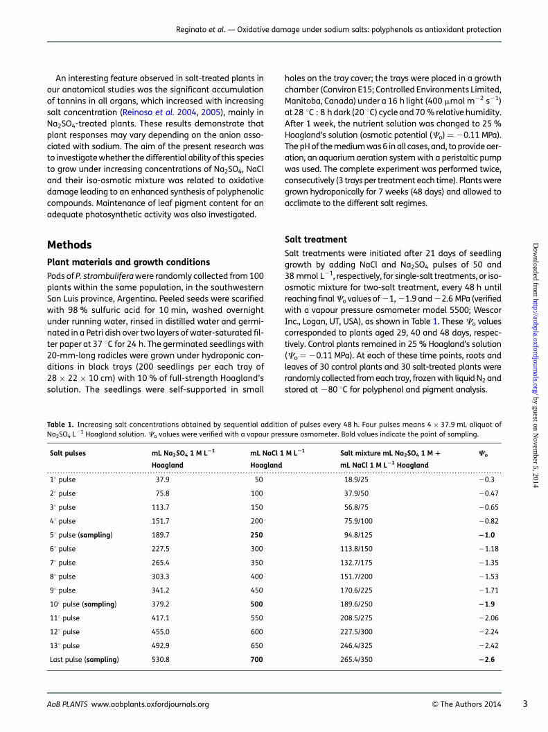
Salt treatment
Salt treatments were initiated after 21 days of seedlinggrowth by adding NaCl and Na2SO4 pulses of 50 and38 mmol L21, respectively, for single-salt treatments, or iso-osmotic mixture for two-salt treatment, every 48 h untilreaching finalCo values of 21, 21.9 and 22.6 MPa (verifiedwith a vapour pressure osmometer model 5500; WescorInc., Logan, UT, USA), as shown in Table 1. These Co valuescorresponded to plants aged 29, 40 and 48 days, respec-tively. Control plants remained in 25 % Hoagland’s solution(Co¼ 20.11 MPa). At each of these time points, roots andleaves of 30 control plants and 30 salt-treated plants wererandomly collected from each tray, frozen with liquid N2 andstored at 280 8C for polyphenol and pigment analysis.
. . . . . . . . . . . . . . . . . . . . . . . . . . . . . . . . . . . . . . . . . . . . . . . . . . . . . . . . . . . . . . . . . . . . . . . . . . . . . . . . . . . . . . . . . . . . . . . . . . . . . . . . . . . . . . . . . . . . . . . . . . . . . . . . . . . . . . . . . . . . . . . . . . . . . . . . . . . . . . . . . . . . . . . . . . . .
Table 1. Increasing salt concentrations obtained by sequential addition of pulses every 48 h. Four pulses means 4 × 37.9 mL aliquot ofNa2SO4 L21 Hoagland solution. Co values were verified with a vapour pressure osmometer. Bold values indicate the point of sampling.
Salt pulses mL Na2SO4 1 M L21
Hoagland
mL NaCl 1 M L21
Hoagland
Salt mixture mL Na2SO4 1 M 1
mL NaCl 1 M L21 Hoagland
Co
18 pulse 37.9 50 18.9/25 20.3
28 pulse 75.8 100 37.9/50 20.47
38 pulse 113.7 150 56.8/75 20.65
48 pulse 151.7 200 75.9/100 20.82
58 pulse (sampling) 189.7 250 94.8/125 21.0
68 pulse 227.5 300 113.8/150 21.18
78 pulse 265.4 350 132.7/175 21.35
88 pulse 303.3 400 151.7/200 21.53
98 pulse 341.2 450 170.6/225 21.71
108 pulse (sampling) 379.2 500 189.6/250 21.9
118 pulse 417.1 550 208.5/275 22.06
128 pulse 455.0 600 227.5/300 22.24
138 pulse 492.9 650 246.4/325 22.42
Last pulse (sampling) 530.8 700 265.4/350 22.6
AoB PLANTS www.aobplants.oxfordjournals.org & The Authors 2014 3
Reginato et al. — Oxidative damage under sodium salts: polyphenols as antioxidant protection
by guest on Novem
ber 5, 2014http://aobpla.oxfordjournals.org/
Dow
nloaded from
Oxidative damage in tissues
Hydrogen peroxide was measured spectrophotometri-cally after reaction with KI according to Alexieva et al.(2001). The reaction mixture consisted of 0.5 mL of0.1 % trichloroacetic acid (TCA), leaf extract supernatant,0.5 mL of 100 mM K-phosphate buffer and 2 mL of re-agent (1 M KI w/v in fresh double-distilled water). Theblank test consisted of 0.1 % TCA in the absence of leafextract. The reaction was developed for 1 h in darknessand absorbance was measured at 390 nm. The amountof hydrogen peroxide was calculated using a standardcurve prepared with known concentrations of H2O2.
Lipid peroxidation was determined by estimating theamount of malondialdehyde (MDA), a product of unsatur-ated fatty acid peroxidation, according to Heath andPacker (1968). Frozen samples (0.15 g) were crushedinto a fine powder in a mortar under liquid nitrogen andthen mixed with 1.5 mL 20 % TCA. The homogenate wascentrifuged at 10 000g for 10 min at 4 8C, with the super-natant being used for MDA determination. A mixture of0.5 mL of extract + 0.5 mL of 0.5 % TBA (thiobarbituricacid) (0.5 g TBA + TCA 20 % to complete to 100 mL) wasproduced, heated at 95 8C for 25 min, cooled and centri-fuged for 10 min. The sample was measured at 532 nmand corrected by non-specific absorption at 600 nm.The concentration of MDA was calculated using an extinc-tion coefficient of 155 mM21 cm21.
Leaf pigment analysis
Pigment concentrations of P. strombulifera leaves were de-termined according to the method reported by Castagnaet al. (2001). Frozen samples were homogenized in thedark in 100 % HPLC-grade acetone with 1 mM sodiumascorbate then filtered through 0.2 mm filters. The analysiswas performed by high-performance liquid chromatog-raphy (HPLC) (HPLC P200; Thermo Fisher Scientific,Waltham, MA, USA) using a non-endcapped column(Zorbax ODS column; Chrompack, Raritan, NJ, USA) for pig-ment separation. Two solvents were used: A (acetonitrile/methanol, 75/25, v/v) and B (methanol/ethylacetate,68/32, v/v). The separation cycle was 1920 s with a flowrate of 16.67 mm3 s21. Pigments were eluted using100 % A for the first 900 s, followed by a 150-s linear gra-dient to 100 % B, which continued isocratically until theend of the cycle. The column was allowed to re-equilibratein 100 % solvent A for 600 s before the next injection. Pig-ments were detected by their absorbance at 445 nm, andtheir quantification was realized by the injection ofknown amounts of pure standard into the HPLC systemand the formulation of an equation correlating peak areato pigment concentration. The latter was expressed asnmol g21 DW.
Calculation of the de-epoxidation (DEPS) index was basedon the contents of antheraxanthin (A), zeaxanthin (Z) andviolaxanthin (V) according to the following equation:
DEPS index = (0.5A + Z)/(V + A + Z)
Polyphenol analysis
Polyphenols extraction. Dry samples (0.5 g) were groundwith liquid N2. The plant material was extracted on amagnetic stirrer three times using a total of 90 mL ofmethanol/water (80 : 20, v/v). The liquid extract wasseparated by centrifugation (14 000g, 15 min) at 4 8C. Thefinal volume was quantified, and the extract, reduced to16 mL by rotary evaporation, was filtered with a 0.45-mmfilter (Minisart) and stored at 280 8C.
Phenol compound quantification. Total phenols weredetermined using the Folin-Ciocalteu method, modified asdescribed by Barbolan et al. (2003). Amounts of 1.85 mL ofdistilled water, 0.125 mL of Folin-Ciocalteu reagent and0.5 mL of a 20 % sodium carbonate solution were addedto 25 mL of liquid extract sample in a test tube, making afinal volume of 2.5 mL. The solution was homogenizedand left to stand for 30 min, and the absorbance wasdetermined at 750 nm. The total phenols were calculatedas milligrams of gallic acid equivalents.
Total flavonoids were determined as described by Kimet al. (2003). Assays contained 60 mL of 5 % NaNO2,40 mL of 10 % AlCl3 and 400 mL of 1 M NaOH in additionto 100 mL of extract. The solution was diluted with200 mL of distilled water, and the absorbance was deter-mined at 510 nm. The flavonoid amount was calculatedas milligrams of catechin equivalents.
Tartaric acid ester and flavonol contents were deter-mined using the method described by Romani et al.(1996). An aliquot of 25 mL of extract was diluted with225 mL of 10 % ethanol and 250 mL of 0.1 % HCl in 95 %ethanol, and 1 mL of 2 % HCl was then added. Thesolution was mixed, and the absorbance determinedat 320 nm for tartaric acid esters and at 360 nm forflavonols. Tartaric acid ester and flavonol amounts werecalculated as milligrams of caffeic acid and quercetin,respectively.
Total flavan-3-ols were determined with p-(dimethyl-amino) cinnamaldehyde (DMACA) reagent, as describedby Nigel and Glories (1991). The sample extract (10 mL)was diluted with 90 mL of methanol. Next, 250 mL of HCl(0.24 N in MeOH), 250 mL of DMACA solution (0.2 % inMeOH) and 250 mL of methanol were added. The absorb-ance was determined at 640 nm, and the total amount offlavan-3-ols was calculated as milligrams of catechinequivalents.
4 AoB PLANTS www.aobplants.oxfordjournals.org & The Authors 2014
Reginato et al. — Oxidative damage under sodium salts: polyphenols as antioxidant protection
by guest on Novem
ber 5, 2014http://aobpla.oxfordjournals.org/
Dow
nloaded from
Condensed tannins (proanthocyanidins) were deter-mined in accordance with the method described byWaterman and Mole (1994). Butanol reagent was pre-pared by mixing 128 mg of FeSO4 . 7H2O with 5 mL of con-centrated HCl and brought to 100 mL with n-butanol. Analiquot of 50 mL of extract sample was mixed with 700 mLof butanol reagent and heated at 95 8C in a water bath for45 min. The sample was cooled, 250 mL of n-butanol wasadded, and the absorbance was measured at 550 nm.The total amount of condensed tannin was calculatedas milligrams of cyanidin equivalents. The assays wereperformed using an Ultro spec 2100 pro UV-visible spec-trophotometer (Amersham Biosciences).
Antioxidant activity of polyphenolic extracts. 2,2′-Azino-bis(3-ethylbenzothiazoline-6-sulfonic acid) (ABTS*) scav-enging ability of polyphenolic extracts was determinedaccording to the method described by Re et al. (1999).ABTS* was generated by reacting an ABTS aqueous solution(7 mM L21) with K2S2O8 (2.45 mM L21, final concentration)in the dark for 16 h and diluting with ethanol to obtain anabsorbance of 0.700+0.020 at 734 nm. About 0.2 mL ofappropriate dilution of the extract was added to 1.0 mLABTS* measuring absorbance at 734 nm after 6 min. TheTrolox equivalent antioxidant capacity was subsequentlycalculated.
Anatomical analyses
Samples were taken from roots, stems and leaves andplaced in FAA (95 % ethanol : glacial acetic acid : 37–40 % formaldehyde : water; 50 : 5 : 10 : 35, v/v) (Reinosoet al. 2004, 2005). The dehydration of samples was car-ried out according to the procedures outlined in Johansen(1940) using graduated solutions of ethanol and xylene.Fully infiltrated tissues were embedded in Histowax(highly purified paraffin wax blended with polymer addi-tives). A series of transverse sections 10-mm-thick wereobtained from the sample blocks using a Minot rotarymicrotome. The sections were triple-stained with haema-toxylin, safranin O and fast green FCF as described byJohansen (1940). A coverslip was added to the slideswith one or two drops of Depex. A standard Zeiss Model16 microscope was used to assess the histologicalpreparations and photomicrographs were taken with aZeiss Axiophot microscope with image capture and digit-alization (AxioVision 4.3 with AxioCam HRc camera). Toidentify tannins, freehand sections were cut from freshmaterial and treated with ferric chloride (D’Ambrogioand Argueso 1986).
Statistical analysis
Data were analysed using InfoStat program (Student Ver-sion 2011, Universidad Nacional de Cordoba, Argentina).
Two-way general linear model ANOVA was used to deter-mine the effect of each treatment at each osmotic poten-tial. Thus, the factors considered for two-way ANOVA wereosmotic potential (Co) (21.0, 21.9 or 22.6 MPa) and salttreatment (control, NaCl, Na2SO4 and salt mixture). Nor-mality was verified with the Shapiro–Wilk test. Homogen-eity of variance was verified with the Levenne test. Whennecessary, data were transformed to meet the assump-tions of ANOVA. For cases in which normality and homo-geneity of variance were not verified, the non-parametricKruskall–Wallis test was used. Post hoc analysis used theBonferroni test to determine differences between means.P values ,0.05 were considered statistically significant.
Results
H2O2 content and lipid peroxidation in tissuesinduced by salt stress
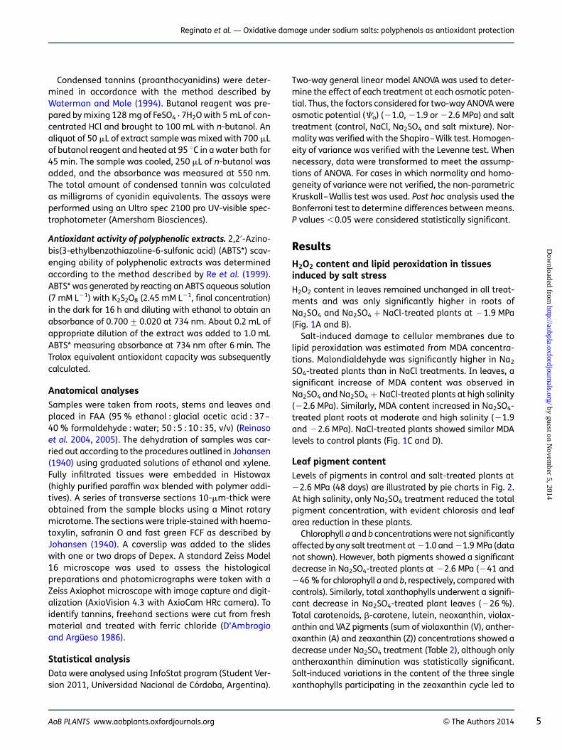
H2O2 content in leaves remained unchanged in all treat-ments and was only significantly higher in roots ofNa2SO4 and Na2SO4 + NaCl-treated plants at 21.9 MPa(Fig. 1A and B).
Salt-induced damage to cellular membranes due tolipid peroxidation was estimated from MDA concentra-tions. Malondialdehyde was significantly higher in Na2
SO4-treated plants than in NaCl treatments. In leaves, asignificant increase of MDA content was observed inNa2SO4 and Na2SO4 + NaCl-treated plants at high salinity(22.6 MPa). Similarly, MDA content increased in Na2SO4-treated plant roots at moderate and high salinity (21.9and 22.6 MPa). NaCl-treated plants showed similar MDAlevels to control plants (Fig. 1C and D).
Leaf pigment content
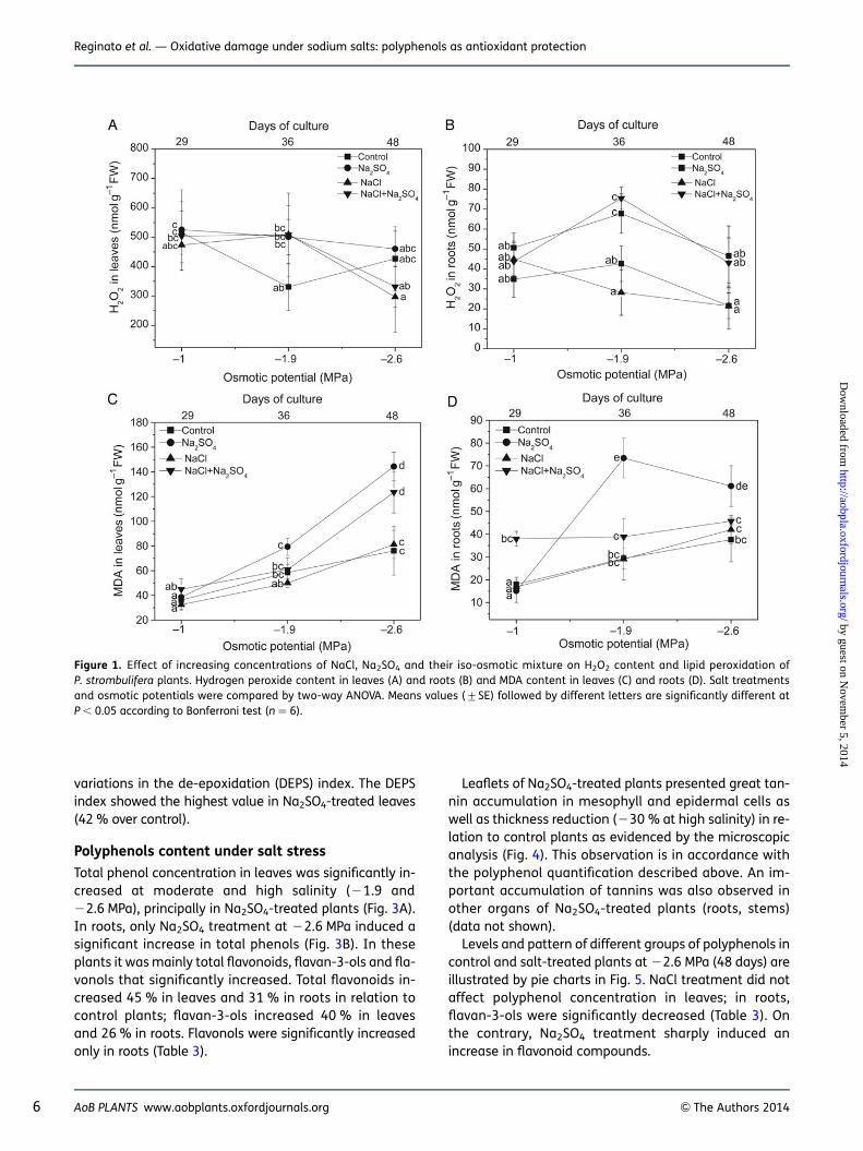
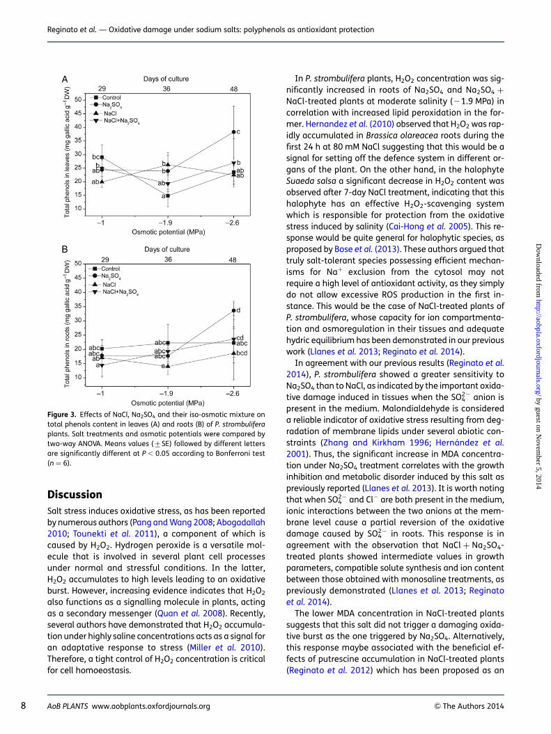
Levels of pigments in control and salt-treated plants at22.6 MPa (48 days) are illustrated by pie charts in Fig. 2.At high salinity, only Na2SO4 treatment reduced the totalpigment concentration, with evident chlorosis and leafarea reduction in these plants.
Chlorophyll a and b concentrations were not significantlyaffected by any salt treatment at 21.0 and 21.9 MPa (datanot shown). However, both pigments showed a significantdecrease in Na2SO4-treated plants at 22.6 MPa (241 and246 % for chlorophyll a and b, respectively, compared withcontrols). Similarly, total xanthophylls underwent a signifi-cant decrease in Na2SO4-treated plant leaves (226 %).Total carotenoids, b-carotene, lutein, neoxanthin, violax-anthin and VAZ pigments (sum of violaxanthin (V), anther-axanthin (A) and zeaxanthin (Z)) concentrations showed adecrease under Na2SO4 treatment (Table 2), although onlyantheraxanthin diminution was statistically significant.Salt-induced variations in the content of the three singlexanthophylls participating in the zeaxanthin cycle led to
AoB PLANTS www.aobplants.oxfordjournals.org & The Authors 2014 5
Reginato et al. — Oxidative damage under sodium salts: polyphenols as antioxidant protection
by guest on Novem
ber 5, 2014http://aobpla.oxfordjournals.org/
Dow
nloaded from
variations in the de-epoxidation (DEPS) index. The DEPSindex showed the highest value in Na2SO4-treated leaves(42 % over control).
Polyphenols content under salt stress
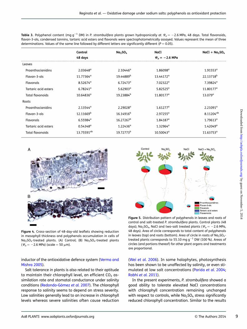
Total phenol concentration in leaves was significantly in-creased at moderate and high salinity (21.9 and22.6 MPa), principally in Na2SO4-treated plants (Fig. 3A).In roots, only Na2SO4 treatment at 22.6 MPa induced asignificant increase in total phenols (Fig. 3B). In theseplants it was mainly total flavonoids, flavan-3-ols and fla-vonols that significantly increased. Total flavonoids in-creased 45 % in leaves and 31 % in roots in relation tocontrol plants; flavan-3-ols increased 40 % in leavesand 26 % in roots. Flavonols were significantly increasedonly in roots (Table 3).
Leaflets of Na2SO4-treated plants presented great tan-nin accumulation in mesophyll and epidermal cells aswell as thickness reduction (230 % at high salinity) in re-lation to control plants as evidenced by the microscopicanalysis (Fig. 4). This observation is in accordance withthe polyphenol quantification described above. An im-portant accumulation of tannins was also observed inother organs of Na2SO4-treated plants (roots, stems)(data not shown).
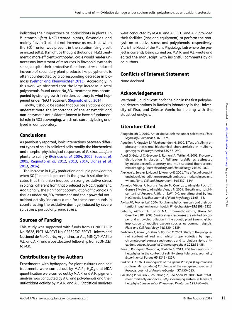
Levels and pattern of different groups of polyphenols incontrol and salt-treated plants at 22.6 MPa (48 days) areillustrated by pie charts in Fig. 5. NaCl treatment did notaffect polyphenol concentration in leaves; in roots,flavan-3-ols were significantly decreased (Table 3). Onthe contrary, Na2SO4 treatment sharply induced anincrease in flavonoid compounds.
Figure 1. Effect of increasing concentrations of NaCl, Na2SO4 and their iso-osmotic mixture on H2O2 content and lipid peroxidation ofP. strombulifera plants. Hydrogen peroxide content in leaves (A) and roots (B) and MDA content in leaves (C) and roots (D). Salt treatmentsand osmotic potentials were compared by two-way ANOVA. Means values (+SE) followed by different letters are significantly different atP , 0.05 according to Bonferroni test (n ¼ 6).
6 AoB PLANTS www.aobplants.oxfordjournals.org & The Authors 2014
Reginato et al. — Oxidative damage under sodium salts: polyphenols as antioxidant protection
by guest on Novem
ber 5, 2014http://aobpla.oxfordjournals.org/
Dow
nloaded from
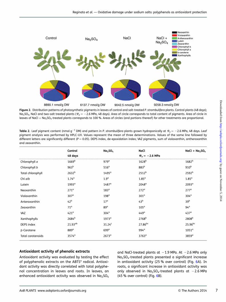
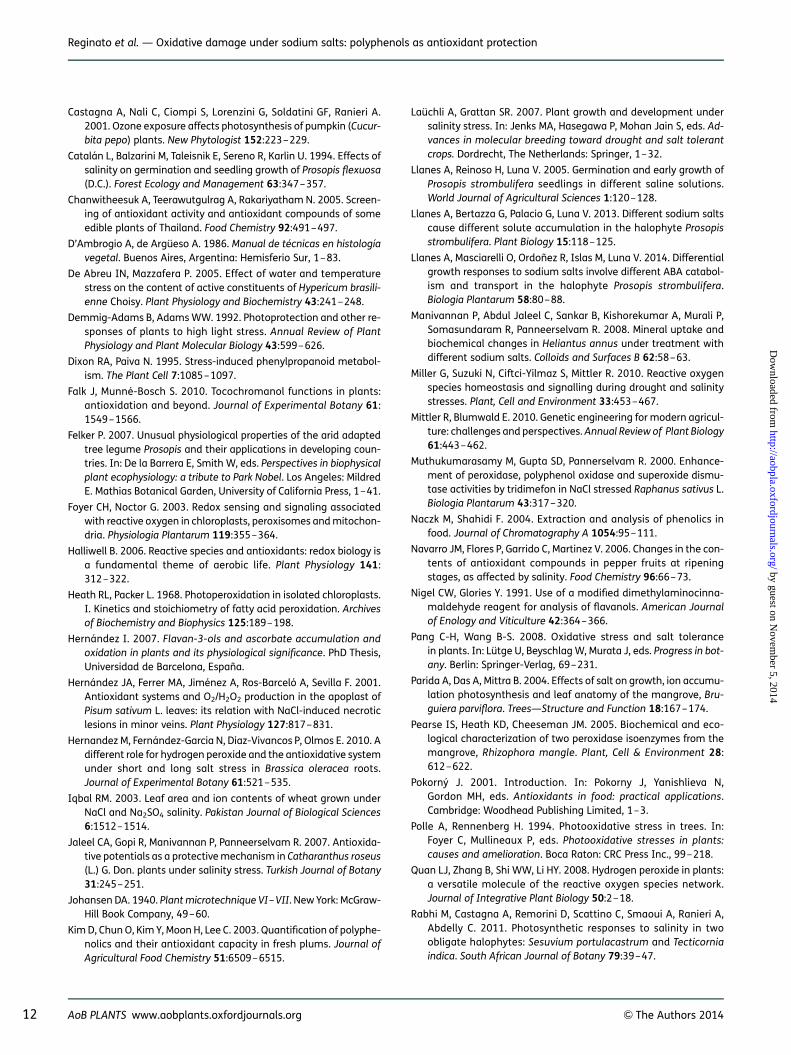
Antioxidant activity of phenolic extracts
Antioxidant activity was evaluated by testing the effectof polyphenolic extracts on the ABTS* radical. Antioxi-dant activity was directly correlated with total polyphe-nol concentration in leaves and roots. In leaves, anenhanced antioxidant activity was observed in Na2SO4
and NaCl-treated plants at 21.9 MPa. At 22.6 MPa onlyNa2SO4-treated plants presented a significant increasein antioxidant activity (25 % over control) (Fig. 6A). Inroots, a significant increase in antioxidant activity wasonly observed in Na2SO4-treated plants at 22.6 MPa(45 % over control) (Fig. 6B).
Figure 2. Distribution patterns of photosynthetic pigments in leaves of control and salt-treated P. strombulifera plants. Control plants (48 days);Na2SO4, NaCl and two-salt treated plants (Co ¼ 22.6 MPa, 48 days). Area of circle corresponds to total content of pigments. Area of circle inleaves of NaCl + Na2SO4-treated plants corresponds to 100 %. Areas of circles (and portions thereof) for other treatments are proportional.
. . . . . . . . . . . . . . . . . . . . . . . . . . . . . . . . . . . . . . . . . . . . . . . . . . . . . . . . . . . . . . . . . . . . . . . . . . . . . . . . . . . . . . . . . . . . . . . . . . . . . . . . . . . . . . . . . . . . . . . . . . . . . . . . . . . . . . . . . . . . . . . . . . . . . . . . . . . . . . . . . . . . . . . . . . . .
Table 2. Leaf pigment content (nmol g21 DM) and pattern in P. strombulifera plants grown hydroponically at Co ¼ 22.6 MPa, 48 days. Leafpigment analysis was performed by HPLC-UV. Values represent the mean of three determinations. Values of the same line followed bydifferent letters are significantly different (P ¼ 0.05). DEPS index, de-epoxidation index; VAZ pigments, sum of violaxanthin, antheraxanthinand zeaxanthin.
Control Na2SO4 NaCl NaCl 1 Na2SO4
48 days Co 5 22.6 MPa
Chlorophyll a 1669b 979a 1628b 1682b
Chlorophyll b 963b 516a 883b 910b
Total chlorophyll 2632b 1495a 2512b 2592b
Chl a/b 1.74a 1.9a 1.85a 1.85a
Lutein 1993a 1487a 2048a 2093a
Neoxanthin 271a 183a 272a 277a
Violaxanthin 307a 198a 301a 304a
Anteraxanthin 42b 17a 43b 39b
Zeaxanthin 71a 89a 105a 94a
VAZ 421a 304a 449a 437a
Xanthophylls 2684b 1973a 2768b 2808b
DEPS index 21.93ab 31.24c 27.86bc 25.96bc
b-Carotene 889a 699a 994a 1051a
Total carotenoids 3574a 2673a 3763a 3859a
AoB PLANTS www.aobplants.oxfordjournals.org & The Authors 2014 7
Reginato et al. — Oxidative damage under sodium salts: polyphenols as antioxidant protection
by guest on Novem
ber 5, 2014http://aobpla.oxfordjournals.org/
Dow
nloaded from
DiscussionSalt stress induces oxidative stress, as has been reportedby numerous authors (Pang and Wang 2008; Abogadallah2010; Tounekti et al. 2011), a component of which iscaused by H2O2. Hydrogen peroxide is a versatile mol-ecule that is involved in several plant cell processesunder normal and stressful conditions. In the latter,H2O2 accumulates to high levels leading to an oxidativeburst. However, increasing evidence indicates that H2O2
also functions as a signalling molecule in plants, actingas a secondary messenger (Quan et al. 2008). Recently,several authors have demonstrated that H2O2 accumula-tion under highly saline concentrations acts as a signal foran adaptative response to stress (Miller et al. 2010).Therefore, a tight control of H2O2 concentration is criticalfor cell homoeostasis.
In P. strombulifera plants, H2O2 concentration was sig-nificantly increased in roots of Na2SO4 and Na2SO4 +NaCl-treated plants at moderate salinity (21.9 MPa) incorrelation with increased lipid peroxidation in the for-mer. Hernandez et al. (2010) observed that H2O2 was rap-idly accumulated in Brassica olareacea roots during thefirst 24 h at 80 mM NaCl suggesting that this would be asignal for setting off the defence system in different or-gans of the plant. On the other hand, in the halophyteSuaeda salsa a significant decrease in H2O2 content wasobserved after 7-day NaCl treatment, indicating that thishalophyte has an effective H2O2-scavenging systemwhich is responsible for protection from the oxidativestress induced by salinity (Cai-Hong et al. 2005). This re-sponse would be quite general for halophytic species, asproposed by Bose et al. (2013). These authors argued thattruly salt-tolerant species possessing efficient mechan-isms for Na+ exclusion from the cytosol may notrequire a high level of antioxidant activity, as they simplydo not allow excessive ROS production in the first in-stance. This would be the case of NaCl-treated plants ofP. strombulifera, whose capacity for ion compartmenta-tion and osmoregulation in their tissues and adequatehydric equilibrium has been demonstrated in our previouswork (Llanes et al. 2013; Reginato et al. 2014).
In agreement with our previous results (Reginato et al.2014), P. strombulifera showed a greater sensitivity toNa2SO4 than to NaCl, as indicated by the important oxida-tive damage induced in tissues when the SO4
22 anion ispresent in the medium. Malondialdehyde is considereda reliable indicator of oxidative stress resulting from deg-radation of membrane lipids under several abiotic con-straints (Zhang and Kirkham 1996; Hernandez et al.2001). Thus, the significant increase in MDA concentra-tion under Na2SO4 treatment correlates with the growthinhibition and metabolic disorder induced by this salt aspreviously reported (Llanes et al. 2013). It is worth notingthat when SO4
22 and Cl2 are both present in the medium,ionic interactions between the two anions at the mem-brane level cause a partial reversion of the oxidativedamage caused by SO4
22 in roots. This response is inagreement with the observation that NaCl + Na2SO4-treated plants showed intermediate values in growthparameters, compatible solute synthesis and ion contentbetween those obtained with monosaline treatments, aspreviously demonstrated (Llanes et al. 2013; Reginatoet al. 2014).
The lower MDA concentration in NaCl-treated plantssuggests that this salt did not trigger a damaging oxida-tive burst as the one triggered by Na2SO4. Alternatively,this response maybe associated with the beneficial ef-fects of putrescine accumulation in NaCl-treated plants(Reginato et al. 2012) which has been proposed as an
Figure 3. Effects of NaCl, Na2SO4 and their iso-osmotic mixture ontotal phenols content in leaves (A) and roots (B) of P. strombuliferaplants. Salt treatments and osmotic potentials were compared bytwo-way ANOVA. Means values (+SE) followed by different lettersare significantly different at P , 0.05 according to Bonferroni test(n ¼ 6).
8 AoB PLANTS www.aobplants.oxfordjournals.org & The Authors 2014
Reginato et al. — Oxidative damage under sodium salts: polyphenols as antioxidant protection
by guest on Novem
ber 5, 2014http://aobpla.oxfordjournals.org/
Dow
nloaded from
inductor of the antioxidative defence system (Verma andMishra 2005).
Salt tolerance in plants is also related to their aptitudeto maintain their chlorophyll level, an efficient CO2 as-similation rate and stomatal conductance under salinityconditions (Redondo-Gomez et al. 2007). The chlorophyllresponse to salinity seems to depend on stress severity.Low salinities generally lead to an increase in chlorophylllevels whereas severe salinities often cause reduction
(Wei et al. 2006). In some halophytes, photosynthesishas been shown to be unaffected by salinity, or even sti-mulated at low salt concentrations (Parida et al. 2004;Rabhi et al. 2011).
In the present experiments, P. strombulifera showed agood ability to tolerate elevated NaCl concentrationswith chlorophyll concentration remaining unchangedwith respect to controls, while Na2SO4 stress significantlyreduced chlorophyll concentration. Similar to the results
. . . . . . . . . . . . . . . . . . . . . . . . . . . . . . . . . . . . . . . . . . . . . . . . . . . . . . . . . . . . . . . . . . . . . . . . . . . . . . . . . . . . . . . . . . . . . . . . . . . . . . . . . . . . . . . . . . . . . . . . . . . . . . . . . . . . . . . . . . . . . . . . . . . . . . . . . . . . . . . . . . . . . . . . . . . .
Table 3. Polyphenol content (mg g21 DM) in P. strombulifera plants grown hydroponically at Co ¼ 22.6 MPa, 48 days. Total flavonoids,flavan-3-ols, condensed tannins, tartaric acid esters and flavonols were spectrophotometrically assayed. Values represent the mean of threedeterminations. Values of the same line followed by different letters are significantly different (P ¼ 0.05).
Control Na2SO4 NaCl NaCl 1 Na2SO4
48 days Co 5 22.6 MPa
Leaves
Proanthocianidins 2.03648a 2.10446a 1.86098a 1.91553a
Flavan-3-ols 11.77364a 19.44889b 13.44172a 22.13718b
Flavonols 8.52674a 6.72473a 7.02322a 7.39824a
Tartaric acid esters 6.78241a 5.62903a 5.82525a 11.80177a
Total flavonoids 10.64836a 19.23884b 11.80177a 13.079a
Roots
Proanthocianidins 2.13544a 2.29028a 1.61277a 2.21091a
Flavan-3-ols 12.11669b 16.14916b 2.97255a 8.11204ab
Flavonols 6.55984a 16.27267b 1.84187a 1.79613a
Tartaric acid esters 0.54348a 1.22436a 1.32964a 1.42049a
Total flavonoids 13.75591ab 19.72773b 10.50043a 11.63753a
Figure 4. Cross-section of 48-day-old leaflets showing reductionin mesophyll thickness and polyphenols accumulation in cells ofNa2SO4-treated plants. (A) Control; (B) Na2SO4-treated plants(Co ¼ 22.6 MPa) (scale ¼ 50 mm).
Figure 5. Distribution pattern of polyphenols in leaves and roots ofcontrol and salt-treated P. strombulifera plants. Control plants (48days); Na2SO4, NaCl and two-salt treated plants (Co ¼ 22.6 MPa,48 days). Area of circle corresponds to total content of polyphenolsin leaves (top) and roots (bottom). Area of circle in roots of Na2SO4-treated plants corresponds to 55.10 mg g21 DW (100 %). Areas ofcircles (and portions thereof) for other plant organs and treatmentsare proportional.
AoB PLANTS www.aobplants.oxfordjournals.org & The Authors 2014 9
Reginato et al. — Oxidative damage under sodium salts: polyphenols as antioxidant protection
by guest on Novem
ber 5, 2014http://aobpla.oxfordjournals.org/
Dow
nloaded from
reported by Ramani et al. (2006), no changes in carotenoidconcentrations were observed. In addition to their role assecondary light-absorbing pigments, carotenoids, andb-carotene in particular, are able to reduce the Chl tripletstate and to prevent the formation of the harmful singletoxygen or to scavenge it after its production by the inter-action of triplet chlorophyll with O2 (Ramani et al. 2006).The unchanged carotenoid concentration, despite reduc-tion in chlorophyll concentration, observed in Na2SO4-grownplants resulting in an increased carotenoid/chlorophyll ratiomay represent a strategy to protect photosystems againstphotooxidation. One of the most effective mechanisms ofexcess energy dissipation is the de-epoxidation of violax-anthin to antheraxanthin and zeaxanthin through the xan-thophyll cycle (VAZ) (Demmig-Adams and Adams 1992).Such a protective mechanism seems to be carried out bysalinized plants of P. strombulifera, principally those grown
in the presence of Na2SO4 with the maximal DEPS index, in-dicating the need to alleviate excessive excitation pressure.Concomitantly, these plants showed a remarkable decreasein the maximal photochemical efficiency (Fv/Fm) and elec-tron transport rate at the end of the experiment (unpubl.res.). If there were inactive units in photosytem II, therewould be great potential for ROS formation.
Plants vary widely in their phenolic content and com-position, with both genetics and environment affectingthe type and level of these compounds (Awika andRooney 2004; De Abreu and Mazzafera 2005). Phenoliccompounds exhibit antioxidant activity in tissues ex-posed to a wide range of environmental stressors byinactivating lipid free radicals or preventing decompos-ition of hydroperoxides into free radicals (Pokorny 2001;Agati et al. 2002; Babu et al. 2003; Pearse et al. 2005).Generally the accumulation of phenolics is stimulated inresponse to biotic/abiotic stresses (Dixon and Paiva 1995;Naczk and Shahidi 2004). Increase in total polyphenolcontent in different tissues under increasing salinity hasbeen reported in a number of plants (Agastian et al.2000; Muthukumarasamy et al. 2000; Navarro et al.2006). In the present study, NaCl treatment did not affectpolyphenol synthesis in P. strombulifera plants, which wasdifferent from the response to Na2SO4 treatment, whichinduced a sharp increase in total phenols and flavonoidcompounds and consequently, in the antioxidant activityin both leaves and roots. As evidenced by the microscopicanalysis, leaflets of Na2SO4-treated plants showed ahighly increased polyphenol accumulation in mesophylland epidermal cells, in agreement with Tattini et al.(2005) who proposed that the main sites of flavonoidaccumulation in plants (including glycosilated forms)are the mesophyll, epidermis and subepidermis of photo-synthetic tissues. Taken together, these observations leadto the proposal of a fundamental role of polyphenols inthe protection of the photosynthetic apparatus undersevere stress. Furthermore, from our results it could beinferred that when other ROS-detoxifying systems suchas the xanthophyll cycle fail or are not effective enough,as in the case of Na2SO4-treated plants, polyphenol pro-duction is increased as an alternative detoxifying system.
In P. strombulifera the pool of total phenols iscomposed mainly by flavan-3-ols, in leaves and roots.Hernandez (2007) reported that the levels of flavan-3-olsincreased significantly after a water deficit treatment inleaves of Cistus clusii in field conditions, and suggestedthat accumulation of flavan-3-ols and proanthocyanidinsmight protect leaves from excess of ROS. These authorsreported that accumulation of monomeric flavan-3-olspreceded accumulation of proanthocyanidins (con-densed tannins) and evidence was provided for in vivooxidation of flavan-3-ols to their respective quinones,
Figure 6. Effects of NaCl, Na2SO4 and their iso-osmotic mixture onantioxidant activity of phenolic extracts obtained from leaves (A)and roots (B) of P. strombulifera plants. Salt treatments and osmoticpotentials were compared by two-way ANOVA. Means values (+SE)followed by different letters are significantly different at P , 0.05 ac-cording to Bonferroni test (n ¼ 6).
10 AoB PLANTS www.aobplants.oxfordjournals.org & The Authors 2014
Reginato et al. — Oxidative damage under sodium salts: polyphenols as antioxidant protection
by guest on Novem
ber 5, 2014http://aobpla.oxfordjournals.org/
Dow
nloaded from
indicating their importance as antioxidants in plants. InP. strombulifera NaCl-treated plants, flavonoids andmainly flavan-3-ols did not increase as much as whenthe SO4
22 anion was present in the solution (single saltor mixed salts). It might be thought that under NaCl treat-ment a more efficient xanthophyll cycle would render un-necessary investment of resources in flavonoid synthesissince, despite their protective functions, stress-inducedincrease of secondary plant products like polyphenols isoften counteracted by a corresponding decrease in bio-mass (Selmar and Kleinwachter 2013). Accordingly, inthis work we observed that the large increase in totalpolyphenols found under Na2SO4 treatment was accom-panied by strong growth inhibition, contrary to what hap-pened under NaCl treatment (Reginato et al. 2014).
Finally, it should be stated that our observations do notunderestimate the importance of the enzymatic andnon-enzymatic antioxidants known to have a fundamen-tal role in ROS scavenging, which are currently being ana-lysed in our laboratory.
ConclusionsAs previously reported, ionic interactions between differ-ent types of salt in salinized soils modify the biochemicaland morpho-physiological responses of P. strombuliferaplants to salinity (Reinoso et al. 2004, 2005; Sosa et al.2005; Reginato et al. 2012, 2013, 2014; Llanes et al.2013, 2014).
The increase in H2O2 production and lipid peroxidationwhen SO4
22 anion is present in the growth solution indi-cates that this anion induced a strong oxidative stressin plants, different from that produced by NaCl treatment.Additionally, the significant accumulation of flavonoids intissues under Na2SO4 treatment and their powerful anti-oxidant activity indicates a role for these compounds incounteracting the oxidative damage induced by severesalt stress, particularly, ionic stress.
Sources of FundingThis study was supported with funds from CONICET PIPNo. 5628, PICT-ANPCYT No. 02232/07, SECYT-UniversidadNacional de Rıo Cuarto, Argentina, to V.L., MINCyT-MAE toV.L. and A.R., and a postdoctoral fellowship from CONICETto M.R.
Contributions by the AuthorsExperiments with hydropony for plant cultures and salttreatments were carried out by M.A.R.; H2O2 and MDAquantification were carried out by M.A.R. and A.F.; pigmentanalysis was conducted by A.C. and polyphenols and theirantioxidant activity by M.A.R. and A.C. Statistical analyses
were conducted by M.A.R. and A.C. S.C. and A.R. providedtheir facilities (labs and equipment) to perform the ana-lysis on oxidative stress and polyphenols, respectively.V.L. is the Head of the Plant Physiology Lab where the pro-ject is currently being carried on. M.A.R. and V.L. wrote andedited the manuscript, with insightful comments by allco-authors.
Conflicts of Interest StatementNone declared.
AcknowledgementsWe thank Claudia Scattino for helping in the first polyphe-nol determinations in Ranieri’s laboratory in the Univer-sity of Pisa, and Celeste Varela for helping with thestatistical analysis.
Literature CitedAbogadallah G. 2010. Antioxidative defense under salt stress. Plant
Signaling & Behavior 5:369–374.
Agastian P, Kingsley SJ, Vivekanandan M. 2000. Effect of salinity onphotosynthesis and biochemical characteristics in mulberrygenotypes. Photosynthetica 38:287–290.
Agati G, Galardi C, Gravano E, Romani A, Tattini M. 2002. Flavonoiddistribution in tissues of Phillyrea latifolia as estimatedby microspectrofluorometry and multispectral fluorescencemicroimaging. Photochemistry and Photobiology 76:350–360.
Alexieva V, Sergiev I, Mapelli S, Karanov E. 2001. The effect of droughtand ultraviolet radiation on growth and stress markers in pea andwheat. Plant, Cell and Environment 24:1337–1344.
Almeida Viegas R, Martins Fausto M, Queiroz J, Almeida Rocha I,Gomez Silveira J, Almeida Viegas P. 2004. Growth and total-Ncontent of Prosopis juliflora (S.W.) D.C. are stimulated by lowNaCl levels. Brazilian Journal of Plant Physiology 16:65–68.
Awika JM, Rooney LW. 2004. Sorghum phytochemicals and their po-tential impact on human health. Phytochemistry 65:1199–1221.
Babu S, Akhtar TA, Lampi MA, Tripuranthakam S, Dixon GR,Greenberg BM. 2003. Similar stress responses are elicited by cop-per and ultraviolet radiation in the aquatic plant Lemma gibba:implication of reactive oxygen species as common signals.Plant and Cell Physiology 44:1320–1329.
Barbolan A, Zorro L, Guillen D, Barroso C. 2003. Study of the polyphe-nol content of red and white grape varieties by liquidchromatography-mass spectrometry and its relationship to anti-oxidant power. Journal of Chromatography A 1012:31–38.
Bose J, Rodriguez Moreno A, Shabala S. 2013. ROS homeostasis inhalophytes in the context of salinity stress tolerance. Journal ofExperimental Botany 65:1241–1257.
Burkart A. 1976. A monograph of the genus Prosopis (Leguminosaesubfam. Mimosoideae) Catalogue of the recognized species ofProsopis. Journal of Arnold Arboretum 57:450–525.
Cai-Hong P, Su-Jun Z, Zhi-Zhong Z, Bao-Shan W. 2005. NaCl treat-ment markedly enhances H2O2-scavenging system in leaves ofhalophyte Suaeda salsa. Physiologia Plantarum 125:490–499.
AoB PLANTS www.aobplants.oxfordjournals.org & The Authors 2014 11
Reginato et al. — Oxidative damage under sodium salts: polyphenols as antioxidant protection
by guest on Novem
ber 5, 2014http://aobpla.oxfordjournals.org/
Dow
nloaded from
Castagna A, Nali C, Ciompi S, Lorenzini G, Soldatini GF, Ranieri A.2001. Ozone exposure affects photosynthesis of pumpkin (Cucur-bita pepo) plants. New Phytologist 152:223–229.
Catalan L, Balzarini M, Taleisnik E, Sereno R, Karlin U. 1994. Effects ofsalinity on germination and seedling growth of Prosopis flexuosa(D.C.). Forest Ecology and Management 63:347–357.
Chanwitheesuk A, Teerawutgulrag A, Rakariyatham N. 2005. Screen-ing of antioxidant activity and antioxidant compounds of someedible plants of Thailand. Food Chemistry 92:491–497.
D’Ambrogio A, de Argueso A. 1986. Manual de tecnicas en histologıavegetal. Buenos Aires, Argentina: Hemisferio Sur, 1–83.
De Abreu IN, Mazzafera P. 2005. Effect of water and temperaturestress on the content of active constituents of Hypericum brasili-enne Choisy. Plant Physiology and Biochemistry 43:241–248.
Demmig-Adams B, Adams WW. 1992. Photoprotection and other re-sponses of plants to high light stress. Annual Review of PlantPhysiology and Plant Molecular Biology 43:599–626.
Dixon RA, Paiva N. 1995. Stress-induced phenylpropanoid metabol-ism. The Plant Cell 7:1085–1097.
Falk J, Munne-Bosch S. 2010. Tocochromanol functions in plants:antioxidation and beyond. Journal of Experimental Botany 61:1549–1566.
Felker P. 2007. Unusual physiological properties of the arid adaptedtree legume Prosopis and their applications in developing coun-tries. In: De la Barrera E, Smith W, eds. Perspectives in biophysicalplant ecophysiology: a tribute to Park Nobel. Los Angeles: MildredE. Mathias Botanical Garden, University of California Press, 1–41.
Foyer CH, Noctor G. 2003. Redox sensing and signaling associatedwith reactive oxygen in chloroplasts, peroxisomes and mitochon-dria. Physiologia Plantarum 119:355–364.
Halliwell B. 2006. Reactive species and antioxidants: redox biology isa fundamental theme of aerobic life. Plant Physiology 141:312–322.
Heath RL, Packer L. 1968. Photoperoxidation in isolated chloroplasts.I. Kinetics and stoichiometry of fatty acid peroxidation. Archivesof Biochemistry and Biophysics 125:189–198.
Hernandez I. 2007. Flavan-3-ols and ascorbate accumulation andoxidation in plants and its physiological significance. PhD Thesis,Universidad de Barcelona, Espana.
Hernandez JA, Ferrer MA, Jimenez A, Ros-Barcelo A, Sevilla F. 2001.Antioxidant systems and O2/H2O2 production in the apoplast ofPisum sativum L. leaves: its relation with NaCl-induced necroticlesions in minor veins. Plant Physiology 127:817–831.
Hernandez M, Fernandez-Garcia N, Diaz-Vivancos P, Olmos E. 2010. Adifferent role for hydrogen peroxide and the antioxidative systemunder short and long salt stress in Brassica oleracea roots.Journal of Experimental Botany 61:521–535.
Iqbal RM. 2003. Leaf area and ion contents of wheat grown underNaCl and Na2SO4 salinity. Pakistan Journal of Biological Sciences6:1512–1514.
Jaleel CA, Gopi R, Manivannan P, Panneerselvam R. 2007. Antioxida-tive potentials as a protective mechanism in Catharanthus roseus(L.) G. Don. plants under salinity stress. Turkish Journal of Botany31:245–251.
Johansen DA. 1940. Plant microtechnique VI–VII. New York: McGraw-Hill Book Company, 49–60.
Kim D, Chun O, Kim Y, Moon H, Lee C. 2003. Quantification of polyphe-nolics and their antioxidant capacity in fresh plums. Journal ofAgricultural Food Chemistry 51:6509–6515.
Lauchli A, Grattan SR. 2007. Plant growth and development undersalinity stress. In: Jenks MA, Hasegawa P, Mohan Jain S, eds. Ad-vances in molecular breeding toward drought and salt tolerantcrops. Dordrecht, The Netherlands: Springer, 1–32.
Llanes A, Reinoso H, Luna V. 2005. Germination and early growth ofProsopis strombulifera seedlings in different saline solutions.World Journal of Agricultural Sciences 1:120–128.
Llanes A, Bertazza G, Palacio G, Luna V. 2013. Different sodium saltscause different solute accumulation in the halophyte Prosopisstrombulifera. Plant Biology 15:118–125.
Llanes A, Masciarelli O, Ordonez R, Islas M, Luna V. 2014. Differentialgrowth responses to sodium salts involve different ABA catabol-ism and transport in the halophyte Prosopis strombulifera.Biologia Plantarum 58:80–88.
Manivannan P, Abdul Jaleel C, Sankar B, Kishorekumar A, Murali P,Somasundaram R, Panneerselvam R. 2008. Mineral uptake andbiochemical changes in Heliantus annus under treatment withdifferent sodium salts. Colloids and Surfaces B 62:58–63.
Miller G, Suzuki N, Ciftci-Yilmaz S, Mittler R. 2010. Reactive oxygenspecies homeostasis and signalling during drought and salinitystresses. Plant, Cell and Environment 33:453–467.
Mittler R, Blumwald E. 2010. Genetic engineering for modern agricul-ture: challenges and perspectives. Annual Review of Plant Biology61:443–462.
Muthukumarasamy M, Gupta SD, Pannerselvam R. 2000. Enhance-ment of peroxidase, polyphenol oxidase and superoxide dismu-tase activities by tridimefon in NaCl stressed Raphanus sativus L.Biologia Plantarum 43:317–320.
Naczk M, Shahidi F. 2004. Extraction and analysis of phenolics infood. Journal of Chromatography A 1054:95–111.
Navarro JM, Flores P, Garrido C, Martinez V. 2006. Changes in the con-tents of antioxidant compounds in pepper fruits at ripeningstages, as affected by salinity. Food Chemistry 96:66–73.
Nigel CW, Glories Y. 1991. Use of a modified dimethylaminocinna-maldehyde reagent for analysis of flavanols. American Journalof Enology and Viticulture 42:364–366.
Pang C-H, Wang B-S. 2008. Oxidative stress and salt tolerancein plants. In: Lutge U, Beyschlag W, Murata J, eds. Progress in bot-any. Berlin: Springer-Verlag, 69–231.
Parida A, Das A, Mittra B. 2004. Effects of salt on growth, ion accumu-lation photosynthesis and leaf anatomy of the mangrove, Bru-guiera parviflora. Trees—Structure and Function 18:167–174.
Pearse IS, Heath KD, Cheeseman JM. 2005. Biochemical and eco-logical characterization of two peroxidase isoenzymes from themangrove, Rhizophora mangle. Plant, Cell & Environment 28:612–622.
Pokorny J. 2001. Introduction. In: Pokorny J, Yanishlieva N,Gordon MH, eds. Antioxidants in food: practical applications.Cambridge: Woodhead Publishing Limited, 1–3.
Polle A, Rennenberg H. 1994. Photooxidative stress in trees. In:Foyer C, Mullineaux P, eds. Photooxidative stresses in plants:causes and amelioration. Boca Raton: CRC Press Inc., 99–218.
Quan LJ, Zhang B, Shi WW, Li HY. 2008. Hydrogen peroxide in plants:a versatile molecule of the reactive oxygen species network.Journal of Integrative Plant Biology 50:2–18.
Rabhi M, Castagna A, Remorini D, Scattino C, Smaoui A, Ranieri A,Abdelly C. 2011. Photosynthetic responses to salinity in twoobligate halophytes: Sesuvium portulacastrum and Tecticorniaindica. South African Journal of Botany 79:39–47.
12 AoB PLANTS www.aobplants.oxfordjournals.org & The Authors 2014
Reginato et al. — Oxidative damage under sodium salts: polyphenols as antioxidant protection
by guest on Novem
ber 5, 2014http://aobpla.oxfordjournals.org/
Dow
nloaded from
Ramani B, Reeck T, Debez A, Stelzerd R, Huchzermeyer B, Schmidt A,Papenbrock J. 2006. Aster tripolium L. and Sesuvium portulacas-trum L.: two halophytes, two strategies to survive in saline habi-tats. Plant Physiology and Biochemistry 44:395–408.
Re R, Pellegrini N, Preoteggente A, Pannala A, Yang M, Rice-Evans C.1999. Antioxidant activity applying an improved ABTS radical cat-ion decolorization assay. Free Radicals in Biology and Medicine 26:121–137.
Redondo-Gomez S, Mateos-Naranjo E, Davy A, Fernandez Munoz F,Castellano E, Luque T, Figueroa M. 2007. Growth and photosyn-thetic responses to salinity of the salt-marsh shrub Atriplex por-tulacoides. Annals of Botany 100:555–563.
Reginato M, Abdala G, Miersch O, Ruiz O, Moschetti E, Luna V. 2012.Changes in the levels of jasmonates and free polyamines in-duced by Na2SO4 and NaCl in roots and leaves of the halophyteProsopis strombulifera. Biologia (Section Botany) 67:689–697.
Reginato M, Reinoso H, Llanes A, Luna V. 2013. Stomatal abundanceand distribution in Prosopis strombulifera plants growing underdifferent iso-osmotic salt treatments. American Journal of PlantSciences, Special Issue Plant Stress Responses 4:80–90.
Reginato M, Sosa L, Llanes A, Hampp E, Vettorazzi N, Reinoso H,Luna V. 2014. Na2SO4 and NaCl determine different growth re-sponses and ion accumulation in the halophytic legume Prosopisstrombulifera. Plant Biology 16:97–106.
Reinoso H, Sosa L, Ramirez L, Luna V. 2004. Salt-induced changes inthe vegetative anatomy of Prosopis strombulifera (Leguminosae).Canadian Journal of Botany 82:618–628.
Reinoso H, Sosa L, Reginato M, Luna V. 2005. Histological alterationsinduced by sodium sulfate in the vegetative anatomy of Prosopisstrombulifera (Lam.) Benth. World Journal of Agricultural Sciences1:109–119.
Romani A, Mancini P, Tatti S, Vincieri F. 1996. Polyphenols and poly-saccharides in Tuscan grapes and wines. Italian Journal of FoodSciences 1:13–24.
Schutzendubel A, Polle A. 2002. Plant responses to abiotic stresses:heavy metal-induced oxidative stress and protection by mycor-rhization. Journal of Experimental Botany 53:1351–1365.
Selmar D, Kleinwachter M. 2013. Influencing the product quality bydeliberately applying drought stress during the cultivation of me-dicinal plants. Industrial Crops and Products 42:558–566.
Shi D, Sheng Y. 2005. Effect of various salt–alkaline mixed stress con-ditions on sunflower seedlings and analysis of their stress factors.Environmental and Experimental Botany 54:8–21.
Sosa L, Llanes A, Reinoso H, Reginato M, Luna V. 2005. Osmotic andspecific ion effects on the germination of Prosopis strombulifera.Annals of Botany 96:261–267.
Tattini M, Guidi L, Morassi-Bonzi L, Pinelli P, Remorini D,Degl’Innocenti E, Giordano C, Massai R, Agati G. 2005. On therole of flavonoids in the integrated mechanisms of response ofLigustrum vulgare and Phillyrea latifolia to high solar radiation.New Phytologist 167:457–470.
Tounekti T, Vadel A, Onate M, Khemira H, Munne-Bosch S. 2011. Salt-induced oxidative stress in rosemary plants: damage or protec-tion? Environmental and Experimental Botany 71:298–305.
Van Breusegem F, Dat JF. 2006. Reactive oxygen species in plant celldeath. Plant Physiology 141:384–390.
Verma S, Mishra SN. 2005. Putrescine alleviation of growth in saltstressed Brassica juncea by inducing antioxidative defense sys-tem. Journal of Plant Physiology 162:669–677.
Waterman PG, Mole S. 1994. Analysis of phenolic plant metabolites.In: Wiley, ed. Methods in ecology. Oxford, UK: Wiley-BlackwellScientific Publications, 83–85.
Wei Y, Xu X, Tao H, Wang P. 2006. Growth performance and physio-logical response in the halophyte Lycium barbarum grown at salt-affected soil. Annals of Applied Biology 149:263–269.
Zhang J, Kirkham MB. 1996. Enzymatic responses of the ascorbate-glutathione cycle to drought in sorghum and sunflower plants.Plant Science 113:139–147.
AoB PLANTS www.aobplants.oxfordjournals.org & The Authors 2014 13
Reginato et al. — Oxidative damage under sodium salts: polyphenols as antioxidant protection
by guest on Novem
ber 5, 2014http://aobpla.oxfordjournals.org/
Dow
nloaded from


