Embed Size (px)
Citation preview

Received: 10 May, 2007. Accepted: 25 August, 2007. Invited Review
Plant Stress ©2007 Global Science Books
Oxidative Stress Mechanisms during Flower Senescence
Ajay Arora1 • V. P. Singh1 • S. S. Sindhu1 • D. N. Rao2 • S. R. Voleti2*
1 Division of Plant Physiology, Indian Agricultural Research Institute, New Delhi-110 012, India
2 Department of Crop Production, Directorate of Rice Research, Hyderabad-500 030, India
Corresponding author: * [email protected]
ABSTRACT Free radicals and other active derivatives of oxygen are inevitable by-products of biological redox reactions and universal under stress situations. Reactive oxygen species inactivate enzymes and damage important cellular processes and subsequently cell components also. Plants and other organisms have evolved a wide range of mechanisms to contend with this problem by promoting the synthesis of antioxidant molecules and enzymes. The effects of the action of free radicals on membranes include the induction of lipid peroxidation and fatty acid de-esterification. Both ethylene biosynthesis and membrane breakdown, which appear to be closely linked, seem to involve free radicals, although the sequence of events generating these free radicals is still poorly understood. The death of petal cells is preceded by a loss in membrane permeability, partly due to increases in reactive oxygen species that are in turn related to the up-regulation of oxi-dative enzymes and to a decrease in activity of protective enzymes. It also consists of losses in soluble proteins, nucleic acids caused by the activation of proteinases and nucleases and enzyme-mediated alterations of carbohydrate polymers. Many of the genes for these senescence-associated enzymes have been cloned. In some flowers, the degradative changes of petal cells are initiated by ethylene; in others, abscisic acid may play a role. _____________________________________________________________________________________________________________ Keywords: abscisic acid, antioxidants, ethylene, free radicals, proteases, reactive oxygen species, senescence-associated genes Abbreviations: APX, ascorbate peroxidase; CAT, catalase; H2O2, hydrogen peroxide; NT, non-transgenic; PS, photosystem; ROS, reactive oxygen species; Rubisco, Ribulose-1,5-bisphosphate carboxylase; SOD, superoxide dismutase CONTENTS INTRODUCTION...................................................................................................................................................................................... 157
Generation of toxic ROS and associated regulatory mechanisms.......................................................................................................... 158 Formation of singlet oxygen.................................................................................................................................................................. 159 Superoxide production........................................................................................................................................................................... 159 Production and scavenging of hydrogen peroxide in chloroplasts......................................................................................................... 159 Superoxide generation in plant mitochondria ........................................................................................................................................ 160 Hydroxyl radical: The most reactive oxidant in cells ............................................................................................................................ 160 Oxidation of organic substrates by hydroxyl radical ............................................................................................................................. 160
FREE RADICALS DURING SENESCENCE........................................................................................................................................... 160 Oxidative stress and gene expression .................................................................................................................................................... 161 Gene expression on a genomic scale ..................................................................................................................................................... 161
PETAL SENESCENCE.............................................................................................................................................................................. 162 Oxidative stress and flower senescence................................................................................................................................................. 163 Biochemical and molecular changes...................................................................................................................................................... 164 Genetic modification to improve postharvest performance ................................................................................................................... 165
PROTEOLYSIS AND FLOWER SENESCENCE..................................................................................................................................... 166 Protein degradation and oxidative stress in plants ................................................................................................................................. 166 Plant peroxisomes and proteases ........................................................................................................................................................... 167 Role of peroxisomal proteases in oxidative stress situations ................................................................................................................. 167 Ubiquitin-proteosome pathway.............................................................................................................................................................. 168
SUMMARY, CONCLUSIONS AND FUTURE RESEARCH................................................................................................................... 168 ACKNOWLEDGEMENTS ....................................................................................................................................................................... 170 REFERENCES........................................................................................................................................................................................... 170 _____________________________________________________________________________________________________________ INTRODUCTION Living organisms are exposed to different kinds of stresses, which may originate from human activities or natural causes such as air pollution, drought, temperature, light in-tensities and nutritional limitation. Since plants have limited mechanisms of stress avoidance, they require flexible means of adaptation to changing environmental conditions. A common feature of different stress factors is their poten-
tial to increase the production of reactive oxygen species (ROS) in plant tissues. ROS are also generated in plant cells during normal metabolic processes (Fridovich 1995; Als-cher et al. 1997). The photosynthetic electron transport sys-tem is the major source of active oxygen in plant tissues (Asada 1994), having the potential to generate singlet oxy-gen, 1O2 and superoxide. The production of active oxygen is an unavoidable consequence of the operation of the photo-synthetic electron transport chain in an oxygen atmosphere.

Plant Stress 1(2), 157-172 ©2007 Global Science Books
The major oxygen-consuming processes associated with photosynthesis are: (a) the oxygenase reaction of ribulose-1,5-bisphosphate carboxylase (Rubisco), which is the initi-ating reaction of the photorespiratory pathway, and (b) direct reduction of molecular oxygen by the photosystem I (PSI) electron transport chain. In addition, certain photo-system II (PSII) components are also capable of converting molecular O2 to high-energy 1O2. A cyanide-insensitive res-piratory pathway in chloroplasts that competes for electrons with photosynthetic electron transport (Bennoun 1994) may also reduce oxygen. Oxidative stress is essentially a regu-lated process, the equilibrium between the oxidative and antioxidative capacities determining the fate of the plant. Under non-stressful conditions the antioxidant defence sys-tem provides adequate protection against active oxygen and free radicals (Asada and Takahashi 1987). Both natural and man-made stress situations provoke increased production of toxic oxygen derivatives. In response, the capacity of the antioxidative defence system is increased (Gressel and Salun 1994). But in most situations the response is mode-rate (Foyer et al. 1994). Furthermore, some important sites such as the reaction centre protein of PSII (DI) and the apo-plastic space appear to have very little protection against oxidative damage (Lwe et al. 1993).
In several flowers pollination appears to initiate the pro-cess of senescence followed by an interaction with hor-mones. Ethylene has long been implicated in the control of the senescence of many cut flower species, but the control of senescence in relation to wild species has received much less attention. The longevity of individual flowers varies greatly from species to species; in some, each flower is open for just a few hours, whilst in others the flower may persist for several weeks, or even months. The functional life of the flower may be terminated by petal wilting, abs-cission or a colour change of all, or part, of the perianth. In some species pollination appears to reduce floral longevity whilst in others, particularly those species having short-lived flowers, the pattern of flower development and senes-cence appears unaffected by pollination. Examples of the various pollination-induced strategies shown by plants are presented and the role of ethylene and other potential medi-ators of senescence in these processes discussed very inter-estingly by Stead (2004).
Transgenic plants are a means to understand the roles played by enzymes involved in establishing linkages with cellular processes associated with senescence. Studies on transformed plants expressing increased activities of single enzymes of the antioxidative defence system indicate that it is possible to confer a degree of tolerance to stress by these means. In order to better understand the role of antioxidant enzymes in plant stress protection mechanisms, transgenic tobacco (Nicotiana tabacum cv. ‘Xanthi’) plants were deve-loped that overexpress both superoxide dismutase (SOD) and ascorbate peroxidase (APX) in chloroplasts. These plants were evaluated for protection against methyl violo-gen (MV, paraquat)-mediated oxidative damage both in leaf discs and whole plants. Transgenic plants that express either chloroplast-targeted CuZnSOD (C) or MnSOD (M) and APX (A) were developed (referred to as CA plants and AM plants, respectively). These plant lines were crossed to pro-duce plants that express all three transgenes (CMA plants and AMC plants). These plants had higher total APX and SOD activities than non-transgenic (NT) plants and exhibit novel APX and SOD isoenzymes not detected in NT plants. As expected, transgenic plants that expressed single SODs showed levels of protection from MV that were only slightly improved compared to NT plants. The expression of either SOD isoform along with APX led to increased protection while expression of both SODs and APX provi-ded the highest levels of protection against membrane da-mage in leaf discs and visual symptoms in whole plants (Kwon et al. 2002)
The advent of plant transformation has made it pos-sible to engineer enhanced stress tolerance through anti-oxidative defense mechanisms. Signal transduction may in-
volve G-proteins (Fujisawa et al. 2001), calcium activity changes (Johannes et al. 2005), ethylene receptors (Arora et al. 2006) and the regulation of protein phosphorylation and dephosphorylation (Fujisawa et al. 2001). Variations in flo-ral architecture are of major evolutionary and economic im-portance, impacting various plant processes such as pollina-tion and gene flow, as well as fruit production and seed dis-persal. However, despite the central role of flowers in plant reproduction and agriculture, questions about the origin and diversification of the flower remain fundamental problems in plant biology. Recent studies in plant developmental genetics and genomics have identified dozens of genes with specific roles in flower development in Arabidopsis and other model organisms. Still, many (if not most) genes with critical roles remain undiscovered, largely because of func-tional redundancy. Because most economically important species are not closely related to model plant species, novel approaches are needed to build upon existing genome pro-jects and transfer knowledge to non-model organisms. Flo-ral genome sequencing project investigates the origin, con-servation, and diversification of the genetic architecture of the flower, and develop conceptual and real tools for evolu-tionary functional genomics in plants. A set of plant 'exem-plars' was selected, based on recent phylogenetic studies, to include the basal angiosperm groups where most flower diversity is found, plus key eudicot lineages where many crop species occur. The Floral Genome Project (http://www. floralgenome.org/) will generate large EST datasets, cap-turing thousands of sequences of genes expressed during early flower development in each species. New sampling approaches will be used to improve the chance of obtaining rare transcripts and of obtaining comparable gene sets from each species. Finished sequencing for up to 300 unique genes per species will allow study of the evolution of key genes and gene families expressed in flowers, and test lead-ing hypotheses about the origin of the flower The Floral Genome Project will examine the site and timing of gene expression for the unique genes detected in each species using a combination of microarray analysis and new me-thods of high throughput in situ hybridization. Expression patterns will be evaluated for hundreds of genes in each species, and summarized in 3-D virtual reconstructions of developing flowers. This project would generate the first comparative data set of expression patterns for a large num-ber of genes across diverse angiosperms. Floral genome se-quencing projects are also providing novel approaches for identifying senescence-associated genes of agricultural im-portance (Buzgo et al. 2005). By devoid of lower inter-ference from the chloroplasts senescence petal systems are best suited to study molecular aspects of organ senescence. The two senescent organs best studied are leaves and flow-ers. An advantage of flower petals, compared to leaves, as a system for studying organ senescence is that the process is irreversible and has tight developmental control. Within the given species it is possible to predict exactly when a bud will open and how rapidly the petals will senesce. In ad-dition, a number of morphological and physiological chan-ges are evident that allow the process to be readily docu-mented. The efficacy of the floral system as well as the re-search tools now available make it likely that important in-formation will soon be generated on the molecular mecha-nisms involved in petal cell death.
There are many review articles on flower senescence on various aspects but very little information on the flower se-nescence in relation to oxidative stress. This chapter is an attempt to cover the general photoxidative stress mecha-nism and their relationship with proteolysis and ultimately into petal senescence. Generation of toxic ROS and associated regulatory mechanisms Molecular oxygen is produced as a result of the oxidation of water by the photosynthetic electron transport chain. The latter, however, can also use oxygen as an electron acceptor.
158

Oxidative stress during flower senescence. Arora et al.
In addition, molecular oxygen is assimilated during photo-respiration producing phosphoglycollate. Both of these re-actions have positive and negative effects. Superoxide, pro-duced by the transport of electrons to oxygen, is not compa-tible with metabolism and must be eliminated by the anti-oxidative defence system while recycling of phosphoglycol-late to phosphoglycerate (in order to re-enter the Benson–Calvin cycle) results in a considerable loss of assimilated carbon (Zelitch 1990). In addition, large amounts of hydro-gen peroxide (H2O2) are produced during the oxidation of glycollate in the peroxisomes (Zelitch 1990). Although much of this H2O2 is destroyed by catalase (CAT), some chemical decarboxylation of keto acids by H2O2 still occurs (Zelitch 1990). Nevertheless, photosynthesis benefits since photorespiration protects the photosynthetic membrane against light-induced damage at times when carbon assimi-lation is limited (Heber et al. 1978). Formation of singlet oxygen The chlorophyll pigments associated with the electron transport system are the primary source of 1O2. Singlet oxy-gen may also arise as a by-product of lipoxygenase activity. Like the hydroxyl radical, 1O2 is highly destructive, reacting with most biological molecules at near diffusion-controlled rates (Knox and Dodge 1985). The lifetime of excited chlo-rophyll singlet state is short within these aggregates, but varies according to physiological conditions (Cadenas 1989). The excited singlet state of chlorophyll is used for the transfer of energy or electrons. However, there are two other possible routes of de-excitation, radiative decay (fluo-rescence) and conversion to the triplet chlorophyll state. The latter interacts with oxygen to produce 1O2. There are two strategies for defence against 1O2 in the thylakoid mem-branes. The first is the regulation of the light-harvesting apparatus to minimize triplet chlorophyll production, and the second is the rapid quenching of both the triplet chloro-phyll state and 1O2 by membrane-bound quenchers. Two major processes decrease the lifetime of excited singlet-state chlorophyll; the first is photochemistry and electron transport in the reaction centers and the second process in-volves thermal dissipation of excess excitation energy that
quenches singlet-excited chlorophyll to the ground state (Fig. 1). Thermal energy dissipation plays a pivotal role in photoprotection since it limits the rate of reduction of the first stable electron acceptor of PSII (QA). Superoxide production Photo-reduction of dioxygen in chloroplasts was first shown by the production of acetaldehyde in the presence of ethanol and CAT and the photo-reduced product was assumed to be hydrogen peroxide (Mehlar 1951). Subsequently, in the 1970s the primary reduced product was identified as the superoxide anion radical. Under most circumstances, the control of electron flow between PSII and PSI regulates the reduction state of the acceptor side of PSI (Fig. 2). This means that the redox state of PSI acceptors does not signi-ficantly limit electron transport (Harbinson and Hedley 1993). The regulated activation of the Benson–Calvin cycle and control of the rate of electron flow are important factors determining the redox state of the ferredoxin pool (Harbin-son et al. 1990). This is important because ferredoxin and the electron carriers on the reducing side of PSI have suf-ficient negative electrochemical potentials to donate elec-trons to oxygen resulting in the formation of a superoxide radical. There are two sites of superoxide production on the reducing side of PSI (Badger 1985). The majority of O2 re-duction in vivo is thought to proceed via reduced ferredoxin (Fdred), which reduces molecular oxygen to the superoxide radical. Hydrogen peroxide (H2O2) is then formed through dismutation of superoxide. The latter occurs spontaneously, but the velocity of the reaction is greatly increased by SOD. Production and scavenging of hydrogen peroxide in chloroplasts Hydrogen peroxide is produced by the dismutation of super-oxide radicals in a reaction mostly catalyzed by SOD (Asada et al. 1984). In leaf cells, CAT is exclusively loca-lized in peroxisomes and has not been found in chloroplasts. The hydrogen peroxide in chloroplasts is scavenged by a peroxidase reaction using the photo-reductant produced in the thylakoid as the electron donor (Asada and Badger
Q
PSII
Z
PQ
PQH2
Cyt B6
PC PC
PSI
Fe/SFd
NADP
O2
O2-
P680 P 700
CHL
CHL+ O2+
O2O2
-
THYLAKOID LUMEN
STROMA
H2O
Fig. 1 Production of superoxide radical and singlet oxygen in chloroplast at the site of PSI and PSII. (Modified from Arora et al. 2002).
NADH FAD Ubiquinone Cyt b Cyt C1 Cyt C Cyt a3½ O2
H2O
O2 O2
e- e-e-e-e-
e-
e-
O2- O2
-
Fig. 2 Sites of superoxide radical formation in mitochondrial electron transfer system. (Modified from Arora et al. 2002).
159

Plant Stress 1(2), 157-172 ©2007 Global Science Books
1984). Thus, diffusion of hydrogen peroxide from chloro-plasts to peroxisomes and its scavenging by CAT are very unlikely to occur. The electron donor for the peroxidase reaction has been identified as ascorbate (Foyer and Halli-well 1976) (Fig. 3). Superoxide generation in plant mitochondria Several pathways of oxygen consumption are potentially available to isolated plant mitochondria or submitochon-drial particles (Rich and Bonner 1978). These can be iden-tified as follows:
1. Oxygen consumption via cytochrome oxidase to pro-duce water, a process which accounts for more than 95% oxygen consumption in normal, cyanide sensitive mito-chondria.
2. Direct reduction of oxygen to superoxide anions in the flavoprotein region of NADH dehydrogenase segment of the respiratory chain. The component responsible is likely to be the flavoprotein (of either internal or external dehydrogenase) or perhaps an iron–sulphur centre. The pro-cess may be identified by its insensitivity to KCN, antimy-cin A and salicylhydroxamic acid and by the sensitivity of the assayed epinephrine oxidation rate to superoxide dismu-tase.
3. Oxygen reduction to superoxide anions in the ubiqui-none–cytochrome region of the respiratory chain. The pro-cess may be identified by its insensitivity to salicylhydroxa-mic acid and antimycin A, its sensitivity to KCN and the sensitivity of the assayed rate to superoxide dismutase. In these schemes, fully-reduced ubiquinone donates an elec-tron to cytochrome C1 and leaves an unstable, highly redu-cing semiquinone species, which would normally reduce cytochrome b566. It is presumably this unstable semiquinone or a closely interacting species which reduces the oxygen to superoxide anion, since only a species at this site would have enough reducing potential for the reaction. (The oxy-gen–superoxide couple has an Em at pH 7 of around – 330 mV). Hydroxyl radical: The most reactive oxidant in cells Hydrogen peroxide and superoxide radical by themselves are relatively less damaging, but they can form species damaging the essential cellular components such as hydro-xyl radicals, that can initiate lipid peroxidation and also attack DNA, proteins and many small molecules. In the pre-
sence of trace amounts of iron ion, superoxide and hydro-gen peroxide will form the destructive hydroxyl radical, and initiate the oxidation of organic substrates. Metal ions such as Cu+, Cu2+ can replace Fe2+, Fe3+ in these reactions. Oxidation of organic substrates by hydroxyl radical Oxidation of organic substrates may proceed by two pos-sible reactions: (1) addition of a hydroxyl ion to an organic molecule, or (2) abstraction of a hydrogen atom from it. In the addition reaction the hydroxyl ion add to organic sub-strate forming a hydroxylated product, which is further oxi-dized by Fe3+ ion, O2 or other agents to a stable oxidized product. The hydroxylated product can also dismutate to form cross-linked products.
In the abstraction reaction, the hydroxyl radical oxidizes the organic substrate by forming water and an organic radi-cal. The latter product has single unpaired electron and thus can react with oxygen in triplet ground state. The addition of triplet oxygen to the organic radical can lead to the for-mation of a peroxy-radical, which can readily abstract hyd-rogen from another organic molecule leading to the forma-tion of a second organic radical. This chain reaction is far more damaging than any other reaction catalyzed by ROS (Arora et al. 2002).
This hydrogen abstraction reaction of hydroxyl radical is best demonstrated by lipid peroxidation of linolenic acid in cell membranes (Frankel 1985). The lipid peroxides (ROOH) are unstable in the presence of Fe2+ or other re-duced metal ions (such as Cu+), as they participate in a Fen-ton reaction leading to the formation of reactive alkoxy radical. This alkoxy radical is as damaging as the hydroxyl radical, thus starting a cascade of oxidative reactions. FREE RADICALS DURING SENESCENCE Membrane breakdown and ethylene biosynthesis, which ap-pear to be closely linked, seem to involve free radicals (Car-los et al. 1996). In vitro studies have suggested that the conversion of ACC to ethylene may involve a peroxidation reaction (Legge and Thompson 1983). Studies performed with Dianthus caryophyllus indicate that senescence can be slowed by retarding peroxidation by neutralizing free radi-cals. Moreover, inhibition of ethylene bursts slows peroxi-dation and prolongs the life of cut carnations, suggesting a relationship between free radical generation and ethylene production (Borochov et al. 1997). Sylvestre et al. (1989)
ASCORBIC ACID
ASCORBATE
PEROXIDASE
MONO DEHYDROASCORBATE
MONO DEHYDRO ASCORBATEREDUCTASE
H2O2
H2O
NADP
NADPH
DEHYDROASCORBATE REDUCTASE
DE HYDROASCORBATE
NON ENZYMATIC REACTION
GLUTATHIONEOXIDISED
GLUTATHIONEREDUCED
GLUTATHIONE REDUCTASE
NADPH
NADP
O2 H2O
NONENZYMATIC
REACTIONSOD
2H+
Fig. 3 Asada–Halliwell pathway of hydrogen peroxide scavenging and ascorbic acid regeneration involving various antioxidant enzymes. (Modi-fied from Arora et al. 2002).
160
Oxidative stress during flower senescence. Arora et al.
showed that during petal development in cut carnation, ethylene content increases simultaneously with peroxidation and the activities of SOD and CAT decrease from the initial stage to blooming. McRae et al. (1982) demonstrated more precisely the role of superoxide anion in this reaction. Baker et al. (1977) reported that the vase-life of carnations was increased by the use of sodium benzoate (a free-radical scavenger) at a concentration of 10–3 M, and that the out-burst of ethylene was inhibited. According to Mayak et al. (1983), the microsomal membranes of carnation petals pro-duce an increasing amount of superoxide radicals during se-nescence, and this increase parallels the decrease in mem-brane fluidity. The addition of a free-radical scavenger, pro-pyl gallate at a concentration of 10–2 M, prevents change in the fluidity of the microsomal membranes. The superoxide anions contribute to breakdown of phospholipids and the fatty acids released may then be peroxidized (Mayak et al. 1983). This phenomenon leads to the rigidification of the membrane in senescent tissues. The following hypothesis concerning the sequence of events (and in particular those believed to affect membrane integrity) has been put forward by Mayak et al. (1983). Initially, a transformation of the lipids leads to membrane breakdown. Free radicals are then produced by peroxidation, and these free radicals promote the burst of ethylene. The effect of the rise in ethylene is, therefore, to accelerate the senescence. This hypothesis is in agreement with the work of Mayak and Adam (1984), who suggested that ethylene synthesis requires membrane dete-rioration so that ACC, a polar molecule, may approach the ethylene forming enzyme, the membrane enzyme that trans-forms ACC into ethylene. Aerobic respiration, which is strongly inhibited by cyanide (CN–) and azide (N3
–) ions, is found to continue even when cytochrome oxidase is blocked by these inhibitors in certain organs and/or tissues, due to the existence of an alternate short branch in the elec-tron transport pathway at the first step involving ubiquinone. This provides a means for continued oxidation of NADH and operation of the TCA cycle. The alternate pathway is highly significant in the respiratory climacteric of ripening fruit and leads to the production of hydrogen peroxide and superoxide, which in turn enhance the oxidation and break-down of membrane, necessary activities in the ripening pro-cess. Oxidative stress and gene expression When oxidative stress occurs, cells function to counteract the resulting oxidant effects of oxidative stress, with anti-oxidant defense enzymes being induced by changes in the levels of H2O2 or O2•-, leading to the activation or silencing of genes encoding defensive enzymes, transcription factors and structural proteins (Dalton et al. 1999). ROS have also been proposed to function as second messengers indepen-dent of oxidative stress and to signal such cellular fates as cell proliferation, necrosis, and apoptosis. Although various observations have led to the suggestion that cells have the means to sense ROS and to induce specific responses (Scandalios 1997), the underlying mechanisms are still not fully understood. Regulators of oxidative stress responses are currently best characterized in bacteria (Storz and Imlay 1999), but progress is now also being made towards under-standing such regulators in higher eukaryotes (Dalton et al. 1999; Guan et al. 2000). The transcriptional network that responds to ROS in eukaryotes is only now being unraveled but the prokaryotic system is quite well understood. Seven-teen years ago, it was shown by two-dimensional gel elec-trophoresis that the expression of approximately 30 proteins was induced in bacteria by H2O2. Of these 30 proteins, 12 were maximally induced within 10 minutes and the other 18 between 10 and 30 minutes (Christman et al. 1985). Subse-quent work led to the discovery of the OxyR regulatory protein, which was shown to regulate the expression of 9 of the 12 rapidly induced proteins. The tetrameric OxyR pro-tein is a member of the LysR family of transcription acti-vators and exists in two forms, reduced and oxidized; the
latter is the only form able to activate transcription. Further studies led to the identification of a number of OxyR-acti-vated genes (Storz and Zheng 2000). Similarly, the super-oxide response or SoxRS regulatory proteins were found to regulate expression of O2•--responsive proteins in bacteria (Wu and Weiss 1991). Regulation of the soxRS regulon occurs by conversion of SoxR protein to an active form, which enhances soxS transcription. The enhanced levels of SoxS, in turn, activate expression of the regulon (Wu and Weiss 1991). In addition to SoxR and OxyR, several other transcriptional regulators modulate the expression of anti-oxidant genes in bacteria, indicative of the complexity and connectivity of overlapping regulatory networks (Storz and Imlay 1999). There are no apparent homologs of OxyR, SoxR, or SoxS in eukaryotes, but a number of other trans-cription factors have been found to play a role in regulating eukaryotic antioxidant genes. In yeast, transcriptional regu-lators of antioxidant genes include ACE1, MAC1, YAP1, YAP2, HAP1, and HAP2/3/4 (Ruis and Schuller 1995). In higher eukaryotes, oxidative stress responses are more com-plex and are modulated by several different regulators. In mammalian systems, nuclear factor �B (NF�B) and activa-tor protein-1 (AP-1) are involved in regulating the oxidative stress response. The antioxidant responsive element (ARE) is present in the promoter region of genes encoding mam-malian glutathione S-transferase (GST), metallothionein-I and manganese SOD (MnSOD), and is responsible for the induction of these genes in response to oxidants (Scandalios 2001). The promoters of antioxidant genes in higher plants also contain the ARE and the NF�B and AP-1 binding sites (Scandalios 1997). The role of these factors is not unique to activation of antioxidant genes, however, NF�B, in particu-lar, is known to play central roles in regulating cellular res-ponses to stresses other than oxidants, as well as regulating normal growth and metabolism. There is substantial evi-dence to suggest that a variety of biotic and abiotic stresses induce H2O2, which serves as a common factor in regulating various signaling pathways (Scandalios 1997; Somssich 1998). Similar stresses also activate mitogen-activated pro-tein (MAP) kinases, with kinetics that either precede or parallel H2O2 production, suggesting that MAP kinases may be among the many converging points in the defense-sig-naling network (Zhang and Klessig 2001). In addition, exo-genous application of several plant hormones and toxins has been shown to induce synthesis of O2•- and H2O2, leading to differential induction of some antioxidant genes and iso-zymes (Ruis and Schuller 1995; Scandalios 1997; Guan et al. 2000). Thus, from a utilitarian viewpoint, the identifica-tion of all genes and proteins regulated by H2O2 is an im-portant step toward treatments that might confer tolerance to multiple, but interrelated, stresses. In addition to induc-tion or repression of antioxidant defense genes, ROS are known to similarly affect expression of a variety of other genes involved in different signaling pathways in microbes (Storz and Imlay 1999), yeast (Causton et al. 2001), plants (Desikan et al. 2000), and animals (Finkel and Holbrook 2000). Gene expression on a genomic scale Efforts to identify ROS-responsive genes on a global scale were limited until the advent of microarray-based gene-ex-pression analysis (Schena et al. 1995): DNA microarrays are now being used to comprehensively examine gene ex-pression networks during oxidative stress. Reports on the stress responses of Escherichia coli (Zheng et al. 2001), yeast (Gasch et al. 2000), and higher plants (Desikan et al. 2001) have now provided significant progress in surveying gene expression in response to H2O2. The transcriptional profile of E. coli cells exposed to H2O2 was examined with a DNA microarray composed of 4169 E. coli open reading frames (Zheng et al. 2001). Gene expression was measured in isogenic wild-type and oxyR-deletion mutants (�oxyR) to confirm that the H2O2-response regulator OxyR activates most of the H2O2-inducible genes. There was a very rapid
161

Plant Stress 1(2), 157-172 ©2007 Global Science Books
and strong induction of a set of OxyR-regulated genes in the wild-type but not in the �oxyR strain, providing an internal validation of the experiment and confirmation of the induction of the oxidative stress genes identified by other means (Christman et al. 1985). In addition, several new H2O2-inducible genes were identified: some are mem-bers of the OxyR regulon and some are induced by an OxyR-independent mechanism. These findings indicate that other H2O2 sensors and regulators are present in E. coli (Zheng et al. 2001). Several genes that are known to be repressed by OxyR were found to be significantly expressed in the �oxyR mutant. Overall, the mRNA of 140 genes in the wild-type and 167 genes in the �oxyR strain were signi-ficantly induced after H2O2 treatment. It was also found that the superoxide response transcription factor gene soxS was induced by H2O2, indicating an overlap with other regula-tory pathways. Also highly induced by H2O2 in both wild-type and �oxyR cells were two genes known to be members of the SoxRS regulon, Fpr and sodA (encoding NADH-fer-redoxin oxidoreductase and manganese-superoxide dismu-tase, respectively). The microarray data revealed an overlap between the oxidative stress and heat-shock and 'SOS' DNA-damage responses (Zheng et al. 2001). Thus, the results from E. coli microarrays clearly indicate that the activities of transcription factors in addition to OxyR and SoxRS are likely to be modulated by oxidative stress.
In addition to bacteria, the transcriptional profile in res-ponse to oxidative stress has also been characterized in eu-karyotes. In one broad-ranging study, the expression profile was examined in Saccharomyces cerevisiae cells exposed to H2O2, in addition to other stresses, and the global set of genes induced or repressed by each environmental signal was identified (Gasch et al. 2000; Causton et al. 2001). The results indicated that about two-thirds of the genome is in-volved in the response to environmental changes and the response to oxidative stress involves about one-third of the yeast genome, with the maximal effects on gene expression occurring slightly later relative to other stresses examined during similar time courses. Most of the transcriptome re-turns to pre-stress levels within 2 hours of exposure to H2O2 (Gasch et al. 2000). Genes that are repressed for approxi-mately 1 hour after exposure to H2O2 are only transiently repressed in other stress time courses. Thus, genes encoding the translation apparatus and its regulators are remarkably coordinated in the responses to each environmental change, although the dynamics of each response are different. The expression programs following H2O2 or O2•- treatment were essentially identical, despite the fact that different ROS are involved. There was strong induction of genes known to be involved in detoxification of both H2O2 and O2•-, such as CAT, superoxide dismutase, and glutathione peroxidase, as well as genes involved in oxidative and reductive reactions (for example, thioredoxin, glutathione reductase, and gluta-redoxin). The genes most strongly induced in response to H2O2 and O2•- were dependent on the transcription factor Yap1 for their induction. Genes that are moderately induced by ROS and other signals are regulated by different trans-cription factors, depending on the conditions, and their res-ponse may be governed by different upstream signaling pathways (Gasch et al. 2000).
Recently, it has also been demonstrated that H2O2 acti-vates the Sty1 (stress-activated MAP kinase) pathway in Schizosaccharomyces pombe in a dose-dependent manner, via two sensing mechanisms (Quin et al. 2002). At low H2O2 levels, Sty1 is regulated by a two-component signal-ing pathway that feeds into either of the two – Wak1 or Win1 – stress-activated MAP kinase kinase kinases up-stream of Sty1. In contrast, at high H2O2 levels, Sty1 activa-tion is controlled mainly by an independent two-component mechanism, requiring the function of both Wak1 and Win1. In addition, the individual bZip transcription factors Pap1 and Atf1 were found to function within a limited range of H2O2 concentrations: Pap1 activates target genes at low H2O2 concentration, whereas Atf1 controls transcriptional responses to high H2O2, with some minor overlap. Some ap-
parent cross-talk among Sty1, Atf1, and Pap1 has been de-tected (Nguyen et al. 2000). Thus, S. pombe deploys a com-bination of stress-responsive regulatory proteins to gauge and trigger the appropriate transcriptional response to in-creasing H2O2 concentrations (Quin et al. 2002). This yeast mounts two separate responses to oxidative stress: an adap-tive response to low-level H2O2 exposure that protects it from subsequent exposures to higher H2O2 levels, and an acute response that allows the cell to survive a sudden, potentially lethal dose of H2O2. Our recent work also showed that ethylene signal transduction pathway affected by the oxidative stress but the complete mechanism has yet to be deciphered (Arora et al. 2006).
The oxidative-stress response has also recently been characterized in an organism more complex than yeast and bacteria. A recent large-scale cDNA microarray analysis of the Arabidopsis transcriptome during oxidative stress iden-tified 175 non-redundant expressed sequence tags (ESTs) from a sample of 11,000 that are regulated by H2O2. Of these, 62 are repressed and 113 are induced; and RNA blots showed that some of the H2O2-regulated genes are also modulated by other signals known to involve oxidative stress (Desikan et al. 2001). Furthermore, a substantial number of these genes have predicted functions in defense responses, cell signaling, transcription, and cell rescue (from environmental insults and developmental arrest), underscoring the pleiotropic effects of H2O2 in the response of plants to stress. Overall, the microarray used was esti-mated to represent only about 30% of the Arabidopsis ge-nome, depending on redundancy, and 1% to 2% of the genes represented in the array are affected by H2O2-im-posed oxidative stress (Desikan et al. 2001), which is com-parable to the situation in yeast (Gasch et al. 2000). Of the 175 genes identified as H2O2-responsive, most have no ob-vious direct role in oxidative stress but may be linked to oxidative stress indirectly, as a consequence of other biotic and abiotic stresses, explaining their sensitivity to H2O2. Among the genes induced by H2O2 were genes encoding transcription factors, suggesting that they may mediate downstream H2O2 responses consistent with genomic stu-dies in other species. Also, expression of the MAP kinases in Arabidopsis is induced by oxidative stress, as in other organisms, which in turn can mediate the induction of oxi-dative stress-responsive genes (Kovtun et al. 2000). PETAL SENESCENCE All cut flowers are destined to die, and the challenge for postharvest researchers is to slow the processes controlling flower death to enable cut flowers to reach distant markets with a display life that will ensure their sale and display, and return custom of the buyers. A thorough understanding of the processes that lead to cell death of floral tissues is integral to achieving this goal.
Postharvest performance of cut flowers is affected by the developmental stage of a flower at harvest, pro-senes-cence signals that originate from specific tissues within the flower (e.g., pollination-induced petal senescence), and stress-related metabolism (in response to temperature, wounding, nutrient starvation). Cut flower stems are re-moved from a source of nutrients, undergo water restric-tions and may be held at undesirable temperatures in the dark for days prior to sale. Plant hormones, membrane sta-bility, water availability, cellular proteolysis and carbohy-drate metabolism act in concert to determine the differential rate of senescence for each floral organ. Currently, flowers can be grouped into several categories based on postharvest technologies that can extend their vase life (e.g., sensitivity to ethylene, chilling sensitivity, leafy stems, multiple/single flowers per stem, woody stems).
Flower petals are ideal tissues for cell death studies as they are short lived, the tissue is relatively homogenous, chemical manipulation can be applied without substantial wounding (i.e., feeding through the vascular tissue), and the process of flower senescence has been shown to be a gene-
162

Oxidative stress during flower senescence. Arora et al.
tically programmed event (Eason et al. 2002). To date, most genetic analyses of floral senescence have focused on chan-ges that occur in mature flowers just prior to wilting or co-lour change. However, senescence of one floral organ (e.g., petal) is part of a developmental continuum in the flower, preceded by tissue differentiation, growth and maturation of the petal, followed by growth and development of seeds, and coordinated by plant hormones. Cell-death processes are thought to be regulated by anti- and pro-death proteins, which may be expressed throughout the life of the flower, providing for the most part a highly regulated homeostatic balance. Future genetic analyses of floral senescence are likely to identify the proteins that function to maintain a non-senescent ‘youthful’ state, and the ‘pro-senescence’ proteins which function to progress cell death.
In petals of cut flowers undergoing senescence, protein content falls, protease activity increases, lipid fluidity in the membranes declines, and respiration rate increases (van Doorn and Stead 1997; reviewed in Teixeira da Silva 2006). Aging of petals is accompanied by a morphological, bioche-mical and biophysical deterioration. Senescing carnation flowers exhibit a climacteric-like rise in ethylene produc-tion and exposure of carnation flowers to exogenous ethy-lene induces in-rolling of petals, triggering ethylene synthe-sis, and inducing chemical and physical changes in micro-somal membrane lipids of senescing petals (Bartoli et al. 1996). In chrysanthemum, which is nonclimacteric, ethy-lene does not play a role in flower senescence, with only minor changes in protein content and the proportion of major polypeptides being observed (Williams et al. 1995), explaining the long post-harvest life of chrysanthemum. Conditions inhibiting the action of, i.e. by the supply of silver salt, sodium benzoate or boric acid, or the synthesis of ethylene, i.e. by the supply of aminooxyacetic acid (AOA), prolong the vase-life of carnations (Serrano et al. 2001); an invertase inhibitor, apparently synthesized in wil-ting petals of a number of flowers (Ipomoea, alstroemeria, carnation, dahlia, gladiolus, petunia and rose) affects the senescence of petals by blocking sucrose hydrolysis to glucose and fructose in the senescing tissue, which may control the translocation of sucrose from wilted petals to other organs of the flower. Petal abscission in rose petals is not affected by the water status unless the plants reach a low water potential early on during vase life, neither in-hibited by low light intensity nor affected by the Pr/Pfr ratio (van Doorn et al. 1996).
The termination of a flower involves at least two, some-times overlapping, mechanisms. In one, the perianth absci-ses before the majority of its cells initiate a cell death prog-ram (van Doorn and Stead 1997). Abscission may occur before or during the mobilization of food reserves to other parts of the plant. Alternatively, the petals may be more per-sistent, so that cell deterioration and food remobilization occur while the petals are still part of the flower. The overall pattern of floral senescence varies widely between plant genera; therefore, a number of senescence parameters have been used to group plants into somewhat arbitrary cate-gories. One distinction that is often made is the relative res-ponse of flowers to ethylene, resulting in the recognition of "ethylene-sensitive" (e.g. Orchidaceae, Campanulaceae, and
Cruciferae) and "ethylene-insensitive" (e.g. Iridaceae, Lili-aceae, and Amaryllidaceae; Woltering and van Doorn 1988; van Doorn and Stead 1994) systems.
The maintenance of cellular integrity and subcellular compartmentation is integral to cell function. However, during senescence of both ethylene-sensitive and –insensi-tive flowers, marked changes occur in the biochemical and biophysical properties of the cell membranes. These result from losses of membrane phospholipids, increases in neutral lipids, increases in sterol to phospholipid ratio, and increa-ses in the saturation:unsaturation index of fatty acids (Les-ham 1992; Thompson et al. 1998). Membrane polyunsatu-rated fatty acids are prone to oxidation either by enzymatic means (LOX, lipoxygenase) or through autoxidative events (non-enzyme catalyzed). In a number of floral tissues, such
as carnation (Dianthus caryophyllus; Fobel et al. 1987), daylily (Hemerocallis hybrid; Panavas and Rubinstein 1998), and rose (Rosa hybrid; Fukuchi-Mizutani et al. 2000), LOX activity increases before the onset of electrolyte leakage (a marker of loss of membrane semipermeability). Increase in lipid peroxidation, usually estimated as TBARS, accompa-nies the increase in LOX activity and the products of per-oxidation are considered to perturb membrane function, in part, at least, by causing increased membrane rigidification
(Thompson et al. 1998). Examples exist of both ethylene-sensitive LOX-mediated peroxidation (e.g. carnation and rose) and ethylene-insensitive LOX-mediated peroxidation, e.g. day-lily (Panavas and Rubinstein 1998) and Gladiolus hybrid (Woltering and van Doorn 1988; Peary and Prince 1990). However, daylily flowers have a particularly short life span of around 24 h (Lay-Yee et al. 1992); therefore
these flowers might be anomalous with respect to other longer lived ethylene-insensitive floral systems. Oxidative stress and flower senescence Flowers are the structures responsible for sexual reproduc-tion, and thus play a crucial role in the perpetuation of the earth’s most dominant group of plants. Many flowers have evolved elaborate, complex corollas which serve to entice the pollinators that aid in reproduction, but the larger and more attractive the petals may be, the more resources are needed for their preservation (Ashman and Schoen 1997) and the more likely the chances are for infection (Shykoff et al. 1996). Indeed, flowers having the showiest petals often have the shortest life times (Ashman and Schoen 1994). The importance for the plant of strictly regulating the death of its flowers makes it obvious that a sensitive, tightly con-trolled program for cell death must exist.
ROS have been linked to stress-induced and normal death of animal and plant cells, including petal cells (Boro-chov and Woodson 1989). In fact, ion leakage of daylily (Hemerocallis spp.) petals does not occur under non-lethal conditions of hypoxia (Panavas and Rubinstein 1998), sug-gesting that the oxygen is required for reactions leading to ROS as has been proposed for leaf senescence of tobacco (Mittler et al. 1996). ROS may also be a by product of LOX activity (Siedow 1991), increases during senescence of car-nation and daylily petals (Panavas and Rubinstein 1998). LOX (EC 1.13.11.12) occurs widely in higher plants and catalyzes the dioxygenation of polyunsaturated fatty acids containing one or more cis,cis-1,4-pentadiene structure(s). The primary products of LOX-catalyzed reactions, fatty acid hydroxyperoxides, are further metabolized into mole-cules functioning in many biological processes, such as plant growth regulators, signal transduction molecules (e.g., jasmonates), and compounds responsible for green odor (hexanals). Many physiological and biochemical changes, including ethylene production and degradation of cellular membranes, occur during flower senescence (Porat et al. 1995) Furthermore, the ROS-gene-rating herbicide paraquat hastens the appearance of ion leakage in whole carnation flowers (Bartoli et al. 1996).
Highly oxygen radicals can be formed from H2O2 during various oxidase reactions (Halliwell 1989). In day-lily petals, exogenous H2O2 increase along with Thiobarbi-turic acid reactive substances (TBARS), which is the mea-surement of lipid peroxidation, before flower opening. Hyd-rogen peroxide also increases in the petals after treatments that accelerate senescence and it decreases when senescence is retarded by the antioxidant sodium benzoate (Panavas and Rubinstein 1998).
Since higher levels of endogenous H2O2 would help sti-mulate senescence, it is important to determine the activity of enzymes that serve to regulate levels of H2O2. One well-known reaction involves reduction of H2O2 by ascorbate peroxidase (APX) and the consequent oxidation of ascor-bate to dehydroascorbate (Asada 1992). In carnation petals, APX actually increases during senescence (Bartoli et al. 1995), but in daylily petals, APX activity decreases sharply,
163
Plant Stress 1(2), 157-172 ©2007 Global Science Books
especially as the flower opens, which is about 18 h before cell deterioration becomes obvious (Panavas and Rubinstein 1998).
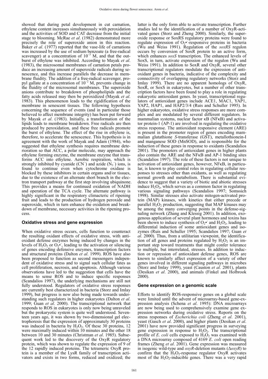
Hossain et al. (2006) studied the flower senescence in gladiolus in terms of antioxidant enzymes (superoxide dis-mutase, SOD; APX; glutathione reductase, GR) activities and membrane integrity. Membrane integrity was studied by measuring lipid peroxidation (TBARS) content and membrane stability index (MSI) percentage and concluded that an increase in endogenous H2O2 level during senes-cence may be the result of a programmed down-regulation of APX enzyme activity, which seems to be the prerequisite factor for initiating senescence process in gladiolus tepal (Fig. 4).
Ezhilmati et al. (2007) studied the effect of 5-sulfosali-cylic acid (5-SSA) on the vase life of cut flowers of Gladi-olus grandiflora. Spikes kept in vase solution containing 5-SSA exhibited lower respiration rates, lipid peroxidation and LOX activity, and higher membrane stability, soluble protein concentration, and activity of SOD and CAT. Re-sults suggest that 5-SSA increases vase life by increasing the ROS scavenging activity of the gladiolus cut flowers.
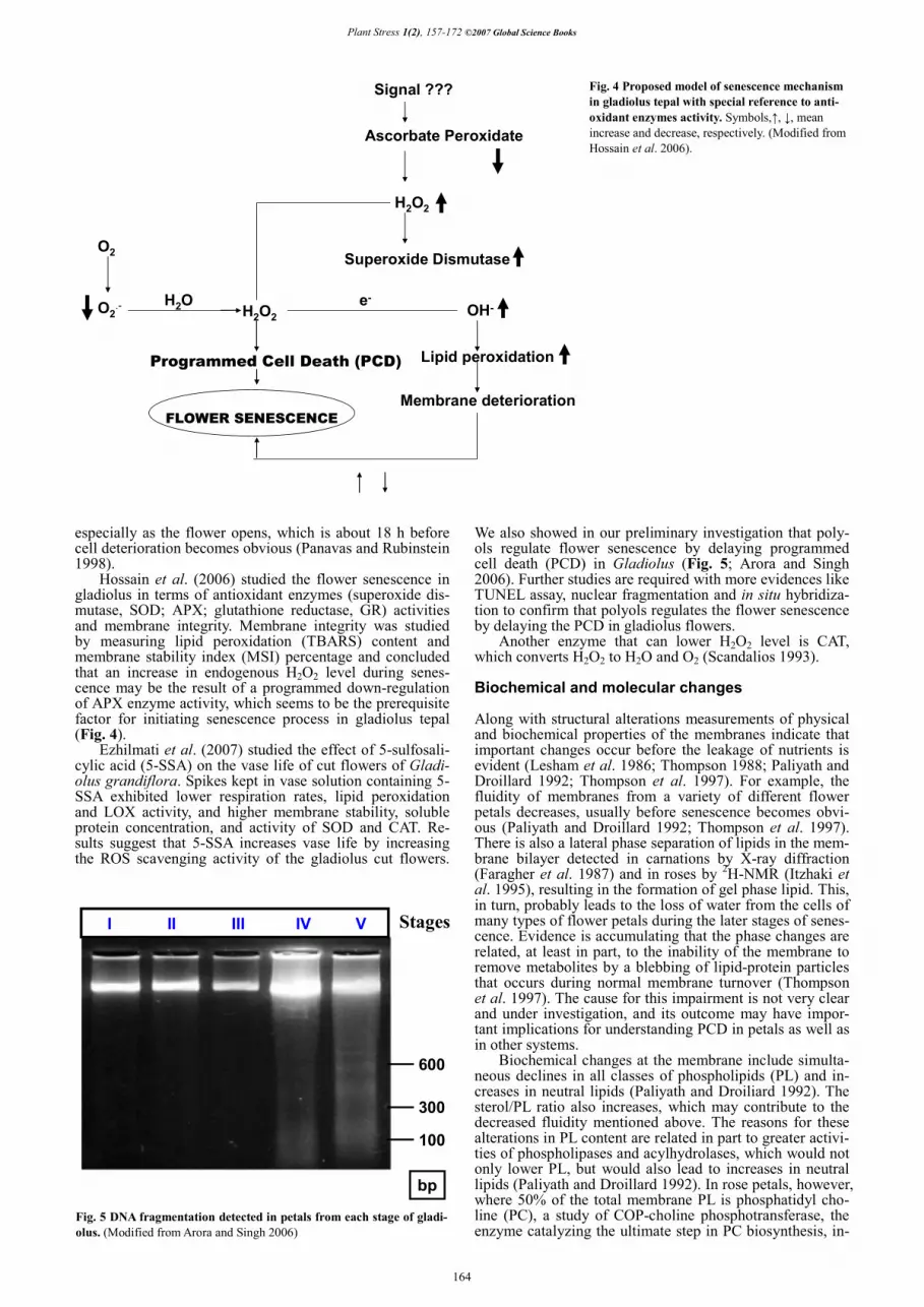
We also showed in our preliminary investigation that poly-ols regulate flower senescence by delaying programmed cell death (PCD) in Gladiolus (Fig. 5; Arora and Singh 2006). Further studies are required with more evidences like TUNEL assay, nuclear fragmentation and in situ hybridiza-tion to confirm that polyols regulates the flower senescence by delaying the PCD in gladiolus flowers.
Another enzyme that can lower H2O2 level is CAT, which converts H2O2 to H2O and O2 (Scandalios 1993). Biochemical and molecular changes Along with structural alterations measurements of physical and biochemical properties of the membranes indicate that important changes occur before the leakage of nutrients is evident (Lesham et al. 1986; Thompson 1988; Paliyath and Droillard 1992; Thompson et al. 1997). For example, the fluidity of membranes from a variety of different flower petals decreases, usually before senescence becomes obvi-ous (Paliyath and Droillard 1992; Thompson et al. 1997). There is also a lateral phase separation of lipids in the mem-brane bilayer detected in carnations by X-ray diffraction (Faragher et al. 1987) and in roses by 2H-NMR (Itzhaki et al. 1995), resulting in the formation of gel phase lipid. This, in turn, probably leads to the loss of water from the cells of many types of flower petals during the later stages of senes-cence. Evidence is accumulating that the phase changes are related, at least in part, to the inability of the membrane to remove metabolites by a blebbing of lipid-protein particles that occurs during normal membrane turnover (Thompson et al. 1997). The cause for this impairment is not very clear and under investigation, and its outcome may have impor-tant implications for understanding PCD in petals as well as in other systems.
Biochemical changes at the membrane include simulta-neous declines in all classes of phospholipids (PL) and in-creases in neutral lipids (Paliyath and Droiliard 1992). The sterol/PL ratio also increases, which may contribute to the decreased fluidity mentioned above. The reasons for these alterations in PL content are related in part to greater activi-ties of phospholipases and acylhydrolases, which would not only lower PL, but would also lead to increases in neutral lipids (Paliyath and Droillard 1992). In rose petals, however, where 50% of the total membrane PL is phosphatidyl cho-line (PC), a study of COP-choline phosphotransferase, the enzyme catalyzing the ultimate step in PC biosynthesis, in-
Signal ???
Ascorbate Peroxidate
H2O2
Superoxide Dismutase
H2O2
Programmed Cell Death (PCD)
OH-
Lipid peroxidation
Membrane deterioration
O2
O2.- H2O e-
FLOWER SENESCENCE
Fig. 4 Proposed model of senescence mechanism in gladiolus tepal with special reference to anti-oxidant enzymes activity. Symbols,�, �, mean increase and decrease, respectively. (Modified from Hossain et al. 2006).
100
300
600
I II III IV V
bp
Stages
Fig. 5 DNA fragmentation detected in petals from each stage of gladi-olus. (Modified from Arora and Singh 2006)
164

Oxidative stress during flower senescence. Arora et al.
dicates that the amount of enzyme and its Vmax decrease during senescence (Itzhaki et al. 1998). In daylily, too, the data suggest that phospholipid synthesis is blocked early in senescence (Bieleski and Reid 1992). An mRNA coding for a carboxyphosphoenolpyruvate/phosphoenolpyruvate mu-tase is up-regulated in carnation petals during senescence; the enzyme could be involved in membrane turnover (Wang et al. 1993). Thus, both a down-regulation of enzymes res-ponsible for PL synthesis and up-regulation of hydrolytic enzymes may cause the membrane breakdown that is an im-portant component of PCD.
Another senescence-associated event that leads to a loss of membrane permeability is the oxidation of existing mem-brane components. For example, lipid peroxidation as mea-sured by TBARS increases during senescence in carnation (Sylvestri et al. 1989; Bartoli et al. 1995), daylily (Panavas and Rubinstein 1998) and gladiolus petals (Ezhilmati et al. 2007). Peroxidation may occur in part by the action of lipo-xygenase, which oxidizes fatty acids liberated from mem-branes (Siedow 1991). LOX activity increases before senes-cence becomes obvious in carnation petals (Sylvestre et al. 1989), and in daylily, the specific activity of LOX increased before the flowers even open, which is about the same time that increases of TBARS occur (Panavas and Rubinstein 1998).
In orchid species, Phalaenopsis hybrid cv. ‘Herbert Hager’, however, even though linoleic and linolenic acids, two substrates of LOX, promoted senescence, no increases in LOX specific activity were detected over time, and inhib-itors of LOX were without effect (Porat et al. 1995). So, even though the peroxidation of lipids that may result from LOX activity precedes loss of membrane integrity, the acti-vity of lipid oxidases other than LOX may be limiting fac-tors for membrane degradation in petals of these species of orchid.
Two cDNA clones from daylily have been sequenced whose gene products may play a role in oxidizing mem-brane lipids and whose message levels are up-regulated prior to or during senescence (Panavas et al. 1999). One clone, detected mainly in petals, shows the greatest simila-rity to an in-chain fatty acid hydroxylase bound to cyto-chrome P450, The translated product of this mRNA may modify fatty acids, leading to their degradation (Cabelio-Hurtado et al. 1998). The other clone is highly similar to an allene oxide synthase, which converts fatty acid hydroper-oxides to allene epoxides, and eventually results in mole-cules that may have signaling capabilities (Song and Brash 1991). The increase in peroxidized lipids already described makes it likely that substrates would be available for this enzyme. No data are available as to whether these messages are translated in day lily petals in proportion to their abun-dance.
Another enzyme that can lower H2O2 levels is CAT, which converts H2O2 to H2O and O2 (Scandalios 1993). Overexpression of the gene for this enzyme protects leaves against ROS (Zelitch et al. 1991) and CAT-deficient plants are more sensitive to a variety of stresses (Willekins et al. 1997). In carnation, CAT activity increases during flower aging and then remains unchanged (Bartoli et al. 1995), but in daylily, the specific activity of the only form of CAT detected on activity gels decreases steadily from about 6 h before flower opening. When senescence is induced prema-turely, CAT activity decreases earlier (Panavas and Rubin-stein 1998).
SOD may also protect against buildup of ROS, but because the product of SOD is H2O2, an effective scaven-ging system must be present if cell damage is to be avoided. SOD decreases in activity in carnation petals (Sylvestre et al. 1989) and increases in daylily (Panavas and Rubinstein 1998), although in both cases the changes occur rather late in the progression of senescence. Since both CAT and APX decrease when daylily flowers open, the H2O2 build-up observed in these petals may in part be a result of increased SOD activity. Thus, in daylily, at past, SOD may be hasten-ing cell death.
Peroxidase activity uses H2O2 as a substrate for several reactions and its specific activity increases in both carnation (Bartoli et al. 1995) and daylily (Panavas and Rubinstein 1998) during senescence. The transcript for an enzyme that detoxifies peroxidized lipids, GST (Meyer et al. 1991), and the corresponding enzyme activity (Sylvestri 1989) is also up-regulated in carnation petals. These data suggest, how-ever, that POX and GST activities are responding to condi-tions of oxidative stress that result from H2O2 accumula-tions, and are thus not directly responsible for cell death.
Because lipid metabolism and ROS are so important to PCD, one must consider a role for peroxisomes. These or-ganelles are the site of lipid breakdown and can produce O2
– and H2O2, especially if protective enzymes are absent. Fur-thermore, as these organelles are converted to glyoxysomes, a rapid oxidation of lipids would ensue. There is an increase in numbers of peroxisomes during senescence of carnation petals (del Rio et al. 1996). A cDNA up-regulated during orchid petal senescence has been cloned that appears to code for a peroxisomal acyl-CoA oxidase (Do and Huang 1997). This particular enzyme may not be a cause of PCD, but it could serve to oxidize the fatty acids resulting from lipid breakdown at the membranes.
Natural antioxidants, such as ascorbate, glutathione and �-tocopherol, also are present in flower petals. All of these substances decline in carnation petals, although �-tocophe-rol decreases only after visual symptoms of senescence appear (Bartoli et al. 1997). In day lily, ascorbate drops by 50% even before the flowers open (Panavas and Rubinstein 1998). Inhibiting senescence of carnation petals with an ethylene synthesis inhibitor maintained higher levels of �-tocopherol and glutathione, and both antioxidants decreased when senescence was accelerated by ethylene or paraquat (Bartoli et al. 1996). Thus, with some exceptions (Bartoli et al. 1997), �-tocopherol levels are correlated with changes occurring later in senescence, but in both carnation and daylily, the decrease in ascorbate precedes many of the parameters associated with senescence. However, it is not possible to determine the involvement of antioxidants or the protective enzymes mentioned above in PCD until more in-formation becomes available about their localization in the cell and the degree of increase or decrease in activity that is needed to affect a particular cellular process.
Membrane proteins may also play a role in petal senes-cence. Membrane protein and the content of thiol groups decline during aging of carnation petals (Borochov and Woodson 1989). Furthermore, in older compared to non-senescent petals, there is a large decrease in vanadate-sensi-tive ATPase activity, presumably the plasma membrane H+ pump. But smaller decreases also occur in activities of cyto-chrome c oxidase, cytochrome c reductase and the nitrate-sensitive ATPase, which is likely the tonoplast H+ pump (Beja-Tal and Borochov 1994). Taken as a whole, the data indicate that the membrane proteins mediating transport and redox reactions decrease in activity during aging. The ques-tion remains, however, if these decreases in activity are cau-sally related to PCD or are just a result of previously trig-gered degradative processes. Genetic modification to improve postharvest performance To date, use of gene transfer technology to delay flower senescence has highlighted the need for tightly regulated transgene expression to avoid affecting other non-target developmental processes, particularly in the modification of plant hormone levels (e.g., poor rooting and lower disease resistance in ethylene-insensitive plants (Clark et al. 1999; Shaw et al. 2002). Thus, the need for tissue-specific promo-ters is paramount for exploiting this avenue of crop deve-lopment in commercially important cultivars. Alternatively, modifying the expression of metabolic genes may produce satisfactory postharvest improvements without the need to alter hormone biosynthesis or perception, which may have pleiotrophic effects. Pollen sterility in flowers innately
165

Plant Stress 1(2), 157-172 ©2007 Global Science Books
lowers pollen-induced senescence signals and may make currently unsuitable flowers suitable for cut flower cropping without needing anti-ethylene treatments. The use of tradi-tional breeding to select for genetic improvement of vase life may progress more rapidly as genetic markers for ‘long life’ are identified and as gene transfer technologies provide a way to improve the postharvest characteristics of crops with low genetic diversity. This theme is dealt with in grea-ter detail in a comprehensive series of reviews (Teixeira da Silva 2006a). PROTEOLYSIS AND FLOWER SENESCENCE Proteolysis is an indispensable process in all living orga-nisms. A continual turnover of proteins removes function-ally impaired proteins (due to biosynthetic errors, improper folding, thermal denaturation, oxidative damage), which if left unchecked may restrict metabolic activities and jeopar-dize a cell’s integrity. Proteases also recycle essential amino acids, and are important in the recovery of valuable nutri-ents. Proteases regulate metabolic pathways and develop-mental programs by affecting the rapid turnover of rate-lim-iting enzymes, and key regulatory proteins (Clarke 2005). Proteolytic cleavage is thought to play a significant role in the senescence of flowers because expression of protease genes is one of the earliest senescence-related gene changes to be identified (Eason et al. 2002). In plants, protein degra-dation linked to different developmental stages, such as ger-mination, differentiation and morphogenesis, senescence, and PCD, has been reported (Huffaker 1990; Vierstra 1996; Beers et al. 2000; Arora 2007a). On the other hand, there are an increasing number of references that report in certain circumstances, proteolysis is also associated to oxidative stress promoted by ROS (Solomon et al. 1999). At cellular level, proteolytic processes commonly take place as a con-sequence of a regulated turnover of most cell components, including organelle biogenesis and autophagy (Klionski and Emr 2000). During organelle biogenesis, translocation of preproteins to their respective target compartments, i.e. mitochondria, chloroplasts, endoplasmic reticulum, etc., of-ten requires the cleavage of a signal peptide. Once the poly-peptide crosses the membrane, it is folded to form the final mature protein. In this event, chaperones are present on both sides of the membrane, thus facilitating the whole pro-cessing of proteins. This mechanism, which has been well studied in mitochondria and chloroplasts, is less understood in peroxisomes, where proteolysis is necessary for the imp-ort of some proteins like thiolase, acyl-CoA oxidase, and malate dehydrogenase (Gietl 1996; Hayashi et al. 1998). Abnormal/misfolded and mistargeted polypeptides occur-ring by mistakes during the translation are also proteolytic-ally degraded. These errors might be a consequence of mu-tations not only in those polypeptides but also in the set of enzymes/proteins involved in their synthesis from trans-cription to the final processing in their target organelles (Adam 1996; Vierstra 1996).
Protein degradation in plants is a complex process in-volving a multitude of proteolytic pathways that can be carried out in different cell compartments. The presence of proteolytic activity has been reported in several cell loci, such as vacuoles, chloroplasts, cell wall, microsomes, mito-chondria, cytosol, and the Golgi apparatus (Distefano et al. 1997; Buchanan and Gruissen 2000).
Among the functions assigned to proteolysis are: (a) the removal of abnormal/misfolded, modified, and mistargeted proteins; (b) the supply of amino acids needed to make new proteins; (c) contribution to the maturation of zymogens and peptide hormones by limited cleavages; (d) the control of metabolism and homeostasis by reducing the abundance of key enzymes and regulatory proteins; and (e) the clea-vage of targeting signals from proteins prior to their final integration into organelles (Vierstra 1996). These molecular mechanisms form a part of more sophisticated global pro-cesses related to plant growth and development. Thus, the role of proteases in some developmental stages, such as ger-
mination, morphogenesis and cell biogenesis, senescence, and PCD, is essential.
Most proteases act either on the interior of peptide chains (endopeptidases, EP) or on their termini (exopepti-dases). Exopeptidases have been differentiated according to their substrate specificity as aminopeptidases (AP), which are able to cleave peptides at the N-terminus, and carboxy-peptidases (CP), which degrade peptides at the C-terminus (Dalling 1986; Huffaker 1990).
EP is classified according to their catalytic mechanism, which implies specificity in the enzyme active sites. It has been suggested that the term EP should be used synony-mously with proteinase. In plants, four classes of endopepti-dases have been described by Huffaker (1990): serine pro-teinases (EC 3.4.21), cysteine-proteinases (EC 3.4.22), as-partic-proteinases (EC 3.4.23), and metallo-proteinases (EC 3.4.24).
Several senescence-induced cDNAs have been se-quenced whose identities suggest close homology to cloned proteinase genes. For example, a cysteine proteinase was cloned from carnation by amplifying a specific cDNA by PCR (Jones et al. 1995). RNA gel blot analyses indicate that the transcript increases after the pollination-induced burst of ethylene production that leads to senescence. Fur-thermore, appearance of the transcript is stimulated by adding ethylene to pre-senescent petals, and the ethylene action inhibitor NBD prevents the appearance of the trans-cript (Jones et al. 1995).
Differential screening of a cDNA library yields two cDNAs from daylily petals whose derived amino acid se-quences show a strong homology to cysteine proteinases (Valpuesta et al. 1995; Guerrero et al. 1998). They appear to have an ER retention signal and an ERFNIN sequence that is consistent with activation by cleavage (Guerrero et al. 1998). Northern blot analysis indicates that message level have two peaks, one at 12 h and one at 19 h after flower opening. These transcripts were also found in leaves, but the mRNAs decreased at senescence (Guerrero et al. 1998).
Certain cysteine proteinases, collectively called cas-pases, are enzymes leading to apoptosis in animal cells, re-gardless of the initiating factor or the nature of the signal transduction pathway (Cryns and Yuan 1998). Caspase-like activity is detected in tobacco leaves and caspase-specific inhibitors prevent the hypersensitive response (del Pozo and Lam 1998). A cDNA for a putative aspartic proteinase with several family members has been cloned from daylily petals (Panavas et al. 1999). The steady state level of the message as determined by the RNase protection assay increases steadily from flower opening and the message is present only at much smaller levels in roots and leaves; in the latter, there is no correlation with senescence. Perhaps aspartic proteinases, whose message levels also increase in senes-cing leaves (Buchanan-Wollaston 1997; Griffiths et al. 1997), are involved in enzyme precursor processing as they are in some other plant systems (Mutlu and Gal 1999). However, it remains to be shown if aspartic proteinase activity increases during petal senescence. Protein degradation and oxidative stress in plants In addition to the involvement of proteases in the biological processes described, protein degradation also occurs under conditions that induce oxidative stress. In fact, a number of reports have shown that cells exhibit increased rates of proteolysis following exposure to oxidative stress-inducing agents (Pacifici et al. 1989; Grune et al. 1997). The wor-king hypothesis for these reports implies that intracellular proteins are oxidatively modified by free radicals and/or related oxidants, and these modified proteins are selectively recognized and preferentially degraded by intracellular pro-teolytic enzymes (Grune et al. 1997). This model has been proved to be valid in all eukaryotic organisms, including plants (Solomon et al. 1999).
The protein modification promoted by oxidative stress is characterized by the production of carbonyl groups in the
166

Oxidative stress during flower senescence. Arora et al.
molecule (Levine et al. 1994; Reinheckel et al. 1998). Basal levels of carbonyl groups are detected in plants, as a result of their generation as byproducts of normal physiological processes. However, increases in carbonyl contents have been observed in maize seedlings after chilling-induced oxi-dative stress (Prasad 1996; Kingston-Smith and Foyer 2000), in senescing nodules from pea and bean (Matamoros et al. 1999), in isolated chloroplasts exposed to oxygen ra-dical-generating systems (Stieger and Feller 1997), in thyla-koid proteins from water-stressed leaves of wheat (Tam-bussi et al. 2000), and in leaves of pea plants grown under toxic Cd concentrations (Romero-Puertas et al. 2002). In the case of Cd, it was found that this metal induced oxida-tive stress in pea plants (Sandalio et al. 2001), and some peptides, such as Rubisco, glutathione reductase, manga-nese superoxide dismutase, and CAT, were specifically oxi-dized by treatment with Cd. It was also found that Cd en-hanced the proteolytic activity, and by using specific anti-bodies, it was demonstrated that the oxidized proteins were more efficiently digested by proteases (Romero-Puertas et al. 2002). Lascano and colleagues (1998) also reported that exposing chloroplastic glutathione reductase to an ·OH generating system brought about the breakdown of the enzyme by a sulfhydryl- and metal-containing protease. Works by other workers have associated the expression of different proteases and/or their proteolytic activity to condi-tions that usually induce oxidative stress, i.e. anoxia (Sub-baiah et al. 2000), drought and temperature stress (Stroeher and Maclagan 1997), and the pathogen-promoting hyper-sensitive response (Yano et al. 1999). However, the specific combination oxidative stress–proteolysis was not directly addressed.
Different evidence obtained substantiated the idea that PCD and senescence (Quirino et al. 2000) are the physiolo-gical archetypes where controlled proteolysis is linked to the oxidative stress generated by ROS. PCD is one of the biological processes most thoroughly studied nowadays. It is generally accepted that ROS trigger PCD and antioxi-dants inhibit mechanisms leading to apoptosis (Jabs 1999; Solomon et al. 1999). During apoptosis, protein degradation also takes place in a modulated way (Beers et al. 2000; Fukuda 1997). In recent years, several proteases (Cys-EPs and Ser-EPs) have been shown to be involved in plant PCD (Arora and Ezura 2003; Arora and Singh 2004).
Flower senescence has been postulated to be a genetic-ally regulated process, controlled by internal and external signals, in which proteases play a key role (Buchanan-Wol-laston 1997). Moreover, the ability of cells to switch from one developmental state to another or to adapt to new envi-ronmental conditions often requires the rapid dismantle-ment of existing regulatory networks through proteolysis (Vierstra 1996).
At the subcellular level, most of the information avail-able on the combined action of oxidative stress plus proteo-lysis has been obtained from studies on chloroplasts. Thus, Ishida and colleagues (Ishida et al. 1998) reported that, in wheat chloroplasts, the large subunit (LSU) of Rubisco is broken down by ROS into 37- and 16-kDa polypeptides. Similar results were also found in pea plants (Roulin and Feller 1998). In this plant species, the degradation of chlo-roplastic phosphoglycolate phosphatase, glutamine synthe-tase, and other enzymes took place as well (Stieger and Feller 1997). These studies also revealed that the degrada-tion of the stroma proteins was light dependent, although some light-independent degradation may also occur (Roulin and Feller 1998). The fact that proteins that become non-functional due to interactions with oxygen species are fur-ther degraded by proteolysis is particularly significant in the thylakoid membranes. In these membranes, enzymes ope-rate in a highly oxidizing environment and are susceptible to structure and function impairments (Lindahl et al. 2000). Within the thylakoid membrane, photosystem II (PSII) is the most susceptible component to oxidative damage. The degradation of the PSII D1 protein has been found to be carried out by the metallo-peptidase FtSH in a reaction that
requires GTP (Lindahl et al. 2000). Plant peroxisomes and proteases In plants, the presence of proteolytic activity has been re-ported in several cell compartments, such as vacuoles, chlo-roplasts, the cell wall, microsomes, mitochondria, cytosol, and the Golgi apparatus (Buchanan et al. 1997; Distefano et al. 1997). However, the demonstration of the presence of proteolytic activity in plant peroxisomes is recent, and the information in this field is still scarce. Peroxisomes are sub-cellular respiratory organelles, containing, as basic enzyma-tic constituents, CAT and H2O2-producing flavin oxidases (del Río et al. 2002). These organelles have an essentially oxidative type of metabolism, and it has become increa-singly clear that they carry out vital functions in plant cells and play an important role in the generation of signal mole-cules (del Río et al. 2002).
Several metabolic processes support the idea that some proteolytic activity must exist in peroxisomes. The conver-sion of glyoxysomes into leaf peroxisomes during the ger-mination of seeds, as well as the opposite process conver-ting leaf peroxisomes into glyoxysomes, implies the degra-dation of the organelle’s pre-existing proteins (Nishimura et al. 1996). During the translocation of some polypeptides into the peroxisome, a proteolytic cleavage of the N-termi-nus takes place (Olsen and Harada 1995; Mullen et al. 2001). This partial proteolysis has been observed in in vitro import assays with some peroxisomal proteins, such as thio-lase, MDH, and acyl-CoA oxidase, whose molecular mass inside the organelle is lower than that of the translation pro-duct (Gietl 1996; Hayashi et al. 1998). The cleaved peptidic sequence corresponds to the peroxisomal targeting signal located at the N-terminus (PTS2), and the process is accom-plished in a way similar to that reported for mitochondrial and chloroplastic proteins (Bar-Peled et al. 1996). On the other hand, it has been reported that plant peroxisomes con-tain the xanthine oxidoreductase system (del Río et al. 2002). Xanthine oxidoreductase is an FAD-, molybdenum-, iron-, and sulfur-containing hydroxylase that have been found as two interconvertible forms (D and O). The conver-sion of form D (xanthine dehydrogenase, XDH) into form O (xanthine oxidase, XOD) is carried out by either rever-sible or irreversible pathways. In the irreversible step, XDH is converted into XOD by a proteolytic cleavage (Nishino 1994; Distefano et al. 1999). In peroxisomes from senes-cent pea leaves, a much higher increase of XOD activity is observed compared to XDH activity (Pastori and del Río 1997). This suggests that the conversion of XDH into XOD seems to occur inside peroxisomes, as has been observed in mitochondria (Saksela et al. 1999).
The presence of EP activity in peroxisomes has been investigated in pea leaves. In assays with peroxisomes from young leaves in the presence of azocasein, a specific sub-strate for endoproteases, a low EP activity of approximately 167 nKat mg–1 proteins was measured. By contrast, an up to six-fold higher endoproteolytic activity was observed in se-nescent leaves (Distefano et al. 1997). Through analysis by SDS–PAGE in gelatin-containing gels, three EP isozymes were detected in peroxisomes from young leaves, which were designated EP2, EP4, and EP5. However, four ad-ditional isoenzymes (EP1, EP3, EP6, and EP7) were detec-ted in senescent plants (Distefano et al. 1997). The endo-peptidases were characterized by using different class-spe-cific inhibitors. All proteases were neutral, and the serine-proteinases (EP1, EP3, and EP4) represented about 70% of the total EP activity of peroxisomes from senescent leaves. Besides, these serine-proteinases showed a notable thermal stability, not being inhibited by incubation at 50°C (Diste-fano et al. 1997). Role of peroxisomal proteases in oxidative stress situations The higher proteolytic activity found in peroxisomes from
167

Plant Stress 1(2), 157-172 ©2007 Global Science Books
senescent leaves suggested a role for peroxisomal EPs at that developmental stage. Senescence in plants is associated to several metabolic events similar to those that occur at the subcellular level during oxidative stress situations. In parti-cular, plant peroxisomes were postulated to play an acti-vated oxygen-mediated function during natural senescence (Pastori and del Río 1997). In order to know how peroxi-somal endopeptidases participate in the processes occurring during senescence, the degradation of peroxisomal and non-peroxisomal proteins by the endoproteases of purified per-oxisomes from senescent pea leaves was investigated (Distefano et al. 1999). It was found that most peroxisomal proteins were endoproteolytically degraded, and this was specifically demonstrated in the case of CAT, glycolate oxi-dase, and glucose-6-phosphate dehydrogenase. Peroxisomal proteases were also able to cleave nonperoxisomal proteins like Rubisco and urease (Distefano et al. 1999). These results indicate that proteases from plant peroxisomes might play an important role in the turnover of peroxisomal pro-teins during senescence. This means that the organelle’s own proteolytic machinery could participate in the mecha-nism of conversion of leaf peroxisomes into glyoxysomes, which is known to occur in senescent tissues (Distefano et al. 1999). Also, peroxisomal EPs might be involved in the turnover of proteins located in other cell compartments during advanced stages of senescence, when deterioration of membranes and leakage of the organelles’ soluble frac-tion takes place. In experiments carried out in vitro, it was demonstrated that bacterial XDH was converted into XOD in the presence of peroxisomes from senescent leaves (Di-stefano et al. 1999). These data suggest that peroxisomal EPs could also participate in regulatory mechanisms that do not necessarily imply full degradation of proteins.
On the other hand, it was reported that 50 �M CdCl2 produced an enhancement of the hydrogen peroxide con-centration in peroxisomes and also an increase in the acti-vity of some antioxidative enzymes of the ascorbate–gluta-thione cycle and NADP-dependent dehydrogenases located in these cell organelles (Romero-Puertas et al. 1999). Fur-thermore, in western blotting assays using an antibody against dinitrophenylhydrazone (DNPH), which recognizes oxidatively modified proteins, a higher level of oxidized proteins was found in peroxisomes from Cd-treated plants (Romero-Puertas et al. 2002). Under the same experimental conditions, Cd induced senescence symptoms in leaf per-oxisomes, with an enhancement of the endogenous proteo-lytic activity and the activity of the glyoxylate cycle en-zymes malate synthase and isocitrate lyase (McCarthy et al. 2001). Taken together, these results indicate that the peroxi-somal proteases could participate in the metabolic changes produced by Cd, by degrading the oxidized proteins, and possibly also in the transition of leaf peroxisomes into gly-oxysomes, which is known to occur during senescence (Nishimura et al. 1996; del Río et al. 1998). Ubiquitin-proteosome pathway The ubiquitin-proteosome pathway is a major cytoplasmic proteolytic system for the degradation of short-lived and abnormal proteins (Vierstra 1993). In plants, changes in the level of ubiquitinated proteins have been associated with cell death during the development of vascular tissue (Bach-meier et al. 1990; Stephenson et al. 1996; Woffenden et al. 1998) as well as during senescence of leaves (Garbarino et al. 1995; Pinedo et al. 1996). In immunoblots of daylily petals, protein-ubiquitin conjugates were detected, some of which increased and some decreased during senescence. RNA gel blots revealed several ubiquitin transcripts that in-creased, decreased or stayed unchanged during flower opening and senescence (Courtney et al. 1994). Analysis in daylily of enzymatic components of the ubiquitin proteo-some pathway (ubiquitin-activating enzyme, ubiquitin-con-jugating enzymes, a subunit of the multiubiquitin binding protein) show detectable but steadily decreasing quantities during senescence when expressed on a per petal basis (Ste-
phenson and Rubinstein 1998). Because proteinase activity increases during senescence, but banding patterns on PAGE gels remain sharp and well defined, the ubiquitin-proteo-some system may be efficiently breaking down the cleaved proteins to amino acids rather than initiating senescence. However, inhibitors of proteosome activity prevent cell death leading to tracheid formation (Woffenden et al. 1998) and delay the ion leakage associated with senescence in daylily petals (Stephenson and Rubinstein 1998). This im-plies that there may be a causal role for the ubiquitin-pro-teosome system in petal senescence. SUMMARY, CONCLUSIONS AND FUTURE RESEARCH Petal senescence provides a facile system for studying PCD of plant organs. In many cases isolated petals behave simi-larly to those on the plant, and the process occurs relatively quickly, synchronously and uniformly throughout the tissue. The most extensively investigated flowers; however, are generally not the species that lend themselves readily to genetic manipulation, mutagenesis, or transformation. But it is likely that more tractable systems such as Arabidopsis, tomato or tobacco can also be employed to further analyze the role of particular pathways in petal PCD.
The fact that carbohydrates delays petal senescence demonstrates that the synthesis of particular suicide proteins orchestrates the cell death program. It is necessary, however, to show that these proteins and their products are not just correlated with PCD, but actually play a crucial role. For example, particular protein may serve only to mobilize materials during transport out during cell, or a substance may be produced as a result of a perceived stress associated with senescence, and there is little direct effect on those processes most closely related to cell death. After a causal role is established, one must then determine if the substance acts alone or in concert with others.
With this in mind, several enzymatic reactions have been identified in petals that may directly affect senescence. These reactions result in the oxidation of membrane com-ponents and the breakdown of proteins and nucleic acids. Genes for several enzymes that mediate these events have been cloned and the mRNAs may be up-regulated prior to, or during, senescence. If the higher steady-state message level is a result of transcriptional regulation, techniques exist to examine the regulatory regions of these genes for consensus sequences that may indicate the factor(s) that enhances transcription. Other regulatory mechanisms that may be investigated include senescence-associated trans-cription factors, and a role for protease inhibitor genes (Solomon et al. 1999). Characterization of genes that are down-regulated during senescence should also yield useful information. For example, expression of the DAD gene, which appears to protect cells against cell death, decreases in pea and gladiolus petals during senescence (Orzaez and Granell 1997; Yamada et al. 2004).
The endogenous and environmental triggers that initiate suicide genes are also being identified. Senescence in many flowers is controlled in part by ethylene, but whereas other factors may also play a role, one must determine for the particular flower if only the production of ethylene is a limiting factor for senescence or if the sensitivity to ethy-lene is also important (Fig. 6; Arora 2005). An enhanced evolution of ethylene may be related to higher levels of the mRNAs for the enzymes responsible for synthesis of this hormone. The up-regulation of the ethylene-binding protein also implicates this process in an increased sensitivity to ethylene. The crucial question remains, however, as to how these messages are regulated (Arora 2005).
There are also flowers in several families that are insen-sitive to ethylene. Even though ABA may be the senes-cence-inducing factor, many species should be tested before a generalization can be made. Furthermore, it is important to show that depletion of ABA delays senescence in these flowers. There may also be ABA-responsive sequences on
168

Oxidative stress during flower senescence. Arora et al.
the regulatory regions of the genes that directly or indirectly cause PCD.
Finally, pollination, injury, dehydration and light are environmental triggers of petal senescence that may act first on other parts of the flower. The transportable signal, which may be ACC or ethylene, leads to elevation of ethylene evolution (or ABA production in the case of water stress) in the petals. Different mechanisms may exist for different species.
An area that is ripe for exploration is the identification of components of the signal transduction chain resulting in PCD due to oxidative stress. Various factors and processes known to be important for developmental processes, inclu-ding PCD, have been implicated in petal senescence, such as G-protein activation, inositol phosphate metabolism, al-terations in cytosolic free calcium, and phosphorylation/de-phosphorylation reactions. However, events such as calcium level changes need to be investigated on the cell level in living material, and genes involved in producing signal transduction components such as kinases and phosphatases must be identified and their regulation studied. It is impor-tant to keep in mind that different species may have dif-ferent signal transduction systems.
The impact of characterizing PCD in petals allows an understanding of a process crucial to the survival of plants and other plant organs as well as providing economic bene-fits. The ease of working with flowers and the exciting ad-vancements made in techniques at both the molecular and cell levels should lead to the identification of particular mu-tants, the cloning of more suicide genes and the production of transformed plants, all of which make it very likely that great strides will be made in the next few years.
At present, one of the most studied aspects of peroxi-somes biology is the biogenesis of these organelles. Peroxi-somal proteins (PTS) are synthesized in free polyribosomes, with a targeting signal that drives proteins to their organelle. Much is known about the different targeting signals of PTS
and the required cofactors for the translocation inside the organelle (Olsen and Harada 1995; Mullen et al. 2001). Two main PTSs have been reported so far. PTS1 resides in the C-terminus with a tripeptide and PTS2 is in the N-ter-minus and is usually cleaved upon the translocation of poly-peptides through the membrane. This process possibly im-plies the involvement of an aminopeptidase. The occurrence of a Leu-AP in peroxisomes suggests that it might be in-volved in the processing of imported precursor polypeptides in peroxisomes. The purification of the peroxisomal Leu-AP, and its further characterization and assays with its specific substrates, will help in clarifying the mechanisms of pro-cessing of polypeptides till their mature form during the translocation to the peroxisome. On the other hand, the study of the combined mechanism of oxidation plus proteo-lytic degradation of proteins under certain conditions is gaining relevance in plant biology. Senescence and metal toxicity by cadmium is representative of an oxidative stress taking place together with an enhanced proteolytic activity (Sandalio et al. 2001; Romero-Puertas et al. 2002). In these experimental conditions, a parallel behavior was observed in the metabolism of leaf peroxisomes. The molecular and biochemical characterization of peroxisomal proteases will allow the comparison of their properties with those avail-able in databanks. It will provide information on their cata-lytic mechanism and their putative substrates. Studies on the role of peroxisomal proteases in oxidative stress situa-tions can be accomplished by the incubation of enzymes with different cell components and specifically in petal tis-sues. This is particularly relevant in the light of recent re-sults in Arabidopsis plants that demonstrated that oxidative stress induced PEX genes involved in the peroxisome bio-genesis (Lopez-Huertas et al. 2000). The control of pro-teolysis will be also of interest in terms of increasing plant productivity. This control could be established by using specific inhibitors. Plants are known to contain important levels of protease inhibitors that regulate enzyme activity.
ETHYLENE
CTR1
EIN2
EIN3
AtEBPsERFs
TARGET TARGETGENES
GENES
Nuclear Membrane
ETHYLENE
CTR1
EIN2
EIN3AtEBPSERFs
EIN3
AtEBPSERFs
TARGETGENES
TARGETGENES
RESPONSENO RESPONSE
Fig. 6 Schematic diagram of the pathway of ethylene signal transduction in Arabidopsis. In the receptor inhibition model, receptor isoforms signal to CTR1 in the absence of ethylene. Ethylene inhibits this signalling and CTR1 is inactivated. Then, EIN2, EIN3 and EILs are activated and ethylene responses occur. Mutants that have lost ethylene-binding ability would continue to activate CTR1 in the presence of ethylene and thus suppress response pathways downstream from CTR1. (Modified from Arora 2005).
169

Plant Stress 1(2), 157-172 ©2007 Global Science Books
This strategy of manipulating senescence could overlap other strategies such as that of breeding or genetic engineer-ing to improve crop yields by keeping leaves photosynthe-tically active for longer.
Today, much attention is being devoted to the investiga-tion of PCD in plants, a physiological stage characterized by a notable oxidative stress and the activation of several proteolytic pathways. At the subcellular level, there is still little information on the events that occur in PCD processes in plants. Taking into account that senescence is a type of PCD, the cellular and molecular study of the possible in-volvement of peroxisomal proteases in apoptosis during flower senescence could provide deeper insights into this important physiological process. ACKNOWLEDGEMENTS The first author (AA) acknowledges funding from Department of Biotechnology (DBT), Ministry of Science and Technology, New Delhi, India. REFERENCES Adam Z (1996) Protein stability and degradation in chloroplasts. Plant Molecu-
lar Biology 32, 773-783 Alscher RG, Donahue JL, Cramer CL (1997) Reactive oxygen species and
antioxidants: relationships in green cells. Physiologia Plantarum 100, 224-233
Arora A (2005) Ethylene receptors and molecular mechanism of ethylene sensi-tivity in plants. Current Science 89, 1348-1361
Arora A (2007a) Biochemistry of flower senescence. In: Paliyath G, Murr DP, Handa AK, Lurie S (Eds) Postharvest Biology and Technology of Fruits, Vegetables and Flowers, Blackwell Publishing, Iowa, USA, in press
Arora A (2007b) Programmed cell death during plant senescence. In: Paliyath G, Murr DP, Handa AK, Lurie S (Eds) Postharvest Biology and Technology of Fruits, Vegetables and Flowers, Blackwell Publishing, Iowa, USA, in press
Arora A, Ezura H (2003) Isolation, molecular characterization and regulation of cysteine protease gene in Gladiolus grandiflora. Molecular and Cellular Proteomics 2, 746
Arora A, Singh VP (2004) Cysteine protease gene expression and proteolytic activity during floral development and senescence in ethylene-insensitive gladiolus. Journal of Plant Biochemistry and Biotechnology 13, 123-126
Arora A, Singh VP (2006) Polyols regulate the flower senescence by delaying programmed cell death in Gladiolus. Journal of Plant Biochemistry and Bio-technology 15, 139-142
Arora A, Sairam RK, Srivastava GC (2002) Oxidative stress and antioxida-tive system in plants. Current Science 82, 1227-1238
Arora A, Watanabe S, Ma B, Takada K, Ezura H (2006) A novel ethylene re-ceptor homolog gene isolated from ethylene-insensitive flowers of gladiolus (Gladiolus grandiflora Hort.). Biochemical and Biophysical Research Com-munications 351, 739-44
Asada K (1992) Ascorbate peroxidase-a hydrogen peroxide-scavenging enzyme in plants. Physiologia Plantarum 85, 235-241
Asada K (1994) Production and action of active oxygen species in photosyn-thetic tissues. In: Foyer CH, Mullineaux PM (Eds) Causes of Photooxidative Stress in Plants and Amelioration of Defence Systems, CRC Press, Florida, USA, pp 77-104
Asada K, Badger MR (1984) Photoreduction of 18O2 and H218O2 with conco-
mitant evolution of 16O2 in intact spinach chloroplasts: evidence for scaven-ging of hydrogen peroxide by peroxidase. Plant Cell Physiology 25, 1169-1179
Asada K, Kiso K, Yoshikawa K (1984) Univalent reduction of molecular oxy-gen by spinach chloroplasts on illumination. The Journal of Biological Che-mistry 249, 2175-2181
Asada K, Takahashi M, Kyle DJ, Osmond CB, Arntzen CHJ (Eds) (1987) Topics in Photosynthesis, Photoinhibition (Vol 9), Elsevier Science Publi-shers B.V., Amsterdam, pp 227-288
Ashman TL, Schoen DJ (1994) How long should flowers live? Nature 371, 788-791
Ashman TL, Schoen DJ (1997) The cost of floral longevity in Clarkia temblo-riensis: an experimental investigation. Evolution and Ecology 11, 289-300
Bachmair A, Becker F, Masterson RV, Schell J (1990) Perturbation of the ubiquitin system causes leaf curling, vascular issue alterations and necrotic lesion in a higher plant. The EMBO Journal 9, 4543-4549
Badger MR (1985) Photosynthetic oxygen exchange. Annual Review of Plant Physiology 36, 27-53
Baker JE, Wang CY, Lieberman M, Hardenburg R (1977) Delay of senes-cence in carnations by a rhizobitoxine analog and sodium benzoate. Horticul-tural Sciences 12, 38-39
Bartoli CG, Simontacchi M, Guiamet J, Montaldi E, Puntarulo S (1995)
Antioxidant enzymes and lipid peroxidation during aging of Chrysanthemum morifolium RAM petals. Plant Science 104, 161-168
Bar-Peled M, Bassham DC, Raikhel NV (1996) Transport of proteins in euka-ryotic cells: more questions ahead. Plant Molecular Biology 32, 223-249
Bartoli CG, Simontacchi M, Montaldi E, Puntarulo S (1996) Oxidative stress, antioxidant capacity and ethylene production during ageing of cut carnation of cut carnation (Dianthus caryophyllus). Journal of Experimental Botany 47, 595-601
Bartoli CG, Simontacchi M, Montaldi E, Puntarulo S (1997) Oxidants and antioxidant during ageing of chrysanthemum petals. Plant Science 129, 157-165
Beers EP, Woffenden BJ, Zhao C (2000) Plant proteolytic enzymes: possible roles during programmed cell death. Plant Molecular Biology 44, 399-415
Beja-Tal S, Borochov A (1994) Age-related changes in bio-chemical and phy-sical properties of carnation petal plasma membranes. Journal of Plant Phy-siology 143, 195-199
Bennoun P (1994) Chlororespiration revisited: mitochondrial-plastid interac-tions in Chlamydomonas. Biochimica et Biophysica Acta 1186, 59-66
Bieleski RL, Reid MS (1992) Physiological changes ac- accompanying senes-cence in the ephemeral daylily flower. Plant Physiology 98, 1042-1049
Borochov A, Woodson WR (1989) Physiology and biochemistry of flower petal senescence. Horticulture Reviews 11, 15-43
Borochov A, Spiegelstein H, Philosoph-Hadas S (1997) Ethylene and flower petal senescence: interrelationship with membrane lipid catabolism. Physio-logia Plantarum 100, 606-612
Buchanan BB, Gruissen W, Jones RL (2000) Biochemistry and Molecular Biology of Plants, American Society of Plant Physiologists Rockville, MD, 1367 pp
Buchanan-Wollaston V (1997) The molecular biology of leaf senescence. Journal of Experimental Botany 48, 181-199
Buzgo M, Soltis PS, Kim S, Soltis DE (2005) The making of the flower. The Biologist 52, 149-154
Cabelio-Hurtado F, Batard Y, Salaun JP, Durst F, Pinot F,d Werck-Reich-art D (1998) Cloning, expression in yeast and functional characterization of CYP81B1, a plant cytochrome P450 that catalyzes in-chain hydroxylation of fatty acids. The Journal of Biological Chemistry 273, 7260-7267
Cadenas E (1989) Biochemistry of oxygen toxicity. Annual Review of Bioche-mistry 58, 79-110
Carlos G, Bartoli M, Montaldi SE, Puntarulo S (1996) Oxidative stress, anti-oxidant capacity and ethylene production during ageing of cut carnation (Di-anthus caryophyllus) petals. Journal of Experimental Botany 47, 595-601
Causton H, Bing R, Koh S, Harbison C, Kanin E, Jennings E, Lee T, True H, Lander E, Young R (2001) Remodeling of yeast genome expression in response to environmental changes. Molecular Biology of the Cell 12, 323-337
Christman MF, Morgan RW, Jacobson FS, Ames BN (1985) Positive control of a regulon for defenses against oxidative stress and some heat-shock pro-teins in Salmonella typhimurium. Cell 41, 753-762
Clarke AK (2005) Plant proteases – an appetite for destruction. Physiologia Plantarum 123, 359-361
Clark DG, Gubrium EK, Barrett JE, Nell TA, Klee HJ (1999) Root forma-tion in ethylene-insensitive plants. Plant Physiology 121, 53-59
Courtney S, Rider CC, Stead AD (1994) Changes in protein ubiquitination and the expression of ubiquitin-encoding transcripts in daylily petals during floral development and senescence. Physiologia Plantarum 91, 196-204
Cryns V, Yuan J (1998) Proteases to die for. Genes and Development 12, 1551-1570
Dalling MJ (1986) Plant Proteolytic Enzymes (Vols I, II), CRC Press, Boca Raton, FL
Dalton TP, Shertzer HG, Puga A (1999) Regulation of gene expression by reactive oxygen. Annul Review of Pharmacology and Toxicology 39, 67-101
del Pozo O, Lam E (1998) Caspases and programmed cell death in the hyper-sensitive response of plants to pathogens. Current Biology 8, 1129-1132
del Río LA, Sandalio LM, Palma JM, Corpas FJ, Lopez-Huertas E, Ro-mero-Puertas MC, McCarthy I (2002) Peroxisomes, reactive oxygen meta-bolism, and stress-related enzyme activities. In: Baker A, Graham I (Eds) Plant Peroxisomes, Kluwer Academic Publishers, Dordrecht, The Nether-lands, pp 221-258
del Río LA, Palma JM, Sandaiio LM, Corpas FJ, Pastcri GM, Bueno P, Lopez-Huertas E (1996) Peroxisomes as a source of superoxide and hydro-gen peroxide in stressed plants. Biochemical Society Transactions 24, 434-438
Desikan R, Mackerness A, Hancock JT, Neill SJ (2001) Regulation of the Arabidopsis transcriptome by oxidative stress. Plant Physiology 127, 159-172
Desikan R, Neill SJ, Hancock JT (2000) Hydrogen peroxide-induced gene ex-pression in Arabidopsis thaliana. Free Radical Biology and Medicine 28, 773-778
Distefano S, Palma JM, Gomez M, del Río LA (1997) Characterization of endoproteases from plant peroxisomes. Biochemical Journal 327, 399-405
Do YY, Huang PL (1997) Gene structure of PAC01, a petal senescence-related gene from Phalaenopsis encoding peroxisomal acyl-CoA oxidase homolog. Biochemistry and Molecular Biology International 41, 609-617
170

Oxidative stress during flower senescence. Arora et al.
Eason JR, Ryan DJ, Pinkey TT, Donoghue EMO (2002) Programmed cell death during flower senescence: isolation and characterization of cysteine proteinases from Sandersonia aurantiaca. Functional Plant Biology 29, 1055-1064
Ezhilmathi K, Singh VP, Arora A, Sairam RK (2007) Effect of 5-sulfosali-cylic acid on antioxidant activity in relation to vase life of Gladiolus cut flowers. Plant Growth Regulation 51, 99-108
Faragher JD, Wachtel E, Mayak S (1987) Changes in the physical stale of membrane lipids during senescence of rose petals. Plant Physiology 83, 1037-1042
Finkel T, Holbrook NJ (2000) Oxidants, oxidative stress and the biology of aging. Nature 408, 239-247
Foyer CH, Halliwell B (1976) The presence of glutathione and glutathione re-ductase in chloroplasts: a proposed role in ascorbic acid metabolism. Planta 133, 21-25
Foyer CH, Descourvieres P, Kunert KJ (1994) Protection against oxygen radicals: important defence mechanism studied in transgenic plants. Plant, Cell and Environment 17, 507-523
Frankel EN (1985) Chemistry of free radical and singlet oxidation of lipids. Progress in Lipid Research 23, 197-221
Fridovich I (1995) Superoxide radical and superoxide dismutases. Annual Re-view of Biochemistry 64, 97-112
Fujisawa Y, Kato H, Iwasaki Y (2001) Structure and function of heterotri-meric G proteins in plants. Plant and Cell Physiology 42, 789-794
Fukuda H (1997) Programmed cell death during vascular system formation. Cell Death Differentiation 4, 684-688
Garbarino JE, Oosumi T, Belknap WR (1995) Isolation of a polyubiquitin promoter and its expression in transgenic potato plants. Plant Physiology 109, 1371-1378
Gasch A, Spellman P, Kao C, Harel O, Eisen M, Storz G, Botstein D, Brown P (2000) Genomic expression programs in the response of yeast cells to envi-ronmental changes. Molecular Biology Cell 11, 4241-4257
Gietl C (1996) Protein targeting and import into plant peroxisomes. Physiologia Plantarum 97, 599-608
Gressel J, Galun E (1994) Genetic controls of photooxidative tolerance. Foyer CH, Mullineaux PM (Eds) In: Causes of Photooxidative Stress and Amelio-ration of Defence Systems in Plants, CRC Press, Boca Raton, FL, pp 237-274
Griffiths CM, Hosken SE, Oliver D, Chojecki J, Thomas H (1997) Sequen-cing expression pattern and RFLP mapping of a senescence-enhanced cDNA from Zea mays with high homology to oryzain � and aleurain. Plant Molecu-lar Biology 34, 81-82
Grune T, Reinheckek T, Davies KJA (1997) Degradation of oxidized proteins in mammalian cells. FASEB Journal 11, 526-534
Guan LM, Zhao J, Scandalios JG (2000) Cis-elements and trans-factors that regulate expression of the maize Cat1 antioxidant gene in response to ABA and osmotic stress: H2O2 is the likely intermediary signaling molecule for the response. The Plant Journal 22, 87-95
Guerrero C, de la Calle M, Reid MS, Valpuesta V (1998) Analysis of the ex-pression of two thiolprotease genes from daylily (Hemerocallis spp.) during flower senescence. Plant Molecular Biology 36, 565-571
Halliwell B, Gutteridge JMC (1989) Free Radicals in Biology and Medicine, Clarendon Press, Oxford, UK, pp 450-499
Harbinson J, Hedley CL (1993) Changes in P-700 oxidation during the early stages of the induction of photosynthesis. Plant Physiology 103, 649-660
Harbinson J, Genty B, Foyer CH (1990) Relationship between photosynthetic electron transport and stromal enzyme activity in pea. Plant Physiology 94, 545-553
Hayashi H, de Bellis L, Yamaguchi K, Kato A, Hayashi M, Nishimura M (1998) Molecular characterization of a glyoxysomal long chain acyl-CoA oxidase that is synthesized as a precursor of higher molecular mass in pump-kin. The Journal of Biological Chemistry 273, 8301-8307
Heber U, Egneus H, Hanch U, Jensen M, Koster S (1978) Regulation of pho-tosynthetic electron transport and photophosphorylation in intact chloroplasts and leaves of Spinacia oleracea L. Planta 143, 41-49
Hossain Z, Mandal AKA, Datta SK, Biswas AK (2006) Decline in ascorbate peroxidase activity – A prerequisite factor for tepal senescence in Gladiolus. Journal of Plant Physiology 163, 186-194
Huffaker RC (1990) Proteolytic activity during senescence of plants. New Phy-tologist 116, 199-231
Ishida H, Shimizu S, Makino A, Mae T (1998) Light-dependent fragmentation of the large subunit of ribulose-1,5-bisphosphate carboxylase/oxygenase in chloroplasts isolated from wheat leaves. Planta 204, 305-309
Itzhaki H, Davis JH, Borochov A, Mayak S, Pauls KP (1995) Deuterium magnetic resonance studies of senescence related changes in the physical properties of rose petal membrane lipids. Plant Physiology 108, 1029-1033
Itzhaki H, Mayak S, Borochov A (1998) Phosphatidylchpline turnover during senescence of petals. Plant Physiology and Biochemistry 36, 457-462
Jabs T (1999) Reactive oxygen intermediates as mediators of programmed cell death in plants and animals. Biochemical Pharmacology 57, 231-245
Johannes E, Brosnan JM, Sanders D (2005) Calcium channels and signal transduction in plant cells. BioEssays 13, 331-336
Jones ML, Larsen PB, Woodson WR (1995) Ethylene regulated expression of a carnation cysteine proteinase during flower petal senescence. Plant Molecu-
lar Biology 28, 505-512 Kingston-Smith AH, Foyer CH (2000) Bundle sheath proteins are more sensi-
tive to oxidative damage than those of the mesophyll in maize leaves exposed to paraquat or low temperatures. Journal of Experimental Botany 51, 123-130
Klionski DJ, Emr SD (2000) Autophagy as a regulated pathway of cellular degradation. Science 290, 1717-1721
Knox JP, Dodge AD (1985) Singlet oxygen and plants. Phytochemistry 24, 889-896
Kovtun Y, Chiu WL, Tena G, Sheen J (2000) Functional analysis of oxidative stress-activated mitogen-activated protein kinases cascade in plants. Proceed-ings of the National Academy of Sciences USA 97, 2940-2945
Kumar S, Sinha SK (1992) Alternative respiration and heat production in ripening banana fruits (Musa paradisiaca). Journal of Experimental Botany 43, 1639-1642
Kumar S, Patil BC, Sinha SK (1990) Cyanide resistant respiration is involved in temperature rise in ripening mangoes. Biochemical and Biophysical Re-search Communications 168, 818-822
Kwon SY, Jeong YJ, Lee HS, Kim JS, Cho KY, Allen RD, Kwak SS (2002) Enhanced tolerances of transgenic tobacco plants expressing both superoxide dismutase and ascorbate peroxidase in chloroplasts against methyl viologen-mediated oxidative stress. Plant Cell and Environment 25, 873-882
Lascano HR, Gomez LD, Casano LM, Trippi VS (1998) Changes in gluta-thione reductase activity and protein content in wheat leaves and chloroplasts exposed to photooxidative stress. Plant Physiology and Biochemistry 36, 321-329
Legge RI, Thompson JE (1983) Involvement of hydroperoxides and an ACC-derived free radical in the formation of ethylene. Phytochemistry 22, 2161-2166
Leshem Y, Halevy AH, Frenkel C (1986) Process and control of plant senes-cence. Developmental Crop Science 8, 142-161
Levine RL, Williams JA, Stadtman ER, Shacter E (1994) Carbonyl assays for determination of oxidatively modified proteins. Methods Enzymology 233, 346-363
Lindahl M, Spetea C, Hundal T, Oppenheim AB, Adam Z, Andersson B (2000) The thylakoid FtsH protease plays a role in the light-induced turnover of the photosystem II D1 protein. Plant Cell 12, 419-431
Lopez-Huertas E, Charlton WL, Johnson B, Graham I, Baker A (2000) Stress induces peroxisome biogenesis genes. The EMBO Journal 19, 6770-6777
Lwe MWF, Takkahama U, Heber U (1993) Role of ascorbate in detoxifying ozone in the apoplast of spinach (Spinacia oleracea L.) leaves. Plant Physio-logy 101, 969-976
Matamoros MA, Baird LD, Escuredo PR, Dalton DA, Minchin FR, Iturbe-Ormaetxe I, Rubio MC, Moran JF, Gordon AJ, Becana M (1999) Stress-induced legume root nodule senescence. Physiological and structural altera-tions. Plant Physiology 121, 97-111
Mayak S, Adam Z (1984) Accelerated ethylene production by a microsomal membrane fraction from carnation petals in vitro. Plant Science Letters 33, 345-352
Mayak S, Legge RI, Thompson JE (1983) Superoxide radical production by microsomal membranes from senescing carnation flowers: an effect on mem-brane fluidity. Phytochemistry 22, 1375-1380
McCarthy I, Romero-Puertas MC, Palma JM, Sandalio LM, Corpas FJ, Gomez M, del Río LA (2001) Cadmium induces senescence symptoms in leaf peroxisomes of pea plants. Plant Cell and Environment 24, 1065-1073
McRae DG, Baker JE, Thompson JE (1982) Evidence for involvement of the superoxide radical in the conversion of 1-aminocyclopropane-1-carboxylic acid to ethylene by pea microsomal membranes. Plant Cell Physiology 23, 375-383
Mehlar AH (1951) Studies on reactions of illuminated chloroplasts. I. Mecha-nism of the reduction of oxygen and other Hill reagents. Archives of Bioche-mistry 33, 65-77
Meyer RC, Goldsbrough PB, Woodson WR (1991) An ethylene-responsive flower senescence-related gene from carnation encodes a protein homologous to glutathione S-transferases. Plant Molecular Biology 17, 277-281
Mittler R, Shulaev Y, Seskar M, Lam E (1996) Inhibition of programmed cell death in tobacco plants during pathogen induced hypersensitive response at low oxygen pressure. Plant Cell 8, 1991-2001
Mullen, RT, Flynn, CR, Trelease RN (2001) How are peroxisomes formed? The role of the endoplasmic reticulum and peroxins. Trends in Plant Science 6, 256-261
Mutlu A, Gal S (1999) Plant aspartic proteinases: enzymes on the way to a function. Physiologia Plantarum 105, 569-576
Nguyen AN, Lee A, Place W, Shinozaki K (2000) Multistep phosphorelay pro-teins transmit oxidative stress signals to the fission yeast stress-activated pro-tein kinase. Molecular Biology Cell 11, 1169-1181
Nishimura M, Hayashi M, Kato A, Yamaguchi K, Mano S (1996) Functional transformation of microbodies in higher plant cells. Cell Structure and Func-tion 21, 387-393
Nishino T (1994) The conversion of xanthine dehydrogenase to xanthine oxi-dase and the role of the enzyme in reperfusion injury. Journal of Biochemis-try 116, 1-6
171

Plant Stress 1(2), 157-172 ©2007 Global Science Books
Olsen L, Harada JJ (1995) Peroxisomes and their assembly in higher plants. Annual Review of Plant Physiology and Plant Molecular Biology 46, 123-146
Orzaez D, Granell A (1997) The plant homologue of the defender against apoptotic death gene is down-regulated during senescence of flower petals. FEBS Letters 404, 275-278
Pacifici RE, Salo DC, Davies KJA (1989) Macroxyproteinase (M.O.P.): a 670 kDa proteinase complex that degrades oxidatively denatured proteins in red blood cells. Free Radical Biology: Medicines 7, 521-536
Paliyath G, Droillard MJ (1992) The mechanisms of membrane deterioration and disassembly during senescence. Plant Physiology and Biochemistry 30, 789-812
Panavas T, Rubinstein B (1998) Oxidative events during programmed cell death of daylily (Hemerocallis hybrid) petals. Plant Science 133, 125-138
Panavas T, Pikula A, Reid PD, Rubinstein B, Walker EL (1999) Identifica-tion of senescence-associated genes from daylily petals. Plant Molecular Biology 40, 237-248
Pastori GM, del Río LA (1997) Natural senescence of pea leaves: an activated oxygen-mediated function for peroxisomes. Plant Physiology 113, 411-418
Pinedo ML, Goicoechea SM, Lamattina L, Conde RD (1996) Estimation of ubiquitin and ubiquitin mRNA content in dark senescing wheat leaves. Bio-logia Plantarum 33, 321-328
Porat R, Reiss N, Atzorn R, Halevy AH, Borochov A (1995) Examination of the possible involvement of lipoxygenase and jasmonates in pollination-in-duced senescence of Phalaenopsis and Dendrobium orchid flowers. Physiolo-gia Plantarum 94, 205-210
Prasad TK (1996) Mechanisms of chilling-induced oxidative stress injury and tolerance: changes in antioxidant systems, oxidation of proteins and lipids and protease activities. Plant Journal 10, 1017-1026
Quin J, Findlay VJ, Dawson K, Millar JB, Jones N, Morgan BA, Toone WM (2002) Distinct regulatory proteins control the graded transcriptional response to increasing H2O2 levels in fission yeast Schizosaccharomyces pombe. Molecular Biology of the Cell 13, 805-816
Quirino BF, Noh YS, Himelblau E, Amasino RM (2000) Molecular aspects of leaf senescence. Trends in Plant Science 5, 278-282
Reinheckel T, Noack H, Lorenz S, Wiswedel I, Augustin W (1998) Compari-son of protein oxidation and aldehyde formation during oxidative stress in isolated mitochondria. Free Radical Research 29, 297-305
Rich PR, Bonner WD (1978) The sites of superoxide anion generation in higher plant mitochondria. Archives in Biochemistry and Biophysics 188, 206-213
Romero-Puertas MC, McCarthy I, Sandalio LM, Palma JM, Corpas FJ, Gomez M, del Río LA (1999) Cadmium toxicity and oxidative metabolism of pea leaf peroxisomes. Free Radical Research 31, S25-S31
Romero-Puertas MC, Sandalio LM, Palma JM, Gomez M, del Rio LA (2002) Cadmium causes the oxidative modification of proteins in pea plants. Plant Cell and Environment 25, 677-686
Roulin S, Feller U (1998) Dithiothreitol triggers photooxidative stress and frag-mentation of the large subunit of ribulose-1,5-bisphosphate carboxylase/oxy-genase in intact pea chloroplasts. Plant Physiology and Biochemistry 36, 849-856
Ruis H, Schuller C (1995) Stress signaling in yeast. BioEssays 17, 959-965 Saksela M, Lapatto R, Raivio KO (1999) Irreversible conversion of xanthine
dehydrogenase into xanthine oxidase by a mitochondrial protease. FEBS Let-ters 443, 117-120
Sandalio LM, Dalurzo HC, Gomez M, Romero-Puertas MC, del Río LA (2001) Cadmium-induced changes in the growth and oxidative metabolism of pea plants. Journal of Experimental Botany 52, 2115-2126
Scandalios JG (1993) Oxygen stress and superoxide dismutases. Plant Physio-logy 101, 7-12
Scandalios JG (1997) Oxidative Stress and the Molecular Biology of Antioxi-dant Defenses, Cold Spring Harbor Laboratory Press, New York, pp 527-568
Schena M, Shalon D, Davis R, Brown P (1995) Quantitative monitoring of gene expression patterns with complementary DNA microarray. Science 270, 467-470
Shaw J-F, Chen H-H, Tsai M-F, Kuo C-I, Huang L-C (2002) Extended flower longevity of Petunia hybrida plants transformed with boers, a mutated ERS gene of Brassica oleracea. Molecular Breeding 9, 211-216
Shykoff JA, Bucheli E, Kaltz O (1996) Flower lifespan and disease risk. Na-ture 379, 779-780
Siedow JN (1991) Plant lipoxygenase: structure and function. Annual Review of Plant Physiology and Plant Molecular Biology 42, 145-188
Solomon M, Belenghi B, Delledonne M, Menachem E, Levine A (1999) The involvement of cysteine proteases and protease inhibitor genes in the regula-tion of programmed cell death in plants. Plant Cell 11, 431-443
Solomos T (1977) Cyanide-resistant respiration in higher plants. Annual Review Plant Physiology 28, 279
Somssich IE, Halbrock K (1998) Pathogen defence in plants-a paradigm of biological complexity. Trends in Plant Science 3, 86-90
Song WC, Brash AR (1991) Purification of an allene oxide synthase and iden-
tification of the enzyme as a cytochrome P-450. Science 253, 781-784 Stead AD (1992) Pollination-induced flower senescence: A review. Plant
Growth Regulation 11, 13-20 Stephenson P, Rubinstein B (1998) Characterization of proteolytic activity
during senescence in daylilies. Physiologia Plantarum 104, 463-473 Stephenson P, Collins BA, Reid PD, Rubinstein B (1996) Localization of ubi-
quitin to differentiating vascular tissues. American Journal of Botany 83, 140-147
Stieger PA, Feller U (1997) Degradation of stromal proteins in pea (Pisum sati-vum L.) chloroplasts under oxidising conditions. Journal of Plant Physiology 151, 556-562
Storz G, Imlay JA (1999) Oxidative stress. Current Opinion in Microbiology 2,188-194
Storz G, Zheng M (2000) Oxidative stress. In: Storz G, Henge-Aronis R (Eds) Bacterial Stress Responses, ASM Press, Washington DC, pp 47-59
Stroeher VL, Maclagan JL, Good AG (1997) Molecular cloning of a Brassica napus cysteine protease gene inducible by drought and low temperature stress. Physiologia Plantarum 101, 389-397
Subbaiah CC, Kollipara KP, Sachs MM (2000) A Ca2+-dependent cysteine protease is associated with anoxia-induced root tip death in maize. Journal of Experimental Botany 51, 721-730
Sylvestre I, Droillard MJ, Bureau JM, Paulin A (1989) Effects of the ethy-lene rise on the peroxidation of membrane lipids during the senescence of cut carnations. Plant Physiology and Biochemistry 27, 407-413
Sylvestre I, Droillard MJ, Bureau JM, Paulin A (1989) Effects of the ethy-lene rise on the peroxidation of membrane lipids during the senescence of cut carnations. Plant Physiology and Biochemistry 27, 407-413
Tambussi EA, Bartoli CG, Beltrano J, Guiamet JJ,d Araus JL (2000) Oxi-dative damage to thylakoid proteins in water-stressed leaves of wheat (Triti-cum aestivum). Physiologia Plantarum 108, 398-404
Teixeira da Silva JA (Ed) (2006) Floriculture, Ornamental and Plant Biotech-nology: Advances and Topical Issues (Vols I-IV), Global Science Books, Isle-worth, UK, 2481 pp
Thompson JE (1988) The molecular basis for membrane deterioration during senescence. In: Nooden LD, Leopold AC (Eds) Senescence and Aging in Plants, Academic Press, New York, pp 51-83
Thompson JE, Froese CD, Hong Y, Hudak KA, Smith MD (1997) Mem-brane deterioration during senescence. Canadian Journal of Botany 75, 867-879
Valpuesta V, Lange NE, Cuerrero C, Reid MS (1995) Upregulation of a cys-teine protease accompanies the ethylene-insensitive senescence of daylily (Hemerocallis) flowers. Plant Molecular Biology 28, 575-582
Vierstra RD (1993) Protein degradation in plants. Annual Review of Plant Phy-siology and Plant Molecular Biology 44, 385-410
Vierstra RD (1996) Proteolysis in plants: mechanisms and functions. Plant Molecular Biology 32, 275-302
Wang H, Brandt AS, Woodson WR (1993) A flower senescence-related mRNA from carnation encodes a novel protein related to enzymes involved in phosphonate biosynthesis. Plant Molecular Biology 22, 719-724
Willekens H, Chamnongpol S, Davey M, Schrauder M, Langebartels C, van Montagu M, Inzé D, van Camp W (1997) Catalase is a sink for H2O2 and is indispensable for stress defence in C3 plants. The EMBO Journal 16, 4806-4816
Woffenden BJ, Freeman TB, Beers EP (1998) Proteasome inhibitors prevent tracheary element differentiation in Zinnia mesophyll cell cultures. Plant Physiology 118, 419-430
Wu J, Weiss B (1991) Two divergently transcribed genes, soxR and soxS, con-trol a superoxide response regulon of Escherichia coli. Journal of Bacterio-logy 173, 2864-2871
Wu J, Nelnanis S, Heber U (1991) Photorespiration is more effective than the Mehler reaction in protecting the photosynthetic apparatus against photoinhi-bition. Botanica Acta 104, 283-291
Yano A, Suzuki K, Shinshi H (1999) A signalling pathway, independent of the oxidative burst that leads to hypersensitive cell death in cultured tobacco cells includes a serine protease. Plant Journal 18, 105-109
Yanru Z, Pandey M, Prasad NK, Srivastava GC (1996) Hydrolysing en-zymes and respiration during ripening of tomato (Lycopersicon esculentum) fruits. Current Science 70, 1017-1018
Zelitch I (1990) Oxygen resistant photosynthesis in tobacco plants selected for oxygen resistant growth. In: Zelitch I (Ed) Perspectives in Biochemical and Genetic Regulation of Photosynthesis, Liss Inc., New York, pp 239-252
Zelitch I, Havir EA, McGonigle B, McHale NA, Nelson T (1991) Leaf cata-lase mRNA and catalase-protein levels in a high-catalase tobacco mutant with O2-resistant photosynthesis. Plant Physiology 97, 1592-1595
Zhang S, Klessig D (2001) MAPK cascades in plant defense signaling. Trends in Plant Science 6, 520-527
Zheng M, Wang X, Templeton L, Smulski D, la Rossa R, Storz G (2001) DNA microarray-mediated transcriptional profiling of the Escherichia coli response to hydrogen peroxide. Journal of Bacteriology 183, 4562-4570
172


