Embed Size (px)
Citation preview
Cancer Cell
Article
p53-Mediated Senescence Impairsthe Apoptotic Response to Chemotherapyand Clinical Outcome in Breast CancerJames G. Jackson,1 Vinod Pant,1 Qin Li,1,5 Leslie L. Chang,1 Alfonso Quintas-Cardama,2 Daniel Garza,1 Omid Tavana,3
Peirong Yang,1 Taghi Manshouri,2 Yi Li,6 Adel K. El-Naggar,4 and Guillermina Lozano1,*1Department of Genetics2Department of Leukemia3Department of Immunology4Department of Pathology
The University of Texas MD Anderson Cancer Center, Houston, TX 77030, USA5The University of Texas Graduate School of Biomedical Sciences Program in Genes and Development, Houston, TX 77030, USA6Lester & Sue Smith Breast Center and Department of Molecular and Cell Biology, Baylor College of Medicine, Houston, TX 77030, USA
*Correspondence: [email protected]
DOI 10.1016/j.ccr.2012.04.027
SUMMARY
Studies on the role of TP53 mutation in breast cancer response to chemotherapy are conflicting. Here, weshow that, contrary to dogma, MMTV-Wnt1 mammary tumors with mutant p53 exhibited a superior clinicalresponse compared to tumors with wild-type p53. Doxorubicin-treated p53 mutant tumors failed to arrestproliferation, leading to abnormal mitoses and cell death, whereas p53 wild-type tumors arrested, avoidingmitotic catastrophe. Senescent tumor cells persisted, secreting senescence-associated cytokines exhibitingautocrine/paracrine activity and mitogenic potential. Wild-type p53 still mediated arrest and inhibited drugresponse even in the context of heterozygous p53 point mutations or absence of p21. Thus, we show thatwild-type p53 activity hinders chemotherapy response and demonstrate the need to reassess the paradigmfor p53 in cancer therapy.
INTRODUCTION
The tumor suppressor TP53 is mutated or inactivated in the
majority of cancers (Soussi and Lozano, 2005). p53 exerts its
effects by binding specific promoter sequences following
cellular stress and activating transcription of genes involved in
cell-cycle arrest, senescence, and apoptosis (Riley et al.,
2008). DNA damage, such as that induced by radiation or
chemotherapy drugs, is a potent activator of p53.
Classic studies using mouse models have demonstrated an
in vivo role for p53 in the induction of apoptosis following DNA
damage (Jackson et al., 2011). Thymocytes from p53 null mice
do not undergo apoptosis after radiation (Clarke et al., 1993;
Lowe et al., 1993b), and the embryonic neural tube in p53 null
Significance
Approximately one-third of human breast cancers harbormutatdigm that wild-type p53 mediates apoptosis resulting in a favobecause many reports conflict, including some suggesting thaHere, we show that wild-type p53 activity is paradoxically detp53 tumors, p53wild-type tumors can avoid aberrantmitoses bkines in senescent cells that can stimulate cell proliferation anorder to accurately predict clinical response of TP53-mutated
mice is similarly resistant (Lang et al., 2004; Liu et al., 2004).
p53 also contributes to response after exposure to DNA-
damaging drugs by inducing apoptosis in E1A/Ras-transformed
mouse embryo fibroblasts (Lowe et al., 1993a, 1994).
Interestingly, studies examining the paradigm that wild-type
p53 activity improves drug response are lacking in tumors arising
from epithelial tissues (Brown and Attardi, 2005). This deficiency
is exemplified in breast cancer. Some reports on the response of
breast cancer with or without TP53 mutation to chemotherapy
drugs have been inconclusive (Bonnefoi et al., 2011; Makris
et al., 1995; Mathieu et al., 1995), whereas others show that
wild-type TP53 activity is beneficial to response (Aas et al.,
1996; Berns et al., 2000; Kroger et al., 2006; Rahko et al.,
2003). Intriguingly, still other reports have shown that TP53
ions in the tumor suppressor gene TP53. The long-held para-rable chemotherapy response is less clear in breast cancert tumors harboring TP53mutations respond more favorably.rimental to chemotherapy response because, unlike mutanty undergoing arrest, which is followed by expression of cyto-d tumor relapse. Furthermore, our data demonstrate that intumors, the status of the second allele must be determined.
Cancer Cell 21, 793–806, June 12, 2012 ª2012 Elsevier Inc. 793
Cancer Cell
Wild-type p53 Activity Impairs Drug Response
mutant tumors respond better (Bertheau et al., 2002, 2007;
Mathieu et al., 1995). It is unclear why these reports reached
different conclusions, and importantly, the notion that wild-
type TP53 activity would hinder response, given its known
apoptotic and tumor suppressive functions, is controversial
and lacks a mechanistic explanation. Thus, it is desirable to
have a controlled setting to examine if wild-type p53 activity is
beneficial in breast cancer response, and if not, as some have
suggested (Bertheau et al., 2008), then why?
RESULTS
Superior Response of p53 Mutant Mammary Tumorsto Doxorubicin TreatmentIn order to address the role of the p53 response in breast cancer,
we bred MMTV-Wnt1 (Tsukamoto et al., 1988) mice into p53
wild-type as well as p53�/� (Jacks et al., 1994) and p53R172H
(Lang et al., 2004) (‘‘p53 mutant’’ herein) backgrounds.
p53R172H is a structurally defective mutant that cannot bind
DNA, and mice that are homozygous or heterozygous for this
mutation are identical to p53�/� or p53+/� mice, respectively,
in survival (Lang et al., 2004) and for results presented here.
After mammary tumors formed, mice were treated with doxoru-
bicin and monitored. We found that, despite heterogeneity of
responses (see Figure S1A available online), tumors from mice
in the wild-type p53 background generally underwent minimal
tumor regression, stabilized for several days, and quickly
relapsed (Figures 1A and 1E).
We next examined p53 mutant MMTV-Wnt1 mammary
tumors. In most human tumors, biallelic point mutations or
deletions of p53 are relatively rare. More typically, a p53 point
mutation is acquired in a single allele, and the other allele is
subsequently retained or lost. Thus, we treated MMTV-Wnt1
p53 heterozygous mutant mice (p53R172H/+ genotype) bearing
tumors, of which approximately 40% undergo loss of heterozy-
gosity (LOH) for the wild-type p53 allele (Donehower et al.,
1995). Surprisingly, we found that tumors in p53R172H/+ MMTV-
Wnt1 mice that lost the wild-type allele (rendering the tumor
p53R172H/0 and functionally null) showed greater tumor regres-
sion and longer time to relapse (Figures 1B, 1E, and S1B).
Also to our surprise, we found that as long as tumors from
p53R172H/+ mice retained the wild-type p53 allele, they exhibited
a response typical of p53wild-type tumors (Figures 1C and S1C),
despite the presence of a mutant allele with reported dominant-
negative activity (Lang et al., 2004; Willis et al., 2004). We also
observed dramatic tumor volume reductions in MMTV-Wnt1
mammary tumors from treated p53 homozygous mutant mice
(Figures 1D and S1D), although these mice were typically har-
vested within 1 week following the final doxorubicin treatment
(Figure 1D) due to gastrointestinal syndrome (Komarova et al.,
2004). Use of the p53 homozygous mutant mice was also limited
by the difficulty of acquiring females in the cohort (Sah et al.,
1995). In sum although tumors of all genotypes eventually
relapsed, treated tumors lacking all p53 activity, whether homo-
zygous null or heterozygous mutant with LOH, had significantly
greater decrease in tumor volume and significantly longer time
to relapse compared to tumors retaining p53 activity (Figure 1E).
To assess the acute biochemical response to treatment, we
examined p53 function in MMTV-Wnt1 tumors 24 hr after the
794 Cancer Cell 21, 793–806, June 12, 2012 ª2012 Elsevier Inc.
final doxorubicin dose. p53 target genes p21, Puma, and
Ccng1 were induced 4- to 6-fold compared to untreated tumors
(Figure 1F), demonstrating retention of wild-type p53 in MMTV-
Wnt1 tumors from p53+/+ mice, as others have shown (Done-
hower et al., 1995). Predictably, p53 homozygousmutant tumors
did not induce target genes (Figure 1F). Noxa levels, however,
were elevated in some tumors in a p53-independent fashion.
p53 heterozygous mutant MMTV-Wnt1 tumors that retained
the wild-type allele also induced p53 target genes, in contrast
to tumors with p53 LOH (Figure 1G).
To address the mechanism of response in p53 wild-type
versus mutant tumors, we examined growth arrest and
apoptosis in tumors 24 hr after doxorubicin treatment. Exam-
ining proliferation, we found that untreated tumors of all geno-
types were highly positive for Ki67, a marker of cells outside
G0 of the cell cycle. Following treatment, only p53 wild-type
MMTV-Wnt1 tumors had large areas that were Ki67 negative
or sparsely positive, demonstrating cell-cycle exit, whereas
p53 mutant tumors remained positive for Ki67 (Figure 2A).
Furthermore, tumors from parallel orthotopic transplants of
a p53 wild-type MMTV-Wnt1 tumor ceased incorporating
bromodeoxyuridine (BrdU) following treatment, whereas trans-
planted p53 mutant tumors continued to enter S phase (Fig-
ure 2B). This failure to arrest in the presence of DNA damage
resulted in aberrant mitoses, as evidenced by anaphase bridges,
in treated p53 mutant tumors, whereas p53 wild-type tumors
arrested and, thus, were not mitotic (Figures 2C–2E). The p53
mutant MMTV-Wnt1 tumors also showed a significant increase
in both cleaved caspase-3 and terminal deoxynucleotidyl trans-
ferase dUTP nick end labeling (TUNEL)-positive cells after doxo-
rubicin treatment (Figure 2F). p53wild-type tumors, however, did
not undergo apoptosis following treatment (Figure 2F). Taken
together, our data show that despite induction of apoptotic
genes such as Puma and Noxa, growth arrest, not apoptosis,
was acutely induced in p53 wild-type tumors following doxoru-
bicin treatment, and lack of arrest in mutant tumors resulted in
aberrant mitoses, cell death, and ultimately, a superior clinical
response.
Doxorubicin Induces a Senescent-like Phenotype in p53
Wild-type Mammary TumorsTo further address the mechanism responsible for the subop-
timal response in p53 wild-type tumors, we harvested p53
wild-type or heterozygous MMTV-Wnt1 tumors 5–7 days post-
treatment (Figures S2A–S2C) and examined them for markers
of senescence. We quantitatively determined that many markers
of senescence (p21, Dcr2, p16, p15, Dec1, and Pml) (Collado
et al., 2005; te Poele et al., 2002) were elevated in many stably
arrested MMTV-Wnt1 tumors that were p53 wild-type or hetero-
zygous with retention of the wild-type allele, when compared to
untreated tumors (Figures 3A, 3B, and 3D). Treated and har-
vested p53 mutant tumors (Figure S2C) exhibited heterogeneity
in expression levels of several senescence markers, although
only Pml was consistently elevated to a degree similar to that
observed in p53 wild-type tumors 5 days following treatment
(Figure 3C). Dcr2 was induced slightly following treatment in
p53R172H/0 tumors, possibly attributable to stromal cells within
the tumor that still have the p53 wild-type allele. We used ortho-
topic transplants of p53 wild-type and mutant MMTV-Wnt1
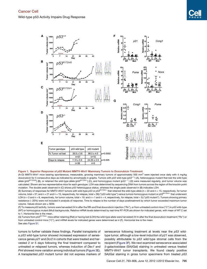
Figure 1. Superior Response of p53 Mutant MMTV-Wnt1 Mammary Tumors to Doxorubicin Treatment(A–D) MMTV-Wnt1 mice bearing spontaneous, measurable, growing mammary tumors of approximately 500 mm3 were injected once daily with 4 mg/kg
doxorubicin for 5 consecutive days as indicated by arrowheads in graphs. Tumors with p53 wild-type (p53+/+) (A), heterozygous mutant that lost the wild-type
allele (p53R172H/0) (B), or retained the wild-type allele (p53R172H/+) (C), and homozygous mutant (p53�/�) (D) were measured regularly, and tumor volume was
calculated. Shown are two representative mice for each genotype. LOH was determined by sequencing DNA from tumors across the region of the knockin point
mutation. The double peak observed in (C) shows p53 heterozygous status, whereas the single peak observed in (B) indicates LOH.
(E) Summary of responses for MMTV-Wnt1 tumors with wild-type p53 or p53R172H/+ that retained the wild-type allele (n = 22 and n = 15, respectively, for tumor
volume, total = 37; and n = 21 and n = 15, respectively, for relapse, total = 36) (‘‘p53 wild-type’’) versus tumors homozygous mutant or p53R172H/+ that underwent
LOH (n = 2 and n = 8, respectively, for tumor volume, total = 10; and n = 1 and n = 4, respectively, for relapse, total = 5) (‘‘p53 mutant’’). Tumors showing primary
resistance (�20%) were not included in analysis of response. Time to relapse is the number of days posttreatment by which tumor exceeded maximum tumor
volume. Values shown are ± SEM.
(F) Tomeasure p53 activity, tumors were harvested 24 hr after the fifth and final doxorubicin injection (‘‘Rx’’), or from untreated control mice (‘‘C’’) in p53wild-type
(WT) or homozygous mutant (Mut) backgrounds. Relative mRNA levels determined by real-time RT-PCR are shown for indicated genes, with mean of WT C set
to 1. Horizontal line is the mean.
(G) Tumors from p53R172H/+ mice either retaining (Ret) or having lost (LOH) the wild-type allele were harvested 24 hr after the final doxorubicin treatment (‘‘Rx’’) or
from untreated control mice (‘‘C’’), and mRNA levels for indicated genes were determined as in (F). Horizontal line is the mean.
See also Figure S1.
Cancer Cell
Wild-type p53 Activity Impairs Drug Response
tumors to further validate these findings. Parallel transplants of
a p53 wild-type tumor showed increased expression of senes-
cence genes p21 and Dcr2 in cohorts that were treated and har-
vested 2 or 5 days following the final treatment compared to
untreated or relapsed tumors, whereas induction of Dec1 and
Pml showedmore variation among individual tumors (Figure 3E).
A transplanted p53 mutant tumor did not express markers of
senescence following treatment at levels near the p53 wild-
type tumor, although a low-level induction of p21 was observed,
possibly attributable to p53 wild-type stromal cells from the
recipient (Figure 3F). We next examined senescence-associated
b-galactosidase (SAbGal) staining in untreated versus treated
MMTV-Wnt1 tumor transplants. We found clearly positive
SAbGal staining in gross tumor specimens from treated p53
Cancer Cell 21, 793–806, June 12, 2012 ª2012 Elsevier Inc. 795
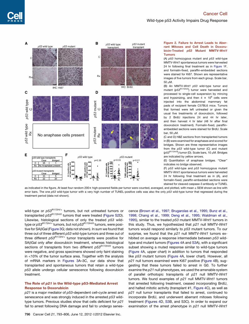
Figure 2. Failure to Arrest Leads to Aber-
rant Mitoses and Cell Death in Doxoru-
bicin-Treated p53 Mutant MMTV-Wnt1
Tumors
(A) p53 homozygous mutant and p53 wild-type
MMTV-Wnt1 spontaneous tumors were harvested
24 hr following final treatment as in Figure 1F,
and formalin-fixed, paraffin-embedded sections
were stained for Ki67. Shown are representative
images of five tumors from each group. Scale bar,
50 mM.
(B) An MMTV-Wnt1 p53 wild-type tumor and
mutant (p53R172H/0) tumor were harvested and
processed to single-cell suspension by mincing
and trypsinizing, and then 4 3 106 cells were
injected into the abdominal mammary fat
pads of recipient female C57BL6 mice. Tumors
that formed were left untreated or given the
usual five treatments of doxorubicin, followed
by 2 BrdU injections 24 and 44 hr later,
and then harvest 4 hr later (48 hr after final
doxorubicin treatment). Formalin-fixed, paraffin-
embedded sections were stained for BrdU. Scale
bar, 50 mM.
(C and D) H&E sections from transplanted tumors
in (B) were examined for anaphases and scored for
bridges. Shown are three representative images
from the p53 wild-type tumor (C) and mutant
(p53R172H/0) tumor (D). Scale bars, 10 mM. Bridges
are indicated by yellow arrows.
(E) Quantitation of anaphase bridges. ‘‘Clear’’
indicates no bridge observed.
(F) p53 wild-type and p53 homozygous mutant
MMTV-Wnt1 spontaneous tumors were harvested
24 hr following final treatment as in (A), and
formalin-fixed, paraffin-embedded sections were
stained for cleaved caspase-3 antibody or TUNEL
as indicated in the figure. At least four random 2003 high-powered fields per tumor were counted, averaged, and plotted, with mean ± SEM shown as line with
error bars. The one p53 wild-type tumor with a very high number of TUNEL-positive cells was also the only p53 wild-type tumor that regressed during the
treatment period (data not shown).
Cancer Cell
Wild-type p53 Activity Impairs Drug Response
wild-type or p53R172H/+ tumors, but not untreated tumors or
transplanted p53R172H/0 tumors that were treated (Figure S2D).
Likewise, histological sections of only the treated p53 wild-
type or p53R172H/+ tumors, but not p53R172H/0 tumors, were posi-
tive for SAbGal (Figure 3G; data not shown). In sumwe found that
three out of three different p53wild-type tumors and three out of
three different p53R172H/+ tumor transplants were positive for
SAbGal only after doxorubicin treatment, whereas histological
sections of transplants from two different p53R172H/0 tumors
were negative, and gross specimens showed only faint staining
in <10% of the tumor surface area. Together with the analysis
of mRNA markers in Figures 3A–3C, our data show that
transplanted and spontaneous tumors that retain a wild-type
p53 allele undergo cellular senescence following doxorubicin
treatment.
The Role of p21 in the Wild-type p53-Mediated ArrestResponse to Doxorubicinp21 is a major mediator of p53-dependent cell-cycle arrest and
senescence and was strongly induced in the arrested p53 wild-
type tumors. Previous studies show that cells deficient for p21
fail to arrest following DNA damage and fail to undergo senes-
796 Cancer Cell 21, 793–806, June 12, 2012 ª2012 Elsevier Inc.
cence (Brown et al., 1997; Brugarolas et al., 1995; Bunz et al.,
1998; Chang et al., 1999; Deng et al., 1995; Waldman et al.,
1995), similar to the treated p53 mutant MMTV-Wnt1 tumors in
this study. Thus, we hypothesized that p21 null MMTV-Wnt1
tumors would respond similarly to p53 mutant tumors. To our
surprise, we found that the p21 null MMTV-Wnt1 tumors ex-
hibited on average a response intermediate between p53 wild-
type and mutant tumors (Figures 4A and S3A), with a significant
subset showing a muted response similar to wild-type tumors
(Figure 4A, upper chart) in addition to tumors that responded
like p53 mutant tumors (Figure 4A, lower chart). However, all
p21 null tumors examined were Ki67 positive (Figure 4B), sug-
gesting that these tumors failed to arrest in G0. To further
examine the p21 null phenotypes, we used the amenable system
of parallel orthotopic transplants of p21 null MMTV-Wnt1
tumors. We found examples of p21 null MMTV-Wnt1 tumors
that arrested following treatment, ceased incorporating BrdU,
and halted mitotic activity (transplant #1, Figure 4C), as well as
p21 null tumor transplants that failed to arrest, continued to
incorporate BrdU, and underwent aberrant mitoses following
treatment (Figures 4D, S3B, and S3C). In order to expand our
examination of the arrest phenotype in p21 null MMTV-Wnt1
Cancer Cell
Wild-type p53 Activity Impairs Drug Response
tumors beyond these parallel transplants, we quantitated mitotic
activity in ten spontaneous p21 null MMTV-Wnt1 tumors 24 hr
following treatment to assess the fraction of these tumors that
undergo arrest following treatment, compared to p53 wild-type
and mutant tumors. We found that six of ten tumors were essen-
tially in mitotic arrest 24 hr following treatment, similar to five out
of five p53 wild-type-treated tumors (Figure 4E). However,
mitotic activity persisted in four of ten treated tumors at a level
similar to untreated tumors (Figure 4E). p53 mutant MMTV-
Wnt1 tumors failed to arrest in all cases examined because
mitotic figures were abundant in all eight treated spontaneous
tumors. These data show that p53 can mediate arrest in the
absence of p21 in a subset of tumors, providing a plausible
explanation for the observation that some p21 null tumors
respond like p53 wild-type tumors that arrest, and some like
mutant tumors that fail to arrest.
The aforementioned results show that some p21 null MMTV-
Wnt1 tumors arrested mitotic activity and cease incorporating
BrdU after treatment, but all treated tumors examined were
Ki67 positive (Figure 4B). This suggests that tumor cells were
arrested somewhere in the cell cycle other than in G0. Indeed,
FACS analysis revealed that orthotopic transplants of the
BrdU-negative p21 null tumor in Figure 4C arrested with 4n
DNA content after treatment, consistent with a G2 arrest (Fig-
ure 5B), whereas the p21/p53 wild-type transplanted MMTV-
Wnt1 tumor responded to treatment by a predominantly G1
arrest (Figure 5A). DNA content histograms of proliferating,
relapsed tumors resembled those of proliferating untreated
tumors (Figures 5A and 5B).
To address the mechanism of the G2 arrest in the two types of
p21 null responses, we examined G2 regulators. The p21 null
transplanted tumor that ceased DNA synthesis and mitosis (Fig-
ure 4C) had reduced levels of G2 regulators such as Cyclin B,
Cdc2, and Stathmin1 (Figure 5C, upper chart, transplant #1). In
contrast, p21 null tumors that were BrdU positive after treatment
and had aberrant mitoses (Figure 4D) failed to downregulate
genes that promote transition through mitosis (Figure 5C, middle
chart, transplant #2; Figure S4), similar to a transplanted p53
mutant tumor (Figure 5C, lower chart). Thus, p53, in the absence
of the cyclin-dependent kinase inhibitor p21, can still direct a G2
cell-cycle arrest in treated tumors, preventing the mitotic catas-
trophes associated with the superior response of p53 mutant
tumors. Although we were able to perform the extensive analysis
using parallel orthotopic transplants of p21 null tumors in only
a limited number of tumors, our finding that even the p21 null
spontaneous tumors that lacked mitotic figures remained Ki67
positive suggests that these tumors likewise arrested outside
of G0.
Senescence-Associated Cytokines Are Expressedin Doxorubicin-Treated Mammary TumorsTo further address the reason for early relapse in the arrested,
senescent, p53 wild-type MMTV-Wnt1 tumors, we examined
an array of cytokines and their receptors and cofactors that
others have shown to be produced by normal cells made senes-
cent after oncogenic stress (Acosta et al., 2008; Coppe et al.,
2008; Kuilman et al., 2008; Wajapeyee et al., 2008). We found
that our stably arrested, senescent MMTV-Wnt1 tumors, either
p53 wild-type or heterozygous with retention of the wild-type
allele (from Figures 3A and 3B), expressed elevated levels of
a cytokine signaling network that included ligands and receptors
(Figures 6A and 6B). Most tumors with mutant p53 did not
have significantly elevated levels of cytokines following treat-
ment, with the exception of Rantes and Tnfa, although some
approached significance (Figure 6C).
The senescent, cytokine-producing cells of the treated p53
wild-type tumors did not undergo apoptosis, and the tumors
did not lose significant volume as did p53mutant tumors (Figures
1 and 2). Therefore, we next tested whether the cytokines
produced by the persistent senescent tumor cells could be de-
tected in the sera of treated mice. Interestingly, we found no
changes in serum levels of cytokines in treated p53 wild-type
mice with and without MMTV-Wnt1 tumors at various stages
before and following treatment (Figures S5A–S5D). This led us
to test whether the expressed cytokines in treated tumors might
be acting in a paracrine/autocrine manner. Stat transcription
factors are activated by many of the cytokines elevated in
treated, senescent, p53 wild-type tumors and are important
mediators of cytokine signaling (Clevenger, 2004; Novakova
et al., 2010). Staining serial tumor sections, we found that
untreated tumors were largely phospho-Stat3 negative, or
showed only light nuclear staining, but were highly positive for
the proliferation marker Ki67 (Figures S5E and S5F). Treated
tumors, however, had regions of intense nuclear phospho-
Stat3 staining that were Ki67 negative (Figures 6D, S5G, and
S5H). Conversely, some regions in treated tumors were phos-
pho-Stat3 negative, and Ki67 positive (Figure 6E). In fact many
tumors had adjacent regions in the same field of view with this
inverse phospho-Stat3/Ki67 staining (Figures 6F, 6G, S5I, and
S5J). Interestingly, we also observed small areas within treated
tumors that contained a mixed population of phospho-Stat3
and Ki67-positive cells along the borders of the inversely staining
areas (Figure 6F, red outline; Figures S5K and S5L). This sug-
gested the possibility that cytokines secreted by senescent cells
could induce proliferation in neighboring, nonsenescent cells.
Indeed, we found that Eotaxin and, to a lesser extent, Cxcl5
and Rantes could induce proliferation in cultured MMTV-Wnt1
mammary tumor cells (Figure 6H, stimulation by knownmitogens
epidermal growth factor plus insulin is shown for comparison),
suggesting a functional role for the cytokines produced by
senescent cells in promoting tumor cell proliferation leading to
clinical relapse.
To determine the role of p21 in the induction of the senes-
cence-associated cytokines, we examined treated, p21 null
MMTV-Wnt1 tumors for expression of these genes. Treated
spontaneous tumors did not show a clear induction of senes-
cence markers, including cytokines and chemokines, when
compared to untreated tumors (Figures S5M and S5N). It is
possible that combining p21 null MMTV-Wnt1 tumors that arrest
with those that continue proliferation following treatment could
confound the interpretation of results in the spontaneous tumors.
Therefore, we examined three parallel orthotopic transplants.
We found that the p21 null tumor that arrested following treat-
ment (transplant #1) induced some senescent markers and cyto-
kines following treatment (Figures S5O and S5P). Of the two
tumors that failed to arrest following treatment, we found that
one failed to induce any senescent markers or cytokines (Figures
S5S and S5T), whereas the other strongly induced several
Cancer Cell 21, 793–806, June 12, 2012 ª2012 Elsevier Inc. 797
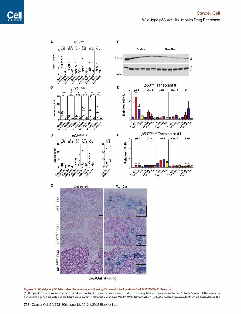
Figure 3. Wild-type p53 Mediates Senescence following Doxorubicin Treatment of MMTV-Wnt1 Tumors
(A–C) Spontaneous tumors were harvested from untreated mice or from mice 5–7 days following final doxorubicin treatment (‘‘Stable’’), and mRNA levels for
senescence genes indicated in the figure were determined for p53 wild-type MMTV-Wnt1 tumors (p53+/+) (A), p53 heterozygous mutant tumors that retained the
Cancer Cell
Wild-type p53 Activity Impairs Drug Response
798 Cancer Cell 21, 793–806, June 12, 2012 ª2012 Elsevier Inc.
Cancer Cell
Wild-type p53 Activity Impairs Drug Response
senescence-associated cytokines (Figures S5Q and S5R). The
fact that some treated p21 null MMTV-Wnt1 tumors retain the
ability to induce senescence-associated cytokines following
treatment could be a contributing factor in their early relapse.
Wild-type p53-Mediated Arrest Impairs DoxorubicinResponse in Human Breast Cancer Cell LinesWe next investigated p53-p21-mediated response to doxoru-
bicin in human breast cancer cells in culture. We found that
MCF-7 and ZR-75.1 cells (TP53 wild-type) treated with doxoru-
bicin were positive for SAbGal activity, and induced cytokines
following treatment that led to phosphorylation of STAT3
(Figures 7A–7C and S6A–S6C). Both cell lines also induced
p53 and p21, ceased incorporating BrdU, and adopted a flat-
tened morphology following doxorubicin treatment (Figures
7D–7F, S6D, and S6E), as previously described by Jackson
and Pereira-Smith (2006). However, similar to the treated
MMTV-Wnt1 tumors lacking functional p53, MCF-7 and ZR-
75.1 cells with TP53 knockdown failed to arrest, adopted
a rounded morphology consistent with cell death, and had fewer
viable cells present following treatment than Si control-trans-
fected, treated cells that adopted a senescent-like phenotype
(Figures 7D–7G and S6D–S6F). Also consistent with our results
in p53 wild-type and mutant MMTV-Wnt1 tumors, both cell lines
exhibited numerous aberrant mitoses, including anaphase
bridges, and stark evidence of cell death such as micronuclei
and condensed nuclei when treated after TP53 or p21 knock-
down (Figures 7H and S6G). The response of MCF-7 and ZR-
75.1 cells with p21 knockdown was consistent with the subset
of p21 null MMTV-Wnt1 tumors that continued to proliferate
and enter mitosis following treatment. These data in breast
cancer cell lines are similar to observations in other cell lines,
such as the isogenic variants of the HCT-116 colon cancer cell
line that lack p53 or p21 (Bunz et al., 1999;Waldman et al., 1997).
DISCUSSION
In this study we show that a robust response of breast cancer to
chemotherapy is highly dependent on the absence of p53-medi-
ated arrest. Functional p53 activated a cell-cycle arrest/senes-
cence program, preventing mitosis in the presence of DNA
strand breaks. Treated tumors with mutant p53 proceeded
through the cell cycle and into aberrant mitoses. Another recent
study observed that a p53 mutant xenografted cell line showed
wild-type allele (p53R172H/+) (B), or lost the wild-type allele (p53R172H/0) (C). p16 is
graphs in (A)–(C) are relative to the mean of untreated p53wild-type tumors set to
treatment (5 day Rx) (the time point when p53 wild-type tumors express senesce
nonsignificant comparisons are shown above the gene symbol. Horizontal line is
(D) p21 protein levels in six of the tumors from (A) compared to growing, relapsed
Student’s t test of densitometric analysis of the two groups, p = 0.0045.
(E and F) Parallel transplanted tumors as in Figure 2B were untreated (‘‘C’’) or doxo
followed until relapse (‘‘Rel’’). mRNA levels for senescence genes indicated in the fi
in at least four tumors for each treatment group. Values in mutant tumors in (F) are
can be discerned. *p < 0.05 by ANOVA and Newman-Keuls posttest for compar
(G) SAbGal staining in histological sections of untreated and treated orthotopic
donor and two different p53R172H/+ donors, with and without doxorubicin treatme
three out of three p53R172H/+-transplanted tumors. Insets shown in bottom-right co
the image. Scale bar, 50 mM.
See also Figure S2.
an increase in aberrant mitotic figures after chemotherapy treat-
ment, whereas a p53 wild-type xenograft had increased SAbGal
staining; however, outcome was not examined in this study
(Varna et al., 2009).
Our data are consistent with retrospective human breast
cancer studies showing that tumors with functional p53 respond
worse to dose-dense doxorubicin-based chemotherapy than
tumors with nonfunctional p53 (Bertheau et al., 2002, 2007).
The delivery of a high dose of DNA-damaging agent is critical
for this effect (Lehmann-Che et al., 2010). In addition to differing
dose regimens used in conflicting studies, none of these reports
stratified responses by LOH status of TP53. Our data presented
here show that determining LOH status is critical for predicting
response. We found that MMTV-Wnt1 p53R172H/+ tumors that
retained the wild-type allele, despite the presence of the ‘‘domi-
nant negative’’ point mutant (Lang et al., 2004; Willis et al., 2004),
exhibited minimal tumor regression and had a transcriptional
profile very similar to p53 wild-type MMTV-Wnt1 tumors, with
acute expression of p53 targets and long-term expression of
senescence markers. This finding has important clinical implica-
tions for using TP53 status to risk stratify patients with breast
cancer, where the wild-type allele is often retained (Mazars
et al., 1992). Thus, identifying a TP53mutation without assessing
LOH is a confounding factor likely responsible for the contradic-
tory results observed in multiple studies analyzing the role of
TP53 mutational status in breast cancer response (Aas et al.,
1996; Berns et al., 2000; Bertheau et al., 2002, 2007, 2008;
Kroger et al., 2006; Makris et al., 1995; Mathieu et al., 1995).
Furthermore, our results suggest that TP53mutations that occur
in basal-like breast cancers contribute to the relatively high rate
of complete responses observed in this subset (Carey et al.,
2007; Straver et al., 2010) and also imply that these tumors are
likely to undergo LOH. Conversely, in the luminal subtypes of
breast cancer, which are mostly TP53 wild-type, p53 activity
could contribute to the lower frequency of complete remissions
(Carey et al., 2007; Straver et al., 2010), and in instances where
a TP53 mutation is present in this subtype, the wild-type allele
is likely to be retained. It will also be interesting to determine if
the p53-mediated arrest phenotype we describe here is also
predictive of poor chemotherapy response in other cancers.
Cells induced by doxorubicin to undergo senescence
evidently persisted and likely contributed to the relapse. Our
immunohistochemical analysis suggests that the cytokines
secreted by senescent tumors operate in an autocrine or
shown on a separate axis due to the aberrantly high values in some tumors. All
a value of 1. For p53mutant tumors, responding tumors harvested 5 days after
nce markers) were used. **p < 0.005; *p < 0.05 by Student’s t test; p values of
the mean.
tumors as determined by western blot, with Vinculin (Vinc) used as a control.
rubicin treated (‘‘Rx’’) and harvested 48 hr or 5 days after the final treatment, or
gure were determined for p53wild-type (E) andmutant (F) transplanted tumors,
relative to untreated tumors in (E) and shown on a smaller scale so differences
ison to untreated. Error bars are ± SEM.
tumor transplants. Shown are representative sections from one p53 wild-type
nt. Similar results were observed in a total of three out of three wild-type and
rner are higher-magnification images of the area shown outlined in black within
Cancer Cell 21, 793–806, June 12, 2012 ª2012 Elsevier Inc. 799

Figure 4. Dichotomous Responses in Doxorubicin-Treated p21 null MMTV-Wnt1Mice: Subsets of Tumors Retain Arrest Capability, whereas
Others Continue Proliferation
(A) p21 null MMTV-Wnt1 mice bearing spontaneous, measurable, growing mammary tumors of approximately 500 mm3 were injected once daily with 4 mg/kg
doxorubicin for 5 consecutive days as indicated by red arrowheads in graphs. Shown are representative charts frommice with poor (top) and favorable (bottom)
responses, and mean tumor regression ± SEM.
(B) Ki67 IHC staining of untreated (‘‘C’’) and treated (‘‘Rx’’) p21 null MMTV-Wnt1 tumors harvested 24 hr following the final treatment is presented. Shown are
representative images from seven treated and six untreated or relapsed tumors. Scale bar, 50 mM.
(C and D) Two different p21 null MMTV-Wnt1 tumors were transplanted, treated in parallel, and BrdU incorporation was determined as in Figure 2B. Shown are
representative images of BrdU staining (scale bar, 50 mM), the corresponding tumor volume charts for parallel transplanted tumors that were followed (blue arrow
indicates the 48 hr time point when parallel transplants were harvested for analysis), representative anaphases (scale bar, 5 mM), and a table quantitating the
anaphase data.
(E) H&E sections from untreated (‘‘C’’) and treated (‘‘Rx’’) tumors of the indicated genotypes were quantitated for mitotic figures. Each data point represents the
average count of mitotic figures in ten 4003 high-power field views. p21 null MMTV-Wnt1-treated tumors were separated into arrested (open circles) and mitotic
(open triangles) groups. Mean (horizontal line) with error bars (SEM) is shown.
See also Figure S3.
Cancer Cell
Wild-type p53 Activity Impairs Drug Response
800 Cancer Cell 21, 793–806, June 12, 2012 ª2012 Elsevier Inc.
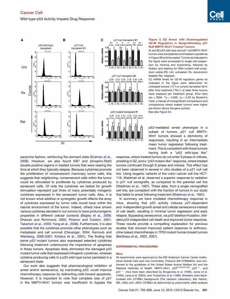
Figure 5. G2 Arrest with Downregulated
G2-M Regulators in Nonproliferating p21
Null MMTV-Wnt1-Treated Tumors
(A and B) p53 wild-type and p21 null MMTV-Wnt1
tumorswere transplanted and treated in parallel as
in Figure 2B and harvested. Tumors as indicated in
the figure were processed to single-cell suspen-
sion by mincing and trypsinizing, followed by
fixation and staining for DNA content with propi-
dium iodide (PI). Unt, untreated; Rx, doxorubicin
treated; Rel, relapsed.
(C) mRNA levels for G2-M regulatory genes as
indicated in the figure were determined for
untreated tumors (‘‘C’’) or tumors harvested 48 hr
after final treatment (‘‘Rx’’); at least three tumors
were analyzed per treatment group. Error bars
are ± SEM. **p < 0.005, *p < 0.05 by Student’s
t test. p Values of nonsignificant comparisons and
comparisons where treated tumors were higher
are shown above the gene symbol.
See also Figure S4.
Cancer Cell
Wild-type p53 Activity Impairs Drug Response
paracrine fashion, reinforcing the dormant state (Kuilman et al.,
2008). However, we also found Ki67 and phospho-Stat3
double-positive regions in treated tumors that were nearing the
time at which they typically relapse. Because cytokines promote
the proliferation of nonsenescent mammary tumor cells, this
suggests that neighboring, nonsenescent cells within the tumor
could be stimulated to proliferate by cytokines produced by
senescent cells. Of note the cytokines we tested for growth
stimulation represent just three of many potentially mitogenic
cytokines expressed in the senescent tumor cells. Also, it is
not known what additive or synergistic growth effects the array
of cytokines expressed by tumor cells would have within the
natural environment of the tumor. Indeed, others have shown
various cytokines elevated in our tumors to have protumorigenic
properties in different cellular contexts (Begley et al., 2008;
Dhawan and Richmond, 2002; Pirianov and Colston, 2001;
Takamori et al., 2000; Yang et al., 2006). Furthermore, it is also
possible that the cytokines promote other phenotypes such as
metastasis and cell survival (Clevenger, 2004; Karnoub and
Weinberg, 2006-2007; Krtolica et al., 2001). The finding that
some p53 mutant tumors also expressed selected cytokines
following treatment underscores the importance of apoptosis
in these tumors. Apoptosis likely eliminated the damaged p53
mutant tumor cells that expressedmitogenic cytokines, whereas
cytokine producing cells in a p53 wild-type tumor persisted in a
senescent state.
Our work also suggests that pharmacological inhibition of
arrest and/or senescence, by inactivating p53, could improve
chemotherapy response by redirecting cells toward apoptosis.
However, it is important to note that complete loss of p21
in the MMTV-Wnt1 tumors was insufficient to bypass the
Cancer Cell 21, 793–8
p53-mediated arrest phenotype in a
subset of tumors. p21 null MMTV-
Wnt1 tumors showed a dichotomy of
responses, resulting in an intermediate
mean tumor regression following treat-
ment. This is consistent with these tumors
having both a ‘‘p53 wild-type like’’
response, where treated tumors do not enter S phase or mitosis,
arresting in G2, and a ‘‘p53mutant-like’’ response, where treated
tumors continued through S phase and mitosis. This effect has
not been observed in several in vitro studies of a p21 null cell
line. Using isogenic variants of the colon cancer cell line HCT-
116, Waldman et al. observed a superior response to radiation
in p21 null xenografts, as compared to the parental cell line
(Waldman et al., 1997). These data, from a single xenografted
cell line, are consistent with the fraction of tumors in our study
that failed to arrest following treatment (Waldman et al., 1997).
In summary we have modeled chemotherapy response in
mice, showing that p53 activity induces p21-dependent
and -independent growth arrest and cellular senescence instead
of cell death, resulting in minimal tumor regression and early
relapse. Bypassing senescence, via p53 deletion/mutation, initi-
ated p53-independent cell death and improved tumor response.
These results provide a compelling explanation for previous
studies that showed improved patient response to anthracy-
cline-based chemotherapy in TP53mutant human breast tumors
(Bertheau et al., 2002, 2007).
EXPERIMENTAL PROCEDURES
Mice
All experiments were approved by the MD Anderson Cancer Center Institu-
tional Animal Care and Use Committee, Protocol ID# 079906634, and con-
formed to the guidelines of the United States Animal Welfare Act and the
National Institutes of Health. MMTV-Wnt1, p53R172H/R172H, p53�/�, and
p21�/� mice have been described by Brugarolas et al. (1995), Jacks et al.
(1994), Lang et al. (2004), and Tsukamoto et al. (1988). Breeders were back-
crossed into C57Bl6J background (The Jackson Laboratory, Bar Harbor,
ME, USA) until >90% C57Bl6J as determined by polymorphic allele analysis
06, June 12, 2012 ª2012 Elsevier Inc. 801
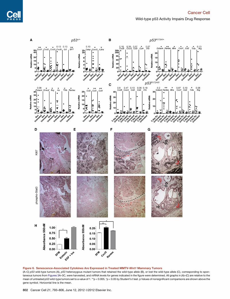
Figure 6. Senescence-Associated Cytokines Are Expressed in Treated MMTV-Wnt1 Mammary Tumors
(A–C) p53 wild-type tumors (A), p53 heterozygous mutant tumors that retained the wild-type allele (B), or lost the wild-type allele (C), corresponding to spon-
taneous tumors from Figures 3A–3C, were harvested, and mRNA levels for genes indicated in the figure were determined. All graphs in (A)–(C) are relative to the
mean of untreated p53wild-type tumors set to a value of 1. **p < 0.005, *p < 0.05 by Student’s t test. p Values of nonsignificant comparisons are shown above the
gene symbol. Horizontal line is the mean.
Cancer Cell
Wild-type p53 Activity Impairs Drug Response
802 Cancer Cell 21, 793–806, June 12, 2012 ª2012 Elsevier Inc.
Cancer Cell
Wild-type p53 Activity Impairs Drug Response
by the Research Animal Support Facility-Smithville, Genetic Services.
Subsequent breedings produced MMTV-Wnt1 mice in p53 wild-type,
p53R172H/R172H, p53�/�, p53R172H/+, and p21�/� backgrounds. Homozygous
p53 mutant mice, particularly females, were not born at Mendelian ratio,
consistent with other reports by Sah et al. (1995), and were thus difficult to
acquire in the cohort. Mammary tumors formed in MMTV-Wnt1 mice in
our study with median latency of 185 days. p53R172H/R172H, p53�/� and
p53R172H/+mice hadmedian latencies of 108, 111, and 173 days, respectively.
Mice were monitored frequently for tumor formation and tumors measured
regularly using digital calipers: tumor volume in mm3 = (width2 3 length)/2
(Bearss et al., 2000). Histologically, all of the MMTV-Wnt1 tumors were ductal
carcinomas, generally of solid, cystic, or mixed subtypes. p53 wild-type,
p53R172H/+, p21�/� were all 68%–79% mixed, whereas p53R172H/R172H and
p53R172H/0 were 61% solid. When tumors reached a volume of �500 mm3
and were growing, 4 mg/kg doxorubicin (Sigma-Aldrich, St. Louis) in PBS
was injected intraperitoneally once daily for 5 consecutive days. Treatments
were well tolerated in p53 wild-type and heterozygous mutant mice as well
as p21�/� mice, with minimal (less than 10%) or no weight loss during or after
treatment. p53 homozygous mutant mice, however, did show signs of toxicity,
primarily weight loss �4 days after treatment, likely due to GI syndrome, as
previously described in p53 mutant mice by Komarova et al. (2004). This
toxicity did not appear to contribute to tumor regression because p53 hetero-
zygous mutant mice with LOH in the tumor had no toxicity and had similar
tumor regression as the homozygous mutant mice. At the defined endpoint
for each mouse, tumors were harvested, and portions were fixed in formalin
for 48 hr and paraffin embedded (FFPE), and also flash frozen for biochemical
analysis. LOH analysis was performed exactly as previously described by Post
et al. (2010).
Real-Time RT-PCR
RNA was extracted from frozen, pulverized tumors using TRIzol reagent
(Invitrogen, Carlsbad, CA,USA), subjected to DNase treatment (Roche, Indian-
apolis, IN, USA), and then reverse transcribed using a kit (GE Healthcare,
Piscataway, NJ, USA). Real-time PCR using Sybr green (BioRad, Valencia,
CA, USA) was performed as previously described by Jackson and Pereira-
Smith (2006). Expression was normalized to Gapdh and verified with Rplp0.
Primer sequences are available on request.
Immunohistochemistry
Cleaved caspase staining was performed as previously described by Post
et al. (2010), and staining for phospho-Stat3 polyclonal antibody (1:100; Cell
Signaling, Danvers, MA, USA) and Ki67 (1:100; Leica, Newcastle Upon
Tyne, UK, Ki67-MM1) were performed similarly. For phospho-Stat3 mono-
clonal, antigen retrieval was in Tris EDTA buffer (pH 9). For detecting incorpo-
ration of BrdU, tumor-bearing mice were injected with BrdU (Invitrogen)
according to manufacturer’s instructions, 24 and 4 hr before harvest, followed
by standard fixation and processing. Denaturation was in 2 N HCl for 90 min,
followed by neutralization in 0.1 M Na2B4O7, standard processing, and then
incubation with anti-BrdU (BD Immunocytometry Systems, San Jose, CA,
USA) at 1:40 for 1 hr (McGinley et al., 2000). Antigen detection for immunohis-
tochemistry (IHC) was performed using a VECTASTAIN kit and ABC (Vector
Laboratories, Burlingame, CA, USA). Images were acquired on a Nikon 80i
microscope equipped with a Nikon DS-Fi1 color camera using the 103/0.45
objective and Nikon Elements software. Some images were processed
minimally in Adobe Photoshop only by histogram stretching and gamma
adjustment. At least four random fields were manually counted for cleaved
(D–G) Phospho-Stat3 and Ki67 staining in treated, senescent tumors. Serial sectio
for Ki67 (upper panels) or phosphoTyr705-Stat3 (antibody clone D3A7) (lower pa
phospho-Stat3 (antibody clone D3A7) and Ki67 from treated tumors. (F) Inverse st
positivity marked by red dashed line. (G) Similar staining results with a second p
positive/Ki67-negative areas (indicated by black arrows) and less intense phospho
(H) Tumor cells isolated from an MMTV-Wnt1 mammary tumor and cultured we
changed to serum-free media (SFM), or 100 ng/ml cytokine. EGF and insulin, know
number was determined at day 4 by MTT assay. Error bars are ± SEM. For compa
Cxcl5, and Rantes, p < 0.005 by ANOVA and Newman-Keuls.
See also Figure S5.
caspase-3 experiments. Number of positive cells was averaged for the four
fields.
TUNEL staining was performed using the FragEL DNA fragmentation detec-
tion kit (Calbiochem, Darmstadt, Germany) and quantitated as above. SAbGal
assay was performed essentially as described by Dimri et al. (1995) for in vitro
and in vivo experiments. Tumor segments�1mm thick were fixed in 2% form-
aldehyde, 0.2% glutaraldehyde in PBS, stained for SAbGal, cut to 6 mm
sections, and counterstained with Nuclear Fast Red (Vector Laboratories).
These results were verified by staining of frozen sections in parallel.
Western Blotting
Frozen pulverized tumors were lysed and separated by SDS-PAGE as previ-
ously described by Pant et al. (2011). Antibodies and dilutions were p21 (for
mouse p21 detection), 1:1,000, #556431 (BD PharMingen, San Jose, CA,
USA); Vinculin, 1:1,000, #V-9131 (Sigma-Aldrich); p21 (for human p21 detec-
tion), 1:200, SC6246 (Santa Cruz Biotechnology, Santa Cruz, CA, USA);
and mouse monoclonal p53, 1:200, OP09 (EMD Biosciences, Darmstadt,
Germany).
Statistical Analysis
Two-tailed Student’s t tests and ANOVA using Newman-Keuls posttest were
performed using GraphPad Prism software (La Jolla, CA, USA).
Anaphase Bridges and Mitotic Activity
H&EMMTV-Wnt1 tumor sections were scanned on amicroscope at 4003, and
cells in anaphase were photographed. Two different observers identified the
presence or absence of bridges. For mitotic figures, ten random, 4003,
high-powered fields for each tumor were selected, and mitotic figures were
identified and counted by two different observers.
Flow Cytometry
Tumors were harvested and processed as for transplants (above). After
filtering, cells were washed in PBS, then fixed in 70% ethanol for at least
24 hr at �20�C. Propidium iodide staining was performed as previously
described by Jackson and Pereira-Smith (2006).
Transplants
Primary MMTV-Wnt1 tumors were removed from euthanized mice, minced
thoroughly with a scalpel blade, and then trypsinized for 10 min at 37�C.Trypsin was inactivated with DMEM plus 10% fetal calf serum, followed by
passage through a 40 mM filter. After PBS washing, cells were resuspended
in Matrigel/PBS (BD Biosciences) at a concentration of 4 3 106/50 ml. The
50 ml solution was injected into each abdominal mammary fat pad of recipient
C57Bl6 mice using a 30G needle. Tumors were detectable at �2 weeks,
typically.
SUPPLEMENTAL INFORMATION
Supplemental Information includes six figures and Supplemental Experimental
Procedures and can be found with this article online at doi:10.1016/j.ccr.2012.
04.027.
ACKNOWLEDGMENTS
The authors wish to thankMDACC Animal Support Facility-Smithville, the DNA
Analysis Facility, the Flow Cytometry & Imaging Core Facility, and the
ns from formalin-fixed paraffin-embedded tumors from Figure 3A were stained
nels). (D–F) Representative examples of positive/negative or negative/positive
aining of adjacent areas within the same tumor sections, and an area of double
olyclonal phosphoTyr705-Stat3 antibody, with more intense phospho-Stat3-
-Stat3 staining/Ki67-positive areas (indicated by red arrows). Scale bar, 50 mM.
re plated at 8,000 cells per well in a 24-well plate overnight, then media were
n mitogens in breast cancer, were at 20 ng/ml and 10 mg/ml, respectively. Cell
rison of SFM to Eotaxin, *p < 0.05 by Student’s t test. For comparison of SFM,
Cancer Cell 21, 793–806, June 12, 2012 ª2012 Elsevier Inc. 803
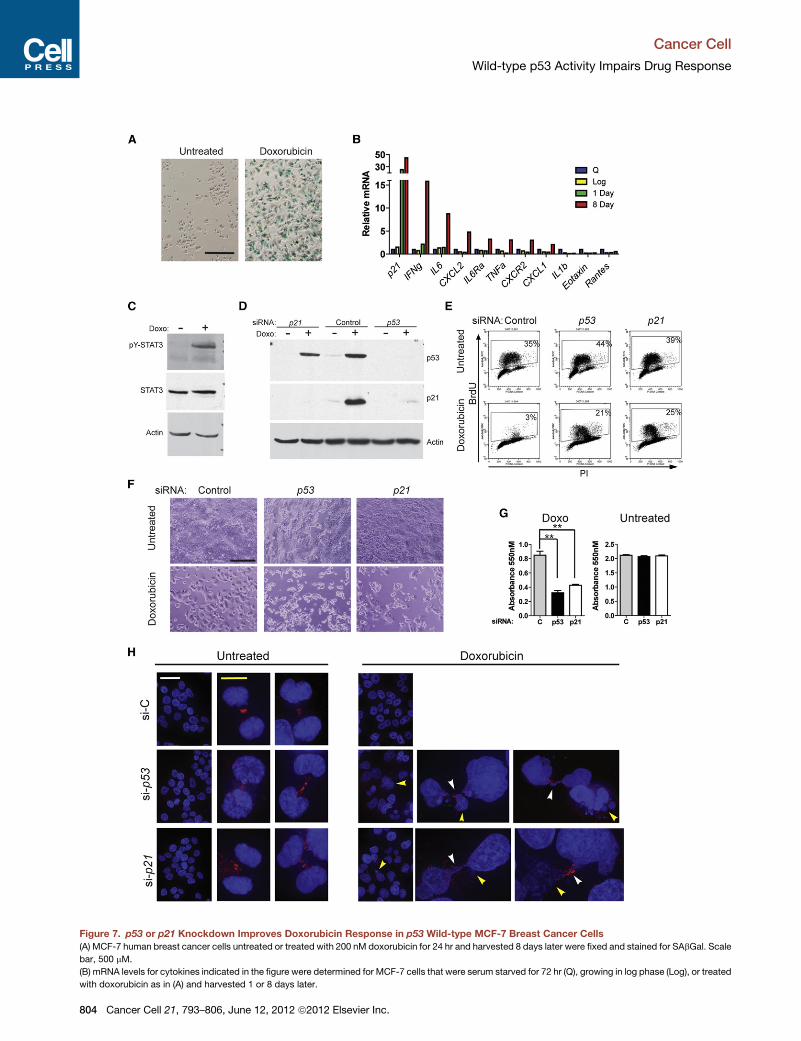
Figure 7. p53 or p21 Knockdown Improves Doxorubicin Response in p53 Wild-type MCF-7 Breast Cancer Cells
(A) MCF-7 human breast cancer cells untreated or treated with 200 nM doxorubicin for 24 hr and harvested 8 days later were fixed and stained for SAbGal. Scale
bar, 500 mM.
(B) mRNA levels for cytokines indicated in the figure were determined for MCF-7 cells that were serum starved for 72 hr (Q), growing in log phase (Log), or treated
with doxorubicin as in (A) and harvested 1 or 8 days later.
Cancer Cell
Wild-type p53 Activity Impairs Drug Response
804 Cancer Cell 21, 793–806, June 12, 2012 ª2012 Elsevier Inc.
Cancer Cell
Wild-type p53 Activity Impairs Drug Response
Department of Veterinary Medicine and Surgery and the Histology Core
Research Lab, each supported by NCI Grant CA16672. We also acknowledge
Courtney Vallien for histological sectioning, Henry P. Adams for technical help
with microscopy, and Archana Sidalaghatta Nagaraja for assistance with
mouse necropsy and RNA preparation. J.G.J. was funded as an Odyssey
Scholar by the Theodore N. Law Endowment for Scientific Achievement,
a Dodie P. Hawn Fellowship in Genetics, and by NIH Grant U01DE019765-
01. Q.L. and V.P. are supported by NIH Grant CA47296. G.L. is supported
by NIH Grants CA34936 and CA82577.
J.G.J. conceived, designed, and performed all the experiments except
where noted, and wrote the manuscript. V.P. and J.G.J. performed protein
expression analysis. J.G.J., Q.L., and V.P. performed mRNA analysis. J.G.J.,
L.L.C., Q.L., D.G., and A.Q.-C. performed immunohistochemical analyses.
T.M. performed the Bio-Plex cytokine array. J.G.J., D.G., and A.Q.-C. per-
formed quantitative analysis of mitosis. Y.L. developed cell lines. J.G.J. and
O.T. performed senescence associated b-galactosidase studies. P.Y. and
J.G.J. determined loss of heterozygosity in tumors. A.K.E-N. performed path-
ological analysis. A.Q.-C. edited the manuscript. G.L. was the principal inves-
tigator for the study.
Received: October 1, 2011
Revised: March 2, 2012
Accepted: April 14, 2012
Published: June 11, 2012
REFERENCES
Aas, T., Børresen, A.L., Geisler, S., Smith-Sørensen, B., Johnsen, H., Varhaug,
J.E., Akslen, L.A., and Lønning, P.E. (1996). Specific P53 mutations are asso-
ciated with de novo resistance to doxorubicin in breast cancer patients. Nat.
Med. 2, 811–814.
Acosta, J.C., O’Loghlen, A., Banito, A., Guijarro, M.V., Augert, A., Raguz, S.,
Fumagalli, M., Da Costa, M., Brown, C., Popov, N., et al. (2008). Chemokine
signaling via the CXCR2 receptor reinforces senescence. Cell 133, 1006–1018.
Bearss, D.J., Subler, M.A., Hundley, J.E., Troyer, D.A., Salinas, R.A., and
Windle, J.J. (2000). Genetic determinants of response to chemotherapy in
transgenic mouse mammary and salivary tumors. Oncogene 19, 1114–1122.
Begley, L.A., Kasina, S., Mehra, R., Adsule, S., Admon, A.J., Lonigro, R.J.,
Chinnaiyan, A.M., andMacoska, J.A. (2008). CXCL5 promotes prostate cancer
progression. Neoplasia 10, 244–254.
Berns, E.M., Foekens, J.A., Vossen, R., Look, M.P., Devilee, P., Henzen-
Logmans, S.C., van Staveren, I.L., van Putten, W.L., Inganas, M., Meijer-van
Gelder, M.E., et al. (2000). Complete sequencing of TP53 predicts poor
response to systemic therapy of advanced breast cancer. Cancer Res. 60,
2155–2162.
Bertheau, P., Plassa, F., Espie, M., Turpin, E., de Roquancourt, A., Marty, M.,
Lerebours, F., Beuzard, Y., Janin, A., and de The, H. (2002). Effect of mutated
TP53 on response of advanced breast cancers to high-dose chemotherapy.
Lancet 360, 852–854.
Bertheau, P., Turpin, E., Rickman, D.S., Espie, M., de Reynies, A., Feugeas,
J.P., Plassa, L.F., Soliman, H., Varna, M., de Roquancourt, A., et al. (2007).
Exquisite sensitivity of TP53 mutant and basal breast cancers to a dose-dense
epirubicin-cyclophosphamide regimen. PLoS Med. 4, e90.
(C) MCF-7 cells untreated or 8 days following treatment as in (A) were harvested
antibodies.
(D–F) MCF-7 cells were transfected with nontargeting siRNA (Control) or siRNA ta
untreated as indicated. (D) Cells were harvested 24 hr after treatment for western
24 hr after doxorubicin treatment, fixed, stained with anti-BrdU FITC, and sorted b
days following treatment and media change, cells were photographed using brig
(G) Cell number was determined by MTT assay 9 days following treatment. Error
(H) MCF-7 cells were untreated or treated with 200 nM doxorubicin for 24 hr, then
and 10 mM (yellow). Yellow arrows indicate micronuclei and white arrows anapha
See also Figure S6.
Bertheau, P., Espie, M., Turpin, E., Lehmann, J., Plassa, L.F., Varna, M., Janin,
A., and de The, H. (2008). TP53 status and response to chemotherapy in breast
cancer. Pathobiology 75, 132–139.
Bonnefoi, H., Piccart, M., Bogaerts, J., Mauriac, L., Fumoleau, P., Brain, E.,
Petit, T., Rouanet, P., Jassem, J., Blot, E., et al; EORTC 10994/BIG 1-00
Study Investigators. (2011). TP53 status for prediction of sensitivity to taxane
versus non-taxane neoadjuvant chemotherapy in breast cancer (EORTC
10994/BIG 1-00): a randomised phase 3 trial. Lancet Oncol. 12, 527–539.
Brown, J.M., and Attardi, L.D. (2005). The role of apoptosis in cancer develop-
ment and treatment response. Nat. Rev. Cancer 5, 231–237.
Brown, J.P., Wei, W., and Sedivy, J.M. (1997). Bypass of senescence after
disruption of p21CIP1/WAF1 gene in normal diploid human fibroblasts.
Science 277, 831–834.
Brugarolas, J., Chandrasekaran, C., Gordon, J.I., Beach, D., Jacks, T., and
Hannon, G.J. (1995). Radiation-induced cell cycle arrest compromised by
p21 deficiency. Nature 377, 552–557.
Bunz, F., Dutriaux, A., Lengauer, C., Waldman, T., Zhou, S., Brown, J.P.,
Sedivy, J.M., Kinzler, K.W., and Vogelstein, B. (1998). Requirement for p53
and p21 to sustain G2 arrest after DNA damage. Science 282, 1497–1501.
Bunz, F., Hwang, P.M., Torrance, C., Waldman, T., Zhang, Y., Dillehay, L.,
Williams, J., Lengauer, C., Kinzler, K.W., and Vogelstein, B. (1999).
Disruption of p53 in human cancer cells alters the responses to therapeutic
agents. J. Clin. Invest. 104, 263–269.
Carey, L.A., Dees, E.C., Sawyer, L., Gatti, L., Moore, D.T., Collichio, F., Ollila,
D.W., Sartor, C.I., Graham, M.L., and Perou, C.M. (2007). The triple negative
paradox: primary tumor chemosensitivity of breast cancer subtypes. Clin.
Cancer Res. 13, 2329–2334.
Chang, B.D., Xuan, Y., Broude, E.V., Zhu, H., Schott, B., Fang, J., and
Roninson, I.B. (1999). Role of p53 and p21waf1/cip1 in senescence-like
terminal proliferation arrest induced in human tumor cells by chemothera-
peutic drugs. Oncogene 18, 4808–4818.
Clarke, A.R., Purdie, C.A., Harrison, D.J., Morris, R.G., Bird, C.C., Hooper,
M.L., and Wyllie, A.H. (1993). Thymocyte apoptosis induced by p53-depen-
dent and independent pathways. Nature 362, 849–852.
Clevenger, C.V. (2004). Roles and regulation of stat family transcription factors
in human breast cancer. Am. J. Pathol. 165, 1449–1460.
Collado, M., Gil, J., Efeyan, A., Guerra, C., Schuhmacher, A.J., Barradas, M.,
Bengurıa, A., Zaballos, A., Flores, J.M., Barbacid, M., et al. (2005). Tumour
biology: senescence in premalignant tumours. Nature 436, 642.
Coppe, J.P., Patil, C.K., Rodier, F., Sun, Y., Munoz, D.P., Goldstein, J., Nelson,
P.S., Desprez, P.Y., and Campisi, J. (2008). Senescence-associated secretory
phenotypes reveal cell-nonautonomous functions of oncogenic RAS and the
p53 tumor suppressor. PLoS Biol. 6, 2853–2868.
Deng, C., Zhang, P., Harper, J.W., Elledge, S.J., and Leder, P. (1995). Mice
lacking p21CIP1/WAF1 undergo normal development, but are defective in
G1 checkpoint control. Cell 82, 675–684.
Dhawan, P., and Richmond, A. (2002). Role of CXCL1 in tumorigenesis of
melanoma. J. Leukoc. Biol. 72, 9–18.
Dimri, G.P., Lee, X., Basile, G., Acosta, M., Scott, G., Roskelley, C., Medrano,
E.E., Linskens, M., Rubelj, I., Pereira-Smith, O., et al. (1995). A biomarker that
identifies senescent human cells in culture and in aging skin in vivo. Proc. Natl.
Acad. Sci. USA 92, 9363–9367.
48 hr after a media change, and western blots were performed with indicated
rgeting p53 or p21. After 24 hr, cells were treated with doxorubicin for 24 hr, or
blot with p53, p21, or actin antibodies. (E) Cells were pulsed for 1 hr with BrdU
y flow cytometry. Percent BrdU-positive cells are indicated in the figure. (F) Four
ht-field microscopy (scale bar, 500 mM).
bars are ± SEM. **p < 0.005 by ANOVA and Newman-Keuls posttest.
fixed and stained for a-tubulin and DAPI 72 hr later. Scale bars, 40 mM (white)
se bridges.
Cancer Cell 21, 793–806, June 12, 2012 ª2012 Elsevier Inc. 805
Cancer Cell
Wild-type p53 Activity Impairs Drug Response
Donehower, L.A., Godley, L.A., Aldaz, C.M., Pyle, R., Shi, Y.P., Pinkel, D.,
Gray, J., Bradley, A., Medina, D., and Varmus, H.E. (1995). Deficiency of p53
accelerates mammary tumorigenesis in Wnt-1 transgenic mice and promotes
chromosomal instability. Genes Dev. 9, 882–895.
Jacks, T., Remington, L.,Williams, B.O., Schmitt, E.M., Halachmi, S., Bronson,
R.T., andWeinberg, R.A. (1994). Tumor spectrum analysis in p53-mutant mice.
Curr. Biol. 4, 1–7.
Jackson, J.G., and Pereira-Smith, O.M. (2006). Primary and compensatory
roles for RB family members at cell cycle gene promoters that are deacetylated
and downregulated in doxorubicin-induced senescence of breast cancer cells.
Mol. Cell. Biol. 26, 2501–2510.
Jackson, J.G., Post, S.M., and Lozano, G. (2011). Regulation of tissue- and
stimulus-specific cell fate decisions by p53 in vivo. J. Pathol. 223, 127–136.
Karnoub, A.E., and Weinberg, R.A. (2006–2007). Chemokine networks and
breast cancer metastasis. Breast Dis. 26, 75–85.
Komarova, E.A., Kondratov, R.V., Wang, K., Christov, K., Golovkina, T.V.,
Goldblum, J.R., and Gudkov, A.V. (2004). Dual effect of p53 on radiation sensi-
tivity in vivo: p53 promotes hematopoietic injury, but protects from gastro-
intestinal syndrome in mice. Oncogene 23, 3265–3271.
Kroger, N., Milde-Langosch, K., Riethdorf, S., Schmoor, C., Schumacher, M.,
Zander, A.R., and Loning, T. (2006). Prognostic and predictive effects of immu-
nohistochemical factors in high-risk primary breast cancer patients. Clin.
Cancer Res. 12, 159–168.
Krtolica, A., Parrinello, S., Lockett, S., Desprez, P.Y., and Campisi, J. (2001).
Senescent fibroblasts promote epithelial cell growth and tumorigenesis:
a link between cancer and aging. Proc. Natl. Acad. Sci. USA 98, 12072–12077.
Kuilman, T., Michaloglou, C., Vredeveld, L.C., Douma, S., van Doorn, R.,
Desmet, C.J., Aarden, L.A., Mooi, W.J., and Peeper, D.S. (2008). Oncogene-
induced senescence relayed by an interleukin-dependent inflammatory
network. Cell 133, 1019–1031.
Lang, G.A., Iwakuma, T., Suh, Y.A., Liu, G., Rao, V.A., Parant, J.M., Valentin-
Vega, Y.A., Terzian, T., Caldwell, L.C., Strong, L.C., et al. (2004). Gain of func-
tion of a p53 hot spot mutation in a mouse model of Li-Fraumeni syndrome.
Cell 119, 861–872.
Lehmann-Che, J., Andre, F., Desmedt, C., Mazouni, C., Giacchetti, S., Turpin,
E., Espie, M., Plassa, L.F., Marty, M., Bertheau, P., et al. (2010).
Cyclophosphamide dose intensification may circumvent anthracycline resis-
tance of p53 mutant breast cancers. Oncologist 15, 246–252.
Liu, G., Parant, J.M., Lang, G., Chau, P., Chavez-Reyes, A., El-Naggar, A.K.,
Multani, A., Chang, S., and Lozano, G. (2004). Chromosome stability, in the
absence of apoptosis, is critical for suppression of tumorigenesis in Trp53
mutant mice. Nat. Genet. 36, 63–68.
Lowe, S.W., Ruley, H.E., Jacks, T., and Housman, D.E. (1993a). p53-depen-
dent apoptosis modulates the cytotoxicity of anticancer agents. Cell 74,
957–967.
Lowe, S.W., Schmitt, E.M., Smith, S.W., Osborne, B.A., and Jacks, T. (1993b).
p53 is required for radiation-induced apoptosis in mouse thymocytes. Nature
362, 847–849.
Lowe, S.W., Bodis, S., McClatchey, A., Remington, L., Ruley, H.E., Fisher,
D.E., Housman, D.E., and Jacks, T. (1994). p53 status and the efficacy of
cancer therapy in vivo. Science 266, 807–810.
Makris, A., Powles, T.J., Dowsett, M., and Allred, C. (1995). p53 protein over-
expression and chemosensitivity in breast cancer. Lancet 345, 1181–1182.
Mathieu, M.C., Koscielny, S., Le Bihan, M.L., Spielmann, M., and Arriagada,
R.; Institut Gustave-Roussy Breast Cancer Group. (1995). p53 protein overex-
pression and chemosensitivity in breast cancer. Lancet 345, 1182.
Mazars, R., Spinardi, L., BenCheikh, M., Simony-Lafontaine, J., Jeanteur, P.,
and Theillet, C. (1992). p53 mutations occur in aggressive breast cancer.
Cancer Res. 52, 3918–3923.
806 Cancer Cell 21, 793–806, June 12, 2012 ª2012 Elsevier Inc.
McGinley, J.N., Knott, K.K., and Thompson, H.J. (2000). Effect of fixation and
epitope retrieval on BrdU indices in mammary carcinomas. J. Histochem.
Cytochem. 48, 355–362.
Novakova, Z., Hubackova, S., Kosar, M., Janderova-Rossmeislova, L.,
Dobrovolna, J., Vasicova, P., Vancurova, M., Horejsi, Z., Hozak, P., Bartek,
J., and Hodny, Z. (2010). Cytokine expression and signaling in drug-induced
cellular senescence. Oncogene 29, 273–284.
Pant, V., Xiong, S., Iwakuma, T., Quintas-Cardama, A., and Lozano, G. (2011).
Heterodimerization of Mdm2 and Mdm4 is critical for regulating p53 activity
during embryogenesis but dispensable for p53 and Mdm2 stability. Proc.
Natl. Acad. Sci. USA 108, 11995–12000.
Pirianov, G., and Colston, K.W. (2001). Interactions of vitamin D analogue
CB1093, TNFalpha and ceramide on breast cancer cell apoptosis. Mol. Cell.
Endocrinol. 172, 69–78.
Post, S.M., Quintas-Cardama, A., Terzian, T., Smith, C., Eischen, C.M., and
Lozano, G. (2010). p53-dependent senescence delays Emu-myc-induced
B-cell lymphomagenesis. Oncogene 29, 1260–1269.
Rahko, E., Blanco, G., Soini, Y., Bloigu, R., and Jukkola, A. (2003). A mutant
TP53 gene status is associated with a poor prognosis and anthracycline-resis-
tance in breast cancer patients. Eur. J. Cancer 39, 447–453.
Riley, T., Sontag, E., Chen, P., and Levine, A. (2008). Transcriptional control of
human p53-regulated genes. Nat. Rev. Mol. Cell Biol. 9, 402–412.
Sah, V.P., Attardi, L.D., Mulligan, G.J., Williams, B.O., Bronson, R.T., and
Jacks, T. (1995). A subset of p53-deficient embryos exhibit exencephaly.
Nat. Genet. 10, 175–180.
Soussi, T., and Lozano, G. (2005). p53 mutation heterogeneity in cancer.
Biochem. Biophys. Res. Commun. 331, 834–842.
Straver, M.E., Glas, A.M., Hannemann, J., Wesseling, J., van de Vijver, M.J.,
Rutgers, E.J., Vrancken Peeters, M.J., van Tinteren, H., Van’t Veer, L.J., and
Rodenhuis, S. (2010). The 70-gene signature as a response predictor for neo-
adjuvant chemotherapy in breast cancer. Breast Cancer Res. Treat. 119,
551–558.
Takamori, H., Oades, Z.G., Hoch, O.C., Burger, M., and Schraufstatter, I.U.
(2000). Autocrine growth effect of IL-8 and GROalpha on a human pancreatic
cancer cell line, Capan-1. Pancreas 21, 52–56.
te Poele, R.H., Okorokov, A.L., Jardine, L., Cummings, J., and Joel, S.P.
(2002). DNA damage is able to induce senescence in tumor cells in vitro and
in vivo. Cancer Res. 62, 1876–1883.
Tsukamoto, A.S., Grosschedl, R., Guzman, R.C., Parslow, T., and Varmus,
H.E. (1988). Expression of the int-1 gene in transgenic mice is associated
with mammary gland hyperplasia and adenocarcinomas in male and female
mice. Cell 55, 619–625.
Varna, M., Lehmann-Che, J., Turpin, E., Marangoni, E., El-Bouchtaoui, M.,
Jeanne, M., Grigoriu, C., Ratajczak, P., Leboeuf, C., Plassa, L.F., et al.
(2009). p53 dependent cell-cycle arrest triggered by chemotherapy in xeno-
grafted breast tumors. Int. J. Cancer 124, 991–997.
Wajapeyee, N., Serra, R.W., Zhu, X., Mahalingam, M., and Green, M.R. (2008).
Oncogenic BRAF induces senescence and apoptosis through pathways
mediated by the secreted protein IGFBP7. Cell 132, 363–374.
Waldman, T., Kinzler, K.W., and Vogelstein, B. (1995). p21 is necessary for the
p53-mediated G1 arrest in human cancer cells. Cancer Res. 55, 5187–5190.
Waldman, T., Zhang, Y., Dillehay, L., Yu, J., Kinzler, K., Vogelstein, B., and
Williams, J. (1997). Cell-cycle arrest versus cell death in cancer therapy. Nat.
Med. 3, 1034–1036.
Willis, A., Jung, E.J., Wakefield, T., and Chen, X. (2004). Mutant p53 exerts
a dominant negative effect by preventing wild-type p53 from binding to the
promoter of its target genes. Oncogene 23, 2330–2338.
Yang, X., Lu, P., Fujii, C., Nakamoto, Y., Gao, J.L., Kaneko, S., Murphy, P.M.,
and Mukaida, N. (2006). Essential contribution of a chemokine, CCL3, and its
receptor, CCR1, to hepatocellular carcinoma progression. Int. J. Cancer 118,
1869–1876.