Embed Size (px)
Citation preview
www.palaeontologyonline.com
Published on: 01/11/2013| Published by: Palaeontology [online]
Title: Fossil Focus: Heterostraci
Author(s): Joseph N. Keating *1 Volume: 3 Article: 11 Page(s): 1-9 Published Date: 01/11/2013
PermaLink: http://www.palaeontologyonline.com/articles/2013/fossil-focus-heterostraci/
IMPORTANT
Your use of the Palaeontology [online] archive indicates your acceptance of Palaeontology [online]'s Terms and
Conditions of Use, available at http://www.palaeontologyonline.com/site-information/terms-and-conditions/.
COPYRIGHT
Palaeontology [online] (www.palaeontologyonline.com) publishes all work, unless otherwise stated, under the Creative Commons Attribution 3.0 Unported (CC BY 3.0) license.
This license lets others distribute, remix, tweak, and build upon the published work, even commercially, as long as they credit Palaeontology[online] for the original creation. This is the most accommodating of licenses offered by Creative Commons and is recommended for maximum dissemination of published material.
Further details are available at http://www.palaeontologyonline.com/site-information/copyright/.
CITATION OF ARTICLE
Please cite the following published work as:
Keating, Joseph N. 2013. Fossil Focus: Heterostraci, Palaeontology Online, Volume 3, Article 11, 1-
9.
www.palaeontologyonline.com |Page 1
Published by: Palaeontology [online]
Fossil Focus: Heterostraci by Joseph N. Keating*1
Introduction: The Heterostraci (which means ‘different shield’) make up an extinct group of jawless fish that lived
during the early to middle Palaeozoic era, approximately 440 million to 359 million years ago. They
were exceptionally diverse, with over 300 species currently described from marine and freshwater
sediments of North America, Europe and Siberia. Heterostracans are characterized by their external
armour of distinct plates, which are composed mainly of bone anddentine (a hard-tissue component
of teeth in vertebrates). Most heterostracans can be classified into two major groups, the cyathaspids
and the pteraspids, which differ with respect to the structure, number and arrangement of their
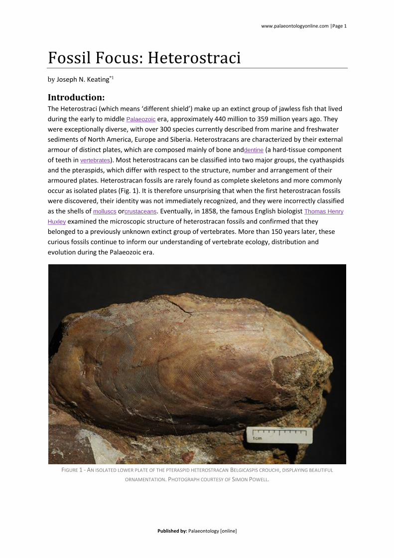
armoured plates. Heterostracan fossils are rarely found as complete skeletons and more commonly
occur as isolated plates (Fig. 1). It is therefore unsurprising that when the first heterostracan fossils
were discovered, their identity was not immediately recognized, and they were incorrectly classified
as the shells of molluscs orcrustaceans. Eventually, in 1858, the famous English biologist Thomas Henry
Huxley examined the microscopic structure of heterostracan fossils and confirmed that they
belonged to a previously unknown extinct group of vertebrates. More than 150 years later, these
curious fossils continue to inform our understanding of vertebrate ecology, distribution and
evolution during the Palaeozoic era.
FIGURE 1 - AN ISOLATED LOWER PLATE OF THE PTERASPID HETEROSTRACAN BELGICASPIS CROUCHI, DISPLAYING BEAUTIFUL
ORNAMENTATION. PHOTOGRAPH COURTESY OF SIMON POWELL.
www.palaeontologyonline.com |Page 2
Published by: Palaeontology [online]
Anatomy:
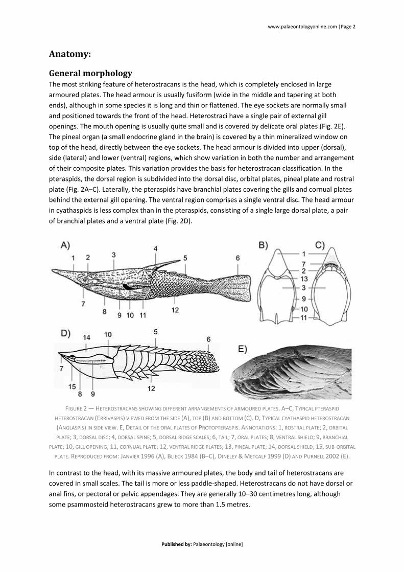
General morphology The most striking feature of heterostracans is the head, which is completely enclosed in large
armoured plates. The head armour is usually fusiform (wide in the middle and tapering at both
ends), although in some species it is long and thin or flattened. The eye sockets are normally small
and positioned towards the front of the head. Heterostraci have a single pair of external gill
openings. The mouth opening is usually quite small and is covered by delicate oral plates (Fig. 2E).
The pineal organ (a small endocrine gland in the brain) is covered by a thin mineralized window on
top of the head, directly between the eye sockets. The head armour is divided into upper (dorsal),
side (lateral) and lower (ventral) regions, which show variation in both the number and arrangement
of their composite plates. This variation provides the basis for heterostracan classification. In the
pteraspids, the dorsal region is subdivided into the dorsal disc, orbital plates, pineal plate and rostral
plate (Fig. 2A–C). Laterally, the pteraspids have branchial plates covering the gills and cornual plates
behind the external gill opening. The ventral region comprises a single ventral disc. The head armour
in cyathaspids is less complex than in the pteraspids, consisting of a single large dorsal plate, a pair
of branchial plates and a ventral plate (Fig. 2D).
FIGURE 2 — HETEROSTRACANS SHOWING DIFFERENT ARRANGEMENTS OF ARMOURED PLATES. A–C, TYPICAL PTERASPID
HETEROSTRACAN (ERRIVASPIS) VIEWED FROM THE SIDE (A), TOP (B) AND BOTTOM (C). D, TYPICAL CYATHASPID HETEROSTRACAN
(ANGLASPIS) IN SIDE VIEW. E, DETAIL OF THE ORAL PLATES OF PROTOPTERASPIS. ANNOTATIONS: 1, ROSTRAL PLATE; 2, ORBITAL
PLATE; 3, DORSAL DISC; 4, DORSAL SPINE; 5, DORSAL RIDGE SCALES; 6, TAIL; 7, ORAL PLATES; 8, VENTRAL SHIELD; 9, BRANCHIAL
PLATE; 10, GILL OPENING; 11, CORNUAL PLATE; 12, VENTRAL RIDGE PLATES; 13, PINEAL PLATE; 14, DORSAL SHIELD; 15, SUB-ORBITAL
PLATE. REPRODUCED FROM: JANVIER 1996 (A), BLIECK 1984 (B–C), DINELEY & METCALF 1999 (D) AND PURNELL 2002 (E).
In contrast to the head, with its massive armoured plates, the body and tail of heterostracans are
covered in small scales. The tail is more or less paddle-shaped. Heterostracans do not have dorsal or
anal fins, or pectoral or pelvic appendages. They are generally 10–30 centimetres long, although
some psammosteid heterostracans grew to more than 1.5 metres.
www.palaeontologyonline.com |Page 3
Published by: Palaeontology [online]
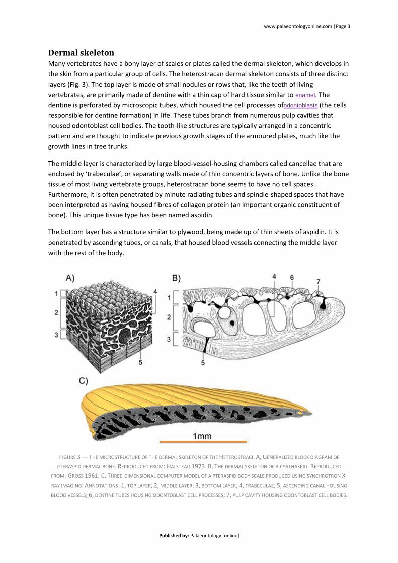
Dermal skeleton Many vertebrates have a bony layer of scales or plates called the dermal skeleton, which develops in
the skin from a particular group of cells. The heterostracan dermal skeleton consists of three distinct
layers (Fig. 3). The top layer is made of small nodules or rows that, like the teeth of living
vertebrates, are primarily made of dentine with a thin cap of hard tissue similar to enamel. The
dentine is perforated by microscopic tubes, which housed the cell processes ofodontoblasts (the cells
responsible for dentine formation) in life. These tubes branch from numerous pulp cavities that
housed odontoblast cell bodies. The tooth-like structures are typically arranged in a concentric
pattern and are thought to indicate previous growth stages of the armoured plates, much like the
growth lines in tree trunks.
The middle layer is characterized by large blood-vessel-housing chambers called cancellae that are
enclosed by ‘trabeculae’, or separating walls made of thin concentric layers of bone. Unlike the bone
tissue of most living vertebrate groups, heterostracan bone seems to have no cell spaces.
Furthermore, it is often penetrated by minute radiating tubes and spindle-shaped spaces that have
been interpreted as having housed fibres of collagen protein (an important organic constituent of
bone). This unique tissue type has been named aspidin.
The bottom layer has a structure similar to plywood, being made up of thin sheets of aspidin. It is
penetrated by ascending tubes, or canals, that housed blood vessels connecting the middle layer
with the rest of the body.
FIGURE 3 — THE MICROSTRUCTURE OF THE DERMAL SKELETON OF THE HETEROSTRACI. A, GENERALIZED BLOCK DIAGRAM OF
PTERASPID DERMAL BONE. REPRODUCED FROM: HALSTEAD 1973. B, THE DERMAL SKELETON OF A CYATHASPID. REPRODUCED
FROM: GROSS 1961. C, THREE-DIMENSIONAL COMPUTER MODEL OF A PTERASPID BODY SCALE PRODUCED USING SYNCHROTRON X-
RAY IMAGING. ANNOTATIONS: 1, TOP LAYER; 2, MIDDLE LAYER; 3, BOTTOM LAYER; 4, TRABECULAE; 5, ASCENDING CANAL HOUSING
BLOOD VESSELS; 6, DENTINE TUBES HOUSING ODONTOBLAST CELL PROCESSES; 7, PULP CAVITY HOUSING ODONTOBLAST CELL BODIES.
www.palaeontologyonline.com |Page 4
Published by: Palaeontology [online]
Internal anatomy The internal anatomy of the Heterostraci is known mostly from internal casts, which provide
impressions of the inside surface of the head armour. This evidence suggests that the heterostraci
had two distinct vertical semi-circular canals in the inner ear, which are important for sensing
rotation. The brain was small and elongated. The eyeballs were small and slightly conical.
The olfactory system, which provides vertebrates with a sense of smell, was paired and relatively large.
This is similar to the olfactory system of living jawed vertebrates, which possess paired nasal sacs,
and rather unlike the single nasal sac present in living jawless lampreys. The gill apparatus is often
preserved as a series of 8–10 furrows next to a series of small impressions.
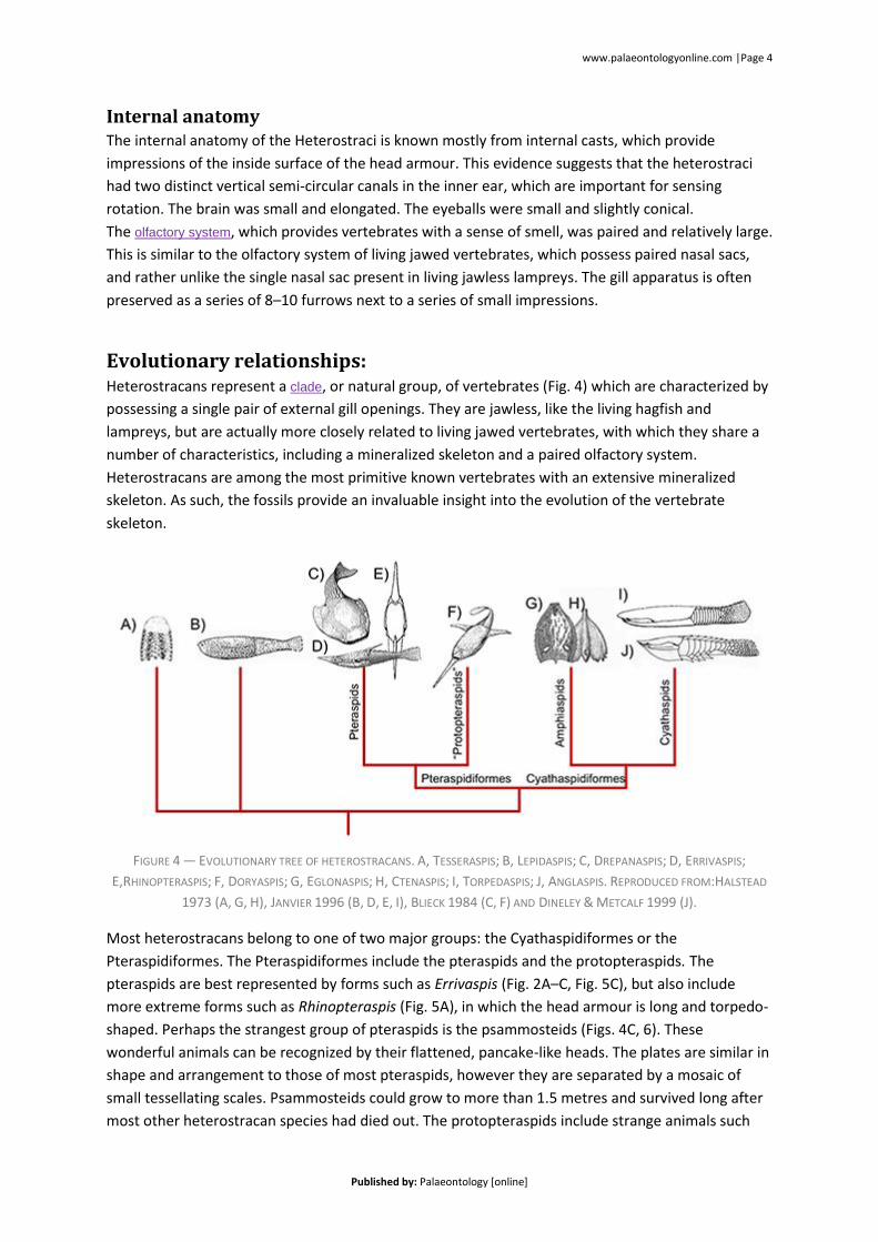
Evolutionary relationships: Heterostracans represent a clade, or natural group, of vertebrates (Fig. 4) which are characterized by
possessing a single pair of external gill openings. They are jawless, like the living hagfish and
lampreys, but are actually more closely related to living jawed vertebrates, with which they share a
number of characteristics, including a mineralized skeleton and a paired olfactory system.
Heterostracans are among the most primitive known vertebrates with an extensive mineralized
skeleton. As such, the fossils provide an invaluable insight into the evolution of the vertebrate
skeleton.
FIGURE 4 — EVOLUTIONARY TREE OF HETEROSTRACANS. A, TESSERASPIS; B, LEPIDASPIS; C, DREPANASPIS; D, ERRIVASPIS;
E,RHINOPTERASPIS; F, DORYASPIS; G, EGLONASPIS; H, CTENASPIS; I, TORPEDASPIS; J, ANGLASPIS. REPRODUCED FROM:HALSTEAD
1973 (A, G, H), JANVIER 1996 (B, D, E, I), BLIECK 1984 (C, F) AND DINELEY & METCALF 1999 (J).
Most heterostracans belong to one of two major groups: the Cyathaspidiformes or the
Pteraspidiformes. The Pteraspidiformes include the pteraspids and the protopteraspids. The
pteraspids are best represented by forms such as Errivaspis (Fig. 2A–C, Fig. 5C), but also include
more extreme forms such as Rhinopteraspis (Fig. 5A), in which the head armour is long and torpedo-
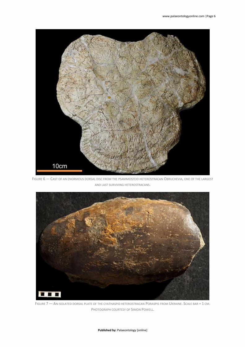
shaped. Perhaps the strangest group of pteraspids is the psammosteids (Figs. 4C, 6). These
wonderful animals can be recognized by their flattened, pancake-like heads. The plates are similar in
shape and arrangement to those of most pteraspids, however they are separated by a mosaic of
small tessellating scales. Psammosteids could grow to more than 1.5 metres and survived long after
most other heterostracan species had died out. The protopteraspids include strange animals such
www.palaeontologyonline.com |Page 5
Published by: Palaeontology [online]
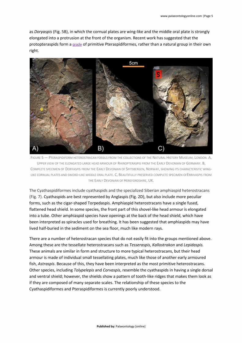
as Doryaspis (Fig. 5B), in which the cornual plates are wing-like and the middle oral plate is strongly
elongated into a protrusion at the front of the organism. Recent work has suggested that the
protopteraspids form a grade of primitive Pteraspidiformes, rather than a natural group in their own
right.
FIGURE 5 — PTERASPIDIFORM HETEROSTRACAN FOSSILS FROM THE COLLECTIONS OF THE NATURAL HISTORY MUSEUM, LONDON. A,
UPPER VIEW OF THE ELONGATED LARGE HEAD ARMOUR OF RHINOPTERASPIS FROM THE EARLY DEVONIAN OF GERMANY. B,
COMPLETE SPECIMEN OF DORYASPIS FROM THE EARLY DEVONIAN OF SPITSBERGEN, NORWAY, SHOWING ITS CHARACTERISTIC WING-
LIKE CORNUAL PLATES AND SWORD-LIKE MIDDLE ORAL PLATE. C, BEAUTIFULLY PRESERVED COMPLETE SPECIMEN OFERRIVASPIS FROM
THE EARLY DEVONIAN OF HEREFORDSHIRE, UK.
The Cyathaspidiformes include cyathaspids and the specialized Siberian amphiaspid heterostracans
(Fig. 7). Cyathaspids are best represented by Anglaspis (Fig. 2D), but also include more peculiar
forms, such as the cigar-shaped Torpedaspis. Amphiaspid heterostracans have a single fused,
flattened head shield. In some species, the front part of this shovel-like head armour is elongated
into a tube. Other amphiaspid species have openings at the back of the head shield, which have
been interpreted as spiracles used for breathing. It has been suggested that amphiaspids may have
lived half-buried in the sediment on the sea floor, much like modern rays.
There are a number of heterostracan species that do not easily fit into the groups mentioned above.
Among these are the tessellate heterostracans such as Tesseraspis, Kallostrakon and Lepidaspis.
These animals are similar in form and structure to more typical heterostracans, but their head
armour is made of individual small tessellating plates, much like those of another early armoured
fish, Astraspis. Because of this, they have been interpreted as the most primitive heterostracans.
Other species, including Tolypelepis and Corvaspis, resemble the cyathaspids in having a single dorsal
and ventral shield; however, the shields show a pattern of tooth-like ridges that makes them look as
if they are composed of many separate scales. The relationship of these species to the
Cyathaspidiformes and Pteraspidiformes is currently poorly understood.
www.palaeontologyonline.com |Page 6
Published by: Palaeontology [online]
FIGURE 6 — CAST OF AN ENORMOUS DORSAL DISC FROM THE PSAMMOSTEID HETEROSTRACAN OBRUCHEVIA, ONE OF THE LARGEST
AND LAST SURVIVING HETEROSTRACANS.
FIGURE 7 — AN ISOLATED DORSAL PLATE OF THE CYATHASPID HETEROSTRACAN PORASPIS FROM UKRAINE. SCALE BAR = 1 CM.
PHOTOGRAPH COURTESY OF SIMON POWELL.
www.palaeontologyonline.com |Page 7
Published by: Palaeontology [online]
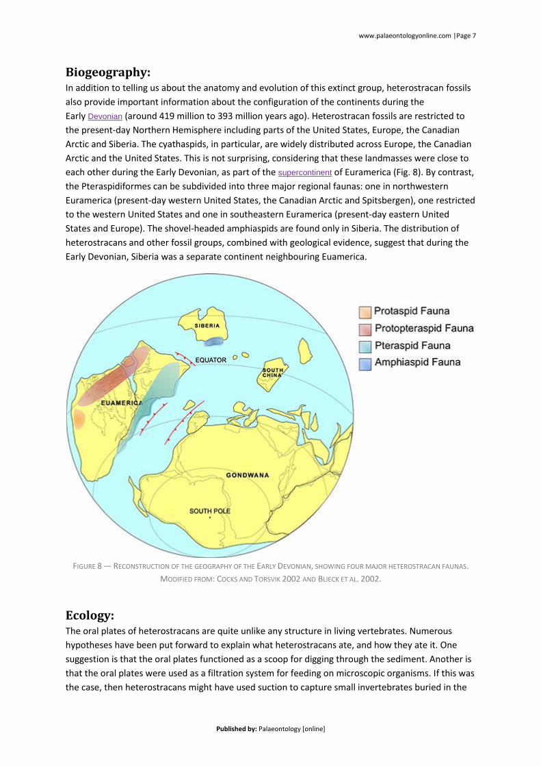
Biogeography: In addition to telling us about the anatomy and evolution of this extinct group, heterostracan fossils
also provide important information about the configuration of the continents during the
Early Devonian (around 419 million to 393 million years ago). Heterostracan fossils are restricted to
the present-day Northern Hemisphere including parts of the United States, Europe, the Canadian
Arctic and Siberia. The cyathaspids, in particular, are widely distributed across Europe, the Canadian
Arctic and the United States. This is not surprising, considering that these landmasses were close to
each other during the Early Devonian, as part of the supercontinent of Euramerica (Fig. 8). By contrast,
the Pteraspidiformes can be subdivided into three major regional faunas: one in northwestern
Euramerica (present-day western United States, the Canadian Arctic and Spitsbergen), one restricted
to the western United States and one in southeastern Euramerica (present-day eastern United
States and Europe). The shovel-headed amphiaspids are found only in Siberia. The distribution of
heterostracans and other fossil groups, combined with geological evidence, suggest that during the
Early Devonian, Siberia was a separate continent neighbouring Euamerica.
FIGURE 8 — RECONSTRUCTION OF THE GEOGRAPHY OF THE EARLY DEVONIAN, SHOWING FOUR MAJOR HETEROSTRACAN FAUNAS.
MODIFIED FROM: COCKS AND TORSVIK 2002 AND BLIECK ET AL. 2002.
Ecology: The oral plates of heterostracans are quite unlike any structure in living vertebrates. Numerous
hypotheses have been put forward to explain what heterostracans ate, and how they ate it. One
suggestion is that the oral plates functioned as a scoop for digging through the sediment. Another is
that the oral plates were used as a filtration system for feeding on microscopic organisms. If this was
the case, then heterostracans might have used suction to capture small invertebrates buried in the
www.palaeontologyonline.com |Page 8
Published by: Palaeontology [online]
sediment; alternatively, the oral plates may have been used to filter microscopic organisms, such as
plankton, that were suspended in the water. The oral plates have also been compared directly to
teeth and it has been suggested that they functioned as biting, grasping, shearing or scraping
structures used to feed on larger prey animals (such as arthropods). Unfortunately, there is little
evidence to support any of these hypotheses. In fact, our current understanding of heterostracan
biology contradicts some of the proposed feeding methods. For instance, without jaws it is unclear
how heterostracans would have produced sufficient suction to capture microorganisms.
One technique we can use to test these hypotheses is to examine the microscopic patterns of wear
on the oral plates. Each of the proposed feeding methods should produce a different pattern of
wear; for instance, if the oral plates were used as a scoop, we would expect the plates to show
heavy scratching and damage. A study by palaeontologist Mark Purnell at the University of Leicester,
UK, showed that oral plates in a variety of heterostracan species are pristinely preserved, with little
evidence of wear. Furthermore, these plates are covered with tiny delicate barbs that would have
been destroyed if the oral plates had been used to bite or scrape in life. Microscopic evidence
therefore effectively falsifies all but one feeding hypothesis: that heterostracans ate microscopic
organisms suspended in the water. This is therefore the current best surmise for how they fed.
Summary: The Heterostraci make up a diverse and interesting group of Palaeozoic fish that occupy an
important position in vertebrate evolution, close to the origin of the mineralized skeleton. Over the
past 150 years, we have begun to build up a picture of heterostracan biology. This has enhanced our
understanding of Palaeozoic geography and ecology, and informed our knowledge of the evolution
of jawed vertebrates. However, we still have much to learn about heterostracan evolution and
diversification. For example, how are the tessellated heterostracans related to other groups? We
also know little about heterostracan growth and the development of their armoured skeleton. A
greater understanding of their skeletal biology may well provide fundamental insights into the
earliest experiments in vertebrate skeleton building. Further study and application of new
techniques will surely continue to enhance our understanding of these peculiar and fascinating
animals.
Acknowledgements: Joe would like to thank Professor Philip Donoghue and Dr Alain Blieck for their helpful comments
and suggestions.
Further Reading:
Blieck, A. R. M., Karatajute-Talimaa, V. N. & Mark-Kurik, E. 2002. Upper Silurian and Devonian
heterostracan pteraspidomorphs (Vertebrata) from Severnaya Zemlya (Russia): a preliminary report
with biogeographical and biostratigraphical implications. Geodiversitas 24, 805–820.
(http://www.mnhn.fr/publication/geodiv/g02n4a6.pdf)
www.palaeontologyonline.com |Page 9
Published by: Palaeontology [online]
Halstead, L. B. 1973. The heterostracan fishes. Biological Reviews 48, 279–332. (doi:10.1111/j.1469-
185X.1973.tb01005.x)
Janvier, P. 1996. Early vertebrates. Clarendon Press, Oxford. (ISBN:0198540477 9780198540472)
Purnell, M. A. 2002. Feeding in extinct jawless heterostracan fishes and testing scenarios of early
vertebrate evolution.Proceedings of the Royal Society of London B 269, 83–88.
(doi:10.1098/rspb.2001.1826)
White, E. I. 1973. Form and growth in Belgicaspis (Heterostraci). Palaeontographica Abteilung A 143,
11–24.
(http://www.schweizerbart.de/papers/pala/detail/A143/70788/Form_and_growth_in_Belgicaspis_Heterostraci)
http://tolweb.org/Heterostraci/16904 — Tree of Life Web Project page on the Heterostraci.
1 School of Earth Sciences, University of Bristol, Wills Memorial Building, Queen’s Road, Bristol BS8 1RJ, UK.


