Embed Size (px)
Citation preview
Proteomes 2014, 2, 169-190; doi:10.3390/proteomes2020169
proteomes ISSN 2227-7382
www.mdpi.com/journal/proteomes
Article
Effect of Aluminum Treatment on Proteomes of Radicles of
Seeds Derived from Al-Treated Tomato Plants
Ikenna Okekeogbu 1, Zhujia Ye
1, Sasikiran Reddy Sangireddy
1, Hui Li
1, Sarabjit Bhatti
1,
Dafeng Hui 1, Suping Zhou
1,*, Kevin J. Howe
2, Tara Fish
2, Yong Yang
2 and
Theodore W. Thannhauser 2,†
1 Department of Agricultural and Environmental Sciences, College of Agriculture,
Human and Natural Sciences, Tennessee State University, 3500 John A Merritt Blvd, Nashville,
TN 37209, USA; E-Mails: [email protected] (I.O.); [email protected] (Z.Y.);
[email protected] (S.R.S.); [email protected] (H.L.); [email protected] (S.B.);
[email protected] (D.H.) 2
Plant, Soil and Nutrition Research Unit, USDA-ARS, Tower Rd, Ithaca, NY 14853, USA;
E-Mails: [email protected] (K.J.H.); [email protected] (T.F.); [email protected] (Y.Y.)
† Correspondence author for proteomics analysis; E-Mail: [email protected].
* Author to whom correspondence should be addressed; E-Mail: [email protected];
Tel.: +1-931-802-1300; Fax: +1-615-963-5888.
Received: 18 February 2014; in revised form: 4 March 2014 / Accepted: 6 March 2014 /
Published: 28 March 2014
Abstract: Aluminum (Al) toxicity is a major constraint to plant growth and crop yield in
acid soils. Tomato cultivars are especially susceptible to excessive Al3+
accumulated in the
root zone. In this study, tomato plants were grown in a hydroponic culture system
supplemented with 50 µM AlK(SO4)2. Seeds harvested from Al-treated plants contained a
significantly higher Al content than those grown in the control hydroponic solution. In this
study, these Al-enriched tomato seeds (harvested from Al-treated tomato plants) were
germinated in 50 µM AlK(SO4)2 solution in a homopiperazine-1,4-bis(2-ethanesulfonic
acid) buffer (pH 4.0), and the control solution which contained the buffer only. Proteomes
of radicles were analyzed quantitatively by mass spectrometry employing isobaric tags for
relative and absolute quantitation (iTRAQ®
). The proteins identified were assigned to
molecular functional groups and cellular metabolic pathways using MapMan. Among the
proteins whose abundance levels changed significantly were: a number of transcription
factors; proteins regulating gene silencing and programmed cell death; proteins in primary
OPEN ACCESS
Proteomes 2014, 2 170
and secondary signaling pathways, including phytohormone signaling and proteins for
enhancing tolerance to abiotic and biotic stress. Among the metabolic pathways, enzymes
in glycolysis and fermentation and sucrolytic pathways were repressed. Secondary
metabolic pathways including the mevalonate pathway and lignin biosynthesis were
induced. Biological reactions in mitochondria seem to be induced due to an increase in the
abundance level of mitochondrial ribosomes and enzymes in the TCA cycle, electron
transport chains and ATP synthesis.
Keywords: iTRAQ; orbitrap mass spectrometry; Al-enriched tomato seeds; seed germination;
root proteomics; functional pathways
1. Introduction
Aluminum is not an essential mineral to plants; Al ions (Al3+
), when at an excessive level, are very
toxic to seed germination in both tolerant and susceptible plants [1,2]. Of all the Al-induced phytotoxic
symptoms, disruption of cell division and growth within the root apex has the most significant impact
on plant growth and yield [3,4]. Thus, many studies are focused on the physiological and molecular
activities in the root tip zone related to reducing Al3+
phytotoxicity. Tolerance to Al is achieved by
avoidance mechanisms (e.g., through secretion of organic acids to bind Al3+
near the vicinity of root
tips) [5–7], and internal resistance, by remodeling cellular processes [8,9] and through apoplastic and
symplastic detoxification of internalized Al [10,11].
Seed germination is the process by which an embryo transitions into a complete plant. It begins as
the root (radicle) becomes the first embryonic organ to emerge from the seed coat; this is followed by
elongation of the hypocotyl and ends in expansion of the cotyledon (s) [12–14]. Radicle emergence
involves both cell division and cell enlargement. Radicle growth (mainly involving cell enlargement) is
sensitive to metal toxicity [15]. A study using Arabidopsis thaliana shows that de novo protein synthesis
from the pool of stored mRNA is essential for the completion of radicle protrusion; however, the
process can proceed even in the absence of transcription (de novo mRNA synthesis) [16]. Pre-incubation
of wheat (Triticum aestivum) seedlings with low doses of Al increased tolerance to subsequent exposure
to lethal concentrations. The study also concluded that synthesis of proteins is essential for acquiring
tolerance to Al because addition of the protein translation inhibitor, cycloheximide, completely
abolished the induced tolerance to Al toxicity [17]. Therefore, proteome changes in the primary root can
directly affect the development of tolerance and may represent the key to understanding the molecular
mechanism involved.
Tomato (Solanum lycopersicum) is among the few species that produce very acidic fruits (pH < 3.0
in ripened tomato fruit), thereby providing an environment capable of shifting the equilibrium from the
benign Al2+
form to the highly toxic Al3+
. In this study, tomato plants were grown in a hydroponic
system supplemented with Al during the reproductive stages (from flowering until fruit ripening).
Tomato seeds produced by these plants were considered to be Al-enriched as they contained a higher
Al content than those harvested from plants growing in a solution without added Al. Subsequently, the
Al-enriched seeds were germinated in an Al solution, and a proteomics analysis of their radicles was
Proteomes 2014, 2 171
performed to identify proteome changes in response to the Al treatment as a means to identify
candidate proteins that could play a key role in acquiring Al tolerance.
2. Experimental
Tomato (S. lycopersicum cv. Micro-Tom) plants were grown in a hydroponic culture system. As
soon as plants started to set fruits (pea-sized fruits were seen on the first fruit cluster), AlK(SO4)2 was
added up to a final concentration of 7.2 µM of Al3+
activity [or 50 µM AlK(SO4)2]. The pH of the
solution was tested daily using pH strips (Fisher Scientific) and the solution was refreshed weekly or
when the pH increased to 5.0. Tomato fruits were harvested periodically when the color turned red. To
collect tomato seeds, fruits were wrapped in paper towels to squeeze out all the tomato juice. After
removal of the gelatinous sack tissues, seeds were soaked in 50% bleach for 5 min followed by three
rinses in autoclaved water. Seeds were stored at 4 °C until analysis. These field experiments were
performed for two seasons. Mineral analysis of seed tissues (embryo and seed coat separately) found
that the Al content of embryo was 10–15 mg per kg dry weight (DW) for seeds derived from Al-treated
plants, and it was 6–8 mg per kg DW for those harvested from plants growing in the same hydroponic
system but without adding AlK(SO4)2. In this experiment, it was noticed that control samples including
roots, leaves and seeds also contained Al although the content level was much lower than the treated
samples. Consistently in the two-season experiments, the Al-treated embryos contained a significantly
(p < 0.01 using t-test) lower amount of boron (B) and iron (Fe) [18].
A seed germination assay was conducted to test the effect of Al treatment on improving Al
tolerance of the next-generation offspring. Seeds (Al-enriched) that were harvested from Al-treated
plants were germinated on wet filter paper soaked in a 50 mM homopiperazine-1,4-bis(2-ethanesulfonic
acid) (Homopipes) buffer containing 50 µM AlK(SO4)2 (pH 4.5–5.0) in the Al-treated group, as
opposed to the buffer only in the control treatment. The germination was carried out at 25 ± 2 °C. The
lengths of radicles were measured on the third day of germination. There was no significant difference
in terms of radicle length between Al-treated (5 ± 1 mm) and untreated groups (4 ± 1 mm). These
results indicate that radicle elongation growth was not affected by the presence of Al in the solution for
these seeds.
For preparation of this study, seeds were germinated under the same conditions, with three
biological replicates for treated and control experiments. Each biological replicate consisted of 100 seeds
with 10 seeds wrapped in one filter paper sandwich. Radicles protruding from seed coat were dissected
using a sharp blade on the third day after germination. Tissues were frozen in liquid nitrogen and
ground into a fine powder immediately after harvest.
2.1. Protein Extraction and Isobaric Tags for Relative and Absolute Quantification Labeling
For protein extraction, tissue powder was washed sequentially in 10% TCA/acetone, 80%
methanol/0.1 M ammonium acetate, and 80% acetone with centrifugation to pellet the powder after
each wash. The protein was then extracted in a phenol (pH 8.0) and dense SDS buffer [30% sucrose,
2% SDS, 0.1 M Tris-HCl, pH 8.0, 5% beta-mercaptoethanol (v/w)]. After incubation at 4 °C for 2 h,
the mixture was centrifuged at 16,000 g at 4 °C for 20 min. Protein in the upper phenol phase was
precipitated in 0.1 M ammonium acetate in methanol after incubation overnight at −20 °C. Protein
Proteomes 2014, 2 172
pellets were washed in methanol and acetone and were then dissolved in a buffer of 500 mM
triethylammonium bicarbonate (TEAB) and 2 M urea, and 0.1% SDS and a proteinase inhibitor
cocktail for plant tissue (100 × dilution in the extraction buffer) (Sigma, St. Louis, MO, USA). Protein
concentration was determined using a Bradford assay kit (Bio-Rad, Hercules, CA, USA).
One hundred µg of protein from each sample was digested with trypsin and then labeled as
previously described [19] following the instructions accompanying the 8-plex iTRAQ®
labeling kit
(AB SCIEX, Framingham, MA, USA). The treated samples were labeled with tags 113, 114 and 115
and the control samples with 116, 117 and 118 were combined. Unbound tags and SDS were removed
through cation exchange cartridge (AB SCIEX), and salts were removed using reverse-phase
solid-phase extraction procedure involving 1-cm3, 50-mg cartridges following the manufacturer’s
instructions (Sep-Pak C18; Waters, Milford, MA, USA). Peptides were eluted in 500 L 50% (v/v)
acetonitrile with 0.1% TFA and dried under vacuum.
These peptide samples were subjected to a first dimension of high pH Ultra Performance Liquid
Chromatography (UPLC) separation using an Acquity UPLC System (Waters) coupled with a robotic
fraction collector (Probot; Dionex, Sunnyvale, CA, USA) [19]. One hundred micrograms of the
multiplexed sample was injected and fractionated into 48 fractions in a 96-well plate. The 48 fractions
were concatenated to yield 16 samples pools by pooling every 16th sample. These were dried at
reduced pressure using a CentiVac Concentrator (LabConco, Kansas City, MO, USA). For the low pH
2nd dimension, low pH reverse-phase (RP) chromatography was employed. Dried samples were
reconstituted with 15 µL of 2% acetonitrile with 0.5% formic acid. Nano-LC separations of tryptic
peptides were performed as described previously [20,21]. The eluent from the analytical column was
delivered to the LTQ-Orbitrap Elite (Thermo-Fisher Scientific,Waltham, MA, USA) via a “Plug and
Play” nano ion source (CorSolutions LLC, Ithaca, NY, USA). The mass spectrometer was externally
calibrated across the m/z range from 375–1,800 with Ultramark 1621 for the FT mass analyzer, and
individual runs were internally calibrated with the background polysiloxane ion at m/z 445.1200025 as
a lock mass.
The Orbitrap Elite was operated in the positive ion mode with nanosource voltage set at 1.7 kV and
source temperature at 250 °C. A parallel DDA mode was used to obtain one MS survey scan with the
FT mass analyzer, followed by isolation and fragmentation of the 15–20 most abundant, multiply-charged
precursor ions with a threshold ion count higher than 50,000 in both the LTQ mass analyzer and the
high energy collisionally induced dissociation (HCD)-based FT mass analyzer at a resolution of 15,000
(fwhm m/z 400). MS survey scans were acquired with resolution set at 60,000 across the survey scan
range (m/z 375–1800). Dynamic exclusion was utilized with repeat count set to 1 with a 40 s repeat
duration; exclusion list size was set to 500, 20–30 s exclusion duration, and low and high exclusion
mass widths set to 1.5. Fragmentation parameters were set with isolation width at 1.5 m/z, normalized
collision energy at 37%, activation Q at 0.25. Activation time for HCD analysis was 0.1 min. All data
were acquired using XCalibur 2.1 (Thermo-Fisher Scientific).
Proteomes 2014, 2 173
2.2. Data Processing, Database Searching and iTRAQ Quantitation
Proteome Discoverer v 1.4 was used to convert raw spectral data files for each iTRAQ experiment
into a merged peak list (mgf format) containing all 2nd dimension fractions for each tomato
experiment for subsequent database searching. Mascot Daemon v. 2.3.2 was used to query .mgf files
against an iTAG 2.3 tomato protein database [22]. Trypsin was selected as the enzyme with 1 missed
cleavage allowed. Methylthiolation of cysteine, oxidation of methionine, and deamidation of
asparagine and glutamine were set as variable modifications. Peptide charge was set to 2+, 3
+, and 4
+.
Precursor tolerance was set to 10 ppm, while fragment tolerance was set to 100 mmu. The instrument
selected was ESI-FTICR. The iTRAQ quantitation method utilized a weighted protein ratio type,
featured outlier removal, and required a minimum of 2 peptides for protein quantitation. Summed
normalization was used. For the iTRAQ 8-plex labeling, N-terminal and lysine modification with
iTRAQ were set as fixed modifications, and tyrosine labeling was set as a variable modification. Upon
completion of searching, each report was opened and results were exported after setting the ion score
filter to 0.1, thereby exporting only results with an expectation value below 0.1, specifying unique
peptides only. Only the highest scoring matches to a particular peptide sequence, listed under the
highest scoring protein containing that match, were considered.
2.3. Protein Quantification, Statistics, and Protein Functional Analysis
For a protein to be included in the quantitative analysis, it was required that at least two unique
peptides (with the normalized intensity levels raw intensity >20) were identified in all the six
biological samples (three biological replicates each in Al-treated and control groups). The normalized
peak intensities of reporter ions of constituent peptides were log2 transformed. Then, log2 fold values
from all constituent peptides were subjected to t-test (general linear model procedure) followed by
false discovery rate (FDR) corrections to test the statistical significance of the difference in normalized
abundance of each protein between Al-treated and control sample groups [19]. The log2 transformed
abundance ratios were then fit to a normal distribution. Two standard deviations (at a 95% confidence
level) of the log2 fold (from treated to untreated control) in protein abundance were used as the cutoff
threshold for significantly changed proteins. Statistical analyses were performed using SAS (version
9.3; SAS Institute, Cary, NC, USA).
MapMan [23] was used to associate the identified tomato proteins with cellular process and
metabolic pathways using iTAG 2.3 tomato protein database downloaded from the MapMan website.
The putative functions of the identified proteins were also discussed based on relevant information
from literature and database searches on tomatoes and other plant species.
3. Results and Discussion
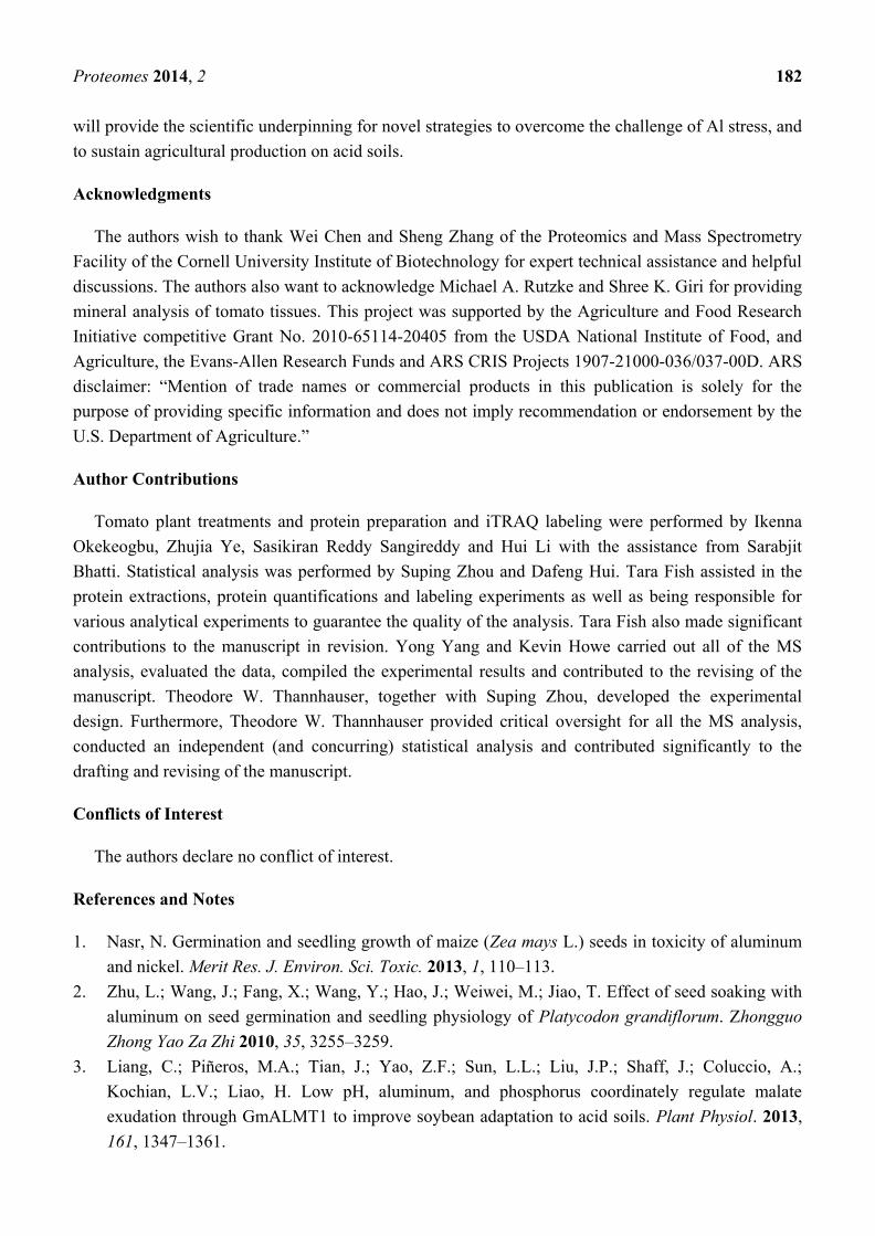
In this study, 3,160 proteins, meeting the quantification analysis criteria of two or more peptides,
were identified in all six biological samples. The spectral intensity of each peptide was transformed
into log (base 2) values, and principal component analysis (PCA) (Figure 1) separated the treated and
control triplicate groups, which indicates that there is a systematic difference in protein composition
between the two groups. The low percentage in component 2 compared to component 1 (2.7% vs.
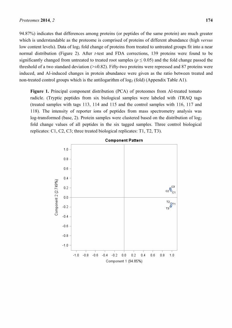
Proteomes 2014, 2 174
94.87%) indicates that differences among proteins (or peptides of the same protein) are much greater
which is understandable as the proteome is comprised of proteins of different abundance (high versus
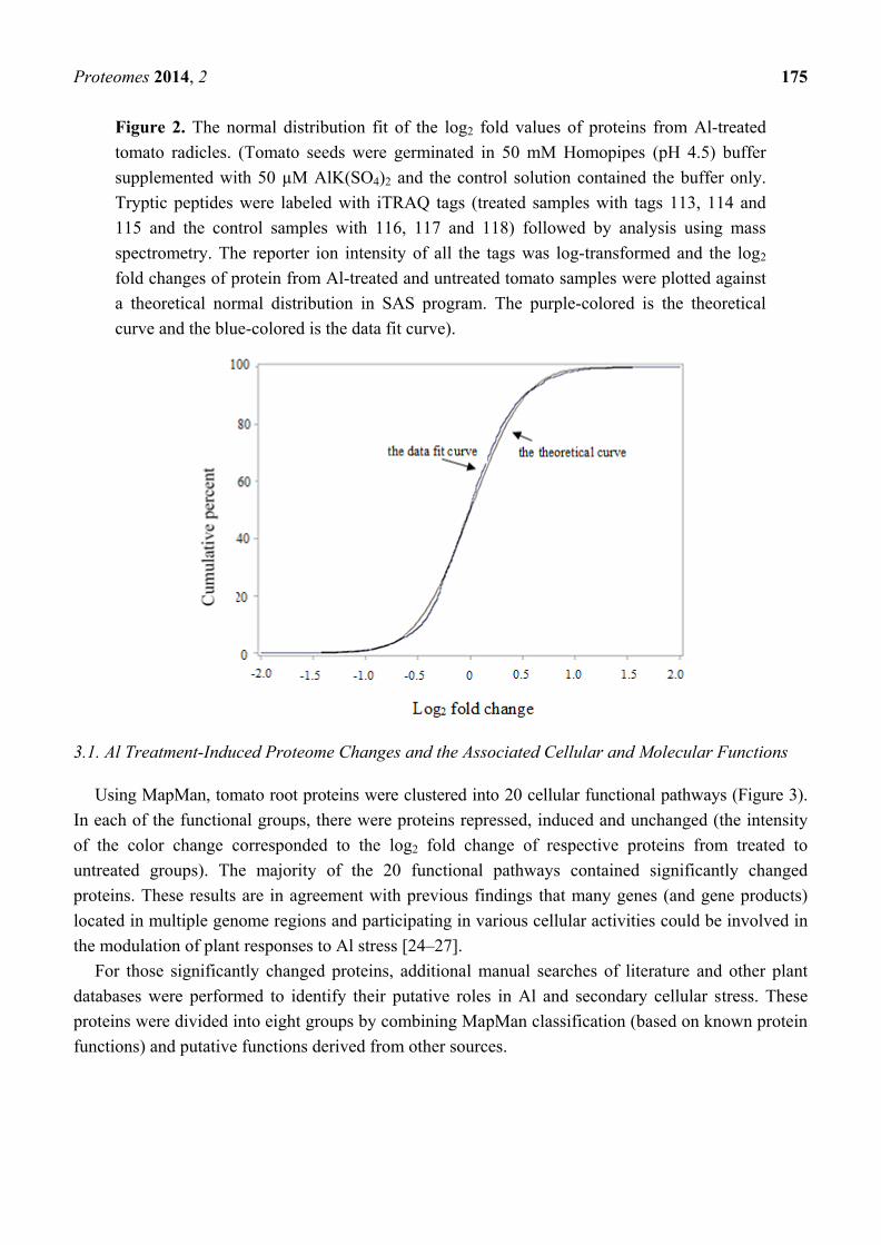
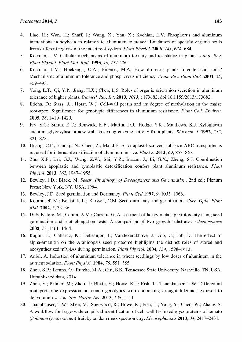
low content levels). Data of log2 fold change of proteins from treated to untreated groups fit into a near
normal distribution (Figure 2). After t-test and FDA corrections, 139 proteins were found to be
significantly changed from untreated to treated root samples (p ≤ 0.05) and the fold change passed the
threshold of a two standard deviation (>±0.82). Fifty-two proteins were repressed and 87 proteins were
induced, and Al-induced changes in protein abundance were given as the ratio between treated and
non-treated control groups which is the antilogarithm of log2 (fold) (Appendix Table A1).
Figure 1. Principal component distribution (PCA) of proteomes from Al-treated tomato
radicle. (Tryptic peptides from six biological samples were labeled with iTRAQ tags
(treated samples with tags 113, 114 and 115 and the control samples with 116, 117 and
118). The intensity of reporter ions of peptides from mass spectrometry analysis was
log-transformed (base, 2). Protein samples were clustered based on the distribution of log2
fold change values of all peptides in the six tagged samples. Three control biological
replicates: C1, C2, C3; three treated biological replicates: T1, T2, T3).
Proteomes 2014, 2 175
Figure 2. The normal distribution fit of the log2 fold values of proteins from Al-treated
tomato radicles. (Tomato seeds were germinated in 50 mM Homopipes (pH 4.5) buffer
supplemented with 50 µM AlK(SO4)2 and the control solution contained the buffer only.
Tryptic peptides were labeled with iTRAQ tags (treated samples with tags 113, 114 and
115 and the control samples with 116, 117 and 118) followed by analysis using mass
spectrometry. The reporter ion intensity of all the tags was log-transformed and the log2
fold changes of protein from Al-treated and untreated tomato samples were plotted against
a theoretical normal distribution in SAS program. The purple-colored is the theoretical
curve and the blue-colored is the data fit curve).
3.1. Al Treatment-Induced Proteome Changes and the Associated Cellular and Molecular Functions
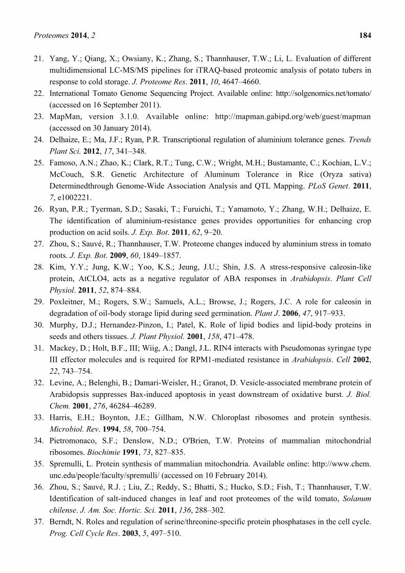
Using MapMan, tomato root proteins were clustered into 20 cellular functional pathways (Figure 3).
In each of the functional groups, there were proteins repressed, induced and unchanged (the intensity
of the color change corresponded to the log2 fold change of respective proteins from treated to
untreated groups). The majority of the 20 functional pathways contained significantly changed
proteins. These results are in agreement with previous findings that many genes (and gene products)
located in multiple genome regions and participating in various cellular activities could be involved in
the modulation of plant responses to Al stress [24–27].
For those significantly changed proteins, additional manual searches of literature and other plant
databases were performed to identify their putative roles in Al and secondary cellular stress. These
proteins were divided into eight groups by combining MapMan classification (based on known protein
functions) and putative functions derived from other sources.

Proteomes 2014, 2 176
Figure 3. Cell function overview of proteins from radicles of Al-treated tomato seeds. (The
graphic was generated using the Cell-Function Overview and Slyc-iTAG2.3 as the
reference database in MapMan. The intensity of the color change corresponds to the scale
created based on log2 fold of protein from Al-treated to untreated groups.)
3.1.1. Mobilization of Seed Storage Proteins in Al-Treated Tomato Radicles
Mature seeds contain many species of hydrophilic proteins, such as dehydrins, globulins, and late
embryogenesis abundant proteins (LEA). During seed germination, these seed proteins play a key role
in maintaining intracellular water balance by controlling water uptake, and they are also recycled to
provide a nitrogen source for the germinating embryo. In this study, Al-treated radicles were found to
contain a lower level of hydrophilic proteins, such as globulin, vicilin, LEA, seed biotin-containing
protein SBP65, dehydrin, and small hydrophilic plant seed protein. These results suggest that the
Al treatment induced more active catabolism of hydrophilic proteins or inhibited transport of those
proteins from cotyledons to the growing radicles. Both processes can result in a low hydrophilic
protein content in the Al-treated radicles.
In addition, two seed oil body-associated proteins, caleosin and oleosin, were also repressed in
Al-treated radicles. These lipid body-binding proteins play a key role in the degradation of storage
lipid during seed germination [28,29]. A decrease in oleosin content can lead to coalescence of lipid
bodies which is harmful to cells, or it may make the radicle more susceptible to dehydration as these
proteins also affect tissue tolerance to desiccation [30]. In summary, changes in proteins of this group
will make tomato more sensitive to dehydration stress, which can happen under excessive salt and
Proteomes 2014, 2 177
drought stresses. This could be one of the major molecular mechanisms by which Al toxicity can
exacerbate the impact of other environmental stress factors during seed germination.
3.1.2. Proteins Involved in Cell Organization, Cell Division and Cell Cycle
In the Al-treated radicles, proteins affecting cell division cycle (protease ftsH homolog) and cell
skeleton structure (actin and tubulin) were repressed. On the other hand, proteins in programmed cell
death (PCD) or related processes were induced. These are the RPM1 interacting proteins which are
essential for hypertensive cell death in reaction to pathogens [31] and the vesicle-associated membrane
family protein which has a critical role in regulating execution of PCD by affecting the rate of
membrane recycling, especially under oxidative stress [32].
3.1.3. Proteins Involved in Regulation of Transcription
Expression of genetic materials provides the basis for all physiological traits. One of the first
critical steps is the regeneration of mature mRNA (gene transcripts). In tomato radicles, a number of
transcription factors were affected by Al treatment. Several C2H2 zinc finger family proteins were
induced, but CCHC zinc finger, CCCH-type zinc finger and ZF-HD class zinc finger-homeodomain
proteins were repressed. Additional induced proteins are Myb transcription factor, BolA-like protein
and bZIP transcription factor. DNA silencing and mRNA decay are both important mechanisms in
regulating gene expression, especially under stress conditions. Proteins in these categories were induced
in the Al-treated tomato radicles, and include DNA (cytosine-5-)-methyltransferase 3, U6 RNA-associated
Sm-like proteins and LSm6. Two proteins in the nonsense-mediated mRNA decay (NMD) pathway
were also identified; they are eukaryotic translation initiation factor SUI1, and eukaryotic translation
initiation factor 4 gamma-MIF4-like. Activation of these mechanisms can help plant cells to rid themselves
of aberrant proteins and transcripts, and ensure a “healthy mRNA pool” in the Al-treated tissues.
3.1.4. Proteins Affecting Protein Synthesis and Post-Translational Modification
Plants have three separate sets of genomes in chloroplasts, mitochondria, and cell nucleus. While
nuclear genes are translated in the cytoplasm, the mitochondria and chloroplasts each contain its own
translation machinery within the respective organelles [33,34]. Changes in ribosomal proteins suggest
that protein translation in plastids and mitochondria may be more active as ribosomal proteins
annotated to those organelles were induced, which include 30S ribosomal protein S7, ribosomal
protein L24 and L27 for chloroplasts, and mitochondrial ribosomal protein L37 and L15 and
peptidyl-tRNA hydrolase ICT1. In mitochondria, all the proteins in the electron transport chain and
ATP synthase are synthesized by mitochondrial ribosomes [35]. The Al-induced ribosomal protein
expression could have some effect on mitochondrial functions (which will be discussed later).
Cytoplasmic ribosomes which are responsible for the translation of nuclear-genome encoded genes
revealed more complex changes. Some proteins were induced including ribosomal protein S21e and
L32e, whereas others were repressed including ribosomal protein S26e, L15e and L19/L19e.
Differential relative abundances of these ribosomal proteins were also found in tomatoes under other
abiotic stresses [19,36].
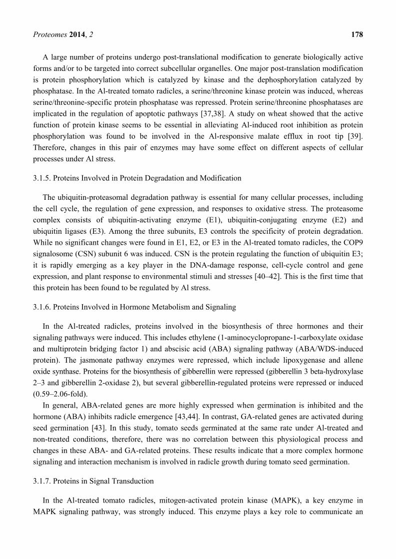
Proteomes 2014, 2 178
A large number of proteins undergo post-translational modification to generate biologically active
forms and/or to be targeted into correct subcellular organelles. One major post-translation modification
is protein phosphorylation which is catalyzed by kinase and the dephosphorylation catalyzed by
phosphatase. In the Al-treated tomato radicles, a serine/threonine kinase protein was induced, whereas
serine/threonine-specific protein phosphatase was repressed. Protein serine/threonine phosphatases are
implicated in the regulation of apoptotic pathways [37,38]. A study on wheat showed that the active
function of protein kinase seems to be essential in alleviating Al-induced root inhibition as protein
phosphorylation was found to be involved in the Al-responsive malate efflux in root tip [39].
Therefore, changes in this pair of enzymes may have some effect on different aspects of cellular
processes under Al stress.
3.1.5. Proteins Involved in Protein Degradation and Modification
The ubiquitin-proteasomal degradation pathway is essential for many cellular processes, including
the cell cycle, the regulation of gene expression, and responses to oxidative stress. The proteasome
complex consists of ubiquitin-activating enzyme (E1), ubiquitin-conjugating enzyme (E2) and
ubiquitin ligases (E3). Among the three subunits, E3 controls the specificity of protein degradation.
While no significant changes were found in E1, E2, or E3 in the Al-treated tomato radicles, the COP9
signalosome (CSN) subunit 6 was induced. CSN is the protein regulating the function of ubiquitin E3;
it is rapidly emerging as a key player in the DNA-damage response, cell-cycle control and gene
expression, and plant response to environmental stimuli and stresses [40–42]. This is the first time that
this protein has been found to be regulated by Al stress.
3.1.6. Proteins Involved in Hormone Metabolism and Signaling
In the Al-treated radicles, proteins involved in the biosynthesis of three hormones and their
signaling pathways were induced. This includes ethylene (1-aminocyclopropane-1-carboxylate oxidase
and multiprotein bridging factor 1) and abscisic acid (ABA) signaling pathway (ABA/WDS-induced
protein). The jasmonate pathway enzymes were repressed, which include lipoxygenase and allene
oxide synthase. Proteins for the biosynthesis of gibberellin were repressed (gibberellin 3 beta-hydroxylase
2–3 and gibberellin 2-oxidase 2), but several gibberellin-regulated proteins were repressed or induced
(0.59–2.06-fold).
In general, ABA-related genes are more highly expressed when germination is inhibited and the
hormone (ABA) inhibits radicle emergence [43,44]. In contrast, GA-related genes are activated during
seed germination [43]. In this study, tomato seeds germinated at the same rate under Al-treated and
non-treated conditions, therefore, there was no correlation between this physiological process and
changes in these ABA- and GA-related proteins. These results indicate that a more complex hormone
signaling and interaction mechanism is involved in radicle growth during tomato seed germination.
3.1.7. Proteins in Signal Transduction
In the Al-treated tomato radicles, mitogen-activated protein kinase (MAPK), a key enzyme in
MAPK signaling pathway, was strongly induced. This enzyme plays a key role to communicate an
Proteomes 2014, 2 179
external signal from a receptor on the surface of the cell to the DNA in the nucleus of the cell [45].
Earlier studies using cell suspension cultures of coffee (Coffea arabica) found that the MAPK was
activated by the oxidative burst induced by Al treatment but this protein is not necessarily associated
with Al tolerance [46,47]. Additionally, several calcium-binding proteins that participate in the
secondary calcium cell signaling pathways to mediate intracellular stress responses were also induced
in the Al-treated tomato radicles. Therefore, the higher abundance of MAPK and Ca-binding proteins
in Al-treated tomato radicles may also have a role in enhancing plant tolerance to the secondary
cellular stresses induced by Al stress.
More importantly, the rapid alkalinization factor (RALF) proteins were induced in the Al-treated
tomato radicles. RALF is a 5-kDa ubiquitous polypeptide initially isolated from tobacco leaves that
induces a rapid alkalinization of the culture medium of tobacco suspension-cultured cells and a
concomitant activation of an intracellular mitogen-activated protein kinase [48]. A synthetic tomato
RALF homolog peptide, when supplied to germinating tomato and Arabidopsis thaliana seeds, caused
an arrest of root growth and development [49]. This is the first finding that Al induced an increase in
the endogenous level of RALF in tomato radicles, which might also have some roles in regulating cell
cycle under the stress condition.
3.1.8. Stress Proteins
The induced proteins include the universal stress protein family, major latex-like proteins and
germin-like protein and wound/stress protein. Some of these proteins were also induced during tomato
seed germination of normal seeds (seeds harvested from non-treated plants) in Al-treated solution [27].
However, it seems that Al-treated radicles contained a lower level of several heat shock proteins,
including Hsp40, DnaJ, DnaJ 2, Hsp 70, ClpB chaperone, class IV HSP and class I HSP, as well as a
low-temperature-induced 65 kDa protein. These important protection proteins are mostly induced by
stress factors, and they may have a different role under Al stress.
Oxidative burst is an important secondary cellular stress induced by Al [50]. Among the antioxidant
enzymes, thioredoxin and superoxide dismutase, and germin (oxalate oxidase) were induced in
Al-treated radicles. In contrast, germin protein was reduced in tomato “Money Maker” treated with the
same type of stress [27]. Previous studies indicate that the Al-induced up-regulation of oxalate oxidase
gene in the root tip of wheat helps roots to get rid of Al-damaged cells and maintain a healthy
epidermal layer of roots, thus protecting the deeper layer of the meristematic and elongation zone that
are essential for root growth [51]. Therefore, the induction of germin and germin-like proteins may
enhance Al tolerance which was acquired during exposure to Al during seed germination.
3.1.9. Enzymes in Cellular Metabolism
Protein changes in different metabolic pathways are shown in Figure 4. In the Al-treated tomato
radicles, fermentation and glycolysis pathways were repressed, due to the reduced abundance in
pyruvate decarboxylase-2, alcohol dehydrogenase, and an additional 10 enyzmes in glycolysis.
Phosphate dikinase in glucogenesis pathway was also repressed.
Proteomes 2014, 2 180
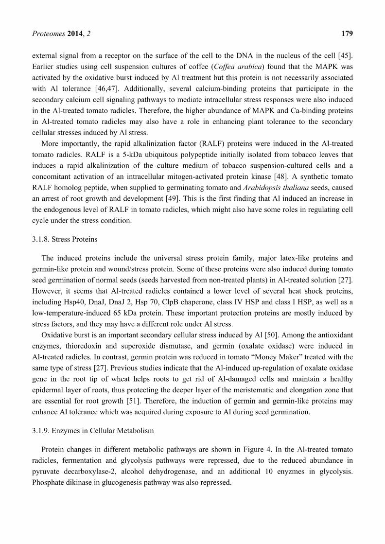
Figure 4. Overview of metabolic pathways in tomato radicles. (The graph was generated
using the Metabolism Overview in MapMan [23]. The intensity of the color change
corresponds to the scale created based on log2 fold of respective protein from Al-treated to
untreated tomato radicle tissues.)
The only pathway that was enhanced in Al-treated radicles is the TCA cycle where malate
dehydrogenases were induced. The most systematic changes were found in the mitochondrial electron
transfer chain (ETC). ETC consists of complex I, II, III, IV, and ATPases for generation of ATP. The
induced proteins are localized in NADH-DH.complex I (NADH ubiquinone dehydrogenase), complex
II (succinate dehydrogenase assembly factor 2), complex III (biquinol-cytochrome C reductase
complex proteins); a class IA/ IB cytochrome to transfer electrons from complex III to complex IV;
complex IV (cytochrome c oxidases), and five ATPase proteins (F0 complex subunit D, F1 complex,
OSCP/delta subunit, epsilon subunit and delta/epsilon subunit). Such changes suggest that metabolic
pathways from sucrose degradation to glycolysis and fermentation could be repressed, but TCA and
ETC in mitochondria were induced in tomato radicles under Al treatment.
Enzymes in glycolysis and fermentation pathways are all encoded by nuclear genes and translated
in cytoplasm. As described above, Al induced the repression of several cytosolic ribosomal proteins,
which could have affected translation of these proteins. Mitochondial ETC proteins are encoded by
mitochondrial genes, and translated within the organelle. Aluminum induced expression of several
Proteomes 2014, 2 181
mitochondrial ribosomal proteins, which may have promoted translation of these proteins in the
Al-treated radicles.
Proteins induced in secondary metabolism include isopentenyl-diphosphate delta-isomerase in the
mevalonate pathway (MVA), several O-methyltransferases, cinnamyl alcohol dehydrogenase-like
protein and caffeoyl-CoA O-methyltransferase in the phenylpropanoids and lignin biosynthesis. These
pathways are activated by various biotic and abiotic stresses [19,52,53]. In a previous study of Al
treatment of tomato roots, none of these proteins were identified [27]. The use of different analytical
methods (2D-DIGE in the previous analysis and iTRAQ in the present one) and variation in the seed
proteomes (regular seeds compared to Al-enriched from two tomato cultivars) may be factor(s) causing
the identification of different proteins in the two experiments.
4. Conclusions
When subjected to excessive Al and other toxic metals, plants have to cope with the direct ion
toxicity and the induced secondary cellular stresses, such as accumulation of reactive oxygen
species [50,54] and toxic metabolic aldehydes [55] among harmful biomolecules. Correspondingly,
multiple and various cellular pathways are affected during the process [5,56], with concomitant
alteration in expression of proteins in multiple functional groups. As shown in this proteomics study,
proteins in nearly all of the 20 functional categories displayed significant changes in abundance in the
Al treated condition. The Al-induced changes in the proteomes of radicles generated from Al-enriched
tomato seeds can be summarized as follows:
1. The Al-treated radicles contained lower abundance of hydrophilic (seed) proteins and oil body
membrane proteins, which could reduce the tolerance to dehydration. This could cause the
tomato seedlings to be more susceptible to drought and salt and other factors;
2. Mitochondria function was enhanced because of the active protein translational machinery
(induction in ribosomal proteins) and TCA–oxidative phosphorylation cycle;
3. The identified proteins include regulatory proteins for gene expression, signaling pathways, cell
cycle and programmed cell death.
Aluminum (Al) is ubiquitous in soil being the most abundant metal in the earth’s crust (>8% by
weight). When solubilized at pH values below 5.0, it is highly toxic to plants as Al3+
. Approximately
50% of the world’s potentially arable lands are acidic [57]. A large proportion of the acid soils occur in
developing countries in the tropics and subtropics and it has been estimated that the humid tropics
account for 60% of the acid soils in the world. Thus, acid soils limit the growth of crops in many
developing countries where food production is critical. Acid soils also have a significant impact on
U.S. agriculture as approximately 135 million hectares of land in the U.S. are highly acidic.
Furthermore, intensive agricultural practices used in the U.S. and in other developed countries,
including the widespread use of N fertilization with anhydrous ammonia, can cause significant
acidification of surface soils [58], thereby exacerbating an already thorny problem. Thus, there is a
genuine need to better understand Al tolerance mechanisms and the genes/proteins that define them, to
sustain and enhance crop production on acid soils. The knowledge gained from this and similar studies
Proteomes 2014, 2 182
will provide the scientific underpinning for novel strategies to overcome the challenge of Al stress, and
to sustain agricultural production on acid soils.
Acknowledgments
The authors wish to thank Wei Chen and Sheng Zhang of the Proteomics and Mass Spectrometry
Facility of the Cornell University Institute of Biotechnology for expert technical assistance and helpful
discussions. The authors also want to acknowledge Michael A. Rutzke and Shree K. Giri for providing
mineral analysis of tomato tissues. This project was supported by the Agriculture and Food Research
Initiative competitive Grant No. 2010-65114-20405 from the USDA National Institute of Food, and
Agriculture, the Evans-Allen Research Funds and ARS CRIS Projects 1907-21000-036/037-00D. ARS
disclaimer: “Mention of trade names or commercial products in this publication is solely for the
purpose of providing specific information and does not imply recommendation or endorsement by the
U.S. Department of Agriculture.”
Author Contributions
Tomato plant treatments and protein preparation and iTRAQ labeling were performed by Ikenna
Okekeogbu, Zhujia Ye, Sasikiran Reddy Sangireddy and Hui Li with the assistance from Sarabjit
Bhatti. Statistical analysis was performed by Suping Zhou and Dafeng Hui. Tara Fish assisted in the
protein extractions, protein quantifications and labeling experiments as well as being responsible for
various analytical experiments to guarantee the quality of the analysis. Tara Fish also made significant
contributions to the manuscript in revision. Yong Yang and Kevin Howe carried out all of the MS
analysis, evaluated the data, compiled the experimental results and contributed to the revising of the
manuscript. Theodore W. Thannhauser, together with Suping Zhou, developed the experimental
design. Furthermore, Theodore W. Thannhauser provided critical oversight for all the MS analysis,
conducted an independent (and concurring) statistical analysis and contributed significantly to the
drafting and revising of the manuscript.
Conflicts of Interest
The authors declare no conflict of interest.
References and Notes
1. Nasr, N. Germination and seedling growth of maize (Zea mays L.) seeds in toxicity of aluminum
and nickel. Merit Res. J. Environ. Sci. Toxic. 2013, 1, 110–113.
2. Zhu, L.; Wang, J.; Fang, X.; Wang, Y.; Hao, J.; Weiwei, M.; Jiao, T. Effect of seed soaking with
aluminum on seed germination and seedling physiology of Platycodon grandiflorum. Zhongguo
Zhong Yao Za Zhi 2010, 35, 3255–3259.
3. Liang, C.; Piñeros, M.A.; Tian, J.; Yao, Z.F.; Sun, L.L.; Liu, J.P.; Shaff, J.; Coluccio, A.;
Kochian, L.V.; Liao, H. Low pH, aluminum, and phosphorus coordinately regulate malate
exudation through GmALMT1 to improve soybean adaptation to acid soils. Plant Physiol. 2013,
161, 1347–1361.
Proteomes 2014, 2 183
4. Liao, H.; Wan, H.; Shaff, J.; Wang, X.; Yan, X.; Kochian, L.V. Phosphorus and aluminum
interactions in soybean in relation to aluminum tolerance: Exudation of specific organic acids
from different regions of the intact root system. Plant Physiol. 2006, 141, 674–684.
5. Kochian, L.V. Cellular mechanisms of aluminum toxicity and resistance in plants. Annu. Rev.
Plant Physiol. Plant Mol. Biol. 1995, 46, 237–260.
6. Kochian, L.V.; Hoekenga, O.A.; Piñeros, M.A. How do crop plants tolerate acid soils?
Mechanisms of aluminum tolerance and phosphorous efficiency. Annu. Rev. Plant Biol. 2004, 55,
459–493.
7. Yang, L.T.; Qi, Y.P.; Jiang, H.X.; Chen, L.S. Roles of organic acid anion secretion in aluminum
tolerance of higher plants. Biomed. Res. Int. 2013, 2013, e173682, doi:10.1155/2013/173682.
8. Eticha, D.; Stass, A.; Horst, W.J. Cell-wall pectin and its degree of methylation in the maize
root-apex: Significance for genotypic differences in aluminium resistance. Plant Cell. Environ.
2005, 28, 1410–1420.
9. Fry, S.C.; Smith, R.C.; Renwick, K.F.; Martin, D.J.; Hodge, S.K.; Matthews, K.J. Xyloglucan
endotransglycosylase, a new wall-loosening enzyme activity from plants. Biochem. J. 1992, 282,
821–828.
10. Huang, C.F.; Yamaji, N.; Chen, Z.; Ma, J.F. A tonoplast-localized half-size ABC transporter is
required for internal detoxification of aluminum in rice. Plant J. 2012, 69, 857–867.
11. Zhu, X.F.; Lei, G.J.; Wang, Z.W.; Shi, Y.Z.; Braam, J.; Li, G.X.; Zheng, S.J. Coordination
between apoplastic and symplastic detoxification confers plant aluminum resistance. Plant
Physiol. 2013, 162, 1947–1955.
12. Bewley, J.D.; Black, M. Seeds, Physiology of Development and Germination, 2nd ed.; Plenum
Press: New York, NY, USA, 1994.
13. Bewley, J.D. Seed germination and Dormancy. Plant Cell 1997, 9, 1055–1066.
14. Koornneef, M.; Bentsink, L.; Karssen, C.M. Seed dormancy and germination. Curr. Opin. Plant
Biol. 2002, 5, 33–36.
15. Di Salvatore, M.; Carafa, A.M.; Carratù, G. Assessment of heavy metals phytotoxicity using seed
germination and root elongation tests: A comparison of two growth substrates. Chemosphere
2008, 73, 1461–1464.
16. Rajjou, L.; Gallardo, K.; Debeaujon, I.; Vandekerckhove, J.; Job, C.; Job, D. The effect of
alpha-amanitin on the Arabidopsis seed proteome highlights the distinct roles of stored and
neosynthesized mRNAs during germination. Plant Physiol. 2004, 134, 1598–1613.
17. Aniol, A. Induction of aluminum tolerance in wheat seedlings by low doses of aluminum in the
nutrient solution. Plant Physiol. 1984, 76, 551–555.
18. Zhou, S.P.; Ikenna, O.; Rutzke, M.A.; Giri, S.K. Tennessee State University: Nashville, TN, USA.
Unpublished data, 2014.
19. Zhou, S.; Palmer, M.; Zhou, J.; Bhatti, S.; Howe, K.J.; Fish, T.; Thannhauser, T.W. Differential
root proteome expression in tomato genotypes with contrasting drought tolerance exposed to
dehydration. J. Am. Soc. Hortic. Sci. 2013, 138, 1–11.
20. Thannhauser, T.W.; Shen, M.; Sherwood, R.; Howe, K.; Fish, T.; Yang, Y.; Chen, W.; Zhang, S.
A workflow for large-scale empirical identification of cell wall N-linked glycoproteins of tomato
(Solanum lycopersicum) fruit by tandem mass spectrometry. Electrophoresis 2013, 34, 2417–2431.
Proteomes 2014, 2 184
21. Yang, Y.; Qiang, X.; Owsiany, K.; Zhang, S.; Thannhauser, T.W.; Li, L. Evaluation of different
multidimensional LC-MS/MS pipelines for iTRAQ-based proteomic analysis of potato tubers in
response to cold storage. J. Proteome Res. 2011, 10, 4647–4660.
22. International Tomato Genome Sequencing Project. Available online: http://solgenomics.net/tomato/
(accessed on 16 September 2011).
23. MapMan, version 3.1.0. Available online: http://mapman.gabipd.org/web/guest/mapman
(accessed on 30 January 2014).
24. Delhaize, E.; Ma, J.F.; Ryan, P.R. Transcriptional regulation of aluminium tolerance genes. Trends
Plant Sci. 2012, 17, 341–348.
25. Famoso, A.N.; Zhao, K.; Clark, R.T.; Tung, C.W.; Wright, M.H.; Bustamante, C.; Kochian, L.V.;
McCouch, S.R. Genetic Architecture of Aluminum Tolerance in Rice (Oryza sativa)
Determinedthrough Genome-Wide Association Analysis and QTL Mapping. PLoS Genet. 2011,
7, e1002221.
26. Ryan, P.R.; Tyerman, S.D.; Sasaki, T.; Furuichi, T.; Yamamoto, Y.; Zhang, W.H.; Delhaize, E.
The identification of aluminium-resistance genes provides opportunities for enhancing crop
production on acid soils. J. Exp. Bot. 2011, 62, 9–20.
27. Zhou, S.; Sauvé, R.; Thannhauser, T.W. Proteome changes induced by aluminium stress in tomato
roots. J. Exp. Bot. 2009, 60, 1849–1857.
28. Kim, Y.Y.; Jung, K.W.; Yoo, K.S.; Jeung, J.U.; Shin, J.S. A stress-responsive caleosin-like
protein, AtCLO4, acts as a negative regulator of ABA responses in Arabidopsis. Plant Cell
Physiol. 2011, 52, 874–884.
29. Poxleitner, M.; Rogers, S.W.; Samuels, A.L.; Browse, J.; Rogers, J.C. A role for caleosin in
degradation of oil-body storage lipid during seed germination. Plant J. 2006, 47, 917–933.
30. Murphy, D.J.; Hernandez-Pinzon, I.; Patel, K. Role of lipid bodies and lipid-body proteins in
seeds and others tissues. J. Plant Physiol. 2001, 158, 471–478.
31. Mackey, D.; Holt, B.F., III; Wiig, A.; Dangl, J.L. RIN4 interacts with Pseudomonas syringae type
III effector molecules and is required for RPM1-mediated resistance in Arabidopsis. Cell 2002,
22, 743–754.
32. Levine, A.; Belenghi, B.; Damari-Weisler, H.; Granot, D. Vesicle-associated membrane protein of
Arabidopsis suppresses Bax-induced apoptosis in yeast downstream of oxidative burst. J. Biol.
Chem. 2001, 276, 46284–46289.
33. Harris, E.H.; Boynton, J.E.; Gillham, N.W. Chloroplast ribosomes and protein synthesis.
Microbiol. Rev. 1994, 58, 700–754.
34. Pietromonaco, S.F.; Denslow, N.D.; O'Brien, T.W. Proteins of mammalian mitochondrial
ribosomes. Biochimie 1991, 73, 827–835.
35. Spremulli, L. Protein synthesis of mammalian mitochondria. Available online: http://www.chem.
unc.edu/people/faculty/spremulli/ (accessed on 10 February 2014).
36. Zhou, S.; Sauvé, R.J. ; Liu, Z.; Reddy, S.; Bhatti, S.; Hucko, S.D.; Fish, T.; Thannhauser, T.W.
Identification of salt-induced changes in leaf and root proteomes of the wild tomato, Solanum
chilense. J. Am. Soc. Hortic. Sci. 2011, 136, 288–302.
37. Berndt, N. Roles and regulation of serine/threonine-specific protein phosphatases in the cell cycle.
Prog. Cell Cycle Res. 2003, 5, 497–510.
Proteomes 2014, 2 185
38. Garcia, A.; Cayla, X.; Guergnon, J.; Dessauge, F.; Hospital, V.; Rebollo, M.P.; Fleischer, A.;
Rebollo, A. Serine/threonine protein phosphatases PP1 and PP2A are key players in apoptosis.
Biochimie 2003, 85, 721–726.
39. Osawa, H.; Matsumoto, H. Possible involvement of protein phosphorylation in aluminum-responsive
malate efflux from wheat root apex. Plant Physiol. 2001, 126, 411–420.
40. Alcaide-Loridan, C.; Jupin, I. Ubiquitin and Plant Viruses, Let’s Play Together! Plant Physiol.
2012, 160, 72–82.
41. Nezames, C.D.; Deng, X.Y. The COP9 Signalosome: Its regulation of Cullin-Based E3 Ubiquitin
ligases and role in photomorphogenesis. Plant Physiol. 2012, 160, 38–46.
42. Wei, N.; Serino, G.; Deng, X.W. The COP9 signalosome: More than a protease. Trends Biochem.
Sci. 2008, 33, 592–600.
43. Argyris, J.; Dahal, P.; Hayashi, E.; Still, D.W.; Bradford, K.J. Genetic Variation for lettuce seed
thermoinhibition is associated with temperature-sensitive expression of abscisic acid, gibberellin,
and ethylene biosynthesis, metabolism, and response genes. Plant Physiol. 2008, 148, 926–947.
44. Finkelstein, R.R.; Lynch, T.J. Abscisic acid inhibition of radicle emergence but not seedling
growth is suppressed by sugars. Plant Physiol. 2000, 122, 1179–1186.
45. Orton, R.J.; Sturm, O.E.; Vyshemirsky, V.; Calder, M.; Gilbert, D.R.; Kolch, W. Computational
modelling of the receptor-tyrosine-kinase-activated MAPK pathway. Biochem. J. 2005, 392,
249–261.
46. Arroyo-Serralta, G.A.; Ku -Gonza lez, A.; Herna ndez-Sotomayor, S.M.T.; Zu´n˜iga-Aguilar, J.J.
Exposure to toxic concentrations of Aluminum activates a MAPK-like protein in cell suspension
cultures of Coffea arabica. Plant Physiol. Biochem. 2005, 43, 27–35.
47. Ramı´rez-Benı´tez, J.E.; Chee-Gonza lez, L.; Herna ndez-Sotomayor, S.M.T. Aluminum induces
changes in organic acids metabolism in Coffea arabica suspension cells with differential
Al-tolerance. J. Inorg. Biochem. 2008, 102, 1631–1637.
48. Schaller, A.; Oecking, C. Modulation of plasma membrane H+-ATPase activity differentially
activates wound and pathogen defense responses in tomato plants. Plant Cell 1999, 11, 263–272.
49. Pearce, G.; Moura, D.S.; Stratmann, J.; Ryan, C.A., Jr. RALF, a 5-kDa ubiquitous polypeptide in
plants, arrests root growth and development. Proc. Natl. Acad. Sci. USA 2001, 98, 12843–12847.
50. Richards, K.D.; Schott, E.J.; Sharma, Y.K.; Davis, K.R.; Gardner, R.C. Aluminum induces
oxidative stress genes in Arabidopsis thaliana. Plant Physiol. 1998, 116, 409–418.
51. Delisle, G.; Champoux, M.; Houde, M. Characterization of oxalate oxidase and cell death in
Al-sensitive and tolerant wheat roots. Plant Cell Physiol. 2001, 42, 324–333.
52. Soto, G.; Stritzler, M.; Lisi, C.; Alleva, K.; Pagano, M.E.; Ardila, F.; Mozzicafreddo, M.;
Cuccioloni, M.; Angeletti, M.; Ayub, N.D. Acetoacetyl-CoA thiolase regulates the mevalonate
pathway during abiotic stress adaptation. J. Exp. Bot. 2011, 62, 5699–5711.
53. Nveawiah-Yoho, P.; Zhou, J.; Palmer, M.; Sauve, R.; Zhou, S.; Howe, K.J.; Fish, T.;
Thannhauser, T.W. Identification of Proteins for Salt Tolerance Using a Comparative Proteomics
Analysis of Tomato Accessions with Contrasting Salt Tolerance. J. Am. Soc. Hortic. Sci. 2013,
138, 382–394.
Proteomes 2014, 2 186
54. Yamamoto, Y.; Kobayashi, Y.; Devi, S.R.; Rikiishi, S.; Matsumoto, H. Aluminum toxicity is
associated with mitochondrial dysfunction and the production of reactive oxygen species in plant
cells. Plant Physiol. 2002, 128, 63–72.
55. Yin, L.; Mano, J.; Wang, S.; Tsuji, W.; Tanaka, K. The involvement of lipid peroxide-derived
aldehydes in aluminum toxicity of tobacco roots. Plant Physiol. 2010, 152, 1406–1417.
56. Von Uexküll, H.R.; Mutert, E. Global extent, development and economic impact of acid soils. In
Plant-Soil Interactions at Low pH: Principles and Management; Date, R.A., Grundon, N.J.,
Raymet, G.E., Probert, M.E., Eds.; Kluwer Academic Publishers: Dordrecht, The Netherlands,
1995; pp. 5–19.
57. Carver, B.F.; Ownby, J.D. Acid soil tolerance in wheat. Adv. Agron. 1995, 54, 117–173.
58. Sawyer, S. Anhydrous ammonia application and dry Soils. Available online: http://www.extension.
iastate.edu/CropNews/2011/1028sawyer.htm (accessed on 1 September 2012).
Proteomes 2014, 2 187
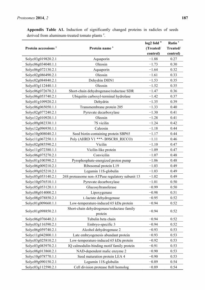
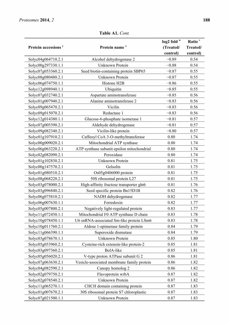
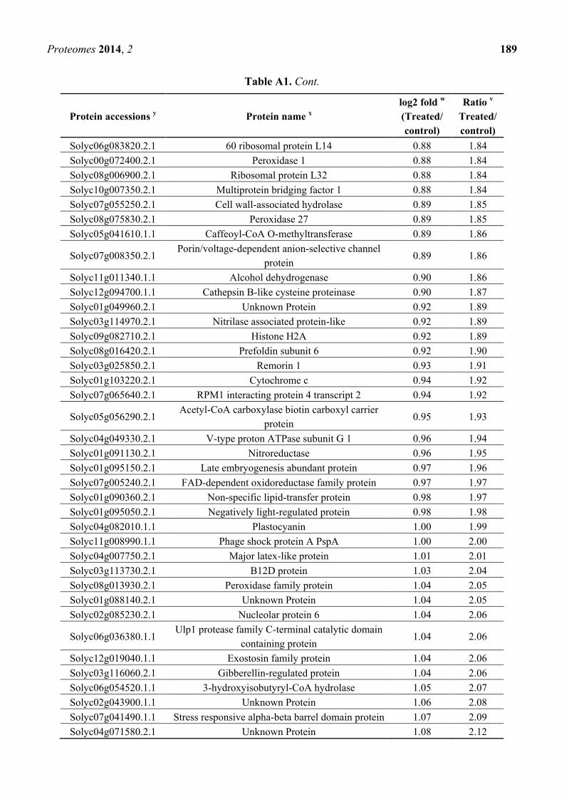
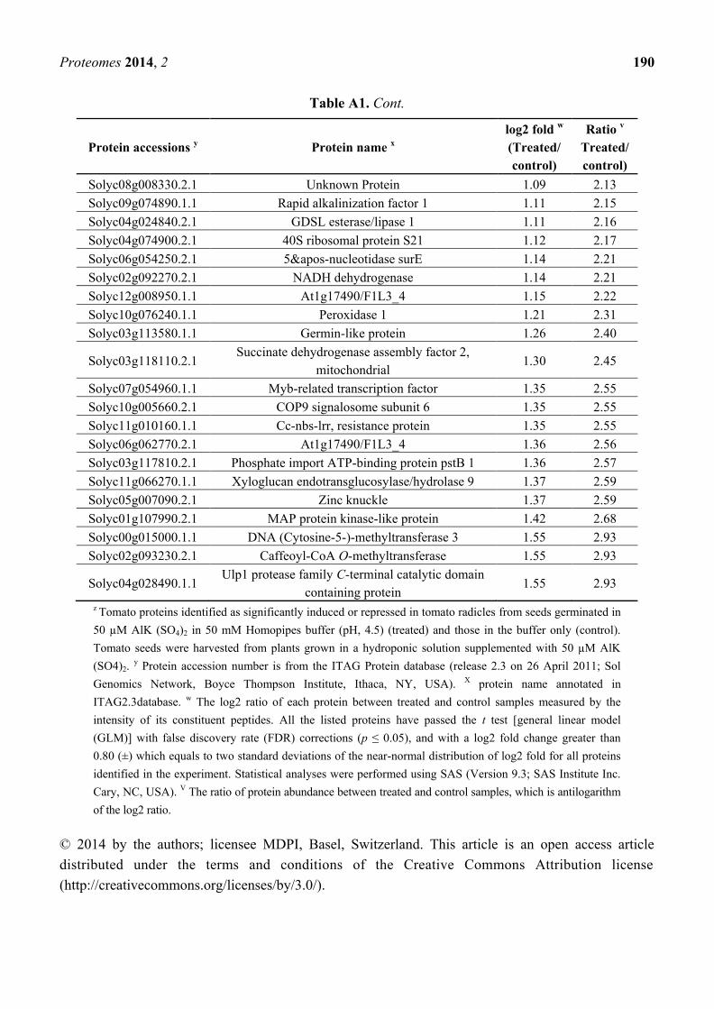
Appendix Table A1. Induction of significantly changed proteins in radicles of seeds
derived from aluminum-treated tomato plants z.
Protein accessions y Protein name
x
log2 fold w
(Treated/
control)
Ratio v
Treated/
control)
Solyc03g019820.2.1 Aquaporin −1.88 0.27
Solyc06g034040.1.1 Oleosin −1.73 0.30
Solyc06g072130.2.1 Aquaporin −1.64 0.32
Solyc02g086490.2.1 Oleosin −1.61 0.33
Solyc02g084840.2.1 Dehydrin DHN1 −1.53 0.35
Solyc03g112440.1.1 Oleosin −1.52 0.35
Solyc06g072670.2.1 Short-chain dehydrogenase/reductase SDR −1.47 0.36
Solyc06g053740.2.1 Ubiquitin carboxyl-terminal hydrolase −1.42 0.37
Solyc01g109920.2.1 Dehydrin −1.35 0.39
Solyc06g065050.1.1 Transmembrane protein 205 −1.33 0.40
Solyc02g077240.2.1 Pyruvate decarboxylase −1.30 0.41
Solyc12g010920.1.1 Oleosin −1.28 0.41
Solyc09g082330.1.1 7S vicilin −1.24 0.42
Solyc12g096930.1.1 Caleosin −1.18 0.44
Solyc10g008040.2.1 Seed biotin-containing protein SBP65 −1.17 0.44
Solyc11g067250.1.1 Poly (AHRD V1 ***- B9SCR8_RICCO) −1.11 0.46
Solyc02g085590.2.1 Vicilin −1.10 0.47
Solyc11g072380.1.1 Vicilin-like protein −1.09 0.47
Solyc06g075270.2.1 Convicilin −1.07 0.48
Solyc01g100390.2.1 Pyrophosphate-energized proton pump −1.06 0.48
Solyc06g009210.2.1 Ribosomal protein L19 −1.03 0.49
Solyc09g025210.2.1 Legumin 11S-globulin −1.03 0.49
Solyc05g053140.2.1 26S proteasome non-ATPase regulatory subunit 13 −1.02 0.49
Solyc10g076510.1.1 Pyruvate decarboxylase −1.01 0.50
Solyc05g053120.1.1 Glucosyltransferase −0.99 0.50
Solyc08g014000.2.1 Lipoxygenase −0.98 0.51
Solyc08g078850.2.1 L-lactate dehydrogenase −0.95 0.52
Solyc01g009660.1.1 Low-temperature-induced 65 kDa protein −0.94 0.52
Solyc01g098850.2.1 Short-chain dehydrogenase/reductase family
protein −0.94 0.52
Solyc06g076640.2.1 Tubulin beta chain −0.94 0.52
Solyc03g116590.2.1 Embryo-specific 3 −0.94 0.52
Solyc06g059740.2.1 Alcohol dehydrogenase 2 −0.93 0.53
Solyc11g042800.1.1 Late embryogenesis abundant protein −0.93 0.53
Solyc03g025810.2.1 Low-temperature-induced 65 kDa protein −0.92 0.53
Solyc03g083970.2.1 IQ calmodulin-binding motif family protein −0.91 0.53
Solyc08g013860.2.1 NAD-dependent malic enzyme 2 −0.90 0.53
Solyc10g078770.1.1 Seed maturation protein LEA 4 −0.90 0.53
Solyc09g090150.2.1 Legumin 11S-globulin −0.89 0.54
Solyc03g112590.2.1 Cell division protease ftsH homolog −0.89 0.54
Proteomes 2014, 2 188
Table A1. Cont.
Protein accessions y Protein name
x
log2 fold w
(Treated/
control)
Ratio v
Treated/
control)
Solyc04g064710.2.1 Alcohol dehydrogenase 2 −0.89 0.54
Solyc00g297330.1.1 Unknown Protein −0.88 0.54
Solyc07g053360.2.1 Seed biotin-containing protein SBP65 −0.87 0.55
Solyc08g080480.2.1 Unknown Protein −0.87 0.55
Solyc06g074750.1.1 Histone H2B −0.86 0.55
Solyc12g098940.1.1 Ubiquitin −0.85 0.55
Solyc07g032740.2.1 Aspartate aminotransferase −0.85 0.56
Solyc01g007940.2.1 Alanine aminotransferase 2 −0.83 0.56
Solyc09g065470.2.1 Vicilin −0.83 0.56
Solyc09g015070.2.1 Reductase 1 −0.83 0.56
Solyc12g014380.1.1 Glucose-6-phosphate isomerase 1 −0.81 0.57
Solyc07g005390.2.1 Aldehyde dehydrogenase −0.81 0.57
Solyc09g082340.2.1 Vicilin-like protein −0.80 0.57
Solyc01g107910.2.1 Caffeoyl CoA 3-O-methyltransferase 0.80 1.74
Solyc00g009020.2.1 Mitochondrial ATP synthase 0.80 1.74
Solyc06g063220.2.1 ATP synthase subunit epsilon mitochondrial 0.80 1.74
Solyc02g082090.2.1 Peroxidase 0.80 1.74
Solyc01g102830.2.1 Unknown Protein 0.81 1.75
Solyc00g147570.2.1 Gelsolin 0.81 1.75
Solyc01g080510.2.1 Os05g0406000 protein 0.81 1.75
Solyc08g068220.2.1 50S ribosomal protein L27 0.81 1.75
Solyc03g078000.2.1 High-affinity fructose transporter ght6 0.81 1.76
Solyc03g096840.2.1 Seed specific protein Bn15D1B 0.82 1.76
Solyc06g075810.2.1 NADH dehydrogenase 0.82 1.77
Solyc06g007630.1.1 Ferredoxin 0.82 1.77
Solyc05g007800.2.1 Negatively light-regulated protein 0.83 1.77
Solyc11g072450.1.1 Mitochondrial F0 ATP synthase D chain 0.83 1.78
Solyc10g078450.1.1 U6 snRNA-associated Sm-like protein LSm6 0.83 1.78
Solyc10g011760.2.1 Aldose 1-epimerase family protein 0.84 1.79
Solyc11g066390.1.1 Superoxide dismutase 0.84 1.79
Solyc03g078670.1.1 Unknown Protein 0.85 1.80
Solyc05g053960.2.1 Cysteine-rich extensin-like protein-2 0.85 1.81
Solyc03g097360.2.1 BolA-like 0.85 1.81
Solyc05g056020.2.1 V-type proton ATPase subunit G 2 0.86 1.81
Solyc07g063630.2.1 Vesicle-associated membrane family protein 0.86 1.82
Solyc04g082590.2.1 Canopy homolog 2 0.86 1.82
Solyc02g079750.2.1 Flavoprotein wrbA 0.87 1.82
Solyc02g078540.2.1 Unknown Protein 0.87 1.82
Solyc11g065270.1.1 CHCH domain containing protein 0.87 1.83
Solyc01g007670.2.1 30S ribosomal protein S7 chloroplastic 0.87 1.83
Solyc07g021500.1.1 Unknown Protein 0.87 1.83
Proteomes 2014, 2 189
Table A1. Cont.
Protein accessions y Protein name
x
log2 fold w
(Treated/
control)
Ratio v
Treated/
control)
Solyc06g083820.2.1 60 ribosomal protein L14 0.88 1.84
Solyc00g072400.2.1 Peroxidase 1 0.88 1.84
Solyc08g006900.2.1 Ribosomal protein L32 0.88 1.84
Solyc10g007350.2.1 Multiprotein bridging factor 1 0.88 1.84
Solyc07g055250.2.1 Cell wall-associated hydrolase 0.89 1.85
Solyc08g075830.2.1 Peroxidase 27 0.89 1.85
Solyc05g041610.1.1 Caffeoyl-CoA O-methyltransferase 0.89 1.86
Solyc07g008350.2.1 Porin/voltage-dependent anion-selective channel
protein 0.89 1.86
Solyc11g011340.1.1 Alcohol dehydrogenase 0.90 1.86
Solyc12g094700.1.1 Cathepsin B-like cysteine proteinase 0.90 1.87
Solyc01g049960.2.1 Unknown Protein 0.92 1.89
Solyc03g114970.2.1 Nitrilase associated protein-like 0.92 1.89
Solyc09g082710.2.1 Histone H2A 0.92 1.89
Solyc08g016420.2.1 Prefoldin subunit 6 0.92 1.90
Solyc03g025850.2.1 Remorin 1 0.93 1.91
Solyc01g103220.2.1 Cytochrome c 0.94 1.92
Solyc07g065640.2.1 RPM1 interacting protein 4 transcript 2 0.94 1.92
Solyc05g056290.2.1 Acetyl-CoA carboxylase biotin carboxyl carrier
protein 0.95 1.93
Solyc04g049330.2.1 V-type proton ATPase subunit G 1 0.96 1.94
Solyc01g091130.2.1 Nitroreductase 0.96 1.95
Solyc01g095150.2.1 Late embryogenesis abundant protein 0.97 1.96
Solyc07g005240.2.1 FAD-dependent oxidoreductase family protein 0.97 1.97
Solyc01g090360.2.1 Non-specific lipid-transfer protein 0.98 1.97
Solyc01g095050.2.1 Negatively light-regulated protein 0.98 1.98
Solyc04g082010.1.1 Plastocyanin 1.00 1.99
Solyc11g008990.1.1 Phage shock protein A PspA 1.00 2.00
Solyc04g007750.2.1 Major latex-like protein 1.01 2.01
Solyc03g113730.2.1 B12D protein 1.03 2.04
Solyc08g013930.2.1 Peroxidase family protein 1.04 2.05
Solyc01g088140.2.1 Unknown Protein 1.04 2.05
Solyc02g085230.2.1 Nucleolar protein 6 1.04 2.06
Solyc06g036380.1.1 Ulp1 protease family C-terminal catalytic domain
containing protein 1.04 2.06
Solyc12g019040.1.1 Exostosin family protein 1.04 2.06
Solyc03g116060.2.1 Gibberellin-regulated protein 1.04 2.06
Solyc06g054520.1.1 3-hydroxyisobutyryl-CoA hydrolase 1.05 2.07
Solyc02g043900.1.1 Unknown Protein 1.06 2.08
Solyc07g041490.1.1 Stress responsive alpha-beta barrel domain protein 1.07 2.09
Solyc04g071580.2.1 Unknown Protein 1.08 2.12
Proteomes 2014, 2 190
Table A1. Cont.
Protein accessions y Protein name
x
log2 fold w
(Treated/
control)
Ratio v
Treated/
control)
Solyc08g008330.2.1 Unknown Protein 1.09 2.13
Solyc09g074890.1.1 Rapid alkalinization factor 1 1.11 2.15
Solyc04g024840.2.1 GDSL esterase/lipase 1 1.11 2.16
Solyc04g074900.2.1 40S ribosomal protein S21 1.12 2.17
Solyc06g054250.2.1 5&apos-nucleotidase surE 1.14 2.21
Solyc02g092270.2.1 NADH dehydrogenase 1.14 2.21
Solyc12g008950.1.1 At1g17490/F1L3_4 1.15 2.22
Solyc10g076240.1.1 Peroxidase 1 1.21 2.31
Solyc03g113580.1.1 Germin-like protein 1.26 2.40
Solyc03g118110.2.1 Succinate dehydrogenase assembly factor 2,
mitochondrial 1.30 2.45
Solyc07g054960.1.1 Myb-related transcription factor 1.35 2.55
Solyc10g005660.2.1 COP9 signalosome subunit 6 1.35 2.55
Solyc11g010160.1.1 Cc-nbs-lrr, resistance protein 1.35 2.55
Solyc06g062770.2.1 At1g17490/F1L3_4 1.36 2.56
Solyc03g117810.2.1 Phosphate import ATP-binding protein pstB 1 1.36 2.57
Solyc11g066270.1.1 Xyloglucan endotransglucosylase/hydrolase 9 1.37 2.59
Solyc05g007090.2.1 Zinc knuckle 1.37 2.59
Solyc01g107990.2.1 MAP protein kinase-like protein 1.42 2.68
Solyc00g015000.1.1 DNA (Cytosine-5-)-methyltransferase 3 1.55 2.93
Solyc02g093230.2.1 Caffeoyl-CoA O-methyltransferase 1.55 2.93
Solyc04g028490.1.1 Ulp1 protease family C-terminal catalytic domain
containing protein 1.55 2.93
z Tomato proteins identified as significantly induced or repressed in tomato radicles from seeds germinated in
50 µM AlK (SO4)2 in 50 mM Homopipes buffer (pH, 4.5) (treated) and those in the buffer only (control).
Tomato seeds were harvested from plants grown in a hydroponic solution supplemented with 50 µM AlK
(SO4)2. y Protein accession number is from the ITAG Protein database (release 2.3 on 26 April 2011; Sol
Genomics Network, Boyce Thompson Institute, Ithaca, NY, USA). X protein name annotated in
ITAG2.3database. w The log2 ratio of each protein between treated and control samples measured by the
intensity of its constituent peptides. All the listed proteins have passed the t test [general linear model
(GLM)] with false discovery rate (FDR) corrections (p ≤ 0.05), and with a log2 fold change greater than
0.80 (±) which equals to two standard deviations of the near-normal distribution of log2 fold for all proteins
identified in the experiment. Statistical analyses were performed using SAS (Version 9.3; SAS Institute Inc.
Cary, NC, USA). V The ratio of protein abundance between treated and control samples, which is antilogarithm
of the log2 ratio.
© 2014 by the authors; licensee MDPI, Basel, Switzerland. This article is an open access article
distributed under the terms and conditions of the Creative Commons Attribution license
(http://creativecommons.org/licenses/by/3.0/).
![Brazilian Health and Agriculture Cooperation in Mozambique: An Overview [BPC-SSC Working Paper, 2014]](https://img.pdfslide.net/doc/110x75/631b0a0cd5372c006e03c316/brazilian-health-and-agriculture-cooperation-in-mozambique-an-overview-bpc-ssc.jpg)


