Embed Size (px)
Citation preview
jstnrlb~il Ecz. Fak. Mec. 33 (2000)
J. Fac. Pharnz. Istanbul 33 (2000)
PARTIAL PURIFICATION and CHARACTERIZATION of ASPARTATE AMINOTRANSFERASE from the
HEPATOPANCREAS of the MUSSEL Mytilus galloprovincialis Lam.
A. CAN*, N. AKEV* and A. ~ K H A S I *
S U M M A R Y
In this study aspartate and alanine aminotransferase enzymes of Mytilrls gallo- pro~~incialis Lam., the mussel species specific to the Turkish coast were examined. Because it was found that aspartate aminotransferase activity in the hepatopancreas was superior to alanine aminotransferase while surveying the activities of these enzymes in . the whole body, hepatopancreas and mantle tissues; among these two enzymes, aspartate aminotransferase was partially purified from hepatopancreas and its kinetic properties were studied.
Molluscs were collected daily from the Rumelikavagl coast of the Bosphonts and hepatopancreases were homogenized with 0.9% NaCl solution after dissection from the body. The fraction obtained after 35%-65% ammonium sulphate precipitation was applied to hydroxylapatite column. The elution performed by increasing molarity gradient of the phosphate buffer, pH 6.8, resulted in the recovery of the aspartate arninotransferase activity in 100 mM phosphate buffer pool. The 83 fold purified enzyme was applied to polyacrylamide gel electrophoresis and the existence of two activity bands indicated two isoenzymes of the aspartate aminotransferase.
(*I Faculty of Pharmacy, Department of Biochemistry. University of Istanbul, Universite, 34452, Istanbul, Turkey
Effect of temperature on the activity of the enzyme was examined and it was found that the enzyme exhibited maximum activities at 25 "C and 40 "C; the activity completely dissappeared at 60 "C. Aspartate aminotransferase activity was maximum at
I pH 7.6, Km values for aspa$ate,and 2-oksoglutarate were 1.7 mM and 6 . 6 ~ 1 0 - ~ mM, Vmax for the same substrates were .I62 U/ml and 0.149 U/ml, respectively.
\ Bu qaligmada, Turkiye sahillerinde bulunan Mytilus gallopi.ovincialis Lam.
midye tiirunun aspartat ve alanin aminotransferazlarl incelendi. Tum doku, hepatopankreas ve manto k~simlannda yapllan aragt~rmada, aspartat aminotransferazln hepatopankreastaki aktivitesinin alanin aminotransferaza gore fazla olmasindan dolayl, midye hepatopankreas~ndan aspartat aminotransferaz kismen saflagtlrildi ve bazl kinetik ozellikleri incelendi.
Deneyin yapilacagl gun 1stanbul Bogazi Rumelikava@i klyilarindan toplanan
midyelerin hepatopankreas k~s~rnlarl qlkanlarak, %0.9 NaCl ile, homojenize edildi. Aspartat aminotransferaz aktivitesi gosteren homojenizatin %35-%65 amonyum sulfat kesiti hidroksilapatit kolona uygulandi. pH's1 6.8 olan ve artan molaritede fosfat tamponu ile yapllan basamaklt elusyon sonucunda, aspartat aminotransferaz 100 mM fosfat tarnponu ile eliie edildi. Bu iglem sonunda, midye hepatopankreaslndan 83 kez safla~tlnlan aspartat aminotransferazln poliakrilamid jel elektroforezinde iki aktivite bandl gostermesi nedeniyle iki izoenzim iqerdiii sonucuna vanldl.
Aspartat aminotransferaz iizerine temperaturun etkisi incelendiginde, enzimin 25 "C ve 40 "C'lerde maksimum aktivite gosterdigi ve aktivitenin 60 "C'de tamamen kayboldugu goriildu. Enzimin optimum pH'slnin pH 7.6'da oldugu; aspartat ve 2- oksoglutarata kargi Km degerlerinin slraslyla, 1.7 mM ve 6 . 6 ~ 1 0 - ~ mM, aynl substratlara kary Vmax degerlerinin ise 0.162 U/ml ve 0.149 U/ml oldugu bulundu.
Key words: Mussel, Myti l~~s galloproviizcialis Lam., aspartate aminotransferase, alanine aminotransferase, transaminase
I N T R O D U C T I O N
Aspartate aminotransferase (EC 2.6.1.1; AAT) which was formerly called glutamic oxaloacetic transaminase (GOT), occurs widely in nature and has been studied most extensively. In eukaryotes, the enzyme exists in two isoenzymic forms, one localized in the cytoplasm and the other in the mitochondria of mammalian cells ( 1 3 . The measurement of these isoenzyrnes in human serum reportedly is of clinical significance
in assessing tissue damage in diseases including myocardial infarction and hepatitis. Characterization of aspartate aminotransferase, through kinetic and clinical investigations is thoroughly reviewed (3).
Aspartate aminotransferase has been found in all tissues of all molluscs investigated. The levels of activity in molluscan tissues vary from very high in muscle of some cephalopods to very low in the tissues of some fresh water bivalves and gastropods (4,5). An intracellular localization and aminotransferase activity in some tissues of the Black Sea mussels were studied and it was shown that alanine aminotransferase (ALAT; GPT) was localized mainly in the cytoplasmic fraction whereas a considerable quantity of aspartate arninotransferase was localized in mitochondria (6). The same workers examined transaminase activity in tissues of Myrillrs gulloprovi~zciulis Lam. in standart and hypoxic conditions and found an alanine /aspartate ratio of 111 (7).
Although aspartate and alanine aminotransferases were purified from a variety of plant and animal sources, they haven't been purified to date from Mytilus galloproviizciulis Lam. which is a mussel specific to the coasts of Turkey. In this study, the occurence of aspartate and alanine aminotransferase activities in the whole tissue, mantle and hepatopancreas of the mussel Myrilus galloprovincialis Lam., collected from Istanbul Bosphorus Rumelikavag~ coasts, were determined and the most active enzyme was purified and characterized from the tissue which contained the highest activity of this enzyme.
M A T E R I A L A N D M E T H O D S
Chenzicals. P-Nicotinamide adenine dinucleotide disodium salt reduced (NADH2-Na2, Fluka 43420), pyridoxal-5'-phosphate (Fluka 82870), 2-oxoglutaric acid were used in enzyme assays. L-aspartic acid (Merck 129), malate dehydrogenase (1500 U/mg; Serva 28338) and L-alanine (Fluka 05 130), lactate dehydrogenase (860 U/mg; Sigma L-2500) were used for the activity measurements of aspartate aminotransferase and alanine aminotransferase respectively. Hydroxylapatite prepared in our laboratory was used in column chromatographies. Bovine serum albumin (Fluka 05470) was used as a standart for protein determination. All other chemicals were of analytical reagent grade.
Protein deter~~lination. Protein contents of the samples during extraction and ammonium sulphate precipitation processes were determined according to the method of Lowry (8) whereas the protein fractions obtained through column chromatography were analyzed for protein by E 2801260 method (9).
E~zzy~~ze assay. The routine enzyme assay involved a nicotinamide adenine dinucleotide (NAD)-dependent malate dehydrogenase-linked reaction to measure aspartate
aminotransferase activity and a lactate dehydrogenase-linked reaction to measure alanine arninotransferase activity, according to the method of Karmen (10) modified by us (11) and Rej et a1 (12). To a spectrophotometer cuvette 2.5 ml of a solution containing 54 mM L-aspartate, 12 Units malate dehydrogenase (for AAT) or 54 mM L-alanine, 160 Units lactate dehyrogenase (for ALAT), 0.06 mM NADH2, 0.033 mM pyridoxal-5'- phosphate, and 113 mM Tris with a final pH of 7.8. For AAT, 0.25 ml of the enzyme solution was first added and the content of the cuvette was mixed with a thin glass rod. The reaction was started by addition of 0.25 ml of a solution containing 45 mM 2- oxoglutarate and 125 mM Tris. For ALAT, 2-oxoglutarate solution was added first and the reaction was initiated by the addition of 0.25 ml of the enzyme solution. The decrease of absorbance at 339 nm during 5 minutes was monitored spectrophotometrically. One unit of aminotransferase activity (Karmen Unit) was expressed as absorbance decrease of 0.001 per mi of the sample per minute of 1 cm light path.
Polyacryla~nide gel electrophoresis (PAGE). Non-denaturing PAGE was performed on a Pleuger electrophoresis apparatus acccording to Ornstein (13) and Davis (14). 10 fold diluted solution of Tris-glycine buffer (3% Tris and 14.4% glycine, pH 8.5) was used during electrophoresis. Electrophoresis was performed in 7.5% polyacrylarnide gels of 6 x 0.6 cm rods. 50p1 samples in 40% saccharose were applicated to the gels by means of a Hamilton injector. Electrophoresis was canied out at 4 OC in a cold chamber, at 5 mAlgel for 50 mins. The gels were stained with Amidoblack 10B for protein and with Fast violet B (15) for the detection of aspartate aminotransferase activity.
Determination of the occurence of aspartate and alartine a~~zinotransferase activities in different tissues of Mytilzrs galloprovincialis Lam. 25 mussels freshly collected at the day of the experiment from Istanbul Bosphorus Rume1ikavali;i coast, were separated from their shells. The whole mussel tissue was washed, dried and weighed (184.13 g). After the addition of 150 rnl cold saline, the whole tissue was homogenized at 14 000 rpm by means of a Bosch homogenizer. An additional I50 ml saline was added to the homogenate and it was stirred via magnetic stirrer for 30 mins in a cold room. After standing overnight at 4 OC, the homogenate was centrifuged at 20 000 rpm at -20 "C for 30 mins (Cryofuge 20-3 Hereaus-Christ). The supernatant was filtered (345 ml) and called mussel whole tissue crude extract.
Mantle and hepatopancreas tissues of another group of 25 mussels were separated. The mantle tissues (47.66 g) were homogenized with a total of I00 ml of saline as previously described for the whole tissue extract. The extract thus obtained (80 ml) was called mussel mantle tissue crude extract. The hepatopancreases were treated with a total of 100 ml of saline and mussel hepatopancreas crude extract was prepared in the same manner.
41
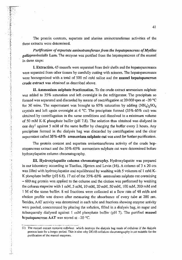
The protein contents, aspartate and alanine aminotransferase activities of the three extracts were determined.
Purification of aspartate alninotransferase from the Itepatopancreas of Mytilus 1 galloprovi~zcialis Lam. The enzyme was purified from the hepatopancreas of the mussel
in three steps:
I. Extraction. 43 mussels were separated from their shells and the hepatopancreases were separated from other tissues by carefully cutting with scissors. The hepatopancreases were homogenized with a total of 500 ml cold saline and the mussel hepatopancreas crude extract was obtained as described above.
II. Ammonium sulphate fractionation. To the crude extract ammonium sulphate was added to 35% saturation and left overnight in the refrigerator. The precipitate so formed was separated and discarded by means of centrifugation at 20 000 rpm at -20 "C for 30 mins. The supernatant was brought to 65% saturation by adding (NH4),S04 crystals and left again overnight at 4 "C. The precipitate formed (35%-65% cut) was obtained by centrifugation in the same conditions and dissolved in a minimum volume of 50 mM K-K phosphate buffer (pH 7.8). The solution thus obtained was dialysed in one day1 against 5 mM of the same buffer by changing the buffer every 2 hours. Any precipitate formed in the dialysis bag was discarded by centrifugation and the clear supematant called 35%-65% ammonium sulphate cut was used for further purification.
The protein content and aspartate aminotransferase activity of the crude hep- atopancreas extract and the 35%-65% ammonium sulphate cut were determined before hydroxylapatite column chromatography.
TCI. Hydroxylapatite column chromatography. Hydroxylapatite was prepared in our laboratory according to Tiselius, Hjerten and Levin (16). A column of 3 x 20 cm was filled with hydroxylapatite and equilibrated by washing with 5 volumes of 1 mM K- K phosphate buffer (pH 6.8). 17 ml of the 35%-65% ammonium sulphate cut containing - 600 mg protein was applied to the column and the elution was performed by washing the column stepwise with 1 mM, 5 mM, 10 mM, 20 mM, 50 mM, I00 mM, 200 mM and 1 M of the same buffer. 8 ml fractions were collected at a flow rate of 48 ml/h and elution profile was drawn after measuring the absorbance of every tube at 280 nm. Besides, AAT activity was determined in each tube and fractions showing enzyme activity were pooled, concentrated by placing the solution, filled in a dialysis bag, in sugar and subsequently dialysed against 1 mM phosphate buffer (pH 7). The purified mussel hepatopancreas AAT was stored at -20 "C.
(1) The mussel extract contains ceilulase which destroys the dialysis bag made of cellulose if the dialysis process lasts for a longer period. This is also why DEAE-cellulose chromatography is not suitable for the purification of the mussel enzymes.
Effect of tenzperatz~re on elzzyme activity. 0.4 ml aliquots of an enzyme solution diluted to 150 U/ml activity, were heated for 30 minutes at temperatures varying between 10 "C - 60 "C with 5 OC intervals. Activities were determined after bringing to room temperature.
Effect of pH orz enzyme activity. The activity of AAT was examined between pH 5-9. 0.1 M K-K phosphate buffer was used between pH 5-7 and 0.1 M Tris-HC1 buffer for pH 7.2-9. The buffer solution of desired pH (3 ml) were mixed with I ml of a neutral aspartate solution, containing NADH2, pyridoxal-5'-phosphate, malate dehydrogenase and Tris. 2.5 ml of this mixture was transferred to a spectrophotometer cuvette, 0.25 ml 2-oxoglutarate solution was added and the reaction was initiated with 0.25 ml of the enzyme solution properly diluted to show an activity of 150 U/ml.
K,l, values. The K, values for two substrates L-aspartate and 2-oxoglutarate were determined at 6 different concentrations of both substrates. The reaction mixtures were the same as used in the standart assay. The difference was that L-aspartate concentration was varied from 0.45 mM to 45 mM when 2-oxoglutarate concentration was kept constant and 2-oxoglutarate concentration was changed from 0.0375 mM to 3.75 mM with constant L-aspartate concentration. Reaction velocities were plotted on double reciprocal plots and K, values were determined by linear regression according to Lineweaver and Burk.
R E S U L T S
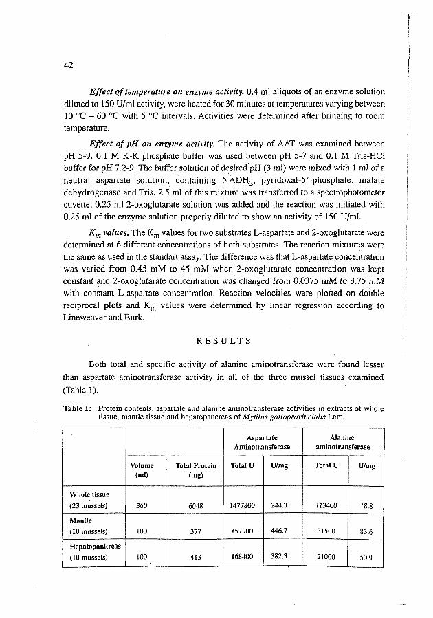
Both total and specific activity of alanine aminotransferase were found lesser
than aspartate aminotransferase activity in all of the three mussel tissues examined
(Table 1).
Table 1: Protein contents, aspartate and alanine aminotransferase activities in extracts of whole tissue, mantle tissue and hepatopancreas of Mytil~u gallnprovi~~cialis Lam.
Whole tissue (23 mussels)
Mantle (10 mussels)
Hepatopankreas (10 mussels)
AIanine aminotransferase
Total U
113400
31500
21000
Aspartate Aminotransferase
Volume (ml)
360
100
100
Ulmg
18.8
83.6
50.9
Total U
1477800
157900
168400
Total Protein (ms)
6048
377
413
Ulmg
244.3
446.7
382.3
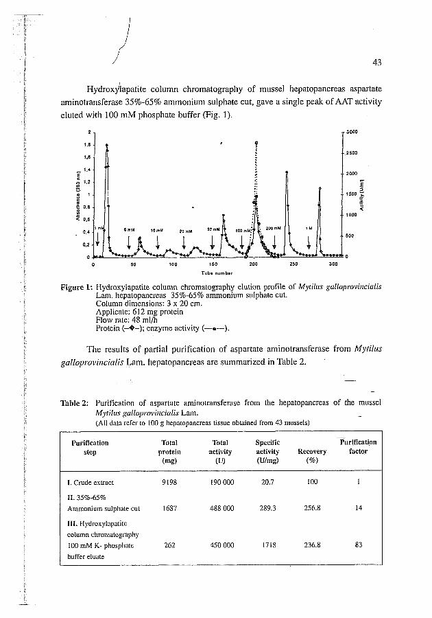
~ ~ d r o x y ' i a ~ a t i t e column chromatography of mussel hepatopancreas aspartate
aminotransferase 35%-65% ammonium sulphate cut, gave a single peak of AAT activity
eluted with 100 mM phosphate buffer (Fig. 1).
Figure 1: Hydroxylapatite column chromatography elution profile of Mytillis galioprovincialis Lam. hepatopancreas 35%-65% ammonium sulphate cut. Column dimensions: 3 x 20 cm. Applicate: 61 2 mg protein Flow rate: 48 ml/h Protein (-44; enzyme activity (---0---).
The results of partial purification of aspartate aminotransferase from Mytillts gallo~~~~ovincialis Lam. hepatopancreas are summarized in Table 2.
Table 2: Purification of aspartate aminotransferase from the hepatopancreas of the mussel Mytillrs gullol~ro~~i~tcialis Lam. - (All data refer to 100 g hcpatopancreas tissue obtained from 43 mussels)
- Purification Total Total Specific PuriBcati.on
step protein activity activity Recovery f a c t ~ r (ms) (U) (Ulmg) (%I
11. 35%-65%
Ammonium sulphnte cul 1687 488 000 289.3 256.8
111. Hydroxylapatite column chromatography 100 mM K- phosphate 262 450 000 1718 236.8 83
buffer eluate
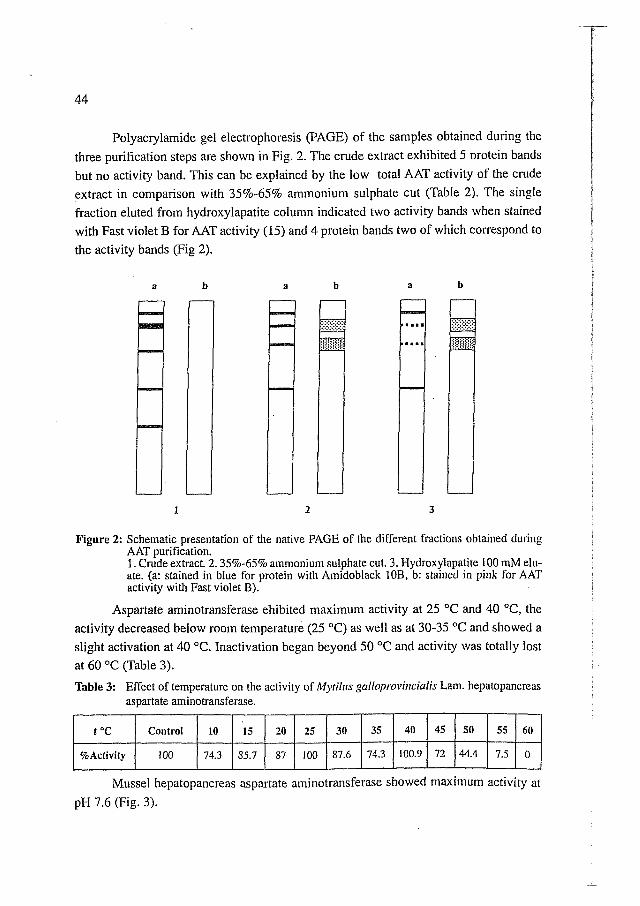
Polyacrylarnide gel electrophoresis (PAGE) of the samples obtained during the three purification steps are shown in Fig. 2. The crude extract exhibited 5 nrotein bands
but no activity band. This can be explained by the low total AAT activity of the crude extract in comparison with 35%-65% ammonium sulphate cut (Table 2). The single fraction eluted from hydroxylapatite column indicated two activity bands when stained with Fast violet B for AAT activity (15) and 4 protein bands two of which correspond to the activity bands (Fig 2).
Figure 2: Schematic presentation of the native PAGE of the different fractions obtained during AAT purification. 1. Crude extract. 2.35%-65% ammonium sulphate cut. 3. Hydroxylapatite 100 mM elu- ate. (a: stained in blue for protein with Amidoblack 10B, b: stained in pink for AAT activity with Fast violet B).
Aspartate aminotransferase ehibited maximum activity at 25 OC and 40 "C, the activity decreased below room temperature (25 "C) as well as at 30-35 OC and showed a slight activation at 40 OC. Inactivation began beyond 50 OC and activity was totally lost at 60 "C (Table 3).
Table 3: Effect of temperature on the activity of Myrihs gallnprm~i~lcialis Lam. hepatopancreas aspartate aminotransferase.
Mussel hepatopancreas aspartate arninotransferase showed maximum activity at
pH 7.6 (Fig. 3).
45
72
t "C
%Activity
35
74.3
40
100.9
SO
44.4
Control
100
55
7.5
10
74.3
60
0
15
85.7
25
100
20
87
30
87.6
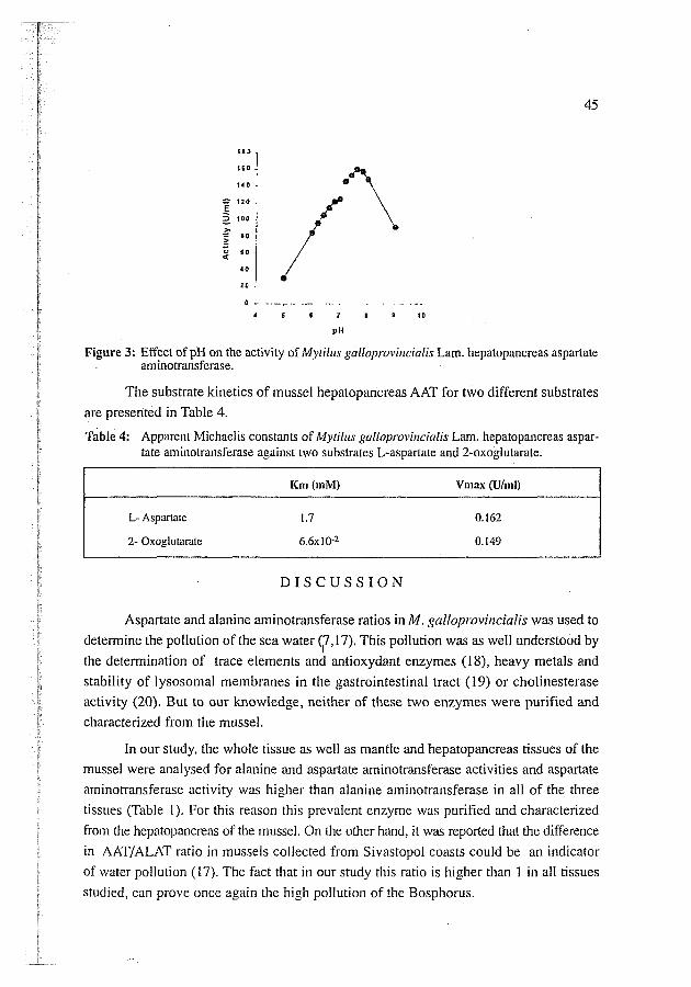
Figure 3: Effect of pH on the activity of Mytillis galloprovi~lcialis Lam. hepatopancreas aspartate aminotransferase.
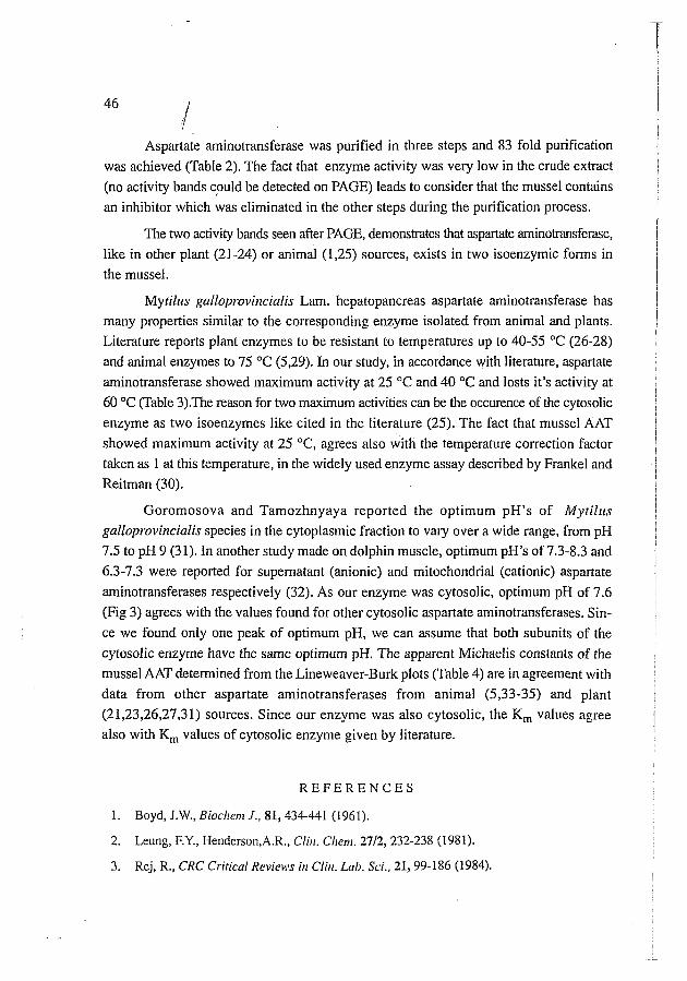
The substrate kinetics of mussel hepatopancreas AAT for two different substrates are presented in Table 4.
Table 4: Apparent Michaclis constants of Mytilus galloprovi~zcialis Lam. hepatopancreas aspar- tate aminolransferase against two substrates L-aspartate and 2-oxoglutarate.
Km (mM) Vmax (Ulml)
L- Aspartate 1.7 0.162
2- Oxoglutarite 6 . 6 ~ 1 0-2 0.149
D I S C U S S I O N
Aspartate and alanine aminotransferase ratios in M. gulloprovinciulis was used to detemiine the pollution of the sea water (7,17). This pollution was as well understood by
I the determination of trace elements and antioxydant enzymes (1 X), heavy metals and
stability of lysosomal membranes in the gastrointestinal tract (19) or cholinesterase activity (20). But to our knowledge, neither of these two enzymes were purified and characterized from the mussel.
In our study, the whole tissue as well as mantle and hepatopancreas tissues of the mussel were analysed for alanine and aspartate aminotransferase activities and aspartate aminotransferase activity was higher than alanine aminotransferase in all of the three
tissues (Table 1). For this reason this prevalent enzyme was purified and characterized
from the hepatopnncreas of the mussel. On the other hand, it was reported that the difference
in AATIALAT ratio in mussels collected from Sivastopol coasts could be an indicator of water pollution (17). The fact that in our study this ratio is higher than 1 in all tissues
studied, can prove once again the high pollution of the Bosphorus.
Aspartate aminotransferase was purified in three steps and 83 fold purification was achieved (Table 2). The fact that enzyme activity was very low in the crude extract (no activity bands could be detected on PAGE) leads to consider that the mussel contains an inhibitor which was eliminated in the other steps during the purification process.
The two activity bands seen after PAGE, demonstrates that aspartate arnin~tr~mferase, like in other plant (21-24) or animal (1,25) sources, exists in two isoenzymic forms in the mussel.
Mytilzts galloprovincialis Lam. hepatopancreas aspartate aminotransferase has many properties similar to the corresponding enzyme isolated from animal and plants. Literature reports plant enzymes to be resistant to temperatures up to 40-55 "C (26-28) and animal enzymes to 75 "C (5,29). In our study, in accordance with literature, aspartate arninotransferase showed maximum activity at 25 "C and 40 "C and losts it's activity at 60 "C (Table 3).The reason for two maximum activities can be the occurence of the cytosolic enzyme as two isoenzymes like cited in the literature (25). The fact that mussel AAT showed maximum activity at 25 "C, agrees also with the temperature correction factor taken as 1 at this temperature, in the widely used enzyme assay described by Frankel and Reitman (30).
Goromosova and Tamozhnyaya reported the optimum pH's of Mytilr~s galloprovincialis species in the cytoplasmic fraction to vary over a wide range, from pH 7.5 to pH 9 (31). In another study made on dolphin muscle, optimum pH's of 7.3-8.3 and 6.3-7.3 were reported for supernatant (anionic) and mitochondria1 (cationic) aspartate aminotransferases respectively (32). As our enzyme was cytosolic, optimum pH of 7.6 (Fig 3) agrees with the values found for other cytosolic aspartate aminotransferases. Sin- ce we found only one peak of optimum pH, we can assume that both subunits of the cytosolic enzyme have the same optimum pH. The apparent Michaeiis constants of the mussel AAT determined from the Lineweaver-Burk plots (Table 4) are in agreement with data from other aspartate aminotransferases from animal (5,33-35) and plant (21,23,26,27,31) sources. Since our enzyme was also cytosolic, the K, values agree also with K, values of cytosolic enzyme given by literature.
R E F E R E N C E S
1. Boyd, J.W., Biockenl J., 81,434-441 (1961).
2. Leung, F.Y., Henderson,A.R., Clirl. Cllen~. 2712, 232-238 (1981).
3. Rej, R., CRC Critical Reviebils it1 Clil~. Lab. Sci., 21, 99-186 (1984).
Bishop, S.H., Ellis, L.L., Burcham, J.M., "The Mollusca, Metabolic Biochemistry and
Molecular Biomechanics" Hochachka, P.W.., Wilbur, K.M., eds. Academic Press Inc., New
York, Vol.1, pp.243-327 (1983).- Ref. McCormic, A., Paynter, K.T., Broday, M.M., Bishop,
S.H., Conrp. Biocl~enz. Plrysiol., 84 B, 163-166 (1986).
Paynter, K.T., Hoffmann, R.J., Ellis, L.L., Bishop, S.H., Tlie Jourtral of Experimental
Zoology. 231, 185-197 (1984).
Goromosova, S.A., Tamozhnyaya, V.A., Biology Marine, 2,62-68 (1980).
Goromosova, S.A., Tamozhnyaya, V.A., Biology Maritre Kiev, 48, 118-122 (1979).
Lowry, O.H., Rosenbrough, N.J., Fan; A.L., Randall, R., J. Biol. Clrenr., 193, 265-275
(1951).
Warburg, O., Christian, W., Biochen~. Z., 310, 384-421 (1941).
Karmen, A., J. Clit~. Invest., 34, 131 -133 (1955).
Akev, N., Can, A., Yanardak, R., I.sratib~~l Ecz. Fak. Mec., 31, 1-9 (1995).
Rej, R., Bretaudiere, J.-P., Graffunder, B., Clitr. Chen~., 27 (4), 535-542 (1981).
Omstein, L., Ann. N.Y. Acad. Sci., 121, 321-349 (1964).
Davis, B.J., Ann. N.Y. Acad. Sci., 121,404-427 (1964). . Schwartz, M.K., Nisselbaum, J.S., Bodansky, O., Americarr J. Clitr. Patliology, 40, 103-
Tiselius, A., HjertCn, S., Levin, O., A ~ l r . Biochenr. Bioplrys., 65, 132-155 (1956).
Goromosova, S.A., Milovidova, N.Yu., Tamozhnyaya, V.A., Shapiro, A.Z., Girdrobiol ZIr.,
23 (I), 61-66 (1987).
Regoli, F., Arch. Etr~~irotr. con tun^. Tmicol., 34 (I), 48-63 (1998).
19. Stien, X., Percic, P., Gnassia-Barelli, M., Romeo, M., Lafaurie, M., Etlvirotl. Pollrrr., 99 (3),
339-345 (1998).
20. Guilhermino, L., Barros, P., Silva, M.C., Soares, A.M.B.M., Bionlarkers, 3 (2), 157-163
(1 998).
21. Wong, K.F.,Cossins, E.A., Pll):tocl?enlisrty, 8, 1327-1338 (1969).
22. Reed, R.E., Hess, J.L., J. Biol.Cl~en~., 250,4456-4461 (1975).
Oner, N., Can, A., Aydeger, N., Tiitem, E., Acta Pharrnacerctica Trircica. 30,43-48 (1988).
Griffith, S.M., Vance, C.P., Plant Plz)isiol., 90, 1622-1629 (1989).
Michuda, C.M., Martinez-Carrion, M., Biochemistry, 8, 1095-1 105 (1969).
Oner, N., Aydeger, N., Can, A., Acta Pharn~acerrtica Turcica, 31,27-32 (1989).
Sauvage, F.X., Romieu, C.G., Flanzy, C., Robin, J.P., Atn. J. E1101.Vitic.. 42,209-218 (1991).
Can, A., Akev, N., Acra Pkar~nacerrtica fiircica, 37, 100-104 (1995).
Jenkins, W.W.T., Yphantis, D.A., Sizer, LW., J. Biol. Cltem., 234,51-57 (1959).
Frankel, S., "Gradwohl's Clinical Laboratory Methods and Diagnosis", Vol.1. p.126.
Frankel, S., Reitman, S., Sonnenwirth, A.C. eds. The C.V. Mosby Company, Saint Louis
(1970).
Goromosova, S.A., Tamozhnyaya, V.A., ZIl. Evol. Biokflinl. Fiziol., 17 (4), 337-341 (1981).
Owen, T.G., Hochachka, P.W., Biochenl.J., 143,541-553 (1974).
Nisselbaum, J.S., Bodansky O., J. Biol. Cllen~., 241,2661-2664 (1966).
Quiroga, C., Busquets, M., Cortes, A., Bozal, J., In[. J. Biochem., 17, 1185-1190 (1985).
McCormic, A., Paynter, K.T., Broday, M.M., Bishop, S.H., Conlp. Biochenl. Pllysiol., 84 B,
163-166 (1986).


