Embed Size (px)
Citation preview

Pathogenic and regulatory roles for B cells in experimentalautoimmune encephalomyelitis
MONICA K. MANN1, AVIJIT RAY1, SREEMANTI BASU1,2, CHRISTOPHER L. KARP3, andBONNIE N. DITTEL1,2
1Blood Research Institute, BloodCenter of Wisconsin, Milwaukee, Wisconsin, USA2Department of Microbiology and Molecular Genetics, Medical College of Wisconsin, Milwaukee,Wisconsin, USA3Division of Molecular Immunology, Cincinnati Children's Hospital Research Foundation and theUniversity of Cincinnati College of Medicine, Cincinnati, Ohio, USA
AbstractA dual role of B cells in experimental autoimmune encephalomyelitis (EAE), the animal model ofthe human autoimmune disease multiple sclerosis (MS), has been established. In the first role, Bcells contribute to the pathogenesis of EAE through the production of anti-myelin antibodies thatcontribute to demyelination. On the contrary, B cells have also been shown to have protectivefunctions in that they play an essential role in the spontaneous recovery from EAE. In this review,we summarize studies conducted in a number of species demonstrating the conditions under whichB cells are pathogenic in EAE. We also discuss the phenotype and anti-inflammatory mechanismsof regulatory B cells.
KeywordsB cell; experimental autoimmune encephalomyelitis; immune regulation; immunoglobulin;multiple sclerosis
IntroductionOne key outcome from autoimmunity is a disruption in the balance between pro- and anti-inflammatory immune responses, ultimately leading to potentially irreparable tissue damage.It has become clear that B lymphocytes can contribute both to the pathogenesis andregulation of autoimmunity [1]. The exact mechanisms of these dual functions have beendifficult to unravel, given that B cells perform a spectrum of functions during aninflammatory immune response. Also, a complicating factor is that are a number of B cellsubsets each with unique functions and anatomical locations. Broadly, B cells are dividedinto the B1 and B2 subpopulations.
B1 cells primarily reside in the peritoneal and pleural cavities, but are also found in thespleen in low numbers and through production of natural IgM, which is often germline
© Informa UK, Ltd.
Correspondence: Bonnie Dittel, Ph.D., BloodCenter of Wisconsin, P.O. Box 2178, Milwaukee, WI 53201-2178, USA. Tel.:414-937-3865. Fax: 414-937-6284, [email protected].
Declaration of interest: The authors report no conflicts of interests. The authors alone are responsible for the content and writing ofthe paper.
NIH Public AccessAuthor ManuscriptAutoimmunity. Author manuscript; available in PMC 2013 April 29.
Published in final edited form as:Autoimmunity. 2012 August ; 45(5): 388–399. doi:10.3109/08916934.2012.665523.
NIH
-PA Author Manuscript
NIH
-PA Author Manuscript
NIH
-PA Author Manuscript

encoded and polyreactive contribute to the early innate defense against pathogens. Alsocontributing to innate immunity are marginal zone B cells that reside in the spleen and dueto their preactivated phenotype can quickly differentiate into antibody secreting plasmablastand plasma cells in the absence of T cell help. The majority of peripheral B2 cells are calledfollicular B cells and are the most prominent B cells in the spleen and lymph nodes.Follicular B cells help form the germinal center where they generate high affinity IgGantibodies in response to T-dependent antigens. Although each subclass of B cells is highlyefficient in the generation of antibodies, they all also have the capacity to present antigenand produce a spectrum of pro- and anti-inflammatory cytokines.
The elucidation of the role of B cells in multiple sclerosis (MS) was greatly facilitated by thegeneration of an animal model now referred to as experimental autoimmuneencephalomyelitis (EAE). Although no animal model completely recapitulates a complexhuman disease, such as MS, the ability to induce EAE in a number of species by a variety ofmechanisms allows for the testing of specific hypotheses in the most relevant model. EAEcan be induced in many species including rabbit, guinea pig, rat, mouse and monkeys.
Although early studies were conducted in rabbit, guinea pig and rat most current studies areconducted in mouse due to their smaller size and the copious reagents and geneticallyaltered mouse strains available. EAE can be induced by both active and passive approaches.For active induction, animals are immunized with self-antigen in the form of spinal cordhomogenate, purified self-protein or as a self-peptide emulsified in adjuvant, typicallycomplete Freund's adjuvant (CFA). Active EAE induction in mice also includes the injectionof two doses of pertussis toxin.
Passive EAE induction involves the adoptive transfer of encephalitogenic T cells withspecificity to self-antigens. The recipient animals are often irradiated to achieve a moreconsistent and predictable disease course [2]. The encephalitogenic T cells are derived fromself-antigen immunized animals or from TCR-transgenic (tg) mice with self-reactivity. EAEonset occurs 7–10 days later and depending on the animal species and strain the diseasecourse can vary from acute and monophasic, acute and chronic, progressive or relapsing andremitting.
Induction of EAE is due to an immune response to self-antigen presented by MHC class IImolecules. In mice the three most prominent myelin antigens used are myelin basic protein(MBP), proteolipid protein (PLP) and myelin oligodendrocyte protein (MOG). The antigenused for EAE induction is dictated by the MHC haplotype of the experimental animal. TableI outlines the strain, antigen usage and typical EAE disease course of the most commonlyused rat and mouse models. The evolution of EAE models over the last century has led us toa better understanding of the complex immune interactions that result in disease and has alsoallowed the discovery of regulatory mechanisms that prevent and control the extent ofdisease. This review will focus on the role of the B lymphocyte outlining our currentunderstanding of their pathogenic and regulatory functions in EAE.
Early evidence for a pathogenic role for B cells and immunoglobulin in EAEThe experimentally induced animal disease now known as EAE was first investigated inrabbits and monkeys [3]. It was in 1949 that EAE induction in mice was successful [4]. Thisachievement literally opened the floodgate regarding EAE research with thousands ofstudies having been published using the mouse model. However, most of the early studiesdefining the role of immune cells in EAE were conducted in the rat. In 1953 the transfer ofEAE from a donor rat immunized with guinea pig spinal cord homogenate in adjuvant toanother rat was first demonstrated by parabiosis, suggesting that the disease was caused byeither soluble and/or cellular components within the blood [5].
MANN et al. Page 2
Autoimmunity. Author manuscript; available in PMC 2013 April 29.
NIH
-PA Author Manuscript
NIH
-PA Author Manuscript
NIH
-PA Author Manuscript

It then took until 1960 to demonstrate that transfer of lymph node cells alone from a rat withEAE was sufficient to lead to EAE in the recipient [6]. These early insights into theimmunological nature of EAE were remarkable given that T and B lymphocytes weren'tdefinitively identified until the mid 1960s [7]. The identification of T cells alone as beingsufficient for EAE induction came from studies conducted in 1981, whereby the adoptivetransfer of MBP-specific T cell lines resulted in disease in syngeneic rats [8]. Although thislater experiment implicated T cells in EAE induction, it did not rule out the possibility thatthe transferred T cells interacted with B cells that then in turn played a pathogenic role inEAE.
The first hard evidence that B cells were involved in EAE pathogenesis came from studies inrats administered anti-IgM resulting in a depletion of B lymphocytes as well as serum IgMthat was accompanied by low levels of IgG. These depleted rats failed to both respond toLPS and make antibodies to sheep red blood cells [9]. These same rats failed to exhibit signsof EAE following immunization with either guinea pig spinal cord homogenate or purifiedMBP [9]. In a follow-up study, the transfer of serum containing anti-MBP into the anti-IgM-treated resistant rats resulted in EAE [10].
Although this experiment suggested a role for Ig in EAE induction and/or pathogenesis, theadoptive transfer of activated lymphocytes from MBP immunized anti-IgM treated rats intoeither WT or anti-IgM-treated rats also lead to EAE [10]. Thus, the final conclusion was thatpassively induced EAE was not dependent upon the presence of B cells and antibodyproduction [10]. Of interest in terms of antibody-mediated pathogenesis is that the studiesconducted by Willenborg and colleagues used MBP as the immunogen. Thus, even if anti-MBP antibodies were generated, how they would invoke pathogenic mechanisms is not cleardue to the intracellular location of MBP.
The answer as to whether or not antibodies play a pathogenic role likely resides in thespecificity of the antibodies. When whole spinal cord homogenate is used, antibodyresponses to multiple myelin antigens is possible, with myelin oligodendrocyte glycoprotein(MOG) being of particular interest because of its cell surface expression making itaccessible to antibody recognition. Indeed, serum containing high levels ofimmunoglobulins specific to MOG, but not MBP, when infused into the subarachnoid spaceof rats resulted in demyelination [11]. Further support for anti-MOG in EAE pathogenesiscame from studies using MOG monoclonal antibodies (mAb), which accelerated EAE inLewis rats and exacerbated passively induced EAE in SJL mice and Lewis rats [12–14].
Similar findings were reported in a primate EAE model, where immunization with wholewhite matter or rat rMOG (aa 1-125) led to inflammation and extensive demyelination [15].In contrast, immunization with the intracellular proteins MBP or proteolipid protein (PLP)resulted in inflammation, but no demyelination [15]. As with rodents, the transfer of anti-MOG into MBP-immunized nonhuman primates lead to fully demyelinated lesions [15].Thus collectively, these early studies indicated that T cells alone could induce and driveEAE disease, but that myelin-specific antibodies could augment EAE pathogenesis if theyrecognize a protein expressed on the cell surface of oligodendrocytes. In other words, thestudies fell short of demonstrating a requirement for B cells or their production ofimmunoglobulin in EAE induction.
Potential mechanisms of antibody augmentation of EAEDemyelination accompanying the transfer of an anti-MOG mAb provided solid evidence ofa pathogenic role for B cells via production of Ig in EAE. However, of interest is that all ofthese early studies used the same antibody clone (8.18.C5) [11–16], which is a mouse IgG1.Thus, it is not surprising that all of the studies obtained consistent results with the induction
MANN et al. Page 3
Autoimmunity. Author manuscript; available in PMC 2013 April 29.
NIH
-PA Author Manuscript
NIH
-PA Author Manuscript
NIH
-PA Author Manuscript

of demyelination being a critical factor. The mechanism by which Ig could inducedemyelination includes complement and Fc receptor (FcR) engagement.
The ability of anti-MOG antibodies to fix complement was directly related to theirpathogenesis with IgG2a mAbs being the most potent [17]. Interestingly, the 8.18.C5 mAb isan IgG1 and consistent with characteristics of this subclass of Ig its ability to fixcomplement and induce pathogenesis was lower that for the IgG2a mAb [17]. As with the8.18.C5 mAb, the IgG2a Z12 mAb, also likely mediated demyelination via a complement-dependent mechanism that did not occur in MOG-deficient animals [18].
Further evidence for a role for complement in antibody-induced demyelination was theblockage of pathogenesis by the administration of a soluble recombinant form of humancomplement receptor 1, which also led to a reduction in the deposition of C1, C3 and C9within the CNS [19]. The issue of a role for complement was definitively resolved by theuse of knockout mice deficient in FcRγ and C1q, which showed that the demyelinatingcapacity of mAb 8.18.C5 relies on complement activation but not FcR that contain the γchain (FcγR1, FcγRIII, Fc1εRI and FcαRI) [20,21].
Further evidence that complement plays a role in antibody-induced demyelination was thefinding that rats deficient in C6, which prevents the formation of the membrane attackcomplex, were not susceptible to exacerbated disease following anti-MOG administration[22]. However, the above experiments did not resolve the finding that FcRγ knockout miceare resistant to EAE induction by rat MOG immunization [23]. Because the FcRγ signalingsubunit is also utilized by proteins other than FcR, the exact role of FcR in EAE inductionremains unresolved.
Because the FcRγ subunit is utilized by a number of FcR, we determined whether FcγRIIIplayed a role in EAE pathogenesis by inducing EAE in mice deficient its ligand binding achain [24]. FcγRIII is a low affinity FcR and plays a role in activating immune cells such asmacrophages and mast cells through binding of Ig. Mice deficient in FcγRIII haddeficiencies in mast cell degranulation, IgG-dependent anaphylaxis, and Arthus reactions[24]. Using a passive transfer model of EAE [2], we found that FcγRIII-deficient mice weresimilar to their WT littermates in day of disease onset, but exhibited a significantly moresevere disease course, indicating that FcγRIII plays a protective or inhibitory role duringEAE progression presumably through the activation of an immune cell (Figure. 1A).However, neither the target cell nor the mechanism of this regulation is known.
Since FcγRIIb is an inhibitory receptor [25], we also determined whether deletion of thisreceptor would result in more severe EAE. However, as shown in Figure 1B, there was nodifference in the EAE disease curves between B10.PL control mice and FcγRIIb+/- andFcγRIIb-/- mice. The FcγRIIb+/+ littermate controls did not recover to the same extent asthe other groups, but this was likely due to this group containing the lowest number ofexperimental mice.
A similar study in DBA/1 mice immunized with rat MOG FcγRIIb-/- mice were similar totheir WT littermates in disease onset, but then the text stated that they exhibited a trendtoward worsened disease [23]. However, no statistics were provided and one caveat to thereport was a potential error in the figure legend that indicated that the WT mice exhibitedthe more severe disease [23]. Nevertheless, the difference in disease severity was notsubstantial and when combined with our data indicates that FcγRIIB plays a minimal to norole in EAE. In our study, using passively induced EAE, in retrospect the lack of a role forFcγRIIB is not surprising since we now know that this model of EAE is antibody-independent.
MANN et al. Page 4
Autoimmunity. Author manuscript; available in PMC 2013 April 29.
NIH
-PA Author Manuscript
NIH
-PA Author Manuscript
NIH
-PA Author Manuscript

Evidence that complement is not required for EAE induction are studies demonstrating thatmice deficient in C3, C3aR or C5 or rats deficient in C6 succumb to MOG-peptide EAEsimilar to WT mice, although the trend was a less severe EAE disease course [26–30]. Onecaveat to the above studies is that they were all performed using an EAE model induced byantigen immunization in CFA. This experimental design does not resolve the issue ofwhether components of the complement system play a role in T cell priming, with reducedpriming leading to reduced EAE severity. This issue was addressed using two approaches. Inthe first, cobra venom factor, which depletes C3 and C5, was administered to Lewis rats ondays 9 and 12 post-immunization leading to attenuation of EAE disease [31].
In the second approach, EAE was induced by adoptive transfer of WT encephalitogenic Tcells into WT or C3-/- recipients, thus avoiding T cell priming in a C3-deficientenvironment, which resulted in attenuated disease in the C3-/- mice. However, the variabilityin disease scores were large and no statistics were provided [32]. Thus while good evidencesupport a role for complement in antibody-induced demyelination, its impact on progressionof EAE disease is likely marginal and it may even play a positive role in remyelination andaxonal survival [29].
Another function of anti-myelin antibodies is the clearance of myelin debris by opsonizationof myelin facilitating its phagocytosis by microglial cells and macrophages [33–35]. Thisclearance of myelin could be beneficial or it could be pathogenic. In support of the formerpossibility, macrophages that had phagocytized myelin were shown to reduce antigen-specific T cell proliferation in a mechanism that involved macrophage production of nitricoxide [36]. However, in this same study, the onset and severity of EAE were higher in micetreated with myelin [36].
Nevertheless, the clearance of myelin debris is likely essential if the lesion is to recover.Although the phagocytosis of myelin is a sign of demyelination, it is not necessarilyindicative of the pathogenic mechanism that led to the demise of the oligodendrocyte. Inaddition, reparative properties of natural IgM with specificity for myelin have beenimplicated by the Rodriguez laboratory in promoting remyelination [37].
Current state of our understanding of B cell pathogenesis in EAEAlthough earlier experiments demonstrated mechanisms for antibody-dependent roles inEAE, we now appreciate more about B cell function, not only in characterization ofsubtypes and phenotypic markers, but also with respect to activation requirements, antigenpresentation, and production of cytokines that are unique to the B cells in immuneresponses. Thus in the late 1990s several groups re-examined the question of whether B cellswere required for the induction of EAE using a mouse deficient in B cells by the disruptionof the IgM heavy chain (μMT) [38].
In the first report, we demonstrated that μMT mice on the B10.PL background weresusceptible to EAE following immunization with MBP peptide [39]. A second studyconducted by Ruddle and colleagues confirmed this result using C57BL/6μMT miceimmunized with MOG peptide [40]. A third study confirmed the Ruddle result, but addedadditional contradictory data in human rMOG-immunized μMT mice, which did notsuccumb to EAE onset (41). In a follow-up study, the Cross group demonstrated arequirement for MOG-specific antibody in EAE pathogenesis in the human rMOG group[42].
Finally, the Ruddle group demonstrated that the species source of the rMOG was critical fordetermining whether antibody was required for EAE. They confirmed that EAE induction byhuman rMOG was B cell-dependent, while rat rMOG was not [43]. Interestingly, the anti-
MANN et al. Page 5
Autoimmunity. Author manuscript; available in PMC 2013 April 29.
NIH
-PA Author Manuscript
NIH
-PA Author Manuscript
NIH
-PA Author Manuscript

MOG titers were not different in the two groups, thus small differences in the sequence ofrat and human MOG dictates as to whether or not pathogenic antibodies are formed. Thesecumulative studies demonstrate that anti-myelin antibodies do play a role in EAEpathogenesis and by extrapolation, MS.
Recently, the above studies have been revisited using a new model of B cell depletion viaanti-CD20. The depletion of B cells prior to EAE onset with human rMOG prevented EAEonset in one study [44] and attenuated EAE severity in a second study [45]. Both studiesreported diminished T cell responses [45,46], which suggests that B cells may function asantigen presenting cells (APC). Evidence for this possibility comes from an elegant studywhereby MOG-TCR transgenic mice on the SJL/J background developed spontaneousdisease accompanied by the generation of anti-MOG antibodies mainly of the IgG1 andIgG2a/b isotypes, which were able to activate complement [47].
Anti-CD20 depletion of B cells largely prevented spontaneous EAE and the production ofanti-MOG antibodies [47]. Even when MOG-TCR transgenic mice were crossed with ananti-MOG BCR transgenic, anti-MOG antibody was generated from the endogenous B cellpool [47]. This result indicated that T cell:B cell interactions are occurring with self-antigenspecific B cells serving as APC facilitating the activation of the autoreactive T cells andtheir ultimate production of autoantibodies. Interestingly, while the MOG-TCR transgenicsuccumbs to spontaneous EAE by expanding endogenous B cells, MOG-BCR transgenicscannot expand endogenous autoreactive T cells to drive spontaneous EAE [48].
Thus, autoreactive T cells are the driving force for the induction of EAE and whileautoreactive B cells can potentially serve as APC, they on their own, even in the presence ofhigh titer myelin-reactive antibody, are not sufficient to induce EAE. Rather B cells seemonly to augment disease caused by T cells.
Evidence for a regulatory role for B cells in EAEOur study whereby we demonstrated that B cells were not required for the onset of EAE,revealed the unexpected finding that B10.PL mice deficient in B cells (μMT) failed tospontaneously recover from EAE [39]. This was the first indication in animal autoimmunemodels that B cells play a regulatory role in downregulating inflammation. In contrast,similar studies in C57BL/6 and (B10.PL × SJL/J)F1 mice did not suggest this novel B cellregulatory mechanism [40,42,49].
When an EAE induction protocol that allowed for recovery from EAE was used, C57BL/6μMT mice also were not able to recover from EAE [50]. This later study also presenteddata indicating that B cell production of IL-10 and expression of CD40 were requirementsfor their regulatory activity [50]. The Tedder laboratory then demonstrated that signalingwas important for regulatory B cell functions since genetic disruption of CD19 exacerbatedMOG-peptide EAE [51].
Interestingly, the transfer of CD19-/- B cells from MOG peptide-primed mice alsoexacerbated disease [51]. The signal transduction pathways affected in CD19-/- B cells inthis study is unclear since CD19 can affect CD40 (discussed above) and TLR signaling inaddition to its well-established affects on the BCR [52]. In regards to TLR signaling, Myd88expression by B cells was required for recovery from EAE in MOG-peptide EAE [53]. Morespecifically, the regulation required TLR2/4. TLR2/4 ligands are present in the CFAadjuvant used to induce EAE and they also induce the production of IL-10 by B cells [53].Thus, it is highly possible that the CFA used to induce EAE is facilitating and/or driving Bcell regulation by inducing them to produce IL-10.
MANN et al. Page 6
Autoimmunity. Author manuscript; available in PMC 2013 April 29.
NIH
-PA Author Manuscript
NIH
-PA Author Manuscript
NIH
-PA Author Manuscript

The μMT mouse, although a powerful tool, has many caveats that include alterations in Tcell function that could affect the interpretations of EAE studies, given that conventionalEAE is mediated by CD4 T cells. However, this concern was recently alleviated by therecapitulation of both the pathogenic and regulatory roles for B cells in EAE using an anti-CD20 depletion strategy. The depletion of B cells prior to immunization with human rMOG(B cell-dependent), prevented EAE onset in the mouse [45]. In the marmoset, anti-CD20depletion of B cells three weeks after EAE induction with human rMOG prevented thedevelopment of neurological deficits [54]. Consistent with a pathogenic role for B cells inthe human rMOG model. The regulatory role of B cells was confirmed in two separatestudies, whereby depletion of B cells prior to EAE induction in the mouse preventedrecovery [45,55].
Because all of the above studies utilized EAE models induced by immunization using CFAand pertussis toxin, we sought to determine whether regulatory B cells functions could berevealed in their absence. To address this question, we used a passive EAE model wherebyin vitro activated encephalitogenic T cells from MBP-TCR transgenic mice were adoptivelytransferred into either WT B10.PL or B10.PLμMT mice [2].
The μMT mice did not recover from EAE, indicating that regulatory B cell functions werenot dependent upon the presence of CFA [56]. In this same study, we showed that B cellexpression of B7 was a requirement for their regulatory activity [56]. The requirement for Bcell expression of both CD40 and B7 is highly suggestive that cognate interactions betweenB cells and T cells required [50,56]. To begin to address this question, we investigatedwhether B cells regulated the presence of CD4+Foxp3+T regulatory cells (Treg) in the CNSand found that in the absence of B cells or B cell expressed B7 the emergence of Treg in theCNS was delayed [50].
Because B cell production of IL-10 was previously implicated in their regulatory activity,we also examined IL-10 within the CNS during EAE and found that it was also delayed[50]. Using an IL-10 reporter mouse [57], we found that CD4+T cells were the mostprominent IL-10 producing cells in the CNS (Figure 2). Of the few B cells that were in theCNS, they were not producing IL-10 (Figure. 2). Our data are consistent with a study byAnderton and colleagues that also reported few B cells in the CNS during EAE andCD4+CD25+ cells being the primary IL-10 producing cells [58].
IL-10 producing B cell subsets and potential mechanisms of B cell-derivedIL-10 suppression during EAE
Although all B cell subsets have the capacity to produce IL-10, especially upon binding ofTLR ligands, of particular interest is the phenotype of the IL-10 producing cell during EAE.A clue came from the Tedder laboratories’ analysis of CD19-/- and human CD19 transgenic(hCD19tg) mice, with the later harboring hyperactive B cells. CD19/- mice exhibit enhancedT cell-mediated inflammation (contact hypersensitivity (CHS)), and inflammation inhCD19tg mice was reduced [59].
When B cell populations were compared in the two mice, it was found that CD1dhiCD5+Bcells were absent from CD19-/- mice and increased in hCD10tg mice [59]. It wassubsequently found that these B cells produce IL-10 and suppress inflammation upontransfer [59]. It should be noted that IL-10 production was observed after stimulation withLPS, PMA and ionomycin while in the presence of monensin. Using an IL-10 reportermouse, the population of splenic B cells with the greatest percentage of IL-10 producers atbaseline was shown to be CD19+B220low/-(35%) [57]. Upon LPS administration, thispercentage increased to 57%. IL-10 production in the conventional CD19+B220+ subset
MANN et al. Page 7
Autoimmunity. Author manuscript; available in PMC 2013 April 29.
NIH
-PA Author Manuscript
NIH
-PA Author Manuscript
NIH
-PA Author Manuscript

increased from 1 to 10% following LPS administration [57]. The CD19+CD138+ populationexhibited the most dramatic change with LPS increasing the percentage of IL-10 producersfrom 7.1 to 65% [57]. These studies confirm that TLR4 signaling is a potent inducer ofIL-10 production by B cells in the absence of any other inflammatory stimuli. However,using these same mice, we were unable to detect such an increase in IL-10 producing B cellin the spleen using an EAE adoptive transfer model that is independent of exogenouslyadded TLR agonists (CFA) (data not shown).
In the CHS model used in the above study, the TLR signal likely occurred through TLR2 asthis receptor has been shown to be essential for oxazolone induced CHS [60]. However, it isnot clear how physiologically relevant the above vitro B stimulation is to EAE or MS sinceit is essentially equivalent to a strong BCR signal occurring simultaneously with a TLRsignal. In EAE induced by immunization the TLR signal likely comes from the CFA.However, the BCR signal is more difficult to explain in models of EAE induced withpeptides or by adoptive transfer in which antibodies are not generated.
Nevertheless, the Tedder laboratory showed that the adoptive transfer of CD1dhiCD5+Bcells from CD19-/- mice or from MOG-sensitized animals prior to EAE induction by MOG-peptide attenuated EAE disease severity [61]. In the later group, B cells from IL-10-/- micedid not reduce disease severity, indicating an IL-10-dependent mechanism [61].
An important question regarding the mechanism of B cell regulation during EAE is whetherantigen specificity is required. For CD1dhiCD5+B cells described by the Tedder laboratorythe absence of MHC class I/II hindered their development. In regards to BCR diversity, theCD1dhiCD5+B cells developed in BCR transgenic mice bearing a transgene specific for henegg lysozyme (HEL) [62], but failed to produce IL-10 when stimulated with a combinationof LPS, ionomycin and PMA [63].
Although it was not addressed whether MHC molecules and BCR diversity are required forthe regulatory activity of CD1dhiCD5+B cells. We investigated whether BCR diversity isrequired for B cells to promote recovery from EAE. To address this question, we bred theHEL BCR transgenic [62] to B10.PLμMT mice generating mice bearing a single IgM BCR.When EAE was induced by adoptive transfer, we found that HELμMT mice exhibited acumulative disease score intermediate between WT and μMT mice (Figure. 3A). As thesedata suggested that BCR diversity did play a role in EAE recovery, we performed the sameexperiment using an IgM heavy chain BCR transgenic mouse that we also bred toB10.PLμMT mice (Vh186.2μMT). This mouse has some limited BCR diversity because theheavy chain can pair with any light chain [64].
Interestingly, we obtained the same result with the Vh186.2μMT mice that also exhibited apartial ability to recovery (Figure. 3B). From these cumulative data, we concluded that themere presence of B cells is not sufficient for their regulatory activity in EAE. However, thebiological context in which B cells require BCR specificity in antibody-independent EAEhas not been explored.
IL-10 is a pleiotropic cytokine that exhibits very potent anti-inflammatory activity [65]. Thelocation, mechanism and cellular targets of B cell-derived IL-10 regulation in EAE are notknown. Precedence for a role for IL-10 in controlling EAE are studies showing that IL-10transcripts reach maximum at the peak of EAE disease just before the animals begin torecover [56,66,67], IL-10-/- mice exhibit a severe EAE disease course [68], IL-10 transgenicmice are resistant to EAE [69] and intracranial injection of IL-10 producing fibroblastsreduced EAE severity [70].
MANN et al. Page 8
Autoimmunity. Author manuscript; available in PMC 2013 April 29.
NIH
-PA Author Manuscript
NIH
-PA Author Manuscript
NIH
-PA Author Manuscript

However, the regulation of EAE by B cell-derived IL-10 production does not likely occur inthe CNS because low numbers of B cells enter the CNS during disease and we have not beenable to detect their IL-10 secretion using an IL-10 reporter mouse (Figure. 2) [57].Interestingly, CD4+T cells were the most prevalent population of IL-10 producers in theCNS (Figure. 2), which are likely Foxp3+ Treg. CD1dhiCD5+B cells are typically classifiedas B1 cells and reside in the peritoneal cavity; however, these cells do recirculate and arealso found in the spleen and likely gut tissues.
Regardless of the site of regulation, based on the known functions of IL-10 the cellulartarget is not likely B cells themselves, because IL-10 enhances B cell functions [65].Because IL-10 effectively downregulates the antigen presentation capacity of dendritic cells[65], this cell type is the likely target in MOG-peptide induced EAE with CFA. Evidence forthis are two studies demonstrating that TLR-stimulated B cells producing IL-10 have beenshown limit the T cell priming/activation capacity of dendritic cells [53,71]. Interestingly,the study by Lo-Man and colleagues detected IL-10 production from CD5+, but not CD5- Bcells, which was recapitulated in later studies by the Tedder group [63].
IL-10-independent mechanisms of regulatory B cellsThe cumulative studies to date convincingly demonstrate that B cell production of IL-10 canlimit the severity of EAE. However, B cell regulation in an IL-10-independent manner hasalso been described. Previously, helminth infection was shown to greatly decrease EAEseverity in SJL/J mice [72]. Skewing of the immune response away from a Th1 and towardsa Th2 response was suggested as the mechanism [72,73]. The transfer of splenic B cellsfrom helminth infected, but not from naïve mice, prior to EAE induction reduced both theincidence and severity of EAE [74].
B cells from the mesenteric lymph node from infected mice were not enriched for CD5, butdid express high levels of CD23 (marker of follicular B cells) and increased production ofIL-6 and IL-10 upon TLR9 signaling [74]. However, the B cell regulation was found to beindependent of IL-10, leaving the mechanism of suppression unknown [74]. Since B cellshave the capacity to produce Th cell polarizing cytokines [75], the sensitized B cells couldhave skewed encephalitogenic T cell priming towards a Th2 phenotype that would result inless severe EAE because Th2 cells are not encephalitogenic.
Another possible mechanism whereby B cells regulate EAE in an IL-10-independent manneris via the induction or regulation of Treg. In B cell deficient mice, we reported that theemergence of Treg into the CNS was delayed as compared to WT mice and was dependentupon B cell expression of B7 [56]. Since B7 is important for Treg development and function[76,77], these studies suggested that B cell:Treg interactions occur during EAE. The natureof this interaction is unknown, but data from others have shown the B cells can drive thegeneration of inducible (i) Treg in vitro in a B7-dependent manner [78]. Also in vitro, Bcells were shown to expand Treg numbers via a TGF-β-dependent mechanism suggestingthe generation of iTreg [79], although the later wasn't definitely demonstrated [80].
An in vivo role for B cells in Treg induction/expansion is supported by several groups thathave reported that μMT have reduced percentages of Treg [80,81]. A similar observationwas observed during EAE in anti-CD20-depleted mice [45]. Additional studies havereported a role for B cells in promoting Treg induction/expansion in an allogeneic manner[82], in oral tolerance [81], during antigen presentation of self-antigen [83] and in arthritisautoimmune models [84]. An importance for Treg in controlling EAE was demonstrated bya reduction in EAE severity following the adoptive transfer of Treg and augmented diseasefollowing the depletion of Treg during EAE [58, 61, 85]. Although the exact location and
MANN et al. Page 9
Autoimmunity. Author manuscript; available in PMC 2013 April 29.
NIH
-PA Author Manuscript
NIH
-PA Author Manuscript
NIH
-PA Author Manuscript

mechanism of Treg suppression during EAE is not known, Treg are found within the CNSduring EAE [45, 56, 58, 61, 86, 87].
ConclusionsAlthough B cells were first discovered as the producers of immunoglobulin ourunderstanding of B cells rapidly expanded to include antigen presentation and cytokineproduction. We now know that each of the three prominent B cell functions can promoteimmunity, including to self-antigens, as well as contribute to the downregulation ofinflammation. However, much still needs to be learned regarding how each B cell functioncan harnessed to either prevent or induce the recovery from MS. B cell depletion therapy inearly MS clinical trials with anti-CD20 showed remarkable efficacy in preventing diseaseprogression [88,89]. Although the animal studies in EAE have shed some light on thepossible immunological mechanisms leading to this encouraging result, the exact role(s) ofB cells in MS remains elusive.
AcknowledgmentsThe authors thank Dr. Mark Shlomchik, Yale University, New Haven, CT for providing the Vh186.2 mice. We alsothank Dr. Eugene Ponomarev for technical assistance and Shelley Morris for assistance with the mice. This workwas supported by grants from the National Multiple Sclerosis Society (RG 3299-A-2) and from the NIH, NIAID(R01 AI069358).
References1. Ray A, Mann MK, Basu S, Dittel BN. A case for regulatory B cells in controlling the severity of
autoimmune-mediated inflammation in experimental autoimmune encephalomyelitis and multiplesclerosis. J Neuroimmunol. 2010; 230:1–9. [PubMed: 21145597]
2. Dittel BN, Merchant RM, Janeway CA Jr. Evidence for Fas-dependent and Fas-independentmechanisms in the pathogenesis of experimental autoimmune encephalomyelitis. J Immunol. 1999;162:6392–6400. [PubMed: 10352252]
3. Baxter AG. The origin and application of experimental autoimmune encephalomyelitis. Nat RevImmunol. 2007; 7:904–912. [PubMed: 17917672]
4. Olitsky PK, Yager RH. Experimental disseminated encephalomyelitis in white mice. J Exp Med.1949; 0:213–224. [PubMed: 18137295]
5. Lipton MM, Freund J. The transfer of experimental allergic encephalomyelitis in the rat by means ofparabiosis. J Immunol. 1953; 71:380–384. [PubMed: 13118176]
6. Paterson PY. Transfer of allergic encephalomyelitis in rats by means of lymph node cells. J ExpMed. 1960; 111:119–136. [PubMed: 14430853]
7. LeBien TW, Tedder TF. B lymphocytes: how they develop and function. Blood. 2008; 112:1570–1580. [PubMed: 18725575]
8. Ben-Nun A, Wekerle H, Cohen IR. The rapid isolation of clonable antigen-specific T lymphocytelines capable of mediating autoimmune encephalomyelitis. Eur J Immunol. 1981; 11:195–199.[PubMed: 6165588]
9. Willenborg DO, Prowse SJ. Immunoglobulin-deficient rats fail to develop experimental allergicencephalomyelitis. J Neuroimmunol. 1983; 5:99–109. [PubMed: 6194180]
10. Willenborg DO, Sjollema P, Danta G. Immunoglobulin deficient rats as donors and recipients ofeffector cells of allergic encephalomyelitis. J Neuroimmunol. 1986; 11:93–103. [PubMed:2419359]
11. Linington C, Lassmann H. Antibody responses in chronic relapsing experimental allergicencephalomyelitis: correlation of serum demyelinating activity with antibody titre to the myelin/oligodendrocyte glycoprotein (MOG). J Neuroimmunol. 1987; 17:61–69. [PubMed: 2445777]
MANN et al. Page 10
Autoimmunity. Author manuscript; available in PMC 2013 April 29.
NIH
-PA Author Manuscript
NIH
-PA Author Manuscript
NIH
-PA Author Manuscript

12. Schluesener HJ, Sobel RA, Linington C, Weiner HL. Monoclonal antibody against a myelinoligodendrocyte glycoprotein induces relapses and demyelination in central nervous systemautoimmune disease. J Immunol. 1987; 139:4016–4021. [PubMed: 3500978]
13. Lassmann H, Brunner C, Bradl M, Linington C. Experimental allergic encephalomyelitis: thebalance between encephalitogenic T lymphocytes and demyelinating antibodies determines sizeand structure of demyelinated lesions. Acta Neuropathol. 1988; 75:566–576. [PubMed: 3259787]
14. Linington C, Bradl M, Lassmann H, Brunner C, Vass K. Augmentation of demyelination in ratacute allergic encephalomyelitis by circulating mouse monoclonal antibodies directed against amyelin/oligodendrocyte glycoprotein. Am J Pathol. 1988; 130:443–454. [PubMed: 2450462]
15. Genain CP, Nguyen MH, Letvin NL, Pearl R, Davis RL, Adelman M, Lees MB, Linington C,Hauser SL. Antibody facilitation of multiple sclerosis-like lesions in a nonhuman primate. J ClinInvest. 1995; 96:2966–2974. [PubMed: 8675668]
16. Linnington C, Webb M, Woodhams PL. A novel myelin-associated glycoprotein defined by amouse monoclonal antibody. J Neuroimmunol. 1984; 6:387–396. [PubMed: 6207204]
17. Piddlesden SJ, Lassmann H, Zimprich F, Morgan BP, Linington C. The demyelinating potential ofantibodies to myelin oligodendrocyte glycoprotein is related to their ability to fix complement. AmJ Pathol. 1993; 143:555–564. [PubMed: 7688186]
18. Morris-Downes MM, Smith PA, Rundle JL, Piddlesden SJ, Baker D, Pham-Dinh D, Heijmans N,Amor S. Pathological and regulatory effects of anti-myelin antibodies in experimental allergicencephalomyelitis in mice. J Neuroimmunol. 2002; 125:114–124. [PubMed: 11960647]
19. Piddlesden SJ, Storch MK, Hibbs M, Freeman AM, Lassmann H, Morgan BP. Solublerecombinant complement receptor 1 inhibits inflammation and demyelination in antibody-mediated demyelinating experimental allergic encephalomyelitis. J Immunol. 1994; 152:5477–5484. [PubMed: 8189065]
20. Urich E, Gutcher I, Prinz M, Becher B. Autoantibody-mediated demyelination depends oncomplement activation but not activatory Fc-receptors. Proc Natl Acad Sci USA. 2006;103:18697–18702. [PubMed: 17121989]
21. Takai T. Roles of Fc receptors in autoimmunity. Nat Rev Immunol. 2002; 2:580–592. [PubMed:12154377]
22. Mead RJ, Singhrao SK, Neal JW, Lassmann H, Morgan BP. The membrane attack complex ofcomplement causes severe demyelination associated with acute axonal injury. J Immunol. 2002;168:458–465. [PubMed: 11751993]
23. Abdul-Majid KB, Stefferl A, Bourquin C, Lassmann H, Linington C, Olsson T, Kleinau S, HarrisRA. Fc receptors are critical for autoimmune inflammatory damage to the central nervous systemin experimental autoimmune encephalomyelitis. Scand J Immunol. 2002; 55:70–81. [PubMed:11841694]
24. Hazenbos WL, Gessner JE, Hofhuis FM, Kuipers H, Meyer D, Heijnen IA, Schmidt RE, SandorM, Capel PJ, Daeron M, van de Winkel JG, Verbeek JS. Impaired IgG-dependent anaphylaxis andArthus reaction in Fc gamma RIII (CD16) deficient mice. Immunity. 1996; 5:181–188. [PubMed:8769481]
25. Takai T, Ono M, Hikida M, Ohmori H, Ravetch JV. Augmented humoral and anaphylacticresponses in Fc gamma RII-deficient mice. Nature. 1996; 379:346–349. [PubMed: 8552190]
26. Nataf S, Carroll SL, Wetsel RA, Szalai AJ, Barnum SR. Attenuation of experimental autoimmunedemyelination in complement-deficient mice. J Immunol. 2000; 165:5867–5873. [PubMed:11067947]
27. Calida DM, Constantinescu C, Purev E, Zhang GX, Ventura ES, Lavi E, Rostami A. Cutting edge:C3, a key component of complement activation, is not required for the development of myelinoligodendrocyte glycoprotein peptide-induced experimental autoimmune encephalomyelitis inmice. J Immunol. 2001; 166:723–726. [PubMed: 11145641]
28. Boos L, Campbell IL, Ames R, Wetsel RA, Barnum SR. Deletion of the complementanaphylatoxin C3a receptor attenuates, whereas ectopic expression of C3a in the brain exacerbates,experimental autoimmune encephalomyelitis. J Immunol. 2004; 173:4708–4714. [PubMed:15383607]
MANN et al. Page 11
Autoimmunity. Author manuscript; available in PMC 2013 April 29.
NIH
-PA Author Manuscript
NIH
-PA Author Manuscript
NIH
-PA Author Manuscript

29. Weerth SH, Rus H, Shin ML, Raine CS. Complement C5 in experimental autoimmuneencephalomyelitis (EAE) facilitates remyelination and prevents gliosis. Am J Pathol. 2003;163:1069–1080. [PubMed: 12937147]
30. Tran GT, Hodgkinson SJ, Carter N, Killingsworth M, Spicer ST, Hall BM. Attenuation ofexperimental allergic encephalomyelitis in complement component 6-deficient rats is associatedwith reduced complement C9 deposition, P-selectin expression, and cellular infiltrate in spinalcords. J Immunol. 2002; 168:4293–4300. [PubMed: 11970970]
31. Piddlesden S, Lassmann H, Laffafian I, Morgan BP, Linington C. Antibody-mediateddemyelination in experimental allergic encephalomyelitis is independent of complementmembrane attack complex formation. Clin Exp Immunol. 1991; 83:245–250. [PubMed: 1993358]
32. Szalai AJ, Hu X, Adams JE, Barnum SR. Complement in experimental autoimmuneencephalomyelitis revisited: C3 is required for development of maximal disease. Mol Immunol.2007; 44:3132–3136. [PubMed: 17353050]
33. Goldenberg PZ, Kwon EE, Benjamins JA, Whitaker JN, Quarles RH, Prineas JW. Opsonization ofnormal myelin by anti-myelin antibodies and normal serum. J Neuroimmunol. 1989; 23:157–166.[PubMed: 2470784]
34. Trotter J, De Jong LJ, Smith ME. Opsonization with antimyelin antibody increases the uptake andintracellular metabolism of myelin in inflammatory macrophages. J Neurochem. 1986; 47:779–789. [PubMed: 3734801]
35. Van der Goes A, Kortekaas M, Hoekstra K, Dijkstra CD, Amor S. The role of anti-myelin (auto)-antibodies in the phagocytosis of myelin by macrophages. J Neuroimmunol. 1999; 101:61–67.[PubMed: 10580814]
36. Bogie JF, Stinissen P, Hellings N, Hendriks JJ. Myelinphagocytosing macrophages modulateautoreactive T cell proliferation. J Neuroinflam. 2011; 8:85.
37. Wright BR, Warrington AE, Edberg DD, Rodriguez M. Cellular mechanisms of central nervoussystem repair by natural autoreactive monoclonal antibodies. Arch Neurol. 2009; 66:1456–1459.[PubMed: 20008649]
38. Kitamura D, Roes J, Kuhn R, Rajewsky K. A B cell-deficient mouse by targeted disruption of themembrane exon of the immunoglobulin mu chain gene. Nature. 1991; 350:423–426. [PubMed:1901381]
39. Wolf SD, Dittel BN, Hardardottir F, Janeway CA Jr. Experimental autoimmune encephalomyelitisinduction in genetically B cell-deficient mice. J Exp Med. 1996; 184:2271–2278. [PubMed:8976182]
40. Hjelmstrom P, Juedes AE, Fjell J, Ruddle NH. B-cell-deficient mice develop experimental allergicencephalomyelitis with demyelination after myelin oligodendrocyte glycoprotein sensitization. JImmunol. 1998; 161:4480–4483. [PubMed: 9794370]
41. Lyons JA, San M, Happ MP, Cross AH. B cells are critical to induction of experimental allergicencephalomyelitis by protein but not by a short encephalitogenic peptide. Eur J Immunol. 1999;29:3432–3439. [PubMed: 10556797]
42. Lyons JA, Ramsbottom MJ, Cross AH. Critical role of antigen-specific antibody in experimentalautoimmune encephalomyelitis induced by recombinant myelin oligodendrocyte glycoprotein. EurJ Immunol. 2002; 32:1905–1913. [PubMed: 12115610]
43. Oliver AR, Lyon GM, Ruddle NH. Rat and human myelin oligodendrocyte glycoproteins induceexperimental auto-immune encephalomyelitis by different mechanisms in C57BL/6 mice. JImmunol. 2003; 171:462–468. [PubMed: 12817031]
44. Monson NL, Cravens P, Hussain R, Harp CT, Cummings M, de Pilar Martin M, Ben LH, Do J,Lyons JA, Lovette-Racke A, Cross AH, Racke MK, Stuve O, Shlomchik M, Eagar TN. Rituximabtherapy reduces organ-specific T cell responses and ameliorates experimental autoimmuneencephalomyelitis. PLoS One. 2011; 6:e17103. [PubMed: 21359213]
45. Weber MS, Prod'homme T, Patarroyo JC, Molnarfi N, Karnezis T, Lehmann-Horn K, DanilenkoDM, Eastham-Anderson J, Slavin AJ, Linington C, Bernard CC, Martin F, Zamvil SS. B-cellactivation influences T-cell polarization and outcome of anti-CD20 B-cell depletion in centralnervous system autoimmunity. Ann Neurol. 2010; 68:369–383. [PubMed: 20641064]
MANN et al. Page 12
Autoimmunity. Author manuscript; available in PMC 2013 April 29.
NIH
-PA Author Manuscript
NIH
-PA Author Manuscript
NIH
-PA Author Manuscript

46. Monson NL, Cravens PD, Frohman EM, Hawker K, Racke MK. Effect of rituximab on theperipheral blood and cerebrospinal fluid B cells in patients with primary progressive multiplesclerosis. Arch Neurol. 2005; 62:258–264. [PubMed: 15710854]
47. Pollinger B, Krishnamoorthy G, Berer K, Lassmann H, Bosl MR, Dunn R, Domingues HS, HolzA, Kurschus FC, Wekerle H. Spontaneous relapsing-remitting EAE in the SJL/J mouse: MOG-reactive transgenic T cells recruit endogenous MOG-specific B cells. J Exp Med. 2009; 206:1303–1316. [PubMed: 19487416]
48. Litzenburger T, Fassler R, Bauer J, Lassmann H, Linington C, Wekerle H, Iglesias A. Blymphocytes producing demyelinating autoantibodies: development and function in gene-targetedtransgenic mice. J Exp Med. 1998; 188:169–180. [PubMed: 9653093]
49. Dittel BN, Urbania TH, Janeway CA Jr. Relapsing and remitting experimental autoimmuneencephalomyelitis in B cell deficient mice. J Autoimmun. 1998; 14:311–318. [PubMed:10882057]
50. Fillatreau S, Sweenie CH, McGeachy MJ, Gray D, Anderton SM. B cells regulate autoimmunityby provision of IL-10. Nat Immunol. 2002; 3:944–950. [PubMed: 12244307]
51. Matsushita T, Fujimoto M, Hasegawa M, Komura K, Takehara K, Tedder TF, Sato S. Inhibitoryrole of CD19 in the progression of experimental autoimmune encephalomyelitis by regulatingcytokine response. Am J Pathol. 2006; 168:812–821. [PubMed: 16507897]
52. Fujimoto M, Poe JC, Inaoki M, Tedder TF. CD19 regulates B lymphocyte responses totransmembrane signals. Semin Immunol. 1998; 10:267–277. [PubMed: 9695183]
53. Lampropoulou V, Hoehlig K, Roch T, Neves P, Calderon Gomez E, Sweenie CH, Hao Y, FreitasAA, Steinhoff U, Anderton SM, Fillatreau S. TLR-activated B cells suppress T cell-mediatedautoimmunity. J Immunol. 2008; 180:4763–4773. [PubMed: 18354200]
54. Kap YS, van Driel N, Blezer E, Parren PW, Bleeker WK, Laman JD, Craigen JL, Hart BA. Late Bcell depletion with a human anti-human CD20 IgG1k monoclonal antibody halts the developmentof experimental autoimmune encephalomyelitis in marmosets. J Immunol. 2010; 185:3990–4003.[PubMed: 20739677]
55. Matsushita T, Yanaba K, Bouaziz JD, Fujimoto M, Tedder TF. Regulatory B cells inhibit EAEinitiation in mice while other B cells promote disease progression. J Clin Invest. 2008; 118:3420–3430. [PubMed: 18802481]
56. Mann MK, Maresz K, Shriver LP, Tan Y, Dittel BN. B cell regulation of CD4+CD25+T regulatorycells and IL-10 via B7 is essential for recovery from experimental autoimmune encephalomyelitis.J Immunol. 2007; 178:3447–3456. [PubMed: 17339439]
57. Madan R, Demircik F, Surianarayanan S, Allen JL, Divanovic S, Trompette A, Yogev N, Gu Y,Khodoun M, Hildeman D, Boespflug N, Fogolin MB, Grobe L, Greweling M, Finkelman FD,Cardin R, Mohrs M, Muller W, Waisman A, Roers A, Karp CL. Nonredundant roles for B cell-derived IL-10 in immune counter-regulation. J Immunol. 2009; 183:2312–2320. [PubMed:19620304]
58. McGeachy MJ, Stephens LA, Anderton SM. Natural recovery and protection from autoimmuneencephalomyelitis: Contribution of CD4+CD25+regulatory cells within the central nervous system.J Immunol. 2005; 175:3025–3032. [PubMed: 16116190]
59. Yanaba K, Bouaziz JD, Haas KM, Poe JC, Fujimoto M, Tedder TF. A regulatory B cell subsetwith a unique CD1dhiCD5+ phenotype controls T cell-dependent inflammatory responses.Immunity. 2008; 28:639–650. [PubMed: 18482568]
60. Jin H, Kumar L, Mathias C, Zurakowski D, Oettgen H, Gorelik L, Geha R. Toll-like receptor 2 isimportant for the T(H)1 response to cutaneous sensitization. J Allergy Clin Immunol. 2009;123:875–882. e871. [PubMed: 19348925]
61. Matsushita T, Horikawa M, Iwata Y, Tedder TF. Regulatory B cells (B10 Cells) and regulatory Tcells have independent roles in controlling experimental autoimmune encephalomyelitis initiationand late-phase immunopathogenesis. J Immunol. 2010; 185:2240–2252. [PubMed: 20624940]
62. Mason DY, Jones M, Goodnow CC. Development and follicular localization of tolerant Blymphocytes in lysozyme/anti-lysozyme IgM/IgD transgenic mice. Int Immunol. 1992; 4:163–175.[PubMed: 1622894]
MANN et al. Page 13
Autoimmunity. Author manuscript; available in PMC 2013 April 29.
NIH
-PA Author Manuscript
NIH
-PA Author Manuscript
NIH
-PA Author Manuscript

63. Yanaba K, Bouaziz JD, Matsushita T, Tsubata T, Tedder TF. The development and function ofregulatory B cells expressing IL-10 (B10 cells) requires antigen receptor diversity and TLRsignals. J Immunol. 2009; 182:7459–7472. [PubMed: 19494269]
64. Hannum LG, Haberman AM, Anderson SM, Shlomchik MJ. Germinal center initiation, variablegene region hypermutation, and mutant B cell selection without detectable immune complexes onfollicular dendritic cells. J Exp Med. 2000; 192:931–942. [PubMed: 11015435]
65. Moore KW, de Waal Malefyt R, Coffman RL, O'Garra A. Interleukin-10 and the interleukin-10receptor. Annu Rev Immunol. 2001; 19:683–765. [PubMed: 11244051]
66. Kennedy MK, Torrance DS, Picha KS, Mohler KM. Analysis of cytokine mRNA expression in thecentral nervous system of mice with experimental autoimmune encephalomyelitis reveals thatIL-10 mRNA expression correlates with recovery. J Immunol. 1992; 149:2496–2505. [PubMed:1527389]
67. Jander S, Pohl J, D'Urso D, Gillen C, Stoll G. Time course and cellular localization ofinterleukin-10 mRNA and protein expression in autoimmune inflammation of the rat centralnervous system. Am J Pathol. 1998; 152:975–982. [PubMed: 9546358]
68. Bettelli E, Das MP, Howard ED, Weiner HL, Sobel RA, Kuchroo VK. IL-10 is critical in theregulation of autoimmune encephalomyelitis as demonstrated by studies of IL-10- and IL-4-deficient and transgenic mice. J Immunol. 1998; 161:3299–3306. [PubMed: 9759845]
69. Cua DJ, Groux H, Hinton DR, Stohlman SA, Coffman RL. Transgenic interleukin 10 preventsinduction of experimental autoimmune encephalomyelitis. J Exp Med. 1999; 189:1005–1010.[PubMed: 10075984]
70. Croxford JL, Feldmann M, Chernajovsky Y, Baker D. Different therapeutic outcomes inexperimental allergic encephalomyelitis dependent upon the mode of delivery of IL-10: acomparison of the effects of protein, adenoviral or retroviral IL-10 delivery into the centralnervous system. J Immunol. 2001; 166:4124–4130. [PubMed: 11238662]
71. Sun CM, Deriaud E, Leclerc C, Lo-Man R. Upon TLR9 signaling, CD5+B cells control the IL-12-dependent Th1-priming capacity of neonatal DCs. Immunity. 2005; 22:467–477. [PubMed:15845451]
72. Sewell D, Qing Z, Reinke E, Elliot D, Weinstock J, Sandor M, Fabry Z. Immunomodulation ofexperimental autoimmune encephalomyelitis by helminth ova immunization. Int Immunol. 2003;15:59–69. [PubMed: 12502726]
73. Gruden-Movsesijan A, Ilic N, Mostarica-Stojkovic M, Stosic-Grujicic S, Milic M, Sofronic-Milosavljevic L. Mechanisms of modulation of experimental autoimmune encephalomyelitis bychronic Trichinella spiralis infection in Dark Agouti rats. Parasite Immunol. 2010; 32:450–459.[PubMed: 20500676]
74. Wilson MS, Taylor MD, O'Gorman MT, Balic A, Barr TA, Filbey K, Anderton SM, Maizels RM.Helminth-induced CD19+CD23hi B cells modulate experimental allergic and autoimmuneinflammation. Eur J Immunol. 2010; 40:1682–1696. [PubMed: 20306466]
75. Harris DP, Haynes L, Sayles PC, Duso DK, Eaton SM, Lepak NM, Johnson L, Swain SL, LundFE. Reciprocal regulation of polarized cytokine production by effector B and T cells. Nat.Immunol. 2000; 1:475–482. [PubMed: 11101868]
76. Salomon B, Lenschow DJ, Rhee L, Ashourian N, Singh B, Sharpe A, Bluestone JA. B7/CD28costimulation is essential for the homeostasis of the CD4+CD25+ immunoregulatory T cells thatcontrol autoimmune diabetes. Immunity. 2000; 12:431–440. [PubMed: 10795741]
77. Read S, Greenwald R, Izcue A, Robinson N, Mandelbrot D, Francisco L, Sharpe AH, Powrie F.Blockade of CTLA-4 on CD4+CD25+ regulatory T cells abrogates their function in vivo. JImmunol. 2006; 177:4376–4383. [PubMed: 16982872]
78. Zhong X, Gao W, Degauque N, Bai C, Lu Y, Kenny J, Oukka M, Strom TB, Rothstein TL.Reciprocal generation of Th1/Th17 and T(reg) cells by B1 and B2 B cells. Eur J Immunol. 2007;37:2400–2404. [PubMed: 17683116]
79. Marie JC, Letterio JJ, Gavin M, Rudensky AY. TGF-β1 maintains suppressor function and Foxp3expression in CD4+CD25+ regulatory T cells. J Exp Med. 2005; 201:1061–1067. [PubMed:15809351]
MANN et al. Page 14
Autoimmunity. Author manuscript; available in PMC 2013 April 29.
NIH
-PA Author Manuscript
NIH
-PA Author Manuscript
NIH
-PA Author Manuscript

80. Shah S, Qiao L. Resting B cells expand a CD4+CD25+-Foxp3+ Treg population via TGF-β3. Eur JImmunol. 2008; 38:2488–2498. [PubMed: 18792402]
81. Sun JB, Flach CF, Czerkinsky C, Holmgren J. B lymphocytes promote expansion of regulatory Tcells in oral tolerance: powerful induction by antigen coupled to cholera toxin B subunit. JImmuno. 2008; 181:8278–8287.
82. Chen X, Jensen PE. Cutting edge: primary B lymphocytes preferentially expand allogeneic FoxP3+
CD4 T cells. J Immunol. 2007; 179:2046–2050. [PubMed: 17675460]
83. Morlacchi S, Soldani C, Viola A, Sarukhan A. Self-antigen presentation by mouse B cells resultsin regulatory T-cell induction rather than anergy or clonal deletion. Blood. 2011; 118:984–991.[PubMed: 21652680]
84. Carter NA, Vasconcellos R, Rosser EC, Tulone C, Munoz-Suano A, Kamanaka M, Ehrenstein MR,Flavell RA, Mauri C. Mice lacking endogenous IL-10-producing regulatory B cells developexacerbated disease and present with an increased frequency of Th1/Th17 but a decrease inregulatory T cells. J Immunol. 2011; 186:5569–5579. [PubMed: 21464089]
85. Kohm AP, Carpentier PA, Anger HA, Miller SD. Cutting edge: CD4+CD25+ regulatory T cellssuppress antigen-specific autoreactive immune responses and central nervous system inflammationduring active experimental autoimmune encephalomyelitis. J Immunol. 2002; 169:4712–4716.[PubMed: 12391178]
86. O'Connor RA, Malpass KH, Anderton SM. The inflamed central nervous system drives theactivation and rapid proliferation of Foxp3+ regulatory T cells. J Immunol. 2007; 179:958–966.[PubMed: 17617587]
87. Korn T, Reddy J, Gao W, Bettelli E, Awasthi A, Petersen TR, Backstrom BT, Sobel RA,Wucherpfennig KW, Strom TB, Oukka M, Kuchroo VK. Myelin-specific regulatory T cellsaccumulate in the CNS but fail to control autoimmune inflammation. Nat Med. 2007; 13:423–431.[PubMed: 17384649]
88. Hauser SL, Waubant E, Arnold DL, Vollmer T, Antel J, Fox RJ, Bar-Or A, Panzara M, Sarkar N,Agarwal S, Langer-Gould A, Smith CH. B-cell depletion with rituximab in relapsingremittingmultiple sclerosis. N Engl J Med. 2008; 358:676–688. [PubMed: 18272891]
89. Bar-Or A, Calabresi PA, Arnold D, Markowitz C, Shafer S, Kasper LH, Waubant E, Gazda S, FoxRJ, Panzara M, Sarkar N, Agarwal S, Smith CH. Rituximab in relapsing-remitting multiplesclerosis: a 72-week, open-label, phase I trial. Ann Neurol. 2008; 63:395–400. [PubMed:18383069]
MANN et al. Page 15
Autoimmunity. Author manuscript; available in PMC 2013 April 29.
NIH
-PA Author Manuscript
NIH
-PA Author Manuscript
NIH
-PA Author Manuscript

Figure 1.B6.129P2-Fcgr3tm1Sjv/J and B6;129S-Fcgr2btm1tk/J mice were obtained from JacksonLaboratories (Bar Harbor, ME) and backcrossed to B10.PL mice for three generations togenerate H-2uxu mice that were then intercrossed to generate WT, heterozygous andknockout mice. EAE was induced in littermates by adoptive transfer of 1 × 10⁶
encephalitogenic T cells generated in vitro from the spleen of B10.PL mice bearing a TCRtransgene specific for MBP. Mice were scored for signs of disease daily starting on day 4using a 5-point scale: 0- no disease; 1- tail weakness or wobbly walk; 1.5-tail weakness andwobbly walk; 2-hind limb paresis; 2.5- paralysis in one hind limb; 3- hind limb paralysis; 4-hind and forelimb paralysis and 5- death. A) Data are the mean daily scores of 24FcγRIII+/+ and 43 FcγRIII–/ – mice from 5 separate experiments with 4-13 mice in eachexperiment. The FcγRIII–/ – group contains two mice that died on day 13 and 16 and werescored as 5 for the remaining timepoints. p = 0.0001. B) Data are the mean daily scores of14 B10.PL, 9 FcγRIIb+/+, 18 FcγRIIb+/ – and 20 FcγRIIb–/ – mice from 4 separateexperiments with 1-8 mice in each experiment. Mice that did not receive a score of at least1.5 were not included in the disease curves.
MANN et al. Page 16
Autoimmunity. Author manuscript; available in PMC 2013 April 29.
NIH
-PA Author Manuscript
NIH
-PA Author Manuscript
NIH
-PA Author Manuscript
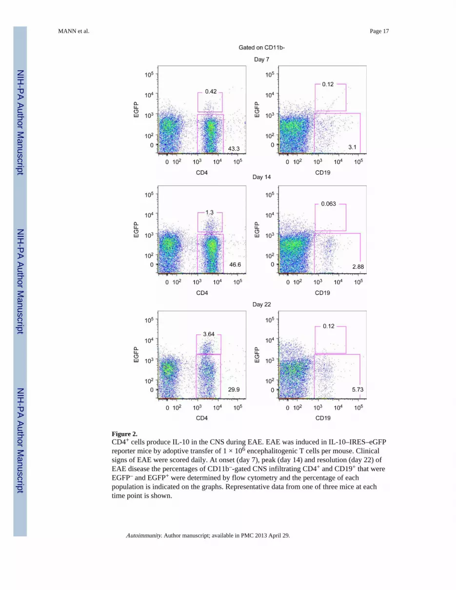
Figure 2.CD4+ cells produce IL-10 in the CNS during EAE. EAE was induced in IL-10–IRES–eGFPreporter mice by adoptive transfer of 1 × 10⁶ encephalitogenic T cells per mouse. Clinicalsigns of EAE were scored daily. At onset (day 7), peak (day 14) and resolution (day 22) ofEAE disease the percentages of CD11b–-gated CNS infiltrating CD4+ and CD19+ that wereEGFP– and EGFP+ were determined by flow cytometry and the percentage of eachpopulation is indicated on the graphs. Representative data from one of three mice at eachtime point is shown.
MANN et al. Page 17
Autoimmunity. Author manuscript; available in PMC 2013 April 29.
NIH
-PA Author Manuscript
NIH
-PA Author Manuscript
NIH
-PA Author Manuscript

Figure 3.C57BL/6-Tg(IghelMD4)4Ccg/J (Jackson Laboratories, Bar Harbor, ME) and Vh186.2 BCRtransgenic mice, which were kindly provided by Dr. M. Shlomchik, Yale University, NewHaven, CT, were bred to B10.PLμMT mice to generate transgenic positive H-2uxu mice thatwere then intercrossed with transgenic negative mice to generate transgene positive μMTmice. EAE was induced as for Figure 1. A) Data are the mean daily scores of 9 B10.PL, 13HEL-negative μMT (littermate control) and 15 HELμMT mice from two experiments forthe B10.PL and three experiments for the μMT and HELμMT mice with 3-9 mice pergroup. B) Data are the mean daily scores of 3 B10.PL, 19 μMT and 18 Vh186.2 BCR micefrom one experiment for the B10.PL and two experiments for the μMT and Vh186.2 BCRmice and each experiment contained 3–15 mice per group.
MANN et al. Page 18
Autoimmunity. Author manuscript; available in PMC 2013 April 29.
NIH
-PA Author Manuscript
NIH
-PA Author Manuscript
NIH
-PA Author Manuscript

NIH
-PA Author Manuscript
NIH
-PA Author Manuscript
NIH
-PA Author Manuscript
MANN et al. Page 19
Tabl
e I
Com
mon
mod
els
of r
at a
nd m
ouse
EA
E
Spec
ies
Stra
inA
ntig
enE
AE
indu
ctio
nD
isea
se c
ours
eB
cel
l rol
eR
efer
ence
sa
Rat
Lew
isM
BP
prot
ein
Act
ive
imm
uniz
atio
nA
cute
Mon
opha
sic
Rec
over
yE
AE
ons
et, A
ntib
ody?
[9,1
0,14
]
Mou
seC
57B
L/6
MO
G35
-55
pept
ide
Act
ive
imm
uniz
atio
nA
cute
Mon
opha
sic
Rec
over
y or
chr
onic
Reg
ulat
ory
role
in r
ecov
ery
[28,
43,5
0,61
]
Mou
seC
57B
L/6
rhM
OG
pro
tein
Act
ive
imm
uniz
atio
nA
cute
Mon
opha
sic
Chr
onic
EA
E o
nset
, Ant
ibod
y[4
1–43
,45]
Mou
seSJ
L/J
PLP 1
39-1
51 p
eptid
eA
ctiv
e im
mun
izat
ion
Rel
apsi
ng a
nd r
emitt
ing
Chr
onic
Non
e kn
own
[49]
Mou
seB
10.P
LM
BPA
c1-1
1 pe
ptid
eA
ctiv
e im
mun
izat
ion
Acu
te M
onop
hasi
c R
ecov
ery
Reg
ulat
ory
role
in r
ecov
ery
[39]
Mou
seB
10.P
LM
BPA
c1-1
1 pe
ptid
ePa
ssiv
e tr
ansf
er o
f M
BP
Ac1
-11-
spec
ific
T c
ells
Acu
te M
onop
hasi
c R
ecov
ery
Reg
ulat
ory
role
in r
ecov
ery
[2, 5
6]
a Ref
eren
ces
are
sele
cted
fro
m th
is r
evie
w a
nd a
re n
ot n
eces
sari
ly th
e fi
rst h
isto
rica
l rep
ort.
Autoimmunity. Author manuscript; available in PMC 2013 April 29.


