Embed Size (px)
Citation preview

PCB-153 Shows Different Dynamics of Mobilisation fromDifferentiated Rat Adipocytes during Lipolysis inComparison with PCB-28 and PCB-118Caroline Louis1, Gilles Tinant1, Eric Mignolet1, Jean-Pierre Thome2, Cathy Debier1*
1 Institut des Sciences de la Vie, Universite catholique de Louvain, Louvain-la-Neuve, Belgium, 2 Laboratoire d’Ecologie animale et d’Ecotoxicologie, Universite de Liege,
Liege, Belgium
Abstract
Background: Polychlorinated biphenyls (PCBs) are persistent organic pollutants. Due to their lipophilic character, they arepreferentially stored within the adipose tissue. During the mobilisation of lipids, PCBs might be released from adipocytesinto the bloodstream. However, the mechanisms associated with the release of PCBs have been poorly studied. Several invivo studies followed their dynamics of release but the complexity of the in vivo situation, which is characterised by a largerange of pollutants, does not allow understanding precisely the behaviour of individual congeners. The present in vitroexperiment studied the impact of (i) the number and position of chlorine atoms of PCBs on their release from adipocytesand (ii) the presence of other PCB congeners on the mobilisation rate of such molecules.
Methodology/Principal Findings: Differentiated rat adipocytes were used to compare the behaviour of PCB-28, -118 and -153. Cells were contaminated with the three congeners, alone or in cocktail, and a lipolysis was then induced withisoproterenol during 12 hours. Our data indicate that the three congeners were efficiently released from adipocytes andaccumulated in the medium during the lipolysis. Interestingly, for a same level of cell lipids, PCB-153, a hexa-CB with twochlorine atoms in ortho-position, was mobilised slower than PCB-28, a tri-CB, and PCB-118, a penta-CB, which are bothcharacterised by one chlorine atom in ortho-position. It suggests an impact of the chemical properties of pollutants on theirmobilisation during periods of negative energy balance. Moreover, the mobilisation of PCB congeners, taken individually,did not seem to be influenced by the presence of other congeners within adipocytes.
Conclusion/Significance: These results not only highlight the obvious mobilisation of PCBs from adipocytes during lipolysis,in parallel to lipids, but also demonstrate that the structure of congeners defines their rate of release from adipocytes.
Citation: Louis C, Tinant G, Mignolet E, Thome J-P, Debier C (2014) PCB-153 Shows Different Dynamics of Mobilisation from Differentiated Rat Adipocytes duringLipolysis in Comparison with PCB-28 and PCB-118. PLoS ONE 9(9): e106495. doi:10.1371/journal.pone.0106495
Editor: Arun Rishi, Wayne State University, United States of America
Received April 25, 2014; Accepted July 31, 2014; Published September 11, 2014
Copyright: � 2014 Louis et al. This is an open-access article distributed under the terms of the Creative Commons Attribution License, which permitsunrestricted use, distribution, and reproduction in any medium, provided the original author and source are credited.
Data Availability: The authors confirm that all data underlying the findings are fully available without restriction. All relevant data are within the paper and itsSupporting Information files.
Funding: The authors have no support or funding to report.
Competing Interests: The authors have declared that no competing interests exist.
* Email: [email protected]
Introduction
Polychlorinated biphenyls (PCBs) are a class of environmentally
persistent pollutants that biomagnify throughout food chains.
Ingestion of contaminated food, and especially fat-rich animal
products, represents 90% of the mean uptake of humans [1].
Adipose tissue is then the main reservoir for the storage of these
highly lipophilic molecules [2]. The cytoplasm of adipocytes is
almost exclusively composed of lipid droplets (LDs) [3], which
appear to be the principal targets for PCBs [4]. These cells
therefore have an enormous capacity to accumulate lipophilic
pollutants [5].
During periods of weight loss in humans, lipids from adipose
tissue are mobilised, leading to an increase of PCB concentrations
in this tissue [6,7]. Evidence from wildlife indicates same trends
[8–14]. This phenomenon suggests that PCBs are less efficiently
mobilised from adipocytes than fatty acids. However, a release of
PCBs in the blood circulation does occur during such periods of
weight loss and appears to become more important when the
adipose stores are already significantly reduced [2,6,15–18]. In
addition to being a reservoir, the adipose tissue can thus also be an
internal source of lipophilic pollutants for the rest of the body [2].
Once in the bloodstream, pollutants are able to contaminate other
tissues or be transferred in maternal milk. The exposure to PCBs is
associated to adverse effects on human and animal health [19,20].
Among others, PCBs are involved in endocrine disruption,
immuno- and neuro-toxicity as well as in the development of
cardiovascular diseases and type-2 diabetes [21–25]. A correlation
between the rise of persistent organic pollutants (POPs), such as
PCBs, in the serum and the alterations of skeletal muscle oxidative
capacity has been suggested in humans [16]. Furthermore,
individuals who underwent bariatric surgery exhibited a positive
association between POP serum levels and a diminished improve-
ment of lipid values and liver markers [6].
PLOS ONE | www.plosone.org 1 September 2014 | Volume 9 | Issue 9 | e106495

Even if several in vivo studies report a release of PCBs from
adipose tissue during lipolytic process [6,10,13–15], little is known
concerning the chemical and biochemical factors that govern their
mobilisation and transfer into the circulation. In vivo studies on
long-term fasting wild animals report an impact of the fasting stage
as well as the degree of lipophilicity of PCB congeners on their
dynamics of mobilisation from the adipose tissue [8,10,13,14]. The
transfer from adipose tissue into the blood circulation appears to
be selective and strongly dependent on the log Kow value of the
compounds, with less lipophilic PCBs being more efficiently
released. In vivo models being usually complex, the in vitrocultures of adipocytes would be useful to precisely understand the
mobilisation of PCB congeners as a function of their chemical
structure. A recent study from our group investigated the
dynamics of accumulation of three PCB congeners, differing in
the position and number of their chlorine atoms (PCB-28, log
Kow = 5.71; PCB-118, log Kow = 6.57 and PCB-153, log
Kow = 6.80) in cultured adipocytes [26]. The accumulation profile
revealed significant differences between PCB congeners. Their
release during lipolysis was however not investigated.
In this study, we followed and compared the dynamics of
mobilisation of PCB-28, PCB-118 and PCB-153 from in vitrodifferentiated rat adipocytes. Cells were contaminated by the three
congeners, added individually or in cocktail, at the same
concentrations in the culture medium. Lipolysis was then triggered
over 12 hours with a lipolytic medium supplemented with
isoproterenol, a well-known synthetic catecholamine [27,28].
The levels of PCBs in the extracellular medium and adipocytes
were regularly assessed. The present experiment allowed (i) to
estimate the impact of the number and position of chlorine atoms
of PCBs on their release from adipocytes and (ii) to assess the
impact of the presence of other PCB congeners on the mobilisation
dynamics of such molecules.
Experimental Procedures
Primary cultures of rat adipocytesDifferentiated rat adipocytes were obtained and cultured as
described previously [5,28]. Experimental procedures in animals
were approved by the Animal Care and Use Committee of the
Universite catholique de Louvain (#103201) and were performed
in accordance with the ‘‘Principles of Laboratory Animal Care’’
(NIH Publication 85–23). Two-month-old male Wistar rats
(Centre d’Elevage Janvier, Le Genest Saint Isle, France) were
sacrificed by decapitation. The fat tissue of the stromal-vascular
fraction was sampled and then digested in a solution of collagenase
(1250 U/ml type II; Sigma-Aldrich, Bornem, Belgium). The
digested tissue underwent three filtrations and three centrifuga-
tions in order to obtain a final pellet of stromal-vascular cells,
which was suspended in a medium composed of Dulbecco’s
Modified Eagle Medium (DMEM, 4.5 g/l glucose, Gibco-
Invitrogen, Merelbeke, Belgium), 10% (v:v) heat-inactivated foetal
bovine serum (FBS, PAA, A&E Scientific, Marcq, Belgium) and an
antibiotic and antifungal mixture. Cells were seeded at a mean
density of 18,000 cells per cm2 on 6-well plates (Corning
CellBIND Surface, Corning, Elscolab, Kruibeke, Belgium) (day
0) and incubated at 37uC in a humidified atmosphere containing
10% CO2 in air for 24 hours to allow cell sedimentation and
adhesion. Twenty-four hours after the isolation of progenitor cells
(day 1), the medium was replaced by a differentiation medium
composed of DMEM (4.5 g/l glucose), 10% (v:v) heat-inactivated
FBS, 100 U/ml penicillin – 100 U/ml streptomycin – 250 ng/ml
amphotericin B mixture (Lonza, Verviers, Belgium), 10 nM
dexamethasone (Sigma-Aldrich), 10 mM ciglitizone (Sigma-Al-
drich) and 5 mg/ml insulin (Sigma-Aldrich). This medium was
renewed every 48 hours until day 10 in order to obtain
differentiated adipocytes.
Cell treatmentAt day 10, adipocytes were incubated with a medium
supplemented with PCB congeners (Dr. Ehrenstorfer GmbH,
Ausburg, DE) during 12 hours (37uC – 10% CO2 in air). The
PCBs were added to the culture medium as ethanolic solution and
four conditions of PCB contamination were tested: (i) 2,4,49-
trichlorobiphenyl (PCB-28); (ii) 2,39,4,49,5-pentachlorobiphenyl
(PCB-118); (iii) 2,29,4,49,5,59-hexachlorobiphenyl (PCB-153); (iv)
an equimolar mixture of three PCB congeners (PCB-28, -118 and
-153), also called cocktail of PCBs. In all conditions of
contamination, each PCB was added to the culture medium at a
concentration of 300 nM, which is within the range of concen-
trations found in in vivo and in vitro studies [4,29,30]. Impact of
the ethanol vehicle was tested earlier [4].
Lipolysis experimentAt day 11, lipolytic process was induced to differentiated
adipocytes as previously described in Louis et al. [28]. The
differentiation medium in contact with adipocytes was removed
and replaced by a lipolytic medium composed of DMEM (1.0 g/l
glucose, Gibco–Invitrogen), 5% (v:v) heat-inactivated FBS, 2%
(w:v) bovine albumin (Sigma-Aldrich) and 1 mM isoproterenol
(Sigma-Aldrich). The lipolytic medium was renewed every 3 hours
and the process was carried out for 12 hours. In the same way as
in Louis et al. [28], cells from one plate (i.e. cells coming from the
same PCB contamination) were collected every 3 hours and
pooled in order to assess the cellular PCB and protein contents as
well as the levels of fatty acids of cellular neutral lipids (NLs).
Likewise, free fatty acids (FFAs), glycerol and PCBs released in the
extracellular medium were quantified every 3 hours in all
conditions.
Cellular protein assessmentEvery 3 hours, cells were washed with phosphate-buffer saline
(Sigma-Aldrich) at 37uC and then collected in an aqueous solution
composed of 35 mM sodium dodecyl sulfate (Merck, Darmstadt,
Germany), 60 mM Tris buffer (Merck) and 10 mM ethylenedi-
aminetetraacetic acid (Sigma-Aldrich). After homogenisation, the
cellular protein content was quantified by using the Bicinchoninic
Acid Protein Assay kit (Sigma-Aldrich) with bovine serum albumin
(Sigma-Aldrich) as calibration curve [28].
Cellular neutral lipid assessmentCells were collected as described for the determination of
protein content. The method used for the extraction and the
isolation of the NL fraction (i.e. triglycerides (TGs), diglycerides,
monoglycerides and cholesterol esters) from cell lysates is described
in details in Louis et al. [28]. Briefly, the lipids were extracted with
a mixture of chloroform/methanol/water (2:2:1, v:v:v) (Biosolve,
Valkenswaard, The Netherlands) containing triheptadecanoin
(Larodan, Malmo, Sweden) used as internal standard. After
centrifugation, the supernatant was discarded; the chloroform
phase was evaporated and samples were then suspended into
200 ml chloroform. In order to isolate NLs, samples were loaded
on solid-phase extraction columns (Bond Elut NH2, 200 mg,
Varian, Middelburg, The Netherlands). The NL fraction collected
with chloroform/2-propanol (2:1, v:v) (Biosolve) was evaporated
and a methylation step was performed by adding 0.1 M KOH
(Sigma-Aldrich) in methanol at 70uC for 1 hour and then, by
PCB Congeners Show Different Dynamics of Mobilisation from Adipocytes
PLOS ONE | www.plosone.org 2 September 2014 | Volume 9 | Issue 9 | e106495

adding 1.2 M HCl in methanol at 70uC for 15 min. The addition
of hexane followed by deionized water allowed extracting the fatty
acid methyl esters by a centrifugation step. Those were separated
by gas chromatography [31]. Each peak was then identified and
quantified by comparison with pure methyl ester standards
(Larodan and Nu-Check Prep, Elysian, MN, USA). Data were
processed with the ChromQuest 4.2 software (ThermoFinnigan,
Milan, Italy). Thereafter, results were expressed by moles of fatty
acids in cellular NLs. For the sake of simplicity, we refer to mmol
NLs/mg protein in the text [28].
Extracellular free fatty acid assessmentFFA contents in the lipolytic medium were quantified with the
Wako NEFA HR kit (Sopachem, Eke, Belgium) following the
manufacturer’s instructions [28].
Extracellular glycerol assessmentGlycerol released in the extracellular medium was measured
with an in vitro enzymatic colorimetric test using glycerol-3-
phosphate-oxidase (Diasys Free Glycerol FS kit, Sopachem)
according to the manufacturer’s instructions.
PCB assessmentAt each studied time of the lipolysis, cells and extracellular
medium were collected in EPA vials (Alltech, Lokeren, BE) with
5 ml of n-hexane (Biosolve) in order to perform a liquid-liquid
extraction by a 10-min shaking. The hexane phase was transferred
into a tube and PCB-112 (Dr. Ehrenstorfer GmbH) was added as
internal standard. All samples were then purified by acid and
Florisil clean-up steps as described in Debier et al. [8]. Purified
samples were collected in n-hexane. Five ml of anhydrous nonane
(Sigma-Aldrich) were added to samples and solvent was then
evaporated under a gentle stream of nitrogen. The purified
extracts were suspended into a hexane solution of Mirex (200 pg/
ml) (Dr. Ehrenstorfer GmbH) used as internal standard for the
correction of the extract volume injected in GC/MS. PCB
congeners were separated and quantified with a gas chromato-
graph (GC Trace, ThermoFinnigan) equipped with an automatic
split/splitless type injector (CTC Analytics, Zwingen, Switzerland),
a fused silica capillary column (30 m 6 0.25 mm internal
diameter; 0.25 mm film) (Rxi-5ms, Restek, Bellefonte, PA, USA)
and a mass spectrometer (Trace DSQ, ThermoFinnigan). The
system used helium as carrier gas at a constant flow rate of 1.1 ml
per minute. The temperature of injector was 230uC. The oven
temperature program was as follows: 2 min at 60uC, gradual
heating from 60 to 140uC at the rate of 20uC per minute, 1 minute
at 140uC, gradual heating from 140 to 290uC at the rate of 2.5uCper minute, 10 minutes at 290uC and gradual cooling from 290uCto 60uC at the rate of 10uC per minute. Molecules were sent to
mass spectrometer by the line transfer at 290uC. The ion source of
the detector was kept at 230uC. PCBs were identified according to
their retention time. Data were recorded using XCalibur 1.3
software (ThermoFinnigan). Quantification was performed by
comparison to an external standard composed of 28 congeners
(IUPAC numbers: PCB-8, -18, -28, -44, -52, -66, -77, -81, -101, -
105, -114, -118, -123, -126, -128, -138, -153, -156, -157, -167, -
169, -170, -180, -187, -189, -195, -206 and -209) in a certified
calibration mixture (AccuStandard, New Haven, CT, USA). Five
dilutions (concentration ranging from 25 to 500 pg/ml) were used
in order to draw a linear calibration curve for each PCB.
Quality controlBlanks were run with sample series to control extraction and
clean-up steps. The PCB recovery was calculated on the basis of
the internal standard, PCB-112. Results were accepted only if the
recoveries were between 70 and 130% according to EC [32]. All
results were corrected to obtain 100% recovery [8]. The quality
control was assessed through an interlaboratory comparison.
Cytotoxicity assessmentThe potential cytotoxicities of the PCBs and the lipolytic
treatment were assessed by measuring the release of lactate
dehydrogenase (LDH) in the extracellular medium. The activity of
LDH was determined using the cytotoxicity detection kit (Roche
Diagnostics, Vilvoorde, Belgium) according to the manufacturer’s
instructions in (i) the differentiation medium after 12 hours of PCB
exposure and (ii) the lipolytic media collected every 3 hours during
the lipolytic process. Before the PCB exposure and the lipolysis,
some cells were lysed with 1% Triton X-100 (Sigma-Aldrich) and
were used as full toxicity control [28]. No treatments appeared
toxic (, 5% of control) as compared to the full toxicity control
(results not shown).
Statistical analysisData are presented as means 6 SEM of three independent
experiments for the conditions with PCB congeners alone and
means 6 SEM of five independent experiments for the condition
with the cocktail of PCBs. The statistical analysis was performed
by SAS 9.3 software (SAS Institute Inc., Cary, USA). Differences
between treatments were assessed with mixed linear models and a
Tukey’s test [28]. Differences were deemed significant at p-
values,0.05.
Results
1. Incorporation of PCBs in adipocytesAt day 10, differentiated rat adipocytes were exposed to four
different treatments of PCBs during a 12-hour period. Three PCB
congeners (PCB-28, -118 and -153) were added to the culture
medium at a concentration of 300 nM, alone or in cocktail.
Assessment of PCBs within adipocytes was carried out before the
induction of lipolysis. The concentration of each congener in
adipocytes, expressed as nmol per unit of cellular protein, was
statistically similar, whatever the kind of contamination (alone or
in cocktail) (0.328,p,1.000) (Table 1). Same conclusions were
drawn when the results were expressed per unit of NLs (0.207,p,
1.000). Since the dynamics of accumulation of PCBs in cells vary
with cellular lipid content [26], the levels of cellular NLs were
quantified and no statistical difference was noted (0.457,p,
0.978) (Table 1).
2. The time-course of lipolysisAt day 11, adipocytes contaminated with PCBs (alone or in
cocktail) were incubated with a lipolytic medium supplemented
with 1 mM isoproterenol, which was replaced every 3 hours for
12 hours. Since the aim of this work was to study the kinetics of
PCB release during the mobilisation of lipids in adipocytes, we
firstly ensured of the efficiency of lipolysis. Cellular NLs as well as
FFAs and glycerol released in the culture medium were quantified
over 12 hours (Figure 1). The FFA and glycerol initially present in
the lipolytic medium, before being in contact with adipocytes,
were subtracted from each result, leading to a value of 0 at 0 hour
(Figure 1B–C). The total FFAs and glycerol released by the
adipocytes throughout a given period were calculated by adding
the quantities measured at each period of 3 hours. For example,
PCB Congeners Show Different Dynamics of Mobilisation from Adipocytes
PLOS ONE | www.plosone.org 3 September 2014 | Volume 9 | Issue 9 | e106495
the amount of total FFAs or glycerol released after 6 hours of
lipolysis corresponds to the sum of FFAs or glycerol released
between 0 and 3 hours and between 3 and 6 hours [28].
A significant decrease of cellular NLs was observed between
early and late lipolysis for the conditions with the PCB-28
(p = 0.005) and cocktail of PCBs (p = 0.029). The cell NLs slightly
decreased over 12 hours within adipocytes contaminated with
PCB-118 (p = 0.298) and PCB-153 (p = 0.097). For all conditions,
the greatest reduction occurred during the first 3 hours of the
experiment (Figure 1A). Indeed, a mean loss of 5763% initial NLs
was noted between 0 and 12 hours, whereas after 3 hours of
lipolysis, adipocytes already lost 3366% of NLs on average,
compared to the initial levels (0.372,p,0.721 for all conditions of
contamination between 0 and 3 hours). The comparison of NL
levels between the four PCB treatments at a given period did not
highlight any difference (0.255,p,1.000).
The decrease of cellular lipid contents was accompanied by a
significant increase of total FFAs in the lipolytic medium over the
12-hour period (p,0.001) (Figure 1B). The increase of total FFAs
was more pronounced during the first 3 hours of lipolysis, during
which an average release of 6364% of total FFAs occurred (p,
0.001). FFA concentrations then continued to increase slightly, but
not significantly, between the subsequent consecutive periods
(0.491,p,0.985 for all conditions of contamination between 3
and 6 hours, 6 and 9 hours, 9 and 12 hours). No difference in the
release of FFAs was noted between the four conditions of
contamination at a given time of the lipolytic process (0.273,
p,1.000). As for total FFAs, contaminated adipocytes released a
significant amount of total glycerol throughout the lipolytic process
(p,0.001 between 0 and 12 hours) (Figure 1C). The major part of
the release (4963%) also occurred during the first 3 hours of
lipolysis for all conditions (p,0.001) (Figure 1C). Here again, no
difference in the release of glycerol was noted between the four
conditions of PCB contamination over the 12-hour period
(0.119,p,0.999). Taking all conditions of PCB contamination
together, the FFA/glycerol ratios lied between 1.4 and 1.8.
3. Comparative dynamics of mobilisation of PCB-28, -118and -153 present in cocktail in adipocytes
In the present section, we compared the dynamics of
mobilisation of PCB-28, -118 and -153 (present in cocktail within
the cells) from the same adipocytes undergoing a lipolytic process
over a 12-hour period. In order to strictly compare the different
mobilisations between congeners, we expressed the results as
percentages of the amounts initially present in adipocytes (i.e. at
0 hour). The potential presence of PCBs in the lipolytic medium,
before being in contact with the cells, has been tested and was
negligible (result not shown). The proportions of PCBs in the
lipolytic medium at 0 hour were thus set at 0%. Regarding the
subsequent studied times of the lipolysis (3, 6, 9 and 12 hours), the
PCB levels in the medium were calculated by adding the quantities
evaluated at each period of 3 hours. For example, the amounts of
each PCB congener released after 6 hours of lipolytic treatment
correspond to the sum of PCB released between 0 and 3 hours and
between 3 and 6 hours.
In parallel to the lipid mobilisation, there was a release of PCBs
from adipocytes into the culture medium (Figure 2). An important
drop of the PCB cellular content occurred during the first half of
the lipolytic process (p,0.001 between 0 and 6 hours for the three
PCBs). It was followed by a slight, but still significant reduction of
PCB-28, -118 and -153 in adipocytes during the second part of the
lipolysis (p,0.001 between 6 and 12 hours for the three PCBs).
Accordingly, a reversed trend was noted in the lipolytic medium:
the proportions of PCB-28, -118 and -153 accrued during the first
6 hours (p,0.001 between 0 and 6 hours for the three PCBs). The
PCB accumulation in the culture medium was more moderate, but
still significant (0.001,p,0.006), during the second part of the
lipolytic process.
Even if all congeners dropped in the cells and increased in the
culture medium, the dynamics of release of PCB-153 somewhat
differed from those of PCB-28 and -118. Indeed, despite the fact
that the amounts of the three congeners within adipocytes were
statistically similar before the beginning of the lipolysis (Table 1),
the percentages of PCB-153 in adipocytes remained higher than
the ones of PCB-28 and PCB-118 at 3, 6 and 9 hours of lipolysis
(0.001,p,0.032 for PCB-28; 0.031,p,0.037 for PCB-118). The
difference disappeared at 12 hours between PCB-153 and PCB-28
(p = 0.139), but remained significant between PCB-153 and PCB-
118 (p = 0.027). On the other hand, the cellular percentage of
PCB-28 and PCB-118 did not differ between each other
throughout the lipolytic process (0.373,p,1.000). The slower
mobilisation of PCB-153 from adipocytes was reflected by a slower
increase in the culture medium as compared to PCB-28 and PCB-
118 after 3 hours of lipolysis (p,0.001 for the both comparisons).
After 6 hours, the accumulation in the medium was weaker for
PCB-153 than PCB-28 (p = 0.027) and similar between PCB-153
and PCB-118 (p = 0.178). The proportions of PCB-153 were then
similar to those of PCB-28 and -118 for the subsequent hours
(0.275,p,0.986 at 9 and 12 hours). On the other hand, the
percentages of PCB-28 and -118 in the extracellular medium were
similar to each other over the lipolysis (0.429,p,1.000).
4. Impact of the presence of other PCB congeners on thedynamics of mobilisation of PCB-28, -118 and -153
We investigated if the dynamics of mobilisation of one PCB
congener was influenced by the presence of the other congeners.
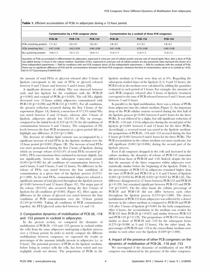
Table 1. Efficient accumulation of PCBs in adipocytes during a 12-hour period.
Contamination by a PCB congener alone: Contamination by a cocktail of three PCB congeners:
PCB-28 PCB-118 PCB-153 PCB-28 PCB-118 PCB-153
PCBs (nmol/mg protein) 1.760.1 2.060.1 1.860.1 1.860.1 2.160.1 1.860.1
PCBs (nmol/mg NLs) 0.4760.08 0.6260.09 0.4860.08 0.6160.08 0.7060.08 0.6060.08
NLs (mmol/mg protein) 13.661.1 13.161.3 15.661.1 11.461.1 11.461.1 11.461.1
Quantities of PCBs accumulated in differentiated rat adipocytes, expressed in nmol per unit of cellular protein and per unit of neutral lipids (NLs), after a dose of PCBswas added during 12 hours in the culture medium. Quantities of NLs, expressed in mmol per unit of cellular protein are also presented. Data represent the means of (i)three independent experiments 6 SEM for conditions with one PCB congener alone, (ii) five independent experiments 6 SEM for conditions with the cocktail of PCBs.There was no significant difference of PCB and NL concentrations between the three PCB congeners, whatever the kind of contamination, alone or in cocktail (p.0.05).doi:10.1371/journal.pone.0106495.t001
PCB Congeners Show Different Dynamics of Mobilisation from Adipocytes
PLOS ONE | www.plosone.org 4 September 2014 | Volume 9 | Issue 9 | e106495

PCB Congeners Show Different Dynamics of Mobilisation from Adipocytes
PLOS ONE | www.plosone.org 5 September 2014 | Volume 9 | Issue 9 | e106495

To achieve this goal, we contaminated adipocytes either with one
of the three PCB congeners (PCB-28, -118 or -153) or with a
cocktail of the three congeners. Here also, the results are expressed
as percentages of the amounts initially present in adipocytes. As
previously described, the proportions of PCBs just before the
lipolytic process (i.e. 0 hour) was set to 0% since no PCB congener
was quantified in the initial lipolytic medium. Here also, the total
PCB congeners released by the adipocytes throughout a given
period were calculated by adding the quantities measured at each
period of 3 hours.
At each studied time throughout the 12-hour period, similar
proportions of PCB-28 (Figure 3A), PCB-118 (Figure 3B) and
PCB-153 (Figure 3C) within adipocytes were quantified between
both conditions of contamination (i.e. congeners alone and in
cocktail) (0.134,p,0.934). Accordingly, no difference was noted
between the percentages of PCBs released in the lipolytic medium
in contact with adipocytes in both conditions of contamination
(0.122,p,0.916), except after 3 hours of lipolysis, where the
proportion of PCB-153 measured in the medium were higher
when it was added in cocktail than when it was added alone
(p = 0.046).
Discussion
Considerable accumulation of PCBs within adipocytesDifferentiated rat adipocytes were exposed to three targeted
PCB congeners (PCB-28, -118 and -153), which differ by the
number and the position of the chlorine atoms [19,33]. After
12 hours, similar concentrations of PCBs were stored within
adipocytes. Same observations were already drawn after 4 hours
of exposure for the same congeners [26]. The different molecular
structures of PCBs did thus not seem to influence their accretion
within adipocytes on long term. Previous studies from our group
highlighted the importance of the amount of cellular NLs, acting
as a trap for the accumulation of PCBs within adipocytes [5,26].
The fact that cellular NL levels were similar between our
Figure 1. Lipolytic treatment decreased cellular neutral lipids and increased extracellular fatty acids and glycerol. At day 11,differentiated rat adipocytes, which were previously contaminated with PCBs, were incubated with a lipolytic medium supplemented with 1 mMisoproterenol. We renewed the lipolytic medium every 3 hours for 12 hours. Cellular neutral lipids (corresponding to mmol of fatty acids in cellularneutral lipids) were expressed per mg of total cell protein (A). A significant decrease of cellular lipid contents was noted throughout the lipolyticprocess for the conditions with PCB-28 (p = 0.005) and the cocktail of PCBs (p = 0.029). The decrease was slighter for the conditions with PCB-118(p = 0.298) and PCB-153 (p = 0.097). Total extracellular free fatty acids (B) and total extracellular glycerol (C) were expressed per ml of medium.Quantities of total free fatty acids and glycerol in the medium were obtained by adding the quantities released during the periods of 3 hours (e.g.total free fatty acids at 6 hours correspond to the sum of total free fatty acids released between 0 and 3 hours and between 3 and 6 hours). Asignificant increase of total free FAs and total glycerol was observed over the 12-hour lipolytic treatment (p,0.001 for all conditions). Data representthe means of (i) three independent experiments 6 SEM for conditions with one PCB congener alone, (ii) five independent experiments 6 SEM forconditions with the cocktail of PCBs.doi:10.1371/journal.pone.0106495.g001
Figure 2. Lipolytic treatment decontaminated the adipocytes, inducing an accumulation of PCB congeners in the extracellularmedium. At day 11, contaminated adipocytes underwent a lipolytic process. The PCB contents as well as the proportions of PCBs released in theextracellular medium were assessed every 3 hours. For each PCB congener, all results were expressed in percentage of the amounts initially presentin the cells. Proportions of one PCB congener in the medium were obtained by adding the amounts released during periods of 3 hours (e.g.proportion of one congener at 6 hours corresponds to the sum of this congener released between 0 and 3 hours and between 3 and 6 hours). Animportant drop of cell PCB proportions was observed during the first 6 hours of lipolysis (p,0.001) and was followed by a more moderate decreaseof the cell PCB percentages during the last 6 hours (p,0.001). In parallel, an important increase of PCBs was noted in the extracellular medium duringthe first half of lipolysis (p,0.001) and was followed by a slower accumulation during the second half of lipolysis (0.001,p,0.006). Data representthe means of five independent experiments 6 SEM.doi:10.1371/journal.pone.0106495.g002
PCB Congeners Show Different Dynamics of Mobilisation from Adipocytes
PLOS ONE | www.plosone.org 6 September 2014 | Volume 9 | Issue 9 | e106495

PCB Congeners Show Different Dynamics of Mobilisation from Adipocytes
PLOS ONE | www.plosone.org 7 September 2014 | Volume 9 | Issue 9 | e106495

experimental conditions is most probably at the origin of the
identical accumulation of PCBs within adipocytes. While PCB
concentrations in the culture medium were in the same range than
those measured in the human serum [29,30], the PCB levels found
in cultured adipocytes after 12 hours of incubation (data from
Table 1 are equivalent ,175 ng PCB congeners per mg NLs)
were much higher than those measured in vivo, in the human
adipose tissue (from 0.02 to 0.66 ng total PCBs per mg lipids) [34–
38]. It reflects a high propensity of differentiated rat adipocytes to
store PCBs [4]. Such differences have already been noticed
previously and the reasons are discussed in details elsewhere [5].
Briefly, the higher in vitro concentrations of PCBs within
adipocytes could result from the extended contact between the
cells and the contaminated culture medium (12 hours). On the
contrary, in the in vivo situation, the PCB congeners, transported
in the circulation by lipoproteins and plasma albumin [33,39,40],
are in continual movement thanks to the blood flow. In addition,
the culture medium contains only a low concentration of serum
(10%) [4], and therefore very low levels of lipoproteins and
albumin, which could contribute to a smaller retention of PCBs in
this hydrophilic compartment and a higher storage in the
lipophilic compartment represented by adipocytes. The differen-
tiated rat adipocytes are also organised as a monolayer whereas invivo adipose tissue shows a complex 3D-structure. Furthermore,
lipolysis, which occurs regularly in vivo, may lead to the
mobilisation of PCBs from adipocytes. Finally, the circulating
PCBs may be taken up by other tissues such as the liver.
Similar dynamics of mobilisation between cellular lipidsand PCBs
Once the lipolytic pathway was induced, adipocytes started to
mobilise their lipid content. A decrease of the cellular NLs could
be observed throughout the 12-hour experiment, with a more
pronounced lipolytic action during the first 3 hours. This sharper
decrease of NL content at early lipolysis was in accordance with
our previous study [28]. As a result of the mobilisation of cellular
NLs, FFAs and glycerol were released in the extracellular medium.
The lipolytic treatment also led to the release of PCBs from
adipocytes to the extracellular medium. The dynamics of
mobilisation of PCBs exhibited some parallelism with those of
cellular lipids, as the major part of PCB discharge occurred during
the first hours of lipolysis as well. Previously, it was shown that invitro epididymal adipocytes isolated from rats also unloaded PCB-
153 during a lipolytic treatment of 50 min with 0.8 mM
isoproterenol [41]. The release of PCBs might accompany the
mobilisation of cell lipids, which agrees with previous studies on
the behaviour of dioxins [42,43]. In addition, cellular TG content
is an important parameter governing the accumulation of PCBs in
adipocytes [5,26]. PCBs are stored almost exclusively within the
LDs [4]. As this lipophilic pool is reduced during lipolysis [28], the
capacity of storage is thus also lessened, promoting the release of
PCBs in the extracellular medium, where they could be tightly
associated with diverse lipoproteins (present in the 5% serum) and
bovine albumin (2%).
Although an obvious release of PCBs occurred from adipocytes
during the lipolytic experiment, some molecules of PCBs could be
taken up again by the cells as previously suggested in in vivostudies [13,14]. This phenomenon is well known for FFAs, which
are partly reabsorbed by adipocytes and re-esterified into newly
synthesized TGs [28,44]. A complete hydrolysis of one mole of
TGs leads to the release of three moles of free FAs and one mole of
glycerol. This could be translated by a free FA/glycerol ratio
equivalent to 3.0. However, free FA/glycerol ratios were lower
than 3.0 in our experiments, which likely reflects a reuptake of free
FAs by the cells. Nevertheless, this phenomenon might have been
somewhat limited in our experimental conditions, because of the
renewal of the lipolytic medium every 3 hours [28].
Studies investigating the release of POPs from adipose tissue
during periods of weight loss in animals and humans usually report
an increase of the concentrations of PCBs and related compounds
in adipose tissue, despite their significant discharge in the blood
circulation [6,14,15,18]. This increase suggests a less efficient
mobilisation of PCBs from this tissue than lipids and a
concentration of these lipophilic pollutants in the remaining
amount of fat cells. The adipose tissue is a macroscopic structure
that is irrigated by blood vessels. During adipose tissue lipolysis, it
is possible that PCBs are transferred to adipocytes that still contain
significant amounts of lipids in their LDs instead of being all
released into the circulation. It is also possible that PCBs are
released in the bloodstream together with the lipids and then
reabsorbed by the adipose tissue as a result of their higher affinity
for the remaining lipids present in the cells [13]. Our in vitromodel differs from the in vivo situation among others by the fact
that it is characterised by only one layer of cells, which is in direct
contact with the extracellular medium that is regularly renewed. A
reuptake of PCBs by the cells and/or a migration of PCBs to
deeper adipocyte layers that are still filled with fat are thus not
possible. Moreover, the high PCB concentrations, which were
found in the cultivated adipocytes before the lipolytic induction,
might also promote the massive release of congeners in the
extracellular medium.
Differences of release according to the kind of PCBcongener
When the three congeners were added in cocktail to the culture
medium, we could observe that PCB-153 was less efficiently
mobilised from adipocytes than PCB-28 and PCB-118 during the
first part of the lipolytic process. This difference however
disappeared at 12 hours of lipolysis. The slower mobilisation of
PCB-153 from adipocytes reflects the fact that, besides the cellular
lipid content, the rate of release is also governed by the physico-
chemical properties of the congeners, which are defined by the
number and the position of chlorine atoms on the biphenyl core
[2]. If we consider the electrostatic potentials of PCBs [26], PCB-
Figure 3. Presence of other congeners did not influence the dynamic of PCB mobilisation. At day 11, differentiated rat adipocytes, whichwere previously contaminated with either individual PCB congeners or with a cocktail of PCBs, underwent a lipolytic process. The cellular levels ofPCBs before the lipolytic process were quantified and set at 100%. During 12-hour period of lipolysis, the contents of PCB congeners withinadipocytes and in the extracellular medium were assessed every 3 hours. The results for PCB-28 (A), PCB-118 (B) and PCB-153 (C) were expressed bythe percentage of initial amounts of each congener. Within a condition of contamination, proportions of one PCB congener in the medium wereobtained by adding the quantities released during the periods of 3 hours (e.g. proportion of one congener at 6 hours corresponds to the sum of thiscongener released between 0 and 3 hours and between 3 and 6 hours). At each given time of lipolytic treatment, no differences were noted betweenthe proportions of each PCB (i.e. PCB-28, -118 and -153) in both conditions of contamination (i.e. congeners alone or in cocktail), either in the cells orin the lipolytic medium (0.122,p,0.916). Only the percentage of PCB-153 in the lipolytic medium was lower when taken alone as compared to thecondition in cocktail after 3 hours of lipolysis (p = 0.046). Data represent the means of (i) three independent experiments 6 SEM for conditions withone PCB congener alone, (ii) five independent experiments 6 SEM for conditions with the cocktail of PCBs.doi:10.1371/journal.pone.0106495.g003
PCB Congeners Show Different Dynamics of Mobilisation from Adipocytes
PLOS ONE | www.plosone.org 8 September 2014 | Volume 9 | Issue 9 | e106495

153 exhibits a large electron-deficient zone. This characteristic
makes this congener rather lipophilic, which is reflected by the
higher partition coefficient n-octanol/water (log Kow = 6.80). On
the other hand, PCB-28 and PCB-118 have a reduced electron-
deficient zone, translated by lower log Kow (PCB-28: log
Kow = 5.71 and PCB-118: log Kow = 6.57). PCB-153 could thus
be more trapped within LDs than PCB-28 and PCB-118 and as a
consequence, be released more slowly.
In addition, it was previously observed that a small proportion
of PCB-153 was sequestrated in the cell membranes when isolated
primary adipocytes absorbed the PCB congeners present in the
culture medium for 2 hours [4]. It was not the case for PCB-28
and -118. Likewise, several studies showed that PCB-52 and -153,
two di-ortho-substituted PCBs, intercalate between membrane
phospholipids similarly to cholesterol and have an impact on the
membrane fluidity in fish, rodent and chicken cells [45–49]. In the
present study, the release of PCB-153 from adipocytes could thus
be slowed down by its association with cell membranes, as
compared to PCB-28 and PCB-118, two mono-ortho-substituted
congeners. The fact that PCB-153 has two chlorine atoms in the
ortho position on the biphenyl core induces a more perpendicular
layout of the phenyl rings. It means that PCB-153 occupies a
larger bulk than PCB-28 and PCB-118, which could be involved
in the sequestration of PCB-153 within membranes and its slower
mobilisation from adipocytes.
Previous findings from our group, investigating the uptake of
PCBs by differentiated adipocytes, highlighted that PCB-28 enters
the cells more rapidly than PCB-118 and PCB-153 [4]. If, during
lipolysis, a reuptake of PCBs by the cells occurs, PCB-28 might
thus be taken up more rapidly than the other congeners, which
could lead to an underestimation of the differences of mobilisation
kinetics during the lipolytic treatment.
Similar rate of release when PCBs are present alone or incocktail
In the in vivo situation, tissues are exposed to a cocktail of
contaminants that might interact with each other’s, regarding
either the toxicokinetics or the toxicodynamics of the molecules.
Here, we investigated the effect of a simple combination of PCBs
(three congeners) on their release by adipocytes during lipolysis.
To do this, the discharge of PCB-28, -118 and -153 from
adipocytes was followed either alone, or in cocktail (i.e. with two
other congeners). In the two conditions of contamination, the
dynamics of PCB release were similar, meaning that the
mobilisation of PCB congeners was not influenced by the presence
of other congeners within adipocytes in these experimental
conditions. As noted above, PCB-153 influences the properties
of cell membranes. One could thus have expected that this
congener could influence the dynamics of release of other PCBs
from the cells.
Conclusion
Our results showed an efficient accumulation of PCB-28, -118
and -153 in adipocytes. Once lipolysis was induced, the congeners
were massively mobilised from cells into the culture medium, in
parallel with the release of lipids. The dynamics of discharge
however differed between the three investigated congeners. The
release of PCB-153 was slightly but significantly slower than the
ones of PCB-28 and -118. The phenomenon might be explained
by the fact that PCB-153 is more lipophilic than the two other
congeners and could thus be more trapped in LDs. In addition,
PCB-153 being a di-ortho-substituted congener, it is more bulky,
which could be involved in its partial sequestration within cell
membrane [5] and its slower mobilisation from adipocytes. On the
other hand, the dynamics of mobilisation was not influenced by
the presence of the other two congeners.
Acknowledgments
The authors are very grateful to Coralie Piget and Marie-Therese Ahn
from ‘‘Institut des Sciences de la Vie’’ (ISV), UCLouvain, for technical
assistance. Members of ‘‘Support en methodologie et calcul statistique’’
(Institut multidisciplinaire pour la modelisation et l’analyse quantitative,
UCLouvain, Belgium) are gratefully acknowledged for the collaboration in
the statistical analyses. We also greatly appreciated the help and advice of
Guillaume Bernard for picture processing.
Author Contributions
Conceived and designed the experiments: CL GT. Performed the
experiments: GT. Analyzed the data: CL GT. Contributed reagents/
materials/analysis tools: EM JPT CD. Contributed to the writing of the
manuscript: CL. Engaged in active discussions: CL GT EM JPT CD.
References
1. Djien Liem AK, Furst P, Rappe C (2000) Exposure of populations to dioxins and
related compounds. Food Additives & Contaminants 17: 241–259.
2. La Merrill M, Emond C, Kim MJ, Antignac JP, Le Bizec B, et al. (2013)
Toxicological function of adipose tissue: focus on persistent organic pollutants.
Environmental Health Perspectives 121: 162–169.
3. Sbarbati A, Accorsi D, Benati D, Marchetti L, Orsini G, et al. (2010)
Subcutaneous adipose tissue classification. European Journal of Histochemistry
54: e48.
4. Bourez S, Le Lay S, Van den Daelen C, Louis C, Larondelle Y, et al. (2012)
Accumulation of polychlorinated biphenyls in adipocytes: Selective targeting to
lipid droplets and role of caveolin-1. PLoS ONE 7: e31834.
5. Bourez S, Joly A, Covaci A, Remacle C, Larondelle Y, et al. (2012)
Accumulation capacity of primary cultures of adipocytes for PCB-126: Influence
of cell differentiation stage and triglyceride levels. Toxicology Letters 214: 243–
250.
6. Kim MJ, Marchand P, Henegar C, Antignac JP, Alili R, et al. (2011) Fate and
complex pathogenic effects of dioxins and polychlorinated biphenyls in obese
subjects before and after drastic weight loss. Environmental Health Perspectives
119: 377–383.
7. Chevrier J, Dewailly E, Ayotte P, Mauriege P, Despres JP, et al. (2000) Body
weight loss increases plasma and adipose tissue concentrations of potentially
toxic pollutants in obese individuals. International Journal of Obesity and
Related Metabolic Disorders 24: 1272–1278.
8. Debier C, Pomeroy PP, Dupont C, Joiris C, Comblin V, et al. (2003)
Quantitative dynamics of PCB transfer from mother to pup during lactation in
UK grey seals Halichoerus grypus. Marine Ecology Progress Series 247: 237–
248.
9. Debier C, Pomeroy PP, Thome JP, Mignolet E, de Tillesse T, et al. (2004) An
unexpected parallelism between Vitamin A and PCBs in seal milk. Aquatic
Toxicology 68: 179–183.
10. Debier C, Chalon C, Le Bœuf BJ, de Tillesse T, Larondelle Y, et al. (2006)
Mobilization of PCBs from blubber to blood in northern elephant seals
(Mirounga angustirostris) during the post-weaning fast. Aquatic Toxicology 80:
149–157.
11. Vanden Berghe M, Mat A, Arriola A, Polain S, Stekke V, et al. (2010)
Relationships between vitamin A and PCBs in grey seal mothers and pups
during lactation. Environmental Pollution 158: 1570–1575.
12. Debier C, Crocker DE, Houser DS, Vanden Berghe M, Fowler M, et al. (2012)
Differential changes of fat-soluble vitamins and pollutants during lactation in
northern elephant seal mother–pup pairs. Comparative Biochemistry and
Physiology Part A 162: 323–330.
13. Vanden Berghe M, Weijs L, Habran S, Das K, Bugli C, et al. (2012) Selective
transfer of persistent organic pollutants and their metabolites in grey seals during
lactation. Environment International 46: 6–15.
14. Louis C, Dirtu AC, Stas M, Guiot Y, Malarvannan G, et al. (2014) Mobilisation
of lipophilic pollutants from blubber in northern elephant seal pups (Miroungaangustirostris) during the post-weaning fast. Environmental Research 132: 438–
448.
15. Hue O, Marcotte J, Berrigan F, Simoneau M, Dore J, et al. (2006) Increased
plasma levels of toxic pollutants accompanying weight loss induced by
hypocaloric diet or by bariatric surgery. Obesity Surgery 16: 1145–1154.
PCB Congeners Show Different Dynamics of Mobilisation from Adipocytes
PLOS ONE | www.plosone.org 9 September 2014 | Volume 9 | Issue 9 | e106495
16. Imbeault P, Tremblay A, Simoneau J-A, Joanisse DR (2002) Weight loss-
induced rise in plasma pollutant is associated with reduced skeletal muscleoxidative capacity. American Journal of Physiology 282: E574–E579.
17. Debier C, Le Boeuf BJ, Ikonomou MG, de Tillesse T, Larondelle Y, et al. (2005)
Polychlorinated biphenyls, dioxins, and furans in weaned, free-ranging northernelephant seal pups from central California, USA. Environmental Toxicology and
Chemistry 24: 629–633.18. Dirtu AC, Dirinck E, Malarvannan G, Neels H, Van Gaal L, et al. (2013)
Dynamics of organohalogenated contaminants in human serum from obese
individuals during one year of weight loss treatment. Environmental Science &Technology 47: 12441–12449.
19. Carpenter DO (2006) Polychlorinated biphenyls (PCBs): routes of exposure andeffects on human health. Reviews on Environmental Health 21: 1–23.
20. Robertson L, Hansen L (2001) PCBs: Recent advances in environmentaltoxicology and health effects. The University Press of Kentucky, Lexington,
Kentucky.
21. Kester MH, Bulduk S, Tibboel D, Meinl W, Glatt H, et al. (2000) Potentinhibition of estrogen sulfotransferase by hydroxylated PCB metabolites: A novel
pathway explaining the estrogenic activity of PCBs. Endocrinology 141: 1897–1900.
22. Gupta C (2000) Reproductive malformation of the male offspring following
maternal exposure to estrogenic chemicals. Proceedings of the Society forExperimental Biology and Medicine 224: 61–68.
23. Schantz SL, Widholm JJ, Rice DC (2003) Effects of PCB exposure onneuropsychological function in children. Environmental Health Perspectives
111: 357–576.24. Lee DH, Steffes MW, Sjodin A, Jones RS, Needham LL, et al. (2011) Low dose
organochlorine pesticides and polychlorinated biphenyls predict obesity,
dyslipidemia, and insulin resistance among people free of diabetes. PLoSONE 6: e15977.
25. Dirinck E, Jorens PG, Covaci A, Geens T, Roosens L, et al. (2011) Obesity andpersistent organic pollutants: Possible obesogenic effect of organochlorine
pesticides and polychlorinated biphenyls. Obesity 19: 709–714.
26. Bourez S, Van den Daelen C, Le Lay S, Poupaert J, Larondelle Y, et al. (2013)The dynamics of accumulation of PCBs in cultured adipocytes vary with the cell
lipid content and the lipophilicity of the congener. Toxicology Letters 216: 40–46.
27. Zhou L, Wang X, Yang Y, Wu L, Li F, et al. (2011) Berberine attenuates cAMP-induced lipolysis via reducing the inhibition of phosphodiesterase in 3T3-L1
adipocytes. Biochimica et Biophysica Acta (BBA) 1812: 527–535.
28. Louis C, Van den Daelen C, Bourez S, Donnay I, Larondelle Y, et al. (2014)Efficient in vitro adipocyte model of long-term lipolysis: A tool to study the
behaviour of lipophilic compounds. In Vitro Cellular & Developmental Biology -Animal 50: 507–518.
29. Wassermann M, Wassermann D, Cucos S, Miller HJ (1979) World PCBs map:
Storage and effects in man and his biologic environment in the 1970s. Annals ofthe New York Academy of Sciences 320: 69–124.
30. Meeker JD, Maity A, Missmer SA, Williams PL, Mahalingaiah S, et al. (2011)Serum concentrations of polychlorinated biphenyls in relation to in vitrofertilization outcomes. Environmental Health Perspectives 119: 1010–1016.
31. Dang Van QC, Focant M, Mignolet E, Turu C, Froidmont E, et al. (2011)
Influence of the diet structure on ruminal biohydrogenation and milk fatty acid
composition of cows fed extruded linseed. Animal Feed Science and Technology169: 1–10.
32. EC (2002) European Commission. Council L221/8. Official Journal of the
European Communities 262: 8–36.33. Matthews HB, Surles JR, Carver JG, Anderson MW (1984) Halogenated
biphenyl transport by blood components. Fundamental and Applied Toxicology
4: 420–428.34. Wang N, Kong D, Cai D, Shi L, Cao Y, et al. (2010) Levels of polychlorinated
biphenyls in human adipose tissue samples from Southeast China. Environ-mental Science & Technology 44: 4334–4340.
35. De Saeger S, Sergeant H, Piette M, Bruneel N, Van de Voorde W, et al. (2005)
Monitoring of polychlorinated biphenyls in Belgian human adipose tissuesamples. Chemosphere 58: 953–960.
36. Moon HB, Lee DH, Lee Y, Choi M, Choi HG, et al. (2012) Polybrominateddiphenyl ethers, polychlorinated biphenyls, and organochlorine pesticides in
adipose tissues of Korean women. Archives of Environmental Contaminationand Toxicology 62: 176–184.
37. Arrebola JP, Cuellar M, Claure E, Quevedo M, Antelo SR, et al. (2012)
Concentrations of organochlorine pesticides and polychlorinated biphenyls inhuman serum and adipose tissue from Bolivia. Environmental Research 112:
40–47.38. Malarvannan G, Dirinck E, Dirtu AC, Pereira-Fernandes A, Neels H, et al.
(2013) Distribution of persistent organic pollutants in two different fat
compartments from obese individuals. Environment International 55: 33–42.39. Becker MM, Gamble W (1982) Determination of the binding of 2,4,5,2’,4’,5’-
hexachlorobiphenyl by low density lipoprotein and bovine serum albumin.Journal of Toxicology and Environmental Health 9: 225–234.
40. Spindler-Vomachka M, Vodicnik MJ, Lech JJ (1984) Transport of 2,4,5,2’,4’,5’-hexachlorobiphenyl by lipoproteins in vivo. Toxicology and Applied Pharma-
cology 74: 70–77.
41. Gallenberg LA, Ring BJ, Vodicnik MJ (1987) Influence of lipolysis on themobilization of 2,4,5,2’,4’,5’-hexachlorobiphenyl from adipocytes in vitro.
Journal of Toxicology and Environmental Health 20: 163–171.42. Koppe JG (1995) Nutrition and breast-feeding. European Journal of Obstetrics
& Gynecology and Reproductive Biology 61: 73–78.
43. Irigaray P, Mejean L, Laurent F (2005) Behaviour of dioxin in pig adipocytes.Food and Chemical Toxicology 43: 457–460.
44. Edens NK, Leibel RL, Hirsch J (1990) Mechanism of free fatty acid re-esterification in human adipocytes in vitro. Journal of Lipid Research 31: 1423–
1431.45. Lopez-Aparicio P, Merino MJ, Sanchez E, Recio MN, Perez-Albarsanz MA
(1997) Effect of Aroclor 1248 and two pure PCB congeners upon the membrane
fluidity of rat renal tubular cell cultures. Pesticide Biochemistry and Physiology57: 54–62.
46. Gonzalez A, Odjele A, Weber JM (2013) PCB-153 and temperature causerestructuring of goldfish membranes: Homeoviscous response to a chemical
fluidiser. Aquatic Toxicology 144–145: 11–18.
47. Yilmaz B, Sandal S, Chen CH, Carpenter DO (2006) Effects of PCB 52 andPCB 77 on cell viability, [Ca2+]i levels and membrane fluidity in mouse
thymocytes. Toxicology 217: 184–193.48. Campbell AS, Yu Y, Granick S, Gewirth AA (2008) PCB association with model
phospholipid bilayers. Environmental Science & Technology 42: 7496–7501.49. Katynski AL, Vijayan MM, Kennedy SW, Moon TW (2004) 3,3’,4,4’,5-
pentachlorobiphenyl (PCB 126) impacts hepatic lipid peroxidation, membrane
fluidity and b-adrenoceptor kinetics in chick embryos. Comparative Biochem-istry and Physiology Part C 137: 81–93.
PCB Congeners Show Different Dynamics of Mobilisation from Adipocytes
PLOS ONE | www.plosone.org 10 September 2014 | Volume 9 | Issue 9 | e106495


